Tubular apocrine adenoma of the axilla: A rare adnexal tumor
Salsabil Attafi, Ines Smichi, Wafa Rkik
Pathology Department of Siliana Hospital, Siliana, Tunis, Tunisia
We report a 52-year-old woman who presented with a painless mobile nodule of the axilla of unknown duration. The physical examination found a small, mobile, painless and circumscribed nodule of the axilla. There was no associated regional lymphadenopathy. The patient had a simple excision of the nodule.
Gross examination revealed a well-circumscribed nodule measuring 1 cm of diameter. Histologically, the tumor was composed of lobules of well-differentiated dilated tubular structures situated in the dermis and the subcutis (Figs. 1 and 2). The tubules were double-lined by an inner layer of typical apocrine epithelial cells and an outer layer of myoepithelial cells (Fig. 3). A focal ductal differentiation was noted. The intervening stroma was scanty and fibrous with minimal inflammatory cells (Fig. 1). There was no evidence of comedolike channels, hyaline, or clear cell differentiation. The diagnosis of tubular apocrine adenoma was retained. The patient received no further therapy and was well during the 3 months period after excision.

Figure 1: Lobules of well-differentiated dilated tubular structures situated in the dermis and separated by a scanty fibrous stroma
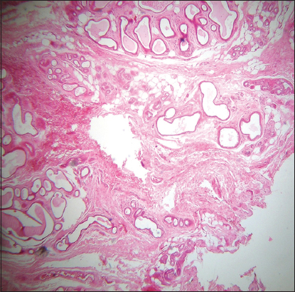
Figure 2: The tubules structures reach the subcutis
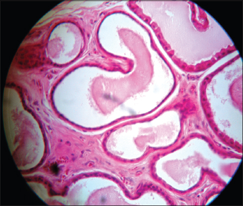
Figure 3: The tubules are double-lined by an inner layer of apocrine cells and an outer layer of myoepithelial cells
Prior to the study, patient gave written consent to the examination and biopsy after having been informed about the procedure.
Tubular apocrine adenoma also called apocrine adenoma, tubulopapillary hidradenoma and papillary tubular adenoma is a rare benign tumor. It predominates in women with a sex ratio of 2:1. The wide age distribution ranges between 18 and 78 years. The scalp is most commonly affected although lesions have been described at a variety of other sites including the face, eyelid, axilla, leg, and genitalia. In the scalp, lesions often arise in a background of nevus sebaceus and may be associated with syringocystadenoma papilliferum. Clinically, the tumor generally presents as a dermal nodule 1–2 cm in diameter or pedunculated lesion, frequently of many years duration. Histologically, tubular apocrine adenoma presents most often as lobules of well-differentiated tubular structures located in the dermis and sometimes the subcutis. The tubules are lined by an inner layer of tall columnar cells, which show decapitation secretion, and frequently show an outer layer of cuboidal cells. Comedo-like channels that extend into the epidermis and connect with some of the tubular structures are occasionally seen. The stroma is composed of fibrous tissue with few inflammatory cells. This is in contrast to syringocystadenoma papilliferum, which contains numerous inflammatory cells in the stroma.
The tubular adenoma is benign and recurrence following excision is uncommon.
CONSENT
The examination of the patient was conducted according to the Declaration of Helsinki principles.
Notes
Source of Support: Nil,
Conflict of Interest: None declared.
Comments are closed.