Published online Apr 26, 2016. doi: 10.4330/wjc.v8.i4.317
Peer-review started: June 29, 2015
First decision: July 31, 2015
Revised: January 29, 2016
Accepted: February 16, 2016
Article in press: February 17, 2016
Published online: April 26, 2016
AIM: To investigate the impact of using computed tomography (CT) and contact force (CF) technology on recurrence of atrial tachyarrhythmia after atrial fibrillation (AF) ablation.
METHODS: This non-randomized study included 2 groups of patients. All patients had symptomatic recurrent paroxysmal or persistent AF and were treated with at least 1 anti arrhythmic medication or intolerant to medication. The first group included 33 patients who underwent circumferential pulmonary veins isolation (PVI) for AF during 2012 and 2013 guided by CT image integration (Cartomerge, Biosense Webster, Diamond Bar, CA, United States) of left atrium and pulmonary veins into an electroanatomic mapping (EAM) system (CT group) using standard irrigated radiofrequency catheter (ThermoCool, Carto, Biosense Webster, Diamond Bar, CA, United States) or irrigated catheter with integrated CF sensor (Smart Touch, Carto, Biosense Webster, Diamond Bar, CA, United States). The second group included immediately preceding 32 patients who had circumferential PVI by standard irrigated catheter (ThermoCool) using only EAM (Carto) system (EAM group). Linear lesions were performed according to the discretion of operator.
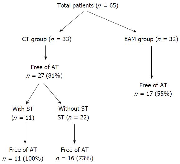
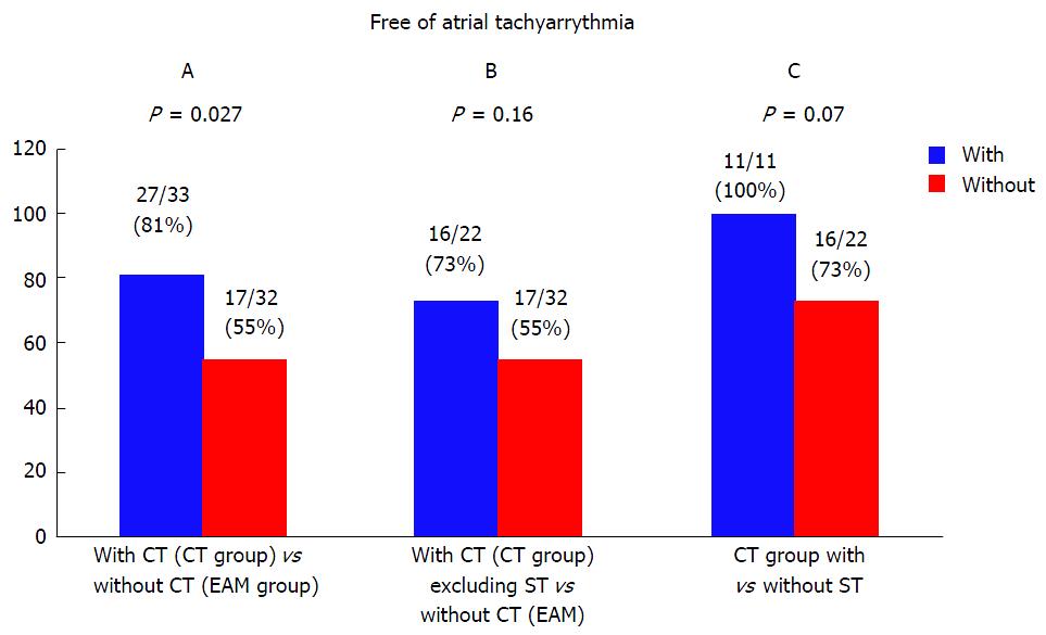
RESULTS: Sex, age, and persistent AF were not different between groups. PVI was achieved in all patients in both groups. Linear ablations including cavo-tricuspid isthmus and or roof line ablation were not different between groups. Free of atrial tachyarrhythmia during follow-up of 24 mo was significantly higher among CT group compared to EAM group (81% vs 55%; respectively; P = 0.027). When 11 patients from CT group who had ablation using Smart Touch catheter were excluded, the difference between CT group and EAM became non significant (73% vs 55%; respectively; P = 0.16). Sub analysis of CT group showed that patients who had ablation using Smart Touch catheter tend to be more free of atrial tachyarrhythmia compared to patients who had ablation using standard irrigated catheter during follow-up (100% vs 73%; respectively; P = 0.07). Major complications (pericardial effusion, cerebrovascular accident/transient ischemic attack, vascular access injury requiring intervention) did not occurred in both groups.
CONCLUSION: These preliminary results suggest that CT image integration and CF technology may reduce the recurrence of atrial tachyarrhythmia after catheter ablation for AF.
Core tip: The aim of this nonrandomized study was to determine the impact of integrating computed tomography (CT) image of left atrium into electroanatomical mapping (EAM) system and using of contact force (CF) technology on recurrence of atrial tachyarrhythmia after atrial fibrillation (AF) ablation. We found that combination of CT image integration into EAM and CF technology might reduce the recurrence of atrial tachyarrhythmia after catheter ablation for AF during follow-up period of 24 mo.
- Citation: Marai I, Suleiman M, Blich M, Lessick J, Abadi S, Boulos M. Impact of computed tomography image and contact force technology on catheter ablation for atrial fibrillation. World J Cardiol 2016; 8(4): 317-322
- URL: https://www.wjgnet.com/1949-8462/full/v8/i4/317.htm
- DOI: https://dx.doi.org/10.4330/wjc.v8.i4.317
In treatment of symptomatic and drug refractory atrial fibrillation (AF), catheter-based pulmonary veins isolation (PVI) has been established as a standard procedure by using a single-tip ablation catheter for creating linear lesion surround ipsilateral pulmonary veins (PVs)[1]. Nevertheless, repeat procedures are required in a significant number of cases and recurrent PV conduction is responsible for most ablation failures in paroxysmal AF[2].
The durability of PVI and clinical outcome after radiofrequency (RF) ablation is affected by the contact force (CF) between the catheter tip and the tissue. Insufficient CF may result in an ineffective lesion, where as excessive CF may result in complications[3]. Catheter ablation using real-time CF technology was reported to be safe for the treatment of supraventricular tachycardia and AF[3]. In addition, understanding the anatomy of left atrium (LA) and PVs is essential for the safety and effectiveness of Procedure. Pre-procedural cardiac computed tomography (CT) helps to evaluate the three dimension (3D) and complex anatomy of LA and PVs[4].
We assume that integrating CT Image of LA/PVs into electroanatomical mapping (EAM) system and using CF technology may reduce the clinical recurrence of atrial tachyarrhythmia after ablation of AF.
We summarized all patients who underwent circumferential PVI for AF during 2012 and 2013 guided by CT image integration into an EAM (Cartomerge, Biosense Webster, Diamond Bar, CA, United States) system (CT group). This group was compared to immediately preceding patients who had PVI using only EAM (Carto, Biosense Webster, Diamond Bar, CA, United States) system (EAM group).
All patients had symptomatic recurrent paroxysmal AF or persistent AF (less than 3 mo duration) who were treated with at least 1 anti arrhythmic drug (AAD) or intolerant to medication. All patients with paroxysmal AF were treated with IC AADs, and all patients with persistent AF were treated with amiodarone.
The procedure was performed during deep sedation with fentanyl and midazolam. Double trans-septal punctures were done with guidance of intra-cardiac echo (ICE): One for circular mapping catheter (Lasso, Biosense Webster, Diamond Bar, CA, United States) and one for ablation catheter.
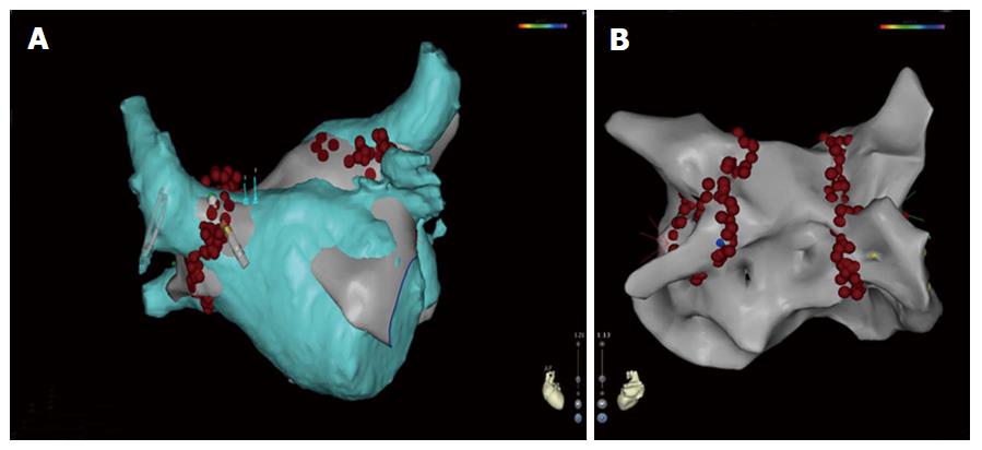
The ablation catheter used was 3.5-mm standard externally - irrigated (ThermoCool, Carto, Biosense Webster, Diamond Bar, CA, United States) for all patients in EAM group and part of patients of CT group. Externally-irrigated Smart Touch catheter (ST, Carto, Biosense Webster, Diamond Bar, CA, United States) was used in the remaining patients of CT group (Figure 1). The Smart Touch catheter is capable of directly assessing CF and showing its absolute value and orientation by means of a 3D vector in real time during the procedure[5].
In the EAM group, wide circumferential ablation of ipsilateral veins pair at the antrum about 1 cm of veno-atrial junction was performed (Figure 1). The ostium and veno-atrial junction of each PV was identified by intra cardiac electrogram, dragging the ablation or Lasso catheter back under fluoroscopic guidance, and ICE. Isolation of each vein was confirmed by lasso catheter. In the CT group, the EAM was merged with 3D-anatomical chamber reconstructions of LA and PVs derived from pre procedure (up to 24 h) cardiac CT. Image integration is based on registration involving landmark points and surface alignment using the Cartomerge software as described previously[6] (Figure 1). The ablation was performed in similar way as in the EAM group by standard irrigated catheter or Smart Touch catheter. When Smart Touch catheter was used, ablation was done only when the force was at least 10 g (optimal range for ablation was considered as 10-40 g). We tried to deliver RF energy when the CF is > 10 g and is stable for at least 20 s.
RF energy was delivered at a maximum power of 25 W at a flow rate of 17 mL/min along the posterior wall, and at a maximum power of 35 W at a flow rate of 30 mL/min along the anterior wall and elsewhere in the atria. The maximum temperature was set at 43 °C. RF ablation was continued at each site until local electrograms were abolished or for 30 s. Linear lesions including roof line and or cavo-tricuspid isthmus line were performed in some patients according to discretion of the operator. All the procedures in both groups were performed by 2 experienced operators.
All patients were followed in the outpatient clinic every 3 mo for 24 mo. Recurrence was defined as any clinical or documented atrial tachyarrhythmia lasts more 30 s after a blanking period of 3 mo. All patients were treated with anticoagulation for at least 3 mo. Anticoagulation was continued after 3 mo in high risk patients. AADs were stopped after 3 mo.
Variables are expressed as mean ± SD. Comparisons between groups were performed with Student’s t test. Categorical variables expressed as numbers and percentages were compared with a χ2 test. Kaplan - Meier survival curve was used for estimation of recurrence of atrial tachyarrhythmia during 24 mo follow-up. A P value < 0.05 was considered statistically significant. The statistical methods of the study were reviewed by biomedical statistician.
The EAM group included 32 patients, and the CT group included 33 patients. Baseline characteristics are similar (Table 1). The AF duration before ablation in both groups was 1-3 years.
| EAM group (n = 32) | CT group (n = 33) | P | |
| Sex (male) | 19 | 24 | 0.3 |
| Age (yr) | 55 ± 8.8 | 56.7 ± 11.6 | 0.6 |
| Persistent AF | 5 | 4 | 0.7 |
| CTI Ablation | 7 | 7 | 0.76 |
| Roof line ablation | 8 | 8 | 0.77 |
| Major complications | 0 | 0 |
Circumferential PVI with confirmation of isolation was performed in all patients in both groups. Cavotricuspid isthmus ablation was performed in the index procedure in 7 patients in the EAM group and in 7 patients in the CT group (P = 0.76). Roof line was performed in the index procedure in 8 patients in the EAM and in 8 patients in the CT group (P = 0.77) (Table 1).
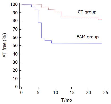
All patients completed the 24 mo follow-up. Free of atrial tachyarrhythmia during 24 mo was significantly higher among CT group compared to EAM group (81% vs 55%; respectively; P = 0.027) (Figures 2-4). When 11 patients from CT group who had ablation using Smart Touch catheter were excluded, the difference became non significant (73% vs 55%; respectively; P = 0.16) (Figures 2 and 3).
Sub analysis of CT group showed that patients who had ablation using Smart Touch catheter tended to be more free of atrial tachyarrhythmia compared to patients who had ablation using standard irrigated catheter (100% vs 73%; respectively; P = 0.07) (Figure 3). Of note, all patients who had recurrence of atrial tachyarrhythmia had AF except 1 patient from CT group and 2 patients from EAM group who had atypical atrial flutter.
Major complications (pericardial effusion/tamponade, cerebrovascular accident/transient ischemic attack, and or vascular access injury requiring intervention) did not occur in both groups.
The main finding of this study was that ablation of AF guided by of CT image integration of LA/PVs into an EAM and the use of CF technology was associated with reduced recurrence of atrial tachyarrhythmia. CT image integration without CF technology was associated with non-significant reduced recurrence of atrial tachyarrhythmia. CF technology tended to reduce recurrence of atrial tachyarrhythmia among patients who underwent ablation of AF guided by of CT image integration.
Kistler et al[7] reported in a nonrandomized study that catheter ablation for AF guided by CT image integration (Cartomerge) was associated with reduced fluoroscopy times, arrhythmia recurrence, and increased restoration of sinus rhythm compared to a similar ablation strategy guided by a 3D mapping. Successful PV electrical isolation did not differ between the two groups. However, Kistler et al[8] reported in another study which was randomized study that CT image integration (Cartomerge) to guide catheter ablation for AF did not significantly improve the procedural and clinical outcome compared to EAM.
As we showed in our non-randomized study, CT image integration without CF technology was associated with non-significant reduced recurrence of atrial tachyarrhythmia compared to EAM. This result is in agreement with the randomized study of Kistler et al[8]. In addition, CF technology tended to reduce recurrence of atrial tachyarrhythmia among patients who underwent ablation of AF guided by CT image integration. Thus, it seems that the contribution of CF technology is significant.
In the TOCCATA study[9], patients with paroxysmal AF underwent PVI by using a RF ablation catheter with a different integrated CF sensor (TactiCath; Endosense, Geneva, Switzerland). The CF during catheter ablation for AF correlated with clinical outcome after 12 mo. Arrhythmia control is best achieved when ablation lesions were placed with an average CF of > 20 g, and clinical failure is universally noted with an average CF of < 10 g.
In the EFFICAS I multicenter study[10], a RF ablation catheter with integrated CF sensor (TactiCath; Endosense, Geneva, Switzerland) was used to perform PVI in patients with paroxysmal AF. At follow-up, an interventional diagnostic procedure was performed to assess gap location as correlated to index procedure ablation parameters. Minimum CF and minimum Force-Time Integral (FTI) values were strong predictors of gap formation. According to this study, optimal CF parameter recommendations are a target CF of 20 g and a minimum FTI of 400 g for each new lesion. In our study, we tried to keep the force at least 10 g at each site for at least 20 s until electrogram abolition or at least for 30 s.
Recently, Sciarra et al[5] studied 3 types of irrigated-tip ablation catheters. Sixty-three patients with paroxysmal AF underwent ablation by standard ThermoCool catheter, Smart Touch catheter, or Surround Flow catheter (Biosense Webster, Diamond Bar, CA, United States). The percentage of isolated PVs was comparable between groups. Both the Smart Touch catheter and the Surround Flow catheter significantly reduced radiofrequency and fluoroscopy times, as well as PVs reconnection rate at 30 min. Moreover, the Smart Touch catheter reduced overall duration of the procedure. However, the long term clinical significance of these results is not known.
We do not know the exact mechanism why combination of technologies is more useful. Image integration could improve clinical outcome because it helps to understand the 3D complex anatomy of LA/PV and appreciate the variant anatomy of PVs including common trunks or more than 4 veins. In addition, it could help to make the lesion set more precise. CF technology could lead to durable lesions. We think that the results of our study emphasized the fact that AF is a complex arrhythmia. The AF ablation is also a complex procedure with relatively high rate of recurrence due to PV reconnection. Many technologies were introduced to overcome this issue like steerable sheath[11]. Recently, a novel irrigated multi electrode mapping and ablation catheter (nMARQ catheter, Biosense Webster, Diamond Bar, CA, United States) was introduced for PVI with promising results[12]. However, there is no specific technology that is significantly more useful than others. We think, that combination of technologies rather than single one can lead to the best results as in this study. In addition centers and operators should choose the technologies according to their experience and according to the 3D anatomy of LA/PVs as detected by pre-procedure echocardiography, CT and or other modalities.
This is a small non-randomized study comparing AF ablation using integrated CT/EAM with AF ablation using EAM only. We used two types of ablation catheters in the CT group, the standard irrigated catheter as in EAM group and CF catheter. Only the combination of CT and CF technology was associated with significant reduction of atrial tachyarrhythmia. Thus, we could not determine the relative contribution of these technologies. Future randomized studies are needed to determine the optimal combination of technologies that gives the best procedural and clinical results of AF ablation.
In summary, these preliminary results suggest that CT image integration into EAM in combination with CF technology may reduce the recurrence of atrial tachyarrhythmia after catheter ablation for AF.
Recurrence of atrial arrhythmia after pulmonary vein isolation (PVI) is mainly due to recurrent pulmonary veins conduction. The durability of PVI and clinical outcome after radiofrequency ablation is affected by the contact force (CF) between the catheter tip and the tissue. In addition, understanding the anatomy of left atrium (LA) and PVs is essential for the safety and effectiveness of procedure. Pre-procedural cardiac computed tomography (CT) helps to evaluate the three dimension (3D) and complex anatomy of LA and PVs. In this study, the authors evaluated the impact of integrating CT image of LA/PVs into electroanatomical mapping system and using CF technology on clinical recurrence of atrial tachyarrhythmia after ablation for atrial fibrillation (AF).
CT image integration of cardiac chamber into electroanatomic mapping (EAM) is widely used to guide catheter ablation for AF and related arrhythmias. Some studies showed that it improved the procedural and clinical outcome compared to EAM but others did not. In addition CF technology was found recently to associated with better clinical outcomes. Research is focused now in defining the parameters of CF that are universally associated with better clinical outcomes.
The authors found that combination of CT image integration into EAM and CF technology may reduce the recurrence of atrial tachyarrhythmia after catheter ablation for AF during follow-up period of 24 mo.
The authors think, that combination of technologies rather than single one can lead to the best results as in this study. Centers and operators should choose the technologies according to their experience and according to the 3D anatomy of LA/PVs as detected by pre-procedure echocardiography, CT and or other modalities.
EAM: Electroanatomic mapping enables reconstruction of 3D anatomy of LA and PVs. Image integration: A technique to integrate a CT image of LA/PVs into EAM. CF technology: Enables assessing contact between tip of catheter and tissue and showing its absolute value and orientation by means of a 3D vector in real time during the procedure.
This is an interesting article analysing the influence of novel technologies (image integration and CF evaluation) on the outcome of catheter ablation of AF.
P- Reviewer: De Ponti R, Kettering K, Sakabe K, Shimizu Y S- Editor: Ji FF L- Editor: A E- Editor: Li D
| 1. | Calkins H, Kuck KH, Cappato R, Brugada J, Camm AJ, Chen SA, Crijns HJ, Damiano RJ, Davies DW, DiMarco J, Edgerton J, Ellenbogen K, Ezekowitz MD, Haines DE, Haissaguerre M, Hindricks G, Iesaka Y, Jackman W, Jalife J, Jais P, Kalman J, Keane D, Kim YH, Kirchhof P, Klein G, Kottkamp H, Kumagai K, Lindsay BD, Mansour M, Marchlinski FE, McCarthy PM, Mont JL, Morady F, Nademanee K, Nakagawa H, Natale A, Nattel S, Packer DL, Pappone C, Prystowsky E, Raviele A, Reddy V, Ruskin JN, Shemin RJ, Tsao HM, Wilber D. 2012 HRS/EHRA/ECAS expert consensus statement on catheter and surgical ablation of atrial fibrillation: recommendations for patient selection, procedural techniques, patient management and follow-up, definitions, endpoints, and research trial design: a report of the Heart Rhythm Society (HRS) Task Force on Catheter and Surgical Ablation of Atrial Fibrillation. Developed in partnership with the European Heart Rhythm Association (EHRA), a registered branch of the European Society of Cardiology (ESC) and the European Cardiac Arrhythmia Society (ECAS); and in collaboration with the American College of Cardiology (ACC), American Heart Association (AHA), the Asia Pacific Heart Rhythm Society (APHRS), and the Society of Thoracic Surgeons (STS). Endorsed by the governing bodies of the American College of Cardiology Foundation, the American Heart Association, the European Cardiac Arrhythmia Society, the European Heart Rhythm Association, the Society of Thoracic Surgeons, the Asia Pacific Heart Rhythm Society, and the Heart Rhythm Society. Heart Rhythm. 2012;9:632-696.e21. [PubMed] [DOI] [Cited in This Article: ] [Cited by in Crossref: 1237] [Cited by in F6Publishing: 1284] [Article Influence: 107.0] [Reference Citation Analysis (0)] |
| 2. | Roten L, Derval N, Pascale P, Scherr D, Komatsu Y, Shah A, Ramoul K, Denis A, Sacher F, Hocini M. Current hot potatoes in atrial fibrillation ablation. Curr Cardiol Rev. 2012;8:327-346. [PubMed] [Cited in This Article: ] |
| 3. | Kuck KH, Reddy VY, Schmidt B, Natale A, Neuzil P, Saoudi N, Kautzner J, Herrera C, Hindricks G, Jaïs P. A novel radiofrequency ablation catheter using contact force sensing: Toccata study. Heart Rhythm. 2012;9:18-23. [PubMed] [DOI] [Cited in This Article: ] [Cited by in Crossref: 234] [Cited by in F6Publishing: 211] [Article Influence: 16.2] [Reference Citation Analysis (0)] |
| 4. | Piorkowski C, Kircher S, Arya A, Gaspar T, Esato M, Riahi S, Bollmann A, Husser D, Staab C, Sommer P. Computed tomography model-based treatment of atrial fibrillation and atrial macro-re-entrant tachycardia. Europace. 2008;10:939-948. [PubMed] [DOI] [Cited in This Article: ] [Cited by in Crossref: 25] [Cited by in F6Publishing: 27] [Article Influence: 1.7] [Reference Citation Analysis (0)] |
| 5. | Sciarra L, Golia P, Natalizia A, De Ruvo E, Dottori S, Scarà A, Borrelli A, De Luca L, Rebecchi M, Fagagnini A. Which is the best catheter to perform atrial fibrillation ablation? A comparison between standard ThermoCool, SmartTouch, and Surround Flow catheters. J Interv Card Electrophysiol. 2014;39:193-200. [PubMed] [DOI] [Cited in This Article: ] [Cited by in Crossref: 3] [Cited by in F6Publishing: 19] [Article Influence: 1.9] [Reference Citation Analysis (0)] |
| 6. | Kistler PM, Earley MJ, Harris S, Abrams D, Ellis S, Sporton SC, Schilling RJ. Validation of three-dimensional cardiac image integration: use of integrated CT image into electroanatomic mapping system to perform catheter ablation of atrial fibrillation. J Cardiovasc Electrophysiol. 2006;17:341-348. [PubMed] [Cited in This Article: ] |
| 7. | Kistler PM, Rajappan K, Jahngir M, Earley MJ, Harris S, Abrams D, Gupta D, Liew R, Ellis S, Sporton SC. The impact of CT image integration into an electroanatomic mapping system on clinical outcomes of catheter ablation of atrial fibrillation. J Cardiovasc Electrophysiol. 2006;17:1093-1101. [PubMed] [Cited in This Article: ] |
| 8. | Kistler PM, Rajappan K, Harris S, Earley MJ, Richmond L, Sporton SC, Schilling RJ. The impact of image integration on catheter ablation of atrial fibrillation using electroanatomic mapping: a prospective randomized study. Eur Heart J. 2008;29:3029-3036. [PubMed] [DOI] [Cited in This Article: ] [Cited by in Crossref: 96] [Cited by in F6Publishing: 106] [Article Influence: 6.6] [Reference Citation Analysis (0)] |
| 9. | Reddy VY, Shah D, Kautzner J, Schmidt B, Saoudi N, Herrera C, Jaïs P, Hindricks G, Peichl P, Yulzari A. The relationship between contact force and clinical outcome during radiofrequency catheter ablation of atrial fibrillation in the TOCCATA study. Heart Rhythm. 2012;9:1789-1795. [PubMed] [DOI] [Cited in This Article: ] [Cited by in Crossref: 352] [Cited by in F6Publishing: 357] [Article Influence: 29.8] [Reference Citation Analysis (0)] |
| 10. | Neuzil P, Reddy VY, Kautzner J, Petru J, Wichterle D, Shah D, Lambert H, Yulzari A, Wissner E, Kuck KH. Electrical reconnection after pulmonary vein isolation is contingent on contact force during initial treatment: results from the EFFICAS I study. Circ Arrhythm Electrophysiol. 2013;6:327-333. [PubMed] [DOI] [Cited in This Article: ] [Cited by in Crossref: 338] [Cited by in F6Publishing: 362] [Article Influence: 32.9] [Reference Citation Analysis (0)] |
| 11. | Piorkowski C, Eitel C, Rolf S, Bode K, Sommer P, Gaspar T, Kircher S, Wetzel U, Parwani AS, Boldt LH. Steerable versus nonsteerable sheath technology in atrial fibrillation ablation: a prospective, randomized study. Circ Arrhythm Electrophysiol. 2011;4:157-165. [PubMed] [DOI] [Cited in This Article: ] [Cited by in Crossref: 83] [Cited by in F6Publishing: 88] [Article Influence: 6.8] [Reference Citation Analysis (0)] |
| 12. | Shin DI, Kirmanoglou K, Eickholt C, Schmidt J, Clasen L, Butzbach B, Rassaf T, Merx M, Kelm M, Meyer C. Initial results of using a novel irrigated multielectrode mapping and ablation catheter for pulmonary vein isolation. Heart Rhythm. 2014;11:375-383. [PubMed] [DOI] [Cited in This Article: ] [Cited by in Crossref: 33] [Cited by in F6Publishing: 33] [Article Influence: 3.0] [Reference Citation Analysis (0)] |


