A Stillborn Multiple Organs’ Investigation from a Maternal DENV-4 Infection: Histopathological and Inflammatory Mediators Characterization

 ,
,  ,
,  and
and 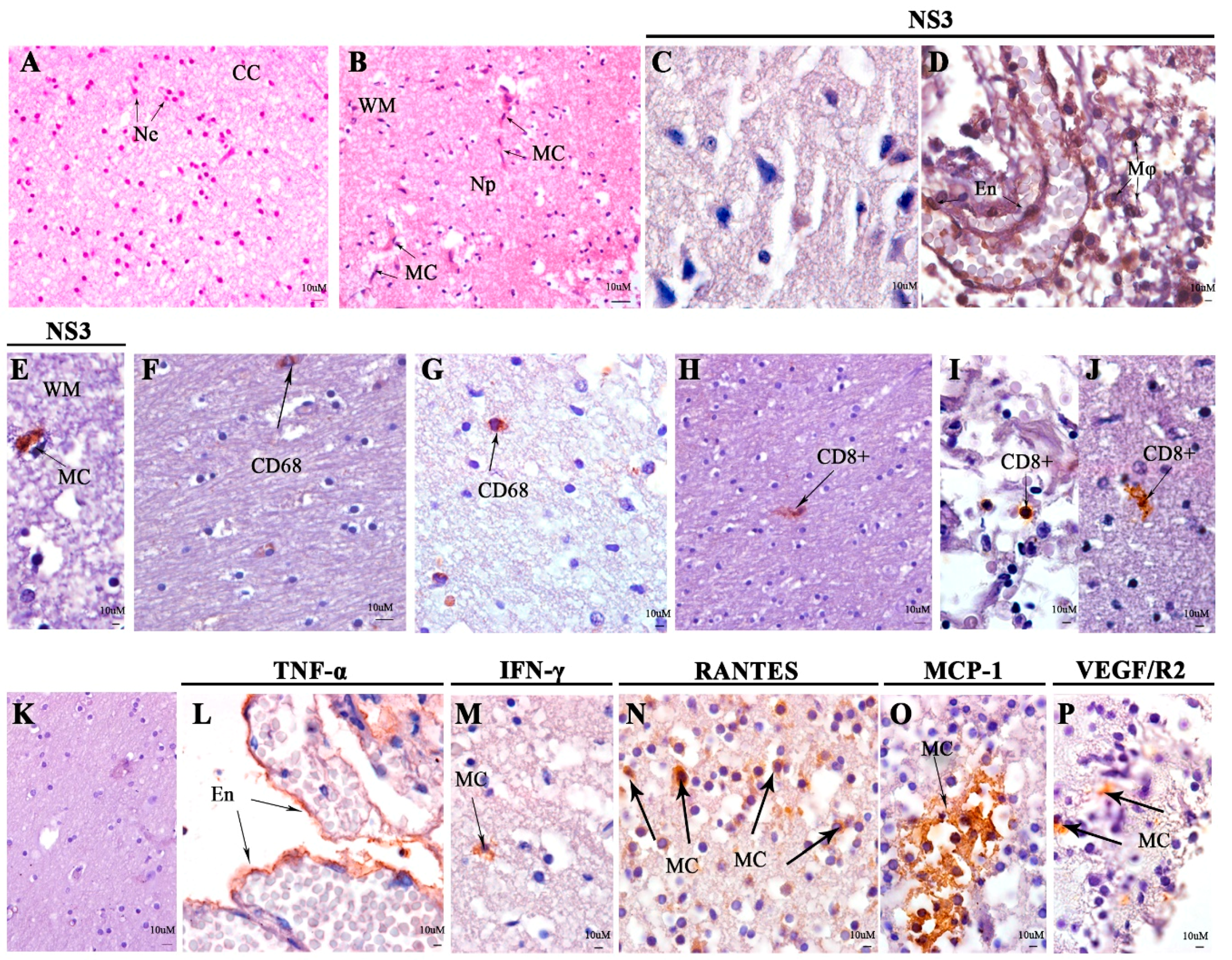
Abstract
:1. Introduction
2. Materials and Methods
2.1. Case
2.2. Ethical Considerations
2.3. Molecular Diagnosis, Histopathological Analysis, and Immunohistochemistry
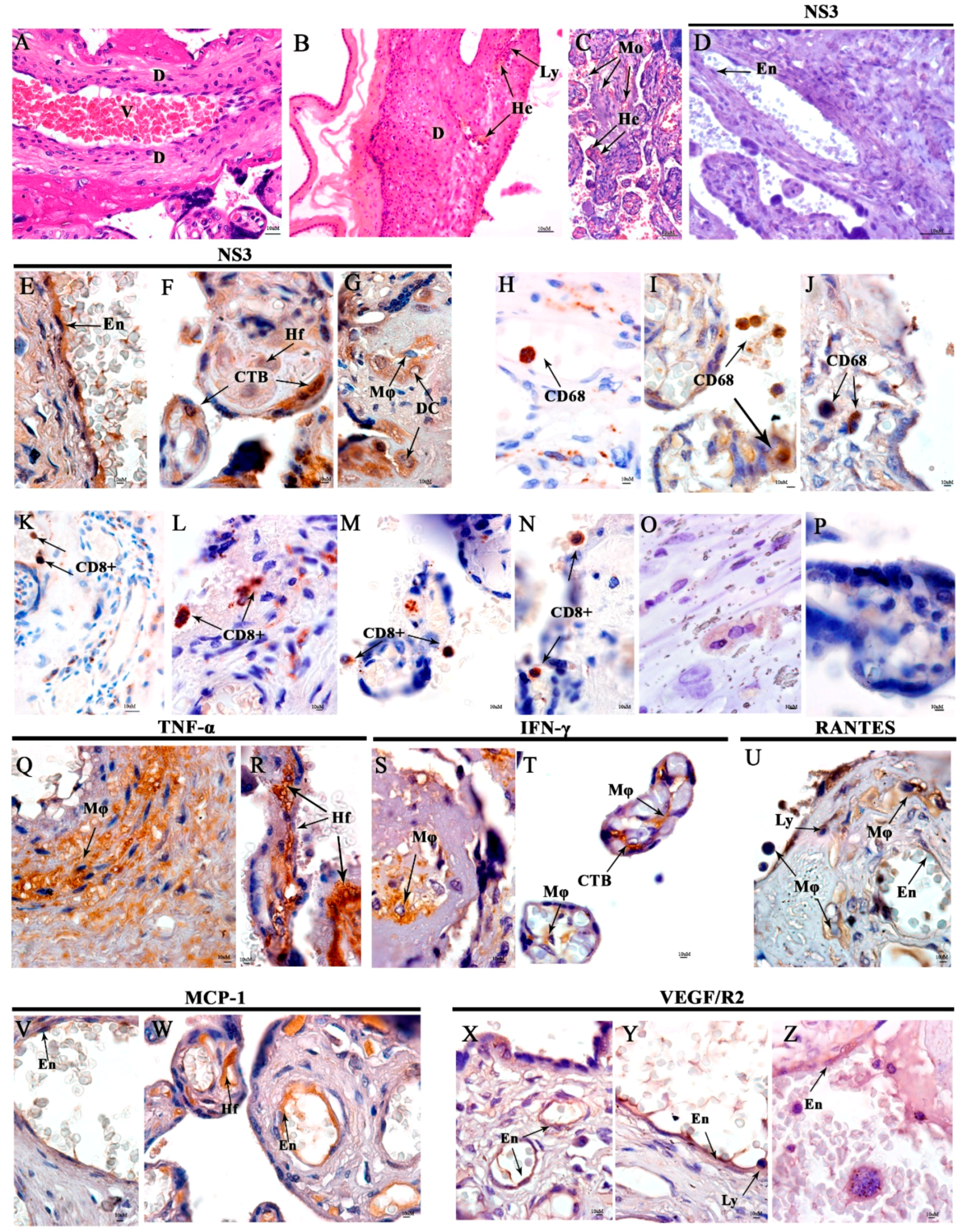
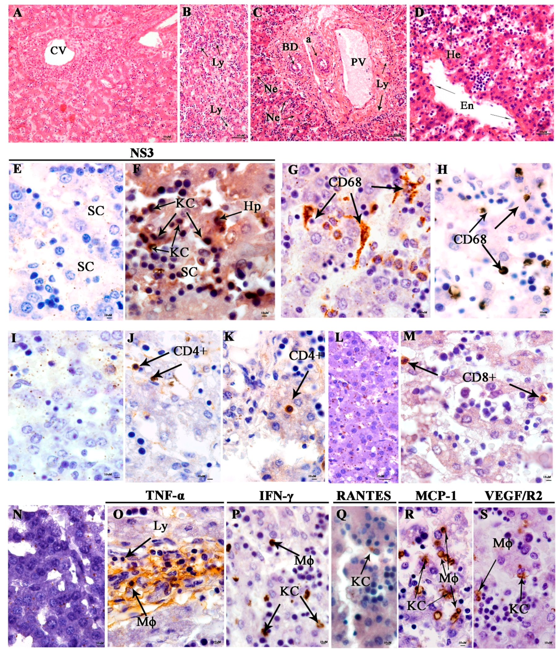
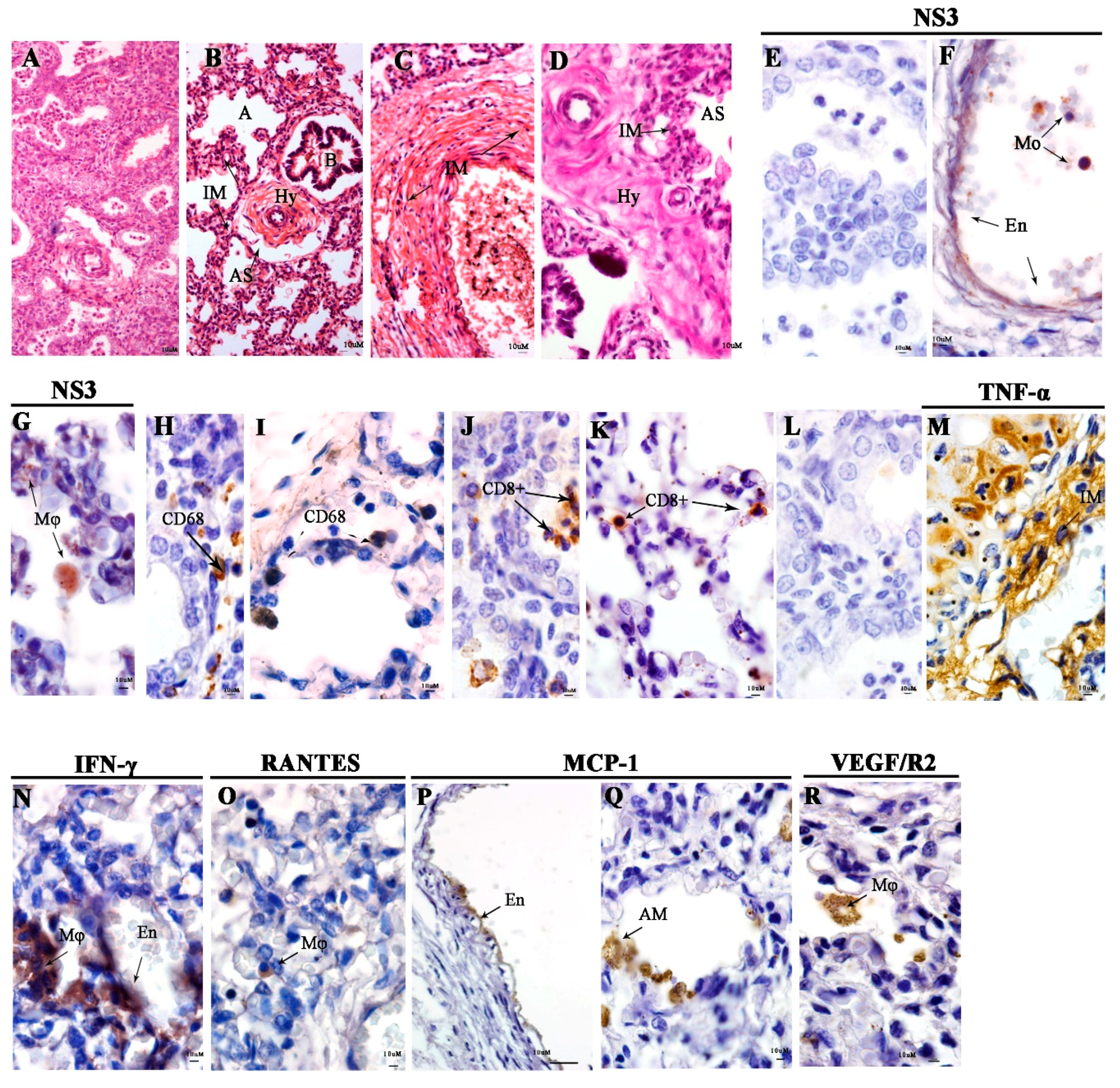
3. Results
4. Discussion
5. Conclusions
Author Contributions
Funding
Acknowledgments
Conflicts of Interest
References
- Gubler, D.J. The economic burden of dengue. Am. J. Trop. Med. Hyg. 2012, 86, 743–744. [Google Scholar] [CrossRef] [PubMed]
- WHO Dengue and severe dengue. Available online: http://www.who.int/news-room/fact-sheets/detail/dengue-and-severe-dengue (accessed on 15 July 2018).
- Schatzmayr, H.G.; Nogueira, R.M.; Travassos da Rosa, A.P. An outbreak of dengue virus at Rio de Janeiro--1986. Mem. Inst. Oswaldo Cruz 1986, 81, 245–246. [Google Scholar] [CrossRef] [PubMed]
- PAHO PAHO/WHO Data—Dengue cases. Available online: http://www.paho.org/data/index.php/en/mnu-topics/indicadores-dengue-en/dengue-nacional-en/252-dengue-pais-ano-en.html (accessed on 15 July 2018).
- Osanai, C.H.; Travassos da Rosa, A.P.; Tang, A.T.; do Amaral, R.S.; Passos, A.D.; Tauil, P.L. [Dengue outbreak in Boa Vista, Roraima. Preliminary report]. Rev. Inst. Med. Trop. Sao Paulo 1983, 25, 53–54. [Google Scholar] [PubMed]
- Temporao, J.G.; Penna, G.O.; Carmo, E.H.; Coelho, G.E.; Azevedo, R.D.S.S.; Nunes, M.R.T.; da Costa Vasconcelos, P.F. Dengue Virus Serotype 4, Roraima State, Brazil. Emerg. Infect. Dis. 2011, 17, 938–940. [Google Scholar] [CrossRef] [Green Version]
- Nogueira, R.M.R.; Eppinghaus, A.L.F. Dengue virus type 4 arrives in the state of Rio de Janeiro: A challenge for epidemiological surveillance and control. Mem. Inst. Oswaldo Cruz 2011, 106, 255–256. [Google Scholar] [CrossRef]
- Heringer, M.; Souza, T.M.A.; Monique da Rocha, Q.L.; Nunes, P.C.G.; da C Faria, N.R.; de Bruycker-Nogueira, F.; Chouin-Carneiro, T.; Nogueira, R.M.R.; Dos Santos, F.B. Dengue type 4 in Rio de Janeiro, Brazil: Case characterization following its introduction in an endemic region. BMC Infect. Dis. 2017, 17, 410. [Google Scholar] [CrossRef] [PubMed]
- SES/SINAN Boletim Epidemiológico. Dengue: Monitoramento até a Semana Epidemiológica (SE) 52 de 2013. Available online: http://portalarquivos2.saude.gov.br/images/pdf/2013/dezembro/52/Dengue-monitoramento-2013.pdf (accessed on 17 December 2018).
- MS. Dengue: Diagnóstico e manejo clínico: Adulto e criança, 5th ed.; Ministério da Saúde, Secretaria de Vigilância em Saúde, Departamento de Vigilância das Doenças Transmissíveis: Brasília, Brazil, 2016; ISBN 978-85-334-2344-2. Available online: http://portalarquivos2.saude.gov.br/images/pdf/2016/janeiro/14/dengue-manejo-adulto-crianca-5d.pdf (accessed on 23 January 2019).
- World Health Organization (WHO). Dengue Guidelines for Diagnosis, Treatment, Prevention and Control; WHO: Geneva, Switzerland, 2009. [Google Scholar]
- Moraes, G.H.; de Fátima Duarte, E.; Duarte, E.C. Determinants of mortality from severe dengue in Brazil: A population-based case-control study. Am. J. Trop. Med. Hyg. 2013, 88, 670–676. [Google Scholar] [CrossRef]
- Bhamarapravati, N.; Tuchinda, P.; Boonyapaknavik, V. Pathology of Thailand haemorrhagic fever: A study of 100 autopsy cases. Ann. Trop. Med. Parasitol. 1967, 61, 500–510. [Google Scholar] [CrossRef] [PubMed]
- Huerre, M.R.; Lan, N.T.; Marianneau, P.; Hue, N.B.; Khun, H.; Hung, N.T.; Khen, N.T.; Drouet, M.T.; Huong, V.T.; Ha, D.Q.; et al. Liver histopathology and biological correlates in five cases of fatal dengue fever in Vietnamese children. Virchows Arch. 2001, 438, 107–115. [Google Scholar] [PubMed]
- Basílio-de-Oliveira, C.A.; Aguiar, G.R.; Baldanza, M.S.; Barth, O.M.; Eyer-Silva, W.A.; Paes, M.V. Pathologic study of a fatal case of dengue-3 virus infection in Rio de Janeiro, Brazil. Braz. J. Infect. Dis. 2005, 9, 341–347. [Google Scholar] [CrossRef] [Green Version]
- Salgado, D.M.; Eltit, J.M.; Mansfield, K.; Panqueba, C.; Castro, D.; Vega, M.R.; Xhaja, K.; Schmidt, D.; Martin, K.J.; Allen, P.D.; et al. Heart and skeletal muscle are targets of dengue virus infection. Pediatr. Infect. Dis. J. 2010, 29, 238–242. [Google Scholar] [CrossRef] [PubMed]
- Bhaskar, E.; Moorthy, S. Spontaneous splenic rupture in dengue fever with non-fatal outcome in an adult. J. Infect. Dev. Ctries 2012, 6, 369–372. [Google Scholar] [CrossRef] [PubMed]
- Póvoa, T.F.; Alves, A.M.B.; Oliveira, C.A.B.; Nuovo, G.J.; Chagas, V.L.A.; Paes, M.V. The pathology of severe dengue in multiple organs of human fatal cases: Histopathology, ultrastructure and virus replication. PLoS ONE 2014, 9, e83386. [Google Scholar] [CrossRef] [PubMed]
- Burke, T. Dengue haemorrhagic fever: A pathological study. Trans. R. Soc. Trop. Med. Hyg. 1968, 62, 682–692. [Google Scholar] [CrossRef]
- Setlik, R.F.; Ouellette, D.; Morgan, J.; McAllister, C.K.; Dorsey, D.; Agan, B.K.; Horvath, L.; Zimmerman, M.K.; Purcell, B. Pulmonary hemorrhage syndrome associated with an autochthonous case of dengue hemorrhagic fever. South. Med. J. 2004, 97, 688–691. [Google Scholar] [CrossRef]
- Rao, S.; Kumar, M.; Ghosh, S.; Gadpayle, A.K. A rare case of dengue encephalitis. BMJ Case Rep. 2013, 2013, bcr2012008229. [Google Scholar] [CrossRef] [PubMed]
- Wakimoto, M.D.; Camacho, L.A.B.; Guaraldo, L.; Damasceno, L.S.; Brasil, P. Dengue in children: A systematic review of clinical and laboratory factors associated with severity. Expert Rev. Anti-Infect. Ther. 2015, 13, 1441–1456. [Google Scholar] [CrossRef] [PubMed]
- Paixao, E.S.; Harron, K.; Campbell, O.; Teixeira, M.G.; Maria da Conceição, N.C.; Barreto, M.L.; Rodrigues, L.C. Dengue in pregnancy and maternal mortality: A cohort analysis using routine data. Sci. Rep. 2018, 8, 9938. [Google Scholar] [CrossRef] [PubMed]
- Basurko, C.; Carles, G.; Youssef, M.; Guindi, W.E.L. Maternal and fetal consequences of dengue fever during pregnancy. Eur. J. Obstet. Gynecol. Reprod. Biol. 2009, 147, 29–32. [Google Scholar] [CrossRef]
- Chansamouth, V.; Thammasack, S.; Phetsouvanh, R.; Keoluangkot, V.; Moore, C.E.; Blacksell, S.D.; Castonguay-Vanier, J.; Dubot-Pérès, A.; Tangkhabuanbutra, J.; Tongyoo, N.; et al. The Aetiologies and Impact of Fever in Pregnant Inpatients in Vientiane, Laos. PLoS. Negl. Trop. Dis. 2016, 10, e0004577. [Google Scholar] [CrossRef]
- Sharma, S.; Jain, S.; Rajaram, S. Spectrum of Maternofetal Outcomes during Dengue Infection in Pregnancy: An Insight. Infect. Dis. Obstet. Gynecol. 2016, 2016, 5046091. [Google Scholar] [CrossRef]
- Brasil, P.; Lupi, O. Dengue infection during pregnancy and risk of preterm birth. Lancet Infect. Dis. 2017, 17, 885–886. [Google Scholar] [CrossRef]
- Tien Dat, T.; Kotani, T.; Yamamoto, E.; Shibata, K.; Moriyama, Y.; Tsuda, H.; Yamashita, M.; Kajiyama, H.; Duc Thien Minh, D.; Quang Thanh, L.; et al. Dengue fever during pregnancy. Nagoya J. Med. Sci. 2018, 80, 241–247. [Google Scholar]
- Machado, C.R.; Machado, E.S.; Rohloff, R.D.; Azevedo, M.; Campos, D.P.; de Oliveira, R.B.; Brasil, P. Is pregnancy associated with severe dengue? A review of data from the Rio de Janeiro surveillance information system. PLoS Negl. Trop. Dis. 2013, 7, e2217. [Google Scholar] [CrossRef] [PubMed]
- Feitoza, H.A.C.; Koifman, S.; Koifman, R.J.; Saraceni, V. Dengue infection during pregnancy and adverse maternal, fetal, and infant health outcomes in Rio Branco, Acre State, Brazil, 2007-2012. Cad Saude Publica 2017, 33, e00178915. [Google Scholar] [PubMed]
- León-Juárez, M.; Martínez-Castillo, M.; González-García, L.D.; Helguera-Repetto, A.C.; Zaga-Clavellina, V.; García-Cordero, J.; Flores-Pliego, A.; Herrera-Salazar, A.; Vázquez-Martínez, E.R.; Reyes-Muñoz, E. Cellular and molecular mechanisms of viral infection in the human placenta. Pathog. Dis. 2017, 75. [Google Scholar] [CrossRef] [Green Version]
- Argolo, A.F.L.T.; Féres, V.C.R.; Silveira, L.A.; Oliveira, A.C.M.; Pereira, L.A.; Júnior, J.B.S.; Braga, C.; Martelli, C.M.T. Prevalence and incidence of dengue virus and antibody placental transfer during late pregnancy in central Brazil. BMC Infect. Dis. 2013, 13, 254. [Google Scholar] [CrossRef] [PubMed]
- Leite, R.C.; Souza, A.I.; Castanha, P.M.S.; Cordeiro, M.T.; Martelli, C.T.; Ferreira, A.L.G.; Katz, L.; Braga, C. Dengue infection in pregnancy and transplacental transfer of anti-dengue antibodies in Northeast, Brazil. J. Clin. Virol. 2014, 60, 16–21. [Google Scholar] [CrossRef] [PubMed]
- Castanha, P.M.S.; Braga, C.; Cordeiro, M.T.; Souza, A.I.; Silva, C.D.; Martelli, C.M.T.; van Panhuis, W.G.; Nascimento, E.J.M.; Marques, E.T.A. Placental Transfer of Dengue Virus (DENV)-Specific Antibodies and Kinetics of DENV Infection-Enhancing Activity in Brazilian Infants. J. Infect. Dis. 2016, 214, 265–272. [Google Scholar] [CrossRef]
- Ribeiro, C.F.; Lopes, V.G.S.; Brasil, P.; Silva, L.E.D.; Ribeiro, P.H.F.J.; Ugenti, L.C.; Nogueira, R.M.R. Dengue During Pregnancy: Association with Low Birth Weight and Prematurity. Rev. Inst. Med. Trop. Sao Paulo 2016, 58, 8. [Google Scholar] [CrossRef]
- Nascimento, L.B.D.; Siqueira, C.M.; Coelho, G.E.; Siqueira, J.B. Dengue in pregnant women: Characterization of cases in Brazil, 2007–2015. Epidemiol. Serv. Saude 2017, 26, 433–442. [Google Scholar] [CrossRef]
- Pengsaa, K.; Luxemburger, C.; Sabchareon, A.; Limkittikul, K.; Yoksan, S.; Chambonneau, L.; Chaovarind, U.; Sirivichayakul, C.; Lapphra, K.; Chanthavanich, P.; et al. Dengue virus infections in the first 2 years of life and the kinetics of transplacentally transferred dengue neutralizing antibodies in thai children. J. Infect. Dis. 2006, 194, 1570–1576. [Google Scholar] [CrossRef] [PubMed]
- Chau, T.N.B.; Hieu, N.T.; Anders, K.L.; Wolbers, M.; Lien, L.B.; Hieu, L.T.M.; Hien, T.T.; Hung, N.T.; Farrar, J.; Whitehead, S.; et al. Dengue virus infections and maternal antibody decay in a prospective birth cohort study of Vietnamese infants. J. Infect. Dis. 2009, 200, 1893–1900. [Google Scholar] [CrossRef] [PubMed]
- Libraty, D.H.; Acosta, L.P.; Tallo, V.; Segubre-Mercado, E.; Bautista, A.; Potts, J.A.; Jarman, R.G.; Yoon, I.-K.; Gibbons, R.V.; Brion, J.D.; et al. A prospective nested case-control study of Dengue in infants: Rethinking and refining the antibody-dependent enhancement dengue hemorrhagic fever model. PLoS Med. 2009, 6, e1000171. [Google Scholar] [CrossRef] [PubMed]
- Kariyawasam, S.; Senanayake, H. Dengue infections during pregnancy: Case series from a tertiary care hospital in Sri Lanka. J. Infect. Dev. Ctries 2010, 4, 767–775. [Google Scholar] [CrossRef]
- Adam, I.; Jumaa, A.M.; Elbashir, H.M.; Karsany, M.S. Maternal and perinatal outcomes of dengue in PortSudan, Eastern Sudan. Virol. J. 2010, 7, 153. [Google Scholar] [CrossRef] [PubMed]
- Lanciotti, R.S.; Calisher, C.H.; Gubler, D.J.; Chang, G.J.; Vorndam, A.V. Rapid detection and typing of dengue viruses from clinical samples by using reverse transcriptase-polymerase chain reaction. J. Clin. Microbiol. 1992, 30, 545–551. [Google Scholar] [PubMed]
- Pouliot, S.H.; Xiong, X.; Harville, E.; Paz-Soldan, V.; Tomashek, K.M.; Breart, G.; Buekens, P. Maternal dengue and pregnancy outcomes: A systematic review. Obstet. Gynecol. Surv. 2010, 65, 107–118. [Google Scholar]
- Paixão, E.S.; Teixeira, M.G.; Maria da Conceição, N.C.; Rodrigues, L.C. Dengue during pregnancy and adverse fetal outcomes: A systematic review and meta-analysis. Lancet Infect. Dis. 2016, 16, 857–865. [Google Scholar] [CrossRef]
- Goldenberg, R.L.; McClure, E.M. Dengue and stillbirth. Lancet Infect. Dis. 2017, 17, 886–888. [Google Scholar] [CrossRef]
- Agarwal, K.; Malik, S.; Mittal, P. A retrospective analysis of the symptoms and course of dengue infection during pregnancy. Int. J. Gynaecol. Obstet. 2017, 139, 4–8. [Google Scholar] [CrossRef] [PubMed]
- Ahsan, J.; Ahmad, S.Q.; Rafi, T. Postmortem Findings in Fatal Dengue Haemorrhagic Fever. J. Coll. Phys. Surg. Pak. 2018, 28, S137–S139. [Google Scholar] [CrossRef] [PubMed]
- Nunes, P.C.G.; Paes, M.V.; de Oliveira, C.A.B.; Soares, A.C.G.; de Filippis, A.M.B.; Lima, M.R.; de Barcelos Alves, A.M.; da Silva, J.F.A.; de Oliveira Coelho, J.M.C.; de Carvalho Rodrigues, F.D.; et al. Detection of dengue NS1 and NS3 proteins in placenta and umbilical cord in fetal and maternal death. J. Med. Virol. 2016, 88, 1448–1452. [Google Scholar] [CrossRef] [PubMed]
- Ribeiro, C.F.; Lopes, V.G.S.; Brasil, P.; Pires, A.R.C.; Rohloff, R.; Nogueira, R.M.R. Dengue infection in pregnancy and its impact on the placenta. Int. J. Infect. Dis. 2017, 55, 109–112. [Google Scholar] [CrossRef] [PubMed]
- Rabelo, K.; de Souza Campos Fernandes, R.C.; de Souza, L.J.; Louvain de Souza, T.; Dos Santos, F.B.; Guerra Nunes, P.C.; de Azeredo, E.L.; Salomão, N.G.; Trindade, G.F.; Basílio-de-Oliveira, C.A.; et al. Placental Histopathology and Clinical Presentation of Severe Congenital Zika Syndrome in a Human Immunodeficiency Virus-Exposed Uninfected Infant. Front. Immunol. 2017, 8, 1704. [Google Scholar] [CrossRef] [Green Version]
- Delorme-Axford, E.; Sadovsky, Y.; Coyne, C.B. The Placenta as a Barrier to Viral Infections. Annu. Rev. Virol. 2014, 1, 133–146. [Google Scholar] [CrossRef]
- Coyne, C.B. The Tree(s) of Life: The Human Placenta and My Journey to Learn More about It. PLoS Pathog. 2016, 12, e1005515. [Google Scholar] [CrossRef]
- McClure, E.M.; Goldenberg, R.L. Infection and stillbirth. Semin. Fetal Neonatal Med. 2009, 14, 182–189. [Google Scholar] [CrossRef] [PubMed] [Green Version]
- Lee, Y.H.; Leong, W.-Y.; Wilder-Smith, A. Markers of dengue severity: A systematic review of cytokines and chemokines. J. Gen. Virol. 2016, 97, 3103–3119. [Google Scholar] [CrossRef]
- Kurane, I.; Innis, B.L.; Nisalak, A.; Hoke, C.; Nimmannitya, S.; Meager, A.; Ennis, F.A. Human T cell responses to dengue virus antigens. Proliferative responses and interferon gamma production. J. Clin. Investig. 1989, 83, 506–513. [Google Scholar] [CrossRef]
- Kurane, I.; Matsutani, T.; Suzuki, R.; Takasaki, T.; Kalayanarooj, S.; Green, S.; Rothman, A.L.; Ennis, F.A. T-cell responses to dengue virus in humans. Trop. Med. Health 2011, 39, 45–51. [Google Scholar] [CrossRef] [PubMed]
- Gagnon, S.J.; Ennis, F.A.; Rothman, A.L. Bystander target cell lysis and cytokine production by dengue virus-specific human CD4(+) cytotoxic T-lymphocyte clones. J. Virol. 1999, 73, 3623–3629. [Google Scholar] [PubMed]
- Locksley, R.M.; Killeen, N.; Lenardo, M.J. The TNF and TNF receptor superfamilies: Integrating mammalian biology. Cell 2001, 104, 487–501. [Google Scholar] [CrossRef]
- Braga, E.L.; Moura, P.; Pinto, L.M.; Ignácio, S.R.; Oliveira, M.J.; Cordeiro, M.T.; Kubelka, C.F. Detection of circulant tumor necrosis factor-alpha, soluble tumor necrosis factor p75 and interferon-gamma in Brazilian patients with dengue fever and dengue hemorrhagic fever. Mem. Inst. Oswaldo Cruz 2001, 96, 229–232. [Google Scholar] [CrossRef] [PubMed]
- Bozza, F.A.; Cruz, O.G.; Zagne, S.M.O.; Azeredo, E.L.; Nogueira, R.M.R.; Assis, E.F.; Bozza, P.T.; Kubelka, C.F. Multiplex cytokine profile from dengue patients: MIP-1beta and IFN-gamma as predictive factors for severity. BMC Infect. Dis. 2008, 8, 86. [Google Scholar] [CrossRef] [PubMed]
- Kubelka, C.F.; Azeredo, E.L.; Gandini, M.; Oliveira-Pinto, L.M.; Barbosa, L.S.; Damasco, P.V.; Avila, C.A.L.; Motta-Castro, A.R.C.; Cunha, R.V.; Cruz, O.G. Metalloproteinases are produced during dengue fever and MMP9 is associated with severity. J. Infect. 2010, 61, 501–505. [Google Scholar] [CrossRef]
- Wegmann, T.G.; Lin, H.; Guilbert, L.; Mosmann, T.R. Bidirectional cytokine interactions in the maternal-fetal relationship: Is successful pregnancy a TH2 phenomenon? Immunol. Today 1993, 14, 353–356. [Google Scholar] [CrossRef]
- Haider, S.; Knöfler, M. Human tumour necrosis factor: Physiological and pathological roles in placenta and endometrium. Placenta 2009, 30, 111–123. [Google Scholar] [CrossRef]
- Aldinucci, D.; Colombatti, A. The inflammatory chemokine CCL5 and cancer progression. Mediators Inflamm. 2014, 2014, 292376. [Google Scholar] [CrossRef]
- De-Oliveira-Pinto, L.M.; Marinho, C.F.; Povoa, T.F.; de Azeredo, E.L.; de Souza, L.A.; Barbosa, L.D.R.; Motta-Castro, A.R.C.; Alves, A.M.B.; Ávila, C.A.L.; de Souza, L.J.; et al. Regulation of inflammatory chemokine receptors on blood T cells associated to the circulating versus liver chemokines in dengue fever. PLoS ONE 2012, 7, e38527. [Google Scholar] [CrossRef]
- Póvoa, T.F.; Oliveira, E.R.A.; Basílio-de-Oliveira, C.A.; Nuovo, G.J.; Chagas, V.L.A.; Salomão, N.G.; Mota, E.M.; Paes, M.V. Peripheral Organs of Dengue Fatal Cases Present Strong Pro-Inflammatory Response with Participation of IFN-Gamma-, TNF-Alpha- and RANTES-Producing Cells. PLoS ONE 2016, 11, e0168973. [Google Scholar] [CrossRef] [PubMed]
- Stamatovic, S.M.; Keep, R.F.; Kunkel, S.L.; Andjelkovic, A.V. Potential role of MCP-1 in endothelial cell tight junction “opening”: Signaling via Rho and Rho kinase. J. Cell. Sci. 2003, 116, 4615–4628. [Google Scholar] [CrossRef] [PubMed]
- Yamada, M.; Kim, S.; Egashira, K.; Takeya, M.; Ikeda, T.; Mimura, O.; Iwao, H. Molecular mechanism and role of endothelial monocyte chemoattractant protein-1 induction by vascular endothelial growth factor. Arterioscler. Thromb. Vasc. Biol. 2003, 23, 1996–2001. [Google Scholar] [CrossRef] [PubMed]
- Gille, H.; Kowalski, J.; Li, B.; LeCouter, J.; Moffat, B.; Zioncheck, T.F.; Pelletier, N.; Ferrara, N. Analysis of biological effects and signaling properties of Flt-1 (VEGFR-1) and KDR (VEGFR-2). A reassessment using novel receptor-specific vascular endothelial growth factor mutants. J. Biol. Chem. 2001, 276, 3222–3230. [Google Scholar] [CrossRef] [PubMed]
- Tseng, C.-S.; Lo, H.-W.; Teng, H.-C.; Lo, W.-C.; Ker, C.-G. Elevated levels of plasma VEGF in patients with dengue hemorrhagic fever. FEMS Immunol. Med. Microbiol. 2005, 43, 99–102. [Google Scholar] [CrossRef] [PubMed] [Green Version]
- Srikiatkhachorn, A.; Ajariyakhajorn, C.; Endy, T.P.; Kalayanarooj, S.; Libraty, D.H.; Green, S.; Ennis, F.A.; Rothman, A.L. Virus-induced decline in soluble vascular endothelial growth receptor 2 is associated with plasma leakage in dengue hemorrhagic Fever. J. Virol. 2007, 81, 1592–1600. [Google Scholar] [CrossRef] [PubMed]


© 2019 by the authors. Licensee MDPI, Basel, Switzerland. This article is an open access article distributed under the terms and conditions of the Creative Commons Attribution (CC BY) license (http://creativecommons.org/licenses/by/4.0/).
Share and Cite
Nunes, P.; Nogueira, R.; Coelho, J.; Rodrigues, F.; Salomão, N.; José, C.; de Carvalho, J.; Rabelo, K.; de Azeredo, E.; Basílio-de-Oliveira, R.; et al. A Stillborn Multiple Organs’ Investigation from a Maternal DENV-4 Infection: Histopathological and Inflammatory Mediators Characterization. Viruses 2019, 11, 319. https://doi.org/10.3390/v11040319
Nunes P, Nogueira R, Coelho J, Rodrigues F, Salomão N, José C, de Carvalho J, Rabelo K, de Azeredo E, Basílio-de-Oliveira R, et al. A Stillborn Multiple Organs’ Investigation from a Maternal DENV-4 Infection: Histopathological and Inflammatory Mediators Characterization. Viruses. 2019; 11(4):319. https://doi.org/10.3390/v11040319
Chicago/Turabian StyleNunes, Priscila, Rita Nogueira, Janice Coelho, Francisco Rodrigues, Natália Salomão, Carollina José, Jorge de Carvalho, Kíssila Rabelo, Elzinandes de Azeredo, Rodrigo Basílio-de-Oliveira, and et al. 2019. "A Stillborn Multiple Organs’ Investigation from a Maternal DENV-4 Infection: Histopathological and Inflammatory Mediators Characterization" Viruses 11, no. 4: 319. https://doi.org/10.3390/v11040319