Technical Note

An Innovative Surgical Technique to Obtain an Adipose-Derived Stromal Cell-Rich Graft for the Treatment of Osteoarthritis: Technical Note

1
Operative Research Unit of Orthopaedic and Trauma Surgery, Fondazione Policlinico Universitario Campus Bio-Medico, Via Alvaro del Portillo 200, 00128 Roma, Italy
2
Research Unit of Orthopaedic and Trauma Surgery, Department of Medicine and Surgery, Università Campus Bio-Medico di Roma, Via Alvaro del Portillo 21, 00128 Roma, Italy
3
Department of Reconstructive and Aesthetic Plastic Surgery, University of Milan, Via Festa del Perdono 7, 20122 Milan, Italy
*
Author to whom correspondence should be addressed.
Surg. Tech. Dev. 2023, 12(2), 60-67; https://doi.org/10.3390/std12020005
Submission received: 27 January 2023 / Revised: 2 March 2023 / Accepted: 20 March 2023 / Published: 25 March 2023

Abstract

:
Osteoarthritis (OA) is one of the main causes of disability worldwide and is caused by the progressive degeneration of joint tissues, ultimately leading to chronic pain and loss of function. Intraarticular delivery of mesenchymal stromal cells, such as adipose-derived stromal cells (ASCs), is being actively investigated due to their trophic properties observed in both preclinical and clinical studies. However, cell expansion and handling involve costly and time-consuming processes that limit their application. Recently, several devices and kits have been developed to isolate and process the stromal vascular fraction (SVF), a high biologically active compound of the adipose tissue, right at the patient’s bedside. In this study, we introduce a novel technique to obtain an SVF graft with a high content of ASCs for intraarticular injection directly from liposuction and with minimal equipment. In this technical note, we describe in detail the steps of the surgical technique as well as strategies to avoid common pitfalls and complications.

1. Introduction

Osteoarthritis (OA) is the most common degenerative joint disorder, and it affects more than 10% of adults older than 60 years of age. It is a debilitating condition characterized by increasing joint pain and stiffness, often leading to disability, with a tremendous negative impact on patients’ overall functionality and quality of life, as well as on healthcare expenditure [1]. OA is characterized by articular cartilage damage and thinning, which are predominantly associated with chondrocyte hypertrophy, tissue inflammation, and extracellular matrix (ECM) degradation. As the process advances, macroscopic lesions, including osteophyte formation, subchondral bone sclerosis, and cyst development, occur. Although several therapeutic options are available, none has demonstrated to halt or ideally reverse the degenerative process [2].
Recently, mesenchymal stromal cell (MSC)-based approaches have been arising as a promising solution to tackle cartilage degeneration in OA. Indeed, adipose-derived stromal cells (ASCs) have demonstrated chondrogenic potential and are able to release several anticatabolic mediators when implanted in the joint environment [3]. However, cell isolation is expensive and requires multiple-stage procedures and dedicated facilities, which limit its application in wider populations [4].
A more efficient method for the collection and transplantation of ASCs is the use of the stromal vascular fraction (SVF), a heterogeneous mix including MSCs, T regulatory cells, macrophages, pericytes, and preadipocytes. Indeed, SVF can be easily acquired through liposuction and processed right at the patient’s bedside [5]. Several in vitro and in vivo studies have confirmed the efficacy and safety of SVF in treating OA, although small sample sizes and lack of control groups limit their clinical interpretation [3]. Furthermore, SVF processing often involves the use of costly fragmentation kits and/or enzymes, which may limit the availability of such a promising technique.
In this study, we describe an innovative technique originally patented by Persichetti and colleagues [6] that allows to obtain a minimally manipulated graft with a higher concentration of ASCs compared to other commercially available devices [7]. This consents to efficiently produce a strongly regenerative graft that can be injected intraarticularly to promote joint anabolism and tackle OA-related degeneration.

2. Preoperative Considerations

Subcutaneous fat is commonly arranged in three layers: superficial, intermediate, and deep. Traditionally, liposuction involves the intermediate and deep layers as disrupting the superficial layer may result in development of adhesions and skin retraction [8]. Due to the ease of harvest, relative abundancy, and safety of supine position, the abdominal subcutaneous fat is one of the most common sites for adipose tissue grafting [9]. Although apparently straightforward and generally safe, abdominal liposuction may be complicated by potentially fatal events, such as perforation. Therefore, particular care should be taken in lean individuals, and the abdomen must be thoroughly inspected preoperatively for hernias, abdominal wall defects, previous liposuction, and preexisting scars [10]. The presence of any of the abovementioned warrants the careful consideration of the incision site or even, in some cases, the selection of a different liposuction region (such as the thigh) [11]. Considering the relatively low volume of lipoaspirate needed for our technique, limited liposuction of periumbilical and lower abdomen fat depots is often adequate to obtain a sufficient amount of graft.
Although liposuction can be performed under general, regional, or local anesthesia [12], considering the small volume required and the short duration of the procedure, local anesthesia with light sedation is usually preferred at our institution. This approach has proved to be safe while shortening recovery time, facilitating earlier discharge, and reducing anesthesia-related costs and complications [13].

3. Surgical Technique

3.1. Equipment

  • 500 mL 0.9% saline;
  • 5 mL lidocaine 10 mg/mL (per incision);
  • 20 mL mepivacaine 10 mg/mL;
  • 0.7 mL 0.1% epinephrine;
  • Surgical scalpel with blade no. 11;
  • Sterile liposuction cannula set;
  • Luer Lock 50 mL syringes × 4;
  • Luer Lock 20 mL syringes × 1;
  • Luer Lock 10 mL syringes × 8;
  • Luer Lock 5 mL syringes × 2;
  • Luer Lock Combi-stoppers × 8;
  • 20-gauge syringe needles × 2;
  • 15-gauge intravenous catheter;
  • Three-way stopcocks × 2;
  • Thermo Scientific™ MediLite™ Centrifuge (Thermo Fisher Scientific, Waltham, MA, USA) or equivalent.

3.2. Patient Positioning and Setup
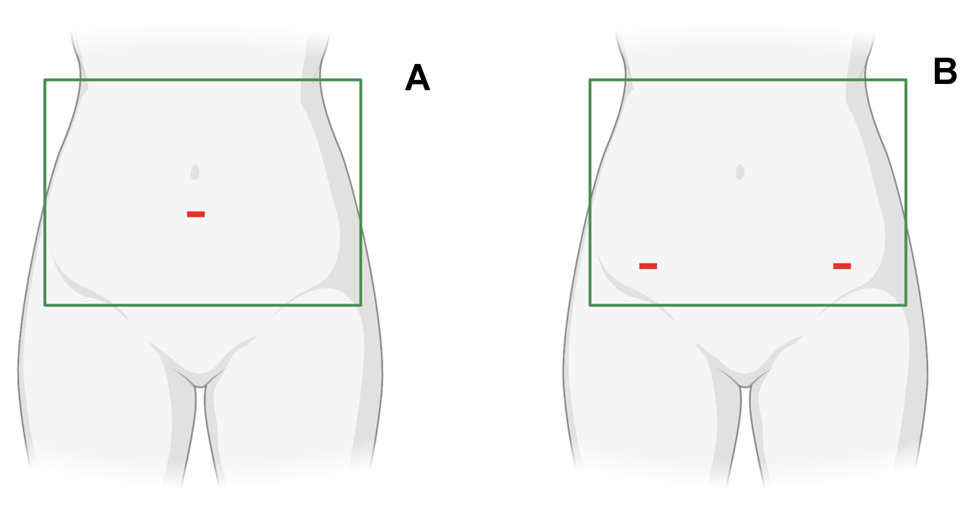
The patient is positioned supine on the operating table with arms abducted at 90° to ensure adequate access to the abdomen. Sterile draping is performed using four towels to delimit a rectangular field between the 12th ribs proximally, the anterior superior iliac spines distally, and the flanks laterally. The surgeon may stand on either side of the surgical field depending on personal preference, presence of scars, incision planning, or concomitant arthroscopy.

3.3. Liposuction

According to Hunstad et al. [11], abdominal liposuction can be performed through a single infraumbilical incision (Figure 1A) or two bilateral incisions at the bikini line (Figure 1B). More specifically, the latter are preferred in case of defects of the abdominal wall at the median line and due to superior aesthetic outcomes as the two small scars can be comfortably covered by underwear. In both cases, subcentimetric incisions will allow the entire lower abdomen to be adequately suctioned.
After selecting the most appropriate surgical site(s), 5 mL of 10 mg/mL lidocaine are subcutaneously injected before incision to reduce wound-related pain. Subsequently, approximately 200 mL of a tumescent solution (500 mL 0.9% saline, 20 mL mepivacaine 10 mg/mL, and 0.7 mL 0.1% epinephrine) are infiltrated in the abdominal subcutaneous fat and, after 10 min, liposuction is performed using the syringe technique [14]. Briefly, a liposuction cannula is connected to a 50 mL Luer Lock syringe and inserted in the subcutaneous fat layer. Subsequently, the plunger is pulled and kept in place using a towel clamp or a locking ring, thus creating the vacuum for suctioning. Liposuction is then performed until an adequate amount of lipoaspirate is obtained for further processing (~50 mL per joint to be injected). Once liposuction has been concluded, skin wounds are closed with a 5-0 nylon suture.

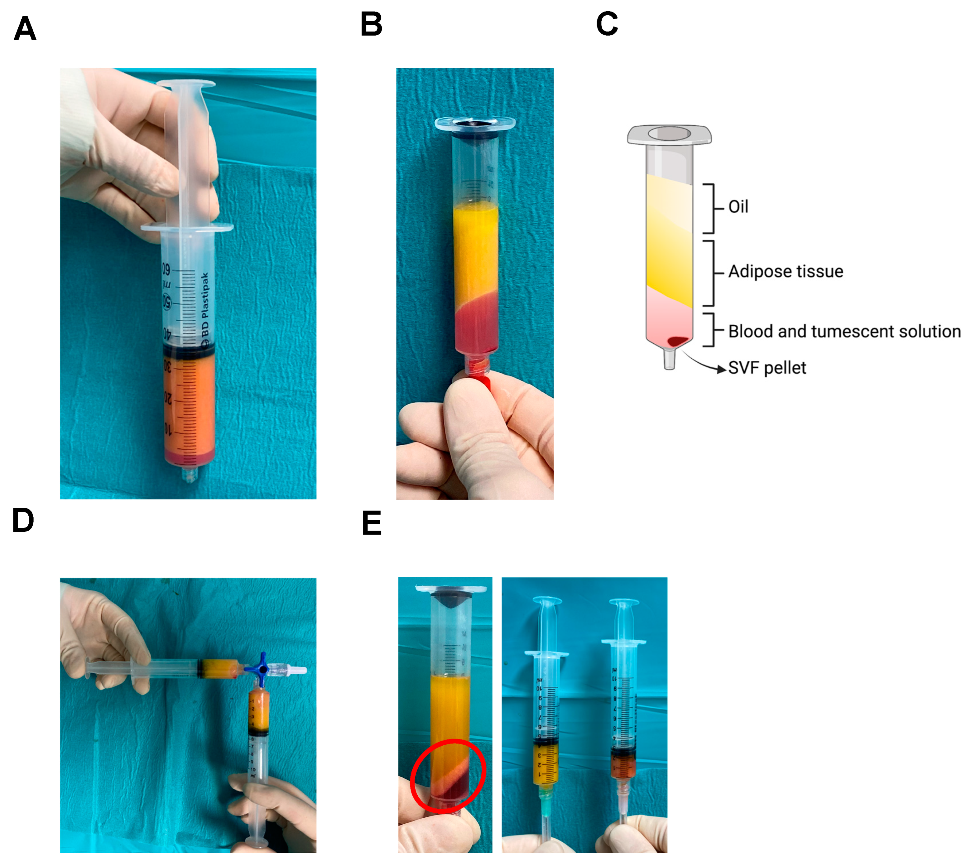
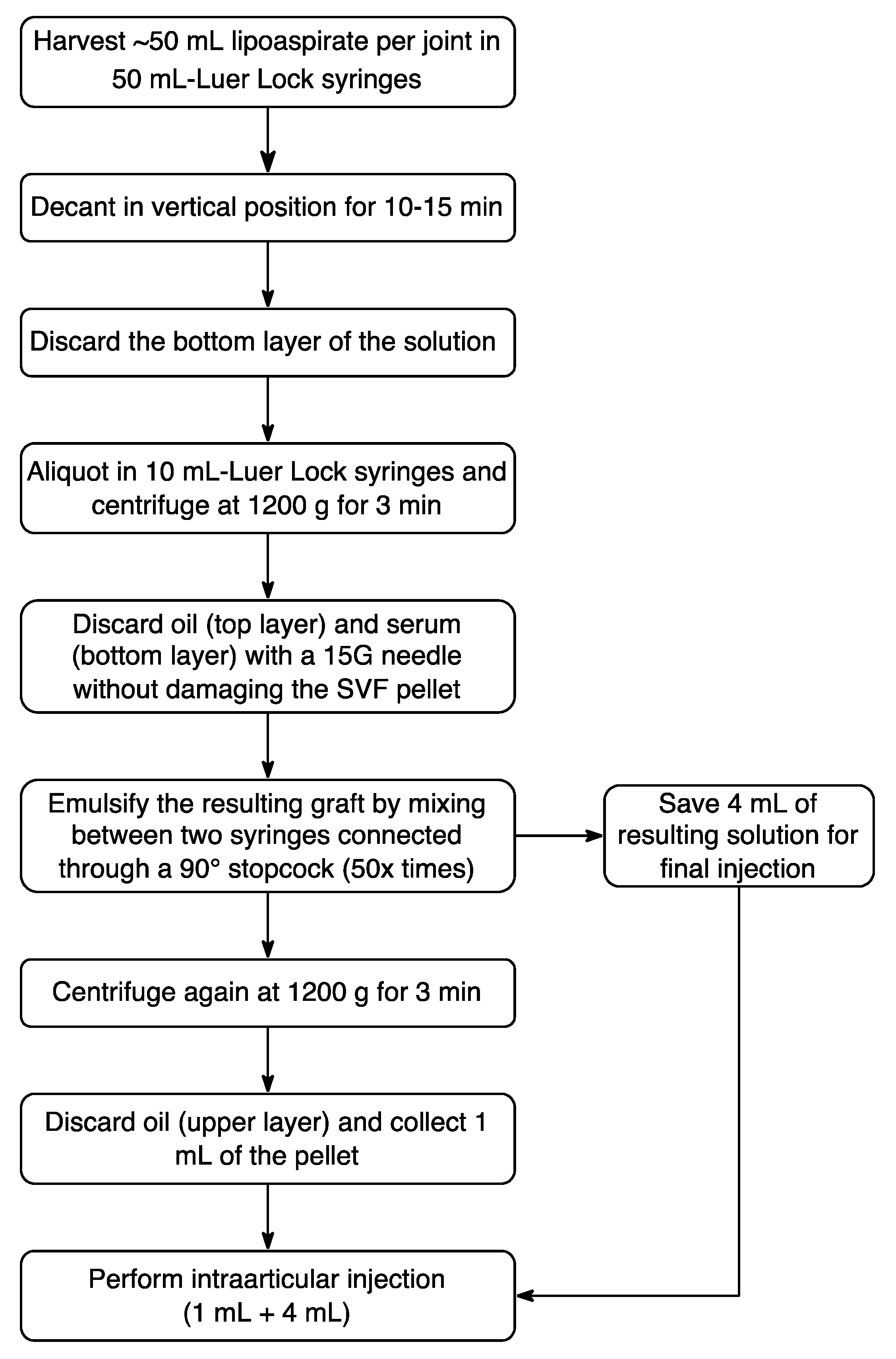
3.4. Graft Preparation

After liposuction, the 50 mL Luer Lock syringes are sealed with Combi-stoppers and kept in vertical position to allow lipoaspirate decantation (Figure 2A). After 10–15 min, the lower layer, mainly composed of tumescent solution and blood, is discarded. Subsequently, the resulting graft is aliquoted in 10 mL Luer Lock syringes and centrifuged at 1200× g for 3 min at room temperature. After this step, a four-phase solution will be obtained (Figure 2B,C): the oil in the upper part and the serum in the lower part are removed using a 15-gauge intravenous catheter needle, carefully keeping the adipose part and the pellet containing the SVF. The remaining graft is then emulsified through energic mixing between two syringes with a 90° stopcock for at least fifty times (Figure 2D). In total, 4 mL of the resulting solution are transferred to a 5 mL Luer Lock syringe for the final injection. The remaining volume is centrifuged again at 1200× g for 3 min at room temperature to obtain a solution composed of a more abundant upper layer of oil, which is discarded, and a lower portion containing the SVF pellet (Figure 2E, left image). The final solution injected consists of 4 mL previously obtained and approximately 1 mL of the pellet, which is transferred to another 5 mL Luer Lock syringe (Figure 2E, right image).

3.5. Intraarticular Injection and Post-Operative Care

Considering the volume obtained and the high viscosity of the graft, we recommend injecting large joints only, such as the shoulder, the knee, the hip, and the ankle. The use of a 20-gauge needle is recommended. Intraarticular injection should be performed in aseptic conditions according to standard protocols. If the procedure is performed in conjunction with arthroscopy, arthroscopic portals can be utilized to perform the injection. In this case, the use of intraarticular drains should be avoided due to the risk of graft loss. A compressive dressing with elastic bandages is applied to the abdomen before leaving the operating room.
Patients are observed for at least 3 h and monitored for perioperative complications, post-operative pain flares, and recovery from anesthesia. Subsequently, they are discharged home the same day and advised to wear an elastic compression garment for the following two weeks to reduce edema and minimize bruising. Low-molecular-weight heparin is prescribed for 14 days in all patients receiving intraarticular injections in weight-bearing joints (e.g., hip, knee, and ankle). Recommendations regarding physical exercise and weight bearing are individualized based on patients’ previous degree of activity and severity of underlying OA. Generally, minimal weight bearing for 2 days and light physical activity for 3 weeks are recommended. Patients are routinely checked back in the office at 1, 2, and 4 weeks. During postoperative visits, pain, discomfort, and range of motion of the injected joint(s) are evaluated, as well as wound healing and related issues, if any. The whole procedure is summarized step by step in the algorithm provided below (Figure 3).

4. Discussion

In recent decades, the use of intraarticularly transplanted MSCs for the treatment of OA has been increasingly reported in both preclinical and clinical studies. Once in the joint environment, these cells have shown the capacity to differentiate towards the chondrogenic lineage, and secrete several growth factors as well as anti-inflammatory cytokines, thus promoting tissue anabolism and relieving pain [15]. Historically, bone-marrow-derived MSCs (BM-MSCs) have been mostly investigated for their promising effects on joint regeneration. Previous evidence has demonstrated that BM-MSCs are able to release soluble factors that reduce tissue inflammation and stimulate resident cell metabolism as well as the differentiation of local progenitors. Furthermore, they are also capable of directly differentiating into chondrocytes and replenishing the cell population at the injected site [16]. However, BM-MSC isolation and transplantation are also affected by considerable disadvantages, including low yield, reduced growth rate, and invasiveness of harvest [17]. Among accessible MSC sources, the adipose tissue is characterized by wide availability, minimal invasiveness of harvesting, and higher MSC yield (>20,000 cells per gram of tissue), compared to the bone marrow (6000–60,000 cells per mL of tissue) [18]. Furthermore, ASCs can be isolated in combination with other biologically potent cells in the form of SVF [5]. More specifically, the SVF is a heterogenous product composed of several cell types, including ASCs, macrophages, endothelial cells, pericytes, preadipocytes, fibroblasts, and blood cells. Collectively, SVF distinct cellular components exert synergistic effects with anabolic, immunomodulatory, anti-apoptotic, anti-inflammatory, and anti-fibrogenic properties [19]. Currently, several devices and kits are available to promptly extract and utilize SVF intraoperatively for regenerative medicine purposes. Generally, the SVF can be harvested from the lipoaspirate obtained during a routine liposuction after proper processing through mechanical fragmentation or enzymatic digestion [20]. However, both approaches usually require costly consumables and dedicated disposable, expensive kits.
In 2017, Persichetti et al. [6] developed a novel technique to obtain a SVF graft with a considerably high concentration of ASCs and without the need of any specific high-end equipment. Indeed, the proposed strategies only require a standard portable centrifuge, Luer Lock syringes, syringe caps, a stopcock, and needles, resulting in a 10-fold to 50-fold lower expense compared to available kits. The described approach is a revision of the technique originally introduced by Tonnard and colleagues, named “nanofat”. Compared to other fragmentation-based strategies, by emulsification through energic mixing between two parallel syringes, nanofat allows to break mature adipocytes while preserving ASC viability. More specifically, the rupture of adipocytes with the release of oil and their subsequent removal further increases ASC concentration within the graft [21]. Nonetheless, the lower amount of mature adipocytes and oil reduces oxidative stress and related inflammation following SVF implantation [22]. Previous studies have demonstrated that the nanofat graft contains a high concentration of CD34+-ASCs, anti-inflammatory cytokines (interleukin [IL]-4, IL-10, IL-1 receptor antagonist), growth factors (platelet-derived growth factor [PDGF], vascular endothelial growth factor [VEGF], insulin-like growth factor 1 [IGF-1], basic fibroblast growth factor [bFGF], transforming growth factor β [TGF-β]), and microvascular fragments [23]. Collectively, these biologically potent components likely mediate nanofat strong regenerative effects described in the recent literature [23]. Compared to the technique described by Tonnard et al. [21] and later modified by Takanobu and coauthors [22], the approach here described, by introducing a 90° emulsification instead of a 180° parallel fragmentation as originally reported, significantly increases ASC concentration up to 69%, thus possibly further enhancing the graft regenerative potential [6]. Indeed, in a recent in vitro study, we have demonstrated that, compared to raw lipoaspirate and mechano-fragmented SVF obtained with a commercially available kit, the graft obtained with this technique is characterized by the highest levels of ECM content, replicating ASCs, stemness gene expression, and the lowest rate of pro-inflammatory cytokines [7].
Safety and effectiveness of SVF in treating OA have also been demonstrated in preliminary clinical studies. Michalek et al. [24] have showed that patients affected by either moderate-to-severe hip or knee OA reported a significant improvement of knee injury and osteoarthritis outcome score/hip disability, and osteoarthritis outcome scores (KOOS/HOOS) in 80.6% of cases at 3 months and 91% of cases at 12 months from SVF injection. Furthermore, no serious side effects were reported at a follow-up of up to 4.5 years.
Although methodologically comparable to other available techniques, the approach here described allows one to obtain a biologically potent product at a very limited cost, which may extend the availability of regenerative treatments to a wider population. However, the safety and effectiveness of our graft needs to be further confirmed in the next future.

5. Conclusions

The intraarticular injection of ASC-rich products is a promising technique to tackle OA degeneration. Compared to available strategies, our approach is easy to perform, cost-effective, and able to produce a graft with a high concentration of ASCs and a potent biological activity. Further studies are needed to validate the efficacy of this technique in a wider clinical setting.

6. Patents

The technique described in this study is adapted from the following patent: Persichetti P, Marangi GF, Segreto F, Pantano F, Tirindelli MC, and Gregorj C. Adipose fabric purification technique to obtain high concentration of adipose stem cells. N. IT201700003805A1.

Author Contributions

Conceptualization, L.A.; writing—original draft preparation, L.A.; writing—review and editing, L.A., S.C., F.R., G.F.P. and G.V.; supervision, R.P. and V.D. All authors have read and agreed to the published version of the manuscript.

Funding

This research received no external funding.

Institutional Review Board Statement

The study was conducted in accordance with the Declaration of Helsinki and approved by the Ethics Committee of Università Campus Bio-Medico di Roma (protocol code n. 21.19 TS).

Informed Consent Statement

Not applicable.

Data Availability Statement

Not applicable.

Conflicts of Interest

The authors declare no conflict of interest.

References

  1. Martel-Pelletier, J.; Barr, A.J.; Cicuttini, F.M.; Conaghan, P.G.; Cooper, C.; Goldring, M.B.; Goldring, S.R.; Jones, G.; Teichtahl, A.J.; Pelletier, J.P. Osteoarthritis. Nat. Rev. Dis. Prim. 2016, 2, 16072. [Google Scholar] [CrossRef] [PubMed] [Green Version]
  2. Glyn-Jones, S.; Palmer, A.J.; Agricola, R.; Price, A.J.; Vincent, T.L.; Weinans, H.; Carr, A.J. Osteoarthritis. Lancet 2015, 386, 376–387. [Google Scholar] [CrossRef] [PubMed]
  3. De Francesco, F.; Gravina, P.; Busato, A.; Farinelli, L.; Soranzo, C.; Vidal, L.; Zingaretti, N.; Zavan, B.; Sbarbati, A.; Riccio, M.; et al. Stem Cells in Autologous Microfragmented Adipose Tissue: Current Perspectives in Osteoarthritis Disease. Int. J. Mol. Sci. 2021, 22, 10197. [Google Scholar] [CrossRef]
  4. Lopa, S.; Colombini, A.; Moretti, M.; de Girolamo, L. Injective mesenchymal stem cell-based treatments for knee osteoarthritis: From mechanisms of action to current clinical evidences. Knee Surg. Sport. Traumatol. Arthrosc. 2018, 27, 2003–2020. [Google Scholar] [CrossRef] [Green Version]
  5. Nguyen, A.; Guo, J.; Banyard, D.A.; Fadavi, D.; Toranto, J.D.; Wirth, G.A.; Paydar, K.Z.; Evans, G.R.D.; Widgerow, A.D. Stromal vascular fraction: A regenerative reality? Part 1: Current concepts and review of the literature. J. Plast. Reconstr. Aesthetic Surg. 2016, 69, 170–179. [Google Scholar] [CrossRef]
  6. Persichetti, P.; Marangi, G.F.; Segreto, F.; Pantano, F.; Tirindelli, M.C.; Gregorj, C. Adipose Tissue Purification Technique to Obtain High Concentration of Adipose Stem Cells. Patent IT201700003805A1, 2017. [Google Scholar]
  7. Cicione, C.; Vadalà, G.; Di Giacomo, G.; Tilotta, V.; Ambrosio, L.; Russo, F.; Zampogna, B.; Cannata, F.; Papalia, R.; Denaro, V. Micro-fragmented and nanofat adipose tissue derivatives: In vitro qualitative and quantitative analysis. Front. Bioeng. Biotechnol. 2023, 11, 911600. [Google Scholar] [CrossRef]
  8. Beidas, O.E.; Gusenoff, J.A. Update on Liposuction: What All Plastic Surgeons Should Know. Plast. Reconstr. Surg. 2021, 147, 658e–668e. [Google Scholar] [CrossRef]
  9. Strong, A.L.; Cederna, P.S.; Rubin, J.P.; Coleman, S.R.; Levi, B. The Current State of Fat Grafting. Plast. Reconstr. Surg. 2015, 136, 897–912. [Google Scholar] [CrossRef] [Green Version]
  10. Kaoutzanis, C.; Gupta, V.; Winocour, J.; Layliev, J.; Ramirez, R.; Grotting, J.C.; Higdon, K. Cosmetic Liposuction: Preoperative Risk Factors, Major Complication Rates, and Safety of Combined Procedures. Aesthetic Surg. J. 2017, 37, 680–694. [Google Scholar] [CrossRef] [PubMed]
  11. Hunstad, J.P.; Aitken, M.E. Liposuction: Techniques and Guidelines. Clin. Plast. Surg. 2006, 33, 13–25. [Google Scholar] [CrossRef]
  12. Kucera, I.J.; Lambert, T.J.; Klein, J.A.; Watkins, R.G.; Hoover, J.M.; Kaye, A.D. Liposuction: Contemporary issues for the anesthesiologist. J. Clin. Anesth. 2006, 18, 379–387. [Google Scholar] [CrossRef]
  13. Boeni, R.; Waechter-Gniadek, P.v. Safety of Tumescent Liposuction Under Local Anesthesia in 9002 Consecutive Patients. Dermatol. Surg. 2021, 47, e184–e187. [Google Scholar] [CrossRef] [PubMed]
  14. Toledo, L.S. Syringe liposculpture: A two-year experience. Aesthetic Plast. Surg. 1991, 15, 321–326. [Google Scholar] [CrossRef] [PubMed]
  15. Tevlin, R.; desJardins-Park, H.; Huber, J.; DiIorio, S.E.; Longaker, M.T.; Wan, D.C. Musculoskeletal tissue engineering: Adipose derived stromal cell implementation for the treatment of osteoarthritis. Biomaterials 2022, 286, 121544. [Google Scholar] [CrossRef]
  16. Doyle, E.C.; Wragg, N.M.; Wilson, S.L. Intraarticular injection of bone marrow-derived mesenchymal stem cells enhances regeneration in knee osteoarthritis. Knee Surg. Sport. Traumatol. Arthrosc. 2020, 28, 3827–3842. [Google Scholar] [CrossRef] [PubMed] [Green Version]
  17. Zhu, C.; Wu, W.; Qu, X. Mesenchymal stem cells in osteoarthritis therapy: A review. Am. J. Transl. Res. 2021, 13, 448–461. [Google Scholar] [PubMed]
  18. Zhu, Y.; Liu, T.; Song, K.; Fan, X.; Ma, X.; Cui, Z. Adipose-derived stem cell: A better stem cell than BMSC. Cell Biochem. Funct. 2008, 26, 664–675. [Google Scholar] [CrossRef]
  19. Boada-Pladellorens, A.; Avellanet, M.; Pages-Bolibar, E.; Veiga, A. Stromal vascular fraction therapy for knee osteoarthritis: A systematic review. Ther. Adv. Musculoskelet. Dis. 2022, 14, 1759720X221117879. [Google Scholar] [CrossRef]
  20. Sharma, S.; Muthu, S.; Jeyaraman, M.; Ranjan, R.; Jha, S.K. Translational products of adipose tissue-derived mesenchymal stem cells: Bench to bedside applications. World J. Stem Cells 2021, 13, 1360–1381. [Google Scholar] [CrossRef]
  21. Tonnard, P.; Verpaele, A.; Peeters, G.; Hamdi, M.; Cornelissen, M.; Declercq, H. Nanofat grafting: Basic research and clinical applications. Plast Reconstr. Surg. 2013, 132, 1017–1026. [Google Scholar] [CrossRef]
  22. Mashiko, T.; Wu, S.H.; Feng, J.; Kanayama, K.; Kinoshita, K.; Sunaga, A.; Narushima, M.; Yoshimura, K. Mechanical Micronization of Lipoaspirates: Squeeze and Emulsification Techniques. Plast Reconstr. Surg. 2017, 139, 79–90. [Google Scholar] [CrossRef] [PubMed]
  23. Jeyaraman, M.; Muthu, S.; Sharma, S.; Ganta, C.; Ranjan, R.; Jha, S.K. Nanofat: A therapeutic paradigm in regenerative medicine. World J. Stem Cells 2021, 13, 1733–1746. [Google Scholar] [CrossRef] [PubMed]
  24. Michalek, J.; Moster, R.; Lukac, L.; Proefrock, K.; Petrasovic, M.; Rybar, J.; Chaloupka, A.; Darinskas, A.; Michalek, J.; Kristek, J.; et al. Stromal Vascular Fraction Cells of Adipose and Connective Tissue in People with Osteoarthritis: A Case Control Prospective Multi-Centric Non-Randomized Study. Glob. Surg. 2017, 3, 1–9. [Google Scholar] [CrossRef]
Figure 1. Abdominal liposuction may be performed using either a single infraumbilical access (A) or two bilateral incisions at the bikini line (B). The green boxes indicate the surgical field. Created with www.biorender.com.
Std 12 00005 g001
Figure 2. Lipoaspirate is decanted in vertical position (A) to separate the fat (upper layer) and from the tumescent solution with blood (lower layer). After centrifugation, four different layers appear (B), as schematically represented in (C). Oil and blood with tumescent solution are discarded, and the remaining graft is emulsified by energic mixing between two syringes using a 90° stopcock (D). After saving 4 mL of the graft, the remaining volume is centrifuged again, hence obtaining a solution with a solid pellet (red circle) on the bottom ((E), left image). The final volume to be injected consists of 4 mL previously obtained and approximately 1 mL of the pellet ((E), right image). Created with www.biorender.com.
Std 12 00005 g002
Figure 3. Step-by-step summary of graft preparation and processing.
Std 12 00005 g003
Disclaimer/Publisher’s Note: The statements, opinions and data contained in all publications are solely those of the individual author(s) and contributor(s) and not of MDPI and/or the editor(s). MDPI and/or the editor(s) disclaim responsibility for any injury to people or property resulting from any ideas, methods, instructions or products referred to in the content.

