
Emerging Optical Materials in Sensing and Discovery of Bioactive Compounds
by
 , and
, and
Raquel Vaz
1,2 ,
,
Beatriz Valpradinhos
2,3,
Manuela F. Frasco
1,2,3,* and
Maria Goreti F. Sales
1,2,3
1
BioMark@UC, Faculty of Sciences and Technology, University of Coimbra, 3030-790 Coimbra, Portugal
2
CEB—Centre of Biological Engineering, University of Minho, 4710-057 Braga, Portugal
3
BioMark@ISEP, School of Engineering, Polytechnic Institute of Porto, 4249-015 Porto, Portugal
*
Author to whom correspondence should be addressed.
Sensors 2021, 21(17), 5784; https://doi.org/10.3390/s21175784
Submission received: 21 July 2021
/
Revised: 20 August 2021
/
Accepted: 22 August 2021
/
Published: 27 August 2021
(This article belongs to the Special Issue Advanced Biosensing of Natural Bioactive Compounds)
Abstract
:Optical biosensors are used in numerous applications and analytical fields. Advances in these sensor platforms offer high sensitivity, selectivity, miniaturization, and real-time analysis, among many other advantages. Research into bioactive natural products serves both to protect against potentially dangerous toxic compounds and to promote pharmacological innovation in drug discovery, as these compounds have unique chemical compositions that may be characterized by greater safety and efficacy. However, conventional methods for detecting these biomolecules have drawbacks, as they are time-consuming and expensive. As an alternative, optical biosensors offer a faster, simpler, and less expensive means of detecting various biomolecules of clinical interest. In this review, an overview of recent developments in optical biosensors for the detection and monitoring of aquatic biotoxins to prevent public health risks is first provided. In addition, the advantages and applicability of these biosensors in the field of drug discovery, including high-throughput screening, are discussed. The contribution of the investigated technological advances in the timely and sensitive detection of biotoxins while deciphering the pathways to discover bioactive compounds with great health-promoting prospects is envisaged to meet the increasing demands of healthcare systems.
1. Introduction
Biotoxins are biological toxic substances produced by various organisms, namely, animals (animal toxins), plants (phytotoxins), and microorganisms (mycotoxins, cyanotoxins, and toxins of dinoflagellates and diatoms). These chemical substances are produced in stress, predation, and defense situations [1]. In particular, aquatic natural toxins, which include freshwater and marine sources (such as algae, coelenterates, reef fish, dinoflagellates, and cyanobacteria), pose a serious threat to public health through exposure by inhalation of aerosolized toxins, dermal absorption, or transmission through the food chain. Ingestion of biotoxins leads to gastrointestinal, neurological, and cardiovascular syndromes, which in severe cases can result in death. To prevent poisoning from aquatic biotoxins, seafood and shellfish meat must be monitored, and the levels of toxin-producing microorganisms in the water must be detected before marketing. In addition to human health, environmental poisoning can also cause fatalities in fish, birds, and marine mammals [2]. For example, microcystins produced by cyanobacteria can kill animals living in eutrophic freshwater ecosystems or accumulate in mollusks, fish, and crayfish consumed by humans [3]. Additionally, increase in harmful algal blooms, possibly due to rising ocean temperatures, anthropogenic pressures, and increasing coastal eutrophication, contaminates various water sources, especially drinking water, and causes poisoning in animals and humans [4].
Aquatic biotoxins and their analogues can be classified according to their chemical structure, source of origin, or mechanism of toxicity (Table 1). In the first classification, biotoxins are usually alkaloids, polyethers, or peptides. For example, tetrodotoxin and saxitoxin are marine alkaloids and are neurotoxins [5]. Regarding the source of the toxin, there are, for example, microcystins from cyanobacteria [6], conotoxin from species of the genus Conus [7], tetrodotoxin from pufferfish [8], and dinophysistoxins from species of the genus Dinophysis [9]. The mechanism of toxicity includes the human syndromes that biotoxins cause: diarrhetic shellfish poisoning (DSP), caused by okadaic acid, dinophysistoxins, pectenotoxin, and yessotoxin; paralytic shellfish poisoning (PSP), caused by saxitoxin; amnesic shellfish poisoning (ASP), caused by domoic acid; azaspiracid shellfish poisoning (AZP), caused by azaspiracid; ciguatera fish poisoning (CFP) due to ciguatoxins; and neurotoxic shellfish poisoning (NSP) due to brevetoxins [5,10].
Although toxins are usually considered harmful, many aquatic biotoxins hold great pharmaceutical potential, the value of which has only been recognized in recent years with the growing need for new drugs. Available drugs are effective in only one-third of diseases, and pathogens have developed resistance to them. Therefore, new biologically active compounds with improved therapeutic activities should be considered. For example, the aquatic biotoxin produced by cone snails, conotoxin, has great therapeutic potential in the discovery of new analgesics to treat visceral pain associated with irritable bowel syndrome and inflammatory bowel disease because it is a selective toxin for the N-type voltage-gated calcium channels, which are among the most important molecular modulators of visceral pain [11].
However, most of the attention has been paid to toxins of cyanobacteria because cultivation of cyanobacteria for drug discovery is less expensive compared with other microorganisms. Cyanobacteria are a group of Gram-negative photoautotrophic prokaryotes that can produce diverse secondary metabolites, including lipopeptides, amino acids, macrolides, amides, among others. This variety of compounds leads to a wide range of bioactivities, such as antibacterial, antifungal, antiviral, and anticarcinogenic [12]. For example, microcystin-LR is a cyclopeptide produced by a wide variety of cyanobacteria. It has been shown to target pancreatic cancer cells overexpressing the organic anion transporting polypeptides 1B1 and 1B3, inhibiting cancer proliferation [13].
Dinoflagellates are unicellular planktonic microalgae that have also been the subject of several studies. They produce a wide range of natural biotoxins, some of which are common to cyanobacteria, such as saxitoxin, having unique and valuable potential for the development of new drugs [14]. For example, saxitoxin acts as a sodium channel blocker, preventing the influx flow of sodium ions. It has been shown to have anesthetic properties [15], which could be used for several days to block the sciatic nerve as a pain treatment with low cytotoxicity [16]. Brevetoxin is a biotoxin that has been previously shown to promote neural repair after ischemic stroke in mice by enhancing dendritic arborization, synapse density, and motor recovery [17]. Pectenotoxin-2 is another relevant toxin produced by dinoflagellates that could be valuable as a chemotherapeutic agent, as it leads to depolymerization of actin filaments and activates an intrinsic pathway of apoptosis in p53-deficient tumor cells [18], while it has also been shown to be more effective against cancer cells than against normal cells of the same tissue [19]. Gymnodimines are a type of macrocyclic imine toxins that can act as cholinergic antagonists and have been shown to decrease the accumulation of intracellular amyloid beta-peptide and hyperphosphorylated forms of tau protein in cortical neurons in vitro, suggesting a potential pharmaceutical approach in Alzheimer’s disease [20]. Other reports indicate that gymnodimines have the potential to sensitize a neuroblastoma cell line to the apoptotic effects of okadaic acid, another algal toxin [21].

Table 1.
Classification of common aquatic biotoxins, their toxicity, and frequently observed adverse symptoms in human poisoning.
Table 1.
Classification of common aquatic biotoxins, their toxicity, and frequently observed adverse symptoms in human poisoning.
| Biotoxin | Chemical Structure | Source | Toxic Syndrome | Toxicological Effects | Ref. |
|---|---|---|---|---|---|
| Actinoporins | Peptide | Anemones, jellyfish | - | Severe pain, hypotension, and cardiac irregularities | [22,23] |
| Amphidinolides | Polyether | Dinoflagellates | - | Cytotoxicity | [24,25] |
| Azaspiracid | Polyether | Dinoflagellates | AZP | Gastrointestinal and neurological symptoms | [26] |
| Brevetoxin | Polyether | Dinoflagellates | NSP | Gastrointestinal and neurological symptoms, respiratory problems, and muscular pain | [27] |
| Ciguatoxin | Polyether | Dinoflagellates | CFP | Gastrointestinal, neurological, and cardiovascular symptoms | [27] |
| Conotoxin | Peptide | Cone snails | - | Muscle paralysis of the diaphragm, and alteration of blood pressure | [7,28] |
| Dinophysistoxins | Polyether | Dinoflagellates | DSP | Gastrointestinal symptoms | [9] |
| Domoic acid | Cyclic amino acid | Diatoms | ASP | Gastrointestinal and neurological symptoms | [29] |
| Gambieric acid | Polyether | Dinoflagellates | - | Cytotoxicity | [30,31] |
| Gambierol | Polyether | Dinoflagellates | CFP | Gastrointestinal disturbances and neurological alterations | [32] |
| Goniodomin A | Polyether | Dinoflagellates | - | Hepatotoxicity | [33] |
| Gymnocin | Polyether | Dinoflagellates | - | Cytotoxicity | [14,34] |
| Gymnodimine | Cyclic imine | Dinoflagellates | - | Neurological symptoms | [35,36] |
| Karlotoxin | Polyether | Dinoflagellates | - | Cytotoxicity | [37,38] |
| Maitotoxin | Polyether | Dinoflagellates | CFP | Neurological symptoms | [27] |
| Microcystin | Peptide | Cyanobacteria | - | Hepatotoxicity | [39] |
| Okadaic acid | Polyether | Dinoflagellates | DSP | Gastrointestinal symptoms | [14] |
| Palytoxin | Polyether | Zoantharians and dinoflagellates | - | Gastrointestinal, cardiac, and respiratory problems | [2,14,40] |
| Pectenotoxin | Polyether | Dinoflagellates | DSP | Gastrointestinal symptoms and hepatotoxicity | [41] |
| Saxitoxin | Alkaloid | Dinoflagellates and cyanobacteria | PSP | Gastrointestinal symptoms and respiratory paralysis | [14] |
| Spirolides | Cyclic imine | Dinoflagellates | - | Neurological symptoms | [35,42] |
| Tetrodotoxin | Alkaloid | Pufferfish and other species (e.g., starfish, gastropods, newts, and crabs) | - | Neurological symptoms | [8] |
| Yessotoxin | Polyether | Dinoflagellates | DSP | Gastrointestinal symptoms | [43] |
Yessotoxin is considered one of the most polar lipophilic toxins due to the presence of two sulfate groups. This toxin elicits several effects, such as modulation of intracellular calcium and cyclic adenosine monophosphate levels, caspase activation, fragmentation of E-cadherin, and alteration of the cytoskeleton [14]. Considering its pharmaceutical advantages, it has been shown to interfere with the apoptotic pathways of cancer cell lines, inhibiting melanoma tumor growth and causing strong toxicity to a lymphocytic leukemia cell line [44]. Yessotoxin can also cause genotoxicity and induce mitotic catastrophe, followed by cell death, including apoptotic and necrosis-like manner, indicating great potential to control tumor progression [45]. Another toxin isolated from dinoflagellates with interesting properties is amphidinol, which has antifungal and hemolytic activities [46]. A potent neurotoxin found primarily in pufferfish, tetrodotoxin is a selective blocker of voltage-gated sodium channels and has been administered intramuscularly to patients with cancer-related pain, showing successful results with mild secondary effects when present [47,48]. In addition, tetrodotoxin has been shown to alleviate symptoms in patients undergoing heroin withdrawal [49].
Unfortunately, there are still many difficulties in the thorough research of aquatic biotoxins and their use in the development of new drugs with higher efficacy, safety, tolerability, and convenience compared with existing drugs [50]. It is estimated that it takes about 14 years from the identification of the active compound and determination of its functionality to its approval in the market, which becomes very expensive and time-consuming. For this reason, it is necessary to implement new drug discovery alternatives that are more efficient, accurate, less expensive, and faster [51].
Biosensors are characterized by high sensitivity, robustness, speed, selectivity, ease of use, and cost-effectiveness. In particular, optical techniques have experienced significant growth in the research community, mainly because they enable low-cost miniaturized systems with reliable and fast responses. Therefore, optical biosensors have become an excellent method for detecting biological systems and are promoting development in drug discovery, clinical diagnosis, environmental monitoring, and other fields [52].
In this review, after introducing common aquatic-derived biotoxins that have been the subject of research as hazardous compounds but also as promising drugs for medicinal purposes, the focus is to provide a comprehensive article on the progress in optical biosensing for the detection of aquatic biotoxins and the technological advances that will enable the rapid development of a new generation of drugs. First, a brief description of the conventional analytical methods that have been used to date for the detection of aquatic bioactive compounds is given. Then, the advantages and importance of replacing conventional methods with optical biosensors are discussed. Their improved performance, miniaturization, and rapid response improve the detection and monitoring of biotoxins and also contribute to the identification of various biomolecules of clinical interest for new drug development. Interesting examples from the literature of the successful use of optical biosensors for these purposes are presented. Another focus is high-throughput screening (HTS) based on optical biosensors used to screen large compound libraries, especially label-free detection systems. It is expected that these technologies can be used to accelerate biomedical research in the field of bioactive natural products.
2. Conventional Methods to Detect Bioactive Natural Compounds
As mentioned above, it is imperative to actively monitor and detect aquatic biotoxins in order to avoid public health risk, contamination of various foods, and potential economic impact, in addition to identifying novel compounds useful for the development of effective drugs. These have the potential to fill the current gaps in pharmacological drugs with anticancer, anti-inflammatory, antifungal, or antibacterial properties without showing toxicity as a side effect. Conventional methods for the detection of biotoxins include animal- and cell-based assays, chemical methods (e.g., liquid chromatography (LC), high-performance liquid chromatography (HPLC), mass spectrometry (MS), and tandem mass spectrometry (MS/MS)), biochemical techniques such as immunoassays, and receptor-based methods [53,54].
2.1. Bioassays
The mouse bioassay (MBA) has been the most common bioassay method. In the MBA, mice are injected intraperitoneally with the sample extract to be tested for lethality. Although animal testing is still used worldwide for research purposes, the MBA has been replaced by alternative detection methods for screening lipophilic marine biotoxins (LMBs) and protecting public health [55]. This is due to ethical concerns, as well as high rates of false-positive and false-negative results, low sensitivity, and reduced reproducibility [55,56,57]. Alternative in vitro cell-based techniques, combined with chemical methods, have been investigated for the detection of LMBs [56]. For example, the neuro-2a assay uses murine neuro-2a neuroblastoma cells to determine cell viability upon exposure to LMBs, such as okadaic acid, dinophysistoxins, pectenotoxin-2, azaspiracids, and yessotoxins. Subsequently, this qualitative screening method is complemented by chemical analysis using LC–MS/MS as a reference method [56,58].
2.2. Chemical Assays
Separation of samples and their chemical analysis by HPLC is the most widely used method for detecting biotoxins because it is a sensitive, reliable, easy-to-calibrate, and accurate method. It also has the great advantage that it can be coupled with various detection systems, such as fluorescence detection and MS [1]. MS measures the mass-to-charge ratio of molecules present in the sample, allowing rapid analysis with high selectivity, a broad spectrum of samples, and identification of analogues of a particular toxin group. Recently, with the continuous advances in coupling LC and MS techniques, the need for toxin standards and derivatization reagents can be eliminated [1]. This is important because, for example, there are at least 24 saxitoxin analogues and 90 yessotoxin analogues, making it incredibly difficult to have reference materials for all of them [56]. Thus, LC–MS methods have been successfully used to detect various lipophilic toxin classes [57,59,60]. The sample can also be pretreated (e.g., with solid-phase extraction), which allows purification and preconcentration of analytes retained on a sorbent cartridge [1].
2.3. Biochemical Assays
An immunoassay is a biochemical assay based on the immunological affinity between an antibody and its antigen. The most commonly used immunoassay for the detection of biotoxins is the enzyme-linked immunosorbent assay (ELISA), in which binding between the biotoxin and its specific antibody is detected based on labeling with an enzyme that converts target recognition into a color reaction upon substrate catalysis. Commonly used labeling enzymes are horseradish peroxidase (HRP) and alkaline phosphatase. The main advantages of ELISA are the simple mechanism, accuracy, and easy equipment operation [1]. Examples of ELISA applications include the detection of yessotoxins in shellfish and algal samples [61], gonyautoxin (a PSP toxin) [62,63], and azaspiracid [64] from shellfish.
In the last decade, new designs of ELISA assays have appeared. For example, Zhang et al. (2012) developed a capillary electrophoresis-based immunoassay with electrochemical detection for saxitoxins in shellfish samples. After a competitive immunoreaction, capillary electrophoresis enabled the separation of the HRP-labeled antibody–antigen complex from the unbound labeled antigen, and then the HRP reaction was followed electrochemically [65]. Kim et al. (2015) prepared a lab-on-a-chip for the detection of saxitoxin using a competitive immunoassay. The biotoxin was captured by functionalized magnetic particles in a sample chamber where the sample and reactants were added. Subsequently, the solid-phase magnetic particles containing the bound toxins were magnetically conducted through the liquid-stationary phase into the detection chamber containing the HRP substrate [66]. Pelin et al. (2018) were able to conjugate cell-based assays with immunoenzymatic detection for the quantification of palytoxin. The biotoxin binds with high affinity to several cell lines and could then be detected by the addition of a specific antibody [67].
Despite advantages in simplicity, sensitivity, and selectivity, immunoassays are still associated with high laboratory and antibody preparation costs, antibody instability, and the possibility of false-positive or false-negative results [68].
3. Optical Biosensors for Detecting Aquatic Biotoxins
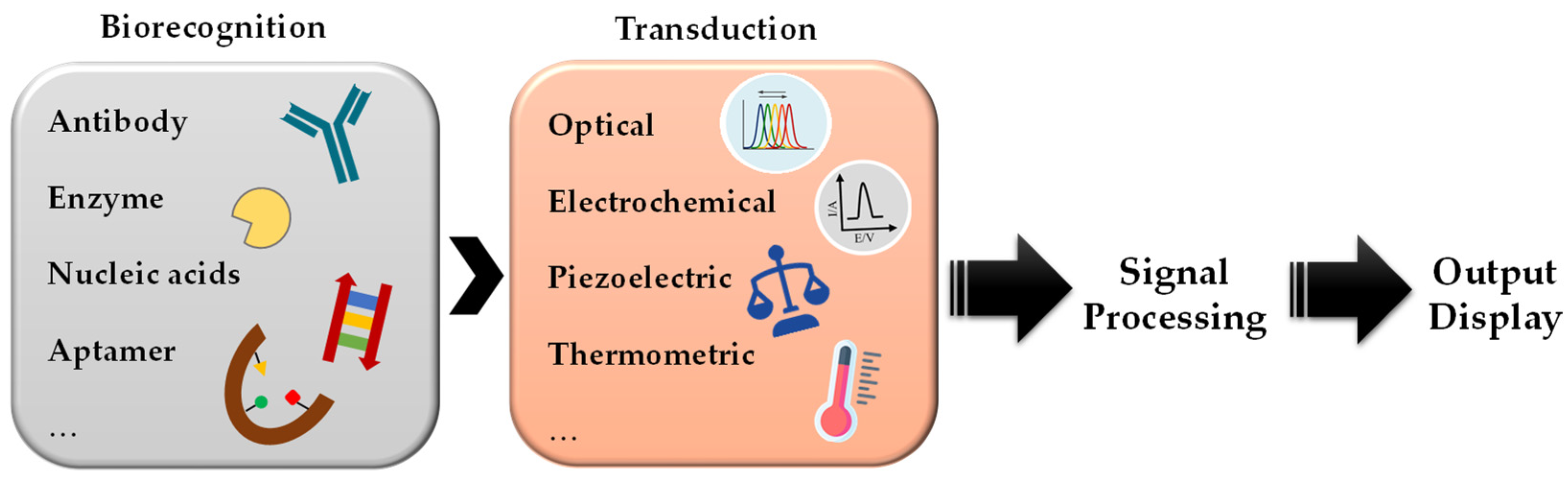
Although the traditional analytical methods described so far have a good detection success rate, there is a need for alternative methods that are cheaper, more sensitive, and faster and can be used as a screening tool to evaluate multiple samples in a reasonable amount of time. Biosensors are an alternative to these classical methods as they can detect a wide range of biotoxins in a sensitive and selective manner and can be used as portable and rapid techniques without the need for qualified personnel [69]. A biosensor is an analytical device consisting of a biorecognition element in conjunction with the transducer responsible for converting the recognition reaction into a measurable signal [70] (Figure 1). In this way, biosensors can be classified according to the bioreceptor element and the transducer type. In the first case, they can be classified as immunosensors, aptasensors, enzymatic sensors, nucleic acid sensors, and cell-based sensors [70]. Biosensors can also be categorized by physicochemical signal transduction, with electrochemical, optical, thermal, and piezoelectric sensors being the most common [71].
Although optical and electrochemical biosensors have the highest sensitivity and selectivity compared with the other methods mentioned, only the optical methods are considered in this work. Optical transducers involve a change in absorption, emission, transmission, scattering, reflection, or refraction of light that is proportional to the concentration of the target analyte. In addition, the optical change can be monitored with a label (e.g., a chromophore or fluorophore) or without a label, in which case they are referred to as label-free biosensors [72]. Optical biosensors have experienced a considerable growth for medical diagnosis, food quality control, and environmental monitoring, primarily because they can be designed as low-cost miniaturized systems with reliable and rapid responses [73]. There are many optical approaches, including colorimetric, photonic, fluorescent, surface-enhanced Raman spectroscopy (SERS), surface plasmon resonance (SPR), interferometers, and microresonators. The most appealing examples of the application of these optical methods for the detection of biotoxins are presented below and summarized in Table 2.
3.1. Colorimetric Biosensors
Colorimetric strategies for the detection of aquatic biotoxins mainly resort to metal nanoparticles and the change in their aggregation state in the presence of the bioanalyte, as well as the inhibition or activation of an enzyme in the presence of the target, both of which result in a color change. For example, microcystin-LR was successfully detected using specific aptamers as linkers for gold nanoparticle (AuNP) dimers. When the biotoxin was present, the aptamer changed its structure to bind the target, disassembling the dimer. As a result, there was a color shift from blue to red [76]. Li et al. (2016) also detected microcystin-LR, reaching a limit of detection (LOD) of 0.37 nmol L−1. In this work, the aptamer binds the AuNPs and protects them from aggregation. Since the aptamer binds the target microcystin-LR with high affinity upon sample loading, there is a displacement of the aptamer, causing the AuNPs to aggregate, resulting in a color change from red to blue [77]. A similar principle using AuNPs and a specific aptamer that reacts with saxitoxin allowed its detection, with a LOD of 10 fmol L−1, through aggregation of AuNPs and shift in color to blue [82].
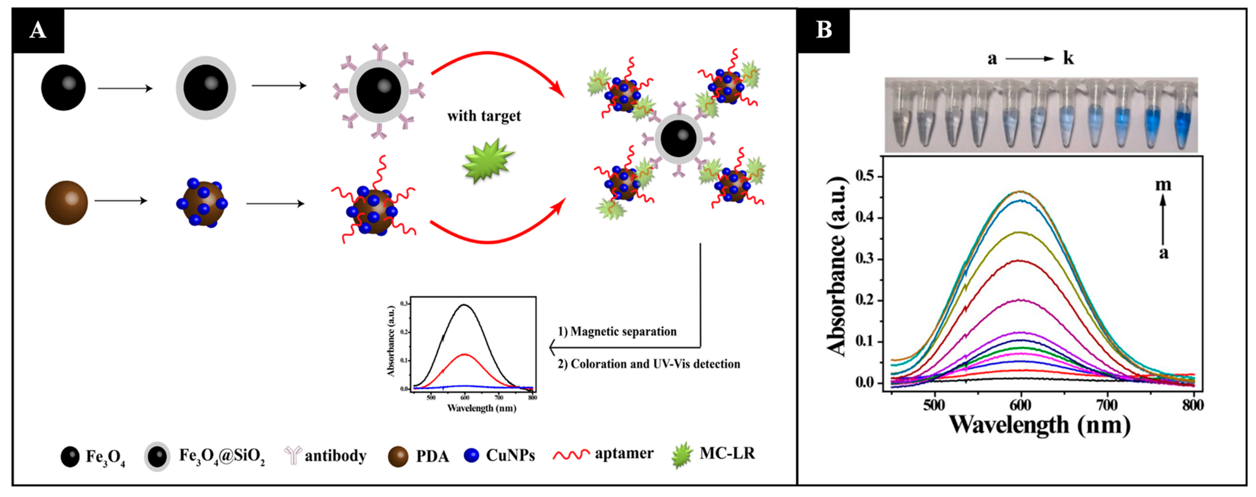
Recently, Tang et al. (2019) achieved a lower LOD of 0.05 nmol L−1 for microcystin-LR using a different strategy: antibody-functionalized silica-coated magnetic nanoparticles (Fe3O4@SiO2) and aptamer-functionalized polydopamine nanospheres decorated with copper nanoparticles (PDA/CuNPs) were developed. Here, in the presence of microcystin-LR in the samples, both nanoparticles were bound in sandwich-like composites and could be magnetically separated. Subsequently, the copper was converted to Cu2+, which reacted with bis(cyclohexanone)oxaldihydrazone, producing color and allowing a quantitative detection of the toxin [78] (Figure 2).
When colorimetric detection is triggered by an enzymatic reaction, the tests may rely on the inhibition of protein phosphatases or phosphodiesterases [79,80,83]. For example, Hayat et al. (2012) used the inhibition of phosphatase 2A in the presence of okadaic acid, which hindered the hydrolysis of p-nitrophenyl phosphate and prevented the formation of the yellow p-nitrophenol [80]. Other colorimetric immunoassays used glucose oxidase (GOx), AuNPs, and blue staining of oxidized 3,3′,5,5′-tetramethylbenzidine (TMB) [74,75]. Lai et al. (2016) presented a technique based on an enzyme-triggered Fenton reaction. In this work, a competitive immunoassay was performed using nanogold labeled with GOx and an antibody for brevetoxin B. When the target brevetoxin B was present, it competed with the immobilized brevetoxin B on magnetic beads for the labeled antibody. After magnetic separation, the carried enzyme oxidized the glucose, forming hydrogen peroxide, which later oxidized iron (II) to iron (III), also forming the radical hydroxyl; and finally, the resulting iron (III) and the radical oxidized TMB, forming a blue product. The absorbance decreased with an increasing concentration of sampled brevetoxin B [75]. In another study, okadaic acid was detected based on a direct competitive enzyme-linked aptamer assay (ELAA). The aptamer was first immobilized on a microplate and hybridized with the complementary sequence labelled with catalase. In the absence of okadaic acid, catalase consumes hydrogen peroxide. As the concentration of hydrogen peroxide decreases, the red solution of gold trichloric acid turns blue due to aggregated nanoparticles. However, in the presence of okadaic acid, the complementary sequence is replaced, resulting in a high concentration of hydrogen peroxide and a nonaggregated red solution of AuNPs [81].
3.2. Fluorescent Biosensors
Fluorescence strategies have been highly studied in the field of biotoxin monitoring. Many examples include the use of indirect detection by competitive assays [85,86,88,89,93], quenching of the fluorescent signal [87,90,127], sandwich assays [84], and aptamer binding [92]. Fluorescent biosensors have been developed not only for single detection but also for multiplex detection. For example, Bickman et al. (2018) designed a multiplex sensor specific for microcystin and cylindrospermopsin cyanotoxins [89]. Liu et al. (2017) were able to detect up to 32 contaminants in lake waters simultaneously, including microcystin-LR. The sensor was based on an integrated multichannel waveguide-based fluorescent sensor functionalized for different contaminants using an indirect competitive immunoassay [91].
The competitive assays were combined with fluorescence quenching/turn-on signal in a study to selectively and sensitively detect tetrodotoxin. For this purpose, the research team used competitive lateral flow immunochromatographic strips (C-LFICSs). The test line contained quantum dot nanobeads (QDNBs) conjugated to the protein BSA, as well as tetrodotoxin–BSA. The QDs were chosen because they have the advantage of higher signal brightness and stability compared with commercial organic fluorophores. Then, gold nanoflowers labeled with an antibody against tetrodotoxin and the sample were added to the C-LFICS so that they could move through the strip by capillarity. If the sample under analysis did not contain tetrodotoxin, the nanoflowers would bind to the tetrodotoxin on the test line, resulting in fluorescence quenching. In the positive case, the nanoflowers recognized the tetrodotoxin on the sample and did not bind to the tetrodotoxin on the test line, causing the fluorescence to remain on [94]. Gholami et al. (2020) based their sensor on fluorescence resonance energy transfer (FRET) between energy donors, carbon QDs (CQDs), and acceptors (AuNPs) for the detection of maitotoxin with low detection limit [87].
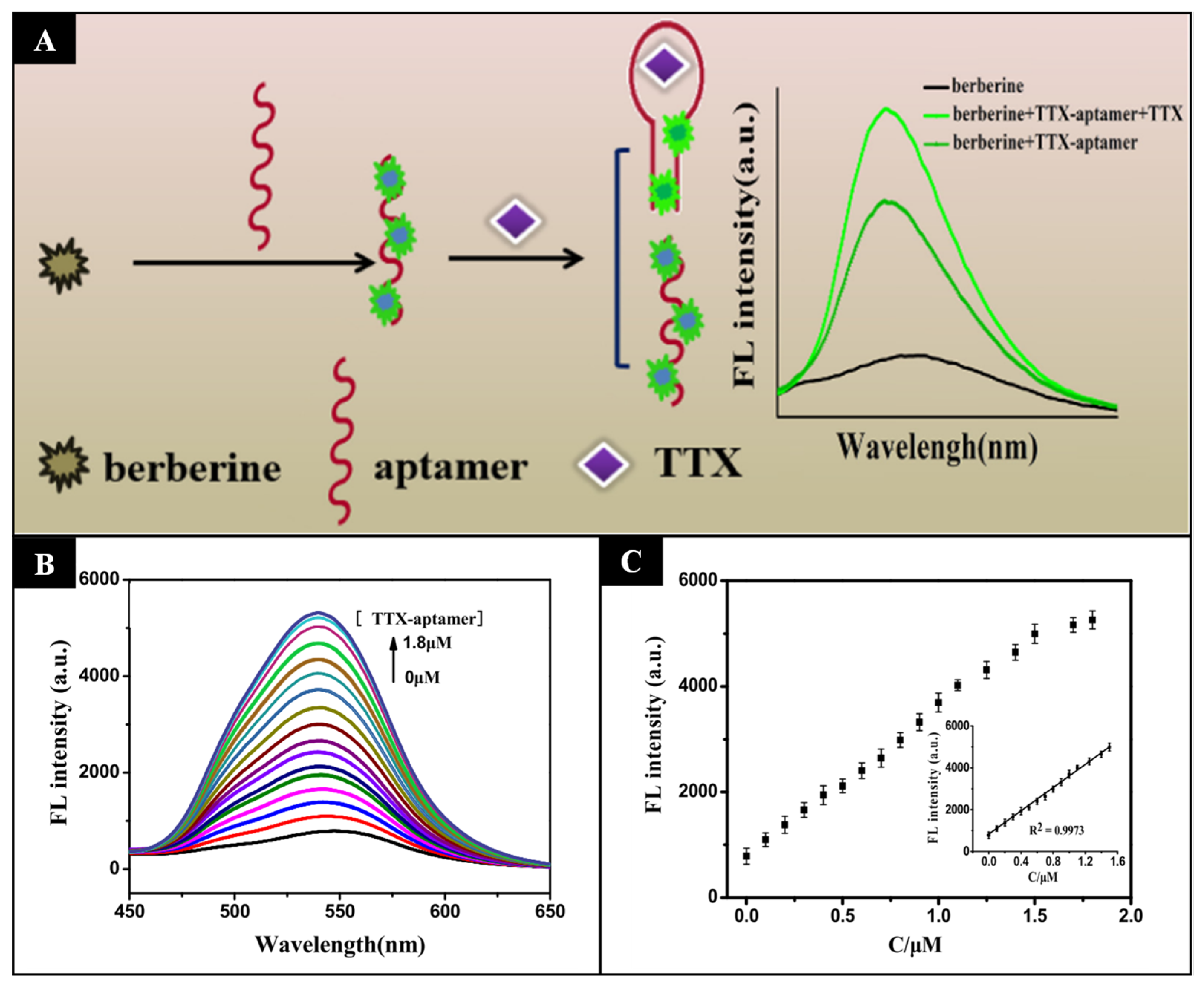
Lan et al. (2019) chose a different strategy and used a specific nucleic acid aptamer for tetrodotoxin that switches its conformational structure from a single-strand random coil to a compact neck ring structure in the presence of the biotoxin. Therefore, insertion of the fluorophore between the random coil leads to changes in the fluorescence signal due to conformational changes of the aptamer that depend on the concentration of tetrodotoxin. Using this method, a LOD of 0.074 nmol L−1 was determined [95] (Figure 3).
3.3. Surface-Enhanced Raman Scattering (SERS) Biosensors
SERS is a technique for enhancing Raman scattering of analyte molecules adsorbed on or in close proximity to SERS-active surfaces (i.e., rough or nanostructured metal surfaces, often classically gold, silver, or copper). This method offers high specificity and sensitivity and has been used to detect various toxins, such as saxitoxin [97,99,100], tetrodotoxin [102,103], domoic acid [97], okadaic acid, dinophysistoxins, yessotoxin [96], and microcystin-LR [98].
Saxitoxin shows a weak affinity for AuNPs or silver nanoparticles (AgNPs). Therefore, a surface modification on the SERS substrate was used, as is the case of cysteine-modified AuNPs (Cys-AuNPs). This modification, in coordination with a new method called dynamic SERS, in which self-assembly of nanoparticles is induced by solvent evaporation, increased the available 3D trapping wells for the biotoxin, resulting in a LOD of 0.1 µmol L−1 [101]. However, it showed lower sensitivity compared with the first reported SERS method for saxitoxin detection, developed by Pearman et al. (2008), who used a colloidal hydrosol of AgNPs as SERS substrate [99]. The sensitivity increased by combining SERS with laser optical tweezers Raman spectroscopy (LTRS) and reached a LOD of 2 nmol L−1. These results arise from the fact that laser optical tweezers are more efficient in capturing numerous AgNPs adsorbed on the saxitoxin molecule, thus enriching the Raman signal [100].
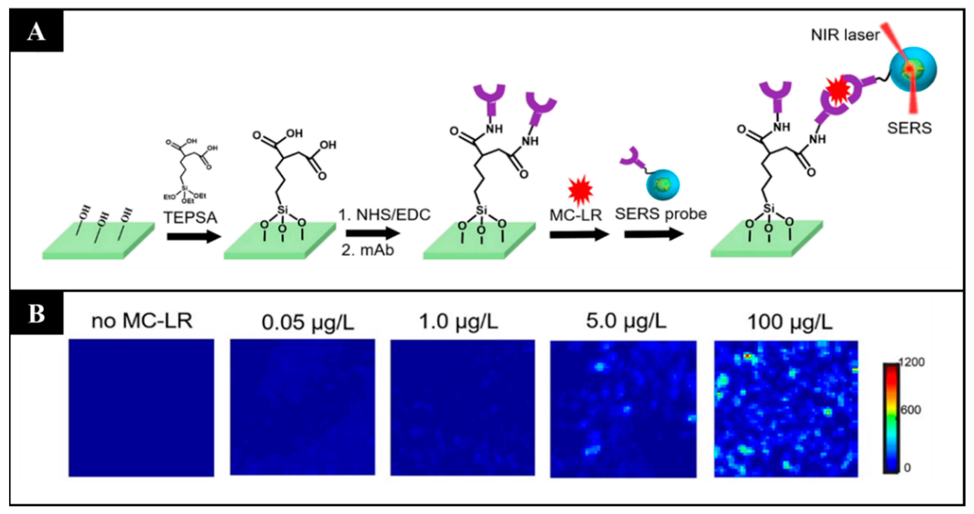
Another interesting study is that of Li et al. (2019), who constructed a biosensor that is more sensitive and has a wider detection range than conventional ELISA kits for the detection of microcystin-LR in aquatic environments. In this work, the authors designed particles with a core of plasmonic gold nanostars with Raman reporter molecules (4-nitrothiophenol) embedded between the core and a protective silica shell. The shell improves the stability and reproducibility of the sensor, and the SERS tags have immobilized antibodies against microcystin-LR for specificity [98] (Figure 4).
3.4. Surface Plasmon Resonance (SPR) Biosensors
The SPR optical signal results from the oscillation of conduction band electrons at the dielectric–metal interface induced by incident light. Binding of analytes on or near the metal surface leads to differences in the refractive index in the immediate vicinity of the metal surface and enables label-free, real-time detection of intermolecular interactions [107,108]. Gold is the metal that is commonly used in SPR. This optical method has been widely explored for the detection of biotoxins in water and food samples: domoic acid [107,108,109], okadaic acid [111,112], palytoxin [113,114], tetrodotoxin [115,116,117], yessotoxin [118], microcystins [110], and multiple toxins simultaneously [104,105,106,128]. When SPR is used for small molecules of low molecular weight, such as toxins, it is generally associated with amplification steps [129], competition-based assays (where the analyte in solution prevents antibody binding to the analytes immobilized on the surface), or displacement-based assays (where antibodies bound to the immobilized analytes on the SPR surface are displaced by the analyte in solution) [107]. However, Yakes et al. (2014) demonstrated the first direct SPR immunosensor for biotoxin detection, tetrodotoxin in a pufferfish matrix as a proof of concept. The antitetrodotoxin was immobilized on the sensor chip, and the analyte was directly injected into the SPR sensor surface, achieving a LOD of 2 ng mL−1 in the pufferfish matrix [117].
There are several approaches to the design of this type of biosensors. For example, Garibo et al. (2014) used magnetic particles functionalized with an antibody against okadaic acid as immobilization supports and carriers of the biotoxin for competitive assay on the SPR immunosensor [112]. Moreover, Stevens et al. (2007) developed a portable six-channel SPR biosensor for the measurement of domoic acid in phosphate-buffered saline and clam extract solutions, reaching a LOD of 10 nmol L−1 [107]. Nevertheless, multiplex analysis is of great importance because samples are usually complex and do not contain only one biotoxin. Microfluidic devices allow not only compartmentalization of the sensor to immobilize the target and reversal of the association event to reuse the biosensor, but also multiplex analysis. For example, Campbell et al. (2011) constructed a biosensor with four flow cells, each with four SPR sensing spots, which allowed simultaneous readout of 16 different interactions at the SPR surface. The biosensor also had a liquid handling system for sample injection and regeneration of the sensor for further measurements [105].
3.5. Interferometric Biosensors
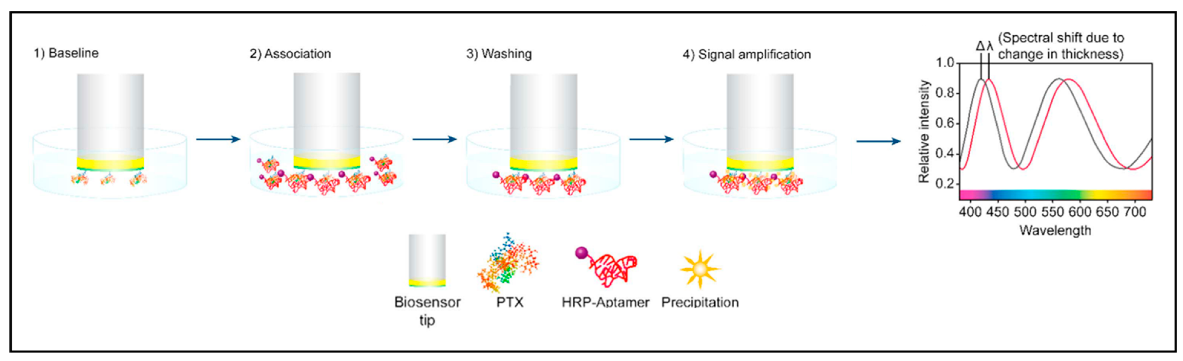
Interferometry is an optical technique that measures the interference pattern of light produced by two light beams, the sensor where the bioconjugation event occurs and the reference beams. Many interferometric configurations have been used for highly sensitive real-time detection of small molecules [130]. Chocarro-Ruiz et al. (2017) created an immunosensor chip based on bimodal waveguide interferometry. The sensor was functionalized with an antibody against okadaic acid and showed a LOD of 0.2 µg L−1 [122]. An interesting approach based on a Fabry–Pérot interferometer was developed by Queirós et al. (2011), who grew a sol–gel molecularly imprinted polymer (MIP) into the tip of the optical fiber by dip-coating, creating a selective membrane for microcystin-LR. The MIP showed a low thermal effect, good for field applications, and excellent selectivity against other coexisting species in the sample [121]. In biolayer interferometry (BLI), the tip of the fiber is coated with a layer of immobilized biomolecules, and the interference pattern of white light reflected from the biolayer and an internal reference surface is analyzed. Any change in the molecules bound to the tip, resulting from analyte recognition, causes a shift in the interference pattern. The wavelength shift is directly related to the distance between the two reflecting surfaces (i.e., it correlates with changes in the thickness of the biolayer resulting from analyte detection) [120]. This technique has been successfully used for the detection of domoic acid [120], palytoxin [123], saxitoxin [124], and dinophysistoxin-1 [119], with low LODs. In the case of palytoxin detection, the signal was amplified by HRP-labeled aptamers specific for palytoxin immobilized on the biosensor surface. HRP aptamers were used as biorecognition receptors to bind competitively with immobilized palytoxin. When the palytoxin/HRP aptamer complex was introduced into a 3,3′-diaminobenzidine solution, a polymeric product precipitated directly in the tip and caused a strong change in the shift of light [123] (Figure 5).
3.6. Resonant Mirror (RM) Biosensors
An RM biosensor is a waveguide-based sensor that uses the evanescent field produced at the sensing surface to follow specific interactions between molecules through changes in the refractive index at the sensing surface. These biosensors enable real-time quantitative measurements [131,132]. To our knowledge, there are only two studies using MR to detect aquatic biotoxins. In summary, the authors investigated the affinity between yessotoxin and various phosphodiesterases, a known target of this toxin, which ensures the specificity of the biosensor. The RM biosensor had aminosilane surfaces to immobilize phosphodiesterases, and increasing concentrations of the biotoxin were added, resulting in a proportional increase in sensor response [125]. This sensor approach also allowed the investigation of the specificity of different phosphodiesterase families for yessotoxin [126].
4. Optical High-Throughput Screening (HTS) Assays for Drug Discovery
Interest in naturally occurring compounds from aquatic organisms with potent pharmacological activity has long led to research efforts. Indeed, isolated compounds from extracts of marine organisms have shown interesting biological activities beyond their known toxicological effects [133,134]. There are many relevant examples of such bioactive natural compounds with antibacterial, anti-inflammatory, antimalarial, and anticancer activities [135,136]. HTS assays are key processes in drug discovery and consist of automated screening of large compound libraries and identification of biologically relevant compounds. Testing a large number of natural or synthetic chemical compounds for a specific biological target is the starting point of a drug design and development pipeline. The advantages of HTS technology are mainly in reducing the cost of drug development and increasing the speed, simplicity, and process efficiency [137]. HTS assays are divided into biochemical assays or cell-based assays. The first group usually relies on enzyme activity or receptor–ligand binding tests, which allow for obtaining highly reproducible miniaturized assays. However, tissue-specific responses may differ from those in biochemical assays because the activity of a small molecule may be different in a cellular context [138]. Therefore, drug screening is evolving into in vitro cell-based assays that are more suitable in the process of validation of new drugs in the preclinical phase. It offers the possibility to study the toxicity of a given drug for both targeted cell populations and nontargeted cells, allowing the selection of potential drugs without harmful side effects [139].
In the techniques used to study biomolecular interactions and the binding of a ligand to its receptor, the labeling steps required in most methods (e.g., fluorescence labeling and radiolabeling) led to some drawbacks involving additional time and cost. In addition, the labels may interfere with the site of molecular interaction, leading to false-negative results, or bind to the background, leading to false-positive results [140]. In this context, there is a growing awareness of novel label-free optical techniques that provide improved data on interaction specificity, kinetics, and affinity in real time. Moreover, their value already extends beyond low-throughput analysis of binding affinity and kinetics, as newly developed optical biosensor arrays for multiplexed detection offer a greater degree of flexibility in experimental design [140]. Optical biosensors have accompanied the shift in drug discovery from a target-directed approach to a systems biology-centered approach by potentiating the development of cell-based biosensors [141]. Among the label-free screening systems, SPR, RM, interferometry, Raman spectroscopy, and photonic crystal (PC)-based biosensors are some of the best-developed methods for HTS applications with the goal of accelerating the drug discovery process [140,141,142,143,144].
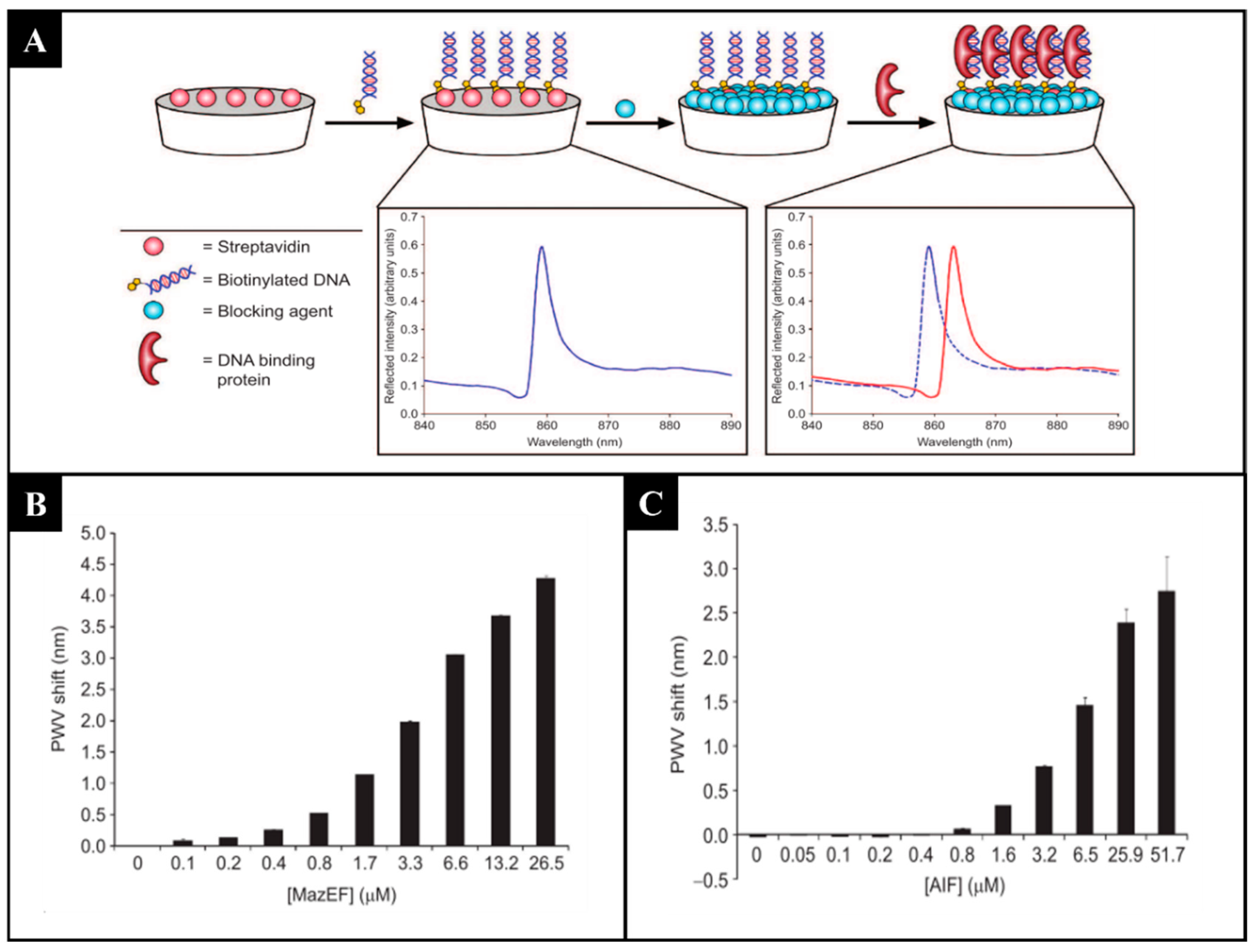
Among the various mentioned label-free methods, PCs show a great potential for incorporation into HTS [145]. Biosensor platforms using PCs present many advantages, such as high sensitivity, flexibility in structural design, cost-effective fabrication with a variety of materials, short testing time, and ability to detect a wide range of analytes [146,147]. PCs are one-, two-, or three-dimensional periodic arrays composed of materials with different refractive indexes. The structural color observed in PCs is explained by the photonic band gap (PBG), where certain wavelengths of light do not propagate through the PC and are reflected. When the PBG is in the visible light range, the PC exhibits unique, vibrant colors. These nanostructures occur in nature (e.g., the bright gold and silver colors of jewel scarabs [148] and the blue color of the wings of Morpho butterflies) [149]. Bioinspired nanostructured materials exhibiting photonic properties and structural colors have been fabricated for various purposes by controlling composition, additives, and arrangement, among others [150]. Proving its usefulness in HTS, the ability of PCs to identify modulators of protein–protein interactions [151], to discover inhibitors of protein–DNA interactions [152] (Figure 6), and to measure antibody–antibody binding [153] has been demonstrated. Another interesting example of the use of PCs for HTS screening is in cell-based assays. A label-free detection system based on PCs incorporated into a 96-well microplate enabled the quantification of the proliferation of cancer cells and cell apoptosis induced by exposure to a cytotoxic compound [154]. The applications are vast because a variety of cell-based assays can be performed in response to chemical and biological stimuli, while cells are quantitatively monitored in their culture environment over time without the use of dyes or stains [145].
5. Conclusions and Future Perspectives
The increasing demand for new detection methods for bioactive natural products, including biotoxins to protect human health, makes optical biosensors an excellent alternative for the detection of these biomolecules compared with conventional methods, such as ELISA, MBA, LC, and MS. Optical biochemical or cell-based assays overcome ethical concerns and irreproducibility associated with animal testing. In addition, optical biosensors exhibit high sensitivity and rapid responses, have lower cost, and are easy to use, often by untrained personnel. The focus of this review was mainly on biosensors for the detection of aquatic biotoxins, as there are great expectations for new, simpler, miniaturized, and improved methods for the real-time detection of various chemicals in water and aquatic organisms. As for other applications, optical biosensors have been successfully used to detect biotoxins even in complex matrices and have shown high sensitivity and selectivity. In addition, new optical detection designs are very promising for HTS of bioactive compounds, as they offer the distinct advantage of being less time-consuming and are cost-effective methods. Label-free optical biosensor arrays will certainly accelerate new areas of drug discovery, as expected, especially regarding the use of HTS for the discovery of aquatic bioactive compounds, and help decipher their pharmaceutical potential.
Funding
This work was supported by the project CY-SENSORS (PTDC/BTA-GES/32359/2017-POCI-01-0145-FEDER-032359), cofunded by the European Regional Development Fund (ERDF) through COMPETE 2020–POCI (Operational Programme for Competitiveness and Internationalization), and by Portuguese funds through Fundação para a Ciência e a Tecnologia (FCT). The author RV also acknowledges FCT for the PhD grant (2020.09673.BD).
Institutional Review Board Statement
Not applicable.
Informed Consent Statement
Not applicable.
Data Availability Statement
Not applicable.
Acknowledgments
The authors gratefully acknowledge funding from the European Regional Development Fund (ERDF) through COMPETE 2020-POCI and Fundação para a Ciência e a Tecnologia (FCT).
Conflicts of Interest
The authors declare no conflict of interest.
References
- Xie, J.; Wang, L.; Wu, W.; Ou, J. Recent Advances on Determination Methods of Mycotoxins and Marine Biotoxins. Med. Res. 2018, 2, 1–7. [Google Scholar]
- Özogul, F.; Hamed, I. Chapter 3—Marine-Based Toxins and Their Health Risk. In Food Quality: Balancing Health and Disease; Academic Press: Cambridge, MA, USA, 2018; pp. 109–144. [Google Scholar]
- Vasconcelos, V.M. Cyanobacterial toxins in Portugal: Effects on aquatic animals and risk for human health. Braz. J. Med. Biol. Res. 1999, 32, 249–254. [Google Scholar] [CrossRef] [PubMed] [Green Version]
- Gobler, C.J. Climate Change and Harmful Algal Blooms: Insights and perspective. Harmful Algae 2020, 2, 101731. [Google Scholar] [CrossRef] [PubMed]
- Jiang, B. Review on Marine Biotoxins Toxicity and Detection Methods. In Proceedings of the 2020 10th International Conference on Biomedical Engineering and Technology, Tokyo, Japan, 15–18 September 2020; Association for Computing Machinery: New York, NY, USA, 2020; pp. 28–33. [Google Scholar]
- Du, X.; Liu, H.; Yuan, L.; Wang, Y.; Ma, Y.; Wang, R.; Chen, X.; Losiewicz, M.D.; Guo, H.; Zhang, H. The Diversity of Cyanobacterial Toxins on Structural Characterization, Distribution and Identification: A Systematic Review. Toxins 2019, 11, 530. [Google Scholar] [CrossRef] [Green Version]
- Jin, A.H.; Muttenthaler, M.; Dutertre, S.; Himaya, S.W.A.; Kaas, Q.; Craik, D.J.; Lewis, R.J.; Alewood, P.F. Conotoxins: Chemistry and Biology. Chem. Rev. 2019, 119, 11510–11549. [Google Scholar] [CrossRef] [PubMed]
- Bane, V.; Lehane, M.; Dikshit, M.; O’Riordan, A.; Furey, A. Tetrodotoxin: Chemistry, toxicity, source, distribution and detection. Toxins 2014, 6, 693–755. [Google Scholar] [CrossRef] [PubMed] [Green Version]
- Fabro, E.; Almandoz, G.O.; Ferrario, M.; Tillmann, U.; Cembella, A.; Krock, B. Distribution of Dinophysis species and their association with lipophilic phycotoxins in plankton from the Argentine Sea. Harmful Algae 2016, 2, 31–41. [Google Scholar] [CrossRef] [PubMed] [Green Version]
- Morabito, S.; Silvestro, S.; Faggio, C. How the marine biotoxins affect human health. Nat. Prod. Res. 2018, 32, 621–631. [Google Scholar] [CrossRef]
- Baj, A.; Bistoletti, M.; Bosi, A.; Moro, E.; Giaroni, C.; Crema, F. Marine Toxins and Nociception: Potential Therapeutic Use in the Treatment of Visceral Pain Associated with Gastrointestinal Disorders. Toxins 2019, 11, 449. [Google Scholar] [CrossRef] [PubMed] [Green Version]
- Singh, R.K.; Tiwari, S.P.; Rai, A.K.; Mohapatra, T.M. Cyanobacteria: An emerging source for drug discovery. J. Antibiot. 2011, 64, 401–412. [Google Scholar] [CrossRef] [Green Version]
- Kounnis, V.; Chondrogiannis, G.; Mantzaris, M.D.; Tzakos, A.G.; Fokas, D.; Papanikolaou, N.A.; Glani, V.; Sainis, I.; Briasoulis, E. Microcystin LR Shows Cytotoxic Activity Against Pancreatic Cancer Cells Expressing the Membrane OATP1B1 and OATP1B3 Transporters. Anticancer Res. 2015, 35, 5857–5865. [Google Scholar]
- Assunção, J.; Guedes, A.C.; Malcata, F.X. Biotechnological and Pharmacological Applications of Biotoxins and Other Bioactive Molecules from Dinoflagellates. Mar. Drugs 2017, 15, 393. [Google Scholar] [CrossRef] [Green Version]
- Kohane, D.; Lu, N.; Gokgolkline, A.; Shubina, M.; Kuang, Y.; Hall, S.; Strichartz, G.; Berde, C. The local anesthetic properties and toxicity of saxitonin homologues for rat sciatic nerve block in vivo. Reg. Anesth. Pain Med. 2000, 25, 52–59. [Google Scholar] [CrossRef]
- Epstein-Barash, H.; Shichor, I.; Kwon, A.H.; Hall, S.; Lawlor, M.W.; Langer, R.; Kohane, D.S. Prolonged duration local anesthesia with minimal toxicity. Proc. Natl. Acad. Sci. USA 2009, 106, 7125–7130. [Google Scholar] [CrossRef] [Green Version]
- Sequeira, E.; Pierce, M.L.; Akasheh, D.; Sellers, S.; Gerwick, W.H.; Baden, D.G.; Murray, T.F. Epicortical Brevetoxin Treatment Promotes Neural Repair and Functional Recovery after Ischemic Stroke. Mar. Drugs 2020, 18, 374. [Google Scholar] [CrossRef]
- Chae, H.D.; Choi, T.S.; Kim, B.M.; Jung, J.H.; Bang, Y.J.; Shin, D.Y. Oocyte-based screening of cytokinesis inhibitors and identification of pectenotoxin-2 that induces Bim/Bax-mediated apoptosis in p53-deficient tumors. Oncogene 2005, 24, 4813–4819. [Google Scholar] [CrossRef] [PubMed] [Green Version]
- Espina, B.; Rubiolo, J.A. Marine toxins and the cytoskeleton: Pectenotoxins, unusual macrolides that disrupt actin. FEBS J. 2008, 275, 6082–6088. [Google Scholar] [CrossRef]
- Alonso, E.; Vale, C.; Vieytes, M.R.; Laferla, F.M.; Gimenez-Llort, L.; Botana, L.M. The cholinergic antagonist gymnodimine improves Abeta and tau neuropathology in an in vitro model of Alzheimer disease. Cell. Physiol. Biochem. 2011, 27, 783–794. [Google Scholar] [CrossRef] [PubMed]
- Dragunow, M.; Trzoss, M.; Brimble, M.A.; Cameron, R.; Beuzenberg, V.; Holland, P.; Mountfort, D. Investigations into the cellular actions of the shellfish toxin gymnodimine and analogues. Environ. Toxicol. Pharmacol. 2005, 20, 305–312. [Google Scholar] [CrossRef]
- Rivera-de-Torre, E.; Palacios-Ortega, J.; Garb, J.E.; Slotte, J.P.; Gavilanes, J.G.; Martinez-Del-Pozo, A. Structural and functional characterization of sticholysin III: A newly discovered actinoporin within the venom of the sea anemone Stichodactyla helianthus. Arch. Biochem. Biophys. 2020, 2, 108435. [Google Scholar] [CrossRef] [PubMed]
- Piontek, M.; Seymour, J.E.; Wong, Y.; Gilstrom, T.; Potriquet, J.; Jennings, E. Nimmo, A.; Miles, J.J. The pathology of Chironex fleckeri venom and known biological mechanisms. Toxicon 2020, 2, 100026. [Google Scholar] [CrossRef] [PubMed]
- Kobayashi, J.; Tsuda, M. Amphidinolides, bioactive macrolides from symbiotic marine dinoflagellates. Nat. Prod. Rep. 2004, 21, 77–93. [Google Scholar] [CrossRef]
- Kobayashi, J. Amphidinolides and its related macrolides from marine dinoflagellates. J. Antibiot. 2008, 61, 271–284. [Google Scholar] [CrossRef] [Green Version]
- Boente-Juncal, A.; Raposo-Garcia, S.; Costas, C.; Louzao, M.C.; Vale, C.; Botana, L.M. Partial Blockade of Human Voltage-Dependent Sodium Channels by the Marine Toxins Azaspiracids. Chem. Res. Toxicol. 2020, 33, 2593–2604. [Google Scholar] [CrossRef]
- Wang, D.Z. Neurotoxins from marine dinoflagellates: A brief review. Mar. Drugs 2008, 6, 349–371. [Google Scholar] [CrossRef] [PubMed]
- Anderson, P.D.; Bokor, G. Conotoxins: Potential Weapons from the Sea. J. Bioterror. Biodef. 2012, 3, 1000120. [Google Scholar]
- Sharma, A.; Gautam, S.; Kumar, S. Phycotoxins. In Encyclopedia of Food Microbiology, 2nd ed.; Academic Press: Cambridge, MA, USA, 2014; pp. 25–29. [Google Scholar]
- Nagai, H.; Mikami, Y.; Yazawa, K.; Gonoi, T.; Yasumoto, T. Biological activities of novel polyether antifungals, gambieric acids A and B from a marine dinoflagellate Gambierdiscus toxicus. J. Antibiot. 1993, 46, 520–522. [Google Scholar] [CrossRef] [Green Version]
- Camacho, F.G.; Rodriguez, J.G.; Miron, A.S.; Garcia, M.C.; Belarbi, E.H.; Chisti, Y.; Grima, E.M. Biotechnological significance of toxic marine dinoflagellates. Biotechnol. Adv. 2007, 25, 176–194. [Google Scholar] [CrossRef] [PubMed]
- Cuypers, E.; Abdel-Mottaleb, Y.; Kopljar, I.; Rainier, J.D.; Raes, A.L.; Snyders, D.J.; Tytgat, J. Gambierol, a toxin produced by the dinoflagellate Gambierdiscus toxicus, is a potent blocker of voltage-gated potassium channels. Toxicon 2008, 51, 974–983. [Google Scholar] [CrossRef] [Green Version]
- Tainter, C.J.; Schley, N.D.; Harris, C.M.; Stec, D.F.; Song, A.K.; Balinski, A.; May, J.C.; McLean, J.A.; Reece, K.S.; Harris, T.M. Algal Toxin Goniodomin a Binds Potassium Ion Selectively to Yield a Conformationally Altered Complex with Potential Biological Consequences. J. Nat. Prod. 2020, 83, 1069–1081. [Google Scholar] [CrossRef] [PubMed]
- Satake, M.; Shoji, M.; Oshima, Y.; Naoki, H.; Fujita, T.; Yasumoto, T. Gymnocin-A, a cytotoxic polyether from the notorious red tide dinoflagellate, Gymnodinium mikimotoi. Tetrahedron Lett. 2002, 43, 5829–5832. [Google Scholar] [CrossRef]
- Zurhelle, C.; Nieva, J.; Tillmann, U.; Harder, T.; Krock, B.; Tebben, J. Identification of Novel Gymnodimines and Spirolides from the Marine Dinoflagellate Alexandrium ostenfeldii. Mar. Drugs 2018, 16, 446. [Google Scholar] [CrossRef] [PubMed] [Green Version]
- Kodama, M. Paralytic Shellfish Poisoning Toxins: Biochemistry and Origin. Aqua-BioSci. Monogr. 2010, 3, 1–38. [Google Scholar] [CrossRef] [Green Version]
- Van Wagoner, R.M.; Deeds, J.R.; Tatters, A.O.; Place, A.R.; Tomas, C.R.; Wright, J.L. Structure and relative potency of several karlotoxins from Karlodinium veneficum. J. Nat. Prod. 2010, 73, 1360–1365. [Google Scholar] [CrossRef] [Green Version]
- Van Wagoner, R.M.; Deeds, J.R.; Satake, M.; Ribeiro, A.A.; Place, A.R.; Wright, J.L. Isolation and characterization of karlotoxin 1, a new amphipathic toxin from Karlodinium veneficum. Tetrahedron Lett. 2008, 49, 6457–6461. [Google Scholar] [CrossRef] [Green Version]
- Massey, I.Y.; Wu, P.; Wei, J.; Luo, J.; Ding, P.; Wei, H.; Yang, F. A Mini-Review on Detection Methods of Microcystins. Toxins 2020, 12, 641. [Google Scholar] [CrossRef]
- Sawelew, L.; Gault, F.; Nuccio, C.; Perez, Y.; Lorquin, J. Characterisation of palytoxin from an undescribed Palythoa (Anthozoa: Zoantharia: Sphenopidae) with significant in vitro cytotoxic effects on cancer cells at picomolar doses. BioRxiv 2018, 292219. [Google Scholar] [CrossRef]
- Suzuki, T.; Walter, J.A.; LeBlanc, P.; MacKinnon, S.; Miles, C.O.; Wilkins, A.L.; Munday, R.; Beuzenberg, V.; MacKenzie, A.L.; Jensen, D.J.; et al. Identification of pectenotoxin-11 as 34S-hydroxypectenotoxin-2, a new pectenotoxin analogue in the toxic dinoflagellate Dinophysis acuta from New Zealand. Chem. Res. Toxicol. 2006, 19, 310–318. [Google Scholar] [CrossRef] [PubMed]
- Munday, R.; Quilliam, M.A.; LeBlanc, P.; Lewis, N.; Gallant, P.; Sperker, S.A.; Ewart, H.S.; MacKinnon, S.L. Investigations into the toxicology of spirolides, a group of marine phycotoxins. Toxins 2012, 4, 1–14. [Google Scholar] [CrossRef] [PubMed]
- Alfonso, A.; Vieytes, M.R.; Botana, L.M. Yessotoxin, a Promising Therapeutic Tool. Mar. Drugs 2016, 14, 30. [Google Scholar] [CrossRef] [PubMed] [Green Version]
- Tobio, A.; Alfonso, A.; Madera-Salcedo, I.; Botana, L.M.; Blank, U. Yessotoxin, a Marine Toxin, Exhibits Anti-Allergic and Anti-Tumoural Activities Inhibiting Melanoma Tumour Growth in a Preclinical Model. PLoS ONE 2016, 11, e0167572. [Google Scholar] [CrossRef] [PubMed]
- Korsnes, M.S.; Korsnes, R. Mitotic Catastrophe in BC3H1 Cells following Yessotoxin Exposure. Front. Cell Dev. Biol. 2017, 5, 1–18. [Google Scholar] [CrossRef] [PubMed] [Green Version]
- Echigoya, R.; Rhodes, L.; Oshima, Y.; Satake, M. The structures of five new antifungal and hemolytic amphidinol analogs from Amphidinium carterae collected in New Zealand. Harmful Algae 2005, 4, 383–389. [Google Scholar] [CrossRef]
- Hagen, N.A.; Fisher, K.M.; Lapointe, B.; du Souich, P.; Chary, S.; Moulin, D.; Sellers, E.; Ngoc, A.H. An open-label, multi-dose efficacy and safety study of intramuscular tetrodotoxin in patients with severe cancer-related pain. J. Pain Symptom. Manag. 2007, 34, 171–182. [Google Scholar] [CrossRef] [PubMed]
- Hagen, N.A.; du Souich, P.; Lapointe, B.; Ong-Lam, M.; Dubuc, B.; Walde, D.; Love, R.; Ngoc, A.H. Tetrodotoxin for moderate to severe cancer pain: A randomized, double blind, parallel design multicenter study. J. Pain Symptom. Manag. 2008, 35, 420–429. [Google Scholar] [CrossRef] [PubMed]
- Song, H.; Li, J.; Lu, C.L.; Kang, L.; Xie, L.; Zhang, Y.Y.; Zhou, X.B.; Zhong, S. Tetrodotoxin alleviates acute heroin withdrawal syndrome: A multicentre, randomized, double-blind, placebo-controlled study. Clin. Exp. Pharmacol. Physiol. 2011, 38, 510–514. [Google Scholar] [CrossRef] [PubMed]
- Mohs, R.C.; Greig, N.H. Drug discovery and development: Role of basic biological research. Alzheimers Dement. 2017, 3, 651–657. [Google Scholar] [CrossRef] [PubMed]
- Batool, M.; Ahmad, B.; Choi, S. A Structure-Based Drug Discovery Paradigm. Int. J. Mol. Sci. 2019, 20, 2783. [Google Scholar] [CrossRef] [PubMed] [Green Version]
- Chen, C.; Wang, J. Optical biosensors: An exhaustive and comprehensive review. Analyst 2020, 145, 1605–1628. [Google Scholar] [CrossRef]
- Vilarino, N.; Louzao, M.C.; Vieytes, M.R.; Botana, L.M. Biological methods for marine toxin detection. Anal. Bioanal. Chem. 2010, 397, 1673–1681. [Google Scholar] [CrossRef]
- Hardison, D.R.; Holland, W.C.; McCall, J.R.; Bourdelais, A.J.; Baden, D.G.; Darius, H.T.; Chinain, M.; Tester, P.A.; Shea, D.; Quintana, H.A.; et al. Fluorescent Receptor Binding Assay for Detecting Ciguatoxins in Fish. PLoS ONE 2016, 11, e0153348. [Google Scholar] [CrossRef] [PubMed]
- Dillon, M.; Zaczek-Moczydlowska, M.A.; Edwards, C.; Turner, A.D.; Miller, P.I.; Moore, H.; McKinney, A.; Lawton, L.; Campbell, K. Current Trends and Challenges for Rapid SMART Diagnostics at Point-of-Site Testing for Marine Toxins. Sensors 2021, 21, 2499. [Google Scholar] [CrossRef]
- Bodero, M.; Bovee, T.F.H.; Wang, S.; Hoogenboom, R.; Klijnstra, M.D.; Portier, L.; Hendriksen, P.J.M.; Gerssen, A. Screening for the presence of lipophilic marine biotoxins in shellfish samples using the neuro-2a bioassay. Food Addit. Contam. Part A Chem. Anal. Control Expo. Risk Assess 2018, 35, 351–365. [Google Scholar] [CrossRef] [PubMed]
- Fang, L.; Qiu, F.; Yu, X. Determination of Lipophilic Marine Biotoxins in Shellfish by Online Turbulent Flow Chromatography Coupled to Liquid Chromatography–Tandem Mass Spectrometry. Chromatographia 2019, 82, 1321–1331. [Google Scholar] [CrossRef]
- Bodero, M.; Gerssen, A.; Portier, L.; Klijnstra, M.D.; Hoogenboom, R.; Guzman, L.; Hendriksen, P.J.M.; Bovee, T.F.H. A Strategy to Replace the Mouse Bioassay for Detecting and Identifying Lipophilic Marine Biotoxins by Combining the Neuro-2a Bioassay and LC-MS/MS Analysis. Mar. Drugs 2018, 16, 501. [Google Scholar] [CrossRef] [PubMed] [Green Version]
- Christian, B.; Luckas, B. Determination of marine biotoxins relevant for regulations: From the mouse bioassay to coupled LC-MS methods. Anal. Bioanal. Chem. 2008, 391, 117–134. [Google Scholar] [CrossRef]
- Blay, P.; Hui, J.P.; Chang, J.; Melanson, J.E. Screening for multiple classes of marine biotoxins by liquid chromatography-high-resolution mass spectrometry. Anal. Bioanal. Chem. 2011, 400, 577–585. [Google Scholar] [CrossRef]
- Briggs, L.R.; Miles, C.O.; Fitzgerald, J.M.; Ross, K.M.; Garthwaite, I.; Towers, N.R. Enzyme-linked immunosorbent assay for the detection of yessotoxin and its analogues. J. Agric. Food Chem. 2004, 52, 5836–5842. [Google Scholar] [CrossRef]
- Hara, Y.; Dong, J.; Ueda, H. Open-sandwich immunoassay for sensitive and broad-range detection of a shellfish toxin gonyautoxin. Anal. Chim. Acta 2013, 2, 107–113. [Google Scholar] [CrossRef]
- Kawatsu, K.; Kanki, M.; Harada, T.; Kumeda, Y. A highly rapid and simple competitive enzyme-linked immunosorbent assay for monitoring paralytic shellfish poisoning toxins in shellfish. Food Chem. 2014, 2, 94–98. [Google Scholar] [CrossRef]
- Samdal, I.A.; Lovberg, K.E.; Kristoffersen, A.B.; Briggs, L.R.; Kilcoyne, J.; Forsyth, C.J.; Miles, C.O. A Practical ELISA for Azaspiracids in Shellfish via Development of a New Plate-Coating Antigen. J. Agric. Food Chem. 2019, 67, 2369–2376. [Google Scholar] [CrossRef] [PubMed]
- Zhang, X.; Zhang, Z. Capillary electrophoresis-based immunoassay with electrochemical detection as rapid method for determination of saxitoxin and decarbamoylsaxitoxin in shellfish samples. J. Food Compost. Anal. 2012, 28, 61–68. [Google Scholar] [CrossRef]
- Kim, M.H.; Choi, S.J. Immunoassay of paralytic shellfish toxins by moving magnetic particles in a stationary liquid-phase lab-on-a-chip. Biosens. Bioelectron. 2015, 2, 136–140. [Google Scholar] [CrossRef] [PubMed]
- Pelin, M.; Sosa, S.; Brovedani, V.; Fusco, L.; Poli, M.; Tubaro, A. A Novel Sensitive Cell-Based Immunoenzymatic Assay for Palytoxin Quantitation in Mussels. Toxins 2018, 10, 329. [Google Scholar] [CrossRef] [PubMed] [Green Version]
- Sakamoto, S.; Putalun, W.; Vimolmangkang, S.; Phoolcharoen, W.; Shoyama, Y.; Tanaka, H.; Morimoto, S. Enzyme-linked immunosorbent assay for the quantitative/qualitative analysis of plant secondary metabolites. J. Nat. Med. 2018, 72, 32–42. [Google Scholar] [CrossRef] [PubMed] [Green Version]
- Vilarino, N.; Fonfria, E.S.; Louzao, M.C.; Botana, L.M. Use of biosensors as alternatives to current regulatory methods for marine biotoxins. Sensors 2009, 9, 9414–9443. [Google Scholar] [CrossRef] [Green Version]
- Kawamura, A.; Miyata, T. Biosensors. In Biomaterials Nanoarchitectonics; Elsevier: Amsterdam, The Netherlands, 2016; Chapter 1; pp. 157–176. [Google Scholar]
- Weller, M.G. Immunoassays and biosensors for the detection of cyanobacterial toxins in water. Sensors 2013, 13, 15085–15112. [Google Scholar] [CrossRef]
- Chen, S.; Shamsi, M.H. Biosensors-on-chip: A topical review. J. Micromech. Microeng. 2017, 27, 1–15. [Google Scholar] [CrossRef]
- Damborsky, P.; Svitel, J.; Katrlik, J. Optical biosensors. Essays Biochem. 2016, 60, 91–100. [Google Scholar]
- Lai, W.; Zhuang, J.; Tang, D. Novel colorimetric immunoassay for ultrasensitive monitoring of brevetoxin B based on enzyme-controlled chemical conversion of sulfite to sulfate. J. Agric. Food Chem. 2015, 63, 1982–1989. [Google Scholar] [CrossRef]
- Lai, W.; Wei, Q.; Zhuang, J.; Lu, M.; Tang, D. Fenton reaction-based colorimetric immunoassay for sensitive detection of brevetoxin B. Biosens. Bioelectron. 2016, 2, 249–256. [Google Scholar] [CrossRef] [PubMed] [Green Version]
- Wang, F.; Liu, S.; Lin, M.; Chen, X.; Lin, S.; Du, X.; Li, H.; Ye, H.; Qiu, B.; Lin, Z.; et al. Colorimetric detection of microcystin-LR based on disassembly of orient-aggregated gold nanoparticle dimers. Biosens. Bioelectron. 2015, 2, 475–480. [Google Scholar] [CrossRef] [PubMed]
- Li, X.; Cheng, R.; Shi, H.; Tang, B.; Xiao, H.; Zhao, G. A simple highly sensitive and selective aptamer-based colorimetric sensor for environmental toxins microcystin-LR in water samples. J. Hazard Mater. 2016, 2, 474–480. [Google Scholar] [CrossRef] [PubMed]
- Tang, X.; Yin, Z.; Lei, X.; Zeng, Y.; Zhang, Z.; Lu, Y.; Zhou, G.; Li, L.; Wu, X. Colorimetric Method for Sensitive Detection of Microcystin-LR Using Surface Copper Nanoparticles of Polydopamine Nanosphere as Turn-On Probe. Nanomaterials 2019, 9, 332. [Google Scholar] [CrossRef] [Green Version]
- Almeida, P.S.; Cogo, K.; Tsai, S.M.; Moon, D.H. Colorimetric test for the monitoring of microcystins in cyanobacterial culture and environmental samples from southeast—Brazil. Braz. J. Microbiol. 2006, 2, 192–198. [Google Scholar] [CrossRef] [Green Version]
- Hayat, A.; Barthelmebs, L.; Marty, J.L. A simple colorimetric enzymatic-assay for okadaic acid detection based on the immobilization of protein phosphatase 2A in sol-gel. Appl. Biochem. Biotechnol. 2012, 166, 47–56. [Google Scholar] [PubMed]
- Gu, H.; Duan, N.; Wu, S.; Hao, L.; Xia, Y.; Ma, X.; Wang, Z. Graphene oxide-assisted non-immobilized SELEX of okdaic acid aptamer and the analytical application of aptasensor. Sci. Rep. 2016, 2, 1–9. [Google Scholar] [CrossRef] [PubMed] [Green Version]
- Qiang, L.; Zhang, Y.; Guo, X.; Gao, Y.; Han, Y.; Sun, J.; Han, L. A rapid and ultrasensitive colorimetric biosensor based on aptamer functionalized Au nanoparticles for detection of saxitoxin. RSC Adv. 2020, 10, 15293–15298. [Google Scholar] [CrossRef]
- Campàs, M.; de la Iglesia, P.; Fernández-Tejedor, M.; Diogène, J. Colorimetric and electrochemical phosphodiesterase inhibition assays for yessotoxin detection: Development and comparison with LC-MS/MS. Anal. Bioanal. Chem. 2010, 396, 2321–2330. [Google Scholar] [CrossRef]
- Tsumuraya, T.; Sato, T.; Hirama, M.; Fujii, I. Highly Sensitive and Practical Fluorescent Sandwich ELISA for Ciguatoxins. Anal. Chem. 2018, 90, 7318–7324. [Google Scholar] [CrossRef]
- Rodríguez, L.P.; Vilariño, N.; Molgó, J.; Aráoz, R.; Antelo, A.; Vieytes, M.R.; Botana, L.M. Solid-phase receptor-based assay for the detection of cyclic imines by chemiluminescence, fluorescence, or colorimetry. Anal. Chem. 2011, 83, 5857–5863. [Google Scholar] [CrossRef] [PubMed]
- McGrath, T.F.; Andersson, K.; Campbell, K.; Fodey, T.L.; Elliott, C.T. Development of a rapid low cost fluorescent biosensor for the detection of food contaminants. Biosens. Bioelectron. 2013, 2, 96–102. [Google Scholar] [CrossRef] [PubMed] [Green Version]
- Gholami, M.; Salmasi, M.A.; Sohouli, E.; Torabi, B.; Sohrabi, M.R.; Rahimi-Nasrabadi, M. A new nano biosensor for maitotoxin with high sensitivity and selectivity based fluorescence resonance energy transfer between carbon quantum dots and gold nanoparticles. J. Photochem. Photobiol. A 2020, 2, 112523. [Google Scholar] [CrossRef]
- Sadik, O.A.; Yan, F. Novel fluorescent biosensor for pathogenic toxins using cyclic polypeptide conjugates. Chem. Commun. 2004, 9, 1136–1137. [Google Scholar] [CrossRef]
- Bickman, S.R.; Campbell, K.; Elliott, C.; Murphy, C.; O’Kennedy, R.; Papst, P.; Lochhead, M.J. An Innovative Portable Biosensor System for the Rapid Detection of Freshwater Cyanobacterial Algal Bloom Toxins. Environ. Sci. Technol. 2018, 52, 11691–11698. [Google Scholar] [CrossRef] [Green Version]
- Shi, Y.; Wu, J.; Sun, Y.; Zhang, Y.; Wen, Z.; Dai, H.; Wang, H.; Li, Z. A graphene oxide based biosensor for microcystins detection by fluorescence resonance energy transfer. Biosens. Bioelectron. 2012, 38, 31–36. [Google Scholar] [CrossRef]
- Liu, L.; Zhou, X.; Wilkinson, J.S.; Hua, P.; Song, B.; Shi, H. Integrated optical waveguide-based fluorescent immunosensor for fast and sensitive detection of microcystin-LR in lakes: Optimization and Analysis. Sci. Rep. 2017, 7, 1–9. [Google Scholar] [CrossRef] [Green Version]
- Alfaro, K.; Bustos, P.; O’Sullivan, C.; Conejeros, P. Facile and Cost-Effective Detection of Saxitoxin Exploiting Aptamer Structural Switching. Food Technol. Biotechnol 2015, 53, 337–341. [Google Scholar] [CrossRef] [PubMed]
- Reverté, L.; Campàs, M.; Yakes, B.J.; Deeds, J.R.; Katikou, P.; Kawatsu, K.; Lochhead, M.; Elliott, C.T.; Campbell, K. Tetrodotoxin detection in puffer fish by a sensitive planar waveguide immunosensor. Sens. Actuators B Chem. 2017, 2, 967–976. [Google Scholar] [CrossRef] [Green Version]
- Shen, H.; Xu, F.; Xiao, M.; Fu, Q.; Cheng, Z.; Zhang, S.; Huang, C.; Tang, Y. A new lateral-flow immunochromatographic strip combined with quantum dot nanobeads and gold nanoflowers for rapid detection of tetrodotoxin. Analyst 2017, 142, 4393–4398. [Google Scholar] [CrossRef] [PubMed]
- Lan, Y.; Qin, G.; Wei, Y.; Dong, C.; Wang, L. Highly sensitive analysis of tetrodotoxin based on free-label fluorescence aptamer sensing system. Spectrochim. Acta A Mol. Biomol. Spectrosc. 2019, 2, 411–418. [Google Scholar] [CrossRef] [PubMed]
- Pinzaru, S.C.; Müller, C.; Ujević, I.; Venter, M.M.; Chis, V.; Glamuzina, B. Lipophilic marine biotoxins SERS sensing in solutions and in mussel tissue. Talanta 2018, 2, 47–58. [Google Scholar] [CrossRef] [PubMed]
- Olson, T.Y.; Schwartzberg, A.M.; Liu, J.L.; Zhang, J.Z. Raman and Surface-Enhanced Raman Detection of Domoic Acid and Saxitoxin. Appl. Spectrosc. 2011, 65, 159–164. [Google Scholar] [CrossRef]
- Li, M.; Paidi, S.K.; Sakowski, E.; Preheim, S.; Barman, I. Ultrasensitive Detection of Hepatotoxic Microcystin Production from Cyanobacteria Using Surface-Enhanced Raman Scattering Immunosensor. ACS Sens. 2019, 4, 1203–1210. [Google Scholar] [CrossRef] [PubMed]
- Pearman, W.F.; Angel, S.M.; Ferry, J.L.; Hall, S. Characterization of the Ag mediated surface-enhanced Raman spectroscopy of saxitoxin. Appl. Spectrosc. 2008, 62, 727–732. [Google Scholar] [CrossRef]
- Huai, Q.-Y.; Gao, C.-L.; Miao, J.-L.; Yao, H.-L.; Wang, Z.-L. Fast detection of saxitoxin using laser tweezers surface enhanced Raman spectroscopy. Anal. Methods 2013, 5, 6870. [Google Scholar] [CrossRef]
- Cao, C.; Li, P.; Liao, H.; Wang, J.; Tang, X.; Yang, L. Cys-functionalized AuNP substrates for improved sensing of the marine toxin STX by dynamic surface-enhanced Raman spectroscopy. Anal. Bioanal. Chem. 2020, 412, 4609–4617. [Google Scholar] [CrossRef]
- Lin, W.-C.; Jen, H.-C.; Chen, C.-L.; Hwang, D.-F.; Chang, R.; Hwang, J.-S.; Chiang, H.-P. SERS Study of Tetrodotoxin (TTX) by Using Silver Nanoparticle Arrays. Plasmonics 2009, 4, 187–192. [Google Scholar] [CrossRef]
- Neng, J.; Wang, X.; Jia, K.; Sun, P. Rapid Detection of Tetrodotoxin Using Surface-Enhanced Raman Spectroscopy and Fe3O4/SiO2/Au Gold/Magnetic Nanoparticles. J. Appl. Spectrosc. 2018, 85, 160–165. [Google Scholar] [CrossRef]
- Fonfría, E.S.; Vilariño, N.; Campbell, K.; Elliott, C.; Haughey, S.A.; Ben-Gigirey, B.; Vieites, J.M.; Kawatsu, K.; Botana, L.M. Paralytic shellfish poisoning detection by surface plasmon resonance-based biosensors in shellfish matrixes. Anal. Chem. 2007, 79, 6303–6311. [Google Scholar] [CrossRef]
- Campbell, K.; McGrath, T.; Sjölander, S.; Hanson, T.; Tidare, M.; Jansson, Ö.; Moberg, A.; Mooney, M.; Elliott, C.; Buijs, J. Use of a novel micro-fluidic device to create arrays for multiplex analysis of large and small molecular weight compounds by surface plasmon resonance. Biosens. Bioelectron. 2011, 26, 3029–3036. [Google Scholar] [CrossRef]
- McNamee, S.E.; Elliott, C.T.; Delahaut, P.; Campbell, K. Multiplex biotoxin surface plasmon resonance method for marine biotoxins in algal and seawater samples. Environ. Sci. Pollut. Res. Int. 2013, 20, 6794–6807. [Google Scholar] [CrossRef]
- Stevens, R.C.; Soelberg, S.D.; Eberhart, B.-T.L.; Spencer, S.; Wekell, J.C.; Chinowsky, T.M.; Trainer, V.L.; Furlong, C.E. Detection of the toxin domoic acid from clam extracts using a portable surface plasmon resonance biosensor. Harmful Algae 2007, 6, 166–174. [Google Scholar] [CrossRef]
- Yu, Q.; Chen, S.; Taylor, A.D.; Homola, J.; Hock, B.; Jiang, S. Detection of low-molecular-weight domoic acid using surface plasmon resonance sensor. Sens. Actuators B Chem. 2005, 107, 193–201. [Google Scholar] [CrossRef]
- Colas, F.; Crassous, M.-P.; Laurent, S.; Litaker, R.W.; Rinnert, E.; Le Gall, E.; Lunven, M.; Delauney, L.; Compère, C. A surface plasmon resonance system for the underwater detection of domoic acid. Limnol. Oceanogr.-Methods 2016, 14, 456–465. [Google Scholar] [CrossRef] [Green Version]
- Herranz, S.; Bocková, M.; Marazuela, M.D.; Homola, J.; Moreno-Bondi, M.C. An SPR biosensor for the detection of microcystins in drinking water. Anal. Bioanal. Chem. 2010, 398, 2625–2634. [Google Scholar] [CrossRef]
- Llamas, N.M.; Stewart, L.; Fodey, T.; Higgins, H.C.; Velasco, M.L.R.; Botana, L.M.; Elliott, C.T. Development of a novel immunobiosensor method for the rapid detection of okadaic acid contamination in shellfish extracts. Anal. Bioanal. Chem. 2007, 389, 581–587. [Google Scholar] [CrossRef]
- Garibo, D.; Campbell, K.; Casanova, A.; de la Iglesia, P.; Fernández-Tejedor, M.; Diogène, J.; Elliott, C.T.; Campàs, M. SPR immunosensor for the detection of okadaic acid in mussels using magnetic particles as antibody carriers. Sens. Actuators B Chem. 2014, 2, 822–828. [Google Scholar] [CrossRef]
- Alfonso, A.; Pazos, M.-J.; Fernández-Araujo, A.; Tobio, A.; Alfonso, C.; Vieytes, M.R.; Botana, L.M. Surface plasmon resonance biosensor method for palytoxin detection based on Na+,K+-ATPase affinity. Toxins 2013, 6, 96–107. [Google Scholar] [CrossRef] [PubMed] [Green Version]
- Yakes, B.J.; DeGrasse, S.L.; Poli, M.; Deeds, J.R. Antibody characterization and immunoassays for palytoxin using an SPR biosensor. Anal. Bioanal. Chem. 2011, 400, 2865–2869. [Google Scholar] [CrossRef]
- Taylor, A.D.; Vaisocherová, H.; Deeds, J.; DeGrasse, S.; Jiang, S. Tetrodotoxin Detection by a Surface Plasmon Resonance Sensor in Pufferfish Matrices and Urine. J. Sens. 2011, 2, 1–10. [Google Scholar] [CrossRef] [Green Version]
- Yakes, B.J.; Deeds, J.; White, K.; DeGrasse, S.L. Evaluation of surface plasmon resonance biosensors for detection of tetrodotoxin in food matrices and comparison to analytical methods. J. Agric. Food Chem. 2011, 59, 839–846. [Google Scholar] [CrossRef] [PubMed]
- Yakes, B.J.; Kanyuck, K.M.; DeGrasse, S.L. First report of a direct surface plasmon resonance immunosensor for a small molecule seafood toxin. Anal. Chem. 2014, 86, 9251–9255. [Google Scholar] [CrossRef] [PubMed]
- Fonfría, E.S.; Vilariño, N.; Vieytes, M.R.; Yasumoto, T.; Botana, L.M. Feasibility of using a surface plasmon resonance-based biosensor to detect and quantify yessotoxin. Anal. Chim. Acta 2008, 617, 167–170. [Google Scholar] [CrossRef] [PubMed]
- Li, Z.; Hu, B.; Zhou, R.; Zhang, X.; Wang, R.; Gao, Y.; Sun, M.; Jiao, B.; Wang, L. Selection and application of aptamers with high-affinity and high-specificity against dinophysistoxin-1. RSC Adv. 2020, 10, 8181–8189. [Google Scholar] [CrossRef]
- McGrath, T.F.; Campbell, K.; Fodey, T.L.; O’Kennedy, R.; Elliott, C.T. An evaluation of the capability of a biolayer interferometry biosensor to detect low-molecular-weight food contaminants. Anal. Bioanal. Chem. 2013, 405, 2535–2544. [Google Scholar] [CrossRef] [PubMed]
- Queirós, R.B.; Silva, S.O.; Noronha, J.P.; Frazão, O.; Jorge, P.; Aguilar, G.; Marques, P.V.S.; Sales, M.G.F. Microcystin-LR detection in water by the Fabry-Perot interferometer using an optical fibre coated with a sol-gel imprinted sensing membrane. Biosens. Bioelectron. 2011, 26, 3932–3937. [Google Scholar] [CrossRef] [Green Version]
- Chocarro-Ruiz, B.; Herranz, S.; Gavela, A.F.; Lechuga, L.M. Nanophotonic interferometric immunosensors for label-free and real-time monitoring of chemical contaminants in marine environment. In Proceedings of the Advanced Environmental, Chemical, and Biological Sensing Technologies XIV, Anaheim, CA, USA, 9–13 April 2017; Volume 10215, p. 1021503. [Google Scholar]
- Gao, S.; Zheng, X.; Hu, B.; Sun, M.; Wu, J.; Jiao, B.; Wang, L. Enzyme-linked, aptamer-based, competitive biolayer interferometry biosensor for palytoxin. Biosens. Bioelectron. 2017, 89, 952–958. [Google Scholar] [CrossRef]
- Gao, S.; Zheng, X.; Wu, J. A biolayer interferometry-based competitive biosensor for rapid and sensitive detection of saxitoxin. Sens. Actuators B Chem. 2017, 2, 169–174. [Google Scholar] [CrossRef]
- Pazos, M.J.; Alfonso, A.; Vieytes, M.R.; Yasumoto, T.; Botana, L.M. Study of the interaction between different phosphodiesterases and yessotoxin using a resonant mirror biosensor. Chem. Res. Toxicol. 2006, 19, 794–800. [Google Scholar] [CrossRef]
- Pazos, M.J.; Alfonso, A.; Vieytes, M.R.; Yasumoto, T.; Vieites, J.M.; Botana, L.M. Resonant mirror biosensor detection method based on yessotoxin-phosphodiesterase interactions. Anal. Biochem. 2004, 335, 112–118. [Google Scholar] [CrossRef]
- Shen, H.; Zhang, S.; Fu, Q.; Xiao, W.; Wang, S.; Yu, S.; Xiao, M.; Bian, H.; Tang, Y. A membrane-based fluorescence-quenching immunochromatographic sensor for the rapid detection of tetrodotoxin. Food Control 2017, 2, 101–106. [Google Scholar] [CrossRef]
- van den Top, H.J.; Elliott, C.T.; Haughey, S.A.; Vilariño, N.; van Egmond, H.P.; Botana, L.M.; Campbell, K. Surface plasmon resonance biosensor screening method for paralytic shellfish poisoning toxins: A pilot interlaboratory study. Anal. Chem. 2011, 83, 4206–4213. [Google Scholar] [CrossRef]
- Hodnik, V.; Anderluh, G. Toxin detection by surface plasmon resonance. Sensors 2009, 9, 1339–1354. [Google Scholar] [CrossRef] [PubMed]
- Peltomaa, R.; Glahn-Martínez, B.; Benito-Peña, E.; Moreno-Bondi, M.C. Optical Biosensors for Label-Free Detection of Small Molecules. Sensors 2018, 18, 4126. [Google Scholar] [CrossRef] [Green Version]
- Zourob, M.; Elwary, S.; Fan, X.; Mohr, S.; Goddard, N.J. Label-free detection with the resonant mirror biosensor. Methods Mol. Biol. 2009, 503, 89–138. [Google Scholar]
- Cush, R.; Cronin, J.M.; Stewart, W.J.; Maule, C.H.; Molloy, J.; Goddard, N.J. The resonant mirror: A novel optical biosensor for direct sensing of biomolecular interactions Part I: Principle of operation and associated instrumentation. Biosens. Bioelectron. 1993, 8, 347–354. [Google Scholar] [CrossRef]
- Kaul, P.N.; Daftari, P. Marine pharmacology: Bioactive molecules from the sea. Annu. Rev. Pharmacol. Toxicol. 1986, 2, 117–142. [Google Scholar] [CrossRef]
- Rocha-Martin, J.; Harrington, C.; Dobson, A.D.W.; O’Gara, F. Emerging strategies and integrated systems microbiology technologies for biodiscovery of marine bioactive compounds. Mar. Drugs 2014, 12, 3516–3559. [Google Scholar] [CrossRef]
- Costa, M.; Garcia, M.; Costa-Rodrigues, J.; Costa, M.S.; Ribeiro, M.J.; Fernandes, M.H.; Barros, P.; Barreiro, A.; Vasconcelos, V.; Martins, R. Exploring bioactive properties of marine cyanobacteria isolated from the Portuguese coast: High potential as a source of anticancer compounds. Mar. Drugs 2013, 12, 98–114. [Google Scholar] [CrossRef] [PubMed] [Green Version]
- Nigam, M.; Suleria, H.A.R.; Farzaei, M.H.; Mishra, A.P. Marine anticancer drugs and their relevant targets: A treasure from the ocean. DARU J. Pharm. Sci. 2019, 27, 491–515. [Google Scholar] [CrossRef]
- Entzeroth, M.; Flotow, H.; Condron, P. Overview of high-throughput screening. Curr. Protoc. Pharmacol. 2009, 44, 9.4.1–9.4.27. [Google Scholar] [CrossRef]
- Zang, R.; Li, D.; Tang, I.-C.; Wang, J.; Yang, S.-T. Cell-Based Assays in High-Throughput Screening for Drug Discovery. Int. J. Biotechnol. Wellness Ind. 2012, 1, 31–51. [Google Scholar]
- Cunningham, B.T.; Laing, L.G. Advantages and application of label-free detection assays in drug screening. Expert Opin. Drug Discov. 2008, 3, 891–901. [Google Scholar] [CrossRef] [PubMed]
- Cooper, M.A. Optical biosensors in drug discovery. Nat. Rev. Drug Discov. 2002, 1, 515–528. [Google Scholar] [CrossRef] [PubMed]
- Fang, Y. Label-free cell-based assays with optical biosensors in drug discovery. Assay Drug Dev. Technol. 2006, 4, 583–595. [Google Scholar] [CrossRef]
- Geschwindner, S.; Carlsson, J.F.; Knecht, W. Application of optical biosensors in small-molecule screening activities. Sensors 2012, 12, 4311–4323. [Google Scholar] [CrossRef] [Green Version]
- Kaminski, T.; Gunnarsson, A.; Geschwindner, S. Harnessing the Versatility of Optical Biosensors for Target-Based Small-Molecule Drug Discovery. ACS Sens. 2017, 2, 10–15. [Google Scholar] [CrossRef] [Green Version]
- Schie, I.W.; Rüger, J.; Mondol, A.S.; Ramoji, A.; Neugebauer, U.; Krafft, C.; Popp, J. High-Throughput Screening Raman Spectroscopy Platform for Label-Free Cellomics. Anal. Chem. 2018, 90, 2023–2030. [Google Scholar] [CrossRef]
- Shamah, S.M.; Cunningham, B.T. Label-free cell-based assays using photonic crystal optical biosensors. Analyst 2011, 136, 1090–1102. [Google Scholar] [CrossRef]
- Inan, H.; Poyraz, M.; Inci, F.; Lifson, M.A.; Baday, M.; Cunningham, B.T.; Demirci, U. Photonic crystals: Emerging biosensors and their promise for point-of-care applications. Chem. Soc. Rev. 2017, 46, 366–388. [Google Scholar] [CrossRef]
- Romano, S.; Lamberti, A.; Masullo, M.; Penzo, E.; Cabrini, S.; Rendina, I.; Mocella, V. Optical Biosensors Based on Photonic Crystals Supporting Bound States in the Continuum. Materials 2018, 11, 526. [Google Scholar] [CrossRef] [Green Version]
- Vargas, W.E.; Avendano, E.; Hernández-Jiménez, M.; Azofeifa, D.E.; Libby, E.; Solís, Á.; Barboza-Aguilar, C. Photonic Crystal Characterization of the Cuticles of Chrysina chrysargyrea and Chrysina optima Jewel Scarab Beetles. Biomimetics 2018, 3, 30. [Google Scholar] [CrossRef] [Green Version]
- Ingram, A.L.; Parker, A.R. A review of the diversity and evolution of photonic structures in butterflies, incorporating the work of John Huxley (The Natural History Museum, London from 1961 to 1990). Philos. Trans. R. Soc. Lond. B Biol. Sci. 2008, 363, 2465–2480. [Google Scholar] [CrossRef] [PubMed] [Green Version]
- Vaz, R.; Frasco, M.F.; Sales, M.G.F. Photonics in nature and bioinspired designs: Sustainable approaches for a colourful world. Nanoscale Adv. 2020, 2, 5106–5129. [Google Scholar] [CrossRef]
- Heeres, J.T.; Kim, S.-H.; Leslie, B.J.; Lidstone, E.A.; Cunningham, B.T.; Hergenrother, P.J. Identifying modulators of protein-protein interactions using photonic crystal biosensors. J. Am. Chem. Soc. 2009, 131, 18202–18203. [Google Scholar] [CrossRef] [PubMed] [Green Version]
- Chan, L.L.; Pineda, M.; Heeres, J.T.; Hergenrother, P.J.; Cunningham, B.T. A general method for discovering inhibitors of protein-DNA interactions using photonic crystal biosensors. ACS Chem. Biol. 2008, 3, 437–448. [Google Scholar] [CrossRef] [PubMed] [Green Version]
- Chakravarty, S.; Lai, W.-C.; Moncivais, K.; Wang, X.; Lin, C.-Y.; Zhang, Z.J.; Chen, R.T. Photonic crystal microarray nanoplatform for high throughput detection of biomolecules. In Proceedings of the Photonic Microdevices/Microstructures for Sensing III, Orlando, FL, USA, 25–29 April 2011; Volume 8034, p. 803402. [Google Scholar]
- Chan, L.L.; Gosangari, S.L.; Watkin, K.L.; Cunningham, B.T. High Throughput Cytotoxicity Screening using Photonic Crystal Biosensors. In Proceedings of the 2007 IEEE International Solid-State Sensors, Actuators and Microsystems Conference, Lyon, France, 10–14 June 2007; pp. 799–802. [Google Scholar]
Figure 1.
Schematic representation of a biosensor.

Figure 2.
(A) Schematic representation of microcystin-LR detection using a colorimetric method based on magnetic separation and copper nanoparticle conversion to Cu2+ ions, which then react with bis(cyclohexanone)oxaldihydrazone. (B) This reaction gives rise to a blue solution, whose color intensity increases with the concentration of microcystin-LR (nmol L−1): (a) 0, (b) 0.05, (c) 1.0, (d) 2.0, (e) 3.0, (f) 4.0, (g) 5.0, (h) 10.0, (i) 15.0, (j) 20.0, (k) 25.0, (l) 30.0, and (m) 35.0. (Reproduced under the terms and conditions of Creative Commons Attribution (CC BY) license [78]).

Figure 3.
(A) Schematic representation of tetrodotoxin detection by a fluorescent method dependent of the aptamer conformational change. (B) Fluorescent spectra obtained with different concentrations of tetrodotoxin and (C) calibration curve. (Reproduced with permission [95], Copyright 2019, Elsevier).

Figure 4.
(A) Schematic representation of microcystin-LR (MC-LR) detection by an immunosensor and SERS probe. (B) Representative SERS images for increasing concentrations of the biotoxin. (Reproduced with permission [98], Copyright 2019, American Society of Chemistry).
Figure 4.
(A) Schematic representation of microcystin-LR (MC-LR) detection by an immunosensor and SERS probe. (B) Representative SERS images for increasing concentrations of the biotoxin. (Reproduced with permission [98], Copyright 2019, American Society of Chemistry).

Figure 5.
Schematic representation of a competitive biolayer interferometric biosensor for palytoxin (PTX) detection using HRP-labeled aptamers as biorecognition receptors. (Reproduced with permission [123] Copyright 2017, Elsevier).
Figure 5.
Schematic representation of a competitive biolayer interferometric biosensor for palytoxin (PTX) detection using HRP-labeled aptamers as biorecognition receptors. (Reproduced with permission [123] Copyright 2017, Elsevier).

Figure 6.
(A) Example of an HTS based on photonic crystals for the study of protein–DNA interactions, where the peak wavelength value (PWV) is measured, in particular for different concentrations of (B) toxin–antitoxin chromosomal module of Escherichia coli (MazEF) and of (C) apoptosis-inducing factor (AIF). (Reproduced with permission [152], Copyright 2008, American Society of Chemistry).

Table 2.
Summary of common optical biosensors applied to detect biotoxins. In cases where more than one biotoxin was studied in the cited article, LOD is linked by symbols to the respective biotoxin.
Table 2.
Summary of common optical biosensors applied to detect biotoxins. In cases where more than one biotoxin was studied in the cited article, LOD is linked by symbols to the respective biotoxin.
| Optical Technique | Biotoxin | Biosensor’s Properties | LOD | Ref. |
|---|---|---|---|---|
| Colorimetric | Brevetoxin | GOx/AuNPs/TMB oxidation | 0.1 ng kg−1 | [74] |
| GOx/AuNPs/TMB oxidation | 0.076 ng kg−1 | [75] | ||
| Microcystins | Disassembly of AuNPs aggregation | 0.05 nmol L−1 | [76] | |
| AuNPs aggregation | 0.37 nmol L−1 | [77] | ||
| Reaction of Cu2+ and bis(cyclohexanone)oxaldihydrazone | 0.05 nmol L−1 | [78] | ||
| Protein phosphatase 1 inhibition | 0.01 ng mL−1 | [79] | ||
| Okadaic acid | Protein phosphatase 2A inhibition | 0.29 ng mL−1 | [80] | |
| Direct competitive ELAA | 0.01 ng mL−1 | [81] | ||
| Saxitoxin | AuNP aggregation | 10 fmol L−1 | [82] | |
| Yessotoxin | Phosphodiesterase inhibition | 0.8 µmol L−1 | [83] | |
| Fluorescent | Ciguatoxins | Sandwich ELISA | 1 pg mL−1 | [84] |
| Cyclic imines | Competitive assay | 10.2 nmol L−1 | [85] | |
| Domoic acid | Competitive immunoassay | - | [86] | |
| Maitotoxin | Fluorescence quenching of CQDs through FRET between CQDs and AuNPs | 0.3 pmol L−1 | [87] | |
| Microcystins | Competitive assay | 10 pmol L−1 | [88] | |
| Competitive immunoassay | 0.4 ng mL−1 | [89] | ||
| Fluorescence quenching of graphene oxide (GO) by FRET between GO and AuNPs to detect microcystin-LR * and microcystin-RR § | * 0.5 ng mL−1 § 0.3 ng mL−1 | [90] | ||
| Integrated optical waveguide-based fluorescent immunosensor | 0.21 ng mL−1 | [91] | ||
| Saxitoxin | Conformational change of aptamer structure | 7.5 ng mL−1 | [92] | |
| Tetrodotoxin | Competitive immunoassay | 2.5 ng mL−1 | [93] | |
| Lateral flow device, based on fluorescence quenching/turn-on signal | 0.2 ng mL−1 | [94] | ||
| Conformational change of aptamer structure | 0.074 nmol L−1 | [95] | ||
| SERS | Dinophysistoxins | In situ SERS analysis using AgNPs | - | [96] |
| Domoic acid | AgNPs as SERS substrate | 0.025 mmol L−1 | [97] | |
| Microcystin-LR | SERS immunosensor | 0.014 ng mL−1 | [98] | |
| Okadaic acid | In situ SERS analysis using AgNPs | - | [96] | |
| Saxitoxin | AgNPs as SERS substrate | 3 nmol L−1 | [99] | |
| AgNPs as SERS substrate | 170 nmol L−1 | [97] | ||
| SERS combined with LTRS | 2 nmol L−1 | [100] | ||
| Dynamic SERS with Cys-AuNPs as substrate | 0.1 µmol L−1 | [101] | ||
| Tetrodotoxin | AgNP arrays as SERS substrate | 0.9 ng mL−1 | [102] | |
| SERS with immunomagnetic concentration | 10 ng mL−1 | [103] | ||
| Yessotoxin | In situ SERS analysis using AgNPs | - | [96] | |
| SPR | Analysis multiplex | SPR inhibition assay for saxitoxins and gonyautoxins in shellfish | - | [104] |
| SPR immunosensor for saxitoxin *, neosaxitoxin §, domoic acid ǂ, and okadaic acid † | * 1.0 ng mL−1 § 1.1 ng mL−1 ǂ 1.0 ng mL−1 † 1.7 ng mL−1 | [105] | ||
| SPR immunosensor for saxitoxin *, okadaic acid §, and domoic acid ǂ | * 0.82 ng mL−1 § 0.36 ng mL−1 ǂ 1.66 ng mL−1 | [106] | ||
| Domoic acid | Competition-based SPR assay | 10 nmol L−1 | [107] | |
| Inhibition-based SPR assay | 0.1 ng mL−1 | [108] | ||
| In situ underwater SPR system | 0.1 ng mL−1 | [109] | ||
| Microcystin-LR | SPR competitive inhibition assay | 73 pg mL−1 | [110] | |
| Okadaic acid | SPR immunosensor for shellfish extracts | 126 ng g−1 | [111] | |
| SPR immunosensor combined with magnetic particles | 2.6 ng mL−1 | [112] | ||
| Palytoxin | SPR method based on Na+, K+-ATPase affinity | 3.73 pg | [113] | |
| SPR immunosensor for seafood matrices, 10% grouper * and 10% clam § | * 2.8 ng mL−1 § 1.4 ng mL−1 | [114] | ||
| Tetrodotoxin | SPR inhibition immunoassay for complex matrices, 10% pufferfish liver extract *, 10% pufferfish muscle extract §, and 10% human urine ǂ | * 1 ng mL−1 § 6 ng mL−1 ǂ 17 ng mL−1 | [115] | |
| SPR inhibition immunoassay for complex matrices, 10% pufferfish extract * and 10% skim milk § | * 6.13 ng mL−1 § 5.02 ng mL−1 | [116] | ||
| SPR immunosensor for pufferfish extract | 2 ng mL−1 | [117] | ||
| Yessotoxin | SPR method based on phosphodiesterase enzymes | - | [118] | |
| Interferometric | Dinophysistoxin | Aptamer-based BLI | 614 pmol L−1 | [119] |
| Domoic acid | BLI with immobilized domoic acid and competitive immunoassay | - | [120] | |
| Microcystin-LR | MIP-based interferometer | - | [121] | |
| Okadaic acid | Bimodal waveguide immunosensor | 0.2 ng mL−1 | [122] | |
| Palytoxin | BLI with immobilized palytoxin and HRP-labeled aptamer competitive assay | 0.04 pg mL−1 | [123] | |
| Saxitoxin | BLI with immobilized saxitoxin and aptamer-based competitive assay | 0.5 ng mL−1 | [124] | |
| RM | Yessotoxin | RM based on phosphodiesterases | - | [125,126] |
Publisher’s Note: MDPI stays neutral with regard to jurisdictional claims in published maps and institutional affiliations. |
© 2021 by the authors. Licensee MDPI, Basel, Switzerland. This article is an open access article distributed under the terms and conditions of the Creative Commons Attribution (CC BY) license (https://creativecommons.org/licenses/by/4.0/).
Share and Cite
MDPI and ACS Style
Vaz, R.; Valpradinhos, B.; Frasco, M.F.; Sales, M.G.F. Emerging Optical Materials in Sensing and Discovery of Bioactive Compounds. Sensors 2021, 21, 5784. https://doi.org/10.3390/s21175784
AMA Style
Vaz R, Valpradinhos B, Frasco MF, Sales MGF. Emerging Optical Materials in Sensing and Discovery of Bioactive Compounds. Sensors. 2021; 21(17):5784. https://doi.org/10.3390/s21175784
Chicago/Turabian StyleVaz, Raquel, Beatriz Valpradinhos, Manuela F. Frasco, and Maria Goreti F. Sales. 2021. "Emerging Optical Materials in Sensing and Discovery of Bioactive Compounds" Sensors 21, no. 17: 5784. https://doi.org/10.3390/s21175784
Note that from the first issue of 2016, this journal uses article numbers instead of page numbers. See further details here.