Recent Advances in Enhancement Strategies for Electrochemical ELISA-Based Immunoassays for Cancer Biomarker Detection


Centre for Biosensors, Bioelectronics and Biodevices (C3Bio) and Department of Electronic & Electrical Engineering, University of Bath, Claverton Down, Bath BA2 7AY, UK
*
Authors to whom correspondence should be addressed.
†
Current address: Gwent Electronic Materials Ltd., Monmouth House, Mamhilad Park, Pontypool, Torfaen NP4 0HZ, UK.
Sensors 2018, 18(7), 2010; https://doi.org/10.3390/s18072010
Submission received: 6 May 2018
/
Revised: 13 June 2018
/
Accepted: 19 June 2018
/
Published: 22 June 2018
(This article belongs to the Section Biosensors)
Abstract
:Electrochemical enzyme-linked immunosorbent assay (ELISA)-based immunoassays for cancer biomarker detection have recently attracted much interest owing to their higher sensitivity, amplification of signal, ease of handling, potential for automation and combination with miniaturized analytical systems, low cost and comparative simplicity for mass production. Their developments have considerably improved the sensitivity required for detection of low concentrations of cancer biomarkers present in bodily fluids in the early stages of the disease. Recently, various attempts have been made in their development and several methods and processes have been described for their development, amplification strategies and testing. The present review mainly focuses on the development of ELISA-based electrochemical immunosensors that may be utilized for cancer diagnosis, prognosis and therapy monitoring. Various fabrication methods and signal enhancement strategies utilized during the last few years for the development of ELISA-based electrochemical immunosensors are described.
1. Introduction
Cancer is one of the major causes of mortality in the world. Many factors, including exposure to cancer-causing reagents, exposure to radiation, infections, genetic modifications, etc., can disrupt the cells and result in their modification and proliferation causing the generation of cancer in different parts of the body. Its diagnosis based on visual symptoms is not recommended as such symptoms appear in later stages of cancer, when there are no efficient therapies. Thus, it is advised to diagnose it in early stages, when useful treatment is possible, in order to achieve longer survival of cancer patients [1]. To achieve early stage diagnosis researchers have proposed the use of proteins and oligonucleotides released in the body during the early stages of cancer and not present in the same concentrations in healthy individuals. Such molecules are known as biomarkers and different types of cancers release different biomarkers, whose detection and estimation can provide very valuable information regarding cancer type and its stage. Thus, it is very important to develop systems, which are simple, low cost and can provide sensitive and specific estimation of such biomarkers [2]. Further, taking into account population and cancer stage variability as well as low levels of biomarkers in early stages in cancer, it is recommended to identify and test panels of multiple biomarkers for better accuracy in diagnosis. Also, it is desired to detect these biomarkers in a non-invasive or minimally invasive manner with high selectivity, sensitively and free from false positives and false negatives. Commonly employed methods of cancer detection such as enzyme-linked immunosorbent assay (ELISA), western blotting, optical, electrochemical, fluorescence or radio immunosensor-based systems also utilize biomarkers for analysis and their estimated levels are related to cancer stage and inform cancer therapy [3,4]. With advances in cancer biology and immunology, researchers have discovered various potential biomarkers specific to particular cancers and related to the bio-mechanism of cancer cells.
Till date, mainly optical sandwich ELISA-based detection of biomolecules is employed in clinical practice and commonly considered as the gold standard method. These assays use antibodies for specific identification and quantification of the desired antigen/biomarker in a process known as immunoassay; sensors used for these assays are known as immunosensors [5,6]. In the medical diagnostics industry, traditional optical ELISA is usually carried out in 96 well plates. Suppliers provide kits of reagents and 96 well plates for desired analytes testing and estimation. In such kits, 96 well plates generally come with a primary antibody coated into the wells of the plate via physical adsorption followed by blocking to prevent non-specific binding. The kits also provide operating procedures. In brief, an antigen sample is first incubated with primary antibodies in the well for the required time to make antibody–antigen complex. After incubation, plate is usually washed with wash buffer provided by the kit provider. After washing antigen-antibody complex is incubated with enzyme tagged detection antibody to form antibody-antigen-antibody sandwich. After incubating for the desired time, followed by washing with wash buffer, the complex is incubated with enzyme substrate and indicator dye. During incubation, the enzymatic reaction results in change of color for indicator dye, which on measurement using optical reader provide the absorbance value. Absorbance value on comparison with standard solution calibration provide the analyte concentration. The whole testing procedure is quite lengthy and often requires an expensive optical reader for analyte estimation. However, the use of a sandwich method provides amplified response and thus results in better detection range. In brief, optical ELISA provides highly reproducible, sensitive and specific, quantitative data that makes it an advantageous biotechnological tool in scientific research and clinical diagnosis. However, optical ELISA suffers from tedious/laborious procedures, necessity for centralized laboratory equipment, and a relatively high sample volume is required. Moreover, the detection limit of conventional ELISA is barely less than the nanomolar concentration level, which is inadequate to reach the clinical threshold of many protein biomarkers, especially in the early stage of diseases.
Electrochemical assays have shown the promise to overcome these issues. Electrochemical assay provides the advantage of easy procedure, portable instrumentation, low volume and faster measurements. However, like for optical ELISA in 96 wells performing large multiplexing simultaneously, not much success has been reported in electrochemical assays. Among electrochemical assays, electrochemical ELISA has shown promise, as it combines the advantages of optical ELISA like sensitive and specific, multiplexing, quantitative data with advantages of an electrochemical assay like faster, lower sample volume, low cost instrumentation, etc. Thus, to shorten the time required and to improve the response and characteristics of traditional optical ELISA, various researchers have proposed newer technologies via development of improved sensor surfaces and detection probes. Also, to reduce cost, easier testing and shorter measurement time, sandwich-based electrochemical ELISAs have been proposed, which utilize the specificity of optical ELISA and advantages of electrochemical measurements to achieve better response and characteristics for desired analyte estimation. In contrast to optical ELISA, electrochemical ELISA uses a potentiostat/galvanostat for signal measurement in research laboratories. Though at present there are not many commercial electrochemical ELISA based immunosensors, the required instrumentation is available and as such there is huge potential for such sensors and their commercialization. Furthermore, the ease of miniaturization of required electronics has potential for smaller, simpler and low cost systems for such measurement. In brief, it has been suggested that, to overcome limitations of optical ELISA, whilst maintaining the advantages of traditional assays, electrochemical immunosensors may provide a workable alternative [7,8,9]. Electrochemical immunosensors utilizing potential, current or impedance based techniques may provide the desired sensitivity in very low volume samples at faster rate of analysis along with ease of fabrication, measurement and mass production at low cost [9,10,11,12]. Keeping these advantages in mind, researchers have recently focused on the development of electrochemical ELISA-based systems to combine the advantages of sandwich assays used in optical ELISA and electrochemical detection [13]. Electrochemical ELISA enjoys the specificity and signal amplification obtained by the use of synchronized binding of the recognition molecule and detection molecule, along with high sensitivity, low detection limit, easy handling and easy detection in miniaturized format provided by electrochemical detection. With evolving material and surface chemistries along with advancing bio- and nano-technologies, electrochemical ELISA based immunosensors have been gaining much interest and promising to replace traditionally used optical ELISA to achieve faster, more sensitive, cheaper and reliable detection of cancer biomarkers in for early stage diagnosis [14,15]. The present review describes various new ways reported by researchers in the last 3 to 4 years for developing and improving sandwich-based electrochemical ELISA for cancer biomarker detection. Authors in many of these reports validated their approaches in spiked/real samples in vitro. There is no information regarding commercialization of any of these sensors at present, however these reports may pave way for better and faster diagnostic of cancer at earlier stages in the near future. Also, there are various useful reviews that have been published for electrochemical immunosensor-based cancer biomarker detection in past using nanoelectrodes, arrays and microfluidics [16,17,18]. Thus, in the future, combining the new advancements in sensor surfaces and detection probes described here with nanoelectrode arrays or microfluidics will further enhance the chances of achieving better sensitivity and detection limits required for early stage measurements of biomarkers.
1.1. Electrochemical Sandwich ELISA
Electrochemical sandwich ELISA is a branch of electrochemical immunoassays where the recognition of a desired target is done using a traditional sandwich assay and detection is achieved using an electrochemical method [19,20,21]. These immunoassays mainly involve three layers: immobilized biorecognition molecule (probe), target analyte that binds specifically to the biorecognition molecule, followed by binding of a secondary recognition molecule with an electrochemically active signal tag. For signal measuring, the electrochemical signal tag either provides the signal directly or a reaction with a substrate is induced afterwards [22,23]. The generated signal is directly proportional to the analyte concentration. This type of sensing involving sandwiching of target analyte between two highly specific capturing and recognition molecules, provides a high level of sensitivity and specificity and makes it suitable for early stage detection of cancer biomarkers [14]. For capturing and recognition molecules one can utilize combinations of suitable molecules ranging from antibodies, aptamers, DNA base sequences, bacteriophages, peptide nucleic acid sequences, etc. [2]. And for sensitive detection researchers have utilized various tags involving redox enzymes, metallic particles, quantum dots, etc., which they have used directly or in combination with another matrix for enhanced loading [6,14,24,25]. Other than these, the activity and recognition ability of developed sensors also depend on how and where the capturing molecule is immobilized and how well it can interact with the target analyte. Thus, innovative methods for binding of capturing agents on the desired surface (hereafter referred to as matrix, to reflect the modification of the electrode with different molecular and polymeric layers) for sensitive analyte capture are required for developing novel immunosensors. Further, the selection and development of a suitable matrix for binding of capturing agent is crucial to achieve optimum response from the assay [5]. Thus, for development of novel electrochemical ELISA-based immunosensors, research groups are working on finding newer and better matrices along with novel methods of binding capturing agents on the desired matrices and have proposed numerous approaches for higher binding of capturing molecules with better retained activity. Researchers have also proposed new and innovative methodologies to amplify the signal generated by binding events of target and recognition molecule. This review will focus mainly on the recent advances made by various groups in methods of making such electrochemical ELISA-based immunosensors for cancer biomarkers detection using innovative surface chemistries and materials along with their measuring methodology and response to analyte. For development of enhancement strategies, researchers have utilized various biomarkers such as CEA, AFP, PSA HER2, SCC, CA 125, CA 19-9, etc. Among these biomarkers CEA, AFP and PSA have gained much attention as model biomarkers for developing enhancement strategies. In a normal person, the cut-off concentration for CEA, AFP, CA 125 and PSA are found to be 3 ng/mL, 10 ng/mL, 46 U/mL and 4 ng/mL, respectively, and higher concentrations are oftern related to cancer stages. Immunosensors and enhancement strategies are normally investigated keeping these ranges in mind; the immunosensor is useful if it can detect concentrations lower than the cut off limit.
1.2. General Mechanism of Enhancement Strategies
In sandwich-based electrochemical ELISA, signals can be enhanced by increasing the capturing efficiency via the use of better antibodies or by their higher loading on sensor surface. Signals can also be enhanced via detection probes containing a larger number of detection tags. Thus, sensor surfaces where capture antibodies are immobilized and detection probes containing detection antibodies and tags play the main roles in achieving enhancement in signal. In general, enhancement in response has been achieved using modified sensor surfaces or improved detection probes. Using modified surfaces, researchers have tried to increase the surface area via use of nanomaterials and their composites, thus resulting in higher loading of antibodies. Also attempts were made to immobilize capturing antibodies in the desired orientation for enhance capturing efficiency, thus resulting in signal enhancement. In use of improved detection probes, researchers have utilized high surface area of nanomaterials and composites for loading of larger number of detection antibodies with tags or catalytic materials. On interaction of detection probe with antibody-antigen complex, one of the detection probe binds to antigen but many more are also available for further catalytic reaction of the detection substrate. In case of enzymatic tags, a larger number of tags results in larger conversion of specific analyte, thus resulting in larger quantity of detection molecules. Also, in the case of nanomaterial based catalytic tags on detection probe, large number of tags result in larger catalytic conversion of target substrate, thus resulting in higher response. Figure 1 shows the general detection strategy for optical and electrochemical ELISA.
2. Matrix Selection, Modification and Development of Immunosensors
The development and properly working immunosensors involves the selection and preparation of a binding matrix followed by immobilization of capturing molecule on the surface of the electrode. The matrix for an electrochemical sensor can comprise monolayers, polymers, carbon based materials, nanomaterials or their composites [13,14,25,26]. Most commonly utilized capturing molecules include antibodies, antibody fragments, DNA/RNA aptamers and peptide aptamers, which are immobilized directly on a conducting/semiconducting electrode surface or on a pre-modified electrode surface via physical, entrapment or covalent methods [27]. Other than these, researchers have also employed oriented biomolecular immobilization approaches either by using engineered capturing molecules, or by using molecules such as protein A, which allows the binding of antibodies in an ‘upright’ position for best activity. In any type of chosen method for binding of capturing molecule, the main emphasis during immobilization is to retain or enhance the capturing molecule activity and stability. Further, binding can also be characterized based on the chosen matrix [27].
In recent years, carbon-based matrices involving graphene oxide (GO) [28], reduced graphene oxide (rGO) [29], graphene sheets [30,31,32], carbon nanotubes [24,33] and their composites with nanoparticles [33,34,35], polymers [36], etc., have recently attracted much attention owing to their high conductivity, large surface area, and stability. In most of the cases, blocking of free surface areas on the sensor chip after capture molecule binding was achieved using BSA solution incubation.
In one example, Gao et al. utilized graphite to prepare GO, which was then reduced and nitrogen doped before coating on a glassy carbon electrode (GCE) for antibody binding via glutaraldehyde chemistry [28]. They further showed the use of β-cyclodextrin-graphene (β-CD-GR) for GCE coating and binding of capture antibody (Ab1)-adamantine (ADA) via physical adsorption [37]. A few other researchers have also utilized the CD-GS based matrix for immunosensor development [38,39]. Li et al. described another approach where single walled carbon nanotubes (SWCNTs) were mixed with l-cysteine modified chitosan (CS) to obtained thiol terminated CSSH-SWCNTs, which can be immobilized on gold surfaces from one side and can be employed for gold nanoparticles (AuNPs) binding on other side for larger surface area and Ab1 binding [40]. Similar to this, Wang et al. reported the use of AuNPs decorated mercapto-functionalized graphene sheets (Au@SH-GS) as matrix on GCE for Ab1 binding [41].
In composites with polymers, Feng et al. described use of hierarchically aloe-like gold microstructures (HAG)/polyaniline (PANI)/rGO, where PANI and HAG were electrochemically deposited on rGO coated GCE [36]. In other study, Kavosi et al. described the development of immunosensor using gold nanoparticles/polyamidoamine dendrimers (AuNPs/PAMAM dendrimer) loaded MWCNTS/CH/ionic liquid (IL) nanocomposite onto GCE surface [42]. Figure 2 shows the schematic for the sensor development and detection procedure. AuNPs-IL-rGO nanocomposite has also been utilized for immunosensor development [43,44]. Other than these, many other composites like gold-(3-aminopropyl)triethoxysilane-GS (Au@APTES-GS) [45], Au-GR [46], AuNPs/thionine(Thi)-CNTs [47], etc. have been utilized and shown to provide novel matrices for immunosensor development.
Other than graphene or CNTs, gold has been used as matrix independently or with many other matrices in composite form for biofunctionalization. Use of such nano and hybrid materials, whose properties can be controlled and tailored in a desired manner have provided a viable opportunity to develop clinically relevant immunoassays in the biomedical arena. Some examples include use of electrochemically deposited AuNPs [48,49,50,51], AuNPs-Chitosan [52,53,54], nanoporous gold (NPG) prepared by acid based removal of silver from silver gold alloy [55], MoS2-Au hybrids [56], Au-multifunctional mesoporous silica (MCM-41) [57], poly(o-phenylenediamine)-AuNPs [58], etc.
Other than these nanomaterials and composites, other materials such as polymers, magnetic materials, electrodeposited films and monolayers has also been utilized for immunosensor development. Wang et al. described the use of Ab1 tagged Dynabeads conjugates [59], while Zhou et al. described CD coated GCE electrode for immunosensor development [60]. Further, PAMAM modified GCE [61], cysteine monolayer on gold [62], etc. have also been utilized for advanced immunosensor development. Table 1 summarizes the various approaches used for immunosensor development.
3. Electrochemical ELISA Based Detection
For signal detection in electrochemical ELISA-based sensors, researchers have explored the use of various electrochemical amperometric and voltammetric techniques including differential pulse voltammetry (DPV), linear sweep voltammetry (LSV), stripping voltammetry and square-wave voltammetry (SWV). In general, once the analyte is captured, the sensor is incubated with the detection molecule tagged with an electroactive agent, such as a redox molecule, nanoparticle, and quantum dot, etc., or with an enzyme capable of generating electroactive species for signal measurement [17]. Use of redox enzyme-based indicator systems is most common and wildly applied in electrochemical ELISA-based immunosensors [105]. Traditionally a 1:1 ratio of redox enzyme and detection molecule is used for amplification and signal measurement. With the advances in material science and chemistry newer nano and hybrid materials have been explored in recent years to enhance the amplification of the signal. These advanced materials act as carriers for loading of multiple enzyme molecules and thus enhance the signal [10,24,25]. However, the use of redox tags and enzyme-mimicking molecules are also receiving much attention in the development of advanced immunosensors. The use of nano labels have also provided the opportunity to achieve better signals and to develop better immunosensors [106]. The signal is usually measured via an amperometric or voltammetric technique using a potentiostat or other low cost electrochemical systems, mainly in a three-electrode configuration. The following sections have been divided in a way to provide more details of different strategies employed for enhanced signal detection. Table 2 shows the details of various strategies used for the development of detection probes for enhanced detection. Furthermore, Table 3 shows the various characteristics of the developed immunoassays using electrochemical ELISA.
3.1. Redox Enzyme Based Detection
The majority of immunosensors till date use redox enzymes for signal amplification and to enhance sensitivity of the immunoassay [22]. In such assays, the detection molecule, which binds to the antigen at a second binding site, is either tagged directly to a redox enzyme or is labeled with a tag capable of binding a modified redox enzyme. After redox enzyme binding, the enzyme catalyzes its substrate and generate an electroactive product, whose measurement give the information regarding the target analyte. Redox enzymes are used either as free molecules or after loading them on metallic or carbon-based nanomaterials for higher signal.
3.1.1. Free Redox Enzyme and Redox Enzyme with Nanomaterial Based Enhancement
In a free redox enzyme and redox enzyme with nanomaterial approach, redox enzyme tagged with detection antibody catalyzes its substrate to generate the electrochemical response. This section describes the various approaches investigated by researchers for loading of redox enzymes onto various nanomaterials for enhancing their concentration during the immunoassay, which in turn increases the signal response. Among various nanomaterials, gold nanoparticles have been investigated most. They have also been used in combination with CNTs and other composites. In the last few years, the use of free enzymes for enhancement strategy is rarely utilized as more advanced strategies have been developed. In one such study of free enzyme based system, Patris et al. utilized the horseradish peroxidase (HRP) tagged detection antibody (Ab2) for signal detection in HER2 immunosensor. For signal enhancement, the antibody-antigen complex was incubated with detection probe for 20 min followed by testing in citrate buffer containing 2.5 mM hydrogen peroxide (H2O2) and the reduction current for added hydroquinone (HQ) was monitored at −280 mV. Results showed detection of HER2 at two concentrations: 1 and 200 µg/mL [63]. For better enhancement, researchers have developed many nanomaterial-tagged redox enzymes based strategies and achieved improved detection limit and sensitivity. In one such study, Feng et al. utilized physically loaded Ab2 and HRP onto AuNPs-PANI@CNTs nanocomposites for signal enhancement in their immunosensor for CEA detection [47]. Detection probe was developed by chemical reduction of aniline in CNT presence followed by electrostatic assembly of AuNPs. During immunoassay, Ab1-antigen complex is incubated with detection probe for 55 min at 37 °C and CEA detection down to 0.008 ng/mL via DPV was achieved in phosphate buffer saline (PBS) containing 4 mM H2O2. They observed two linear ranges, and ascribed the lower concentration range to isadsorption-controlled processes on the electrode, whereas linearity in higher concentrations was attributed to diffusion controlled processes on the electrode. In other study, Kayosi et al. described the use of HRP-prostate specific antigen (PSA) aptamer-modified AuNP-PAMAM conjugate for signal enhancement [70]. They utilized glutaraldehyde chemistry for immobilizing PSA aptamer and HRP-PSA aptamer (prepared using streptavidin-biotin coupling) onto detection probe. With this probe they achieved PSA detection down to 10 fg/mL when tested by DPV scanning. Figure 3 shows the schematic for detection probe development. Further, AuNPs have been utilized by Zhang et al. in the development of AuNPs modified SBA-15 (Au@SBA-15) based detection probe for carbohydrate antigen 19-9 (CA 19-9) estimation [74]. Ab2-HRP was conjugated onto Au@SBA-15 via Au–NH3+ or Au–SH affinity and enhanced direct electron transfer (DET) was utilized for signal enhancement. With such probe they were able to achieve CA 19-9 detection down to 0.01 U/mL.
The use of HRP modified hollow AuNPs has been described by Li et al. for the development of an AFP immunosensor. In their method, hollow AuNPs were synthesized by HAuCl4 reduction in N2 environment using sodium borohydride (NaBH4), sodium citrate and CoCl2∙6H2O mixed solution and then modified with l-cysteine modified HRP-NPs. Physically immobilized Thi-anti-AFP based probe then achieved AFP detection down to 8.3 pg/mL when incubated with Ab1-antigen for 30 min at room temperature and the DPV signal was recorded in the presence of H2O2 [40]. Figure 4 shows the schematic for HRP-HRP-NPs-hollow AuNPs-Thi@anti-AFP bioconjugates development. Moving away from gold, Wang et al. described the use of Fe3O4 and HRP modified mesoporous silica nanoparticles (MSNs) based detection probe for an AFP immunosensor. The detection probe with Ab2 and HRP were immobilized using glutaraldehyde chemistry, and showed AFP detection down to 4 pg/mL when tested via CV in PBS with and without 5 mmol/L H2O2 [64]. In another study, Wang et al. described silver nanoparticles (AgNPs) based detection probe for CEA estimation. For probe development anti-CEA and glucose oxidase (GOD) were physically immobilized onto Ag nanospheres prepared via an ethylene glycol (EG) and poly(vinyl pyrrolidone) (PVP) assisted method. For immunoassay, probe was incubated with Ab1-antigen for 1 h at 4 °C and the resulting complex was tested via DPV. With such probe they achieved CEA detection down to 0.27 pg/mL [56]. Further, Zhou et al. utilized HRP tagged BSA-nanosilver microspheres (Ag@BSA) to quantify CEA. Using enzymatic precipitation and amplification of tyramine signal, they achieved CEA detection down to 5.0 pg/mL via DPV method [108]. Tang et al. utilized HRP and Ab2 modified magnetic nanoparticles (MNPs) based strategy for PSA, PSMA, IL-6, and PF-4 estimation in ab array format. HRP and Ab2 were tagged onto MNP via biotin–streptavidin chemistry. Using HRP tag and added HQ as mediator they detected the target via DPV in the presence of H2O2 and achieved detection down to pg/mL range for all four analytes [81]. In other study, Uludag et al. utilized HRP and anti-PSA tagged AuNPs as detection probe TMB as mediator to achieve detection down to 0.2 ng/mL when tested via amperometric at −0.1 V in the presence of H2O2 [82].
3.1.2. Redox Enzyme with Carbon Material Based Enhancement
To enhance the response of immunosensors in electrochemical ELISA, various carbon-based nanomaterials have been explored to increase the loading of redox enzyme tagged detection antibody probes. This section describes various carbon-based material investigated by researchers to enhance the sensitivity of immunosensor. Among various carbon-based nanomaterial, graphene oxide and reduced graphene oxide have gained maximum attention in recent years. Other than graphene, various carbon-based materials such as CNTs, MWCNTS, nanodots and nanocomposites, etc. have also been utilized for developing detection probes to achieve high sensitivity. Huang et al. described a Ag/Au NPs-graphene based enhancement strategy for CEA immunosensor: a 1,5-diaminonaphthalene (DN)-based Ag/Au–DN–GR probe was prepared simply by mixing Ag/Au (prepared via reduction) with DN-GR. The immunoassay with physically adsorbed anti-CEA onto Ag/Au–DN–GR showed CEA detection down to 8 pg/mL when incubated with Ab1-antigen for 40 min [34]. Similarly, a Au@Pd-GR composite was used by Yang et al. for detection probe development by immobilizing Thi, HRP and anti-CA19-9. It was observed that synergy between Au@Pd-GR and HRP resulted in three times higher response in the presence of H2O2 and the sensor exhibited CA19-9 detection down to 0.006 U/mL [72].
GR-PAMAM dendrimer conjugate based detection probe development was described by Shen et al. GR and PAMAM were conjugated via EDC/NHS chemistry and then utilized for anti-AFP and HRP binding using glutaraldehyde cross-linking. With this simple probe and hydroquinone as detection molecule, they achieved amperometric detection of AFP down to 0.45 ng/mL [50]. In other study, Yang et al. described the development of duel enzyme bio-catalyzed precipitation of 4-CN based immunosensor for α-fetoprotein (AFP) detection. For probe, HRP, GOD and anti-AFP were immobilized via EDC/NHS chemistry on carboxylated SWCNHs. With their probe they achieved AFP detection down to 0.33 pg/mL [73].
3.2. Redox Marker Based Detection
Other than redox enzymes, researchers have employed the use of redox active tags such as nanoparticles, quantum dots or organic/inorganic molecules for measuring signal from sandwich immunoassay. Such molecules are also either tagged directly to detection molecule in free form or after loading to other nanomaterials. Such tags on electrochemical oxidation/reduction provide the information of tags concentration which in turn can be related to the analyte concentration in the immunoassay.
3.2.1. Free Redox Marker and Redox Marker with Metallic Nanomaterial Based Enhancement
This section describes various different types of redox markers either in free form or loaded onto nanomaterials, investigated by researchers to enhance the sensitivity of sandwich-based immunoassays. The higher the presence of redox markers suggests higher responses, thus loading of such markers onto nanomaterials has shown promise in enhancing the sensitivity of immunoassays. Among various nanomaterials, AuNPs have gained maximum attention for achieving higher loading of redox tags. Yang et al. described the use of 6-ferrocenyl hexanethiol tagged AuNPs based probe development for PSA detection. With high physical loading of anti-PSA onto Fc tagged AuNPs, they were able to detect PSA down to 5.4 pg/mL [68]. Lin et al. introduced the use of AuNPs-mesoporous carbon form (MCF) as redox tag. In immunoassay physically tagged anti-CEA on Au/MCF was incubated with Ab1-antigen for 40 min and attached Au/MCF tags were then utilized for silver-deposition by incubating with enhancer solutions in dark for 4 min at 37 °C. Results of CEA detection using anodic stripping analysis revealed detection down to 0.024 pg/mL [71]. Chitosan-AuNP based detection probe was described by Chen and Ma. They utilized CHIT-PB-AuNP and CHIT-FC-AuNP probes for CEA and AFP detection, respectively. Corresponding Ab2 were physically immobilized on desired conjugate and used for immunoassay. For measurement, detection probe mixture was incubated with Ab1-antigen for 45 min at 37 °C and DPV signal was recorded in PBS. With this scheme, they were able to detect AFP and CEA down to 0.03 ng/mL and 0.02 ng/mL, respectively [52]. In another study, Feng et al. described the development of anti-AFP2,2-AuNPs-Thi@rGO and anti-CEA2,1-AuNPs-PB@rGO bioconjugates as detection probe. Probes were easily prepared by mixing and physical adsorption. For measurement, detection probes with physically adsorbed Ab2 were incubated with Ab1-antigen for 50 min at 37 °C and DPV measurements were carried out in PBS. Results indicate that with their probes, CEA and AFP can be estimated simultaneously down to 0.12 ng/mL and 0.08 ng/mL, respectively [36]. Figure 5 shows the preparation process of immunosensing probes. Further, Liu and Ma described PB–CS-Au and Cd–CS-Au based immunoprobes for CEA and AFP detection. With physically adsorbed Ab2, they were able to detect CEA and AFP simultaneously down to 0.006 ng/mL for AFP and 0.01 ng/mL for CEA [44]. Cd2+ modified nanoporous TiO2 has also been utilized for probe development for carbohydrate antigen 15-3 (CA15-3) detection down to 0.008 U/mL [32].
In a different strategy, polymer-nanotags based signal probes were described by Wang et al. for AFP and CEA detection. Probes were prepared by mixing metal ions (Cd2+, Pb2+) modified Apo solution with PLL-Au nanocomposites, which was then utilized for physical adsorption of Ab2. For detection via SWV, they utilized captured metal ions during immunoassay to deposit bismuth film at −1.2 V and estimated AFP and CEA at −0.78 V and −0.53 V, simultaneously. With this scheme they achieved detection down to 4 pg/mL for both targets [59]. Figure 6 shows the preparation process of immunosensing probes. Wang et al. also described the use of PtPNPs-Cd2+ and PtPNPs-Cu2+ hybrids based detection probes for immunosensing. Cd2+ or Cu2+ ions modified PTPNPs were utilized for physical immobilization of Ab2 and DPV signals were recorded for CEA and AFP at −0.736 V and 0.004 V, respectively. Using these probes and incubation for 1 h at 37 °C with Ab1-antigen conjugate, they achieved detection down to 0.002 ng/mL and 0.05 ng/mL for CEA and AFP, respectively [29]. Similarly, Wang et al. described the use of nanocubes of copper and cadmium hexacyanocobaltate based probes for CEA and AFP immunosensing and achieved detection down to 0.0175 ng/mL and 0.0109 ng/mL for CEA and AFP respectively [53].
Use of AuNPs modified mesoporous carbon CMK-3 has been described by Wu et al. to develop detection probes for CEA and SCCA estimation, simultaneously. For detection probe Au@CMK-3-anti-CEA-neutral red and Au@CMK-3-anti-SCCA-thionine conjugate were prepared via EDC/NHS chemistry and assay results using these probes showed detection down to 0.013 ng/mL and 0.010 ng/mL for CEA and SCCA, respectively [66]. Cu2+ and Pb2+ tagged AuNPs have also been used by Xu et al. for CEA and AFP detection down to 4.6 pg/mL and 3.1 pg/mL, respectively [54]. In other approach, Wang et al. described AuNPs modified mesoporous silica KIT-6 (Au@KIT-6) as surface to bind Ab2 (anti-CEA) and toluidine blue (TB) mediator based strategy for immunosensor development for CEA detection. With the developed probe (TB/Au@KIT-6/CMC/ILs-Ab2), they incubated Ab1-antigen complex for 1 h and achieved detection of CEA down to 3.3 fg/mL [45]. Figure 7 shows the preparation process of the immunosensing probes. Furthermore, they have shown that AuNPs modified multifunctional mesoporous silica (MCM-41) can be employed for detection probe development by immobilizing Ab2 and TB. Using such approach they detected AFP down to 0.05 pg/mL [57]. Metal alginate nanobeads (M-Alg), with different metals attached to specific detection antibodies can be employed for simultaneous estimation of biomarkers such as AFP, CEA and PSA [43]. Similarly Metal-Envision copolymer has also been utilized for detection probe preparation to achieve enhanced detection of Ca19-9, AFP and CEA [107].
Zhu et al. described the use of a hybridization chain reaction-based approach for testing four biomarkers simultaneously. For detection probe development, biotin-Ab2 was mixed with gold magnetic particles (Au/Sio2-Fe3O4). The conjugate was then treated in sequence with streptavidin bio-S1, bio-S2 and bio-S3 for bio-dsDNA/SA/bio-Ab2/Au/SiO2-Fe3O4 development via HCR reaction. Product was then modified with redox tag-streptavidin to obtain the detection probe. With such a probe they were able to detect AFP, CEA, CA125 and PSA down to 62, 48, 77 and 60 fg/mL, respectively [46].
A quadruple signal amplification strategy has been described by Zhou et al. for CEA detection. In amplification strategy streptavidin-labeled gold nanoparticles (AuNP-SA) were utilized for immobilizing detection antibody (Ab2) and initiator DNA strands (s0) using avidin-biotin coupling. For amplified electrochemical signal measurement, CEA sandwiched between Ab1 immobilized on sensor surface and modified Ab2 underwent hybridization with s1 and s2 DNA strands to form a concatamer followed by interaction with hemin, which resulted in formation of DNAzyme capable of binding with methylene blue. During DPV measurement, reduction of H2O2 by DNAzyme helped in enhancing methylene signal and sensor for CEA detection exhibited linearity in 1.0 fg/mL to 20 ng/mL range with detection limit of 0.5 fg/mL [60].
Zhang et al. described the development of a sensing strategy using signal tag of PtNP-ferrocenedicarboxylic acid based infinite coordination polymer (ICP) in combination with polyamidoamine dendrimers modified sensor electrode for PSA estimation in a sandwich type electrochemical ELISA. PtNP@ICP tag enhanced the catalytic reduction of H2O2 during the immunoassay to measure PSA. DPV measurements indicated that the sensor is able to detect PSA in the 0.001 to 60 ng/mL range with detection limit (LOD) of 0.3 pg/mL [61]. Shan and Ma described the development of a multiple probe by attaching desired Ab2 with specific redox tag and utilized for simultaneous detection of five biomarkers. Using PBG-Au, PPP-Au, PTBO-Au, PMCP-Au and Cd NCs-based probes they detected CEA, NSE, CA125, Cyfra21–1 and SCCA simultaneously at 0.4 V, 0.15 V, −0.14 V, −0.5 V and −0.75 V in SWV scans and achieved detection down to 0.2 ng/mL for CEA, 0.9 ng/mL for NSE, 0.9 U/mL for CA125, 0.4 ng/mL for Cyfra21–1 and 0.03 ng/mL for SCCA [80]. Zhu et al. described the use of primer-AuNP-PSA aptamer-based probe with RCA reaction-based approach for enhanced detection of PSA. During immunoassay captured primer-AuNP-PSA aptamer was utilized for RCA reaction and CuNP formation in presence of sodium ascorbate and copper sulphate. Formed CuNPs were then extracted in HNO3 and utilized for sensitive detection of PSA down to 0.02 fg/mL via DPSV measurements [83].
3.2.2. Redox Marker with Carbon Material Based Enhancement
Other than metallic nanomaterials, carbon materials in various forms such as graphene, CNTs, etc. have also gained much attention in enhancement strategies for electrochemical ELISA based assays. These materials provide support to load redox marker and detection antibody for signal enhancement. This section summarizes various such approaches described by researchers for enhancing the sensitivity of immunoassays. Using MWCNTs, Chen et al. described a AuNPs/SiO2@MWCNTs-based detection probe. For detection probe development, COOH-MWCNTs (c-MWCNTs) were first treated with PDDA to get positively charged MWCNTs, which were then treated with TEOS to make SiO2@MWCNTs. The obtained SiO2@MWCNTs were again treated with PDDA before incubating in AuNPs solution for 8 h to obtain a AuNPs/SiO2@MWCNTs nanocomposite. The composite was then incubated with thionine followed by an aptamer (Apt) solution, where Apt becomes covalently attached to AuNP via a thiol group. Using this probe, they were able to detect MUC1 down to 1 pM. Figure 8 illustrates the electrochemical sensing strategy for the detection of MUC 1, with the inset showing the preparation of Apt/Thi-AuNPs/SiO2@MWCNTs [58]. PtNPs modified graphene nanocomposites (PGN) were utilized by Jia et al. to develop detection probe for AFP and CEA detection. Using Fc-anti-AFP-PGN or Thi–anti-CEA-PGN they achieved detection down to 1.64 pg/mL and 1.33 pg/mL for CEA and AFP, respectively. Figure 9 illustrates the preparation procedure of PGN-Ab1/2 probes [48]. In another study, Li et al. described 3D graphene sheet (3DGS) prepared from GO reduction using NaI, based detection probe for CEA and AFP detection. On paper based assay with 3DGS@MB and 3DGS@Fc-COOH nanocomposites based probes resulted in CEA and AFP detection down to 0.5 and 0.8 pg/mL, respectively [76].
Xu et al. described the development of carbon and gold (CGN) nanocomposite-based immunoprobes for simultaneous detection of multiple cancer marker. CGN was prepared via glucose carbonization in the presence of sodium citrate followed by microwave reaction-based AuNPs deposition from HAuCl4. Further, redox tags were attached using reactive oxygen groups on CGN via mixing and stirring for 5 h. Physically immobilized antibody-based CGN-Thi-anti-CEA, CGN-DAP-anti-PSA and CGN-Cd2+-anti-AFP probes showed detection limit of 4.8, 2.7 and 3.1 pg/mL for PSA, CEA and AFP, respectively. Figure 10 illustrates the electrochemical probe development [75].
3.2.3. Non-Enzymatic Catalytic Activity and Enzyme-Mimicking Materials Based Signal Amplification Strategies
The following section describes various alternative techniques other than those based on redox enzymes or tags to enhance the response of immunosensors. To get better sensitivity and more stable sensor systems, recently there has been a great interest in the development of detection probes, which can show non-enzymatic catalytic activity, or which can mimic enzymatic behavior. Among this category, various materials like Pt NPs, Fe3O4, etc. exhibiting catalytic activity towards H2O2 have gained maximum attention. In such approach, Cui et al. described mesoporous platinum nanoparticles (M-Pt NPs) as a non-enzymatic label-based immunosensor strategy. Ab2 were immobilized physically and during assay M-Pt showed high catalytic activity toward added H2O2 and the sensor exhibited detection limits of 7.0 pg/mL, 0.001 U/mL and 0.002 U/mL, for CEA, CA153 and CA125, respectively [30]. In another study, Feng et al. described a ferrocene modified ferroferric oxide@silica–amino groups (Fe3O4@SiO2–NH2)-based strategy for signal enhancement and detection of CEA.
For detection probe, Fe3O4 particles prepared by a solvothermal method were first treated with TEOS to obtain Fe3O4@SiO2 particles, which were then again treated with APTES to get Fe3O4@SiO2–NH2. For Fc-COOH and GA binding on prepared particles, Fc-COOH was first activated using EDC/NHS and then incubated with Fe3O4@SiO2–NH2 and GA overnight with stirring. Fe3O4@SiO2/Fc/GA precipitates thus obtained were utilized for Ab2 binding by incubation at 4 °C for 2 h. During immunoassay, Fe3O4 of captured probe provide catalytic activity towards H2O2, which in turn reduce Fc molecules and provide the detection signal. With such approach, they were able to detect CEA down to 0.0002 ng/mL. Figure 11 illustrates preparation of Fe3O4@SiO2–Fc–Ab2/HRP bioconjugate [35].
A polyaniline–Au asymmetric multicomponent nanoparticles (PANI–Au AMNPs)-based strategy for immunosensor development was described by Fan et al. In their approach, captured PANI–Au AMNPs exhibited catalytic activity towards added H2O2 and the sensor showed CA72-4 detection down to 0.10 U/mL. Figure 12 shows the schematic representation of the preparation of the PANI–Au AMNPs-Ab2 [55]. Gao et al. described the use of Pd-Au/C-based probe for SCCA detection. During the immunoassay, Pd-Au helped in achieving higher signal from H2O2 during amperometric measurements and achieved detection down to 1.7 pg/mL [28]. The authors also proposed a Cu@Ag-CD-based enhancement strategy for immunosensor for CEA. In immunoassay Cu@Ag in H2O2 presence generated an enhanced signal and achieved detection down to 20 fg/mL [37]. A palladium nanoparticles/carbon-decorated magnetic microspheres-based strategy for development of immunosensor for AFP was described by Ji et al. In this probe Fe3O4@C@Pd generated an enhanced signal for H2O2 during amperometric measurement in the assay and achieved detection down to 0.16 pg/mL [33]. A Pt@CuO-MWCNTs based probe was described by Jiang et al. for AFP estimation. In presence of H2O2 Pt@CuO-MWCNTs catalyzed the reaction and generated enhanced signal for AFP detection down to 0.33 pg/mL. Figure 13 shows the preparation procedures of Pt@CuO-MWCNTs/Ab2 [38].
Li et al. utilized nanoporous PtFe (NP-PtFe) alloys for probe development and enhanced catalytic conversion of H2O2 during amperometric measurement of CA15-3. With NP-PtFe, they achieved detection down to 3 × 10⁻⁴ U/mL when measured in the presence of 5 mM H2O2 at −0.4V [31]. Li et al. described PdNi/N-GNRs-based probe for H2O2 catalysis. N-GNRs were prepared via microwave-assisted method and modified with PdNi. During assay PdNi enhanced catalysis and helped obtaining a higher signal for AFP detection. With such a probe they achieved detection down to 0.03 pg/mL when measured in the presence of 5 mM H2O2 [39]. Figure 14 shows the synthetic process of N-GNRs from N-MWCNTs and the synthetic process of PdNi/N-GNRs. In another study Li et al. described the use of Pb2+@Au@MWCNTs-Fe3O4 for enhanced H2O2 catalytic conversion for AFP detection down to 3.33 fg/mL [49]. Figure 15 illustrates the preparation procedure of Pb2+@Au@MWCNTs-Fe3O4/Ab2.
Au/Ag/Au core/double shell nanoparticles (Au/Ag/Au NPs) as novel enzyme-mimetic labels for anti-SCCA have been described by Wang et al. The study showed that improved electrocatalytic activity of Au/Ag/Au NPs for H2O2 reduction resulted in enhanced sensitivity and detection of SCCA down to 0.18 pg/mL [41]. For AFP estimation, Wei et al. described the use of anti-AFP tagged GO-CeO2 and Pd nanoparticle-based probes. In such a Pd/APTES-M-CeO2-GS-based probe, Pd octahedral NPs showed enhanced catalytic activity for H2O2 reduction and the sensor achieved detection down to 0.033 pg/mL when measured amperometrically at −0.4 V in the presence of 5 mM H2O2 [51]. Dumbbell shaped Pt–Fe3O4 as labels were described by Wu et al. for SCC estimation. Results indicate that Pt–Fe3O4 improve H2O2 reduction and the immunosensor achieved linearity in the 0.05–18 ng/mL range with a detection limit of 15.3 pg/mL [65]. In other study, Wu et al. described the use of a dumbbell-shaped PtPd-Fe3O4 nanoparticles-based label in designing immunosensor for CA72-4 biomarker for gastric cancer. Results indicated that PtPd–Fe3O4 improve H2O2 reduction and immunosensor achieved linearity in the 0.001–10 U/mL range with detection limit of 0.0003 U/mL [67]. Li et al. described the mesoporous core-shell Pd@Pt nanoparticles loaded by amino group functionalized graphene (M-Pd@Pt/NH2-GS)-based detection. With such a probe, they achieved higher reduction of H2O2 to improve the sensitivity of the immunosensor. In immunoassay for PSA detection they achieved detection down to 3.3 fg/mL [78]. Miao et al. described the use of PVP-stabilized colloidal iridium nanoparticles, prepared via ethanol reduction-based detection probe. Physically immobilized anti-CEA was used for detection and Ir NP-based catalyzed reduction of H2O2 helped in achieving amperometric detection of CEA at −0.6 V down to 0.23 pg/mL [79].
4. Conclusions and Outlook
In the last few years, researchers have shown that electrochemical ELISA-based immunosensors can achieve similar or even better performance when compared to traditional optical ELISA immunoassays and are capable of replacing them in the near future. The innovations in nano- and bio-technologies and in surface and material chemistry have resulted in the development of novel sandwich assays with improved performance and stability. Further, due to the use of electrochemical techniques for testing, they have the advantage of providing faster response and on site testing in either undiluted or treated samples. This review has also described the various approaches which have been attempted by researchers to develop novel electrochemical immunosensors. It is clear that newer matrices and immobilization platforms allow higher capturing molecule loading and thus enhanced signals. Furthermore, the use of carrier materials for detection tags before electrochemical measurement helps in enhancing the sensitivity of the immunosensors. Although there are many success stories, there are a few limitations which need further detailed investigation before these electrochemical ELISA-based sensors can be accepted in clinical practice and able to replace optical ELISA. To improve the shelf life of the systems and to improve their stability, more detailed research is still required to understand the nature of biomolecule bound on flat matrices and on nanomaterials. Further, more studies are required for better surface blocking to prevent non-specific binding, while maintaining conductivity of sensor surface for higher electrochemical signal and better sensitivity. Moreover, newer and better packaging approaches are required to be developed to hold all the required chemicals and reagents required in a multistep ELISA processor, so that the assay can be automated and made less prone to human errors. It is envisioned that further advancements in nano- and bio-technology along with chemistry, material science, physics and electronics will pave the way to solve these issues and result in larger acceptance of these devises in clinical practice.
Author Contributions
Investigation, S.K.A.; Writing, Review & Editing, S.K.A., P.E.; Supervision, P.E.
Funding
S.K.A. was funded by the European Commission Horizon 2020 Programme through a Marie Skłodowska-Curie Individual Fellowship (Grant No. 655176, 2015-2017).
Conflicts of Interest
The authors declare no conflict of interest.
References
- Laocharoensuk, R. Development of electrochemical immunosensors towards point-of-care cancer diagnostics: Clinically relevant studies. Electroanalysis 2016, 28, 1716–1729. [Google Scholar] [CrossRef]
- Jayanthi, V.S.P.K.; Sankara, A.; Das, A.B.; Saxena, U. Recent advances in biosensor development for the detection of cancer biomarkers. Biosens. Bioelectron. 2017, 91, 15–23. [Google Scholar] [CrossRef] [PubMed]
- Ranjan, R.; Esimbekova, E.N.; Kratasyuk, V.A. Rapid biosensing tools for cancer biomarkers. Biosens. Bioelectron. 2017, 87, 918–930. [Google Scholar] [CrossRef] [PubMed]
- Bansi, D.M.; Saurabh, K.; Chandra Mouli, P. Nanomaterials based biosensors for cancer biomarker detection. J. Phys. Conf. Ser. 2016, 704. [Google Scholar] [CrossRef]
- Ezzati Nazhad Dolatabadi, J.; de la Guardia, M. Nanomaterial-based electrochemical immunosensors as advanced diagnostic tools. Anal. Methods 2014, 6, 3891–3900. [Google Scholar] [CrossRef]
- Moro, L.; Turemis, M.; Marini, B.; Ippodrino, R.; Giardi, M.T. Better together: Strategies based on magnetic particles and quantum dots for improved biosensing. Biotechnol. Adv. 2017, 35, 51–63. [Google Scholar] [CrossRef] [PubMed]
- Rama, E.C.; Costa-García, A. Screen-printed electrochemical immunosensors for the detection of cancer and cardiovascular biomarkers. Electroanalysis 2016, 28, 1700–1715. [Google Scholar] [CrossRef]
- Munge, B.S.; Stracensky, T.; Gamez, K.; DiBiase, D.; Rusling, J.F. Multiplex immunosensor arrays for electrochemical detection of cancer biomarker proteins. Electroanalysis 2016, 28, 2644–2658. [Google Scholar] [CrossRef] [PubMed]
- Kokkinos, C.; Economou, A.; Prodromidis, M.I. Electrochemical immunosensors: Critical survey of different architectures and transduction strategies. TrAC Trends Anal. Chem. 2016, 79, 88–105. [Google Scholar] [CrossRef]
- Wen, W.; Yan, X.; Zhu, C.; Du, D.; Lin, Y. Recent advances in electrochemical immunosensors. Anal. Chem. 2017, 89, 138–156. [Google Scholar] [CrossRef] [PubMed]
- Topkaya, S.N.; Azimzadeh, M.; Ozsoz, M. Electrochemical biosensors for cancer biomarkers detection: Recent advances and challenges. Electroanalysis 2016, 28, 1402–1419. [Google Scholar] [CrossRef]
- Dixit, C.K.; Kadimisetty, K.; Otieno, B.A.; Tang, C.; Malla, S.; Krause, C.E.; Rusling, J.F. Electrochemistry-based approaches to low cost, high sensitivity, automated, multiplexed protein immunoassays for cancer diagnostics. Analyst 2016, 141, 536–547. [Google Scholar] [CrossRef] [PubMed] [Green Version]
- Wang, L.; Rong, Q.; Ma, Z. Construction of electrochemical immunosensing interface for multiple cancer biomarkers detection. Electroanalysis 2016, 28, 1692–1699. [Google Scholar] [CrossRef]
- Anik, U.; Timur, S. Towards the electrochemical diagnosis of cancer: Nanomaterial-based immunosensors and cytosensors. RSC Adv. 2016, 6, 111831–111841. [Google Scholar] [CrossRef]
- Felix, F.S.; Angnes, L. Electrochemical immunosensors—A powerful tool for analytical applications. Biosens. Bioelectron. 2018, 102, 470–478. [Google Scholar] [CrossRef] [PubMed]
- Putzbach, W.; Ronkainen, N. Immobilization techniques in the fabrication of nanomaterial-based electrochemical biosensors: A review. Sensors 2013, 13, 4811–4840. [Google Scholar] [CrossRef] [PubMed]
- Chikkaveeraiah, B.V.; Bhirde, A.A.; Morgan, N.Y.; Eden, H.S.; Chen, X. Electrochemical immunosensors for detection of cancer protein biomarkers. ACS Nano 2012, 6, 6546–6561. [Google Scholar] [CrossRef] [PubMed]
- Kimmel, D.W.; LeBlanc, G.; Meschievitz, M.E.; Cliffel, D.E. Electrochemical sensors and biosensors. Anal. Chem. 2012, 84, 685–707. [Google Scholar] [CrossRef] [PubMed]
- Li, Y.; Zhang, Y.; Jiang, L.; Chu, P.K.; Dong, Y.; Wei, Q. A sandwich-type electrochemical immunosensor based on the biotin-streptavidin-biotin structure for detection of human immunoglobulin G. Sci. Rep. 2016, 6. [Google Scholar] [CrossRef] [PubMed]
- Arya, S.K.; Kongsuphol, P.; Park, M.K. On-chip electrochemical immunoassay platform for specific protein biomarker estimation in undiluted serum using off-surface membrane matrix. Biosens. Bioelectron. 2017, 91, 721–727. [Google Scholar] [CrossRef] [PubMed]
- Xiong, P.; Gan, N.; Cao, Y.; Hu, F.; Li, T.; Zheng, L. An ultrasensitive electrochemical immunosensor for alpha-fetoprotein using an envision complex-antibody copolymer as a sensitive label. Materials 2012, 5, 2757–2772. [Google Scholar] [CrossRef]
- Shen, J.; Li, Y.; Gu, H.; Xia, F.; Zuo, X. Recent development of sandwich assay based on the nanobiotechnologies for proteins, nucleic acids, small molecules, and ions. Chem. Rev. 2014, 114, 7631–7677. [Google Scholar] [CrossRef] [PubMed]
- Pei, X.; Zhang, B.; Tang, J.; Liu, B.; Lai, W.; Tang, D. Sandwich-type immunosensors and immunoassays exploiting nanostructure labels: A review. Anal. Chim. Acta 2013, 758, 1–18. [Google Scholar] [CrossRef] [PubMed]
- Feng, T.; Wang, Y.; Qiao, X. Recent advances of carbon nanotubes-based electrochemical immunosensors for the detection of protein cancer biomarkers. Electroanalysis 2016, 28, 1–15. [Google Scholar] [CrossRef]
- Gao, Z.; Xu, M.; Hou, L.; Chen, G.; Tang, D. Magnetic bead-based reverse colorimetric immunoassay strategy for sensing biomolecules. Anal. Chem. 2013, 85, 6945–6952. [Google Scholar] [CrossRef] [PubMed]
- Arduini, F.; Micheli, L.; Moscone, D.; Palleschi, G.; Piermarini, S.; Ricci, F.; Volpe, G. Electrochemical biosensors based on nanomodified screen-printed electrodes: Recent applications in clinical analysis. TrAC Trends Anal. Chem. 2016, 79, 114–126. [Google Scholar] [CrossRef] [Green Version]
- Wan, Y.; Su, Y.; Zhu, X.; Liu, G.; Fan, C. Development of electrochemical immunosensors towards point of care diagnostics. Biosens. Bioelectron. 2013, 47, 1–11. [Google Scholar] [CrossRef] [PubMed]
- Gao, J.; Du, B.; Zhang, X.; Guo, A.; Zhang, Y.; Wu, D.; Ma, H.; Wei, Q. Ultrasensitive enzyme-free immunoassay for squamous cell carcinoma antigen using carbon supported Pd–Au as electrocatalytic labels. Anal. Chim. Acta 2014, 833, 9–14. [Google Scholar] [CrossRef] [PubMed]
- Wang, Z.; Liu, N.; Ma, Z. Platinum porous nanoparticles hybrid with metal ions as probes for simultaneous detection of multiplex cancer biomarkers. Biosens. Bioelectron. 2014, 53, 324–329. [Google Scholar] [CrossRef] [PubMed]
- Cui, Z.; Wu, D.; Zhang, Y.; Ma, H.; Li, H.; Du, B.; Wei, Q.; Ju, H. Ultrasensitive electrochemical immunosensors for multiplexed determination using mesoporous platinum nanoparticles as nonenzymatic labels. Anal. Chim. Acta 2014, 807, 44–50. [Google Scholar] [CrossRef] [PubMed]
- Li, Y.; Xu, C.; Li, H.; Wang, H.; Wu, D.; Ma, H.; Cai, Y.; Du, B.; Wei, Q. Nonenzymatic immunosensor for detection of carbohydrate antigen 15-3 based on hierarchical nanoporous PtFe alloy. Biosens. Bioelectron. 2014, 56, 295–299. [Google Scholar] [CrossRef] [PubMed]
- Zhao, L.; Wei, Q.; Wu, H.; Dou, J.; Li, H. Ionic liquid functionalized graphene based immunosensor for sensitive detection of carbohydrate antigen 15-3 integrated with Cd2+-functionalized nanoporous TiO2 as labels. Biosens. Bioelectron. 2014, 59, 75–80. [Google Scholar] [CrossRef] [PubMed]
- Ji, L.; Guo, Z.; Yan, T.; Ma, H.; Du, B.; Li, Y.; Wei, Q. Ultrasensitive sandwich-type electrochemical immunosensor based on a novel signal amplification strategy using highly loaded palladium nanoparticles/carbon decorated magnetic microspheres as signal labels. Biosens. Bioelectron. 2015, 68, 757–762. [Google Scholar] [CrossRef] [PubMed]
- Huang, J.; Tian, J.; Zhao, Y.; Zhao, S. Ag/Au nanoparticles coated graphene electrochemical sensor for ultrasensitive analysis of carcinoembryonic antigen in clinical immunoassay. Sens. Actuators B Chem. 2015, 206, 570–576. [Google Scholar] [CrossRef]
- Feng, T.; Qiao, X.; Wang, H.; Sun, Z.; Hong, C. A sandwich-type electrochemical immunosensor for carcinoembryonic antigen based on signal amplification strategy of optimized ferrocene functionalized Fe3O4@SiO2 as labels. Biosens. Bioelectron. 2016, 79, 48–54. [Google Scholar] [CrossRef] [PubMed]
- Feng, D.; Li, L.; Han, X.; Fang, X.; Li, X.; Zhang, Y. Simultaneous electrochemical detection of multiple tumor markers using functionalized graphene nanocomposites as non-enzymatic labels. Sens. Actuators B Chem. 2014, 201, 360–368. [Google Scholar] [CrossRef]
- Gao, J.; Guo, Z.; Su, F.; Gao, L.; Pang, X.; Cao, W.; Du, B.; Wei, Q. Ultrasensitive electrochemical immunoassay for CEA through host–guest interaction of β-cyclodextrin functionalized graphene and Cu@Ag core–shell nanoparticles with adamantine-modified antibody. Biosens. Bioelectron. 2015, 63, 465–471. [Google Scholar] [CrossRef] [PubMed]
- Jiang, L.; Han, J.; Li, F.; Gao, J.; Li, Y.; Dong, Y.; Wei, Q. A sandwich-type electrochemical immunosensor based on multiple signal amplification for α-fetoprotein labeled by platinum hybrid multiwalled carbon nanotubes adhered copper oxide. Electrochim. Acta 2015, 160, 7–14. [Google Scholar] [CrossRef]
- Li, N.; Ma, H.; Cao, W.; Wu, D.; Yan, T.; Du, B.; Wei, Q. Highly sensitive electrochemical immunosensor for the detection of alpha fetoprotein based on PdNi nanoparticles and N-doped graphene nanoribbons. Biosens. Bioelectron. 2015, 74, 786–791. [Google Scholar] [CrossRef] [PubMed]
- Li, Y.; Yuan, R.; Chai, Y.; Zhuo, Y.; Su, H.; Zhang, Y. Horseradish peroxidase-loaded nanospheres attached to hollow gold nanoparticles as signal enhancers in an ultrasensitive immunoassay for alpha-fetoprotein. Microchim. Acta 2014, 181, 679–685. [Google Scholar] [CrossRef]
- Wang, Y.; Zhang, Y.; Su, Y.; Li, F.; Ma, H.; Li, H.; Du, B.; Wei, Q. Ultrasensitive non-mediator electrochemical immunosensors using Au/Ag/Au core/double shell nanoparticles as enzyme-mimetic labels. Talanta 2014, 124, 60–66. [Google Scholar] [CrossRef] [PubMed]
- Kavosi, B.; Salimi, A.; Hallaj, R.; Amani, K. A highly sensitive prostate-specific antigen immunosensor based on gold nanoparticles/PAMAM dendrimer loaded on MWCNTS/chitosan/ionic liquid nanocomposite. Biosens. Bioelectron. 2014, 52, 20–28. [Google Scholar] [CrossRef] [PubMed]
- Wang, Z.; Liu, N.; Feng, F.; Ma, Z. Synthesis of cadmium, lead and copper alginate nanobeads as immunosensing probes for the detection of AFP, CEA and PSA. Biosens. Bioelectron. 2015, 70, 98–105. [Google Scholar] [CrossRef] [PubMed]
- Liu, N.; Ma, Z. Au–ionic liquid functionalized reduced graphene oxide immunosensing platform for simultaneous electrochemical detection of multiple analytes. Biosens. Bioelectron. 2014, 51, 184–190. [Google Scholar] [CrossRef] [PubMed]
- Wang, Y.; Li, X.; Cao, W.; Li, Y.; Li, H.; Du, B.; Wei, Q. Ultrasensitive sandwich-type electrochemical immunosensor based on a novel signal amplification strategy using highly loaded toluidine blue/gold nanoparticles decorated KIT-6/carboxymethyl chitosan/ionic liquids as signal labels. Biosens. Bioelectron. 2014, 61, 618–624. [Google Scholar] [CrossRef] [PubMed]
- Zhu, Q.; Chai, Y.; Zhuo, Y.; Yuan, R. Ultrasensitive simultaneous detection of four biomarkers based on hybridization chain reaction and biotin–streptavidin signal amplification strategy. Biosens. Bioelectron. 2015, 68, 42–48. [Google Scholar] [CrossRef] [PubMed]
- Feng, D.; Li, L.; Fang, X.; Han, X.; Zhang, Y. Dual signal amplification of horseradish peroxidase functionalized nanocomposite as trace label for the electrochemical detection of carcinoembryonic antigen. Electrochim. Acta 2014, 127, 334–341. [Google Scholar] [CrossRef]
- Jia, X.; Chen, X.; Han, J.; Ma, J.; Ma, Z. Triple signal amplification using gold nanoparticles, bienzyme and platinum nanoparticles functionalized graphene as enhancers for simultaneous multiple electrochemical immunoassay. Biosens. Bioelectron. 2014, 53, 65–70. [Google Scholar] [CrossRef] [PubMed]
- Li, F.; Han, J.; Jiang, L.; Wang, Y.; Li, Y.; Dong, Y.; Wei, Q. An ultrasensitive sandwich-type electrochemical immunosensor based on signal amplification strategy of gold nanoparticles functionalized magnetic multi-walled carbon nanotubes loaded with lead ions. Biosens. Bioelectron. 2015, 68, 626–632. [Google Scholar] [CrossRef] [PubMed]
- Shen, G.; Hu, X.; Zhang, S. A signal-enhanced electrochemical immunosensor based on dendrimer functionalized-graphene as a label for the detection of α-1-fetoprotein. J. Electroanal. Chem. 2014, 717–718, 172–176. [Google Scholar] [CrossRef]
- Wei, Y.; Li, Y.; Li, N.; Zhang, Y.; Yan, T.; Ma, H.; Wei, Q. Sandwich-type electrochemical immunosensor for the detection of AFP based on Pd octahedral and APTES-M-CeO2-GS as signal labels. Biosens. Bioelectron. 2016, 79, 482–487. [Google Scholar] [CrossRef] [PubMed]
- Chen, X.; Ma, Z. Multiplexed electrochemical immunoassay of biomarkers using chitosan nanocomposites. Biosens. Bioelectron. 2014, 55, 343–349. [Google Scholar] [CrossRef] [PubMed]
- Wang, Z.; Chen, X.; Ma, Z. Chitosan coated copper and cadmium hexacyanocobaltate nanocubes as immunosensing probes for the construction of multiple analytes platform. Biosens. Bioelectron. 2014, 61, 562–568. [Google Scholar] [CrossRef] [PubMed]
- Xu, T.; Jia, X.; Chen, X.; Ma, Z. Simultaneous electrochemical detection of multiple tumor markers using metal ions tagged immunocolloidal gold. Biosens. Bioelectron. 2014, 56, 174–179. [Google Scholar] [CrossRef] [PubMed]
- Fan, H.; Guo, Z.; Gao, L.; Zhang, Y.; Fan, D.; Ji, G.; Du, B.; Wei, Q. Ultrasensitive electrochemical immunosensor for carbohydrate antigen 72-4 based on dual signal amplification strategy of nanoporous gold and polyaniline–Au asymmetric multicomponent nanoparticles. Biosens. Bioelectron. 2015, 64, 51–56. [Google Scholar] [CrossRef] [PubMed]
- Wang, X.; Chu, C.; Shen, L.; Deng, W.; Yan, M.; Ge, S.; Yu, J.; Song, X. An ultrasensitive electrochemical immunosensor based on the catalytical activity of MoS2-Au composite using Ag nanospheres as labels. Sens. Actuators B Chem. 2015, 206, 30–36. [Google Scholar] [CrossRef]
- Wang, Y.; Li, X.; Cao, W.; Li, Y.; Li, H.; Du, B.; Wei, Q. Facile fabrication of an ultrasensitive sandwich-type electrochemical immunosensor for the quantitative detection of alpha fetoprotein using multifunctional mesoporous silica as platform and label for signal amplification. Talanta 2014, 129, 411–416. [Google Scholar] [CrossRef] [PubMed]
- Chen, X.; Zhang, Q.; Qian, C.; Hao, N.; Xu, L.; Yao, C. Electrochemical aptasensor for mucin 1 based on dual signal amplification of poly(o-phenylenediamine) carrier and functionalized carbon nanotubes tracing tag. Biosens. Bioelectron. 2015, 64, 485–492. [Google Scholar] [CrossRef] [PubMed]
- Wang, D.; Li, T.; Gan, N.; Zhang, H.; Long, N.; Hu, F.; Cao, Y.; Jiang, Q.; Jiang, S. Electrochemical coding for multiplexed immunoassays of biomarkers based on bio-based polymer-nanotags. Electrochim. Acta 2015, 163, 238–245. [Google Scholar] [CrossRef]
- Zhou, J.; Lai, W.; Zhuang, J.; Tang, J.; Tang, D. Nanogold-functionalized DNAzyme concatamers with redox-active intercalators for quadruple signal amplification of electrochemical immunoassay. ACS Appl. Mater. Interfaces 2013, 5, 2773–2781. [Google Scholar] [CrossRef] [PubMed]
- Zhang, B.; Liu, B.; Chen, G.; Tang, D. Redox and catalysis ‘all-in-one’ infinite coordination polymer for electrochemical immunosensor of tumor markers. Biosens. Bioelectron. 2015, 64, 6–12. [Google Scholar] [CrossRef] [PubMed]
- Yang, P.; Li, X.; Wang, L.; Wu, Q.; Chen, Z.; Lin, X. Sandwich-type amperometric immunosensor for cancer biomarker based on signal amplification strategy of multiple enzyme-linked antibodies as probes modified with carbon nanotubes and concanavalin A. J. Electroanal. Chem. 2014, 732, 38–45. [Google Scholar] [CrossRef]
- Patris, S.; De Pauw, P.; Vandeput, M.; Huet, J.; Van Antwerpen, P.; Muyldermans, S.; Kauffmann, J.-M. Nanoimmunoassay onto a screen printed electrode for HER2 breast cancer biomarker determination. Talanta 2014, 130, 164–170. [Google Scholar] [CrossRef] [PubMed]
- Wang, H.; Li, X.; Mao, K.; Li, Y.; Du, B.; Zhang, Y.; Wei, Q. Electrochemical immunosensor for α-fetoprotein detection using ferroferric oxide and horseradish peroxidase as signal amplification labels. Anal. Biochem. 2014, 465, 121–126. [Google Scholar] [CrossRef] [PubMed]
- Wu, D.; Fan, H.; Li, Y.; Zhang, Y.; Liang, H.; Wei, Q. Ultrasensitive electrochemical immunoassay for squamous cell carcinoma antigen using dumbbell-like Pt–Fe3O4 nanoparticles as signal amplification. Biosens. Bioelectron. 2013, 46, 91–96. [Google Scholar] [CrossRef] [PubMed]
- Wu, D.; Guo, A.; Guo, Z.; Xie, L.; Wei, Q.; Du, B. Simultaneous electrochemical detection of cervical cancer markers using reduced graphene oxide-tetraethylene pentamine as electrode materials and distinguishable redox probes as labels. Biosens. Bioelectron. 2014, 54, 634–639. [Google Scholar] [CrossRef] [PubMed]
- Wu, D.; Guo, Z.; Liu, Y.; Guo, A.; Lou, W.; Fan, D.; Wei, Q. Sandwich-type electrochemical immunosensor using dumbbell-like nanoparticles for the determination of gastric cancer biomarker CA72-4. Talanta 2015, 134, 305–309. [Google Scholar] [CrossRef] [PubMed]
- Yang, J.; Wen, W.; Zhang, X.; Wang, S. Electrochemical immunosensor for the prostate specific antigen detection based on carbon nanotube and gold nanoparticle amplification strategy. Microchim. Acta 2015, 182, 1855–1861. [Google Scholar] [CrossRef]
- Wu, Y.; Xue, P.; Kang, Y.; Hui, K.M. Paper-based microfluidic electrochemical immunodevice integrated with nanobioprobes onto graphene film for ultrasensitive multiplexed detection of cancer biomarkers. Anal. Chem. 2013, 85, 8661–8668. [Google Scholar] [CrossRef] [PubMed]
- Kavosi, B.; Salimi, A.; Hallaj, R.; Moradi, F. Ultrasensitive electrochemical immunosensor for PSA biomarker detection in prostate cancer cells using gold nanoparticles/PAMAM dendrimer loaded with enzyme linked aptamer as integrated triple signal amplification strategy. Biosens. Bioelectron. 2015, 74, 915–923. [Google Scholar] [CrossRef] [PubMed]
- Lin, D.; Wu, J.; Ju, H.; Yan, F. Nanogold/mesoporous carbon foam-mediated silver enhancement for graphene-enhanced electrochemical immunosensing of carcinoembryonic antigen. Biosens. Bioelectron. 2014, 52, 153–158. [Google Scholar] [CrossRef] [PubMed]
- Yang, F.; Yang, Z.; Zhuo, Y.; Chai, Y.; Yuan, R. Ultrasensitive electrochemical immunosensor for carbohydrate antigen 19-9 using Au/porous graphene nanocomposites as platform and Au@Pd core/shell bimetallic functionalized graphene nanocomposites as signal enhancers. Biosens. Bioelectron. 2015, 66, 356–362. [Google Scholar] [CrossRef] [PubMed]
- Yang, F.; Han, J.; Zhuo, Y.; Yang, Z.; Chai, Y.; Yuan, R. Highly sensitive impedimetric immunosensor based on single-walled carbon nanohorns as labels and bienzyme biocatalyzed precipitation as enhancer for cancer biomarker detection. Biosens. Bioelectron. 2014, 55, 360–365. [Google Scholar] [CrossRef] [PubMed]
- Zhang, Q.; Chen, X.; Tang, Y.; Ge, L.; Guo, B.; Yao, C. Amperometric carbohydrate antigen 19-9 immunosensor based on three dimensional ordered macroporous magnetic Au film coupling direct electrochemistry of horseradish peroxidase. Anal. Chim. Acta 2014, 815, 42–50. [Google Scholar] [CrossRef] [PubMed]
- Xu, T.; Liu, N.; Yuan, J.; Ma, Z. Triple tumor markers assay based on carbon–gold nanocomposite. Biosens. Bioelectron. 2015, 70, 161–166. [Google Scholar] [CrossRef] [PubMed]
- Li, L.; Li, W.; Yang, H.; Ma, C.; Yu, J.; Yan, M.; Song, X. Sensitive origami dual-analyte electrochemical immunodevice based on polyaniline/Au-paper electrode and multi-labeled 3D graphene sheets. Electrochim. Acta 2014, 120, 102–109. [Google Scholar] [CrossRef]
- Jolly, P.; Damborsky, P.; Madaboosi, N.; Soares, R.R.G.; Chu, V.; Conde, J.P.; Katrlik, J.; Estrela, P. DNA aptamer-based sandwich microfluidic assays for dual quantification and multi-glycan profiling of cancer biomarkers. Biosens. Bioelectron. 2016, 79, 313–319. [Google Scholar] [CrossRef] [PubMed] [Green Version]
- Li, M.; Wang, P.; Li, F.; Chu, Q.; Li, Y.; Dong, Y. An ultrasensitive sandwich-type electrochemical immunosensor based on the signal amplification strategy of mesoporous core–shell Pd@Pt nanoparticles/amino group functionalized graphene nanocomposite. Biosens. Bioelectron. 2017, 87, 752–759. [Google Scholar] [CrossRef] [PubMed]
- Miao, L.; Jiao, L.; Zhang, J.; Li, H. Amperometric sandwich immunoassay for the carcinoembryonic antigen using a glassy carbon electrode modified with iridium nanoparticles, polydopamine and reduced graphene oxide. Microchim. Acta 2017, 184, 169–175. [Google Scholar] [CrossRef]
- Shan, J.; Ma, Z. Simultaneous detection of five biomarkers of lung cancer by electrochemical immunoassay. Microchim. Acta 2016, 183, 2889–2897. [Google Scholar] [CrossRef]
- Tang, C.K.; Vaze, A.; Shen, M.; Rusling, J.F. High-throughput electrochemical microfluidic immunoarray for multiplexed detection of cancer biomarker proteins. ACS Sens. 2016, 1, 1036–1043. [Google Scholar] [CrossRef] [PubMed]
- Uludag, Y.; Narter, F.; Sağlam, E.; Köktürk, G.; Gök, M.Y.; Akgün, M.; Barut, S.; Budak, S. An integrated lab-on-a-chip-based electrochemical biosensor for rapid and sensitive detection of cancer biomarkers. Anal. Bioanal. Chem. 2016, 408, 7775–7783. [Google Scholar] [CrossRef] [PubMed]
- Zhu, Y.; Wang, H.; Wang, L.; Zhu, J.; Jiang, W. Cascade signal amplification based on copper nanoparticle-reported rolling circle amplification for ultrasensitive electrochemical detection of the prostate cancer biomarker. ACS Appl. Mater. Interfaces 2016, 8, 2573–2581. [Google Scholar] [CrossRef] [PubMed]
- Zeng, Y.; Bao, J.; Zhao, Y.; Huo, D.; Chen, M.; Qi, Y.; Yang, M.; Fa, H.; Hou, C. A sandwich-type electrochemical immunoassay for ultrasensitive detection of non-small cell lung cancer biomarker CYFRA21-1. Bioelectrochemistry 2018, 120, 183–189. [Google Scholar] [CrossRef] [PubMed]
- Wang, Y.; Zhao, G.; Wang, H.; Cao, W.; Du, B.; Wei, Q. Sandwich-type electrochemical immunoassay based on Co3O4@MnO2-thionine and pseudo-ELISA method toward sensitive detection of alpha fetoprotein. Biosens. Bioelectron. 2018, 106, 179–185. [Google Scholar] [CrossRef] [PubMed]
- Shamsipur, M.; Emami, M.; Farzin, L.; Saber, R. A sandwich-type electrochemical immunosensor based on in situ silver deposition for determination of serum level of HER2 in breast cancer patients. Biosens. Bioelectron. 2018, 103, 54–61. [Google Scholar] [CrossRef] [PubMed]
- Gu, X.; She, Z.; Ma, T.; Tian, S.; Kraatz, H.-B. Electrochemical detection of carcinoembryonic antigen. Biosens. Bioelectron. 2018, 102, 610–616. [Google Scholar] [CrossRef] [PubMed]
- Suresh, L.; Brahman, P.K.; Reddy, K.R.; Bondili, J.S. Development of an electrochemical immunosensor based on gold nanoparticles incorporated chitosan biopolymer nanocomposite film for the detection of prostate cancer using PSA as biomarker. Enzym. Microb. Technol. 2018, 112, 43–51. [Google Scholar] [CrossRef] [PubMed]
- Chen, Y.; Li, Y.; Deng, D.; He, H.; Yan, X.; Wang, Z.; Fan, C.; Luo, L. Effective immobilization of Au nanoparticles on TiO2 loaded graphene for a novel sandwich-type immunosensor. Biosens. Bioelectron. 2018, 102, 301–306. [Google Scholar] [CrossRef] [PubMed]
- Feng, J.; Li, Y.; Li, M.; Li, F.; Han, J.; Dong, Y.; Chen, Z.; Wang, P.; Liu, H.; Wei, Q. A novel sandwich-type electrochemical immunosensor for PSA detection based on PtCu bimetallic hybrid (2D/2D) rGO/g-C3N4. Biosens. Bioelectron. 2017, 91, 441–448. [Google Scholar] [CrossRef] [PubMed]
- Liu, L.; Tian, L.; Zhao, G.; Huang, Y.; Wei, Q.; Cao, W. Ultrasensitive electrochemical immunosensor for alpha fetoprotein detection based on platinum nanoparticles anchored on cobalt oxide/graphene nanosheets for signal amplification. Anal. Chim. Acta 2017, 986, 138–144. [Google Scholar] [CrossRef] [PubMed]
- Li, Y.; Zhang, Y.; Li, F.; Li, M.; Chen, L.; Dong, Y.; Wei, Q. Sandwich-type amperometric immunosensor using functionalized magnetic graphene loaded gold and silver core-shell nanocomposites for the detection of Carcinoembryonic antigen. J. Electroanal. Chem. 2017, 795, 1–9. [Google Scholar] [CrossRef]
- Han, J.; Li, Y.; Feng, J.; Li, M.; Wang, P.; Chen, Z.; Dong, Y. A novel sandwich-type immunosensor for detection of carcino-embryonic antigen using silver hybrid multiwalled carbon nanotubes/manganese dioxide. J. Electroanal. Chem. 2017, 786, 112–119. [Google Scholar] [CrossRef]
- Yang, Z.; Lan, Q.; Li, J.; Wu, J.; Tang, Y.; Hu, X. Efficient streptavidin-functionalized nitrogen-doped graphene for the development of highly sensitive electrochemical immunosensor. Biosens. Bioelectron. 2017, 89, 312–318. [Google Scholar] [CrossRef] [PubMed]
- Jiao, L.; Zhang, L.; Du, W.; Liu, S.; Wei, Q.; Li, H. Robust enzyme-free electrochemical immunoassay of CEA enhanced by porous PdCu nanoparticles. Electrochim. Acta 2017, 252, 374–380. [Google Scholar] [CrossRef]
- Yuan, Y.; Li, S.; Xue, Y.; Liang, J.; Cui, L.; Li, Q.; Zhou, S.; Huang, Y.; Li, G.; Zhao, Y. A Fe3O4@Au-basedpseudo-homogeneous electrochemical immunosensor for AFP measurement using AFP antibody-GNPs-HRP as detection probe. Anal. Biochem. 2017, 534, 56–63. [Google Scholar] [CrossRef] [PubMed]
- Fang, X.; Liu, J.; Wang, J.; Zhao, H.; Ren, H.; Li, Z. Dual signal amplification strategy of Au nanopaticles/ZnO nanorods hybridized reduced graphene nanosheet and multienzyme functionalized Au@ZnO composites for ultrasensitive electrochemical detection of tumor biomarker. Biosens. Bioelectron. 2017, 97, 218–225. [Google Scholar] [CrossRef] [PubMed]
- Duangkaew, P.; Wutikhun, T.; Laocharoensuk, R. Triple signal amplification strategy based on size and shape transformation of ultrasmall sub-10 nm gold nanoparticles tag towards sensitivity improvement of electrochemical immunosensors. Sens. Actuators B Chem. 2017, 239, 430–437. [Google Scholar] [CrossRef]
- Li, Y.; Zhang, Y.; Li, F.; Feng, J.; Li, M.; Chen, L.; Dong, Y. Ultrasensitive electrochemical immunosensor for quantitative detection of SCCA using Co3O4@CeO2-Au@Pt nanocomposite as enzyme-mimetic labels. Biosens. Bioelectron. 2017, 92, 33–39. [Google Scholar] [CrossRef] [PubMed]
- Zhang, X.; Li, Y.; Lv, H.; Feng, J.; Gao, Z.; Wang, P.; Dong, Y.; Liu, Q.; Zhao, Z. Sandwich-type electrochemical immunosensor based on Au@Ag supported on functionalized phenolic resin microporous carbon spheres for ultrasensitive analysis of α-fetoprotein. Biosens. Bioelectron. 2018, 106, 142–148. [Google Scholar] [CrossRef] [PubMed]
- Zhang, D.; Li, W.; Ma, Z. Improved sandwich-format electrochemical immunosensor based on “smart” SiO2@polydopamine nanocarrier. Biosens. Bioelectron. 2018, 109, 171–176. [Google Scholar] [CrossRef] [PubMed]
- Liu, Y.; Weng, X.; Wang, K.-K.; Xue, Y.; Wang, A.-J.; Wu, L.; Feng, J.-J. A novel enzyme-free sandwich-like electrochemical immunosensor for the detection of carbohydrate antigen 15-3 based on hierarchical AuPd nanochain networks. Sens. Actuators B Chem. 2017, 247, 349–356. [Google Scholar] [CrossRef]
- Lv, H.; Li, Y.; Zhang, X.; Gao, Z.; Zhang, C.; Zhang, S.; Dong, Y. Enhanced peroxidase-like properties of Au@Pt DNs/NG/Cu2+ and application of sandwich-type electrochemical immunosensor for highly sensitive detection of CEA. Biosens. Bioelectron. 2018, 112, 1–7. [Google Scholar] [CrossRef] [PubMed]
- Yang, Y.; Yan, Q.; Liu, Q.; Li, Y.; Liu, H.; Wang, P.; Chen, L.; Zhang, D.; Li, Y.; Dong, Y. An ultrasensitive sandwich-type electrochemical immunosensor based on the signal amplification strategy of echinoidea-shaped Au@Ag-Cu2O nanoparticles for prostate specific antigen detection. Biosens. Bioelectron. 2018, 99, 450–457. [Google Scholar] [CrossRef] [PubMed]
- Giri, B.; Pandey, B.; Neupane, B.; Ligler, F.S. Signal amplification strategies for microfluidic immunoassays. TrAC Trends Anal. Chem. 2016, 79, 326–334. [Google Scholar] [CrossRef]
- Hasanzadeh, M.; Shadjou, N. Electrochemical nanobiosensing in whole blood: Recent advances. TrAC Trends Anal. Chem. 2016, 80, 167–176. [Google Scholar] [CrossRef]
- Wang, D.; Gan, N.; Zhou, J.; Xiong, P.; Cao, Y.; Li, T.; Pan, D.; Jiang, S. Signal amplification for multianalyte electrochemical immunoassay with bidirectional stripping voltammetry using metal-enriched polymer nanolabels. Sens. Actuators B Chem. 2014, 197, 244–253. [Google Scholar] [CrossRef]
- Zhou, J.; Tang, J.; Chen, G.; Tang, D. Layer-by-layer multienzyme assembly for highly sensitive electrochemical immunoassay based on tyramine signal amplification strategy. Biosens. Bioelectron. 2014, 54, 323–328. [Google Scholar] [CrossRef] [PubMed]
- Tallapragada, S.D.; Layek, K.; Mukherjee, R.; Mistry, K.K.; Ghosh, M. Development of screen-printed electrode based immunosensor for the detection of HER2 antigen in human serum samples. Bioelectrochemistry 2017, 118, 25–30. [Google Scholar] [CrossRef] [PubMed]
Figure 1.
General schematic for immunosensor functioning and detection.

Figure 2.
Illustration of the stepwise process for PSA immunosensor fabrication. Briefly, to prepare sensor electrode, physical mixture of MWCNTs and IL was coated onto GCE which was then modified with PAMAM decorated gold nanoparticles via phthaloyl chloride chemistry. Phthaloyl chloride chemistry was further utilized for thionine and anti-PSA immobilization. Reproduced with permission from [42].

Figure 3.
Illustration of the stepwise process for PSA immunosensor fabrication. Reproduced with permission from [70].
Figure 3.
Illustration of the stepwise process for PSA immunosensor fabrication. Reproduced with permission from [70].

Figure 4.
Synthesis process of hollow Au-NPs-HRP-NPs and preparation procedure of HRP-hollow Au-NPs-HRP-NPs-Thi@Ab2 bioconjugate. Reproduced with permission from [40].
Figure 4.
Synthesis process of hollow Au-NPs-HRP-NPs and preparation procedure of HRP-hollow Au-NPs-HRP-NPs-Thi@Ab2 bioconjugate. Reproduced with permission from [40].

Figure 5.
Preparation process of immunosensing probes. In brief, reduced GO was first treated with Thi/Pb for their electrostatic binding, which was then utilized for physical adsorption of pre-synthesized AuNPs onto Thi/rGO or PDDA modified PB/rGO. The bound AuNPs were then utilized for physical adsorption of desired antibodies. Reproduced with permission from [36].

Figure 6.
Preparation process of PLL-Au-Cd-Apo-Ab2 and PLL-Au-Pb-Apo-Ab2 signal tags. Reproduced with permission from [59].
Figure 6.
Preparation process of PLL-Au-Cd-Apo-Ab2 and PLL-Au-Pb-Apo-Ab2 signal tags. Reproduced with permission from [59].

Figure 7.
Preparation procedure of the TB/Au@KIT-6/CMC/ILs-Ab2 labels. Reproduced with permission from [45].
Figure 7.
Preparation procedure of the TB/Au@KIT-6/CMC/ILs-Ab2 labels. Reproduced with permission from [45].

Figure 8.
Illustration of the electrochemical sensing strategy for the detection of MUC 1. Inset: Preparation of Apt/Thi-AuNPs/SiO2@MWCNTs. Reproduced with permission from [58].
Figure 8.
Illustration of the electrochemical sensing strategy for the detection of MUC 1. Inset: Preparation of Apt/Thi-AuNPs/SiO2@MWCNTs. Reproduced with permission from [58].

Figure 9.
Preparation procedure of PGN-Ab1/2 probes: (a) modification with PEI to obtain active groups of amino; (b) reducing H2PtCl6 to form PtNPs; (c) labeling PGN with thionine-anti-CEA, ferrocene-anti-AFP, HRP and GOD. Reproduced with permission from [48].
Figure 9.
Preparation procedure of PGN-Ab1/2 probes: (a) modification with PEI to obtain active groups of amino; (b) reducing H2PtCl6 to form PtNPs; (c) labeling PGN with thionine-anti-CEA, ferrocene-anti-AFP, HRP and GOD. Reproduced with permission from [48].

Figure 10.
Schematic illustration of the electrochemical probe development. Reproduced with permission from [75].
Figure 10.
Schematic illustration of the electrochemical probe development. Reproduced with permission from [75].

Figure 11.
Preparation of Fe3O4@SiO2–Fc–Ab2/HRP bioconjugate. Reproduced with permission from [35].
Figure 11.
Preparation of Fe3O4@SiO2–Fc–Ab2/HRP bioconjugate. Reproduced with permission from [35].

Figure 12.
Schematic representation of the preparation of the PANi–Au AMNPs-Ab2. Reproduced with permission from [55].
Figure 12.
Schematic representation of the preparation of the PANi–Au AMNPs-Ab2. Reproduced with permission from [55].

Figure 13.
The preparation procedures of Pt@CuO-MWCNTs/Ab2. Reproduced with permission from [38].
Figure 13.
The preparation procedures of Pt@CuO-MWCNTs/Ab2. Reproduced with permission from [38].

Figure 14.
(A) The synthetic process of N-GNRs from N-MWCNTs; (B) The synthetic process of PdNi/N-GNRs. Reproduced with permission from [39].
Figure 14.
(A) The synthetic process of N-GNRs from N-MWCNTs; (B) The synthetic process of PdNi/N-GNRs. Reproduced with permission from [39].

Figure 15.
The preparation procedure of Pb2+@Au@MWCNTs-Fe3O4/Ab2. Reproduced with permission from [49].
Figure 15.
The preparation procedure of Pb2+@Au@MWCNTs-Fe3O4/Ab2. Reproduced with permission from [49].


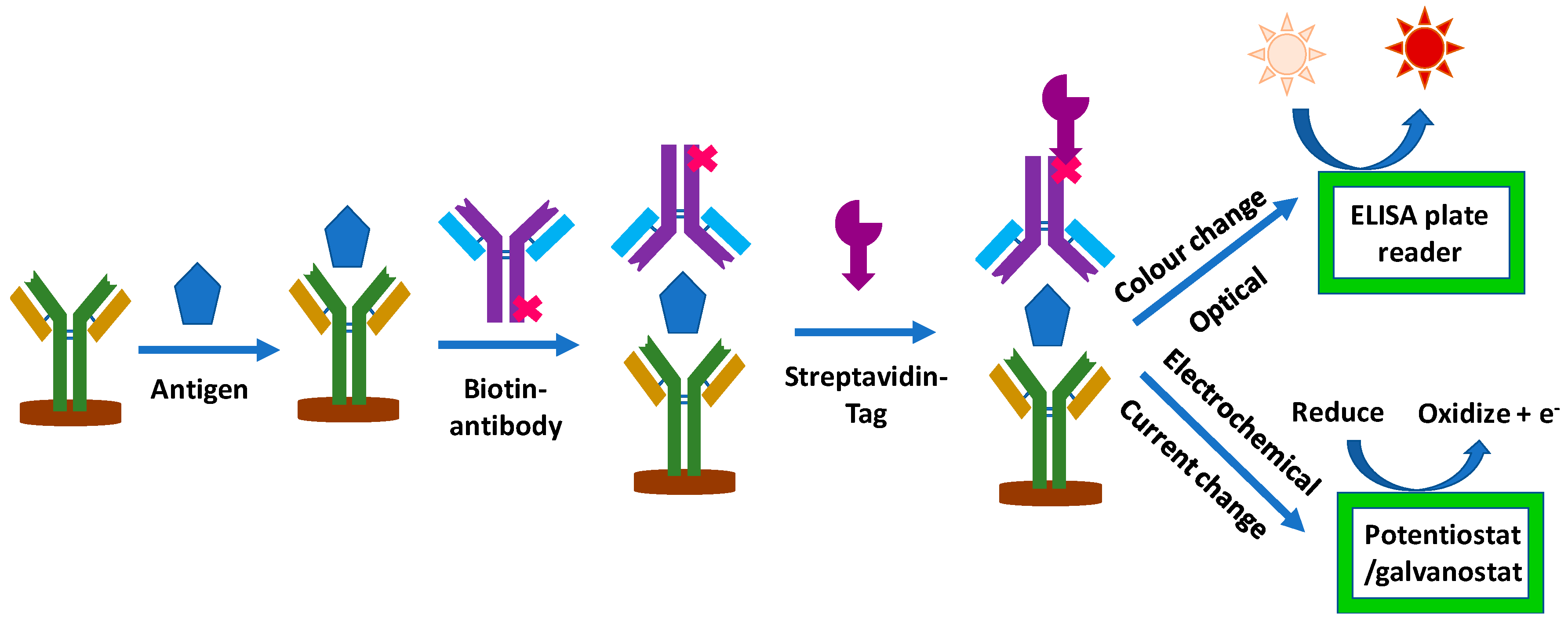
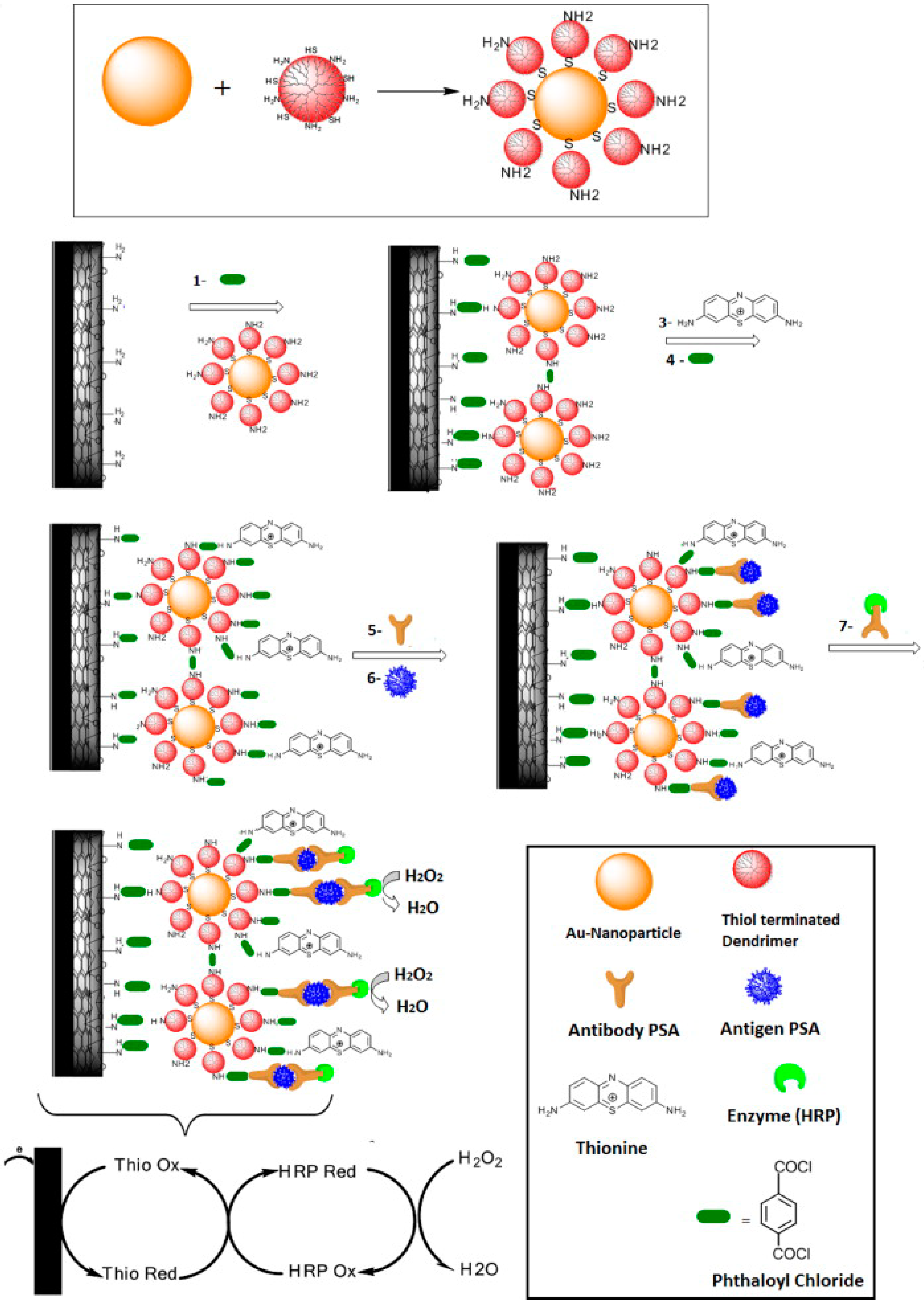
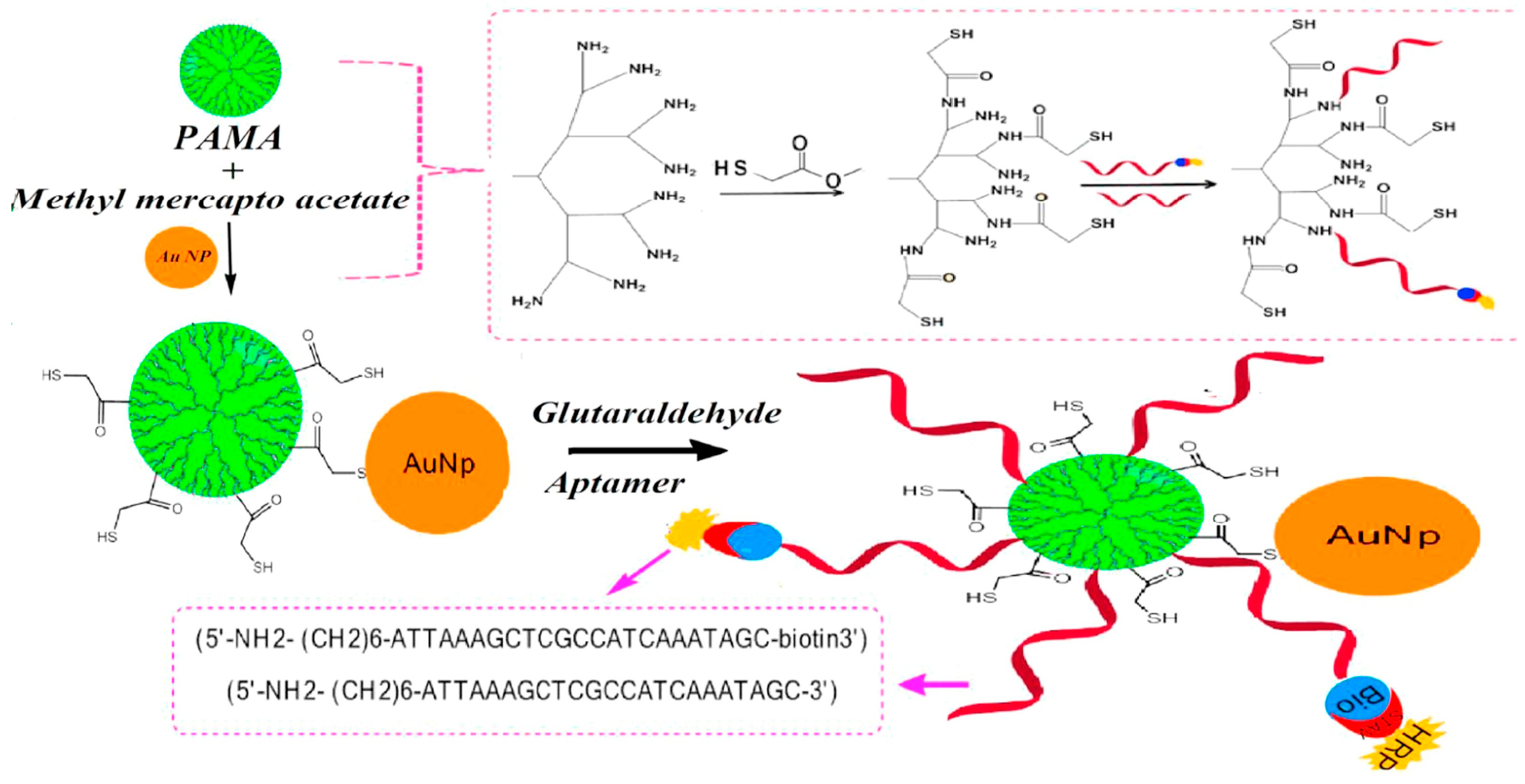
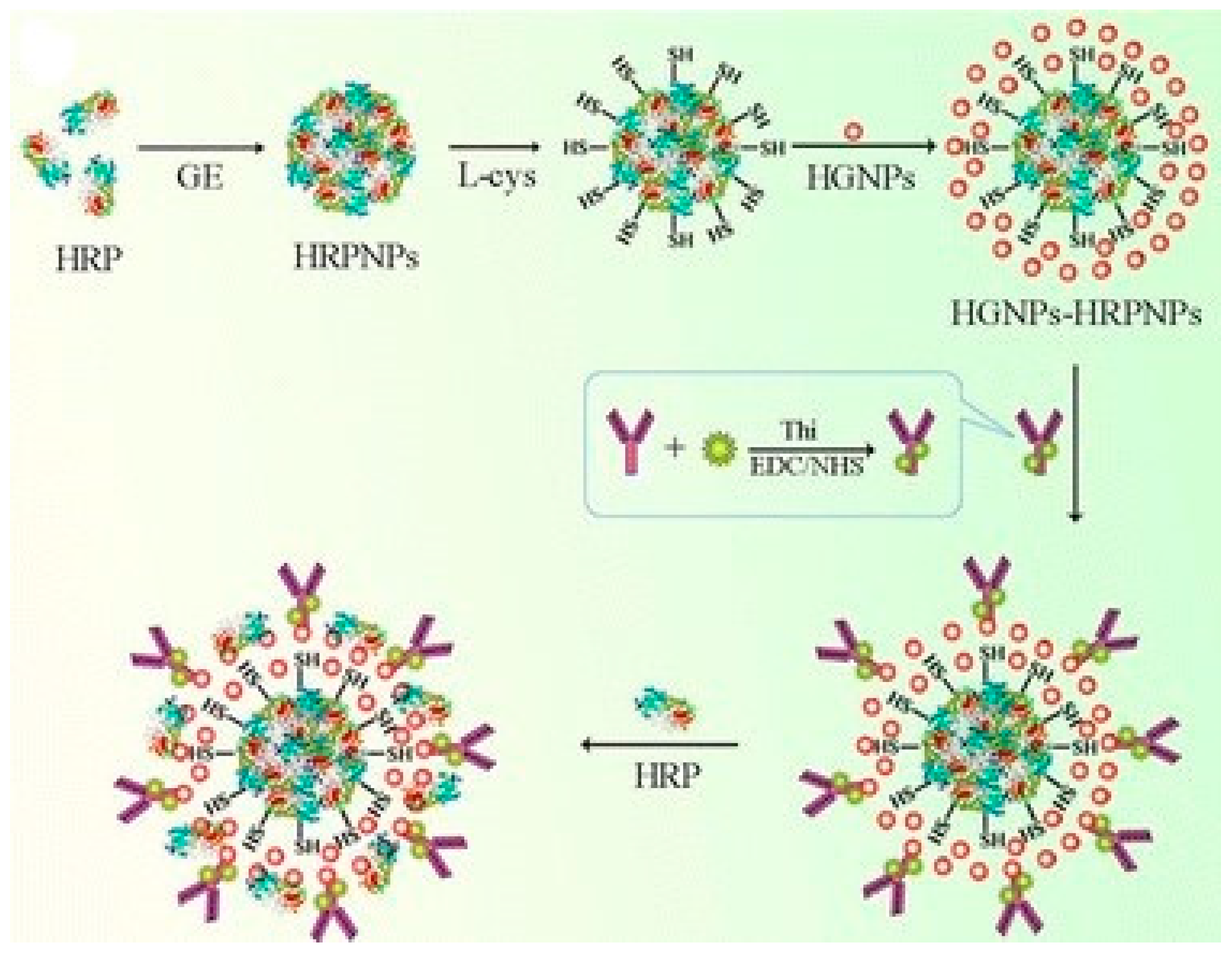
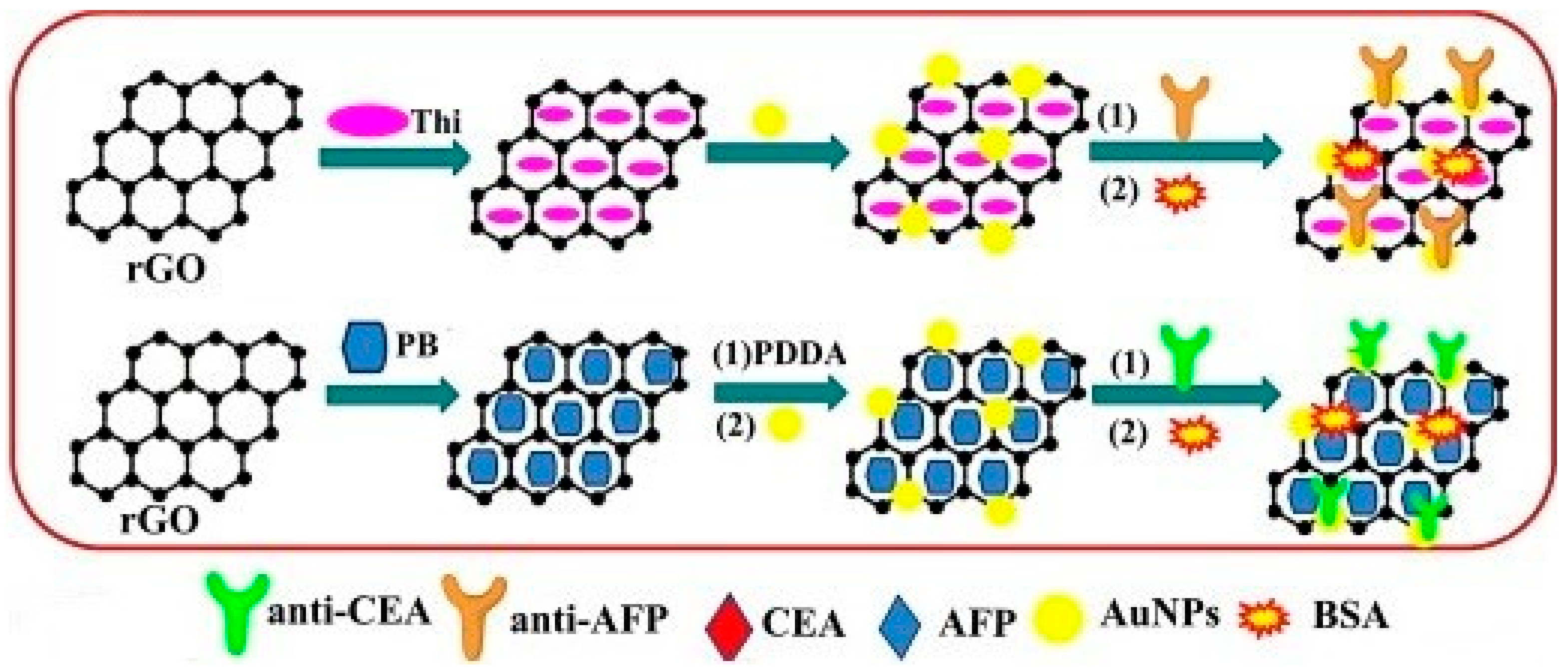
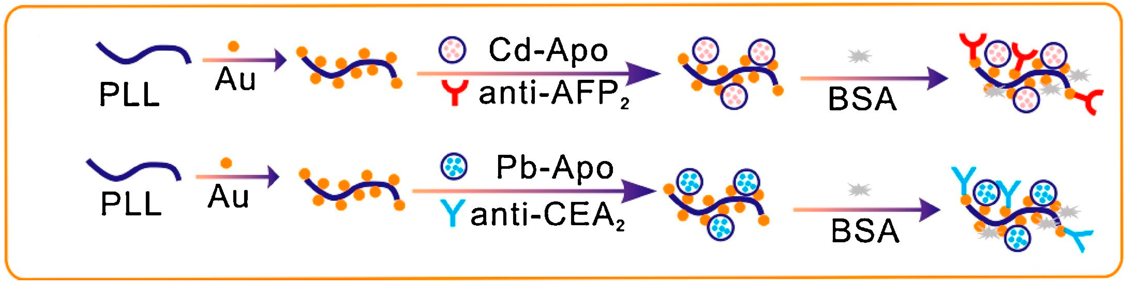
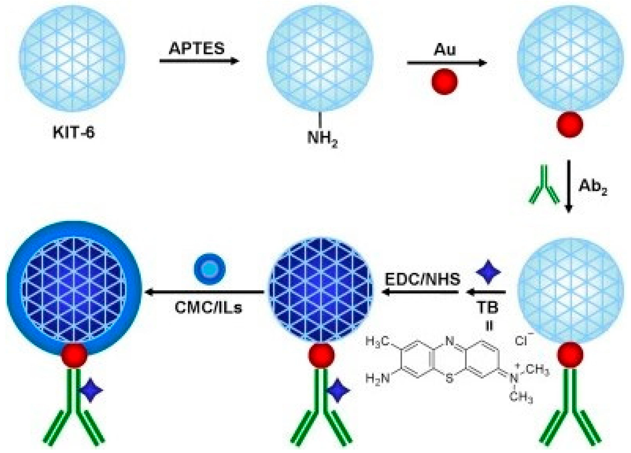
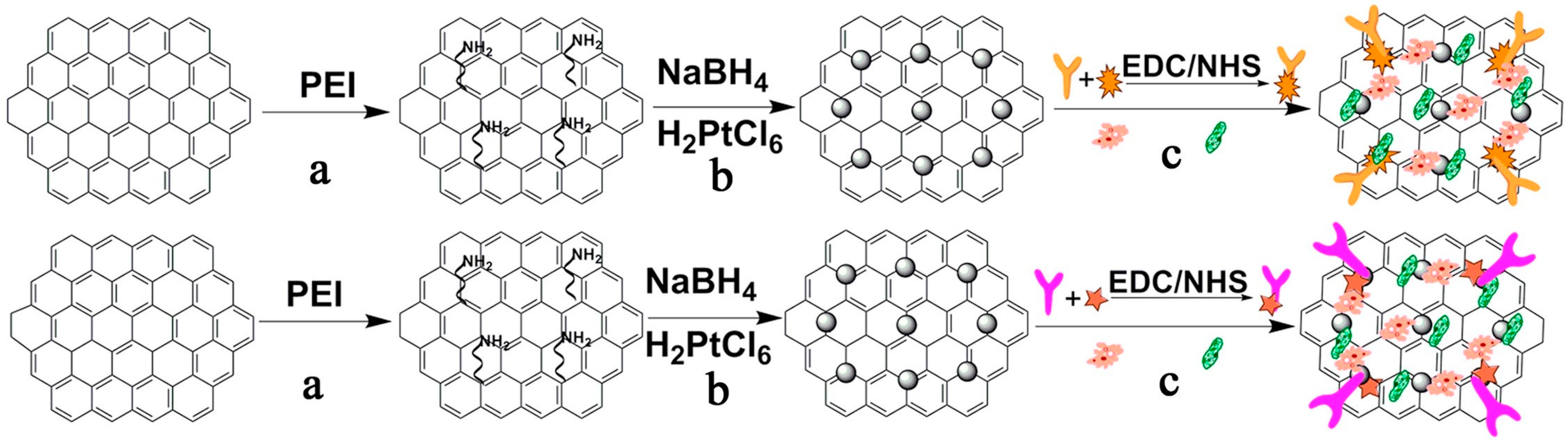
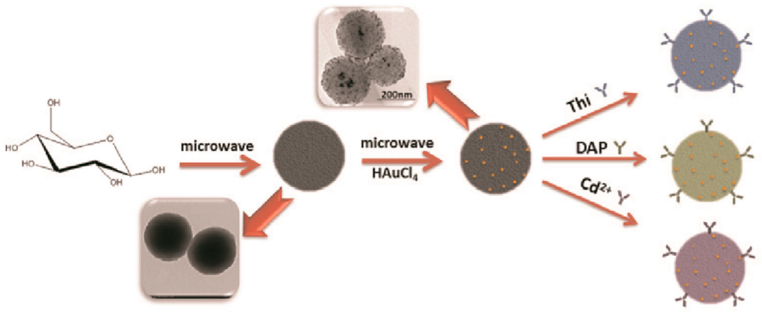
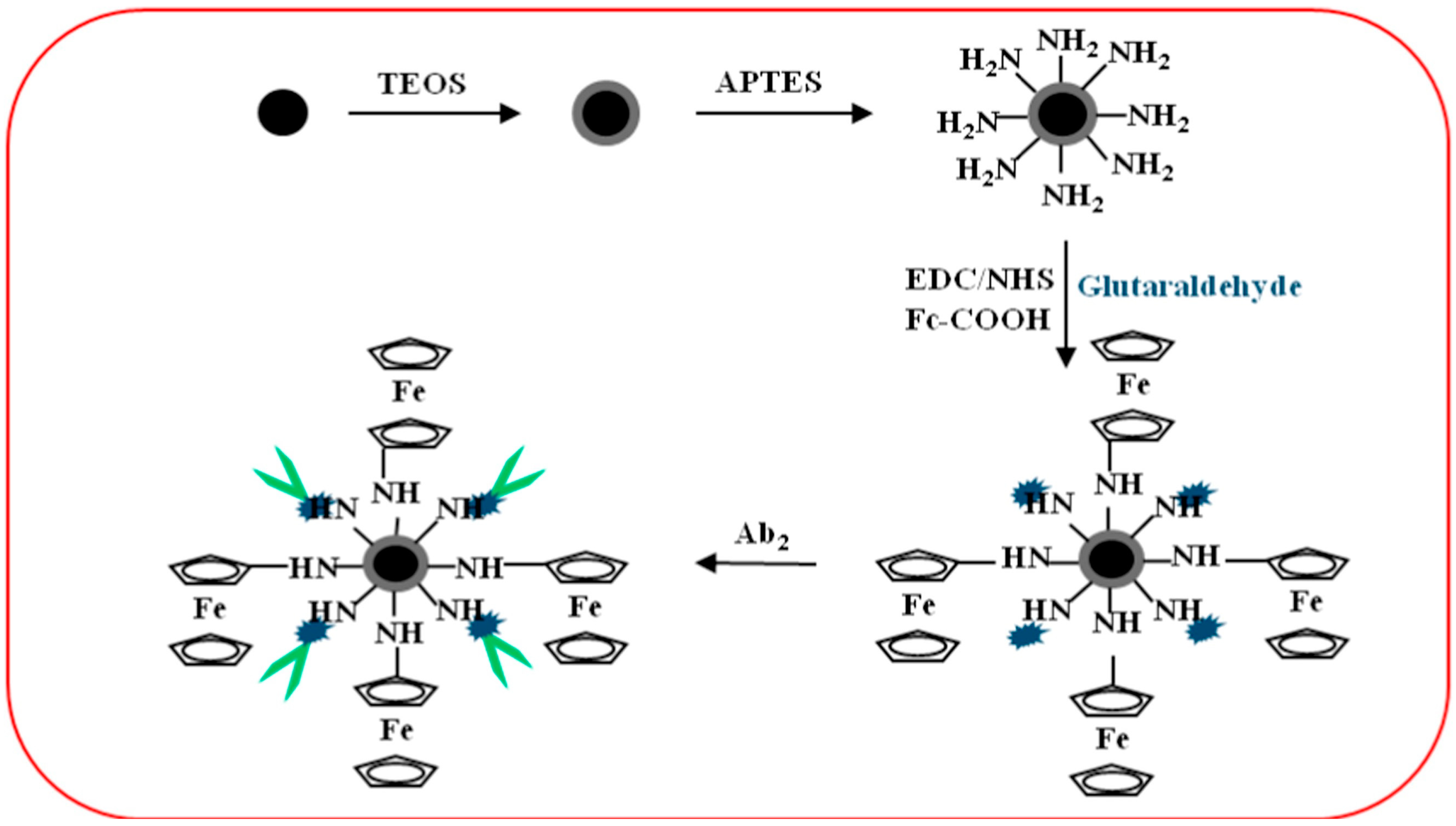
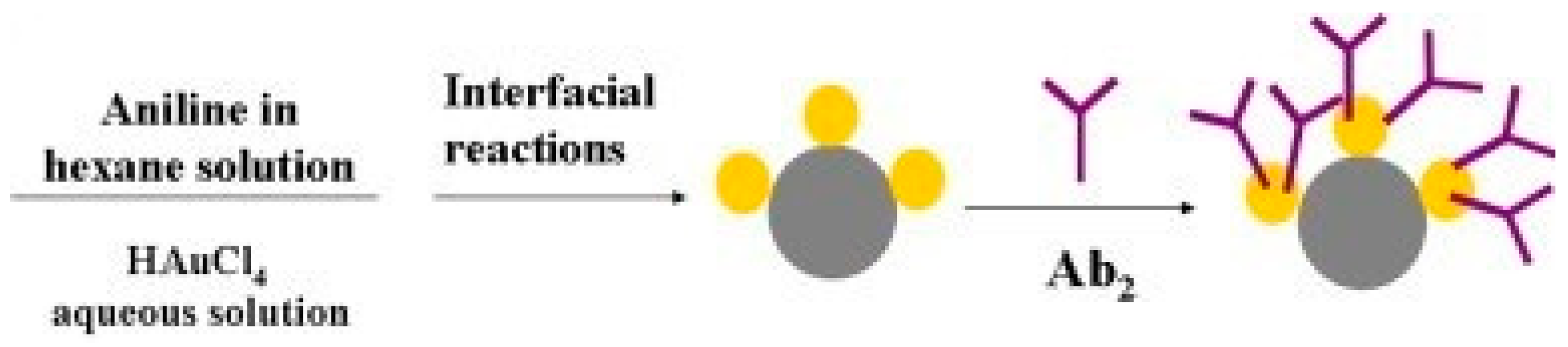
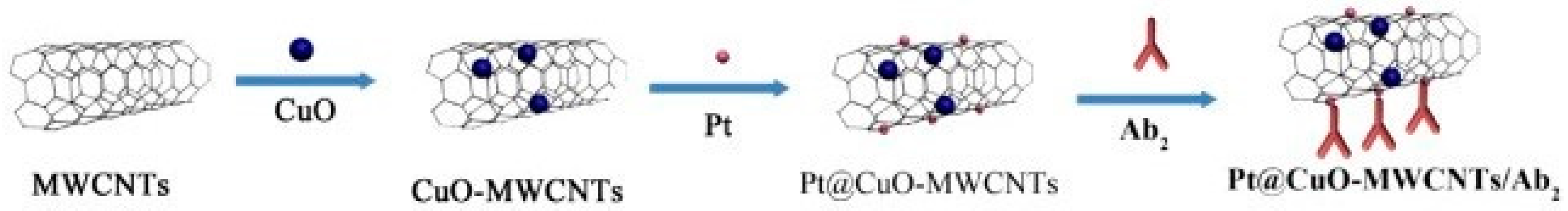
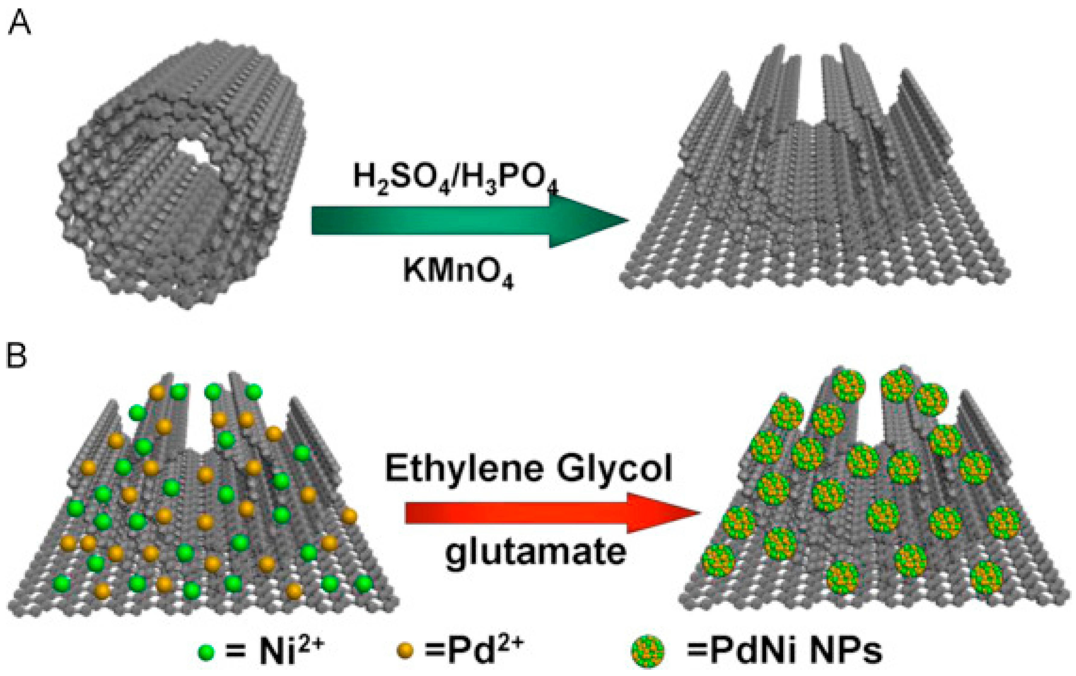
Table 1.
Various immunosensors and their preparation conditions.
| Immunosensor | Components | Preparation Conditions | Binding Technique | Target | Ref. |
|---|---|---|---|---|---|
| Ab1/GS/SPCE | GS | Acid treatment of graphite flakes | EDC/NHS (covalent) | CEA, CA125, CA153 | [30] |
| Ab1/N-GS/GCE | N-GS | GO prepared from acid treatment of graphite and then reduced with DMF at 153 °C to get N-GS | glutaraldehyde (covalent) | SCCA | [28] |
| Ab1-ADA/CD-GN/GCE | Ab1-ADA | EDC/NHS chemistry | physical | CEA | [37] |
| CD-GN | GO prepared from acid treatment of graphite and then reduced with hydrazine in presence of ammonia and β-CD at 60 °C to get CD-GN | ||||
| Ab1/MWCNTs/GCE | MWCNT-COOH | Acid treatment of MWCNTs | EDC/NHS (covalent) | AFP | [33] |
| Ab1/CD-GS/GCE | CD-GS | GO prepared from acid treatment of graphite and then reduced with hydrazine in presence of ammonia and β-CD at 180 °C to get CD-GS | physical | AFP | [38] |
| Ab1-GS/GCE | GS | GO prepared from acid treatment of graphite flakes and then reduced with hydrazine at 100 °C to get GS | EDC/NHS (covalent) | CA 15-3 | [31] |
| Ab1-ADA/CD-GS/GCE | Ab1-ADA | EDC/NHS chemistry | EDC/NHS (covalent) | AFP | [39] |
| CD-GS | GO prepared from acid treatment of graphite and then reduced with hydrazine hydrate in presence of ammonia and β-CD at 60 °C to get CD-GS | ||||
| Ab1-PA/AuNP/CSSH-SWCNTs/Au | AuNPs | Sodium citrate based reduction at 100 °C | PA-antibody interaction | AFP | [40] |
| CSSH | EDC/NHS chemistry | ||||
| anti-HER2 Nb/SPE | COOH-SPE | Acid treatment at 1.6 V | EDC/NHS (covalent) | HER2 | [63] |
| Ab1/GS-Thi/GCE | GS-Thi | Thi adsorption on GS | EDC/NHS (covalent) | AFP | [64] |
| Ab1/IL-rGO/GCE | IL-rGO | treating GO with IL-NH2 in KOH at 80 °C | glutaraldehyde (covalent) | CEA, AFP | [29] |
| Ab1/N-GS-CH/GCE | N-GS-CH | GO prepared from acid treatment of graphite and then undergo thermal annealing in ammonia to get N-GS which was then mixed with CH to get N-GS-CH | glutaraldehyde (covalent) | SCC | [65] |
| Ab1/rGO-TEPA/GCE | rGO-TEPA | rGO-TEPA | EDC/NHS (covalent) | CEA, SCCA | [66] |
| Ab1/rGO-TEPA/GCE | rGO-TEPA | rGO-TEPA | EDC/NHS (covalent) | CA72-4 | [67] |
| Ab1-GS/GCE | GS-COOH | GO prepared from acid treatment of graphite and then undergo thermal exfoliation in quartz tube at 1000 °C to get GS which was then treated chloroacetic acid in basic media to generate GS-COOH | EDC/NHS (covalent) | CA15-3 | [32] |
| Ab1/MWCNTs/DAH/GCE | MWCNT-COOH | Nitric acid treatment of MWCNTs | EDC/NHS (covalent) | PSA | [68] |
| DAH monolayer | CV scans in 0.2 and 1.6 V at 20 mV/s | ||||
| Ab1/CH/rGO/SPC/whatman paper | rGO | GO prepared from acid treatment of graphite and reduced electrochemically at −1.0 V CH coating | glutaraldehyde (covalent) | AFP, CEA, CA125, CA153 | [69] |
| Ab1/nafion-AuNP-DN-GR/GCE | GR | GO prepared from acid treatment of graphite and reduced using NaBH4 at 85 °C | physical | CEA | [34] |
| AuNPs | Sodium citrate based reduction at 97 °C | ||||
| Ab1/thionine/AuNP-PMMA dendrimer/CH-MWCNTs-IL/GCE | AuNP-PMMA dendrimer | AuNPs prepared via citrate method were mixed and incubated with thiol terminated PAMAM prepared via treating amine-terminated PAMAM dendrimer (G4) with methyl mercaptoacetate at 50 °C | phthaloyl chloride (covalent) | PSA | [42] |
| Ab1-thionine/CH/rGO/GCE | rGO | GO prepared from acid treatment of graphite and reduced electrochemically at −1.0 V CH coating | glutaraldehyde (covalent) | PSA | [70] |
| Ab1/Au@SH-GS/GCE | AuNPs | Sodium citrate based reduction in boiling condition | physical | SCCA | [41] |
| SH-GS | GO prepared from acid treatment of graphite was treated with MPTES at 70 °C followed by treating with hydrazine hydrate at 95 °C to get SH-GS | ||||
| Ab1/AuNPs-IL-rGO/GCE | IL-rGO | GO was mixed with IL-NH2 in KOH and reflux at 80 °C to get IL-rGO | physical | AFP, CEA, PSA | [43] |
| AuNPs-IL-rGO | IL-rGO mixed with HAuCl4 was reduced using ascorbic acid to get AuNPs-IL-rGO nanocomposite | ||||
| Ab1/Au@APTES-GS/GCE | APTES-GS | GO treated with APTES at 70 °C followed by treating with hydrazine hydrate at 95 °C to get APTES-GS | physical | CEA | [45] |
| AuNPs | HAuCl4 reduction using NaBH4 in ice bath | ||||
| Ab1/CH/rGO/GCE | rGO | GO prepared from acid treatment of graphite and reduced electrochemically at −1.0 V after CH coating | glutaraldehyde (covalent) | CEA | [71] |
| Ab1/AuNPs-IL-rGO/GCE | IL-rGO | GO was mixed with IL-NH2 in KOH and reflux at 80 °C to get IL-rGO | physical | CEA, AFP | [44] |
| AuNPs | HAuCl4 reduction using NaBH4 and/or sodium citrate | ||||
| Ab1/Au-GR/GCE | Au-GR | Mix HAuCl4 with GO and perform 5 CV scan in −1.5 V to 0 V at 50 mV/s | physical | AFP, CEA, CA125, PSA | [46] |
| Ab1/Au-PGO/GCE | Au-PGO | Treat GO, HAuCl4 and PEG mixture at 180 °C | physical | CA19-9 | [72] |
| Ab1/Au-Gra/GCE | Au-Gra | Treat GO-AA mixture with HAuCl4 at room temperature | physical | AFP | [73] |
| Ab1/AuNPs/CH-Thi-CNTs/GCE | AuNP | Electrochemical deposition at −200 mV | physical | CEA | [47] |
| Thi-CNT | Modify COOH-CNTs with thionine using EDC/NHS chemistry | ||||
| Ab1/GO-AuNP/GCE | GO AuNP | physical | CEA | [35] | |
| Ab1/HAG/PANI/rGO/GCE | HAG | Electrochemically deposited at −200 mV | physical | CEA, AFP | [36] |
| PANI | Electro-polymerization at 0.75 V | ||||
| Ab1/CH-AuNP/GCE | CH-AuNPs | NaBH4 based reduction of CH-HAuCl4 solution | EDC/NHS (covalent) | CEA, AFP | [52] |
| Ab1/NPG/GCE | NPG | Acid based removal of silver from silver gold alloy | physical | CA72-4 | [55] |
| Ab1/AuNPs/GCE | AuNPs | Electrodeposited at −0.2V | physical | CEA, AFP | [48] |
| Ab1/AuNPs/GCE | AuNPs | Electrodeposited at −0.2V | physical | AFP | [49] |
| Ab1/AuNPs/GCE | AuNPs | Sodium citrate based reduction in boiling condition | physical | AFP | [50] |
| AuNPs/GCE | Electrochemical deposition at 1.5 V | ||||
| Ab1/CH-AuNP/GCE | CH-AuNPs | NaBH4 based reduction of CH-HAuCl4 solution | Physical | CEA, AFP | [53] |
| Ab1/MoS2-Au/GCE | MoS2-Au | Citrate based reduction of HAuCl4-MoS2 nano-sheets solution | Physical | CEA | [56] |
| Ab1-TB/Au@MCM-41/GCE | NH2-MCM-41 | Treating MCM-41 with APTES at 70 °C | physical | AFP | [57] |
| AuNPs | HAuCl4 reduction using NaBH4 in ice bath | ||||
| Ab1/CH-AuNPs/GCE | CH-AuNPs | Refluxing CH-HAuCl4 solution for 1 h | glutaraldehyde (covalent) | CEA, AFP | [54] |
| Ab1-biotin/streptavidin/Au–Fe3O4@SiO2/Au/magnet | Fe3O4 | Treating FeCl2, FeCl3, and PEG 4000 mixture with NaOH at 80 °C | streptavidin-biotin interaction | CA 19-9 | [74] |
| Fe3O4@SiO2 | Treating PDDA-Fe3O4 solution pH 11 (using ammonia) with TEOS at room temperature | ||||
| Au–Fe3O4@SiO2 | Treating PDDA-Fe3O4@SiO2 solution with AuNPs solution | ||||
| Ab1/PSS/IL-rGO/GCE | IL-rGO | GO was mixed with IL-NH2 in KOH and treated at 80 °C | electrostatic | CEA, PSA, AFP | [75] |
| aptamer/AuNP/oPD/Au | oPD/Au | Electropolymerized via CV scans in −0.5 and 0.8 V range at 50 mV/s | physical | MUC 1 | [58] |
| Ab1/AuNPs/GCE | AuNPs | Electrodeposited at −0.2 V | physical | AFP | [51] |
| Ab1/PANI/Au/paper | Au | Seed layer using AuNPs prepared via NaBH4, citrate method; Au layer using growth solution of HAuCl4 cetyltrimethyl ammonium chloride | glutaraldehyde (covalent) | CEA, AFP | [76] |
| PANI | 20 CV scans in −0.1 to 0.8 V range at 50 mV/s | ||||
| Ab1/β-CD/GCE | oxidize GCE | 5 CV scans in H2SO4 solution in 0 to 2 V | physical | CEA | [60] |
| Ab1/PAMAM/GCE | PAMAM/GCE | Using infrared light treatment | EDC/NHS (covalent) | PSA | [61] |
| Ab1/cysteine /Au | cysteine /Au | Self-assembled monolayer | EDC/NHS (covalent) | CEA | [62] |
| PSA aptamer/GDPTS/PDMS | GDPTS/PDMS | Self-assembled monolayer | epoxide chemistry | PSA | [77] |
| Ab1/Au@MWCNTs-SO3H/GCE | MWCNTs-SO3H | Refluxing MWCNTs in H2SO4-HNO3 at 120 °C, 30 min | physical | PSA | [78] |
| AuNPs | Sodium citrate based reduction at 100 °C reflux | ||||
| Ab1/PDA-rGO/GCE | PDA-rGO | Mixing dopamine with GO and stirring for 24 h at 25 °C | physical | CEA | [79] |
| Ab1/AuNPs/GCE | AuNPs | Electrodeposited at −0.2 V | physical | CEA, NSE, CA125, Cyfra21–1, SCCA | [80] |
| Ab1/MPA/Au | MPA/Au | Self-assembled monolayer | EDC/NHS (covalent) | PSA, PSMA, IL-6, PF-4 | [81] |
| Ab1/MUDA-mercapto ethanol/Au | MUDA-mercapto ethanol | Self-assembled monolayer | EDC/NHS (covalent) | PSA | [82] |
| Ab1/PS | physical | PSA | [83] | ||
| Ab1/3D-G-CH/GCE | 3D-G | GO was first prepared from natural graphite powder by Hummer’s method followed by autoclaving at 180 °C to get 3D-G. Dried 3D-G was then mixed in 1% CS | glutaraldehyde (covalent) | CYFRA21-1 | [84] |
| Ab1/polystyrene; AgNP/SPCE | AgNPs | Sodium citrate-based reduction of AgNO3 in boiling condition | physical | AFP | [85] |
| antiHER2/APTMS-Fe3O4/GCE | Fe3O4 | Chemical co-precipitation from FeCl3·6H2O and FeCl2·4H2O mix using ammonia solution | glutaraldehyde (covalent) | HER2 | [86] |
| Anti-CEA/LPA/Au | NHS-LPA/Au | Self-assembly | covalent | CEA | [87] |
| Ab1-AuNPs/CHI/SPE | AuNPs | Electrochemical reduction in 0.5 M H2SO4 via CV scans between −1.5 and 0.5 V at a rate of 30 mV/s | physical | PSA | [88] |
| BSA/anti-CEA/AuNPs/GCE | AuNPs | Electrodeposit deposition by cyclic sweeping in the potential range of −0.5 to 0 V (vs. SCE) at 50 mV/s for 50 segments | physical | CEA | [89] |
| Ab1/Au@Th/GO/GCE | Au@Th/GO | GO synthesized using modified Hummers’ method was mixed with Thi and HAuCl4 solution and stir | physical | PSA | [90] |
| Ab1/Au@MWCNTs-SO3H/GCE | AuNPs | Citrate reduction of HAuCl4 solution; | physical | PSA | [78] |
| Au@MWCNT-SO3H | Physical adsorption of AuNPs on MWCNTs-SO3H | ||||
| Ab1/Au@MPTES-GS/GCE | AuNPs | Citrate reduction of HAuCl4 solution | physical | AFP | [91] |
| MPTES-GS | GO synthesized using modified Hummers’ method was treated with MPTES in ethanol at 70 °C for 2 h followed by treatment with hydrazine solution at 95 °C for 1.5 h | ||||
| Ab1/AuNPs/GCE | AuNPs | Electrochemical reduction at −0.2 V, 30 s | physical | CEA | [92] |
| Ab1/β-CD/MWCNT/GCE | β-CD/MWCNTs | Grind rMWCNTs and β-CD in ethanol | physical | CEA | [93] |
| Ab1/streptavidin-NG-CH/GCE | NG-S | GO synthesized using modified Hummers’ method was refluxed with hydrazine at 100 °C, 24 h. Obtained rGO was then mixed with pyrrole and treated with ammonium peroxydisulphate. Obtained PPY-rGO was heat treated till 600 °C, 2 h | Biotin-streptavidin | [94] | |
| Ab1/AuNPs/GCE | AuNPs | Electrochemical reduction at −0.2V, 30 s | physical | CEA | [95] |
| Fe3O4@AuNPs-Ab1 | Fe3O4 | From ferrous complex via hydrothermal method using H2O2 as oxidizer | physical | AFP | [96] |
| Fe3O4@AuNPs | Mixture of Fe3O4 NPs with PEG 20000 and HAuCl4 was treated with hydroxylamine hydrochloride | ||||
| Ab1/Au/ZnO/RGO/GCE | Au/ZnO/RGO | GO synthesized using modified Hummers’ method was mixed with C12N3. Solution was adjusted to pH 12 and mixed with Zn(NO3)2 and HAuCl4 followed by treatment with hydrazine at 105 °C, 5 h | physical | AFP | [97] |
| Ab1/CH/CNT/SPE | CH/CNT/SPE | Acid treated CNTs were mixed with nafion 117 and drop casted on SPE followed by deposition of CH solution | glutaraldehyde (covalent) | PSA | [98] |
| Ab1/AuNP/GCE | AuNPs | Electrochemical reduction at −0.2 V, 30 s | physical | SCCA | [99] |
| Ab1/AuNP/GCE | AuNPs | Electrochemical reduction at −0.2 V, 30 s | physical | AFP | [100] |
| Ab1-BSA/AuNP/PANI/GCE | PANI/GCE | Phytic acid doped polyaniline via electrochemical co-deposition at 0.8 V, 400 s | physical | PSA | [101] |
| AuNPs | Electrodeposit deposition by cyclic sweeping in the potential range of −1 to 0.2 V at 50 mV/s, 10 cycles | ||||
| Ab1/AuPd NCNs/GCE | AuPd NCNs | Add HAuCl4, H2PdCl4 and PVP sequentially into NaOH solution containing T7AA | physical | CA 15-3 | [102] |
| Ab1/Au@PDA/GCE | Au@PDA | Citrate reduced AuNPs were treated with dopamine in tris buffer | physical | CEA | [103] |
| Ab1/Au@N-GQD/GCE | N-GQD | Dicyandiamide and CA solution was autoclaved at 180°C, 12 h | physical | PSA | [104] |
| Au@N-GQD | HAuCl4 was added to N-GQD, pH adjusted to 10 using NaOH followed by autoclaving at 160 °C, 6 h |
Notes: β-CD: β-cyclodextrin; 3D-G: 3-dimensional graphene; AA: ascorbic acid; Ab1: capture antibody; ADA-COOH: adamantine-1-carboxylic acid; AFP: α-fetoprotein; APTES: 3-aminopropyltriethoxysilane; APTMS: 3-aminopropyltrimethoxysilane; CA: citric acid; CA 19-9: carbohydrate antigen 19-9; CA125: carbohydrate antigen 125; CA15-3: carbohydrate antigen 15-3; CEA: carcinoembryonic antigen; CH: chitosan; CSSH: L-cysteine modified chitosan; CV: cyclic voltammetry; Cyfra21–1: cytokeratin 19 fragment antigen 21–1; DAH: 1, 7-diaminoheptane; DMF: dimethylformamide; DN: 1,5-diaminonaphthalene; EDC: 1-Ethyl-3-(3-dimethylaminopropyl)-carbodiimide; GDPTS: (3-glycidyloxypropyl) trimethoxysilane; GN: graphene nanosheet; GS: graphene sheet; IL-6: interleukin-6; IL-NH2: 1-aminopropyl-3-methylimidazolium chloride; LPA: lipoic acid N-hydroxysuccinimide ester; MCM-41: multifunctional mesoporous silica; MPA: mercaptopropionic acid; MUDA: mercaptoundecanoic acid; Nb: nanobody (antigen-binding fragments with single domain); N-GS: nitrogen doped graphene sheet; NHS: N-Hydroxysuccinimide; NPG: nanoporous gold; NSE: neuron specific enolase; oPD: poly(o-phenylenediamine); PA: protein A; PAMAM: polyamidoamine dendrimers; PDA: polydopamine; PDDA: poly(diallyldimethylammonium chloride); PF-4: platelet factor-4; PGO: porous graphene oxide; PS: polystyrene; PSA: prostate specific antigen; PSMA: prostate specific membrane antigen; PSS: poly(sodium-p-styrenesulfonate); SAM: self-assembled monolayer; SCCA: squamous cell carcinoma antigen; SH-GS: mercapto-functionalized graphene sheets; SPC: screen printed carbon; SPCE: screen printed carbon electrode; TB: toluidine blue; TEPA: tetraethylene pentamine; Thi: thionine.
Table 2.
Strategies used for the development of detection probes for enhanced detection.
| Detection Probe | Components | Preparation Conditions | Ref. |
|---|---|---|---|
| HRP, Anti-CEA/AuNPs-PAN@CNTs | PAN@CNTs | (NH4)2S2O8 based polymerization of CNTs and aniline monomers solution in HCl at ice bath | [47] |
| AuNPs | Citrate reduction | ||
| AuNPs-PAN@CNTs | Electrostatic assembly of AuNPs | ||
| HRP-PSA aptamer/AuNP-PAMAM | AuNP | Citrate reduction | [70] |
| thiol-PAMAM | Treating amine-terminated PAMAM dendrimer (G4) with methyl mercaptoacetate at 50 °C for 18 h | ||
| AuNP-PAMAM | Incubation for 5 h at RT | ||
| Thi-Anti-AFP/HRP NPs-hollow AuNPs | hollow AuNPs | HAuCl4 reduction in N2 environment using NaBH4, sodium citrate and CoCl2·6H2O mixed solution | [40] |
| HRP NPs-hollow AuNPs | Self-assembly of L-cysteine modified HRP-NPs prepared via desolvation followed by glutaraldehyde chemistry; | ||
| Thi-Anti-AFP | EDC/NHS chemistry | ||
| HRP, anti-AFP/Fe3O4 NPs-MSNs | Fe3O4 NPs-MSNs | Treating APTES modified MSNs with bromine-functionalized Fe3O4 NPs in EtOH | [64] |
| AgNPs-GOx-anti CEA | AgNPs | Ag nanospheres prepared via ethylene glycol (EG) and poly(vinyl pyrrolidone) (PVP) based reduction | [56] |
| HRP-anti CA 19-9/Au@SBA-15 | Au@SBA-15 | incubating PDDA coated SBA-15 particles with AuNPs solution | [74] |
| Anti-CEA/Ag/Au–DN–GR | Ag/Au–DN–GR | (i) HAuCl4, AgNO3, trisodium citrate dihydrate and SDS mixture reduction using NaBH4, (ii) Mix and incubate Ag/Au with DN-graphene | [34] |
| PAMAM-Gr/anti-AFP-HRP | PAMAM-Gr | EDC/NHS chemistry | [50] |
| HRP, GOD, anti-AFP/SWCNHs | Carboxylated SWCNHs | Acid treatment of SWCNHs | [73] |
| AuNPs-MCF | carboxy-MCF | Refluxing MCF in acid | [71] |
| AuNPs-MCF | NaBH4 based reduction of HAuCl4-MCF mixture | ||
| CHIT-PB-AuNP CHIT-FC-AuNP | CHIT-PB | Treating K3Fe(CN)6 and FeCl3 solution (pH 1.5) in CHIT | [52] |
| CHIT-FC | EDC/NHS chemistry; | ||
| AuNP binding | Physical adsorption by mixing | ||
| anti-AFP2,2-AuNPs-Thi@rGO | AuNPs-Thi@rGO | (i) Incubating rGO and Thi for 12 h, (ii) Incubating Thi@rGO with AuNPs for 24 h, (iii) Mixing FeCl3 and K3Fe(CN)6 solution (pH 1.5) to rGO dispersion | [36] |
| anti-CEA2,1-AuNPs-PB@rGO | AuNPs-PB@rGO | (i) Mixing PB@rGO with PDDA, 30 min, (ii) Incubation with AuNPs | |
| Anti-CEA /PB–CS-Au and Anti-CEA/Cd–CS-Au | PB–CS-Au and Cd–CS-Au | (i) PBNPs and CdNPs were prepared using FeCl3 and CdCl2 were first mixed with CS solution in 1% acetic acid, (ii) Incubating PBNPs and CdNPs with AuNPs | [44] |
| PLL-Au-Cd-Apo-Ab2 and PLL-Au-Pb-Apo-Ab2 | PLL-Au | Incubating PLL with citrate reduced AuNPs | [59] |
| Cd-Apo and Pb-Apo | Dropwise adding metal ions (Cd2+, Pb2+) to Apo solution pH 2 and adjusting pH to 8.5 before stirring for 3 h | ||
| anti-CEA/PtPNPs-Cd2+ and anti-AFP /PtPNPs-Cu2+ | PtPNPs-Cd2+ and PtPNPs-Cu2+ | (i) PtPNPs synthesised from chloroplatinic acid treatment with ascorbic acid in KOH, (ii) Mercapto-ethylamine modification of PtPNPs for capture of Cd2+ or Cu2+ ions | [29] |
| CdNCs–Au–anti-CEA and CuNCs– Au–anti-AFP | CdNCs–Au and CuNCs–Au | (i) Treating CdCl2 or CoCl2 in presence of CS with K3Co(CN)6 dispersed PDDA, (ii) Nanocubes incubation with AuNPs prepared via citrate and NaBH4 reduction | [53] |
| TB/Au@KIT-6/CMC/ILs-anti-CEA | Au@KIT-6 | Treating APTES silanized KIT-6 with AuNPs prepared via NaBH4 reduction; | [45] |
| TB/Au@KIT-6/CMC/ILs-Ab2 | (i) Physical binding of anti-CEA on Au@KIT-6, (ii) TB binding using EDC NHS, (iii) Mixing and incubating with 1-butyl-pyridine tetrafluoroborate (ILs) dissolved in CMC | ||
| Ab2/M-Alg; (M: Cd, Pb and Cu) | M-Alg | (i) Emulsion A: agitate the mixture of triton x-100, 1-hexyl alcohol, n-octane and sodium for more than 30 min RT, (ii) Emulsion B: agitate mixture of triton x-100, 1-hexyl alcohol, n-octane and Metal salt for more than 30 min RT, (iii) Add emulsion A dropwise to emulsion B and stir for 4 h RT, (iv) Break M-Alg using acetone and ethanol to get M-Alg nanobeads | [43] |
| M/Ab2-Envision copolymer; (M: AuNPs, CdS and PbS) | Ab2-Envision copolymer | Mix and incubate Ab2 with Envision (highly branched polymer) at 4 °C, 24 h | [107] |
| M/Ab2-Envision copolymer | AuNP tagging via physical adsorption, CdS and PbS were bound to HRP modified envision-Ab2 via EDC/NHS chemistry | ||
| Au@MCM-41/TB/Ab2 | Au@MCM-41 | APTES modified MCM-41 was mixed with AuNPs prepared via NaBH4 based reduction of HAuCl4 | [57] |
| Au@CMK-3-anti-CEA-neutral red and Au@CMK-3-anti-SCCA-thionine | Au@CMK-3 | AuNPs were prepared from sodium citrate based reduction of HAuCl4 were mixed and stir with mesoporous carbon CMK-3 for 4 h | [66] |
| AuNPs-Ab2-Cu2+ or Pb2+ | AuNPs | Via sodium citrate based reduction of HAuCl4 | [54] |
| Cu2+ and Pb2+ tagging | Cu(NO3)2 or Pb(NO3)2 incubation with AuNPs-Ab2 | ||
| Redox tag bio-dsDNA/SA/bio-Ab2/Au/SiO2-Fe3O4 | Au/SiO2–Fe3O4 | (i) Nano-sized Fe3O4 via treating FeCl2-FeCl3 mixture with NaOH, (ii) Fe3O4–SiO2 via alkaline hydrolysis of TEOS, (iii) Au/SiO2–Fe3O4 via mixing and incubating PDDA treated SiO2–Fe3O4 with for 20 min | [46] |
| bio-dsDNA/SA/bio-Ab2/Au/SiO2-Fe3O4 | (i) Incubation of biotin-Ab2 Au/SiO2-Fe3O4 12 h, 4 °C, (ii) Treatment with streptavidin (SA), initiator bio-S1, bio-S2 and bio-S3 in sequence to form bio-dsDNA/SA/bio-Ab2/Au/SiO2-Fe3O4 via HCR reaction | ||
| Anti-CA15-3–f-TiO2–Cd2+ | nanoporous TiO2 | (i) Mixing and stirring tetrabutoxytitanium (TBOT) and ethylene glycol for 8 h, RT, (ii) Pouring mixture in acetone-water followed by vigorous stirring 1 h, (iii) Ethanol wash and drying at 50 °C, (iv) Mix with water and reflux for 1 h | [32] |
| f-TiO2–Cd2+ | (i) APTMS treatment to get NH2 functionalized nanoporous TiO2 (f-TiO2), (ii) Mixing f-TiO2 with Cd(NO3)2 and stirring for 24 h at 50 °C | ||
| Anti-PSA/Fc-AuNPs | Fc-AuNPs | Self-assembly of 6-ferrocenyl hexanethiol onto AuNPs | [68] |
| Apt/Thi-AuNPs/SiO2@MWCNTs | Apt/Thi-AuNPs/SiO2@MWCNTs | (i) Treat COOH-MWCNTs (c-MWCNTs) with PDDA, (ii) TEOS modification to get SiO2@MWCNTs, (iii) Treatment with PDDA, (iv) Incubation in AuNPs solution to obtain AuNPs/SiO2@MWCNTs, (v) Mixing and incubating with thionine 1 h, RT, (vi) Incubation with SH-Apt solution | [58] |
| Ab2-PGN | rGO | Mix and refluxing GO with PEI | [48] |
| PGN | Mix H2PtCl6 with rGO and treat with NaBH4 | ||
| Anti-CEA/APTES/3DGS@MB and anti-AFP/APTES/3DGS@Fc-COOH | 3DGS | NaI based reduction of GO prepared from graphite | [76] |
| APTES/3DGS@MB, APTES/3DGS@Fc-COOH | (i) Redox tag (MB for CEA and Fc-COOH for AFP) modification by mixing and stirring; (ii) Treatment with APTES to get amino functionalized composites | ||
| CGN-Thi-anti-CEA, CGN-DAP-anti-PSA and CGN-Cd2+-anti-AFP | CGN | (i) Glucose carbonization in presence of sodium citrate, (ii) AuNPs deposition on carbon particles from HAuCl4 using microwave reaction | [75] |
| Thi or DAP or Cd2+/CGN | Mixing Thi or DAP or Cd(NO3)2 with CGN and stirring for 5 h | ||
| M-Pt-Ab2 | M-Pt | Ascorbic acid based reduction of K2PtCl4 | [30] |
| Anti-CA72-4/PANi–Au AMNPs | PANi–Au AMNPs | Mix and incubate aniline in hexane with HAuCl4 aqueous solution at 45 °C overnight | [55] |
| Fe3O4@SiO2/Fc/GA/anti-CEA | Fe3O4@SiO2/Fc/GA/ | (i) Fe3O4 via solvothermal method; (ii) Treatment with TEOS to obtain Fe3O4@SiO2; (iii) Treatment with APTES to get Fe3O4@SiO2–NH2; (iv) Treatment with EDC/NHS activated Fc-COOH followed by treatment with GA | [35] |
| Anti-SCCA/Pd–Au/C | Pd–Au/C | Mixing activated carbon, PdCl2, HAuCl4 and H2O-tetrahydrofuran via ultra-sonicating and stirring followed by treatment with NaBH4 and Na2CO3 | [28] |
| Cu@Ag-CD/anti-CEA | Cu@Ag-CD | CD-ascorbic acid (pH 11) solution based sequential reduction of CuSO4·5H2O and AgNO3 solution in ammonia, followed by mixing and stirring with HS-β-CD overnight; Obtained Cu@Ag-CD was used for EDC/NHS based binding of Ab2 modified ADA-COOH | [37] |
| Fe3O4@C@Pd/anti-AFP | Fe3O4@C@Pd | (i) Fe3O4@C magnetic nanoparticles via hydrothermal process; (ii) Treatment with PDDA followed by mixing and incubation with PDNPs prepared via citrate and NaBH4 based reduction of Na2PdCl4 | [33] |
| Anti-CA15-3/NP-PtFe | NP-PtFe | By removing Al using NaOH from ternary PtFeAl alloy with 80%Al | [31] |
| Anti-AFP/PdNi/N-GNRs | PdNi/N-GNRs | (i) N-GNRs powders via microwave-assisted treatment of N-MWCNTs, (ii) Mix N-GNRs with aqueous solution of Na2PdCl4, NiCl2·6H2O, and glutamate in ethylene glycol (EG), (pH 11) and stirring it for 2h followed by heating at 160 °C for 6 h in autoclave to get PdNi/N-GNRs | [39] |
| Pb2+@Au@MWCNTs-Fe3O4/anti-AFP | Pb2+@Au@MWCNTs-Fe3O4 | (i) MWCNTs-Fe3O4 via autoclaving the mixture of acid treated MWCNTs, FeCl3.6H2O and sodium acetate, (ii) Amino-functionalization via APTES modification, (iii) Mixing and incubation with AuNPs prepared via citrate reduction; (iv) Treatment with lead nitrate solution 24 h to get Pb2+@Au@MWCNTs-Fe3O4 | [49] |
| Au/Ag/Au@anti-SCCA | Au/Ag/Au | (i) Mix AuNPs, ascorbic acid, and AgNO3 in CTAB solution, (ii) Add NaOH dropwise with vigorous stirring to get yellow-golden colored, silver coated Au particles, (iii) Mix with HAuCl4 and ascorbic acid and stirred vigorously to obtain dark-blue Au/Ag/Au NPs solution | [41] |
| Anti-AFP/Pd/APTES-M-CeO2-GS | Pd/APTES-M-CeO2-GS | (i) The Pd octahedral NPs via sonicating followed by heating the mixture of 1-ethenyl-2-pyrrolidinone homopolymer (PVP), citric acid, and Na2PdCl4 dissolved in ethanol and water at 80 °C with stirring and refluxing for 3 h, (ii) M-CeO2-GS prepared by dissolving Ce(NO3)3·6H2O into water followed by adding C2H5COOH, ethylene glycol and GO and then treating at 180 °C for 200 min followed by cooling, centrifuging the ppt and drying at 50 °C for 12 h, (iii) APTES modification of M-CeO2-GS by refluxing, (iv) Pd binding by sonication and stirring to get Pd/APTES-M-CeO2-GS | [51] |
| Anti-SCC-Pt–Fe3O4 | Pt–Fe3O4 | (i) Mix platinum acetylacetonate, oleic acid, oleylamine and octadecane under argon atmosphere followed by heating to 120 °C, (ii) add Fe(CO)5 heat at 280 °C, 20 min, (iii) Precipitation using ethanol addition | [65] |
| Anti-AFP/Pt@CuO-MWCNTs | Pt@CuO-MWCNTs | (i) Acid treated MWCNTs mixed with Cu(CH3COO)2·H2O were grounded and calcinated at 350 °C in argon, followed by addition of NH4OH solution, (ii) MWCNTs addition followed by ageing and calcination to get CuO/MWCNTs composite, (iii) Pt loading by adding CuO/MWCNTs nanocomposites to K2PtCl4 solution followed by Pt salt reduction | [38] |
| M-Pd@Pt/NH2-GS/anti-PSA | NH2-GS | (i) Mix GO and ethylene glycol under ultrasonication followed by ammonia water addition, (ii) Autoclave for solvothermal reaction at 180 °C, 10 h | [78] |
| M-Pd@Pt | (i) Mix Pluronic F127 with aqueous solution of K2PtCl4, Na2PdCl4 and hydrochloric, (ii) Reduction using ascorbic acid at 35 °C for 4 h | ||
| M-Pd@Pt/NH2-GS | Mix and sonicate NH2-GS and M-Pd@Pt for 1 h | ||
| Ir NPs-anti-CEA | PVP stabilized Ir NPs | (i) Add aqueous IrCl3 solution dropwise to ethanol solution containing PVP followed by mixing and stirring at 25 °C for 12 h, (ii) Refluxed in air at 100 °C for 6 h followed by evaporation | [79] |
| PBG-Au-anti-CEA; PPP-Au-anti-NSE; PTBO-Auanti-CA125; PMCP-Au-anti-Cyfra21–1; Cd NCs-Auanti-SCCA | PBG-Au | Add and stir TTAB to brilliant green aqueous solution followed by HAuCl4 addition and agitation for 4 h, RT | [80] |
| PPP-Au | Add water with stirring to DMF solution of N-phenyl-p-phenylenediamine followed by HAuCl4 addition and agitation for 4 h, RT | ||
| PTBO-Au | Add HAuCl4 to toluidine blue o aqueous solution and agitate for about 4 h, RT | ||
| PMCP-Au | Add and stir DTAB to m-cresol purple ethanol solution followed by HAuCl4 addition and agitation for 4 h, RT | ||
| Cd NCs-Au | Mix Cd NCs with gold colloid and stirred for 4 h | ||
| HRP-anti-CYFRA21-1/AuNPs/Thi/MWCNT-NH2 | AuNPs | HAuCl4 reduction using NaBH4 in Thi/MWCNT-NH2 solution | [84] |
| MWCNT-NH2 | Acid treatment of MWCNT to get MWCNT-COOH followed by treatment with HMDA in presence of DCC for 96 h at 120 °C | ||
| anti-AFP-Co3O4@MnO2-Thi | Co3O4@MnO2 | Mixture of Co(CH3COO)2·4H2O and MnO2 nanotubes in ammonium hydroxide autoclaved at 150 °C, 5 h followed by calcination at 300 °C, 1 h | [85] |
| Co3O4@MnO2-Thi | Co3O4@MnO2 treatment with APTES at 70 °C, 1.5 h followed by incubation with Thi at 95 °C, 1 h | ||
| anti-AFP-Co3O4@MnO2-Thi | EDC/NHS chemistry | ||
| antiHER2/Hyd@AuNPs-APTMS-Fe3O4 | antiHER2/Hyd@AuNPs-APTMS-Fe3O4 | AuNPs preparation using HAuCl4 reduction via NaBH4, sodium citrate followed by treatment with APTMS-Fe3O4. Resulting AuNPs-APTMS-Fe3O4 were treated with thiolated anti-HER2 followed by treatment with hydrazine | [86] |
| Anti-CEA-AuNPs-Fc | AuNPs | Reduction of chloroauric acid with trisodium citrate | [87] |
| Anti-CEA-AuNPs-Fc | Physical immobilization of anti-CEA on AuNPs followed by chemisorption of Fc-SH | ||
| HRP-anti-CEA-AuNPs-TiO2-graphene | TiO2-graphene | Sonicate graphene with dopamine for 1 h, followed by stirring with TiO2 | [89] |
| HRP-anti-CEA-AuNPs-TiO2-graphene | Treat TiO2-graphene with HAuCl4 under ultraviolet irradiation followed by physical adsorption of HRP-anti-CEA | ||
| PtCu@rGO/g-C3N4/anti-PSA | PtCu@rGO/g-C3N4/anti-PSA | Physical adsorption of anti-PSA on PtCu@rGO/g-C3N4 | [90] |
| M-Pd@Pt/NH2-GS/anti-PSA | NH2-GS | GO prepared via modified Hummer’s method was mixed with ethylene glycol and ammonia followed by autoclaving at 180 °C for 10 h | [78] |
| M-Pd@Pt | Pluronic F127 was mixed with K2PtCl4 and Na2PdCl4 in HCl followed by reducing with ascorbic acid | ||
| Anti-AFP-Pt NPs/Co3O4/graphene | Pt NPs/Co3O4/graphene | Mix GO and Co(NO3)2·6H2O in ethanol and add ammonia solution followed by autoclaving at 190 °C for 24 h. Obtained Co3O4/graphene was mixed with Na2PtCl4 in ethanol aqueous solution and treat with NaBH4 | [91] |
| GS-Fe3O4/Au@Ag/Ni2+-anti-CEA | NH2-GS-Fe3O4 | GO prepared via modified Hummer’s method was mixed with clear solution of FeCl3·6H2O in ethylene glycol along with NaAc and ethanediamine and autoclaved at 200 °C for 8 h. Resulting GS-Fe3O4 was treated with APTES to get NH2-GS-Fe3O4 | [92] |
| Au@Ag | AuNPs prepared via citrate reduction were mixed with ascorbic acid, AgNO3 and CTAB solution and treated with NaOH | ||
| GS-Fe3O4/Au@Ag/Ni2+-anti-CEA | GS-Fe3O4/Au@Ag made by mixing NH2-GS-Fe3O4 and Au@Ag were dispersed in Ni(NO3)2·6H2O solution and stir for 24 h, anti CEA was immobilized via physical adsorption | ||
| Ag NPs-MWCNTs/MnO2-Anti-CEA | Ag NPs-MWCNTs/MnO2 | Acid treated MWCNTs were dispersed in KMnO4 solution and treated with MnSO4. Obtained MWCNTs/MnO2 were mixed with AgNO3 in water followed by reduction NaBH4 | [93] |
| PdCu-anti-CEA | PdCu | Using AA as reducing agent and HDPC as growth inhibitor | [95] |
| anti-AFP-GNPs-HRP | GNP | Citrate reduction | [96] |
| anti-AFP/HRP-Au@ZnO | Au@ZnO | C18N3 was added to mixture of Zn(NO3)2 and HAuCl4 and heated at 145 °C, 5 h | [97] |
| anti-PSA/AuNPs | AuNPs | Citrate reduction in cold for smaller size and in boiling condition for large size | [98] |
| Co3O4@CeO2-Au@Pt-anti-SCCA | Co3O4@CeO2 | Co(NO3)2·6H2O solution was treated with NaOH at 180 °C, 5 h. Obtained Co3O4 cubes were mixed in ethanol aqueous solution followed by addition of Ce(NO3)3 and HMT and refluxing at 70 °C, 2 h | [99] |
| Au@Pt | Citrate reduced AuNPs were mixed with H2PtCl6 under boiling conditions followed by reduction with AA | ||
| Co3O4@CeO2-Au@Pt | APTES treated Co3O4@CeO2 were mixed with Au@Pt and stir for 12 h at room temperature. | ||
| Anti-AFP/Au@Ag/PDA-PR-MCS | PR-MCS | C6H5OH and HCHO were added to solution containing NH4OH and C2H5OH and autoclaved at 100 °C, 24 h. Product was mixed with KOH and treated at 350 °C, 1 h followed by at 700 °C, 2 h | [100] |
| Au@Ag | Citrate reduced AuNPs were mixed with AgNO3 solution and treated with NaBH4 solution | ||
| Au@Ag/PDA-PR-MCS | PR-MCS dispersed in tris buffer was treated with Dopamine hydrochloride 24 h and mixed with Au@Ag solution | ||
| MSN-MB/PDA-anti-PSA | MSN | Mixture of CTAB and pluronics F127 in ethanol, water and ammonia was treated with TEOS | [101] |
| MSN-MB/PDA | MB loaded MSN was treated with dopamine in tris buffer, pH 8.5 | ||
| Au@Pt DNs/NG/Cu2+-anti-CEA | NG | GO prepared via modified Hummer’s method was treated with ammonia solution at 90 °C, 4 h | [103] |
| Au@Pt DNs | NaBH4 and AA reduced HAuCl4 and CTAB solution was mixed with K2PtCl4 and AA and treated at 60 °C, 12 h | ||
| Au@Ag-Cu2O/anti-PSA | Au@Ag-Cu2O | Citrate reduced AuNPs were mixed with CTAC and AgNO3 followed by reduction using AA at 30 °C, 2 h. Obtained Au@Ag solution was mixed with CuCl2 and SDS followed by treatment with NaOH and NH2OH·HCl | [104] |
Notes: AA: ascorbic acid; Apo: apoferritin; Cd NCs-Au: Cd nanocubes-gold; CdNCs and CuNCs: Cd3[Co(CN)6]2 and Cu3[Co(CN)6]2 nanocubes; CTAB: hexadecyl trimethyl ammonium bromide; CTAC: cetyltrimethylammonium chloride; HDPC: hexadecylpyridinium chloride monohydrate; HMDA: hexamethylenediamine; HMT: hexamethylenetetramine; Ir NPs: iridium nanoparticles; M-Alg: metal alginate nanobeads; MCF: mesoporous carbon form; MCM-41: multifunctional mesoporous silica; M-Pd@Pt/NH2-GS: mesoporous core-shell Pd@Pt nanoparticles loaded by amino group functionalized graphene; M-Pt NPs: mesoporous platinum nanoparticles; MSNs: mesoporous silica nanoparticles; N-MWCNTs: N-doped multi-walled carbon nanotubes; PBG-Au: poly (brilliant green)-gold; PDDA: poly(diallyldimethylammonium chloride); PMCP-Au: poly (m-cresol purple)-gold; PPP-Au: poly (N-phenyl-p-phenylenediamine)-gold; PTBO-Au: poly (toluidine blue o)-gold; PtPNPs: platinum porous nanoparticles; PVP: polyvinylpyrrolidone; SDS: sodium dodecyl sulfate.
Table 3.
Characteristics of the developed immunoassays using electrochemical ELISA.
| Probe | Immunosensor Conditions | Characteristics | Ref. |
|---|---|---|---|
| [DP]: anti-HER2-HRP [Anal]: HER2 [DM]: HQ | [Tran]: amperometry at −280 mV [IC]: (i) anal for 2 min at RT, (ii) [DP] for 20 min [MC]: 2.5 mm H2O2 with HQ in citrate buffer | [L]: 1 and 200 µg/mL, [LgS] [DL]: 1 µg/mL [S]: 18.23 µA/(µg/mL) [SL]: 3 weeks [CR]: 0.9591 | [63] |
| [DP]: anti-CEA-HRP/AuNPs-PAN@CNTs [Anal]: CEA [DM]: H2O2 | [Tran]: DPV in 0.2 to −0.8 V, [PA] 50 mV [IC]: (i) CEA, (ii) [DP] for 55 min at 37 °C, sequentially [MC]: 4 mM H2O2 in 5.0 mL PBS | [L]: (i) 0.02–3.0 ng/mL, (ii) 3.0–80 ng/mL [LS] [DL]: 0.008 ng/mL [S]: (i) 13.9465 µA/(ng/mL), (ii) 0.7342 µA/(ng/mL) [SL]: 30 days [CR]: (i) 0.9875, (ii) 0.9960 | [47] |
| [DP]: AuNP–PAMAM dendrimer/PSA–aptamer-HRP [Anal]: PSA [DM]: thionine | [Tran]: DPV in −0.4 to −0.1 V [IC]: (i) PSA conc. for 15 min, (ii) [DP] for 20 min [MC]: 3 mM H2O2 | [L]: 0.1 pg/mL to 90 ng/mL [LS] [DL]: 10 fg/mL [S]: 0.3635 µA/(pg/mL) [SL]: 3 weeks [CR]: 0.9831 | [70] |
| [DP]: HRP-HRP-NP-hollow Au-NP-Thi@anti-AFP [Anal]: AFP [DM]: thionine | [Tran]: DPV in −0.4 to 0 V [IC]: (i) AFP conc for 16 min, 37 °C, (ii) [DP] for 30 min, RT [MC]: 4.2 mM H2O2 | [L]: 0.025 to 5.0 ng/mL [LgS] [DL]: 8.3 pg/mL [S]: 7.649 µA/(ng/mL) [SL]: 30 days [CR]: 0.9949 | [40] |
| [DP]: anti-AFP, HRP/MSNs-Fe3O4 [Anal]: AFP [DM]: thionine | [Tran]: CV in −0.6 to 0.6 V (vs. SCE) at 100 mV/s in PBS (pH 7.4) [IC]: (i) AFP, (ii) [DP] for 1 h, sequentially [MC]: 5 mmol/L H2O2 in PBS | [L]: 0.01 to 25 ng/mL [LS] [DL]: 4 pg/mL [SL]: 15 days | [64] |
| [DP]: GOx/anti-CEA/AgNPs [Anal]: CEA [DM]: H2O2 | [Tran]: DPV in −0.2 to −0.8 V, [PA]: 50 mV, [PW]: 20 ms [IC]: (i) CEA conc 40 min, RT, (ii) [DP] 1 h, 4 °C [MC]: PBS + 1% glucose | [L]: 1 pg/mL to 50 ng/mL [LgS] [DL]: 0.27 pg/mL [S]: 8.281 µA/(ng/mL) [SL]: 30 days [CR]: 0.9971 | [56] |
| [DP]: HRP-Ag@BSA-anti-CEA [Anal]: CEA [DM]: tyramine | [Tran]: DPV in 0 to -600 mV vs. SCE [PA]: 50mV, [PW]: 50 ms in PBS [IC]: (i) CEA conc 40 min, RT, (ii) [DP] 40 min, RT, (iii) 2 mM H2O2 + HRP-tyramine conjugates 10 min at RT [MC]: 2.5 mM H2O2 in PBS | [L]: 0.005–80 ng/mL [LgS] [DL]: 5.0 pg/mL [S]: 1.617 µA/(ng/mL) [SL]: 28 days [CR]: 0.9867 | [108] |
| [DP]: HRP/HRP-anti-CA 19-9/Au@SBA-15 [Anal]: CA 19-9 [DM]: H2O2 | [Tran]: chronoamperometry PBS pH 6 at −0.2 V [IC]: (i) CA 19-9 conc 1 h, 37 °C; (ii) [DP] 1 h, 37 °C [MC]: 3 mM H2O2 in PBS | [L]: 0.05 to 15.65 U/mL [LgS] [DL]: 0.01 U/mL [S]: 20.51 g/L [SL]: 30 days [CR]: 0.992 | [74] |
| [DP]: anti-CEA –Ag/Au–DN-graphene [Anal]: CEA [DM]: Ag | [Tran]: CV in −0.6 to1.0 V (vs. SCE) at 50 mV/s [IC]: (i) CEA conc 30 min, (ii) [DP]: 40 min [MC]: 0.1M PBS (pH7.0). | [L]: 10 to 1.2 × 10⁵ pg/mL [LgS] [DL]: 8 pg/mL [S]: 0.494 µA/(ng/mL) [CR]: 0.9899 | [34] |
| [DP]: PAMAM-Gr/anti-AFP-HRP [Anal]: AFP [DM]: hydroquinone | [Tran]: (i) amperometric at −0.2 V, (ii) CV −0.5 to +0.5 V, 50 mV/s [IC]: (i) AFP conc 40 min, 37 °C, (ii) [DP]: 40 min, 37 °C [MC]: PBS containing 1 mM hydroquinone + 2 mM H2O2 | [L]: 1.0–100 ng/mL [LS] [DL]: 0.45 ng/mL | [50] |
| [DP]: Au@Pd-Gra/Thi-anti-CA 19-9/HRP [Anal]: CA19-9 [DM]: Thionine | [Tran]: DPV in −0.4–0 V, [PA]: 50 mV, [PW]: 50 ms, [PP]: 0.2 s [IC]: (i) CA 19-9 conc 40 min, 25 °C, (ii) [DP] [MC]: 1.5 mM H2O2 | [L]: 0.015 to 150 U/mL [LgS] [DL]: 0.006 U/mL [S]: 9.8328 µA/(U/mL) [SL]: 30 days [CR]: 0.9982 | [72] |
| [DP]: HRP, GOD, anti-AFP/SWCNHs [Anal]: AFP [DM]: 4-CN | [Tran]: Impedance [IC]: (i) AFP conc 40 min, 37 °C, (ii) [DP] 40 min, 37 °C, (iii) 1.0 mM 4-CN and 10.0 mM glucose in 10mM PBS 15 min, RT [MC]: 0.01M PBS (pH 7.4) containing 5 mM FeCN63−/4− and 0.1M KCl | [L]: 0.001 to 60 ng/mL [LgS] [DL]: 0.33 pg/mL [S]: 230.60 Ω/(ng/mL) [SL]: 30 days [CR]: 0.996 | [73] |
| [DP]: anti-CEA/Au/MCF [Anal]: CEA [DM]: Ag | [Tran]: ASV in −0.08 to 0.2 V, 50 mV/s [IC]: (i) CEA conc 40 min, 37 °C, (ii) [DP] 40 min, 37 °C, (iii) silver-deposition with enhancer solutions 4 min, 37 °C, [MC]: 1.0 M KCl | [L]: 0.05 pg/mL to 1 ng/mL [LgS] [DL]: 0.024 pg/mL [SL]: 15 days [CR]: 0.9997 | [71] |
| [DP]: anti-AFP/CHIT–PB–AuNP; anti-CEA/CHIT–Fc–AuNPs, [Anal]: CEA, AFP [DM]: PB, Fc | [Tran]: DPV in −0.2 to 0.8 V [IC]: 45 min incubation for CEA, AFP concentration [MC]: PBS | [L]: 0.05–100 ng mL−1 for AFP and CEA [LgS] [DL]: 0.03 ng mL−1 and 0.02 ng mL−1 for AFP and CEA [S]: 0.47067 µA/(ng/mL), 0.51106 µA/(ng/mL) for AFP and CEA [CR]: 0.99712 for AFP and 0.99806 for CEA | [52] |
| [DP]: anti-AFP-AuNPs-Thi@rGO and anti-CEA-AuNPs-PB@rGO [Anal]: CEA, AFP [DM]: PB, Thionine | [Tran]: DPV 600 to −600 mV; [PA] 50 mV. [IC]: (i) CEA/AFP conc 50 min, 37 °C, (ii) [DP] 50 min, 37 °C [MC]: PBS pH 6.5 | [L]: 0.6–80 ng/mL for both [LS] [DL]: 0.12 ng/mL and 0.08 ng/mL for CEA and AFP [S]: 0.0188 µA/(ng/mL), 0.0273 µA/(ng/mL) for CEA and AFP [SL]: 30 days [CR]: 0.9908, 0.9936 for CEA and AFP | [36] |
| [DP]: Anti-CEA/PB–CS-Au and anti APF/Cd–CS-Au [Anal]: CEA, AFP [DM]: PB, Cd | [Tran]: DPV in −0.1V to 0.9V (vs. Ag/AgCl), [PA]: 50 mV, [PW]: 50 ms [IC]: (i) CEA/AFP conc 40 min, (ii) [DP] mixture 1:1 [MC]: 0.1 M pH 6.5 phosphate buffered solution (PBS) | [L]: 0.01 to 100 ng/mL range for both [LgS] [DL]: 0.006 ng/mL for AFP and 0.01 ng/mL for CEA [S]: 1.771 µA/(ng/mL), 1.751 µA/(ng/mL) for CEA and AFP [CR]: 0.996 and 0.995 for CEA and AFP | [44] |
| [DP]: PLL-Au-Cd-Apo-anti-AFP and PLL-Au-Pb-Apo-anti-CEA [Anal]: AFP and CEA [DM]: Cd, PB | [Tran]: SWV scan from −1.0 to −0.3 V with frequency of 15 Hz, [PA]: 25 mV, potential step 4 mV, quiet time 2 s to measure AFP and CEA at −0.78 V and −0.53 V [IC]: (i) CEA/AFP conc 20 min, RT, (ii) [DP] 20 min, RT [MC]: (i) immuno-complex in acetate buffer containing 400 µg/L bismuth, (ii) deposition of bismuth film and metal ions in situ at −1.2 V for 120 s | [L]: 0.01–50 ng/mL for both [LgS] [DL]: 4 pg/mL for both [S]: 6.65 µA/(ng/mL), 6.62 µA/(ng/mL), for AFP and CEA [SL]: 25 days [CR]:0.992, 0.994 for AFP and CEA | [59] |
| [DP]: anti-CEA-PtPNP-Cd2+ and anti-AFP-PtPNPs-Cu2+ [Anal]: CEA and AFP [DM]:Cd2+, Cu2+ | [Tran]: DPV in 0.2 to −0.9 V with [PA]: 50 mV, [PW]: 50 ms and quiet time of 2 s were recorded for CEA and AFP at −0.736 V and 0.004 V respectively [IC]: (i) CEA/AFP conc 1 h, 37 °C, (ii) [DP] 1 h, 37 °C [MC]: acetate buffer solution (0.2 M, pH 4.5). | [L]: 0.05 ng/mL to 200 ng/mL range for both CEA and AFP [LgS] [DL]: 0.002 ng/mL and 0.05 ng/mL for CEA and AFP [S]: 2.26 µA/(ng/mL), 1.06 µA/(ng/mL), for CEA and AFP [CR]: 0.997, 0.998 for CEA and AFP | [29] |
| [DP]: CdNCs–Au–anti-CEA and CuNCs–Au– anti-AFP [Anal]: CEA and AFP [DM]: CdNCs and CuNCs | [Tran]: SWV in 0.1 to −0.9 V with [PA]: 25 mV, pulse frequency 15 Hz, were recorded for CEA and AFP at −0.7 V and −0.1 V (vs. Ag/AgCl), [IC]: (i) CEA/AFP conc 50 min, 37 °C, (ii) [DP] 50 min, 37 °C [MC]: acetate buffer solution (0.2 M, pH 6). | [L]: 0.025 to 250 ng/mL range for both [LgS] [DL]: 0.0175 ng/mL and 0.0109 ng/mL for CEA and AFP [S]: 4.31 µA/(ng/mL), 3.858 µA/(ng/mL), for CEA and AFP [CR]: 0.998 for CEA and AFP | [53] |
| [DP]: TB/Au@KIT-6/CMC/ILs-anti-CEA [Anal]: CEA [DM]: TB | [Tran]: DPV in −0.6 V to 0 V [IC]: (i) CEA conc 1 h, RT, (ii) [DP] 1 h [MC]: PBS pH 6.8 | [L]: 10−5 ng/mL to 102 ng/mL [LgS] [DL]: 3.3 fg/mL [S]: 3.32 µA/(ng/mL) [SL]: 2 weeks [CR]: 0.99 | [45] |
| [DP]: Cd-Alg-anti-AFP, Pb-Alg-anti-CEA and Cu-Alg-anti-PSA [Anal]: AFP, CEA and PSA [DM]: Cd, Pb, Cu | [Tran]: DPV in −0.9 to 0.2 V to measure AFP, CEA and PSA at −0.76 V, −0.5 V and 0.12 V (vs. Ag/AgCl) [IC]: (i) CEA/AFP/PSA conc 50 min, 37 °C, (ii) [DP] 50 min, 37 °C [MC]: acetate buffer solution (0.2 M, pH 5). | [L]: 0.01 to 100 ng mL−1 for all [DL]: 0.01, 0.0086 and 0.0075 ng/mL for AFP, CEA and PSA [S]: 5.548 µA/(ng/mL), 3.737 µA/(ng/mL), 4.586 µA/(ng/mL), for AFP, CEA and PSA [SL]: 15 days [CR]: 0.993, 0.994, 0.996 for AFP, CEA and PSA | [43] |
| [DP]: anti-CA19-92/Envision/Au, anti-AFP2/Envision/CdS and anti-CEA2/Envision/PbS [Anal]: CA19-9, CEA and AFP [DM]: Au (via CSV), CdS, PbS (via ASV) | [Tran]: ASV with accumulation at −1.2 V for 120 s, and scanning from −1.0 to −0.3 V, with [PS]: 4 mV, frequency 15 Hz, and [PA]: 25 mV. CSV +1.3 V for 30 s, immediately followed by DPV detection from +0.6 V to 0 V, with [PS]: 4 mV, [PA]: 50 mV, and pulse period of 0.2 s. [IC]: (i) Ca 19-9/CEA/AFP conc 30 min, RT, (ii) [DP] 30 min, RT [MC]: (i) GCE was incubated in pH 2.0 bismuth nitrate solution in acetate and treated at −1.2 V for 120 s, (ii) immune-complex in 0.1 M HCl | [L]: 5 pg/mL–100 ng/mL, 1 pg/mL–50 ng/mL, and 1 pg/mL–50 ng/mL for CA19-9, CEA and AFP [LgS] [DL]: 0.3, 0.05, 0.02 pg/mL for CA19-9, CEA and AFP [S]: 6.65, 7.32, 0.60 µA/(ng/mL) for AFP , CEA and CA19-9 [SL]: 60 days [CR]:0.99, 0.997, 0.993 for AFP , CEA and CA19-9 | [107] |
| [DP]: Au@MCM-41/TB/anti-AFP [Anal]: AFP [DM]: TB | [Tran]: DPV in −0.6 V to 0.2 V. [IC]: (i) AFP conc 1 h, RT, (ii) [DP] 1 h, RT [MC]: PBS pH 6.8 | [L]: 10−4 ng/mL to 103 ng/mL [LgS] [DL]: 0.05 pg/mL [S]: 1.43 µA/(ng/mL), [SL]: 2 weeks [CR]: 0.99 | [57] |
| [DP]: Au@CMK-3-anti-CEA-neutral red and Au@CMK-3-anti-SCCA-thionine [Anal]: CEA and SCCA [DM]: neutral red, thionine | [Tran]: DPV in −0.7 to 1 V for recording −0.62 V (neutral red), and −0.17V (thionine) [IC]: (i) CEA/SCCA conc 1 h, RT, (ii) [DP] 1 h [MC]: PBS pH 7.4 | [L]: 0.05 to 20 ng/mL and 0.03 to 20 ng/mL range for CEA and SCCA [LS] [DL]: 0.013 ng/mL and 0.010 ng/mL for CEA and SCCA [SL]: 10 days | [66] |
| [DP]: AuNPs–anti-CEA–Cu2+ and AuNPs–anti-AFP–Pb2+ [Anal]: CEA and AFP [DM]: Cu2+, Pb2+ | [Tran]: DPV in −0.7 V to 0.3 V (vs. SCE), [PA] 50 mV, [PW] 50 ms [IC]: (i) CEA/AFP conc 35 min, 37 °C, (ii) [DP] 45 min, 37 °C [MC]: HAc/NaAc (0.2 M, pH 3.5) | [L]: 0.01–50 ng/mL for both [LgS] [DL]: 4.6 pg/mL and 3.1 pg/mL for CEA and AFP [S]: 3.3 µA/(ng/mL), 4.86 µA/(ng/mL), for CEA and AFP [CR]:0.9967, 0.9991 for CEA and AFP | [54] |
| [DP]: Aq-SA/bio-dsDNA/SA/bio-anti-AFP/Au/SiO2–Fe3O4, Thi-SA/bio-dsDNA/SA/bio-anti-CEA/Au/SiO2–Fe3O4, Co-SA/bio-dsDNA/SA/bio-anti-CA125/Au/SiO2–Fe3O4, Fc-SA/bio-dsDNA/SA/bio-anti-PSA/Au/SiO2–Fe3O4 [Anal]: AFP, CEA, CA125 and PSA [DM]: Aq, Thi, Co, Fc | [Tran]: DPV in −0.7 to 0.7 V incre: 0.004 V, [PA]: 0.05 V, [PW]: 0.05 s, sampling width: 0.0167 s, pulse period: 0.2 s to record AFP at −0.52 V, CEA at −0.21V, CA125 at 0.0V, PSA at 0.26V [IC]: (i) AFP/CEA/CA125/PSA conc 40 min, 37 °C, (ii) [DP]: 40 min, 37 °C [MC]: PBS 0.1 M, pH: 7.4 | [L]: 0.2 to 800 pg/mL, 0.2 to 600 pg/mL, 0.2 to 1000 pg/mL, and 0.2 to 800 pg/mL for AFP, CEA, CA125 and PSA [LgS] [DL]: 62, 48, 77 and 60 fg/mL for AFP, CEA, CA125 and PSA [S]: 22.71 µA/(pg/mL), 21.91 µA/(pg/mL), 33.69 µA/(pg/mL),21.30 µA/(pg/mL), for for AFP, CEA, CA125 and PSA [SL]: 14 days [CR]: 0.9797, 0.9696, 0.9791, 0.9786 for AFP, CEA, CA125 and PSA | [46] |
| [DP]: anti-CEA-AuNP-So [Anal]: CEA [DM]: MB | [Tran]: DPV 0 to −500 mV vs SCE [IC]: (i) CEA conc + [DP] 40 min, RT, (ii) hybridization mix 80 min, RT, (iii) hemin incubation 50 min RT, (iv) methylene blue incubation 30 min at RT [MC]: PBS (pH 7.0) containing 3.0 mM H2O2 | [L]: 1.0 fg/mL to 20 ng/mL [LgS] [DL]: 0.5 fg/mL [S]: 1.9636 µA/(ng/mL), [SL]: 30 days [CR]: 0.9973 | [60] |
| [DP]: anti-CA15-3–f-TiO2–Cd2+ [Anal]: CA15-3 [DM]: Cd2+ | [Tran]: SWV in −1 to −0.45 V [IC]: (i) CA15-3 conc 1 h, 4 °C, (ii) [DP] 1 h, 4 °C [MC]: PBS (pH 5.4) | [L]: 0.02–60 U/mL [LS] [DL]: 0.008 U/mL [S]: 1.806 µA/(U/mL), [SL]: 4 weeks [CR]: 0.998 | [32] |
| [DP]: PtNP@ICP-anti-PSA [Anal]: PSA [DM]: FcDA | [Tran]: DPV at 0.31 V vs. SCE [IC]: (i) PSA conc 30 min, RT, (ii) [DP] 30 min, RT, [MC]: 5.0 mM H2O2 in the PBS pH 7.0 | [L]: 0.001 to 60 ng/mL [LgS] [DL]: 0.3 pg/mL [S]: 1.85129 µA/(ng/mL), [SL]: 1 week [CR]: 0.988 | [61] |
| [DP]: anti-PSA-Fc-AuNP [Anal]: PSA [DM]: Fc | [Tran]: DPV in 0 to 0.6 V [IC]: (i) PSA conc 75 min, 37 °C, (ii) [DP] 90 min, 37 °C, [MC]: PBS | [L]: 10 pg/mL to 100 ng/mL [LS] [DL]: 5.4 pg/mL [S]: 0.137 µA/(ng/mL), [CR]: 0.9907 | [68] |
| [DP]: Aptamer/Thi-AuNPs/SiO2@MWCNTs [Anal]: MUC 1 [DM]: Thionine | [Tran]: DPV in −0.33 to −0.1 V [IC]: (i) MUC1 conc 40 min, 37 °C, (ii) [DP] 40 min, 37 °C, [MC]: PBS pH 7.4 | [L]: 10−3 to 1 nM, 1–100 nM [LS] [DL]: 1 pM [S]: 1.647 nA/nM [SL]: 30 days [CR]: 0.98 | [58] |
| [DP]: HRP-GOD/Fc-anti-AFP/PGN, HRP-GOD/Thi-anti-CEA/PGN [Anal]: CEA and AFP [DM]: Fc, Thi | [Tran]: SWV in −0.6 V to 0.6 V with a frequency of 15 Hz and a [PA]: of 25 mV (vs. SCE) to record Thi (at −0.15 V) and Fc (at 0.35 V) [IC]: (i) CEA/AFP conc 45 min, 37 °C, (ii) [DP] 45 min, 37 °C, [MC]: PBS (0.1 M pH 6.5) with 4 mM glucose | [L]: 0.01–100 ng/mL for both [LgS] [DL]: 1.64 pg/mL and 1.33 pg/mL for CEA and AFP [SL]: 30 days [CR]: 0.998, 0.994 for CEA and AFP | [48] |
| [DP]: 3DGS@MB-anti-CEA and 3DGS@Fc-anti-AFP [Anal]: CEA and AFP [DM]: MB, Fc | [Tran]: DPV in −0.4 to 0.4 V with [PA]: 50 mV and [PW] 50 ms [IC]: (i) CEA/AFP conc 40 min, RT, (ii) [DP], [MC]: PBS (pH 7.0, containing 0.1 M KCl) | [L]: 0.001 to 100 ng/mL for both [LgS] [DL]: 0.5 and 0.8 pg/mL for CEA and AFP [S]: 11.19, 27.866 µA/(ng/mL), for CEA and AFP [SL]: 10 days [CR]: 0.9985, 0.9957 for CEA and AFP | [76] |
| [DP]: CGN-Thi-anti-CEA, CGN-DAP-anti-PSA and CGN-Cd2+-anti-AFP [Anal]: PSA, CEA, AFP [DM]: DAP, Thi, Cd2+ | [Tran]: SWV −1.2 V to 0.2 V (vs. SCE) [PA] 50 mV [PW] 50 ms to record Thi, DAP and Cd2+ at −0.05 V, −0.35 V and −0.65 V [IC]: (i) PSA/CEA/AFP conc 35 min, 37 °C, (ii) [DP] 45 min, 37 °C, [MC]: PBS (pH 6.5, 0.1 M) | [L]: 0.01–100 ng/mL for all three [LgS] [DL]: 4.8, 2.7 and 3.1 pg/mL for PSA, CEA and AFP [S]: 4.12, 5.84, 5.48 µA/(ng/mL), for PSA, CEA and AFP [SL]: 2 weeks [CR]: 0.997, 0.995, 0.997 for PSA, CEA and AFP | [75] |
| [DP]: M-Pt-anti-CA125, M-Pt-anti-CA153, M-Pt-anti-CEA [Anal]: CEA, CA153, CA125 | [Tran]: DPV in −0.65 to 0.4 V [IC]: (i) CEA, CA153, CA125 conc 1 h, RT, (ii) [DP] 1 h, RT [MC]: PBS (pH 7.4) containing 5 mM H2O2 | [L]: 0.02–20 ng/mL, 0.008–24 U/mL, 0.05–20 U/mL for CEA, CA153 and CA125, [LgS] [DL]: 7.0 pg/mL, 0.001 U/mL and 0.002 U/mL, for CEA, CA153 and CA125 [SL]: one month [CR]: 0.9927, 0.9962, 0.9988 for CEA, CA153 and CA125 | [30] |
| [DP]: anti-CA72-4/PANi–Au AMNPs [Anal]: CA72-4 [DM]: H2O2 | [Tran]: amperometric at −0.4 V [IC]: (i) CA72-4 conc 1 h, RT, (ii) [DP] 1 h, RT [MC]: PBS pH 7.4 with 5.0 mmol/L H2O2 | [L]: 2 to 200 U/mL [LS] [DL]: 0.10 U/mL [S]: 0.814 µA/(U/ml) [SL]: 20 days [CR]: 0.9945 | [55] |
| [DP]: Fe3O4@SiO2–Fc–anti-CEA/HRP [Anal]: CEA [DM]: Fc | [Tran]: DPV in −0.1 to 0.8 V [IC]: (i) CEA conc 40 min, RT, (ii) [DP] [MC]: PBS (pH 7.4) with 4 mM H2O2 | [L]: 0.001 to 80 ng/mL [LS] [DL]: 0.0002 ng/mL [S]: 0.3867 µA/(ng/mL) [SL]: 3 weeks [CR]: 0.99 | [35] |
| [DP]: anti-SCCA/Pd–Au/C [Anal]: SCCA [DM]: H2O2 | [Tran]: amperometric at −0.2 V [IC]: (i) SCCA conc, (ii) [DP] 1 h [MC]: PBS pH 6.8 with H2O2 | [L]: 0.005 to 2 ng/mL [LS] [DL]: 1.7 pg/mL [S]: 4.351 µA/(ng/mL) [SL]: 7 days [CR]: 0.9995 | [28] |
| [DP]: Cu@Ag-CD-ADA-anti-CEA [Anal]: CEA [DM]: H2O2 | [Tran]: amperometric at −0.4 V [IC]: (i) CEA conc, 60 min, RT, (ii) [DP] 60 min, RT [MC]: PBS pH 7.0 with 5mM H2O2 | [L]: 0.0001–20 ng/mL [LS] [DL]: 20 fg/mL [S]: (i) 212.46 µA/(ng/mL) below 0.5 ng/mL, (ii) 5.82 µA/(ng/mL) above 0.5 ng/mL [SL]: 1 week [CR]: (i) 0.9955, (ii) 0.9982 | [37] |
| [DP]: anti-AFP/Fe3O4@C@Pd [Anal]: AFP [DM]: H2O2 | [Tran]: amperometric at −0.4 V [IC]: (i) AFP conc, 1 h, (ii) [DP] [MC]: PBS pH 6.5 with 5 mM H2O2 | [L]: 0.5 pg/mL to 10 ng/mL [LgS] [DL]: 0.16 pg/mL [S]: 45.195 µA/(ng/mL) [SL]: 30 days [CR]: 0.981 | [33] |
| [DP]: anti-CEA/NP-PtFe [Anal]: CA15-3 [DM]: H2O2 | [Tran]: chronoamperometry at −0.4V [IC]: (i) CA15-3 conc, 1 h, RT, (ii) [DP] 1 h, RT [MC]: PBS pH 7.4 with 5 mM H2O2 | [L]: 0.002 to 40 U/mL [LS] [DL]: 3 × 10⁻⁴ U/mL [S]: 1.879 µA/(U/mL) [SL]: 10 days [CR]: 0.9988 | [31] |
| [DP]: anti-AFP/PdNi/N-GNRs [Anal]: AFP [DM]: H2O2 | [Tran]: DPV [IC]: (i) AFP conc, 1 h, RT, (ii) [DP] 1 h, RT [MC]: PBS pH 7.0 with 5 mM H2O2 | [L]: 0.0001–16 ng/mL [LS] [DL]: 0.03 pg/mL [S]: (i) 161.86 µA/(ng/mL) below 0.2 ng/mL, (ii) 9.09 µA/(ng/mL) above 0.2 ng/mL [SL]: 20 days [CR]: (i) 0.9946, (ii) 0.9969 | [39] |
| [DP]: Pb2+@Au@MWCNT-Fe3O4/anti-AFP [Anal]: AFP [DM]: H2O2 | [Tran]: amperometric at −0.4 V [IC]: (i) AFP conc, 1 h; RT (ii) [DP] [MC]: PBS pH 7.4 with 5 mM H2O2 | [L]: 10 fg/mL to 100 ng/mL [LgS] [DL]: 3.33 fg/mL [S]: 11.19 µA/(ng/mL) [SL]: 4 weeks [CR]: 0.9984 | [49] |
| [DP]: anti-SCCA/Au/Ag/Au NPs [Anal]: SCCA [DM]: H2O2 | [Tran]: amperometric at −0.4 V [IC]: (i) SCCA conc, 1 h, RT, (ii) [DP] 1 h, RT [MC]: PBS pH 7.17 with 5 mM H2O2 | [L]: 0.5 pg/mL to 40 ng/mL [LgS] [DL]: 0.18 pg/mL [S]: 25.33 µA/(ng/mL) [SL]: 2 weeks [CR]: 0.9880 | [41] |
| [DP]: anti-AFP/Pd/APTES-M-CeO2-GS [Anal]: AFP [DM]: H2O2 | [Tran]: amperometric at −0.4 V [IC]: (i) AFP conc, 1 h, 4°C, (ii) [DP] 1 h, [MC]: PBS pH 7.4 with 5 mM H2O2 | [L]: 0.1 pg/mL to 50 ng/mL [LgS] [DL]: 0.033 pg/mL [S]: 10.1 µA/(ng/mL) [SL]: 4 weeks [CR]: 0.99 | [51] |
| [DP]: anti-SCC/Pt–Fe3O4 NPs [Anal]: SCC [DM]: H2O2 | [Tran]: amperometric at −0.4 V [IC]: (i) SCC conc, 1 h, (ii) [DP] 1 h, [MC]: PBS pH 7.4 with 5 mM H2O2 | [L]: 0.05 to 18 ng/mL [DL]: 15.3 pg/mL [SL]: 20 days | [65] |
| [DP]: anti-CA72-4/PtPd-Fe3O4 NPs [Anal]: CA72-4 [DM]: H2O2 | [Tran]: amperometric at −0.4 V [IC]: (i) SCC conc, 1 h, 4 °C, (ii) [DP] 1 h, [MC]: PBS pH 7.0 with 5 mM H2O2 | [L]: 0.001–10 U/mL [DL]: 0.0003 U/mL [SL]: 10 days | [67] |
| [DP]: CNTs/PDDA/HRP/ConA/HRP-anti-CEA [Anal]: CEA [DM]: hydroquinone | [Tran]: DPV in −0.4 to 0.2 V (vs. SCE) at a scan rate of 50 mV/s [IC]: (i) CEA conc, 40 min, RT, (ii) [DP] 60 min, RT [MC]: PBS (0.02 M, pH 7.5) with 2 mM H2O2 and 3 mM HQ | [L]: (i) 0.05–5 ng/mL and (ii) 5–200 ng/mL, [LS] [DL]: 0.018 ng/mL [S]: (i) 1.29 µA/(ng/mL), (ii) 0.0315 µA/(ng/mL) [SL]: 15 days [CR]: (i) 0.998, (ii) 0.998 | [62] |
| [DP]: M-Pd@Pt/NH2-GS/anti-PSA [Anal]: PSA [DM]: H2O2 | [Tran]: amperometric at −0.4 V [IC]: (i) PSA conc, 1 h, 4 °C, (ii) [DP] 40 min, RT, [MC]: PBS pH 7.38 with 5 mM H2O2 | [L]: 10 fg/mL–50 ng/mL [LgS] [DL]: 3.3 fg/mL [S]: 11.96 µA/(ng/mL), [SL]: 4 weeks [CR]: 0.9988 | [78] |
| [DP]: Ir NPs-anti-CEA [Anal]: CEA [DM]: H2O2 | [Tran]: amperometric at −0.6 V [IC]: (i) CEA conc, (ii) [DP] 1 h, 37 °C, [MC]: PBS pH 7.4 with 5 mM H2O2 | [L]: 0.5 pg/mL–5 ng/mL [LgS] [DL]: 0.23 pg/mL [S]: 0.435 µA/(ng/mL), [SL]: 30 days [CR]: 0.99 | [79] |
| [DP]: PBG-Au-anti-CEA; PPP-Au-anti-NSE; PTBO-Au anti-CA125; PMCP-Au-anti-Cyfra21–1; Cd NCs-Au anti-SCCA [Anal]: CEA, NSE, CA125, Cyfra21–1, SCCA [DM]: PBG-Au, PPP-Au, PTBO-Au, PMCP-Au and Cd NCs at 0.4 V, 0.15 V, −0.14 V, −0.5 V, −0.75 V | [Tran]: SWV in −1.0 V to 0.8 V to record peaks at 0.4 V, 0.15 V, −0.14 V, −0.5 Vand −0.75 V (vs. Ag/AgCl) for simultaneously detection of CEA, NSE, CA125, Cyfra21–1 and SCCA [IC]: (i) CEA, SCCA, CA125, Cyfra21–1 and NSE mix, (ii) PBG-Au-anti-CEA, PPP-Au-anti-NSE, PTBO-Au-anti-CA125, PMCP-Au-anti-Cyfra21–1, Cd NCs-Au-anti-SCCA probes mixture 45 min, 37 °C. [MC]: PB (0.1 M, pH 6.0). | [L]: 0.1 to 100 ng/mL for SCCA, 1 to 150 ng/mL for CEA, NSE and Cyfra21–1, and 1 to 150 U/mL for CA125 [LgS] [DL]: 0.2 ng/mL for CEA, 0.9 ng/mL for NSE, 0.9 U/mL for CA125, 0.4 ng/mL for Cyfra21–1 and 0.03 ng/mL for SCCA [S]: 3.06 µA/(ng/mL), 4.9 µA/(ng/mL), 3.7 µA/(U/mL), 2.3 µA/(ng/mL), 2.57 µA/(ng/mL), for CEA, NSE, CA125, Cyfra21–1, SCCA [SL]: 4 weeks [CR]: 0.984, 0.983, 0.997, 0.995 and 0.971 for CEA, NSE, CA125, Cyfra21–1, SCCA | [80] |
| [DP]: HRP-MNP-anti-PSA, HRP-MNP-anti-PSMA, HRP-MNP-anti-IL-6, HRP-MNP-anti-PF-4 [Anal]: PSA, PSMA, IL-6, PF-4 [DM]: HQ | [Tran]: DPV from 0.0 V to −0.4 V vs. Ag/AgCl at 4 mV step, 25 mV amplitude, and 0.5 s pulse and 15 Hz [IC]: (i) [Anal]: mix with [DP], (ii) incubation with sensor [MC]: PBS with 1 mM HQ and 100 µM H2O2 | [L]: 2 pg/mL to 200 ng/mL for PSA, 0.05 pg/mL to 5 ng/mL for IL-6, 0.1 pg/mL to 10 pg/mL for PF-4, and 0.15 pg/mL to 15 ng/mL for PSMA [LgS] [S]: 0.84 ± 0.06 nA/(pg/mL), for PSA, 0.90 ± 0.09 nA/(pg/mL), for IL-6, 0.98 ± 0.07 nA/(pg/mL), for PF-4, and 1.1 ± 0.1 nA/(pg/mL), for PSMA [SL]: 7 days | [81] |
| [DP]: AuNP-HRP, anti-PSA [Anal]: PSA [DM]: TMB | [Tran]: amperometric at −0.1 V [IC]: (i) PSA conc 250 µL at 30 µL/min flow, (ii) [DP] [MC]: PBS pH 7.4 with TMB-H2O2 | [L]: 0.2–12.5 ng/mL [LS] [DL]: 0.2 ng/mL [S]: 2.24 nA/(ng/mL) [CR]: 0.94 | [82] |
| [DP]: Primer-AuNP-PSA aptamer [Anal]: PSA [DM]: CuNPs | [Tran]: DPSV in −0.2 to 0.6 V with 4 mV step, amplitude 0.05 V, [PW]: 0.05 s, pulse period 0.5 s, deposition potential −0.5 V, deposition time 300 s [IC]: (i) PSA conc, 1 h, 37 °C, (ii) [DP] 1 h, 37 °C, (iii) RCA reaction, (iv) CuNP formation, 30 min, RT [MC]: Cu2+ in 0.5 M HNO3 | [L]: 0.05–500 fg/mL [LgS] [DL]: 0.02 fg/mL [S]: 3.48 µA/(fg/mL) [CR]: 0.995 | [83] |
| [DP]: anti-CYFRA-1-HRP/AuNPs/Thi/MWCNT-NH2 [Anal]: CYFRA-1 [DM]: Thi | [Tran]: DPV in −0.4 to 0 V [IC]: (i) CYFRA-1 conc for 1 h, 35 °C (ii) [DP] for 1 h, 35 °C [MC]: 2 mM H2O2 | [L]: 0.1–150 ng/mL [LS] [DL]: 43 pg/mL [S]: 0.446 µA/(ng/mL) [SL]: 15 days [CR]: 0.9937 | [84] |
| [DP]: anti-AFP-Co3O4@MnO2-Thi [Anal]: AFP [DM]: Thi | [Tran]: DPV in −0.6 to 0.6 V [IC]: (i) AFP conc, (ii) [DP] [MC]: dried 10 µL unbound [DP] on AgNP/SPEC using 50 µL PBS | [L]: 0.001–100 ng/mL [LgS] [DL]: 0.33 pg/mL [S]: 5.24 µA/(ng/mL) [CR]: 0.9977 | [85] |
| [DP]: antiHER2/Hyd@AuNP-APTMS-Fe3O4 [Anal]: HER2 [DM]: AgNPs | [Tran]: DPV in 0 to 0.6 V [IC]: (i) HER2 conc for 30 min, 37 °C, (ii) [DP] 30 min, 37 °C, 0.01 M AgNO3, 25 min [MC]: 0.01 M AgNO3, 25 min | [L]: 5 × 10⁻⁴ to 50.0 ng/mL [LgS] [DL]: 2.0 × 10⁻⁵ ng/mL [S]: 1.9194 µA/(ng/mL) [CR]: 0.9906 | [86] |
| [DP]: anti-CEA-AuNP-Fc [Anal]: CEA [DM]: Fc | [Tran]: SWV in 0 to 0.6 V [IC]: (i) CEA conc for 45 min, (ii) [DP] for 45 min [MC]: 0.1 M PBS (pH~7.0) | [L]: 0.5 to 10 ng/mL [LS] [DL]: 0.2 ng/mL [S]: 0.4494 µA/(ng/mL) [SL]: 3 weeks [CR]: 0.9968 | [87] |
| [DP]: anti-PSA-HRP [Anal]: PSA [DM]: MB | [Tran]: SWV in −0.4 to 0.15 V [IC]: (i) PSA conc for 25 min, (ii) [DP] for 30 min, [MC]: 1 mM MB + 2.5 mM H2O2 | [L]: 1–18 ng/mL [LS] [DL]: 1 pg/mL [S]: 3.234 µA/(ng/mL) [SL]: 3 weeks [CR]: 0.996 | [88] |
| [DP]: HRP-anti-CEA-AuNP-TiO2-GR [Anal]: CEA [DM]: HQ | [Tran]: DPV in 0.55 to −0.3 V [IC]: (i) CEA conc for 30 min, (ii) [DP] for 1 h, 35 °C [MC]: 2 mM H2O2 + 2.5 mM HQ | [L]: 0.005–200 ng/mL [LgS] [DL]: 3.33 pg/mL [S]: 11.98 µA/(ng/mL) [SL]: 15 days [CR]: 0.994 | [89] |
| [DP]: PtCu@rGO/g-C3N4/anti-PSA [Anal]: PSA [DM]: Thi | [Tran]: amperometric −0.4 V [IC]: (i) PSA conc for 30 min, (ii) [DP] for 50 min, RT [MC]: 5 mM H2O2 | [L]: 50 fg/mL to 40 ng/mL [LgS] [DL]: 16.6 fg/mL [S]: 15.97 µA/(ng/mL) [SL]: 4 weeks [CR]: 0.9913 | [90] |
| [DP]: M-Pd@Pt/NH2-GS/anti-PSA [Anal]: PSA [DM]: H2O2 | [Tran]: amperometric −0.4 V [IC]: (i) PSA conc for 1 h, 4°C, (ii) [DP] for 40 min, RT [MC]: 5 mM H2O2 | [L]: 10 fg/mL to 50 ng/mL [LgS] [DL]: 3.3 fg/mL [S]: 11.96 µA/(ng/mL) [SL]: 4 weeks [CR]: 0.9988 | [78] |
| [DP]: anti-AFP-Pt NPs/Co3O4/graphene [Anal]: AFP [DM]: H2O2 | [Tran]: amperometric −0.4 V [IC]: (i) AFP conc for 1 h, 4 °C, (ii) [DP] for 1 h, 4 °C [MC]: 5 mM H2O2 | [L]: 0.1 pg m/L to 60 ng/mL [LgS] [DL]: 0.029 pg/mL [S]: 9.71 µA/(ng/mL) [SL]: 28 days [CR]: 0.996 | [91] |
| [DP]: GS-Fe3O4/Au@Ag/Ni2+-anti-CEA [Anal]: CEA [DM]: H2O2 | [Tran]: amperometric −0.4 V [IC]: (i) CEA conc for 1 h, RT, (ii) [DP] for 1 h, RT [MC]: 5 mM H2O2 | [L]: 0.1 pg/mL to 100 ng/mL [LgS] [DL]: 0.0697 pg/mL [S]: 6.62 µA/(ng/mL) [SL]: 2 weeks [CR]: 0.998 | [92] |
| [DP]: HRP-anti-CEA [Anal]: CEA [DM]: Thi | [Tran]: DPV in −0.4 to 0.1V [IC]: (i) CEA conc + [DP] for 30 min, RT [MC]: 0.5 mM Thi + 5 mM H2O2in PBS | [L]: 0.02 to 12 ng/mL [LS] [DL]: 0.01 ng/mL [SL]: 4 weeks [CR]: 0.998 | [94] |
| [DP]: PdCu-anti-CEA [Anal]: CEA [DM]: polyaniline | [Tran]: DPV in −0.2 to 0.6 V [IC]: (i) CEA conc for 1 h, (ii) [DP] RT [MC]: aniline polymerization by CV in −1 to 1 V, 200 s, 100 mV/s | [L]: 0.1 pg/mL to 10.0 ng/mL [LgS] [DL]: 0.08 pg/mL [S]: 2.49 µA/(ng/mL) [SL]: 5 weeks [CR]: 0.99 | [95] |
| [DP]: anti-AFP-GNPs-HRP [Anal]: AFP [DM]: HQ | [Tran]: amperometric −0.2 V [IC]: (i) AFP conc + [DP] for 20 min, RT [MC]: 2mM HQ + 2 mM H2O2 | [L]: 20 to 100 ng/mL [LS] [DL]: 0.64 ng/mL [S]: 0.3869 µA/(ng/mL) [SL]: 15 days [CR]: 0.9940 | [96] |
| [DP]: streptavidin-HRP [Anal]: HER2 [DM]: TMB | [Tran]: CV in −0.2 to 0.8 V, 50 mV/s [IC]: (i) HER2 conc for 2 h, RT and dried, (ii) [DP] for 2 h, RT and dried, (iii) streptavidin-HRP 30 min, RT, (iv) TMB-H2O2 20 min | [L]: 5 to 20 ng/mL and 20 to 200 ng/mL [LS] [DL]: 4 ng/mL and 5 ng/mL [S]: 0.087 µA/(ng/mL) and 0.28 µA/(ng/mL) [SL]: 7 days | [109] |
| [DP]: anti-AFP/HRP-Au@ZnO [Anal]: AFP [DM]: TMB | [Tran]: DPV in −0.1 to 0.6 V [IC]: (i) AFP conc for 40 min, 37 °C, (ii) [DP] [MC]: 0.1 mM TMB + 0.2 mM H2O2 | [L]: 0.02 pg/mL to 10ng/mL and 10 to 100 ng/mL [LgS] [DL]: 0.01 pg/mL [S]: 1.48 µA/(10−10 g/mL) and 6.15 µA/(10−10 g/mL) [SL]: 1 week [CR]: 0.9956 and 0.9917 | [97] |
| [DP]: anti-PSA/AuNPs [Anal]: PSA [DM]: Ag (I) ions | [Tran]: Linear sweep anodic striping in −0.2 to 0.5 V, 50 mV/s [IC]: (i) PSA conc for 30 min, RT, (ii) [DP] for 40 min, RT, (iii) 1st Au enhancement, (iv) spiky gold enhancement, (v) silver enhancement [MC]: 1 M KCl | [L]: 1.95 to 125 pg/mL and 0.125 to 10 ng/mL [LS] [DL]: 1.2 pg/mL [S]: 17.51 µA/(ng/mL) and 3.5 µA/(ng/mL) [CR]: 0.99 and 0.9896 | [98] |
| [DP]: Co3O4@CeO2-Au@Pt-anti-SCCA [Anal]: SCCA [DM]: H2O2 | [Tran]: amperometric −0.4 V [IC]: (i) SCCA conc for 1 h, (ii) [DP] for 1 h [MC]: 5 mM H2O2 | [L]: 100 fg/mL to 80 ng/mL [LgS] [DL]: 33 fg/mL [S]: 4.43 µA/(ng/mL) [SL]: 4 weeks [CR]: 0.998 | [99] |
| [DP]: Au@Ag/PDA-PR-MCS [Anal]: AFP [DM]: H2O2 | [Tran]: amperometric −0.4 V [IC]: (i) AFP conc for 30 min, (ii) [DP] for 1 h [MC]: 5 mM H2O2, 30 min | [L]: 20fg/mL to 100 ng/mL [LgS] [DL]: 6.7 fg/mL [S]: 16.07 µA/(ng/mL) [SL]: 1 month [CR]: 0.9987 | [100] |
| [DP]: MSN-MB/PDA-anti-PSA [Anal]: PSA [DM]: MB | [Tran]: SWV −0.7 to 0.3 V [IC]: (i) PSA conc for 50 min, 37 °C, (ii) [DP] for 1 h, 37 °C [MC]: 0.1 M HCl, 40 °C, 15 min | [L]: 10 fg/mL to 100 ng/mL [LgS] [DL]: 1.25 fg/mL [S]: 18.84 µA/(ng/mL) [SL]: 30 days [CR]: 0.992 | [101] |
| [DP]: AuPd NCNs-anti-CA 15-3 [Anal]: CA 15-3 [DM]: H2O2 | [Tran]: amperometric 0.2 V [IC]: (i) CA 15-3 conc. drop and dried 4 °C, (ii) [DP] incubation [MC]: 5 mM H2O2 | [L]: 0.001 pg/mL to 100 ng/mL [LgS] [DL]: 0.35 fg/mL [S]: 14.29 µA/(ng/mL) [SL]: 7 days [CR]: 0.9954 | [102] |
| [DP]: Au@Pt DNa/NG/Cu2+-anti-CEA [Anal]: CEA [DM]: H2O2 | [Tran]: amperometric −0.4 V [IC]: (i) CEA conc, (ii) [DP] for 40 min [MC]: 5 mM H2O2 | [L]: 0.5 pg/mL to 50 ng/mL [LgS] [DL]: 0.167 pg/mL [S]: 17.9 µA/(ng/mL) [SL]: 3 weeks [CR]: 0.9964 | [103] |
| [DP]: Au@Ag-Cu2O/anti-PSA [Anal]: PSA [DM]: 5 mM H2O2 | [Tran]: amperometric −0.4 V [IC]: (i) PSA conc for 1 h, 4 °C, (ii) [DP] [MC]: 5 mM H2O2 | [L]: 0.01 pg/mL to 100 ng/mL [LgS] [DL]: 0.003 pg/mL [S]: 4.98 µA/(ng/mL) [SL]: 16 days [CR]: 0.9998 | [104] |
Notes: [Ab2]: detection antibody; [Anal]: analyte; [CR]: correlation coefficient; [DL]: detection limit; [DM]: detection molecule; [DP]: detection probe; [IC]: incubation conditions; [L]: linearity; [LgS]: log scale; [LS]: linear scale; [MC]: measurement conditions; [PA]: pulse amplitude; [PP]: pulse period; [PW]: pulse width; [S]: sensitivity; [SL]: shelf life; [Tran]: transducer. 4-CN: 4-chloro-1-naphthol; Aq: anthraquinone 2-carboxylic acid; ASV: anodic stripping voltammetry; ASV: square wave anodic stripping voltammetric measurements; Au AMNPs: Au asymmetric multicomponent nanoparticles; CA 19-9: carbohydrate antigen 19-9; CA15-3: carbohydrate antigen 15-3; CA72-4: carbohydrate antigen 72-4; Cd NCs-Au: Cd nanocubes-gold; CGN: nanocomposite of carbon and gold; Co: Tris(2,2′-bipyridine-4,4′-di-carboxylicacid)cobalt(III)(Co(bpy)33þ, expressed as; CSV: cathodic stripping voltammetry; DPSV: differential pulse stripping voltammetry; Fc: ferrocenecarboxylic acid; MCF: mesoporous carbon form; MCM-41: multifunctional mesoporous silica; M-Pd@Pt: mesoporous core-shell Pd@Pt; M-Pt NPs: mesoporous platinum nanoparticles; MSNs: mesoporous silica nanoparticles; MUC 1: Mucin 1; N-GNRs: N-doped graphene nanoribbons; NP-PtFe: nanoporous PtFe; PB: prussian blue; PBG-Au: poly (brilliant green)-gold; PGN: PtNPs modified graphene nanocomposite; PMCP-Au: poly (m-cresol purple)-gold; PPP-Au: poly (N-phenyl-p-phenylenediamine)-gold; PTBO-Au: poly (toluidine blue o)-gold; SWCNHs: single-walled carbon nanohorns; TB: toluidine blue.
© 2018 by the authors. Licensee MDPI, Basel, Switzerland. This article is an open access article distributed under the terms and conditions of the Creative Commons Attribution (CC BY) license (http://creativecommons.org/licenses/by/4.0/).
Share and Cite
MDPI and ACS Style
Arya, S.K.; Estrela, P. Recent Advances in Enhancement Strategies for Electrochemical ELISA-Based Immunoassays for Cancer Biomarker Detection. Sensors 2018, 18, 2010. https://doi.org/10.3390/s18072010
AMA Style
Arya SK, Estrela P. Recent Advances in Enhancement Strategies for Electrochemical ELISA-Based Immunoassays for Cancer Biomarker Detection. Sensors. 2018; 18(7):2010. https://doi.org/10.3390/s18072010
Chicago/Turabian StyleArya, Sunil K., and Pedro Estrela. 2018. "Recent Advances in Enhancement Strategies for Electrochemical ELISA-Based Immunoassays for Cancer Biomarker Detection" Sensors 18, no. 7: 2010. https://doi.org/10.3390/s18072010
Note that from the first issue of 2016, this journal uses article numbers instead of page numbers. See further details here.