3.1. Polycaprolactones Characterization
Before irradiation, the materials were characterized for its molecular weight and physical properties.
In
Figure S1 in the Supporting Information file it is depicted the molecular weight given by the supplier against the molecular weight in the peak maximum obtained by SEC. As it can be seen, the points fitted a straight line except for the polycaprolactone of 37,000 g·mol
−1 molecular weight, that lied significantly outside the line. If all the data were fitted except for the PCL of 37,000 g·mol
−1, the correlation factor was good (0.998) and the difference between the experimental data and the calculated values with the straight line equation was below 7%. Using the equation, the new molecular weight calculated for the PCL of 37,000 g·mol
−1 was lower, 32,000 g·mol
−1. This calculated value was taken for the rest of the study. Polydispersity of the samples was similar in between 1.49 and 1.51 except for the PCL of 80,000 g·mol
−1 that had a value slightly higher of 1.60.
Representative tensile stress-strain curves and tensile stress and strain results for the polycaprolactones are represented in
Figures S2 and S3 in the Supporting Information file respectively. As it can be seen in
Figure S2, the PCL with the lowest molecular weight was fragile whereas the rest of the PCLs were tough, with a yielding point at approximately 20% strain. For the PCL with the highest molecular weight, the tensile specimens became very thin during the test and always escaped from the grip before rupture thus the values in
Figure S3 are the values recorded until the specimens escaped and the real value for tensile stress and strain will be higher. As expected, tensile stress and strain increased almost linearly with the increase in molecular weight until a molecular weight of 50,000 g·mol
−1 and it is awaited to increase further at a molecular weight of 80,000 g·mol
−1.
In semi-crystalline materials, the mechanical properties are strongly affected by the fraction of crystallinity. To evaluate if the variation in the mechanical properties with the molecular weight was influenced by the crystallinity, the thermal properties of the PCLs were evaluated by DSC. In
Figure S4 in the Supporting Information file the crystallinity of the PCLs pristine pellets and after 30 min and 10 days recrystallization at ambient temperature from the melt is represented. Crystallinity in the initial pellets was high and similar for all the PCLs (around 58%) except for PCL of 80,000 g·mol
−1 that showed a slightly lower value. After recrystallization from the melt, crystallinity decreased with the increase in molecular due to the increased restriction to ordering when chain length increased, as observed by other authors [
21]. At longer times, it is expected that crystallinity could reach the values shown by the initial pellets, that is, crystallinity will be similar for all the PCLs. Therefore, the crystallinity is not responsible for the increase in the mechanical properties with the increase in PCL molecular weight and it is the proper increase in the molecular weight of the PCL the main factor influencing the increase in mechanical properties. When PCL molecular weight increases, the entanglement density of the amorphous phase and the interconnectivity of amorphous phase and crystals increase, both effects leading to the increase in mechanical properties [
21].
3.2. Gel Content of Irradiated Samples
It is well known that irradiation with electrons and more generally with ionizing radiation, produces simultaneously chains crosslinking and chain scission [
22]. In order to evaluate the crosslinking of the polycaprolactones, samples were immersed in chloroform to extract the soluble chains and the percentage of the remaining crosslinked material calculated. In
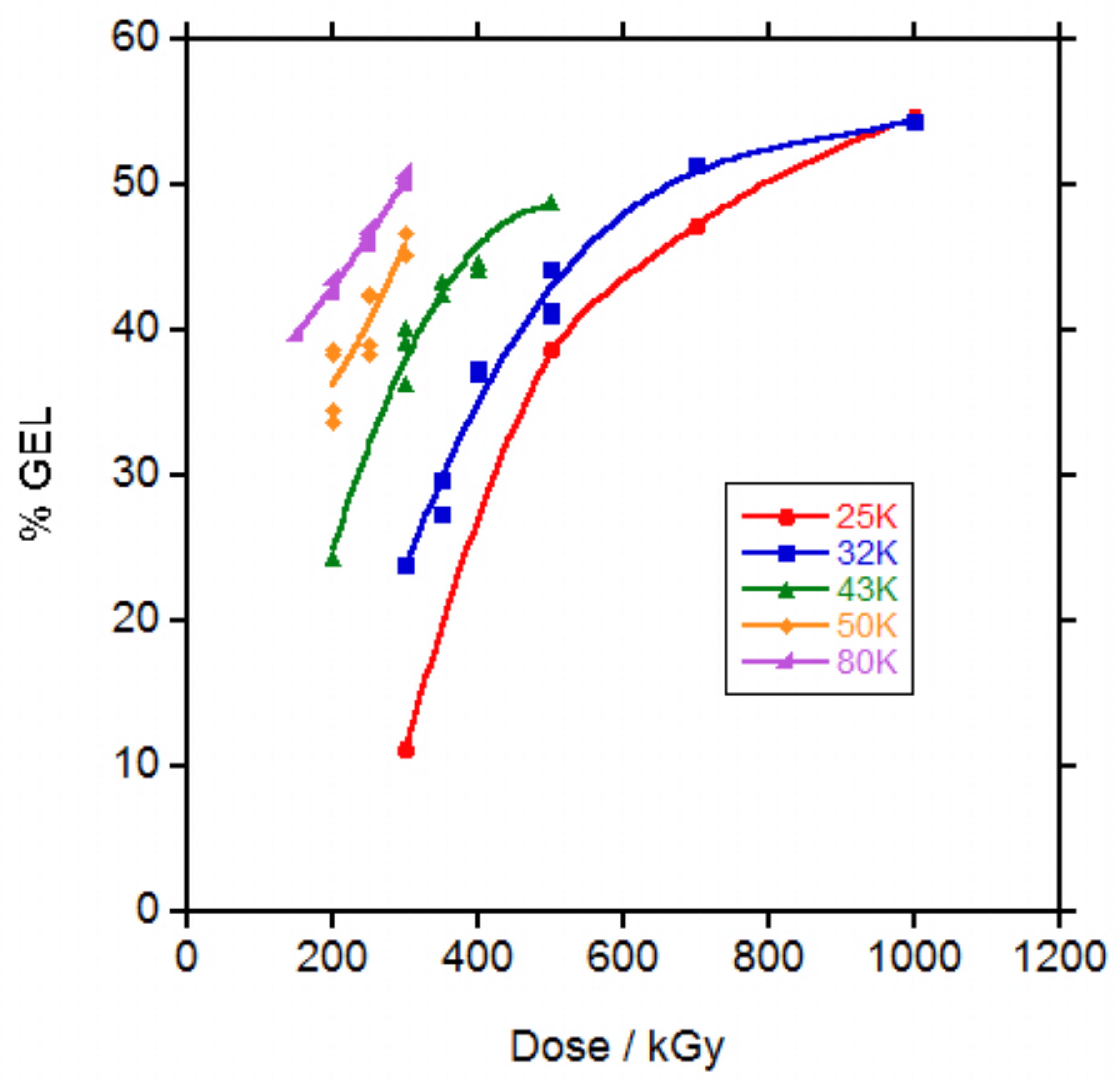
Figure 1, the gel percentage versus irradiation dose is represented.
The percentage of gel at a certain dose was highly dependent on the molecular weight of the polycaprolactone. At 50 kGy dose, none PCL showed evidence of gel. At 100 kGy dose, only PCL 80,000 (80 K) showed gel and at 150 kGy, PCL 50,000 (50 K) and PCL 43,000 (43 K) presented gel. For PCL 32,000 (32 K) and PCL 25,000 (25 K), 200 and 300 kGy were necessary to obtain gel respectively. For some samples at low doses, the gel content could not be measured because the gel could not be separated and only the doses for which the gel content could be measured are represented in
Figure 1. For the same dose, the higher the molecular weight of the PCL the higher the gel content. For the PCLs of lower molecular weight, it seems that at higher doses the gel content tended to a plateau.
It has already been shown that when PCL was irradiated with ionizing radiation (electron beam or gamma rays) in vacuum [
6,
8], inert atmosphere [
12,
13] or in the melted state in supercooled conditions [
8], gel appeared at a lower dose that when irradiation was carried out in the presence of air at ambient temperature. In addition, comparison with results found in literature is difficult because in some cases the molecular weight given is the molecular weight given by the supplier and in other cases is the molecular weight measured by SEC and the difference is very big in between both data al shown in
Figure S1 in the Supporting Information file. Gel content given for a PCL 80 K weight given by the supplier (coincident with the PCL 80 K of this study based on the coincidence in the melt flow index data given by the supplier) irradiated with electron beam in air was 22% at 150 and 200 kGy doses [
17] which is lower than the values of approximately 40% and 43% respectively found in this study. When other authors applied a 270 kGy dose with an electron beam in air at the same PCL 80 K a 55% gel content was found [
13] which is slightly higher than the 45% and 50% values found for 250 and 300 kGy dose respectively in this study. In both cases gel appeared at doses above 50 and 70 kGy respectively, similar to the value in between 50 and 100 kGy necessary for gel formation in the PCL 80 K of this study.
The random radiation crosslinking of thermoplastic polymers is described by the classical Charlesby-Pinner equation:
where
s is sol fraction,
po is degradation density,
qo is crosslinking density,
μ1 is initial molecular weight (
Mn) and
d is radiation dose. Irradiation with electrons causes both random chain scission and random interchain bond formation (crosslinking) and the ratio of scission to cross-linking,
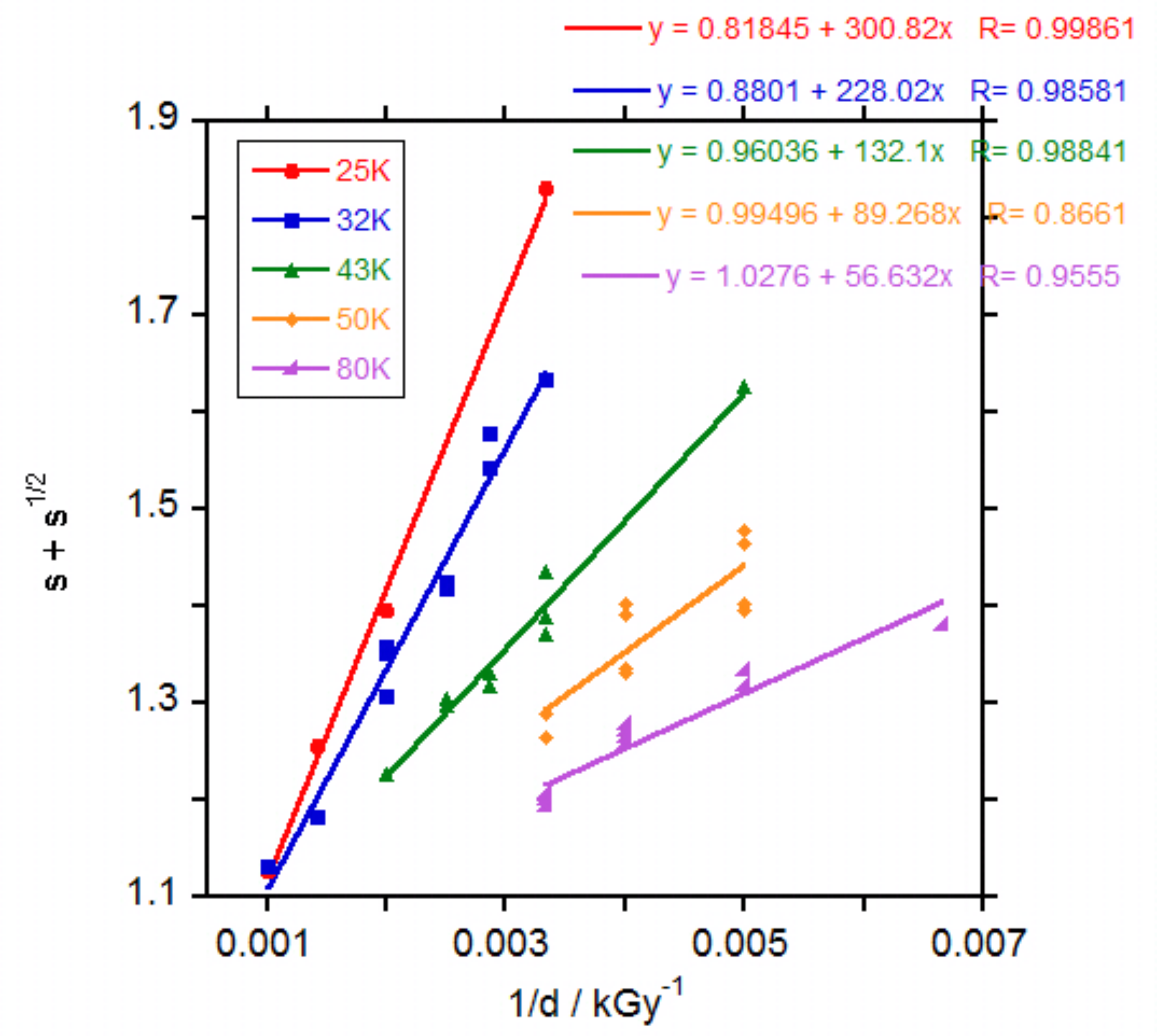
po/qo, represents the inverse crosslinking efficiency of a polymer system at a specific dose. In a classical Charlesby-Pinner analysis plot,
s + s1/2 is plotted against
1/d and a linear fit of the data yields a positively sloping trend line with intercepts at
s + s1/2 equals 2 and
1/d equals 0. The
s + s1/2 equals two intercept represents
do, the minimum dose to gelation and the
1/d equals 0 intercept represents the inverse crosslinking efficiency, represented by
po/qo [
23]. The Chalesby-Pinner plot of the results for the irradiated PCLs,
Figure 2, produced fairly linear fits with the slope of the fit decreasing with the increase in the molecular weight of the PCL, related to a decrease in crosslinking efficiency (
qo/po).
The calculated minimum dose for gelation (
do) increased with the decrease in the molecular weight of the PCL as observed in the measurements of gel percentage. In
Figures S5 and S6 in the Supporting Information file the values for
qo/po and
do versus PCL molecular weight respectively are represented. Calculated values for
d0 (255, 204, 127, 89 and 58 kGy for PCL 25, 32, 43, 50 and 80 K respectively,
Figure S6) agreed with the observed values for the apparition of gel. It was expected that the longer the PCL chains, the lower the number of crosslinks necessary to produce gel, as found by other researchers for PCLs of 40, 50 and 70 K molecular weight as given by the supplier irradiated with gamma rays in air [
10]. The values found for
do were 182, 91 and 41 kGy respectively which are close to the values found in this study for PCL 32, 50 and 80 K respectively.
The calculated crosslinking efficiency (
qo/po) decreased from 1.22 to 0.97 when PCL molecular weight increased (
Figure S5 in the Supporting Information file). This value, close to 1, demonstrates that chain scission and crosslinking have similar contributions without significant predominance of one mechanism over the other. A similar value close to 1 has been found by other authors for PCL irradiated with gamma rays [
6,
7] and only in one work values below 1 (0.6–0.8) were calculated for PCLs of different molecular weight irradiated with gamma rays [
10]. In that work, it was also found that crosslinking efficiency increased with the increase in PCL molecular weight, contrary to our results. For the electron beam irradiation of PCL 80 K weight as giver by the supplier in air or Argon at 20 or 60 °C, when Charlesby-Pinner analysis was performed, the authors found that data did not fit a straight line but bent. They argued that it was necessary an initial molecular weight distribution (
Mw/
Mn) equal to 2 to obtain a straight line. Initial molecular weight distribution for our PCLs was 1.55–1.62 as measured by SEC. Probably due to the limited number of doses in our work the curvature of the data could not be clearly seen and the trend using Charlesby-Pinner analysis produced erroneously the unexpected decay in crosslinking efficiency with the increase in PCL molecular weight seen in
Figure S5. Nevertheless, the crosslinking efficiency values for all the PCLs are around 1 showing the similar contribution of chain scission and crosslinking when PCLs are irradiated with an electron beam. This effect is very different from the effect of peroxides in PCLs for which crosslinking predominates strongly over chain scission, with values for crosslink efficiency of 6.7 or above [
24,
25].
3.3. Molecular Weight of Irradiated PCLs
Molecular weight was measured by SEC for the irradiated PCLs before gelation and for the soluble fraction of the crosslinked PCLs after gelation. In the particular case of PCL 25 K, due to its low mechanical properties, it was excluded from further studies.
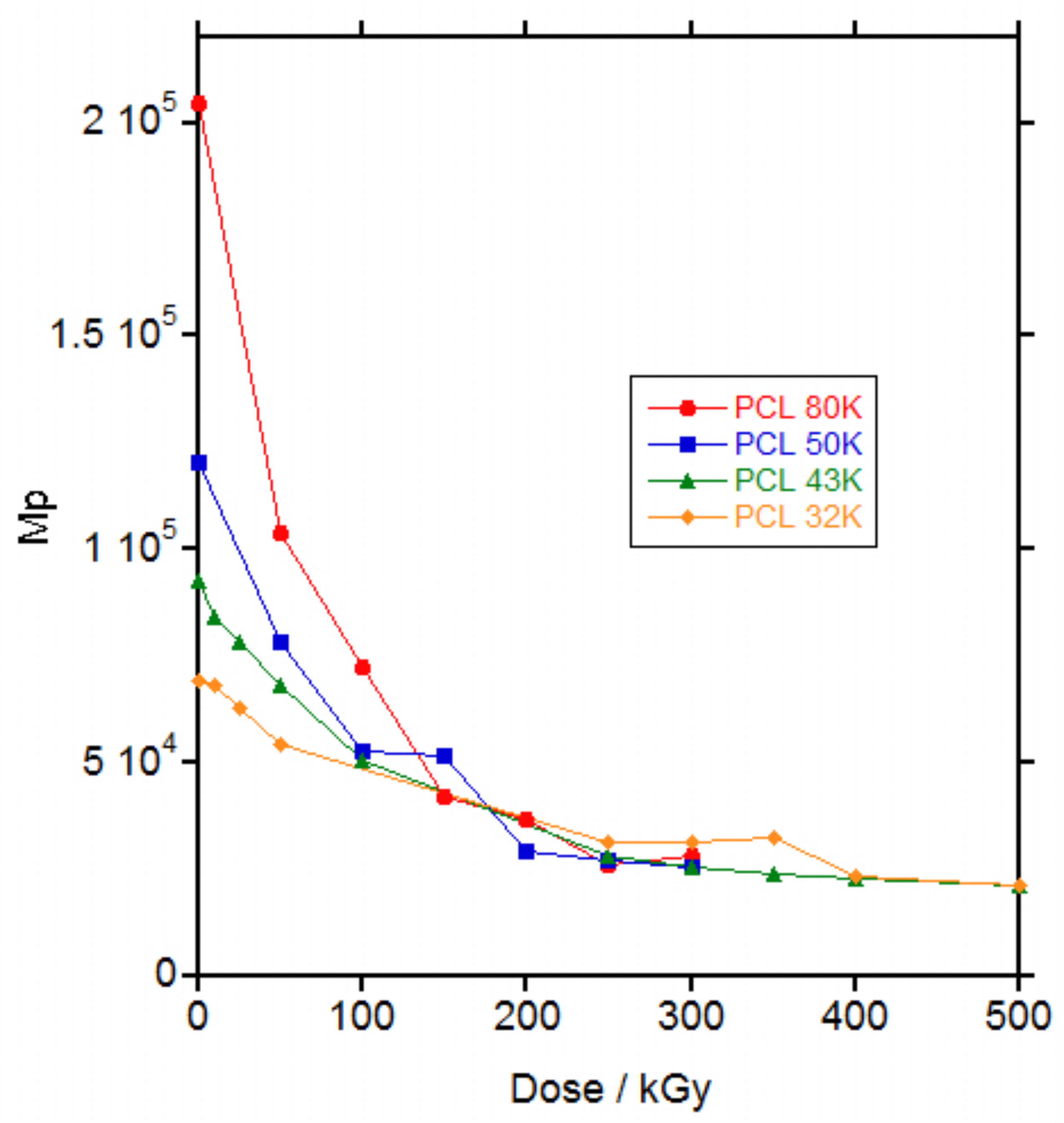
For all the PCLs, irradiation produced a decrease in the molecular weight on the maximum (
Mp) of the SEC curve (
Figure 3) and an increase in the polydispersity of the polymer reaching the total exclusion limit of the columns (
Figures S7–S10 in the Supporting Information file). The increase in the polydispersity reached a maximum at a dose of 50, 100, 100 and 250 kGy for PCL 80, 50, 43 and 32 K, respectively, which is coincident with the maximum dose for gelation (58, 89, 127 and 204 kGy respectively) and then decreased for higher doses when gel was formed.
The increase in polydispersity with the increase in dose until gelation was reached has been already described in literature for PCLs irradiated with gamma rays [
6,
9]. Generally, an increase in molecular weight was described until gelation [
8,
9,
13] followed by a decrease in molecular weight for the soluble part of the material after gelation [
12] and only in one work a decrease in molecular weight was found until gel [
11] as seen in
Figure 3. This apparent contradiction with the results for the PCLs in this study was solved when the weight average molecular weight (
Mw) versus dose was plotted, as in
Figure S11 in the Supporting Information file. Whereas
Mp decreased continuously with the increase in dose,
Figure 3,
Mw increased with the increase in dose until gelation and then decreased in the soluble part of the material,
Figure S11 in the Supporting Information file. Thus, until gelation, branching produced by irradiation leading to an increase in
Mw and polydispersity, dominated over the decrease in molecular weight produced by scission. After gelation, only the fragments produced by scission are soluble and could be measured by SEC and a decrease in molecular weight was found as a consequence of the increase in the scission.
3.4. Proton NMR Spectra of Irradiated PCL
Proton NMR was used to determine the chemical species produced by irradiation. In
Figure S12 in the Supporting Information file the spectra for PCL 50 K non-irradiated and for the soluble part of the PCL 50 K irradiated at 500 and 1000 kGy are presented. Besides the main peaks at 4.05, 2.30, 1.64 and 1.37 ppm for high molecular weight PCL, some new small peaks appeared at 3.64, 2.37 and 0.89 ppm that increased their intensity with the increase in dose, as seen in
Figure S13 in the Supporting Information file. Triplets at 3.64 and 2.37 ppm were assigned to methylenes next to hydroxyl (–CH
2–OH) and carboxylic groups (–CH
2–COOH), respectively, in agreement with the triplets found for α-hydroxyl-
ω-(carboxylic acid) polycaprolactone [
26]. Multiplet at 0.89 ppm was assigned to protons in a saturated hydrocarbon chain. The assignations were confirmed by derivatization of the samples with trifluoroacetic anhydride. Upon derivatization, triplets at 3.64 and 2.37 ppm shifted to 4.34 and 2.63 ppm respectively, as already seen for α-hydroxyl-
ω-(carboxylic acid) polycaprolactone [
26,
27], whereas the multiplet at 0.89 ppm remained unchanged (see
Figure S14 in the Supporting Information file.
Studies on the effect of electron-beam on PCL found in literature demonstrated, by electron paramagnetic resonance (EPR), the existence of radical species of the type –CH
2–CH
2–ĊH–COO– and/or –COO–ĊH–CH
2–CH
2– [
13]. In addition, the analysis of the gases released when PCL was irradiated with gamma rays showed the formation of H
2, CO and CO
2, with a molar ratio of 2.1, 1.0, 1.1 respectively [
7]. From these data and the formed species identified by NMR, a reaction mechanism of PCL with electron beam is proposed as seen in
Figure 4.
Electron beam irradiation of PCL can produce radicals in the polymer chain by hydrogen abstraction and chain scission at any bond within the polymer chain, as seen in
Figure 4. When hydrogen abstraction takes place, the generated carbon radical can recombine with another radical from another chain leading to a branching point that increases the molecular weight and ultimately to a network. The H
2 released when PCL was irradiated with gamma rays, coming from the recombination of two hydrogen radicals [
7], proved that hydrogen abstraction is produced in PCL when irradiated with high energy radiation.
Chain scission can take place at any bond in the PCL chain. When the –COO–CH
2– bond is broken, the oxygen radical can recombine with a hydrogen radical to produce a carboxylic group, as detected by proton NMR, whereas the carbon radical can recombine with another carbon radical to produce branching or with a hydrogen radical to produce a saturated chain end. When the –CH
2–CH
2– bond is broken, the carbon radicals can recombine with another carbon radical or a hydrogen radical. When the –CH
2–COO– bond is broken, the carbon radical can recombine with another carbon radical or a hydrogen radical and the carboxylic radical can decompose to a carbon radical (that can recombine with another carbon radical or a hydrogen radical) with the release of CO
2, as detected for PCL irradiated with gamma rays [
7]. And, when the –CO–O– bond is broken, the carbonylic radical can decompose to a carbon radical (that can recombine with another carbon radical or a hydrogen radical) with the release of CO, as detected for PCL irradiated with gamma rays [
7] and the oxygen radical can recombine with a hydrogen radical to produce a hydroxyl group, as detected by proton NMR.
The molar ratio for CO and CO
2, of 1.0 and 1.1 respectively, found for PCL irradiated with gamma rays [
7], would be interpreted following
Figure 4 as a similar probability of scission for the –CH
2–COO– bond (release of CO
2) and the –CO–O– bond (release of CO).
Proton NMR signals from
Figure S14 were integrated to evaluate the relative abundance of the produced species. Signal at 4.05 ppm from the methylenes next to the oxygen of the PCL ester group (–CH
2–O–CO–) was taken as the internal reference. Signals at 4.34, 2.63 and 0.89 ppm for methylenes next to hydroxyl (–CH
2–OH), methylenes next to carboxylic groups (–CH
2–COOH) and protons in a saturated hydrocarbon chain respectively, were integrated and ratioed to the internal reference. Results are listed in
Table 1.
The most abundant species generated by electron beam irradiation were carboxylic groups whereas the hydroxyl groups and the saturated hydrocarbon bonds had similar presence. From these results, it could be deduced that irradiation breaks preferentially the –COO–CH
2– bond as shown in
Figure 4.
The ratio of the groups does not vary significantly with the increase in dose thus it seems that the mechanism of degradation is independent of the dose.
From the integrals of the signals and by considering that carboxylic and hydroxyls groups are the only terminal groups in the chains and that the chains are linear (which is not true for the irradiated materials because branching was produced by irradiation), the molecular weight of the chains can be calculated. In
Table 1,
Mn calculated by NMR and the changes in
Mn with irradiation as measured by NMR and SEC, are presented. As it can be seen,
Mn for the non-irradiated PCL 50 K was close to 50,000 g∙mol
−1. After irradiation,
Mn of the soluble part of the polymer decreased strongly with calculated values quite similar to
Mn as calculated by SEC despite the facts that branching was not taken into account and the differences in the experimental techniques.
3.5. Thermal Properties
Changes in thermal properties with irradiation doses up to 200 kGy for PCL 50 K and up to 300 kGy for PCL 80 K were measured by DSC. The upper limit for the irradiation dose was chosen after the catastrophic decay in mechanical properties was reached, as it will be shown later. Samples were measured by duplicate to estimate the dispersion of the results.
The general shape of the DSC traces was the same for all the samples. In
Figure S15 in the Supporting Information file, the DSC traces for PCL 80 K irradiated at 200 kGy are shown as an example. In the first heating, a broad endotherm due to the melting of the PCL crystals was observed. On cooling and exotherm demonstrated partial PCL crystallization and in the second heating, a
Tg due to the amorphous part of the PCL was registered at low temperatures followed by a melting endotherm of the PCL crystals at higher temperatures.
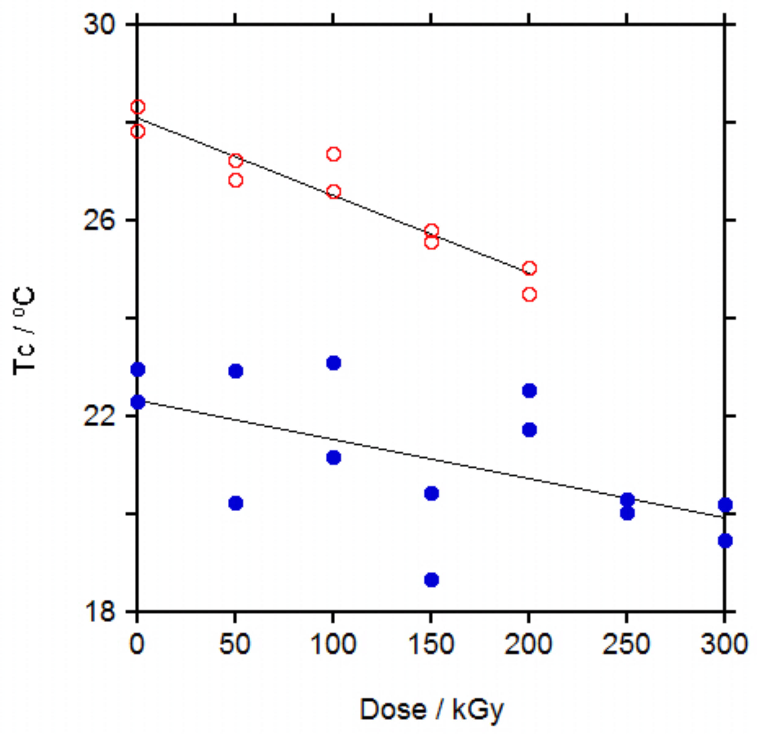
In
Figures S16–S19 in the Supporting Information file the changes in melting point (
Mp), melting enthalpy, glass transition middle point (
Tg) and crystallization enthalpy with dose can be seen and in Figure 6 the changes in crystallization temperature (
Tc) with dose are represented.
No significant changes were observed for the melting point (
Figure S16) in the first heating. In the second heating cycle a slight trend to decrease was observed although the dispersion of the data is high (
Figure S16). For the melting enthalpy (
Figure S17), related to percentage of crystallinity, a slight trend to increase with the increase in dose could be guessed in both heating cycles but the dispersion of the data was too high to consider the trend as significant.
For the glass transition temperature (
Figure S18) and the crystallization enthalpy (
Figure S19), a slight trend to increase their values with the increase in dose can be observed although the big dispersion of the data again masks the trend and makes it statistically not significant. For the glass transition temperature, the change was very small, within a range of 3 °C and although a trend to increase with the increase in dose could be appreciated, the dispersion of the data makes the trend statistically not significant.
Crystallization temperature (
Tc) showed the clearest trend,
Figure 5, with a significant decrease in
Tc with the increase in dose for PCL 50 K. The same trend was found for PCL 80 K but with a higher dispersion in the data.
These trends for PCL are similar to those found for other semicrystalline polymers. For PE irradiated up to 233 kGy,
Mp and crystallinity remained almost unchanged and
Tc decreased slightly with the increase in dose [
28,
29], similar to PCLs in this study. These results were explained by the formation of crosslinking and branching taking place mainly on the amorphous regions and on the boundaries of crystallites, that affects the melting point but more strongly the crystallization temperature as the crosslinks disturbs the formation of crystals on cooling, leading to a delay on the crystallization. For PA-6, when irradiated up to 400 kGy,
Mp and
Tc decreased slightly, more
Tc than
Mp [
30], thus it seems that irradiation affects to the boundaries of crystallites of PA-6 more than to PE and PCL. Changes in
Tg for PA-6 were rather small, as for PCLs, due to the similar effect of the crosslinking leading to an increase in
Tg and the chain scission leading to a decrease in
Tg. The same was found for the crystallinity in PA-6, were the decrease in crystallinity due to crosslinking was compensated by the increase in crystallinity due to the shorter chains produced by chain scission and overall crystallinity did not change significantly with the increase in dose.
When the effect of irradiation with ionizing radiation on the thermal properties of PCL was revised, it was found that the melting point decreased with the increase in dose [
6,
8,
10], in agreement with our results for the melting point in the second heating cycle. For the crystallinity, when PCL was irradiated with gamma rays, it was found that it increased with the increase in dose up to a dose of 500 kGy and then decreased at higher doses [
6], it slightly increased after irradiation at 35 and then slightly decreased after irradiation at 65 kGy and it increased continuously up to a dose of 300 kGy [
4]. When PCL was irradiated with electron beam up to 200 kGy, crystallinity increased continuously with the increase in dose [
16]. This general trend to increase crystallinity with the increase in dose is in agreement with our results (
Figure S17) and the high dispersion of the data observed could account for the apparent initial increase and posterior decrease described in reference 6. Only in one work it was described that crystallinity decreased with the increase in dose when PCL was irradiated with electron beam up to 1000 kGy but in this work crystallinity was evaluated from WAXD spectra [
18]. DSC measurements are more sensitive than WAXD spectra for the determination of the crystallinity of a material and quantification of crystallinity from DSC curves is more precise than quantification from WAXD spectra thus we consider that the decreasing trend in this work is due to the limitation of the technique. The change in crystallization temperature,
Tc, was measured only in two works. In one of them, when PCL was irradiated with gamma rays up to 500 kGy,
Tc decreased with the increase in dose [
8], in agreement with our results, whereas in the other work, when PCL was irradiated with electron beam up to 250 kGy,
Tc increased with the increase in dose, in disagreement with our results [
16].
Similar to PE and PA-6, thermal properties changes in PCL irradiated with gamma rays are explained by crosslinking reactions occurring between chains in their relaxed state, that is, in the amorphous phase, with only little effect on the solid crystallites. Thus, Mp in the first heating cycle would not be affected but after melting, crosslinks would hinder crystal growth, decreasing Tc (slower crystallization) and decreasing Mp in the second heating run (smaller size of the crystals). At the same time, chain scission produces shorter chains that can crystallize better than longer chains and would account for the slight increase in the crystallinity with the increase in dose.
3.6. Mechanical Properties
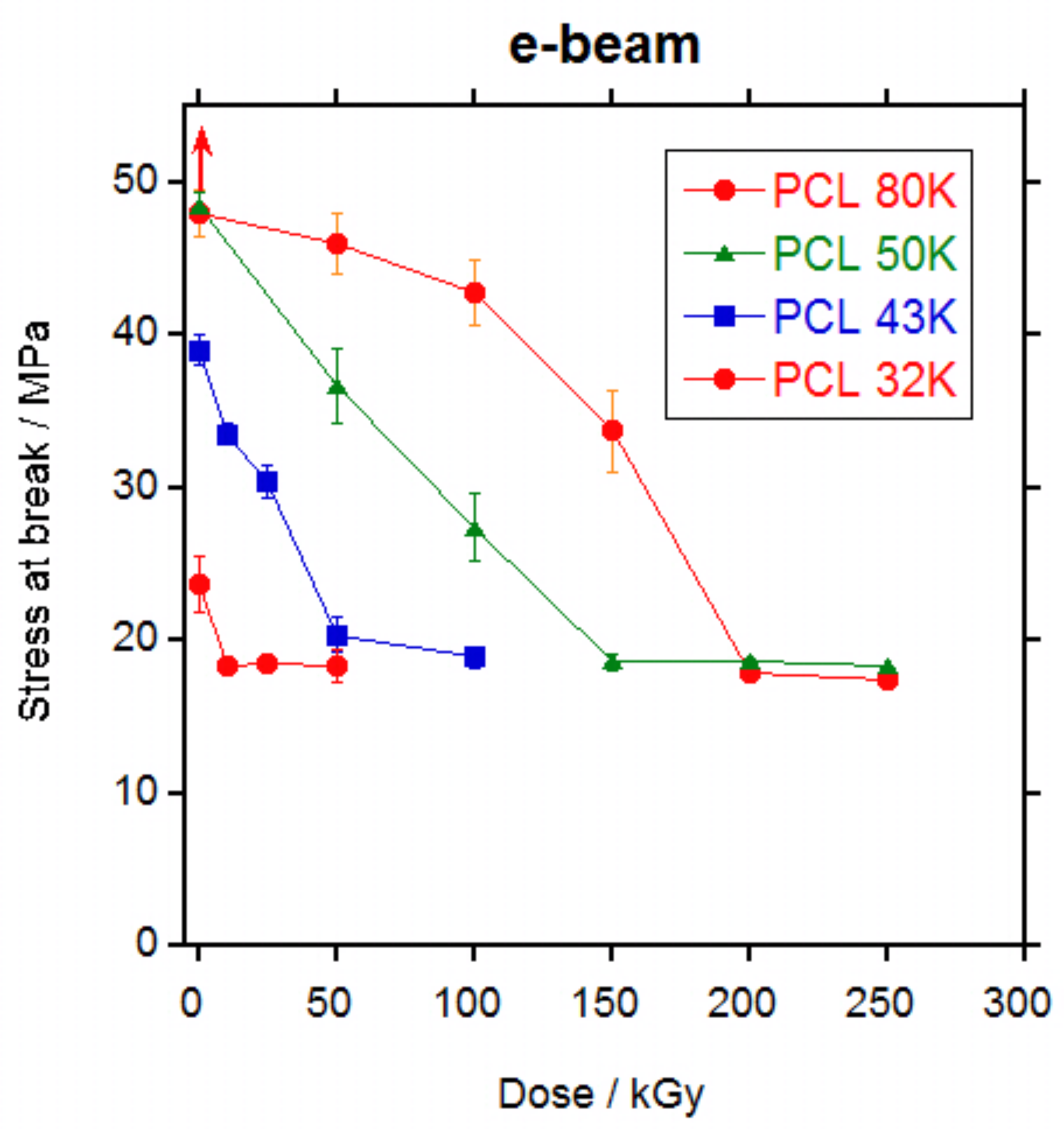
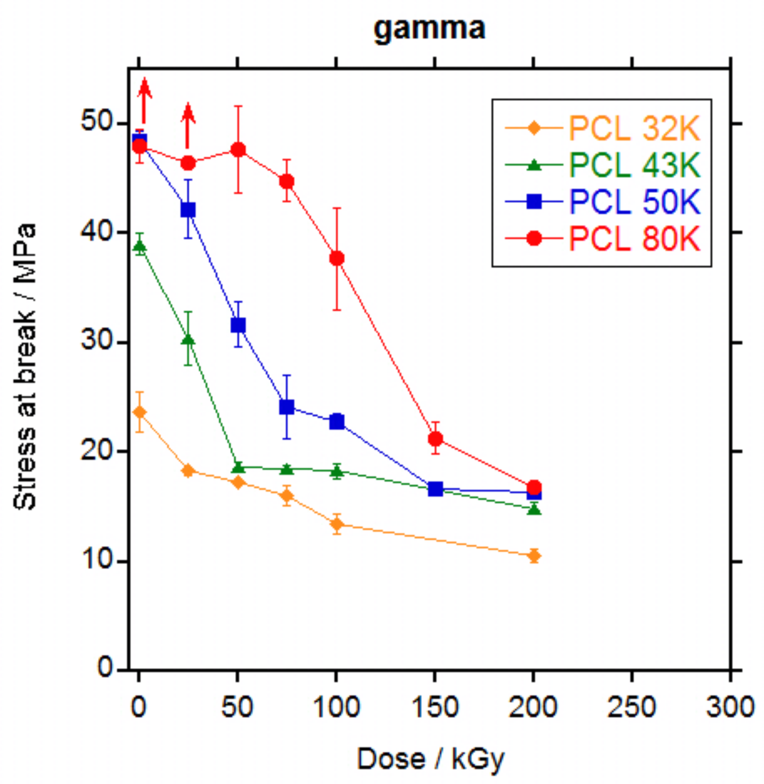
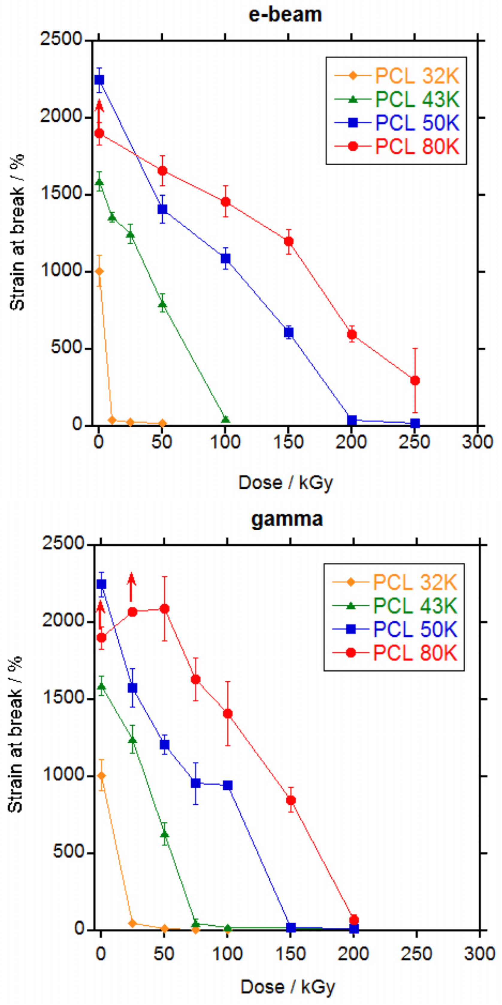
Mechanical properties were measured for PCL 32, 43, 50 and 80 K after irradiation by electron beam up to 250 kGy and by gamma rays up to 200 kGy.
The shape of the stress-strain curve remained the same irrespective of the irradiation dose. The effect of increasing the dose was the decrease of the strain that led to a decrease in the tensile strength.
In
Figure 6 and
Figure 7, the values for tensile stress at break and tensile strain at break versus dose are represented, respectively and in
Tables S1 and S2 in the Supporting Information file are listed. For non-irradiated PCL 80 K, as mentioned previously, specimens escaped from the grip before rupture. The same happened to PCL 80 K irradiated 25 kGy by gamma rays. For these samples, the values represented in
Figure 6 and
Figure 7 are the mean values recorded until the specimens escaped and the real value for tensile stress and strain will be higher, as indicated by the arrows in the Figures.
A continuous decrease in the stress at break (
Figure 6) and in the strain at break (
Figure 7) was observed with the increase in dose for all PCLs. For PCL 32 K, strain dropped sharply to below 50% at the lowest dose (10 kGy for electron beam and 25 kGy for gamma rays); for PCL 43 K the strain dropped to less than 50% at a dose of 100 kGy for electron beam and 75 kGy for gamma rays; for PCL 50 K the strain dropped to less than 50% at a dose of 200 kGy for electron beam and 150 kGy for gamma rays; and for PCL 80 K the strain still did not drop to less than 50% at a dose of 250 kGy for electron beam and dropped to slightly above 50% at a dose of 200 kGy for gamma rays. From the data, it is clear that mechanical properties are adversely affected by ionizing radiation and that the retention of the properties is better when the molecular weight of the PCL is higher. When electron beam and gamma rays are compared, it can be seen that gamma rays produces a slightly higher damage. The slight difference in the results for electron beam and gamma rays is probably due to the differences in dose rate. At high dose rates such as for electron beam, the number of radicals formed by unit time is high and if they do not have enough time to diffuse they would recombine without producing any effect. At low dose rates such as gamma rays, the number of radicals is low and it is more difficult for them to recombine thus they will diffuse and produce crosslinking or scission [
31].
It is well known that crystallinity is one of the primary factors that influence the mechanical properties of polymers [
32]. However, as seen on
Figure S17 in the Supplementary information file, crystallinity does not change significantly with irradiation dose thus changes in the mechanical properties are not due to changes in crystallinity and it is entirely due to the effect of the crosslinking in the amorphous part of the polymer.
When literature data were reviewed, different behaviours were found. In all cases, strain at break decreased with the increase in dose [
6,
7,
15,
17] as found in this work except for one case where a slight increase in strain at break is found for a PCL of unknown molecular weight after irradiation with gamma rays at a 30.8 kGy dose [
3]. For the stress at break, however, an increase in tensile stress was found when PCL was irradiated with electron beam [
15,
17] or gamma rays [
3,
7] up to 160–200 kGy followed by a decrease at higher doses. This is not the case for the results in this work, where a continuous decrease in stress at break was found with the increase in dose either with electron beam or gamma rays, without significant differences in between both ionizing radiations.
The decrease in mechanical properties is explained by the scission of the chains in the amorphous phase that dominates over the crosslinking. Scission of the chains connecting the crystalline domains, that are the load bearing elements in the material, leaves the crystalline domains untied, leading to the decrease in the elongation and the tensile strength, as already stated by other authors [
6].
In
Figures S20 and S21 in the Supplementary information file, the retention of the mechanical properties for PCL 50 K and PCL 80 K are compared with that for other semicrystalline polymers, PA-6 [
30] and PET [
33] and for an almost completely amorphous aliphatic polyurethane [
34]. As it can be seen, irradiation affects much more to PCL than to the other materials and therefore is the material with the lowest resistance to ionizing radiation.
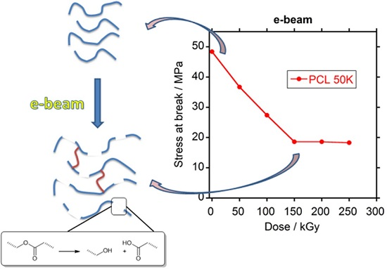
At sterilization doses, PCL 32 K would become very fragile and could not be used in a medical implant if a minimum level of mechanical properties should be required. PCL 43K would become fragile if sterilized at values above 50 kGy, whereas for values below 50 kGy it would retain part of its mechanical properties and depending on the application, PCL 43 K could still be useful after sterilization. For PCL 50 K sterilization doses produce a decay in properties but the level of retention of the mechanical properties is still fair, better than PCL 43 K. PCL 80 K showed by far the best retention of mechanical properties after irradiation at sterilization doses, with very high values of stress and strain even after irradiation at 100 kGy. From these results, it is clear that the higher the molecular weight of the PCL the better the retention of the mechanical properties after irradiation. And that for a medical implant with a minimum requirement of mechanical properties to be sterilized with ionizing radiation, it would be necessary to use a PCL of at least 50,000 g·mol−1 molecular weight.


