Article

Certain Malvaceae Plants Have a Unique Accumulation of myo-Inositol 1,2,4,5,6-Pentakisphosphate

by
Brian Q. Phillippy
1,*,
Imara Y. Perera
1,
Janet L. Donahue
2 and
Glenda E. Gillaspy
2
1
Plant and Microbial Biology, Campus Box 7612, North Carolina State University, Raleigh, NC 27695, USA
2
Biochemistry, Virginia Polytechnic and State University, Blacksburg, VA 24061, USA
*
Author to whom correspondence should be addressed.
Plants 2015, 4(2), 267-283; https://doi.org/10.3390/plants4020267
Submission received: 10 April 2015 / Accepted: 19 May 2015 / Published: 29 May 2015
(This article belongs to the Special Issue Phytic Acid Pathway and Breeding in Plants)

Abstract

:
Methods used to quantify inositol phosphates in seeds lack the sensitivity and specificity necessary to accurately detect the lower concentrations of these compounds contained in the leaves of many plants. In order to measure inositol hexakisphosphate (InsP6) and inositol pentakisphosphate (InsP5) levels in leaves of different plants, a method was developed to concentrate and pre-purify these compounds prior to analysis. Inositol phosphates were extracted from leaves with diluted HCl and concentrated on small anion exchange columns. Reversed-phase solid phase extraction cartridges were used to remove compounds that give peaks that sometimes interfere during HPLC. The method permitted the determination of InsP6 and InsP5 concentrations in leaves as low as 10 µM and 2 µM, respectively. Most plants analyzed contained a high ratio of InsP6 to InsP5. In contrast, certain members of the Malvaceae family, such as cotton (Gossypium) and some hibiscus (Hibiscus) species, had a preponderance of InsP5. Radiolabeling of cotton seedlings also showed increased amounts of InsP5 relative to InsP6. Why some Malvaceae species exhibit a reversal of the typical ratios of these inositol phosphates is an intriguing question for future research.

1. Introduction

Inositol phosphates have been the subject of 30 years of intensive research starting with the unraveling of the function of Ins(1,4,5)P3 (myo-inositol 1,4,5-trisphosphate) as a second messenger for the hormone-triggered release of calcium from the endoplasmic reticulum in animal cells [1]. Dozens of inositol phosphate isomers have been identified in a variety of organisms, and their metabolic importance and relationships have been reported [2,3,4]. Although the majority of studies have focused on Ins(1,4,5)P3, the predominant inositol phosphate in most cells is usually InsP6 (myo-inositol hexakisphosphate, phytic acid), which has been accorded numerous biological activities of its own [5]. It is generally accepted that InsP6 exerts control over nuclear processes, such as DNA repair and mRNA export [6,7], and may be a biologically-relevant antioxidant and regulator of the activities of certain enzymes [8,9]. In plants, InsP6 synthesis plays a critical role in growth and development [10,11].
There has been renewed interest in the metabolism of inositol phosphates in plants resulting from the discovery of novel roles for InsP6 and inositol pentakisphosphate (InsP5). As has been shown for animal cells, InsP6 can stimulate calcium channel activity in plant cells [12] and may in fact ultimately transmit the effects of its precursor, Ins(1,4,5)P3, in plants. InsP6 is co-purified with the auxin receptor [13] and is implicated in defense responses [14]. Similarly, Ins(1,2,4,5,6)P5 (myo-inositol 1,2,4,5,6-pentakisphosphate) was found to be part of the jasmonate co-receptor complex [15], and mutants lacking the kinase that phosphorylates Ins(1,3,4,5,6)P5 (myo-inositol 1,3,4,5,6-pentakisphosphate), IPK1, were also more proficient in jasmonic acid perception [16].
Few studies have been conducted on inositol phosphates in vegetative plant tissues due to their low concentrations and the unavailability of validated methods for their analysis. Seeds have long been known to contain very high amounts of InsP6, approximately 0.5%–1.0% of their dry weight, with lower amounts of InsP5, InsP4 and InsP3 [17]. There are only a few reliable reports in the literature on InsP6 levels in leaves [14,18,19,20], in part because in vegetative tissues, InsP6 is frequently undetectable using regular HPLC (high-performance liquid chromatography) methods [21,22]. Although the predominant inositol phosphate in cabbage (Brassica oleracea) leaves and celery (Apium graveolens) stalks is InsP6, it is present at levels less than 0.02% of their dry weight (weight/dry weight) [23]. Alkarawi and Zotz [24] recently reviewed the literature of InsP6 mass data in plant leaves, but much of it was obtained with methods not specific for InsP6. Following up on the observation that the ratio of phytic acid phosphorus to total phosphorus did not increase with total phosphorus in the compiled data from the leaves of 35 different plant species [24], they performed experiments with dandelions that showed that the ratio of phytic acid phosphorus to total phosphorus had a negative correlation with the total amount of phosphorus, which they interpreted as indicating that InsP6 was not being used as a storage compound in these short-lived leaves [25].
Because the inositol phosphate concentrations of vascular tissues are so low, the study of their metabolism has been limited mostly to the use of radioisotopes. Plant cells grown in the presence of [3H]myo-inositol incorporate radioactivity into inositol phosphates, which can be extracted with acid and analyzed chromatographically [26]. A widely-used Ins(1,4,5)P3 mass assay utilizes competitive binding with [3H]Ins(1,4,5)P3 to an animal Ins(1,4,5)P3 receptor, although data obtained in this manner show somewhat different changes from those obtained chromatographically using radiolabelled cells [27]. Here, we describe a procedure for determining the masses of InsP6 and InsP5 in leaves and seedlings by concentrating and partially purifying acid extracts prior to analysis by high performance ion chromatography. By using this method, we have documented that most plants examined contain very little InsP5 compared to InsP6, but surprisingly, in cotton, more Ins(1,2,4,5,6)P5 than InsP6 was observed.

2. Results and Discussion

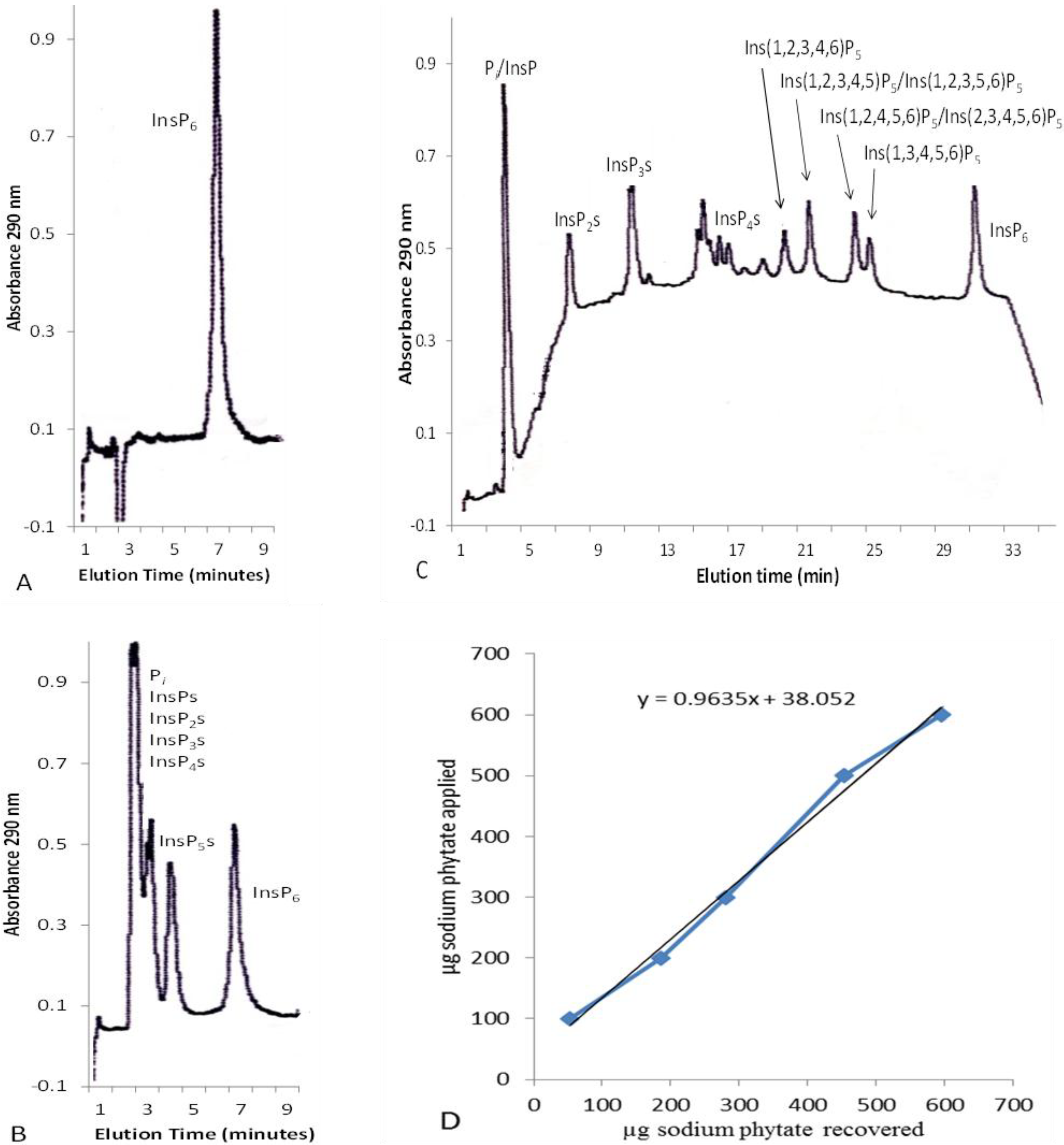
2.1. Concentration of Inositol Phosphates in Leaf Extracts

Whereas seeds contain high levels of InsP6 that are easy to measure by various HPLC methods, the concentration of InsP6 in extracts from leaf tissue can be below the limit of detection when using nonradioactive methods. The most sensitive HPLC/mass spectrometry methods can give quantitative inositol phosphate data from seed extracts [28], but have not yet been adapted to the more challenging task of measuring inositol phosphates in leaves. Because the inositol phosphate composition of leaves and seedlings has rarely been examined except using radioisotopes in in vivo experiments, we decided to partially purify and concentrate the inositol phosphates in leaf extracts in order to obtain basic quantitative data in this heretofore unexplored area.
Isocratic and gradient HPLC of an InsP6 standard and an InsP6 hydrolysate are shown in Figure 1. Upon initial experiments to concentrate InsP6 on AG1-X8 anion exchange resin, it was observed that significant amounts of InsP6 bound in such a manner that they could not be eluted with 1 N HCl. Attempts to presaturate the InsP6 binding sites with InsP6 followed by pre-elution with HCl and re-equilibration with water increased the amount of time for this procedure, thus negating the ability to analyze multiple samples. We decided to start each analysis with a fresh 1-g column of AG1-X8 in a 1.0 cm diameter glass Bio-Rad Econo-column equipped with a flow adaptor. Most of the loaded InsP6 standards could be eluted from these columns with 35 mL 1 N HCl. To compensate for the unrecoverable InsP6, a calibration curve was constructed using 100–600 µg dodecasodium phytate (Figure 1D). The curve was linear with a y-intercept of 38, which indicated that 38 µg of dodecasodium phytate (25 µg InsP6) was essentially irreversibly bound to the column. The equation µg InsP6 loaded = 0.9635 × µg InsP6 recovered + 38.052 was subsequently used to calculate the amount of InsP6 present in leaf extracts. Complete recoveries from the AG1-X8 columns of 50 and 100 µg aliquots of Ins(1,2,4,5,6)P5 in the presence of 132 µg InsP6 were obtained, so leaf InsP5 concentrations were calculated directly from the HPLC data without using a calibration curve.

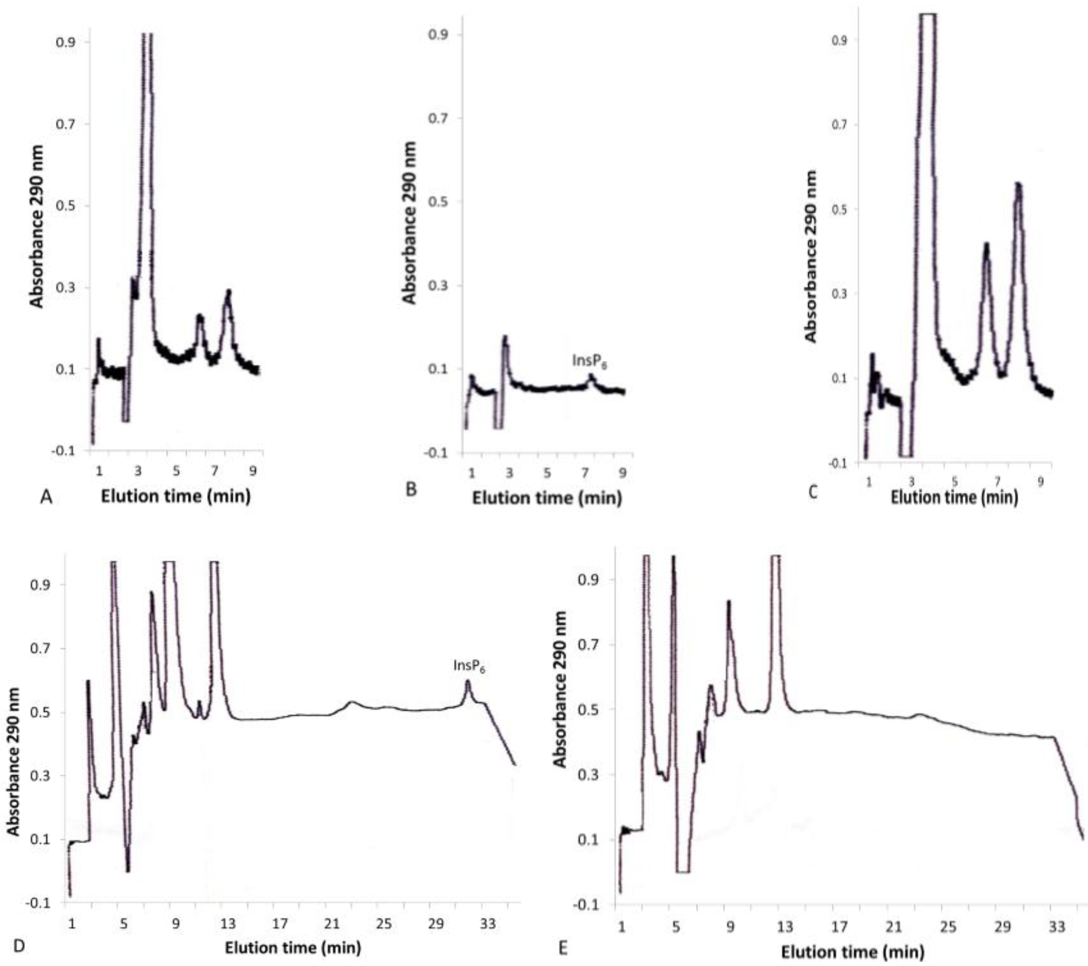
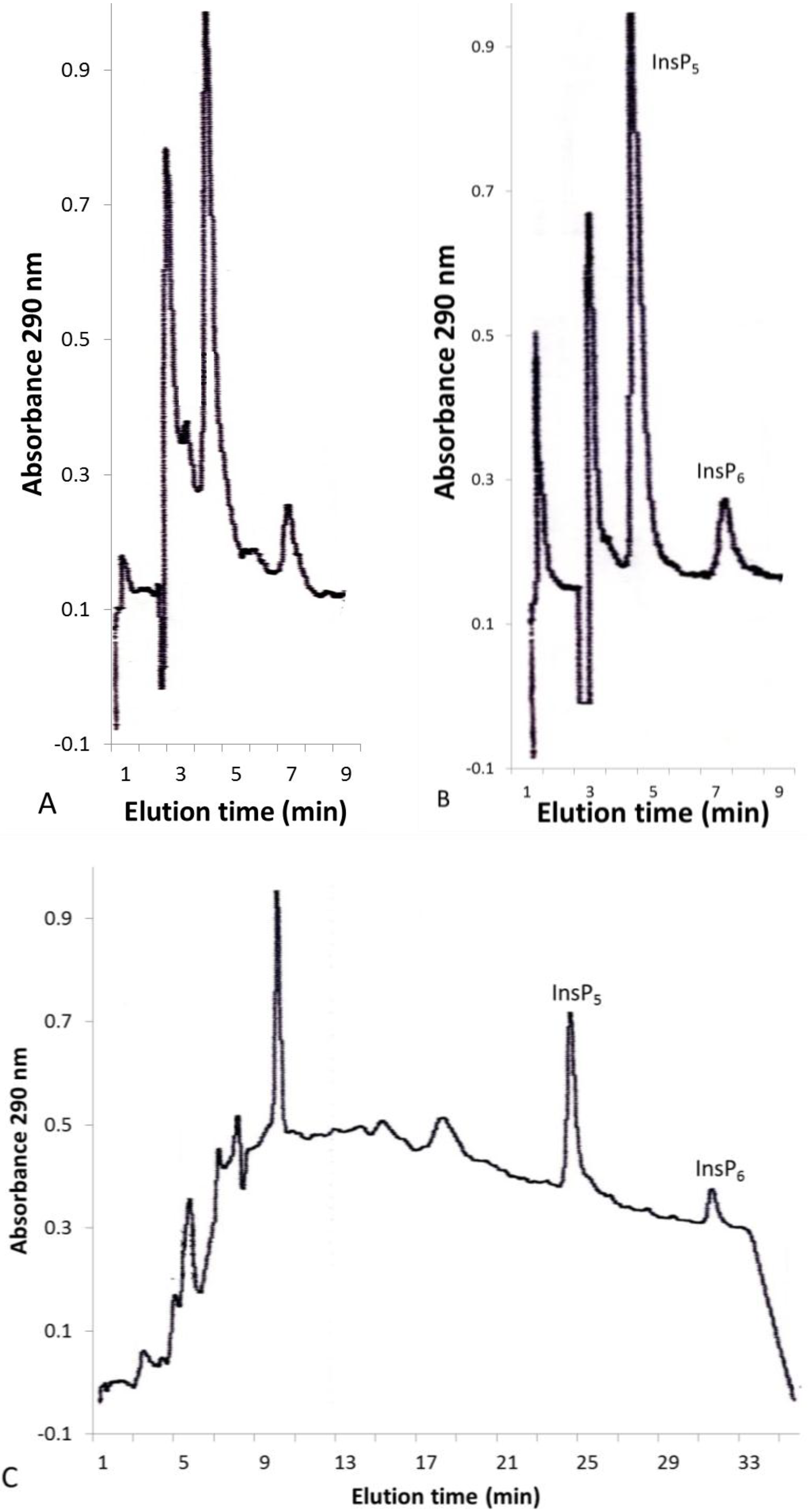
2.2. Solid-Phase Extraction of Inositol Phosphate Concentrates

Inositol phosphates were extracted from leaves by homogenization with 0.37 N HCl followed by centrifugation. When crude leaf extracts were subjected to HPLC, the peaks did not all elute with the same retention times of InsP6 or other inositol phosphate standards (Figure 2A). When the crude extracts were passed through Sep-Pak C-18 or Oasis HLB Plus reversed-phase solid-phase extraction cartridges, which bind hydrophobic compounds, some of the peaks disappeared from the chromatograms, indicating that they were not inositol phosphates (Figure 2B). The identities of these hydrophobic peaks, which could be eluted with 1.5 mL 50% methanol (Figure 2C), are unknown, and the fact that different peaks were present in extracts from different types of leaves indicates that they encompass a variety of compounds. In contrast to the isocratic analysis, where hydrophobic peaks eluted very close to InsP6, in the gradient procedure, InsP6 eluted in 30 min (Figure 2D), whereas the hydrophobic peaks all eluted within 12 min (Figure 2E). Similar peaks were previously observed in extracts from various roots and tubers [29] and from avocado (Persea americana) fruit [30]. In seeds, which contain much higher levels of InsP6 than leaves, it has been assumed that all of the peaks that are strongly retained on ion exchange columns belong to inositol phosphates. The fact that additional compounds may also make up some of these peaks cannot be discounted and may be especially relevant to studies of the elusive inositol polyphosphate pyrophosphates, such as InsP7 in plants [31].
Figure 1. Isocratic and gradient HPLC of inositol hexakisphosphate (InsP6) standard and InsP6 hydrolysate. Chromatographic conditions are described in the Experimental Section. Isocratic HPLC of InsP6 standard (A); isocratic HPLC of InsP6 hydrolysate (B); and gradient HPLC of InsP6 hydrolysate (C); calibration curve to correct for recovery of InsP6 from Ag1X8 anion exchange columns (D).
Plants 04 00267 g001
Figure 2. Isocratic and gradient HPLC of corn (Zea mays) leaves before and after solid phase extraction. Isocratic HPLC of concentrated corn leaf extract (A); isocratic HPLC of concentrated extract that did not bind to the solid phase extraction (SPE) cartridge (B); isocratic HPLC of concentrated extract that bound to the SPE cartridge and was eluted with 50% methanol (C); gradient HPLC of concentrated extract (D); and gradient HPLC of concentrated extract that bound to the SPE cartridge and was eluted with 50% methanol (E).
Plants 04 00267 g002
The only InsP5 observed in any of the leaves analyzed based on retention time was Ins(1,2,4,5,6)P5 and/or its enantiomer Ins(2,3,4,5,6)P5. Cotton (Gossypium hirsutum) leaf extracts contained InsP5 and InsP6 peaks (Figure 3A) that were not removed upon passage through the SPE cartridge (Figure 3B). In the short isocratic high-performance ion chromatography (HPIC) procedure, Ins(1,2,4,5,6)P5 is not well separated from the other InsP5 isomers, but in the longer gradient procedure, the isomer present in cotton was clearly shown to be Ins(1,2,4,5,6)P5 (and/or its enantiomer Ins(2,3,4,5,6)P5) (Figure 3C; see also Figure 1C and [32]). InsP4, InsP3 and InsP2 were not evaluated because the dilution of the 0.37 N HCl extract five-fold with H2O may not have been sufficient to allow them to quantitatively bind to the AG1-X8 column. Hanke et al. [33] have recently reported that the major InsP5 peak in extracts from [3H]inositol-labeled potato (Solanum tuberosum) leaf disks eluted with the retention time of Ins(1,2,4,5,6)P5/Ins(2,3,4,5,6)P5/Ins(1,2,3,4,6)P5. In contrast, duckweed (Spirodela polyrhiza L.) synthesized predominantly Ins(1,3,4,5,6)P5 [34].
Figure 3. HPLC of cotton (Gossypium hirsutum) leaves. Isocratic HPLC of concentrated cotton leaf extract before (A) and after (B) passage through the SPE cartridge. Gradient HPLC of cotton leaf extract (C).
Plants 04 00267 g003
To verify that the entire analytical procedure including the use of the calibration curve described above yielded accurate results, an experiment was performed to measure the recovery of 132 µg InsP6 added to an extract from 5 g cotton leaves that were determined to contain 66 µg InsP6. The recovery, calculated as ((InsPX determined with addition − InsPX determined without addition)/InsPX added) × 100% was 98% (Table 1). A similar experiment performed with the addition of 50 µg Ins(1,2,4,5,6)P5 to a tobacco leaf extract gave a recovery of 49 µg, which also demonstrated the validity of the method for InsP5.
Table 1. Recoveries of Ins(1,2,4,5,6)P5 (myo-inositol 1,2,4,5,6-pentakisphosphate) and InsP6 from tobacco and cotton leaf extracts (see Experimental Section 3.6.).
PlantInsP5 AddedInsP6 AddedInsP5 DeterminedInsP6 DeterminedInsP5 RecoveredInsP6 Recovered
Tobacco0 µg0 µg0 µg37 µg--
Tobacco50 µg0 µg49 µg42 µg49 µg-
Cotton0 µg0 µg40 µg66 µg--
Cotton0 µg132 µg48 µg196 µg-130 µg

2.3. InsP6 and InsP5 Levels in Various Plants

In the procedure used here, the volume of a 50-mL extract is reduced to 1.5 mL, thereby concentrating the inositol phosphates and increasing the sensitivity of the HPIC by a factor of more than 30-fold (Figure 4). This permits accurate quantification of leaf InsP6 concentrations in vegetative tissues as low as 10 µM despite the fact that 25 µg of InsP6 is not retrievable from the 1 g AG1-X8 columns. Because Ins(1,2,4,5,6)P5 was completely recovered, it could be measured in leaves in concentrations as low as 2 µM. In contrast, the limit of detection of the HPIC procedure for unconcentrated extracts corresponds to tissue concentrations of approximately 50 µM for InsP6 and 40 µM for Ins(1,2,4,5,6)P5. Thus, the current method lowers the limit of detection for InsP6 and InsP5 in leaves five-fold and 20-fold, respectively. Although the ultimate improvement in sensitivity gained by concentration is not huge, it does cover the range of InsP6 concentrations commonly found in plants. Also noteworthy is the observation that the sample prepurification step reduces the rate at which the HPLC detector tubing becomes clogged with precipitated compounds, necessitating cleaning or replacement of the tubing.
Figure 4. Overview of the analytical procedure for InsP5 and InsP6 in plant tissues.
Plants 04 00267 g004
For a survey of the InsP6 and InsP5 in plants, leaf samples were obtained from a variety of plants growing indoors and outdoors. In most plants, InsP6 predominated, and only trace amounts of InsP5 were detected (Table 2). The highest levels of InsP6 and InsP5 were observed in members of the Malvaceae (mallow) family, which includes cotton, okra and hibiscus species (Table 3). Cotton and some hibiscus species were atypical in that the amount of InsP5 exceeded that of InsP6 by substantial amounts. In the other plants analyzed, InsP6 predominated when it could be detected. InsP6 was not detected in soybean (G. max) leaves, but this may have resulted from the lack of stimuli (i.e., wind, rain, pests and temperature fluctuation) in the indoor environment growth conditions. However, kudzu (P. labata), a legume related to soybean, also lacked detectable leaf InsP6 in plants growing outdoors (Table 2), indicating the possibility that the lack of fertilizer or developmental constraints are important for InsP6 accumulation in leaves.
Table 2. InsP6 and Ins(1,2,4,5,6)P5 concentrations in leaves of different plants. Values are given as ranges followed by the number of extracts analyzed in parenthesis. The detection limits for InsP6 and InsP5 in leaves were approximately 10 µM and 2µM, respectively (nd = none detected).
PlantTissueInsP6 (µM)Ins(1,2,4,5,6)P5 (µM)
Zea maysleaves101–198 (3)nd
Oryza sativaleaves9–49 (6)nd
Arabidopsis thalianaleaves29–89 (9)2–22 (5)
Arabidopsis thalianaseedlings47–110 (4)nd
Nicotiana tabacumleaves11–24 (3)nd
Solanum lycopersicumleaves39–65 (3)nd
Trifolium repensleaves21 (1)6 (1)
Acer rubrumleaves35 (1)2 (1)
Ginkgo bilobaleaves61 (1)nd
Camelina sativaleaves70 (1)6 (1)
Glycine maxleavesnd (1)nd
Pueraria labataleavesnd (1)nd
Table 3. InsP6 and Ins(1,2,4,5,6)P5 concentrations in tissues of Malvaceae family plants. Values are given as ranges followed by the number of extracts analyzed in parenthesis. The detection limits for InsP6 and InsP5 in leaves were approximately 10 µM and 2 µM, respectively (nd = none detected, dpa = days post-anthesis).
PlantTissueInsP6 (µM)Ins(1,2,4,5,6)P5 (µM)
Gossypium hirsutumleaves11–169 (6)5–510 (6)
Gossypium hirsutumseedlings77–140 (3)86–118 (3)
Gossypium hirsutum14–17 dpa fiber43 (1)nd
Gossypium hirsutum16 dpa seeds105 (1)nd
Gossypium barbadenseleaves60–76 (2)329–507 (2)
Gossypium raimondiileaves71–86 (2)578–589 (2)
Abelmoschus. esculentusleaves90–336 (4)3 (1)
Abelmoschus. esculentuspod (husk)100 (1)nd
Abelmoschus manihotleaves250–822 (4)2–7 (2)
Alcea rosealeaves50–52(2)10(2)
Althea officinalisleaves95–217 (2)38–74 (2)
Hibiscus syriacusleaves27–34 (3)10–19 (3)
Hibiscus moscheutosleaves39–73 (2)219–491 (2)
Hibiscus rosa-sinensisleaves31 (1)392 (1)
Hibiscus cannabinusleaves172–491 (5)19–57 (4)
Hibiscus coccineusleaves97–140 (2)536–647 (2)
Hibiscus sabdariffaleaves153 (1)114 (1)
Hibiscus mutabilisleaves49 (1)46 (1)
To verify the altered InsP5 to InsP6 ratio found in cotton plants, we used a separate radiolabeling method that utilizes incorporation of [3H]myo-inositol into inositol phosphates. We adapted published procedures to label cotton seedlings and leaves. We divided root and shoot tissues after labeling, extracted inositol phosphates and used HPLC for separation. Figure 5 shows representative data and includes Arabidopsis seedlings for comparison. In addition to in-line counting of samples from the HPLC (Figure 5A,B), we also use scintillation counting of fractions, which is more sensitive and can be easily quantified (Figure 5C). We found that cotton seedling shoots, roots and leaves contained relatively more InsP5 as compared to InsP6. Specifically, the ratio of InsP5/InsP6 was 0.097 in Arabidopsis seedlings [31], 3.7 in cotton shoots, 1.9 in cotton roots and 0.75 in cotton leaves (Table 4). We conclude that use of this alternate method validates our finding of altered InsP5 levels in cotton.
Figure 5. HPLC of plant tissue labeled for four days with [2-3H(N)]myo-inositol. (A). Radiolabeled Arabidopsis wild-type seedlings were extracted, separated using anion exchange and quantified via an in-line radioisotope detector. InsP5 elutes at Minutes 40–42 and InsP6 at Minutes 46–48. This chromatogram is representative of seven independent seedling labeling experiments; (B). Extracts were prepared and fractionated by HPLC from radiolabeled G. hirsutum seedling shoot (red trace), root (blue) and young leaf from pre-flowering-stage plant (black). Root and shoot data were repeated two times (C). Scintillation counting of eluted fractions is shown in A and B. (Top) Arabidopsis seedling; (middle) G. hirsutum shoot (red), root (blue); (bottom) young leaf from pre-flowering-stage G. hirsutum.
Plants 04 00267 g005
Table 4. Percentages of InsP5 and InsP6 of total InsP in radiolabeled tissues.
InsP6InsP5InsP5:InsP6
% of Total InsP% of Total InsPRatio
Arabidopsisseedlings 127.82.700.097
G. hirsutumseedling shoot1.184.353.7
G. hirsutumseedling root3.787.201.9
G. hirsutumyoung leaf9.477.070.75
1 Data from [31].
In order to estimate the potential variations in InsP5 and InsP6 levels, cotton (G. hirsutum) leaves from plants at different developmental stages of growth and leaves at different positions on the plant were analyzed. Leaves closer to the cotton bolls had greater InsP5 and InsP6 concentrations than leaves closer to the main stalk or near the bottom of the plant (Table 5). In addition, smaller leaves representing new growth had much higher levels of InsP5 and InsP6 in flowering plants than in those that had ceased flowering. These results could be interpreted to mean that InsP6 in leaves accumulates for reproduction. InsP6 is known to be a storage form of phosphate in seeds to be used upon germination [35,36], and leaf InsP6 may similarly be used as a storage form of phosphate to be mobilized to the developing seeds. An alternative explanation could be that the InsP6 level is simply a reflection of the growth rate of the leaves and is higher in cells undergoing active cell division. Because the data in Table 5 are the results from only one observation from different plants grown in the Phytotron (n = 1), more extensive sampling is needed to delineate with certainty the changes that occur during the life cycles of plants and of individual leaves.
Table 5. InsP5 and InsP6 dependence on cotton (G. Hirsutum) leaf location and developmental stage.
StageLocationInsP6 (µM)InsP5 (µM)
loweringbottom near trunk115
floweringmiddle near trunk2014
floweringmiddle near bolls33126
floweringtop169492
post-anthesistop228
The unusual inositol polyphosphate composition of cotton leaves cannot be easily explained. Because hibiscus species also contain significant amounts of InsP5, the presence of this compound may have little to do with cotton fiber, though the hibiscus seed pods do visually resemble cotton bolls. Since Ins(1,2,4,5,6)P5 is part of the jasmonate receptor [15], which is involved in plant defenses related to wounding and chewing insects [16], it is intriguing to speculate that high Ins(1,2,4,5,6)P5 levels in cotton are related to the jasmonate signaling pathway. The okra (A. esculenta), aibika (Abelmoschus manihot) and kenaf (H. cannabinus) leaves had large quantities of InsP6, but low levels of InsP5 (Table 3). Because these plants exhibit little branching compared to the other Malvaceae species, it is possible that their elevated InsP6 levels may participate in the inhibition of branching. Auxin inhibits branching [37], and InsP6 is part of the auxin receptor [13]. In addition to auxin, branching is known to be regulated by cytokinins, strigolactones, gibberellins, sugars and many other signals [38]. The auxin and jasmonate co-receptor complexes have similar structures [39], so there may be some cross-talk involving InsP6 and Ins(1,2,4,5,6)P5 affecting their signaling functions.
It will be necessary to further investigate what conditions affect and are affected by the inositol phosphates in cotton and related plants. The complex interplay of hormones, including jasmonic acid and auxin, with different environmental conditions may control basal inositol phosphate content, as well as responses to internal and external stimuli. The Malvaceae family members are generally hardy plants that can tolerate challenging environmental conditions, such as heat, drought and even excess water (some hibiscus and marshmallow, i.e., the mallow that grows in the marsh). The common denominator is that all Malvaceae appear to have a reputation for hardiness, and an altered InsP composition may be a possible mechanism for rapid hormonal signaling due to the always present inositol phosphate components of certain hormone receptors. In animal cells there, are several examples of InsP6 and its precursor, Ins(1,3,4,5,6)P5, levels being regulated by physiological stimuli, including neuronal differentiation [40], cell cycle progression [41] and hematopoietic cell differentiation [42].
Another intriguing question has to do with the origin of the elevated Ins(1,2,4,5,6)P5 in cotton. Ins(1,2,4,5,6)P5 may be formed from Ins(1,4,5,6)P4 by the 2-kinase that phosphorylates Ins(1,3,4,5,6)P5 [35]. In animal cells, Ins(1,2,4,5,6)P5 can be produced from InsP6 by multiple inositol polyphosphate phosphatase (MIPP), and its enantiomer, Ins(2,3,4,5,6)P5 can be made from InsP6 by the reverse action of InsP6 kinases in response to decreasing ATP to ADP ratios [43]. The fact that Ins(1,2,4,5,6)P5 is elevated in cotton suggests a unique regulation of its metabolic pathway, or components within this pathway, in cotton. Further studies on the inositol phosphate kinases and phosphatases involved in this pathway in cotton may delineate novel regulatory mechanisms of interest for future plant engineering strategies.

3. Experimental Section

3.1. Plant Material

Cotton (Gossypium hirsutum and Gossypium barbadense) plants were grown in a Phytotron under natural light with the temperature maintained at 26 and 22 °C in the day and night, respectively. Cotton (Gossypium raimondii), rice (Oryza sativa), kenaf (Hibiscus cannabinus), roselle (Hibiscus sabdariffa), Hibiscus rosa-sinensis, hollyhock (Alcea rosea), tobacco (Nicotiana tabacum) and Camelina sativa were grown in a greenhouse. Arabidopsis thaliana, tomato (Lycopersicon esculentum), corn (Zea mays), soybean (Glycine max), marshmallow (Althea officinalis) and okra (Abelmoschus esculentus cv. Clemson spineless) plants were grown at 23 °C under artificial light. Rose-of-Sharon (Hibiscus syriacus), Hibiscus coccineus, Hibiscus mutabilis, aibika (Abelmoschus manihot) and Ginkgo balboa leaves were obtained at the NCSU JC Raulston Arboretum. Leaves from Hibiscus moscheutos and kudzu (Pueraria labata) were harvested from plants growing outside on the NCSU campus. White clover (Trifolium repens) and red maple (Acer rubrum) leaves were taken from a local backyard. The plants from the NCSU campus and local backyard were identified by Brian Phillippy. All of the leaves chosen appeared to be mature and non-senescent, except for those of cotton (Gossypium hirsutum) and Arabidopsis thaliana, which were from varying stages of maturity.

3.2. Inositol Phosphate Standards

Inositol hexaphosphoric acid dodecasodium salt from rice was from Sigma, and phytic acid 50 wt% solution in water was from Aldrich. Inositol 1,2,4,5,6-pentakisphosphate was prepared from InsP6 as follows: five grams of dodecasodium phytate was dissolved in 100 mL distilled deionized H2O, and the pH was adjusted to 4.0 with HCl. The phytate solution was autoclaved 30 min at 121 °C and 20 psi, and 20 mL of the resulting hydrolysate was diluted to 100 mL with H2O. The diluted hydrolysate was loaded on a 2 × 18 cm column packed with AG1X8 chloride form anion exchange resin (Bio-Rad, Hercules, CA, USA) at a flow rate of 2 mL/min. Inositol phosphates were eluted with 800 mL of 0.45 N HCl, and forty 20 mL fractions were collected. The inositol phosphate profile was obtained by combining 200 µL of each fraction with 200 µL H2O and 800 µL 0.05% Fe(NO3)3·9H2O in 1% HClO4 and measuring the absorbance at 290 nm. The inositol phosphates in individual fractions were identified by HPIC of aliquots as described previously [32], and the peak fractions containing only Ins(1,2,4,5,6)P5/Ins(2,3,4,5,6)P5 were combined. Ins(1,2,4,5,6)P5 and Ins(2,3,4,5,6)P5 are enantiomers, which cannot be separated by ion exchange chromatography or distinguished by NMR (nuclear magnetic resonance). The pH of the combined fractions was adjusted to 7.0 with 5 N NaOH, and aliquots were digested with 1 mL 5 N H2SO4 in test tubes with a glass marble on top for 4 h at 150 °C. The inorganic phosphate content of the digests was determined by the procedure of Heinonen and Lahti [44] and was used to calculate the inositol phosphate concentration.

3.3. HCl Extraction and Concentration of Inositol Phosphates

Typically, 5 g of leaves was homogenized 15 s in an Oster Osterizer mini blender jar with 50 mL 0.37 N HCl. Tissues that did not homogenize well, such as rice leaves, were ground in liquid nitrogen with a mortar and pestle, and 5 g of ground tissue was stirred 10 min with 50 mL 0.37 N HCl. The extract was centrifuged 20 min at 20,000× g, and the supernatant was filtered using a Buchner funnel. The filtrate was combined with 1 mg disodium EDTA in 1 mL H2O, diluted to 250 mL with H2O and concentrated on a 1-g Bio-Rad AG1-X8 200–400 mesh chloride form column at 1 mL per min. The column was washed with 10 mL 0.1 N HCl, and inositol phosphates were eluted with 35 mL 1 N HCl. The eluate was dried using a rotary evaporator at 30 °C connected to a vacuum pump, and the residue was dissolved in 1.5 mL H2O.

3.4. Solid-Phase Extraction

The concentrated inositol phosphate fractions from leaves were observed to contain additional compounds that interfered during subsequent ion chromatography. These hydrophobic contaminating substances were removed by absorption on Oasis HLB Plus (225 mg) extraction cartridges (Waters Corp). The residue reconstituted in 1.5 mL H2O after rotary evaporation in the preceding step was passed through an Oasis cartridge attached to a 13 mm diameter 0.45-µm pore size nylon filter using a plastic syringe. Oasis HLB does not bind the hydrophilic inositol phosphates [27].

3.5. HPIC

High-performance ion chromatography (HPIC) was used to quantify InsP6 and InsP5. Aliquots of the sample solutions were separated by isocratic ion chromatography on a Dionex AG7/AS7 (guard/analytical) column combination with 0.25 N HNO3 eluant at a flow rate of 1 mL/min. The eluate was combined with 0.1% Fe(NO3)3 in 2% HClO4 at a total flow rate of 1.5 mL/min in a plastic tee, and the UV absorbance was monitored at 290 nm by a Waters Model 486 Tunable Absorbance Detector [45] (Figure 1A,B). Under these conditions, InsP(1,2,4,5,6)P5 and InsP6 eluted at 3.7 and 7.0 min, respectively. Ten microgram external standards of dodecasodium phytate (6.6 µg InsP6) were analyzed before and after every two sample solutions. The peak height ratio of pure Ins(1,2,4,5,6)P5 to pure InsP6 was 1.5, and this ratio was used to calculate the Ins(1,2,4,5,6)P5 content of the leaves.
To obtain profiles of the total inositol phosphate composition, sample aliquots were separated by gradient ion chromatography [32]. Inositol phosphates were separated on the Dionex AG7/AS7 column combination with a gradient of 5%–100% A (0.25 N HNO3) along with a counter-gradient of 95%–0% B (25 mg/L coumarin) over 30 min followed by 5 min of 100% A at a flow rate of 1 mL/min. The eluate was combined with 0.1% Fe(NO3)3 in 2% HClO4 at a total flow rate of 1.5 mL/min in a plastic tee, and the absorbance was monitored at 290 nm (Figure 1C). In this procedure, the elution times of Ins(1,2,3,4,6)P5, Ins(1,2,3,4,5)P5/Ins(1,2,3,5,6)P5, Ins(1,2,4,5,6)P5/Ins(2,3,4,5,6)P5, Ins(1,3,4,5,6)P5 and InsP6 were 20, 21, 23, 24 and 30 min, respectively.

3.6. Recovery Experiments

Two sets of recovery experiments were performed. First, the columns prepared with new AG1-X8 resin were observed to bind more InsP6 than could be eluted with 1 N HCl. Therefore, a calibration curve for the recovery of InsP6 from the columns was prepared using 100–600 µg dodecasodium myo-inositol hexakisphosphate containing 66–396 µg InsP6 in 50 mL 0.37 N HCl. One milligram of disodium EDTA was added to each of the InsP6 standards, which were then diluted to 250 mL with H2O and concentrated on 1-g AG1-X8 columns for HPIC, as described above. The calibration curve yielded an equation of µg InsP6 loaded = 0.9635 × µg InsP6 recovered + 38.052. This equation was used to calculate the InsP6 content of the leaves from the HPIC data obtained from the leaf extracts.
Recovery experiments using leaves were performed by preparing an extract from 10 g of cotton leaves homogenized with 100 mL 0.37 N HCl. Following centrifugation 20 min at 20,000× g and filtration on a Buchner funnel, the extract was divided into halves. A standard solution of 132 µg InsP6 was added to one half of the extract, and 1 mg of disodium EDTA was added to both halves. The extracts were diluted to 250 mL with H2O and concentrated on 1 g AG1-X8 columns for HPIC. A similar experiment was performed with tobacco leaves by adding 50 µg Ins(1,2,4,5,6)P5 to one half of a 100-mL extract.

3.7. Radiolabeling Plant Tissue with [3H]Inositol

Individual soil-grown, newly-emerged whole cotton (G. hirsutum) seedlings were removed from soil, rinsed and transferred to a bottom-sealed Pasteur pipet containing vermiculite, 1.8 mL 0.5× MS and 100 µL (100 µCi) myo-[2-3H(N)]-inositol, (20 Ci/mmol, ART0116A, American Radiolabeled Chemicals, Inc. Saint Louis, MO, USA). The Pasteur pipet with the unfurling cotyledon emerging from the top was placed inside a small room-temperature incubator with supplemental lighting, for four days, then harvested and frozen at −80 °C. Arabidopsis seedlings and leaf punches from a pre-flowering G. hirsutum plant were labeled as described with 100 µCi myo-[2-3H(N)]-inositol in 150 microliters and 1 mL 0.5× MS, respectively [31]. InsPs were extracted and fractionated by Beckman Gold HPLC using a Partisphere SAX column and a gradient of 1.3 M ammonium phosphate, pH 3.8, as described [46].

4. Conclusions

Vegetative tissues from most plants analyzed contained a high ratio of InsP6 to InsP5. In contrast, certain members of the Malvaceae family, such as cotton and some hibiscus species, contained mostly InsP5. Additional work is needed to explain the reason for this difference.

Acknowledgments

We thank Qian Wu for helpful discussions and for growing the rice, tomato and Camelina plants. This work was supported by the USDA National Institute of Food and Agriculture (Grant No. 67013-21356 2013 to Glenda E. Gillaspy, Imara Y. Perera and Brian Q. Phillippy).

Author Contributions

Brian Q. Phillippy, Glenda E. Gillaspy and Imara Y. Perera designed the research. Brian Q. Phillippy and Janet L. Donahue carried out the experiments. Brian Q. Phillippy and Glenda E. Gillaspy wrote the paper. All authors read and approved the final manuscript.

Conflicts of Interest

The authors declare no conflict of interest.

References

  1. Streb, H.; Irvine, R.F.; Berridge, M.J.; Schulz, I. Release of Ca2+ from a nonmitochondrial intracellular store in pancreatic acinar cells by inositol-1,4,5-trisphosphate. Nature 1983, 306, 67–69. [Google Scholar] [CrossRef] [PubMed]
  2. Stevenson-Paulik, J.; Phillippy, B.Q. Inositol polyphosphates and kinases. In Lipid Signaling in Plants; Book Series: Plant Cell Monographs; Minnik, T., Ed.; Springer-Verlag Berlin: Berlin, Germany, 2010; Volume 16, pp. 161–174. [Google Scholar]
  3. Irvine, R.F. Inositide evolution—Towards turtle domination? J. Physiol. 2005, 566, 295–300. [Google Scholar] [CrossRef] [PubMed]
  4. Irvine, R.F.; Schell, M.J. Back in the water: The return of the inositol phosphates. Nat. Rev. Mol. Cell Biol. 2001, 2, 327–338. [Google Scholar] [CrossRef] [PubMed]
  5. Brehm, M.A.; Schenk, T.M.H.; Zhou, X.; Fanick, W.; Lin, H.; Windhorst, S.; Nalaskowski, M.M.; Kobras, M.; Shears, S.B.; Mayr, G.W. Intracellular localization of human Ins(1,3,4,5,6)P-5 2-kinase. Biochem. J. 2007, 408, 335–345. [Google Scholar] [CrossRef] [PubMed]
  6. Monserrate, J.P.; York, J.D. Inositol phosphate synthesis and the nuclear processes they affect. Curr. Opin. Cell Biol. 2010, 22, 365–373. [Google Scholar] [CrossRef] [PubMed]
  7. Lee, H.S.; Lee, D.H.; Cho, H.K.; Kim, S.H.; Auh, J.H.; Pai, H.S. InsP6-sensitive variants of the Gle1 mRNA export factor rescue growth and fertility defects of the ipk1 low-phytic-acid mutation in Arabidopsis. Plant Cell 2015, 27, 417–431. [Google Scholar] [CrossRef] [PubMed]
  8. Graf, E.; Empson, K.L.; Eaton, J.W. Phytic acid—A natural antioxidant. J. Biol. Chem. 1987, 262, 11647–11650. [Google Scholar] [PubMed]
  9. Doria, E.; Galleschi, L.; Calucci, L.; Pinzino, C.; Pilu, R.; Cassani, E.; Nielsen, E. Phytic acid prevents oxidative stress in seeds: Evidence from a maize (Zea mays L.) low phytic acid mutant. J. Exp. Bot. 2009, 60, 967–978. [Google Scholar] [CrossRef] [PubMed]
  10. Kuo, H.F.; Chang, T.Y.; Chiang, S.F.; Wang, W.D.; Charng, Y.Y.; Chiou, T.J. Arabidopsis inositol pentakisphosphate 2-kinse, AtIPK1, is required for growth and modulates phosphate homeostasis at the transcriptional level. Plant J. 2014, 80, 503–515. [Google Scholar]
  11. Stevenson-Paulik, J.; Bastidas, R.J.; Chiou, S.T.; Frye, R.A.; York, J.D. Generation of phytate-free seeds in Arabidopsis through disruption of inositol polyphosphate kinases. Proc. Natl. Acad. Sci. USA 2005, 102, 12612–12617. [Google Scholar] [CrossRef] [PubMed]
  12. Lemtiri-Chlieh, F.; MacRobbie, E.A.C.; Webb, A.A.R.; Manison, N.F.; Brownlee, C.; Skepper, J.N.; Chen, J.; Prestwich, G.D.; Brearley, C.A. Inositol hexakisphosphate mobilizes an endomembrane store of calcium in guard cells. Proc. Natl. Acad. Sci. USA 2003, 100, 10091–10095. [Google Scholar] [CrossRef] [PubMed]
  13. Tan, X.; Calderon-Villalobos, L.I.A.; Sharon, M.; Zheng, C.X.; Robinson, C.V.; Estelle, M.; Zheng, N. Mechanism of auxin perception by the TIRI1 ubiquitin ligase. Nature 2007, 446, 640–645. [Google Scholar] [CrossRef] [PubMed]
  14. Murphy, A.M.; Otto, B.; Brearley, C.A.; Carr, J.P.; Hanke, D.E. A role for inositol hexakisphosphate in the maintenance of basal resistance to plant pathogens. Plant J. 2008, 56, 638–652. [Google Scholar] [CrossRef] [PubMed]
  15. Sheard, L.B.; Tan, X.; Mao, H.; Withers, J.; Ben-Nissan, G.; Hinds, T.R.; Kobayashi, Y.; Hsu, F.F.; Sharon, M.; Browse, J.; et al. Jasmonate perception by inositol-phosphate-potentiated COI1-JAZ co-receptor. Nature 2010, 468, 400–405. [Google Scholar] [CrossRef] [PubMed]
  16. Mosblech, A.; Thurow, C.; Gatz, C.; Feussner, I.; Heilmann, I. Jasmonic acid perception by COI1 involves inositol polyphosphates in Arabidopsis thaliana. Plant J. 2011, 65, 949–957. [Google Scholar] [CrossRef] [PubMed]
  17. Phillippy, B.Q. Inositol phosphates in foods. Adv. Food Nutr. Res. 2003, 45, 1–60. [Google Scholar] [PubMed]
  18. Bentsink, L.; Yuan, K.; Korneef, D.; Vreugdenhil, D. The genetics of phytate and phosphate accumulation in seeds and leaves of Arabidopsis thaliana, using natural variation. Theor. Appl. Genet. 2003, 106, 1234–1243. [Google Scholar] [PubMed]
  19. Zhang, W.; Gruszewski, H.A.; Chevone, B.I.; Nessler, C.L. An Arabidopsis purple acid phosphatase with phytase activity increases foliar ascorbate. Plant Physiol. 2008, 146, 431–440. [Google Scholar] [CrossRef] [PubMed]
  20. Zhao, J.; Jamar, D.C.L.; Lou, P.; Wang, Y.; Wu, J.; Wang, X.; Bonnema, G.; Koornneef, M.; Vreugdenhil, D. Quantitative trait loci analysis of phytate and phosphate concentrations in seeds and leaves of Brassica rapa. Plant Cell Environ. 2008, 31, 887–900. [Google Scholar] [CrossRef] [PubMed]
  21. Nagy, R.; Grob, H.; Weder, B.; Green, P.; Klein, M.; Frelet-Barrand, A.; Schjoerring, J.K.; Brearley, C.; Martinoia, E. The Arabidopsis ATP-binding cassette protein AtMRP5/AtABCC5 is a high affinity inositol hexakisphosphate transporter involved in guard cell signaling and phytate storage. J. Biol. Chem. 2009, 284, 33614–33622. [Google Scholar] [CrossRef] [PubMed] [Green Version]
  22. Kim, S.I.; Tai, T.H. Identification of genes necessary for wild-type levels of seed phytic acid in Arabidopsis thaliana using a reverse genetics approach. Mol. Genet. Genomics 2011, 286, 119–133. [Google Scholar] [CrossRef] [PubMed]
  23. Harland, B.F.; Morris, E.R. Phytate: A good or a bad food component? Nutr. Res. 1995, 15, 733–754. [Google Scholar] [CrossRef]
  24. Alkarawi, H.H.; Zotz, G. Phytic acid in green leaves. Plant Biol. 2014, 16, 697–701. [Google Scholar] [CrossRef] [PubMed]
  25. Alkarawi, H.H.; Zotz, G. Phytic acid in green leaves of herbaceous plants—Temporal variation in situ and response to different nitrogen/phosphorus fertilizing regimes. AoB Plants 2014, 6. [Google Scholar] [CrossRef] [PubMed]
  26. Stevenson-Paulik, J.; Chiou, S.T.; Frederick, J.P.; dela Cruz, J.; Seeds, A.M.; Otto, J.C.; York, J.D. Inositol phosphate metabolomics: Merging genetic perturbation with modernized radiolabeling methods. Methods 2006, 39, 112–121. [Google Scholar] [CrossRef] [PubMed]
  27. Gillaspy, G.E. The cellular language of myo-inositol signaling. New Phytol. 2011, 192, 823–839. [Google Scholar] [CrossRef] [PubMed]
  28. Liu, X.; Villalta, P.W.; Sturla, S.J. Simultaneous determination of inositol and inositol phosphates in complex biological matrices: Quantitative ion-exchange chromatography/tandem mass spectrometry. Rapid Commun. Mass Spectrom. 2009, 23, 705–712. [Google Scholar] [CrossRef] [PubMed]
  29. Phillippy, B.Q.; Bland, J.M.; Evens, T.J. Ion chromatography of phytate in roots and tubers. J. Agric. Food Chem. 2003, 51, 350–353. [Google Scholar] [CrossRef] [PubMed]
  30. Phillippy, B.Q.; Wyatt, C.J. Degradation of phytate in foods by phytases in fruit and vegetable extracts. J. Food Sci. 2001, 66, 535–539. [Google Scholar] [CrossRef]
  31. Desai, M.; Rangarajan, P.; Donahue, J.A.; Williams, S.P.; Land, E.S.; Mandal, M.K.; Phillippy, B.Q.; Perera, I.Y.; Raboy, V.; Gillaspy, G.E. Two inositol hexakisphosphate kinases drive inositol pyrophosphate synthesis in plants. Plant J. 2014, 80, 642–653. [Google Scholar] [CrossRef] [PubMed]
  32. Phillippy, B.Q.; Bland, J.M. Gradient ion chromatography of inositol phosphates. Anal. Biochem. 1988, 175, 162–166. [Google Scholar] [CrossRef]
  33. Hanke, D.E.; Parmar, P.N.; Caddick, S.E.K.; Green, P.; Brearley, C.A. Synthesis of inositol phosphate ligands of plant hormone-receptor complexes: Pathways of inositol hexakisphosphate turnover. Biochem. J. 2012, 444, 601–609. [Google Scholar] [CrossRef] [PubMed]
  34. Brearley, C.A.; Hanke, D.E. Inositol phosphates in the duckweed Spirodela polyrhiza L. Biochem. J. 1996, 314, 227–233. [Google Scholar] [PubMed]
  35. Phillippy, B.Q.; Ullah, A.H.J.; Ehrlich, K.E. Purification and some properties of inositol 1,3,4,5,6-pentakisphosphate 2-kinase in soybean seeds. J. Biol. Chem. 1994, 269, 28393–28399. [Google Scholar] [PubMed]
  36. Raboy, V. myo-Inositol-1,2,3,4,5,6-hexakisphosphate. Phytochemistry 2003, 64, 1033–1043. [Google Scholar] [CrossRef]
  37. Müller, D.; Leyser, O. Auxin, cytokinin and the control of shoot branching. Ann. Bot. (Lond.) 2011, 107, 1203–1212. [Google Scholar] [CrossRef] [PubMed]
  38. Rameau, C.; Bertheloot, J.; Leduc, N.; Andrieu, B.; Foucher, F.; Sakr, S. Multiple pathways regulate shoot branching. Front. Plant Sci. 2015, 5, 1–15. [Google Scholar] [CrossRef] [PubMed] [Green Version]
  39. Cuḗllar Pḗrez, A.; Goossens, A. Jasmonate signalling: A copycat of auxin signalling? Plant Cell Environ. 2013, 36, 2071–2084. [Google Scholar]
  40. Loss, O.; Wu, C.T.; Riccio, A.; Saiardi, A. Modulation of inositol polyphosphate levels regulates neuronal differentiation. Mol. Biol. Cell 2013, 24, 2981–2989. [Google Scholar] [CrossRef] [PubMed]
  41. Guse, A.H.; Greiner, E.; Emrich, F.; Brand, K. Mass changes of inositol 1,3,4,5,6-pentakisphosphate and inositol hexakisphosphate during cell cycle progression in rat thymocytes. J. Biol. Chem. 1993, 268, 7129–7133. [Google Scholar] [PubMed]
  42. Mountford, J.C.; Bunce, C.M.; French, P.J.; Michell, R.H.; Brown, G. Intracellular concentrations of inositol, glycerophosphoinositol and inositol pentakisphosphate increase during hematopoetic cell differentiation. Biochim. Biophys. Acta 1994, 1222, 101–108. [Google Scholar] [CrossRef] [PubMed]
  43. Wundenberg, T.; Grabinski, N.; Lin, H.Y.; Mayr, G.W. Discovery of InsP(6)-kinases as InsP(6)-dephosphorylating enzymes provides a new mechanism of cytosolic InsP(6) degradation driven by the cellular ATP/ADP ratio. Biochem. J. 2014, 462, 173–184. [Google Scholar] [CrossRef] [PubMed]
  44. Heinonen, J.K.; Lahti, R.J. A new and convenient colorimetric determination of inorganic ortho-phosphate and its application to the assay of inorganic pyrophosphatase. Anal. Biochem. 1981, 113, 313–317. [Google Scholar] [PubMed]
  45. Phillippy, B.Q.; Johnston, M.R. Determination of phytic acid in foods by ion chromatography with post-column derivitization. J. Food Sci. 1985, 50, 541–542. [Google Scholar] [CrossRef]
  46. Azevedo, C.; Saiardi, A. Extraction and analysis of soluble inositol phosphates from yeast. Nat. Protoc. 2006, 1, 2416–2422. [Google Scholar] [CrossRef] [PubMed]

