Article

Antifungal Activity of Glucosinolate-Derived Nitriles and Their Synergistic Activity with Glucosinolate-Derived Isothiocyanates Distinguishes Various Taxa of Brassicaceae Endophytes and Soil Fungi

1
Department of Botany, Division of Pharmacognosy, University of Debrecen, Egyetem tér 1, 4032 Debrecen, Hungary
2
Healthcare Industry Institute, University of Debrecen, 4032 Debrecen, Hungary
3
Doctoral School of Pharmaceutical Sciences, University of Debrecen, 4032 Debrecen, Hungary
4
Department of Genetics and Applied Microbiology, University of Debrecen, 4032 Debrecen, Hungary
5
Department of Organic Chemistry, University of Debrecen, Egyetem tér 1, 4032 Debrecen, Hungary
*
Author to whom correspondence should be addressed.
Plants 2023, 12(14), 2741; https://doi.org/10.3390/plants12142741
Submission received: 21 June 2023 / Revised: 18 July 2023 / Accepted: 19 July 2023 / Published: 24 July 2023

Abstract

:
The glucosinolates of Brassicaceae plants are converted into bioactive isothiocyanates and other volatiles during a challenge by pathogens and other biotic stressors. However, the role of alternative downstream products with weaker potency (e.g., nitriles) is far from being fully understood. This study tested the possible synergistic antifungal interaction between various glucosinolate-derived nitriles and 2-phenylethyl isothiocyanate (PEITC) on 45 fungal strains, including endophytes from horseradish roots (Brassicaceae) and soil fungi, using an airtight system enabling the accurate study of extremely volatile antifungal agents. The median minimal inhibitory concentrations (MICs) were 1.28, 6.10, 27.00 and 49.72 mM for 1H-indole-3-acetonitrile (IAN), 3-phenylpropanenitrile (PPN), 4-(methylsulfanyl)-butanenitrile (MSBN) and 3-butenenitrile (BN, = allyl cyanide), respectively. Thus, nitriles were considerably weaker antifungal agents compared to PEITC with a median MIC of 0.04 mM. For the same nitriles, the median fractional inhibitory concentration indices (FICIs) of the combinations were 0.562, 0.531, 0.562 and 0.625, respectively. Altogether, 47.7%, 56.8%, 50.0% and 27.3% of tested fungal strains showed a synergistic antifungal activity (FICI ≤ 0.5) for the nitrile–isothiocyanate combinations, respectively. Hypocreales strains showed the least sensitivity towards the GSL decomposition products and their combinations. The mean MIC values for PEITC showed 0.0679 ± 0.0358, 0.0400 ± 0.0214, 0.0319 ± 0.0087 and 0.0178 ± 0.0171 mM for Hypocreales, Eurotiales, Glomerellales and Pleosporales, respectively. In addition, nitriles, especially IAN, also showed significant differences. For the same fungi, the median FICI values fell in the ranges of 0.61–0.67, 0.52–0.61, 0.40–0.50 and 0.48–0.67, respectively, depending on the nitrile. Our results suggest that glucosinolate-derived nitriles may enhance isothiocyanate antifungal activity and that they may play an active role in shaping the plant microbiome and contribute to the filtering of microbes by plants.

1. Introduction

Plants of the Brassicaceae (Cruciferae) family are known to possess a potent arsenal of bioactive compounds. The most important class of specialized compounds in these plants is that of the glucosinolates (GSLs) and their downstream products. GSLs are β-D-thioglucosides of O-sulfated (Z)-thiohydroximates. As they are biosynthesized from various amino acids, there is a considerable variance in GSL side chains. More than 100 GSLs are described in the literature; most structures are supported by both NMR and MS data [1]. Based on the side chain, GSLs are usually classified as indolic, benzenic and aliphatic GSLs. The latter group also includes sulfur-containing side chains as well. Their biosynthetic apparatus is relatively well characterized; the interested reader should consult one of the recent reviews [2].
After deglucosylation by thioglucosydase (myrosinase), the resulting unstable aglycon undergoes spontaneous rearrangement to form volatile constituents; by default, these are isothiocyanates (ITCs, Figure 1), but non-enzymatic decomposition pathways have also been described [3,4]. If so-called specifier proteins are present, alternative decomposition products, such as nitriles, epithionitriles or thiocyanates, are formed (Figure 1). These primary decomposition products can be starting points for the biosynthesis of various other phytoalexins and downstream products, as we have recently discussed [5]. Furthermore, these decomposition products are thought to be the actual bioactive agents that are produced in planta, on demand. Various functions of GSL decomposition products have been covered in reviews and include activity against bacteria [6], fungi [2,7], oomycetes [6], as well as against nematodes [8] and insects [2].
The literature on nitriles and other downstream products is dwarfed by that on ITCs for most bioactivities. Plant–fungus interactions are no exception; while ITCs have been shown to have a strong antifungal effect against a wide range of fungi with various functions [7], there are many fewer reports on nitriles. The role of nitriles in fungal arrest in planta is suggested by evidence of increased biosynthesis following fungal challenge [9,10,11], and some glucosinolate-derived nitriles have also been shown to be antifungal agents in vitro against a few plant pathogen models ([5] and references therein). Nevertheless, the composition of the actual antifungal agents acting in situ remains to be fully characterized [5]; many papers have concluded that currently unknown decomposition products may also contribute to in vivo antifungal activity [12,13,14,15].
Only a few studies have been published that directly compare the antifungal activity of ITCs and other GSL hydrolysis products [5]. These have concluded that nitriles are much less potent antifungal agents when directly compared to ITCs. Not only do nitriles show lower efficacy against fungi, but insects also perform better on nitrile-producing varieties. This raises the question of why plants invest in specifier proteins to enable the production of nitriles, which appear to provide less benefit than ITCs [16].
A handful of studies provide evidence for the use of nitriles. Although 1H-indole-3-acetonitrile (IAN) can give rise to various downstream products such as 1H-indole-3-carbaldehyde and 1H-indole-3-carboxylic acid, these products are even weaker antifungal agents compared to nitriles [5]. In fact, they are typical fungal detoxification products of the above-mentioned compound. Nitriles could be a way for the plant to decompose GSLs without production of ITCs, thereby avoiding autotoxicity. This is relevant as downstream products of GSLs, including IAN from glucobrassicin, can also act as a biosynthetic intermediate towards generation of 1H-indole-3-acetic acid [17]. The ability to recover sulfur from GSLs for protein synthesis has also been recently documented, though this happens via downstream products of ITCs [18]. An additional possible role of non-isothiocyanate products could be interference with the attraction of ovipositing herbivores [16]. Other results suggest that nitriles (being volatile organic constituents) play a role in indirect defense responses; for example, the recruitment of parasitoids or predators of herbivores of Brassicaceae plants, or inter-plant communication. Exogenous application of a GSL-derived nitrile, 3-butenenitrile (=allyl cyanide), has been shown to enhance tolerance against a necrotrophic fungus, Botrytis cinerea in Arabidopsis thaliana [19]. As the downstream product mixture is sometimes dominated by nitriles [20,21,22], the above results unfortunately do not fully explain why the plant would benefit from a considerably reduced antifungal potency.
To obtain additional data on the interaction between fungi and the compounds of the glucosinolate system, we aimed to (1) assess antifungal activity of four glucosinolate-derived nitriles in a range of different fungi, (2) assess whether there is a synergistic antifungal activity between these nitriles and a model isothiocyanate and (3) assess whether any of these phenomena show differences among groups of endophytes and soil fungi, or along taxonomic groups of the strains tested. The study was carried out using endophytes from Armoracia rusticana (horseradish) root endophytes and soil fungi from the same site.

2. Results

2.1. Isolation and Taxonomy of Fungi Based on Their ITS Barcode

The taxonomic identification of isolates was performed using the common ITS (internal transcribed spacer) barcode [23] for the entire region, which is widely used for fungal identification. A double-level check was performed; within NCBI Genbank, we searched the RefSeq database [24] and BLAST filtered for type materials [25]. The RefSeq database contains only validated type material and associated sequences and is considered to be the most reliable taxonomically [26]. The predicted taxonomic thresholds for genus- and species-level identification of filamentous fungi based on ITS barcodes were 94.3% and 99.61%, respectively [27]. The isolated strains were uniformly identified at the genus level (Table 1). Apparently, most fungi belonged to the orders Glomerellales, Pleosporales, Hypocreales and Eurotiales.
Importantly, Glomerellales, Pleosporales and Hypocreales were the three most abundant fungal orders in our recent study on root endosphere microbiomes of field-grown horseradish (Armoracia rusticana), together accounting for 17.5–76.7% of all reads, while Eurotiales had a minor contribution of 0–1.6% [28]. The same orders also contributed significantly to the microbiome of Brassicales in another study comparing 33 plant orders [29].

2.2. Antifungal Potency of Glucosinolate-Derived Nitriles

The literature on ITCs covers fungi with various functions [7]. However, previous studies reporting on the antifungal effects of nitriles are limited to plant pathogens and most studies have only tested IAN [5]. In contrast, our approach enabled accurate determination of MIC and FICI values for compounds with very high volatility on a collection of 45 fungal strains. Airtight apparatuses are rarely used for antimicrobial studies [30,31,32,33,34], but compiled data on plant essential oil MIC values suggest that significant evaporation loss can occur in non-airtight systems [35], even with agents that are less volatile than those tested in this study. Although there is no explicit study on evaporation loss in the literature (see, e.g., a paper on yeasts [36] that found no activity for BN), our study also showed that the most volatile nitrile has the lowest potency.
While the MIC value for PEITC had a median of 0.0402 mM, the same values were 1.28, 6.11, 27.00 and 49.72 mM for the tested glucosinolate-derived nitriles, IAN, PPN, MSBN and BN, respectively (Table 1, Figure S1). This not only shows that the antifungal activity of nitriles is much lower compared to a relatively potent ITC [37], but also presents the relative potency of the indolic and aromatic nitriles compared to the aliphatic ones (MSBN, BN).
Correlation values (Spearman’s rho) between the MIC and FICI values of various strains ranged from −0.2366 to 0.6491 (Table S1). Except for PPN (which is the most similar structure to PEITC), the MIC values of nitriles showed a relatively low correlation to PEITC MIC values.

2.3. Synergism between ITCs and Nitriles

All four tested nitriles showed synergistic activity when combined with PEITC. The median FICI values for all fungi were 0.562, 0.531, 0.562 and 0.625 for IAN, PPN, MSBN and BN, respectively. A FICI value indicating synergy (FICI ≤ 0.5) [38,39] was found in 21, 25, 22 and 12 cases (47.7%, 56.8%, 50.0% and 27.3%) for these agents, respectively. The histogram plots of the distribution of values also show that almost all fungi showed synergistic or nearly synergistic interactions (Figure S2).
Relatively high multi-correlation (0.6034–0.6491) between the FICI values of IAN, PPN and MSBN combinations suggested similar efficacy for the nitriles of various GSL subclasses, while the BN combination was less efficient.

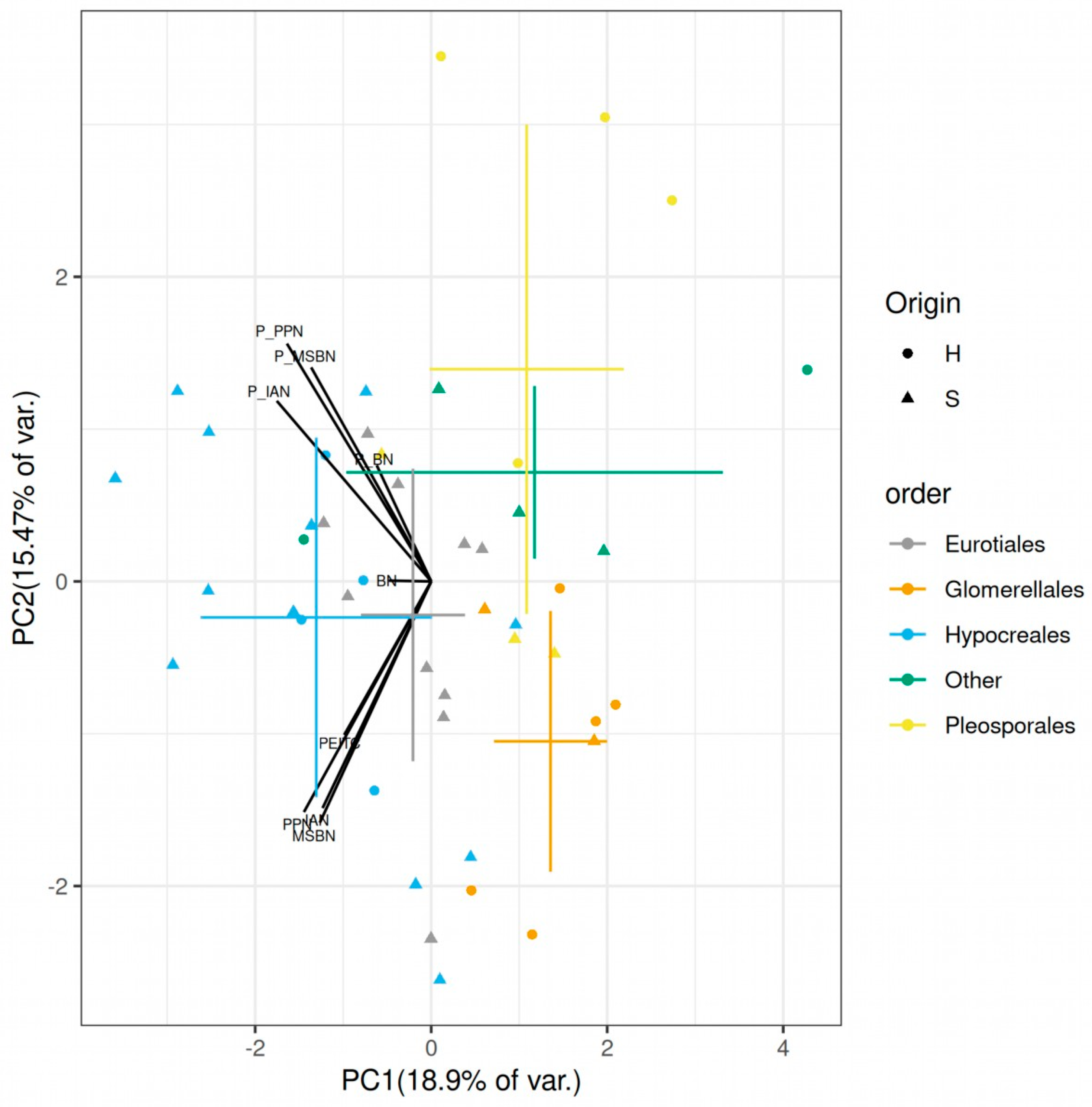
2.4. Differences among Endophytes and Soil Fungi, Fungal Taxa and Possible Microbiome Implications

Unlike in our study on a smaller set of fungi [37], no clear difference between the soil and endophytic fungal groups was revealed. This was mainly because both endophytes and soil fungi contained genera from various taxonomic clades, and there were groups found to be relatively distinct in their sensitivities towards various glucosinolate decomposition products. Due to the relatively multi-correlating nature of the dataset (Table S1), the separation of orders on the biplot of principal component analysis of the scaled MIC and FICI values was apparent (Figure 2); Hypocreales, Glomerellales and Pleosporales strains show distinct sensitivities (n = 16, 7, 7, respectively).
MIC values of individual GSL decomposition products varied considerably among different fungal orders (Figure S1); sensitivities to PEITC, IAN, PPN and MSBN were significantly different among fungal orders, while BN sensitivities were not (p = 0.0006, 0.0221, 0.0458, 0.0242 and 0.4377, respectively; Kruskal–Wallis test). Hypocreales was the least sensitive to pure compounds. Overall, the mean MIC values for Eurotiales, Glomerellales and Pleosporales were 1.03–1.69-fold, 1.11–2.13-fold and 1.05–3.81-fold lower for different compounds (Table 1). The most spectacular was the case of PEITC; Pleosporales, Glomerellales, Eurotiales and Hypocreales showed 0.0178 ± 0.0171, 0.0319 ± 0.0087, 0.0400 ± 0.0214 and 0.0679 ± 0.0358 mM for mean MIC values, respectively (p = 0.04658).
When checked between taxonomic orders, the FICI differences were also relatively pronounced (Figure 1 and Figure S2). The individual orders showed significantly different FICI indices for IAN, PPN and MSBN, but not for BN (p = 0.0073, 0.0076, 0.0102 and 0.1917, respectively, Kruskal–Wallis test). Hypocreales showed higher FICI values compared to other fungal groups. While its mean FICI values were 0.62 ± 0.16, 0.63 ± 0.16 and 0.61 ± 0.15 for IAN, PPN and MSBN, the same values for Glomerellales were only 0.44 ± 0.07, 0.40 ± 0.06 and 0.44 ± 0.05, respectively. In other words, almost all Glomerellales strains were synergistically affected by the combinations, while most of the Hypocreales strains did not show any synergy as per accepted criteria (Figure S2). The origin did not significantly affect the FICI values (p > 0.05), possibly because all relatively tolerant Eurotiales strains were from the soil, clustered close to the Hypocreales strains (Figure 2).
Overall, Hypocreales strains were distinguished from other clades by relatively high mean FICI values of 0.61–0.67, depending on the nitrile. In contrast, Eurotiales, Glomerellales and Pleosporales showed lower mean FICI values in the ranges of 0.52–0.61, 0.40–0.50 and 0.48–0.67, respectively.

3. Discussion

Although they have only been tested on a few pathogenic fungi in previous literature, the nitriles derived from indolic GSLs have been shown to be more potent antifungal agents compared to nitriles from other GSLs [5,9,10,11]. A detailed comparison of the effect of nitriles on a large number of strains is not available in the literature [5].
In the absence of specific studies, we speculate that the tested nitriles also act through aspecific target(s) and exert their toxicity via membrane disruption due to their low polarity, like other relatively nonpolar natural products of the plant kingdom [40,41]. An increase in membrane permeability might enhance the uptake of ITCs, thereby complementing their specific oxidative-stress-generating potential [7]. This is well reflected by the correlation between the logP values of nitriles and their median MIC values, which was −0.9197.
Though reports on plant volatiles acting synergistically against microbes [42,43] and a few other plant natural products [39] do exist, the direct chemical and biological interactions between the GSL decomposition products are relatively unknown and only a very limited number of available studies have tested their combinations. The homogenates of Brassicaceae species have also been shown to contain various proportions of nitriles (see Plaszkó et al. [5] and references therein). Although some of these papers also reported antifungal activity, they cannot be considered clear evidence of synergistic activity because individual compounds and their binary combinations were not tested.
Rare reports on pure compounds include descriptions of the synergistic antifungal effect of vapor phase combinations of ITCs [44,45]. Furthermore, a recent publication by Popović et al. showed synergism between PEITC, PPN and allyl ITC, involving the three fungal species Candida albicans, Penicillium citrinum and Aspergillus niger [46]. In the absence of binary combination tests, it is unclear whether we are seeing allyl ITC–phenylethyl ITC synergy or nitrile–ITC synergy.
Our results suggest that nitriles can be active participants in the potentiation of ITC antifungal activity under specific circumstances. Translation of the components of FICI values into real concentrations shows that synergy can occur even in mixtures with a relatively large amount of nitriles. An FICI ≤ 0.5 means that, in the presence of a relatively large amount of nitriles, a fold change of about four in ITC sensitivity can be observed. As ITCs have short half-lives due to their chemical reactivity [47,48], residual nitriles with a considerably higher stability might extend the time frame of ITC potency to some extent.
The results also explain the viability of plant accessions with nitrile-dominant GSL decomposition profiles, as the reduction in antifungal activity is likely to be much smaller than previously thought. The variance in the proportion of nitriles in the overall GSL decomposition product mixture can be extremely high, even within a species. For example, in a study on broccoli seed GSL decomposition products, the above-mentioned ratio ranged from 1.37% to 93.83% in accessions without alkenic GSLs and epithionitriles [22]. The same study found similar numbers for accessions with alkenic GSLs. In another study on A. thaliana accessions [21], nitriles accounted for 0–100% of the GSL decomposition products. Other studies have shown that levels of indolic GSL-derived nitriles increase after fungal challenge [9,10,11]. This supports our suggestion that nitriles or their combinations are likely to play a role in limiting fungal colonization in vivo. Although the generation of the bioactive GSL decomposition products in the vicinity of a fungal challenge site has also been documented [49,50,51], the relative proportion and in situ concentrations of various products are unknown [5].
In a more detailed analysis on A. thaliana, various plant accessions were challenged with Verticillium longisporum [20]. The authors found that the different accessions responded to the fungal challenge with different ratios of GSL decomposition product classes. Because the authors added water to lyophilized plant tissue to generate the volatiles, their results are indicative of changes in the abundance of specifier proteins [52]. While the nitrile-dominant Ler-0 showed a shift towards ITCs and a significant reduction in IAN and its 4-methoxy derivative, the isothiocyanate-dominant Hi-0 responded with increased biosynthesis of both 2-propenyl ITC and its nitrile counterpart (BN) in its leaves. The accession Bur-0 with diverse GSLs increased the production ITCs and various nitriles as well. The authors [20] speculated that nitrile levels may be more relevant in deterring fungal infection of roots. In the light of our results, a possible explanation is that Verticillium spp. is more sensitive to the nitrile–ITC combination than to ITCs alone. Overall, as the various accessions responded to a specific pathogen with alterations in the nitrile–isothiocyanate mixture [20], it appears that the plant is fine-tuning the synergistic mixture on demand.
The clear separation of different orders (Figure 2) shows different sensitivities to PEITC, the tested nitriles as well as their combinations. Due to the high variability in sensitivity among the 45 strains tested (Table 1, Figure S2), it is reasonable to conclude that GSL-decomposition product nitriles can modulate the plant microbiome, either alone or as adjuvants to ITCs. This leads us to the suggestion that the distinct sensitivity patterns of taxa are likely contributors to “root filtering” [53], i.e., the differences in the taxonomic composition of bulk-soil and endosphere microbiomes. The differences in the specialized metabolite patterns likely also contribute to the differences in the microbiomes of various plant families [29].
The better tolerance of Hypocreales against PEITC and the nitrile–ITC combinations also explains the findings of Ishimoto et al. [54] and those of Wand and Zhou [55]. The former group found Fusarium to be a dominant member of the rhizosphere of Brassicaceae plants, while the latter concluded that using Brassica green manures increases the relative abundance of Fusarium in the rhizosphere soils of cucumber. The synergistic effect might also be one of the mechanisms behind the large differences in glucosinolate–fungal-microbiome correlations in nitrile- and ITC-dominant A. thaliana accessions challenged by Fusarium oxysporum from Hypocreales [56].
Future research should take into account that the ratio and total amounts of GSL decomposition products are likely to be much better descriptors of the outcome of plant–fungus interactions than data on native glucosinolates alone. Direct testing of the effects of pure nitriles or combinations of nitriles and ITCs on soil microbiomes may provide additional insight into the relative sensitivities of fungal taxa, but to gain a deeper understanding of in vivo effects, studies comparing the microbiomes of plant accessions with different nitrile–ITC ratios are required. Even more data could be obtained from a well-designed comparison of wild-type plants with plants specifically compromised in nitrile generation.

4. Materials and Methods

4.1. Chemicals

The reagents were of analytical purity. The compounds 3-butenenitrile (BN, = Allyl cyanide), 2-phenylethyl isothiocyanate (PEITC), 3-phenylpropionitrile (PPN) and 1H-indole-3-acetonitrile (IAN, = indole-3-acetonitrile) were from Merck (Darmstadt, Germany), while 4-(methylsulfanyl)-butanenitrile (MSBN, = methyl thiobutyl nitrile) was purchased from ChemSpace (Riga, Latvia). The media components malt extract and peptone were obtained from Reanal (Budapest, Hungary), while glucose-monohydrate was from VWR. Dimethyl sulfoxide was from Fisher Scientific (Pittsburgh, PA, USA). Type I water was used throughout the study (18.2 MΩ), produced by a Zeneer Power I Water Purification System (Human corporation, Seoul, Republic of Korea). ACD ChemSketch v2020.2.1 was used for logP calculation.

4.2. Fungi

Endophytic fungi from horseradish (Armoracia rusticana G. Gaertn., B. Mey. & Scherb.) and soil fungi from the soil of the same site were used for this study. The isolation procedure for both endophytes and soil fungi has already been described previously [37]. Preparation of fungal inoculums was carried out as in our recent study [57]. The identification procedure was based on amplification and sequencing a characteristic barcode sequence, as previously described [37]. In addition to the fungi already identified in the aforementioned publication, a number of additional horseradish endophyte and soil fungi were identified using the ITS1-F_KYO2/ITS4_KYO3 primer pair [58]. The ITS sequences were deposited in NCBI GenBank under the accession numbers of OR019707-OR019746. All fungal strains were maintained on 2% Malt Extract Agar medium [37] during the study.

4.3. Antimicrobial Assay

The antifungal inhibition assay could not be conducted in 96-well plates (checkerboard assay [59]) with sufficient accuracy. In spite of testing several precautions described for studying volatile antimicrobials in [35], GC-MS evaluation of the microplate setup discovered a significant evaporation loss of several agents (BN, MSBN and, to a minor extent, PPN) within 4 days (see Supplementary Materials for additional details).
Therefore, the antifungal experiments were run in autoclaved, airtight 1.5 mL borosilicate glass screw-cap vials with 300 µL autoclaved inserts. Screw caps were disinfected using UV light. The lack of loss by evaporation was verified by a 4-day incubation study with 50 µL of diethyl ether and the agents of interest. Advantages of the current method include no evaporation loss and no cross-contamination between adjacent cells, as well as an exact in-liquid concentration of the antifungal agent.
In each vial insert, an aliquot of 196 µL Saboraud Glucose Broth (SGB, 40 g/L glucose monohydrate, peptone 10 g/L, pH 5.6 ± 0.2) was inoculated with 20 µg dry weight equivalent of fungal inoculum [37] per vial, and DMSO stocks of different agents were added so that the total amount of added volume was 4 µL. To controls, 4 µL DMSO was added. The vial sets were incubated in darkness at 20/18 °C (16/8 h). All vials were evaluated manually on day 4 for visible mycelial growth. For all fungi, at least two biological replicates were obtained for all agents, with 8–12 fungi run in parallel. Fungi with poorly reproducible MIC values for any agent, and fungi with a poorly reproducible generation of inoculation material, were eliminated from further analyses.

4.4. Minimum Inhibitory Concentration Measurement

For MIC determination, serial dilutions from the different agent concentrations were tested for the ability to inhibit the growth of mycelia (0.000452–0.858 mM for PEITC, 0.622–124.3 mM for BN, 0.844–84.4 mM MSBN, 0.19–48.8 mM for PPN and 0.0204–20.49 mM for IAN).

4.5. Synergism Experiments

The fractional inhibitory concentration index (FICI) value is considered the standard reference parameter that can quantitatively describe pairwise drug interactions in antimicrobial assays [59]. This approach was used to assess the strength of the interaction between the agents. The following conservative interpretation of FICI values [38,39] was applied: ‘synergy’ (FICI ≤ 0.5), ‘antagonism’ (FICI ≥ 4.0) and ‘no interaction’ (4.0 > FICI > 0.5).
In preliminary tests, no antagonism was found, so only combinations to assess synergies were run. In brief, after the determination of the MIC values, 0.125, 0.25 and 0.5 MIC equivalents for each agent were mixed in a 3 × 3 matrix to test growth in all combinations, so the FICI range of 0.25–1.00 was tested. As positive controls, 1 MIC concentrations from all pure agents were run in parallel. The FICI was calculated from the data of the 3 × 3 matrix using the “lowest FICI” method [60] using in-house scripts in R 4.2 [61].

4.6. Statistical Tests

The mean of biological replicates was submitted to all statistical tests so that data on each individual fungal-strain–sensitivity pair was considered an observation. The Kruskal–Wallis test was used to determine statistically significant differences between fungal groups for both MIC values and FICI parameters. Tests were implemented in R 4.2 [61]. When comparison of all taxonomic groups was carried out with the Kruskal–Wallis test, fungi from “other” orders were omitted from the dataset.

5. Conclusions

In the current work, we have shown that glucosinolate-derived nitriles act as synergistic enhancers of the antifungal efficacy of the main glucosinolate decomposition products, the isothiocyanates. This synergy was detected in approximately 50% of the tested strains, regardless of glucosinolate class. Moreover, different fungal taxa had different sensitivities towards glucosinolate-derived nitriles or their synergistic combination, as quantified by minimal inhibitory concentration (MIC) and fractional inhibitory concentration index (FICI) values. Hypocreales (many Fusarium strains) emerged as a more tolerant group, compared to fungi from Glomerellales, Pleosporales and Eurotiales. Thus, our study provided in vitro quantitative evidence of the effects of nitriles on various members of the plant microbiome.
This contribution of nitriles to antifungal efficacy explains the difficulty in finding actual bioactive agents that limit fungal colonization in Brassicaceae, which includes many crops and Arabidopsis. Although our results support the frequently described phenomenon of increased nitrile biosynthesis following fungal challenge, it draws our attention to why the plants biosynthesize different mixtures of glucosinolate downstream to cope with the challenge by different fungi.
As glucosinolate-derived nitriles and ITCs are likely to act together during fungal challenge, further studies are needed to shed light on the role of nitriles during in vivo fungal colonization of plants. Targeted studies comparing the microbiome of plant accessions with different nitrile–ITC ratios, as well as a critical comparison of the microbiomes of wild-type plants with those of plants compromised in glucosinolate-derived nitrile biosynthesis, are warranted.

Supplementary Materials

The following supporting information can be downloaded at: https://www.mdpi.com/article/10.3390/plants12142741/s1: Loss of volatile nitriles PPN and MSBN in parafilm-covered 96-well plates (additional description); Figure S1: Histogram showing distribution of the minimal inhibitory concentration (MIC) values of 2-phenylethyl isothiocyanate and glucosinolate-derived nitriles for fungal orders of the current study; Figure S2: Histogram showing distribution of the fractional inhibition concentration indices (FICI) values of synergistic combinations of 2-phenylethyl isothiocyanate (PEITC) and glucosinolate-derived; Table S1; Spearman’s rho correlation values between individual MIC and FICI sensitivity values; Table S2. GenBank accession numbers of fungi used throughout the study.

Author Contributions

This study was conceptualized and designed by S.G. The experiments were conducted by T.P., E.B. and Z.S.; fungal identification was carried out by H.C. and L.Á.-S.; and GC-MS evaluation was performed by A.K.-S. The data were evaluated by S.G. and Z.S. and G.V. provided a critical revision. Funding was obtained by S.G. The draft was prepared by S.G. All authors have read and agreed to the published version of the manuscript.

Funding

This study was supported by the Hungarian Scientific Research Fund (OTKA) 128021 grant of the National Research, Development and Innovation Office, which is greatly appreciated.

Data Availability Statement

Additional data are available on request.

Conflicts of Interest

The authors declare no conflict of interest.

References

  1. Blažević, I.; Montaut, S.; Burčul, F.; Olsen, C.E.; Burow, M.; Rollin, P.; Agerbirk, N. Glucosinolate Structural Diversity, Identification, Chemical Synthesis and Metabolism in Plants. Phytochemistry 2020, 169, 112100. [Google Scholar] [CrossRef] [PubMed]
  2. Abuyusuf, M.; Rubel, M.H.; Kim, H.-T.; Jung, H.-J.; Nou, I.-S.; Park, J.-I. Glucosinolates and Biotic Stress Tolerance in Brassicaceae with Emphasis on Cabbage: A Review. Biochem. Genet. 2023, 61, 451–470. [Google Scholar] [CrossRef] [PubMed]
  3. Hanschen, F.S.; Lamy, E.; Schreiner, M.; Rohn, S. Reactivity and Stability of Glucosinolates and Their Breakdown Products in Foods. Angew. Chem. Int. Ed. 2014, 53, 11430–11450. [Google Scholar] [CrossRef] [PubMed]
  4. Abdel-Massih, R.M.; Debs, E.; Othman, L.; Attieh, J.; Cabrerizo, F.M. Glucosinolates, a Natural Chemical Arsenal: More to Tell than the Myrosinase Story. Front. Microbiol. 2023, 14, 1130208. [Google Scholar] [CrossRef] [PubMed]
  5. Plaszkó, T.; Szűcs, Z.; Vasas, G.; Gonda, S. Interactions of Fungi with Non-Isothiocyanate Products of the Plant Glucosinolate Pathway: A Review on Product Formation, Antifungal Activity, Mode of Action and Biotransformation. Phytochemistry 2022, 200, 113245. [Google Scholar] [CrossRef] [PubMed]
  6. Poveda, J.; Eugui, D.; Velasco, P. Natural Control of Plant Pathogens through Glucosinolates: An Effective Strategy against Fungi and Oomycetes. Phytochem. Rev. 2020, 19, 1045–1059. [Google Scholar] [CrossRef]
  7. Plaszkó, T.; Szűcs, Z.; Vasas, G.; Gonda, S. Effects of Glucosinolate-Derived Isothiocyanates on Fungi: A Comprehensive Review on Direct Effects, Mechanisms, Structure-Activity Relationship Data and Possible Agricultural Applications. J. Fungi 2021, 7, 539. [Google Scholar] [CrossRef]
  8. Eugui, D.; Escobar, C.; Velasco, P.; Poveda, J. Glucosinolates as an Effective Tool in Plant-Parasitic Nematodes Control: Exploiting Natural Plant Defenses. Appl. Soil Ecol. 2022, 176, 104497. [Google Scholar] [CrossRef]
  9. Pedras, M.S.C.; Ahiahonu, P.W.K. Phytotoxin Production and Phytoalexin Elicitation by the Phytopathogenic Fungus Sclerotinia sclerotiorum. J. Chem. Ecol. 2004, 30, 2163–2179. [Google Scholar] [CrossRef]
  10. Hiruma, K.; Fukunaga, S.; Bednarek, P.; Piślewska-Bednarek, M.; Watanabe, S.; Narusaka, Y.; Shirasu, K.; Takano, Y. Glutathione and Tryptophan Metabolism Are Required for Arabidopsis Immunity during the Hypersensitive Response to Hemibiotrophs. Proc. Natl. Acad. Sci. USA 2013, 110, 9589–9594. [Google Scholar] [CrossRef]
  11. Pedras, M.S.C.; Zheng, Q.-A. Metabolic Responses of Thellungiella Halophila/Salsuginea to Biotic and Abiotic Stresses: Metabolite Profiles and Quantitative Analyses. Phytochemistry 2010, 71, 581–589. [Google Scholar] [CrossRef]
  12. Frerigmann, H.; Piślewska-Bednarek, M.; Sánchez-Vallet, A.; Molina, A.; Glawischnig, E.; Gigolashvili, T.; Bednarek, P. Regulation of Pathogen-Triggered Tryptophan Metabolism in Arabidopsis Thaliana by MYB Transcription Factors and Indole Glucosinolate Conversion Products. Mol. Plant 2016, 9, 682–695. [Google Scholar] [CrossRef] [Green Version]
  13. He, Y.; Xu, J.; Wang, X.; He, X.; Wang, Y.; Zhou, J.; Zhang, S.; Meng, X. The Arabidopsis Pleiotropic Drug Resistance Transporters PEN3 and PDR12 Mediate Camalexin Secretion for Resistance to Botrytis cinerea. Plant Cell 2019, 31, 2206–2222. [Google Scholar] [CrossRef]
  14. Kuhn, H.; Lorek, J.; Kwaaitaal, M.; Consonni, C.; Becker, K.; Micali, C.; Ver Loren van Themaat, E.; Bednarek, P.; Raaymakers, T.M.; Appiano, M.; et al. Key Components of Different Plant Defense Pathways are Dispensable for Powdery Mildew Resistance of the Arabidopsis Mlo2 Mlo6 Mlo12 Triple Mutant. Front. Plant Sci. 2017, 8, 1006. [Google Scholar] [CrossRef]
  15. Yun, H.S.; Kang, B.G.; Kwon, C. Arabidopsis Immune Secretory Pathways to Powdery Mildew Fungi. Plant Signal. Behav. 2016, 11, e1226456. [Google Scholar] [CrossRef] [Green Version]
  16. Wittstock, U.; Kurzbach, E.; Herfurth, A.-M.; Stauber, E.J. Glucosinolate Breakdown. Adv. Bot. Res. 2016, 80, 125–169. [Google Scholar] [CrossRef]
  17. Vik, D.; Mitarai, N.; Wulff, N.; Halkier, B.A.; Burow, M. Dynamic Modeling of Indole Glucosinolate Hydrolysis and Its Impact on Auxin Signaling. Front. Plant Sci. 2018, 9, 550. [Google Scholar] [CrossRef] [Green Version]
  18. Sugiyama, R.; Li, R.; Kuwahara, A.; Nakabayashi, R.; Sotta, N.; Mori, T.; Ito, T.; Ohkama-Ohtsu, N.; Fujiwara, T.; Saito, K.; et al. Retrograde Sulfur Flow from Glucosinolates to Cysteine in Arabidopsis thaliana. Proc. Natl. Acad. Sci. USA 2021, 118, e2017890118. [Google Scholar] [CrossRef]
  19. Ting, H.-M.; Cheah, B.H.; Chen, Y.-C.; Yeh, P.-M.; Cheng, C.-P.; Yeo, F.K.S.; Vie, A.K.; Rohloff, J.; Winge, P.; Bones, A.M.; et al. The Role of a Glucosinolate-Derived Nitrile in Plant Immune Responses. Front. Plant Sci. 2020, 11, 257. [Google Scholar] [CrossRef]
  20. Witzel, K.; Hanschen, F.S.; Klopsch, R.; Ruppel, S.; Schreiner, M.; Grosch, R. Verticillium Longisporum Infection Induces Organ-Specific Glucosinolate Degradation in Arabidopsis thaliana. Front. Plant Sci. 2015, 6, 508. [Google Scholar] [CrossRef] [Green Version]
  21. Hanschen, F.S.; Pfitzmann, M.; Witzel, K.; Stützel, H.; Schreiner, M.; Zrenner, R. Differences in the Enzymatic Hydrolysis of Glucosinolates Increase the Defense Metabolite Diversity in 19 Arabidopsis thaliana Accessions. Plant Physiol. Biochem. 2018, 124, 126–135. [Google Scholar] [CrossRef] [PubMed]
  22. Wang, J.; Yu, H.; Zhao, Z.; Sheng, X.; Shen, Y.; Gu, H. Natural Variation of Glucosinolates and Their Breakdown Products in Broccoli (Brassica Oleracea Var. Italica) Seeds. J. Agric. Food Chem. 2019, 67, 12528–12537. [Google Scholar] [CrossRef] [PubMed]
  23. Schoch, C.L.; Seifert, K.A.; Huhndorf, S.; Robert, V.; Spouge, J.L.; Levesque, C.A.; Chen, W.; Fungal Barcoding Consortium; Fungal Barcoding Consortium Author List; Bolchacova, E.; et al. Nuclear Ribosomal Internal Transcribed Spacer (ITS) Region as a Universal DNA Barcode Marker for Fungi. Proc. Natl. Acad. Sci. USA 2012, 109, 6241–6246. [Google Scholar] [CrossRef] [PubMed]
  24. Benson, D.A.; Cavanaugh, M.; Clark, K.; Karsch-Mizrachi, I.; Lipman, D.J.; Ostell, J.; Sayers, E.W. GenBank. Nucleic Acids Res. 2017, 45, D37–D42. [Google Scholar] [CrossRef] [PubMed] [Green Version]
  25. Federhen, S. Type Material in the NCBI Taxonomy Database. Nucleic Acids Res. 2015, 43, D1086–D1098. [Google Scholar] [CrossRef] [Green Version]
  26. Xu, J. Fungal DNA Barcoding. Genome 2016, 59, 913–932. [Google Scholar] [CrossRef] [Green Version]
  27. Vu, D.; Groenewald, M.; de Vries, M.; Gehrmann, T.; Stielow, B.; Eberhardt, U.; Al-Hatmi, A.; Groenewald, J.Z.; Cardinali, G.; Houbraken, J.; et al. Large-Scale Generation and Analysis of Filamentous Fungal DNA Barcodes Boosts Coverage for Kingdom Fungi and Reveals Thresholds for Fungal Species and Higher Taxon Delimitation. Stud. Mycol. 2019, 92, 135–154. [Google Scholar] [CrossRef]
  28. Plaszkó, T.; Szűcs, Z.; Cziáky, Z.; Ács-Szabó, L.; Csoma, H.; Géczi, L.; Vasas, G.; Gonda, S. Correlations Between the Metabolome and the Endophytic Fungal Metagenome Suggests Importance of Various Metabolite Classes in Community Assembly in Horseradish (Armoracia rusticana, Brassicaceae) Roots. Front. Plant Sci. 2022, 13, 921008. [Google Scholar] [CrossRef]
  29. Toju, H.; Kurokawa, H.; Kenta, T. Factors Influencing Leaf- and Root-Associated Communities of Bacteria and Fungi across 33 Plant Orders in a Grassland. Front. Microbiol. 2019, 10, 241. [Google Scholar] [CrossRef] [Green Version]
  30. Shofran, B.G.; Purrington, S.T.; Breidt, F.; Fleming, H.P. Antimicrobial Properties of Sinigrin and Its Hydrolysis Products. J. Food Sci. 1998, 63, 621–624. [Google Scholar] [CrossRef]
  31. Szűcs, Z.; Plaszkó, T.; Cziáky, Z.; Kiss-Szikszai, A.; Emri, T.; Bertóti, R.; Sinka, L.T.; Vasas, G.; Gonda, S. Endophytic Fungi from the Roots of Horseradish (Armoracia rusticana) and Their Interactions with the Defensive Metabolites of the Glucosinolate—Myrosinase—Isothiocyanate System. BMC Plant Biol. 2018, 18, 85. [Google Scholar] [CrossRef] [Green Version]
  32. Odds, F.C. Synergy, Antagonism, and What the Chequerboard Puts between Them. J. Antimicrob. Chemother. 2003, 52, 1. [Google Scholar] [CrossRef]
  33. Caesar, L.K.; Cech, N.B. Synergy and Antagonism in Natural Product Extracts: When 1 + 1 Does Not Equal 2. Nat. Prod. Rep. 2019, 36, 869–888. [Google Scholar] [CrossRef] [Green Version]
  34. Hu, Z.; Yuan, K.; Zhou, Q.; Lu, C.; Du, L.; Liu, F. Mechanism of Antifungal Activity of Perilla Frutescens Essential Oil against Aspergillus Flavus by Transcriptomic Analysis. Food Control 2021, 123, 107703. [Google Scholar] [CrossRef]
  35. Wu, T.-L.; Zhang, B.-Q.; Luo, X.-F.; Li, A.-P.; Zhang, S.-Y.; An, J.-X.; Zhang, Z.-J.; Liu, Y.-Q. Antifungal Efficacy of Sixty Essential Oils and Mechanism of Oregano Essential Oil against Rhizoctonia Solani. Ind. Crops Prod. 2023, 191, 115975. [Google Scholar] [CrossRef]
  36. Clemente, I.; Aznar, M.; Nerín, C. Synergistic Properties of Mustard and Cinnamon Essential Oils for the Inactivation of Foodborne Moulds in Vitro and on Spanish Bread. Int. J. Food Microbiol. 2019, 298, 44–50. [Google Scholar] [CrossRef]
  37. Liu, Q.; Niu, H.; Zhang, W.; Mu, H.; Sun, C.; Duan, J. Synergy among Thymol, Eugenol, Berberine, Cinnamaldehyde and Streptomycin against Planktonic and Biofilm-Associated Food-Borne Pathogens. Lett. Appl. Microbiol. 2015, 60, 421–430. [Google Scholar] [CrossRef]
  38. Tunc, S.; Chollet, E.; Chalier, P.; Preziosi-Belloy, L.; Gontard, N. Combined Effect of Volatile Antimicrobial Agents on the Growth of Penicillium Notatum. Int. J. Food Microbiol. 2007, 113, 263–270. [Google Scholar] [CrossRef]
  39. Wu, H.; Zhang, X.; Zhang, G.-A.; Zeng, S.-Y.; Lin, K.-C. Antifungal Vapour-Phase Activity of a Combination of Allyl Isothiocyanate and Ethyl Isothiocyanate Against Botrytis Cinerea and Penicillium Expansum Infection on Apples. J. Phytopathol. 2011, 159, 450–455. [Google Scholar] [CrossRef]
  40. Popović, M.; Maravić, A.; Čulić, V.Č.; Đulović, A.; Burčul, F.; Blažević, I. Biological Effects of Glucosinolate Degradation Products from Horseradish: A Horse That Wins the Race. Biomolecules 2020, 10, 343. [Google Scholar] [CrossRef] [Green Version]
  41. Andini, S.; Araya-Cloutier, C.; Waardenburg, L.; den Besten, H.M.W.; Vincken, J.-P. The Interplay between Antimicrobial Activity and Reactivity of Isothiocyanates. LWT 2020, 134, 109843. [Google Scholar] [CrossRef]
  42. Van Wambeke, E.; Ceustermans, A.; De Landtsheer, A.; Coosemans, J. Combinations of Soil Fumigants for Methyl-Bromide Replacement. Commun. Agric. Appl. Biol. Sci. 2009, 74, 75–84. [Google Scholar]
  43. Hiruma, K.; Onozawa-Komori, M.; Takahashi, F.; Asakura, M.; Bednarek, P.; Okuno, T.; Schulze-Lefert, P.; Takanoa, Y. Entry Mode-Dependent Function of an Indole Glucosinolate Pathway in Arabidopsis for Nonhost Resistance against Anthracnose Pathogens. Plant Cell 2010, 22, 2429–2443. [Google Scholar] [CrossRef] [PubMed] [Green Version]
  44. Lipka, U.; Fuchs, R.; Kuhns, C.; Petutschnig, E.; Lipka, V. Live and Let Die—Arabidopsis Nonhost Resistance to Powdery Mildews. Eur. J. Cell Biol. 2010, 89, 194–199. [Google Scholar] [CrossRef] [PubMed]
  45. Fuchs, R.; Kopischke, M.; Klapprodt, C.; Hause, G.; Meyer, A.J.; Schwarzländer, M.; Fricker, M.D.; Lipka, V. Immobilized Subpopulations of Leaf Epidermal Mitochondria Mediate PENETRATION2-Dependent Pathogen Entry Control in Arabidopsis. Plant Cell 2016, 28, 130–145. [Google Scholar] [CrossRef] [Green Version]
  46. Lambrix, V.; Reichelt, M.; Mitchell-Olds, T.; Kliebenstein, D.J.; Gershenzon, J. The Arabidopsis Epithiospecifier Protein Promotes the Hydrolysis of Glucosinolates to Nitriles and Influences Trichoplusia Ni Herbivory. Plant Cell 2001, 13, 2793–2808. [Google Scholar] [CrossRef] [Green Version]
  47. Maciá-Vicente, J.G.; Nam, B.; Thines, M. Root Filtering, Rather than Host Identity or Age, Determines the Composition of Root-Associated Fungi and Oomycetes in Three Naturally Co-Occurring Brassicaceae. Soil Biol. Biochem. 2020, 146, 107806. [Google Scholar] [CrossRef]
  48. Ishimoto, H.; Fukushi, Y.; Yoshida, T.; Tahara, S. Rhizopus and Fusarium are Selected as Dominant Fungal Genera in Rhizospheres of Brassicaceae. J. Chem. Ecol. 2000, 26, 2387–2399. [Google Scholar] [CrossRef]
  49. Wang, Y.; Zhou, X. Effects of Green Manures on Rhizosphere Fungal Community Composition of Cucumber Seedlings. Curr. Microbiol. 2023, 80, 87. [Google Scholar] [CrossRef]
  50. Kudjordjie, E.N.; Hooshmand, K.; Sapkota, R.; Darbani, B.; Fomsgaard, I.S.; Nicolaisen, M. Fusarium Oxysporum Disrupts Microbiome-Metabolome Networks in Arabidopsis Thaliana Roots. Microbiol. Spectr. 2022, 10, e0122622. [Google Scholar] [CrossRef]
  51. Plaszkó, T.; Szűcs, Z.; Kállai, Z.; Csoma, H.; Vasas, G.; Gonda, S. Volatile Organic Compounds (VOCs) of Endophytic Fungi Growing on Extracts of the Host, Horseradish (Armoracia rusticana). Metabolites 2020, 10, 451. [Google Scholar] [CrossRef]
  52. Toju, H.; Tanabe, A.S.; Yamamoto, S.; Sato, H. High-Coverage ITS Primers for the DNA-Based Identification of Ascomycetes and Basidiomycetes in Environmental Samples. PLoS ONE 2012, 7, e40863. [Google Scholar] [CrossRef] [Green Version]
  53. Gómara, M.; Ramón-García, S. The FICI Paradigm: Correcting Flaws in Antimicrobial in Vitro Synergy Screens at Their Inception. Biochem. Pharmacol. 2019, 163, 299–307. [Google Scholar] [CrossRef]
  54. Houdkova, M.; Kokoska, L. Volatile Antimicrobial Agents and in Vitro Methods for Evaluating Their Activity in the Vapour Phase: A Review. Planta Medica 2020, 86, 822–857. [Google Scholar] [CrossRef]
  55. Inouye, S.; Takizawa, T.; Yamaguchi, H. Antibacterial Activity of Essential Oils and Their Major Constituents against Respiratory Tract Pathogens by Gaseous Contact. J. Antimicrob. Chemother. 2001, 47, 565–573. [Google Scholar] [CrossRef] [Green Version]
  56. Nakahara, K.; Alzoreky, N.S.; Yoshihashi, T.; Nguyen, H.T.; Trakoontivakorn, G. Chemical Composition and Antifungal Activity of Essential Oil from Cymbopogon nardus (Citronella Grass). Jpn. Agric. Res. Q. JARQ 2013, 37, 249–252. [Google Scholar] [CrossRef] [Green Version]
  57. Seo, H.-S.; Beuchat, L.R.; Kim, H.; Ryu, J.-H. Development of an Experimental Apparatus and Protocol for Determining Antimicrobial Activities of Gaseous Plant Essential Oils. Int. J. Food Microbiol. 2015, 215, 95–100. [Google Scholar] [CrossRef]
  58. Houdkova, M.; Rondevaldova, J.; Doskocil, I.; Kokoska, L. Evaluation of Antibacterial Potential and Toxicity of Plant Volatile Compounds Using New Broth Microdilution Volatilization Method and Modified MTT Assay. Fitoterapia 2017, 118, 56–62. [Google Scholar] [CrossRef]
  59. Ji, H.; Kim, H.; Beuchat, L.R.; Ryu, J.-H. Synergistic Antimicrobial Activities of Essential Oil Vapours against Penicillium corylophilum on a Laboratory Medium and Beef Jerky. Int. J. Food Microbiol. 2019, 291, 104–110. [Google Scholar] [CrossRef]
  60. Bonapace, C.R.; Bosso, J.A.; Friedrich, L.V.; White, R.L. Comparison of Methods of Interpretation of Checkerboard Synergy Testing. Diagn. Microbiol. Infect. Dis. 2002, 44, 363–366. [Google Scholar] [CrossRef]
  61. R Core Team. R: A Language and Environment for Statistical Computing; R Foundation for Statistical Computing: Vienna, Austria, 2021; ISBN 3-900051-07-0. [Google Scholar]
Figure 1. Effects of specifier proteins on the main routes of glucosinolate (GSL) decomposition. In the absence of any specifier proteins, isothiocyanates are formed. Data compiled from [5] and references therein. Abbreviation: ESP: epithiospecifier protein; TFP: thiocyanate-forming protein; ESM: epithiospecifier modifier protein; NSP: nitrile-specifier protein.
Plants 12 02741 g001
Figure 2. Biplot of principal component analysis scores of fungal sensitivities to glucosinolate decomposition products (nitriles and 2-phenylethyl isothiocyanate) and their combinations. Crosshairs show the average ± standard deviation for different taxonomic groups. The labeled vectors represent the relative contribution of loadings to the two principal components, PC1–2. Strains from various fungal orders are shown with different colors. Abbreviations for loading vectors: BN, IAN, MSBN, PEITC and PPN denote MIC values for individual agents; P_BN, P_IAN, P_MSBN and P_PPN denote FICI values for nitrile-PEITC combinations. Fungi of different origin are shown with circles and triangles. Abbreviations for origin: H, horseradish endophytes; S, soil.
Plants 12 02741 g002
Table 1. Antifungal activity and synergistic potency of 2-phenylethyl isothiocyanate and its combinations with glucosinolate-derived nitriles in vitro against endophytic and soil fungi.
IDGenusOrderOriginMIC (mM)FICI (PEITC + Nitrile)
PEITCIANPPNMSBNBNIANPPNMSBNBN
F1FusariumHyH0.05362.566.112779.60.560.750.560.63
F2FusariumHyH0.08041.286.112744.80.630.630.631.06
F3PhomaPlH0.00420.322.2913.523.20.630.50.50.56
F4PhomaPlH0.00670.435.0911.3101.90.630.750.750.75
F5Oidiodendron-H0.00500.271.2715.849.70.380.50.250.5
F6FusariumHyH0.05361.286.113629.80.540.810.440.69
F7FusariumHyH0.08041.286.1140.5101.90.560.50.50.52
F8ParaphomaPlH0.01341.284.072759.70.440.560.630.63
F9PlectosphaerellaGlH0.04021.284.582779.60.440.380.380.5
F10PlectosphaerellaGlH0.02681.715.0920.346.40.380.440.380.5
F11PseudopyrenochaetaPlH0.00670.432.5413.541.40.50.560.750.69
F12PlectosphaerellaGlH0.02681.928.1427530.380.380.440.31
F13Phomopsis-H0.02011.289.162779.60.690.630.630.69
F14PlectosphaerellaGlH0.01791.494.072759.70.460.460.460.67
F15PlectosphaerellaGlH0.03131.717.123679.60.50.310.460.54
F16StagonosporopsisPlS0.05360.646.112749.70.440.440.50.5
F17CurvulariaPlS0.02011.716.112756.40.380.5610.69
F18PenicilliumEuS0.08041.286.112779.60.810.560.560.63
F19AspergillusEuS0.02012.998.1429.350.440.380.560.56
F20FusariumHyS0.05361.286.113666.30.630.560.810.63
F21FusariumHyS0.16081.926.112744.80.380.380.381.25
F22PenicilliumEuS0.02011.924.5840.544.80.560.440.560.56
F23FusariumHyS0.04021.926.115479.60.690.810.630.63
F24PenicilliumEuS0.04021.286.112719.90.690.690.630.63
F25PenicilliumEuS0.02011.286.112759.70.50.50.630.69
F26FusariumHyS0.03571.289.1640.5820.440.380.440.47
F27PenicilliumEuS0.05361.926.112779.60.690.560.560.63
F28PenicilliumEuS0.04471.286.113626.50.580.560.50.56
F29FusariumHyS0.13401.286.112779.60.440.440.440.44
F30PenicilliumEuS0.01341.285.093644.80.630.630.630.63
F31Cadophora-S0.02011.284.0722.544.80.440.440.50.56
F32ClonostachysHyS0.04021.924.5820.3101.90.630.690.690.57
F33PlectosphaerellaGlS0.04020.966.112749.70.380.380.50.38
F34Cladosporium-S0.02010.646.112743.10.560.440.50.81
F35FusariumHyS0.08041.926.112779.60.810.810.750.69
F36FusariumHyS0.08041.926.112759.70.810.690.880.5
F37PenicilliumEuS0.05361.284.582729.80.630.440.630.5
F38PenicilliumEuS0.05361.926.112719.90.560.440.560.63
F39PlectosphaerellaGlS0.04021.286.112714.90.560.50.50.63
F40Cladosporium-S0.02680.646.112736.50.630.50.631
F41PurpureocilliumHyS0.03131.286.112714.90.50.440.50.75
F42ClonostachysHyS0.06262.566.112759.70.560.630.810.5
F43AaosphaeriaPlS0.02011.286.112779.60.380.560.560.38
F44FusariumHyS0.03572.138.143626.50.690.750.690.75
F45ClonostachysHyS0.06261.289.163639.810.810.630.69
FICI values are the sum of MIC equivalents for members of the tested combination that result in inhibition of growth. Abbreviations: BN, 3-butenenitrile (allyl cyanide); IAN, 1H-indol-3-yl acetonitrile; MSBN, 4-(methylsulfanyl)-butanenitrile; PEITC, 2-phenylethyl isothiocyanate; PPN, phenylpropionitrile. All results are a mean of 2 measurements (nitriles’ MIC values and FICI values) or 6 measurements (MIC of PEITC). Orders: Eu, Eurotiales; Gl, Glomerellales; Hy, Hypocreales; Pl, Pleosporales; -, other. Abbreviations for origin: H, horseradish endophyte; S, soil.
Disclaimer/Publisher’s Note: The statements, opinions and data contained in all publications are solely those of the individual author(s) and contributor(s) and not of MDPI and/or the editor(s). MDPI and/or the editor(s) disclaim responsibility for any injury to people or property resulting from any ideas, methods, instructions or products referred to in the content.

