Article

Genome-Wide Identification and Expression Profiling of Pathogenesis-Related Protein 1 (PR-1) Genes in Durum Wheat (Triticum durum Desf.)

1
Laboratory of Biotechnology and Plant Improvement, Centre of Biotechnology of Sfax, P.O. Box 1177, Sfax 3018, Tunisia
2
Department of Biology, College of Sciences, University of Hail, P.O. Box 2440, Ha’il City 81451, Saudi Arabia
*
Author to whom correspondence should be addressed.
Plants 2023, 12(10), 1998; https://doi.org/10.3390/plants12101998
Submission received: 4 April 2023 / Revised: 3 May 2023 / Accepted: 9 May 2023 / Published: 16 May 2023
(This article belongs to the Special Issue Applications of Bioinformatics in Plant Resources and Omics)

Abstract

:
Pathogen-related proteins (PRs) are diversified proteins with a low molecular weight implicated in plant response to biotic and abiotic stress as well in regulating different functions in plant maturation. Interestingly, no systematical study has been conducted in durum wheat (Triticum turgidum subsp. durum). In the present study, 12 PR-1 genes encoding a CAP superfamily domain were identified in the genome of Triticum turgidum subsp. durum, which is an important cereal, using in silico approaches. Additionally, phylogenetic analysis showed that the PR-1 genes were classified into three groups based on their isoelectric point and the conserved motif domain. Moreover, our analysis showed that most of the TdPR-1 proteins presented an N-terminal signal peptide. Expression patterns analysis showed that the PR-1 gene family presented temporal and spatial specificity and was induced by different abiotic stresses. This is the first report describing the genome-scale analysis of the durum wheat PR-1 gene family, and these data will help further study the roles of PR-1 genes during stress responses, leading to crop improvement.

1. Introduction

Plants grow under constantly varying biotic and abiotic (salty soils, drought, waterlogging, UV lights, extreme temperatures, heavy metals, and many others) stress conditions. These factors are detrimental to plant maturation as they cause many metabolic perturbances and are the main cause of cell death [1,2]. To overcome such constraints, plants have evolved different mechanisms such as the accumulation of different phytohormones (abscisic acid (ABA), ethylene (ET), jasmonic acid (JA), methyl jasmonate (MeJA) and salicylic acid (SA), phytoalexins, and different defense genes (antioxidants, Mitogen Activatd proteins Kinase). These proteins control the accumulation of small proteins known as Pathogen-related proteins (PR proteins) which inhibit pathogen multiplication in uninfected plant organs [3].
PRs are a group of proteins presenting low molecular weight that accumulate in plants during plant maturation, gene expression, flowering, plasmolysis, and senescence [4,5]. Moreover, several studies have shown that PR proteins are accumulated following pathogen infection. They control plant production of different peptides, proteins, or compounds which prevent pathogen infections where they start or act as toxins to pathogens [6]. As a result, plants increase their tolerance to protect themselves against pathogens [5]. In addition, PR proteins respond to various types of abiotic stresses such as drought, freezing, UV-lights, salinity, light, and osmotic stress [7,8] and hormones such as salicylic acid (SA) [9], abscissic acid (ABA), jasmonic acid (JA), and auxin (IAA) [8,10,11]. The PR proteins are thermostable, protease-resistant proteins that have a molecular weight of ~5–43 kDa. In addition, the PR-genes are expressed in all plant organs. Interestingly, they constitute about 5–10% of the total protein in leaves [12]. PR proteins have been classified into 19 different families based on their main properties such as their protein sequence similarities, enzymatic activities [13], pH (acidic or alkaline), antigen 5, cysteine-rich secretory proteins, and pathogenesis-related-1 (CAP) proteins [14]. Among the different PR classes, PR-1 proteins were the first discovered proteins and the most studied. In 1970, PR-1 proteins were identified in Nicotiana tabacum plants infected with Tobacco Mosaic Virus (TMV) [15]. Subsequently, different PR proteins, especially PR-1 proteins, have been isolated in many plant species. In fact, 32 and 22 PR-1 genes were identified in rice and Arabidopsis thaliana, respectively [16], 17 in tea [17], 23 in wheat [18], 11 in black pepper [19], 24 in soybean [20], 21 in grape [21], 18 and 14 in cassava and black cottonwood, respectively [22], 19 in sugarcane [23], 13 in tomato [7], and 15 and 11 in banana (A and B genomes, respectively) [24].
PR-1 are ubiquitous proteins used as markers for systemic acquired resistance (SAR) by inducing programmed cell death. Thus, they ensure the reduction of pathogen growth [25,26]. Moreover, those proteins have anti-fungal, anti-bacterial, anti-viral, anti-insecticidal, and anti-nematode activities [5,9]. Cysteine-rich secretory protein, antigen 5, and pathogenesis-related 1 (CAP) are the conserved domains characteristic of all PR-1 proteins [4]. The CAP domain is known by its antimicrobial activity due to the caveolin-binding motif. Thus, it is responsible for the binding of sterols present in the membrane of the pathogen [27,28]. Structurally, the CAP domain conserved structure folds into four α-helices and one–four strands of beta-sheets, stabilized by disulfide bridges. Similar structure characteristics were shown overall in PR-1 proteins [16,29,30]. Such unique structures are indispensable for their biological roles in response to different biotic and abiotic stresses [31]. In fact, in sugarcane, 19 different PR-1 proteins were identified that respond to a wide range of stresses such as infection with Acidovorax avenae subsp. avenae (Aaa), as well as other abiotic stresses such as NaCl, PEG6000, and SA treatments [23]. In Arabidopsis, it has been shown that an ELF18-INDUCED LONG NONCODING RNA 1 (ELENA1) acts as a positive regulator of immune responsive genes during their transcription [32]. Moreover, ELENA1 associate with a Mediator subunit 19a (MED19a) to enhance the enrichment of the complex on the PATHOGENESIS-RELATED GENE 1 (PR-1) promoter, whereas FIBRILLARIN 2 (FIB2) are negative transcriptional regulators of different immune responsive genes, such as PR-1. ELENA1 can dissociate the FIB2/MED19a complex. Thus, it releases a FIB2 transcriptional regulator from the PR-1 promoter and enhances PR-1 expression [32]. SlPR-1, a tomato PR-1 protein, was induced after plant treatment with SA and infection with Meloidogyne incognita nematode [7], whereas PR-1b1 was induced after plant exposure to chilling temperatures [33]. An accurate tolerance to fungi and bacteria has been proved by over-expression of PR-1 in transgenic plants [26,34,35]. In the bread wheat, TaPR-1-1 gene expression was induced by osmotic stresses, freezing, and salinity. Interestingly, the overexpression of TaPR1-1 positively regulated plant tolerance to those stresses in yeast and Arabidopsis [36]. In oat (Avena sativa L.), AvPR-1 was induced after plant exposure to different phytohormone applications and various abiotic stresses such as NaCl, mannitol, PEG, and heat [10]. Interestingly, and despite the extensive work on PR-1 proteins, little is known about their regulation. In Arabidopsis, it has been demonstrated that PR-1 protein interacts with AtWRKY50, which is considered the most effective WRKY activator of PR-1 gene expression [37]. In fact, AtWRKY50 interacts with the PR-1 promoter via its C-terminal portion. This interaction occurs simultaneously in the presence of TGA2 or TGA5 and AtWRKY stimulates this binding [37]. The same result was also found in tobacco. In fact, NtWRKY12 and TGA2.2 interact in the regulation of tobacco PR-1a promoter activity [38].
The tetraploid wheat (Triticum turgidum subsp. durum (Desf.) Husn. is an important staple crop for food security around the world [39].
About ten thousand years ago, the origin of durum wheat cultivation was concentrated in the fertile crescent [40]. Durum wheat was almost the principle constituent of both popular local dishes such as bulgur, couscous, semolina, frike, pasta, and bread wheat flour, and national identities [41].
Since then, durum wheat cropping has constituted an important industrial and economical crop in those regions. For instance, North African countries are considered among the largest exporters of pasta to Sub-Saharian Africa by using 100% durum semolina to respond to the international standard of ‘pasta’. Its high production and quality are linked to the adaptation and resistance of durum wheat to climatic stresses [39].
During human invasion, interesting durum wheat varieties (yielding, tasty, resistant to different environmental conditions) were domesticated and selected by farmers as well as by nature, which affected the genetic evolution by adapting to the environmental conditions in the Mediterranean areas [42]. Concerning the genetic structure of Tunisian durum wheat, it had been constructed not only by selection but also by the seed exchanges between farmers [41].
Indeed, Tunisian durum is known by its allelic richness and ability to resist drought [42] and to tolerate biotic stresses, such as the Fusarium head infection [43] and Septoria tritici blotch disease (STB), the causal agent of the most agronomically and economically destructive fungi, Zymoseptoria tritici [44]. The latest agent can cause a loss of yield with an average of 5–35% [45]. Durum wheat (Tunisian varieties) is also resistant to the wheat stem rust disease caused by Puccinia graminis f.sp. tritici [46] and to the cereal cyst nematode attack via the (TRITD2Av1G265560) gene, a potential candidate for improving wheat resistance to nematode infections [47].
After the green revolution, traditional varieties were abandoned and farms replaced these landraces with uniform modern cultivars; therefore, genetic erosion has occurred [48]. Recently, scientists have highlighted the problem of the unexplored Tunisian durum accessions to exploit the Tunisian genes’ quality and quantity [42,47]. Regarding its nutritive importance in comparison with common wheat, we investigated the genetic modification of a Tunisian variety of durum wheat known by its higher grain hardness, yield of gluten proteins, and minerals contained in endosperm [49].
During their growth, wheat plants are faced with various pathogens and unfavorable environmental conditions which negatively affect their yielding. Recently, the first PR-1 gene was isolated from a durum wheat genome called TdPR1.2. This gene was activated upon plant treatment with exogenous SA application [31]. Moreover, TdPR1.2 physically interacted with the CaM/Ca2+ complex in a calcium-dependent manner. This interaction enhanced the catalytic activity of TdPR-1.2 in vitro. This activity was further enhanced in the presence of Mn2+ cations [31]. On the other hand, TdPR1.2 conferred abiotic stress tolerance (salt, osmotic, and heavy metal stress) to E. coli [31].
In this paper, we carried out a comprehensive genome-wide analysis of PR-1 genes from durum wheat with bioinformatics analyses to boost our understanding of the molecular pathways of TdPR1 in stressful conditions and to enhance the exploitation of genetic resources in durum wheat. Structural analysis, the modeling of proteins, ligand–protein interactions, and expression profiling by qRT–PCR (under different stresses) were performed on the deduced TdPR1 proteins. These results highlight the crucial roles played by PR-1 proteins in T. durum, which provides clues towards their diversified roles in plants.

2. Results

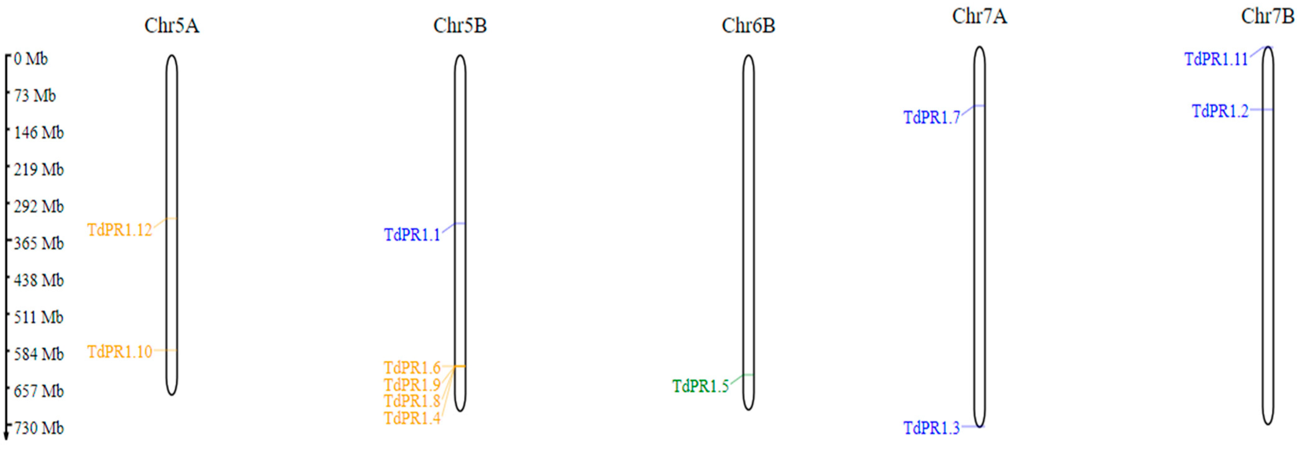
2.1. Identification, Distribution, Gene Structures, and Conserved Motifs of PR-1 in Triticum durum

After using BLASTp v 2.13.0 searches, the verification of the possession of CAP domain (cysteine-rich secretory proteins, antigen 5, and pathogenesis-related 1) (Figure 1), and the elimination of redundant sequences, twelve TdPR1 sequences were obtained from the Triticum durum genome (named TdPR1.1 to TdPR1.12). Moreover, the analyses of the exon–intron organization were performed to understand the evolution of the TdPR1 genes. We found that the selected PR-1 genes shared the same structure. In fact, only one gene (TdPR1.12) had two exons and one intron, whereas the other 11 TdPR1 genes possessed a single exon. Furthermore, no 5′-UTR or 3′-UTR region was detected in the gene sequences (Figure 1). By using the Multiple Em for Motif Elicitation (MEME) (version 5.5.1), five motifs were identified. Four of them were presented in all the selected TdPR1 proteins (Motif 1, 2, 3, and 4), whereas motif 5 (presented by red boxes) was missing in TdPR1.5 (Figure 1).
The genomic features of TdPR1 members are presented in Table 1. The distribution of TdPR-1 genes were on five different chromosomes. Two genes were located on Chr5A, Chr7A, and Chr7B, whereas only one gene was located on chromosome 6B. The rest (about a third of the selected genes) were clustered on chromosome 5B (Figure 2, Table 1).

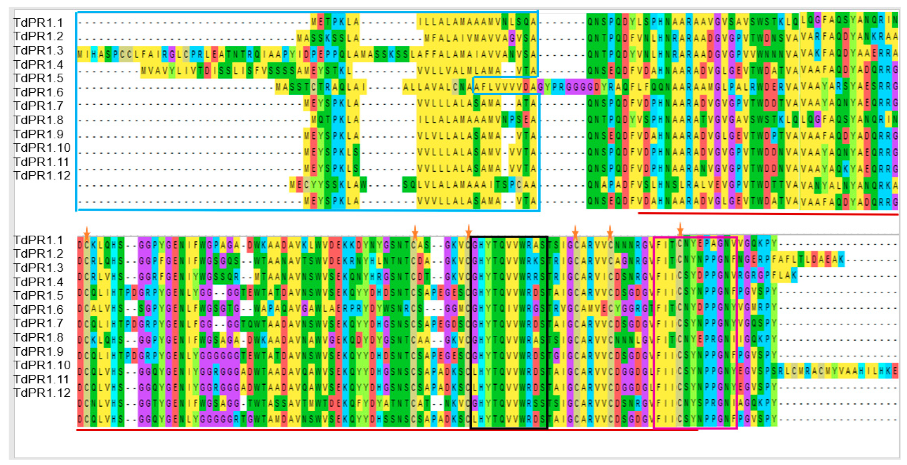
2.2. Multiple Alignment and Phylogenetic Relationship among the TdPR1 Genes

In order to understand the evolutional history of the candidate’s proteins, a phylogenetic tree was reconstructed. TdPR-1 genes were distributed into three groups: Group I comprising genes encoding all acid proteins, Group III included five basic proteins and the six-basic protein, and the only TdPR1.5 located on Chr6B were clustered in Group II. Our findings show that this gene did not have the conserved motif 5 and had a different cleavage site sequence CNA-AF (Table 2; Figure 3).
Based on the multiple alignment performed by the Muscle algorithm, we found that TdPR1 protein sequences are homologous. Indeed, the analysis indicated the presence of the Pfam0018 domain (CAP) in all the TdPR 1 proteins, (shown by red lines in the Figure 4). In addition, the blue box indicates the presence of the signal peptide (SP) regions (located at the N-terminal portion of the proteins; 24–31; Table 2). These peptides were found in all identified TdPR1 proteins except for TdPR1.3 and TdPR1.4. Moreover, only TdPR1.5 showed a cleavage site (between two alanine residues), whereas the others presented a common cleavage site between alanine (A) and glutamine (Q) amino-acid residues (Table 2).
According to the results given by the PROSITE database, TdPR1 sequences contain two conserved domain CRISPs (cysteine rich secretory proteins) in the C-terminal: CRISP family signature 1 (CRISP_1) (shown by black box Figure 4;) and CRISP family signature 2 (CRISP_2) (shown as pink box Figure 4; Supplemental Figure S1).

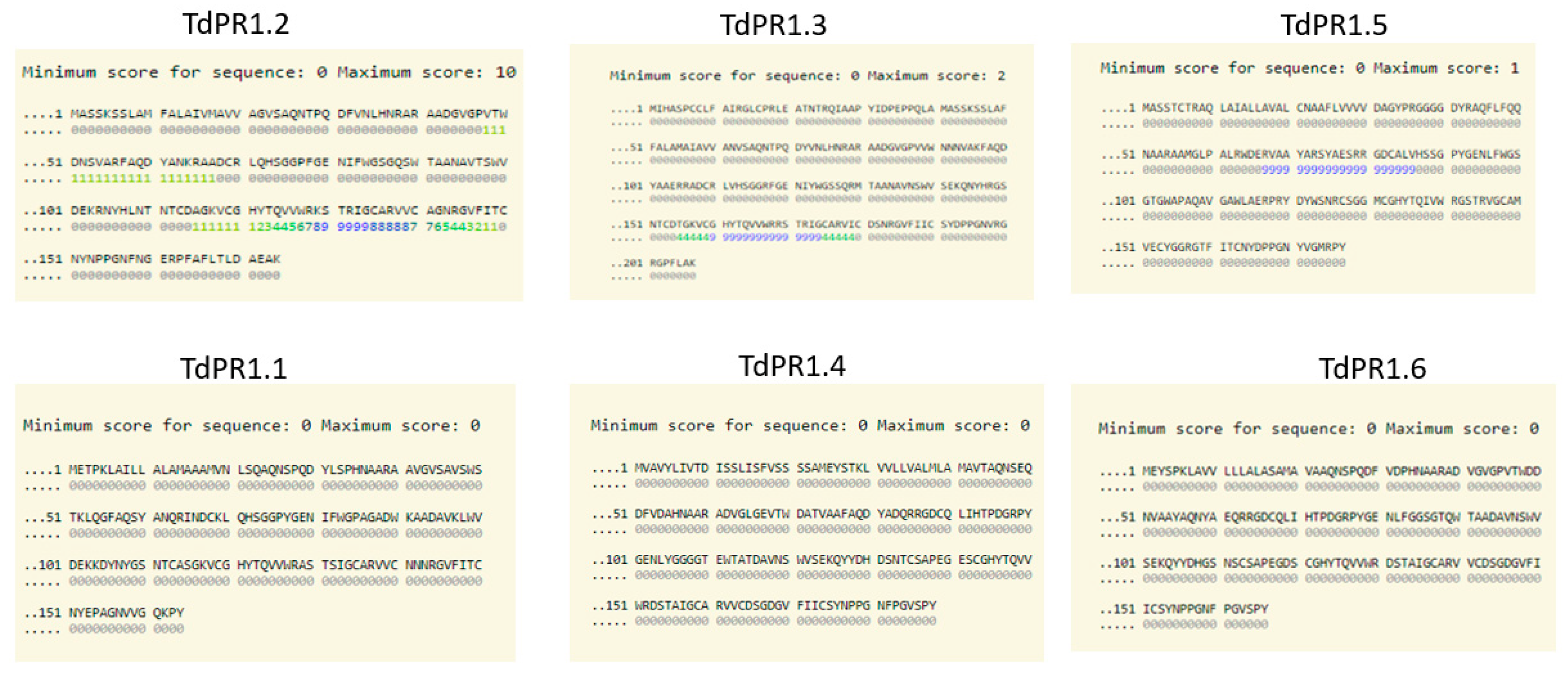
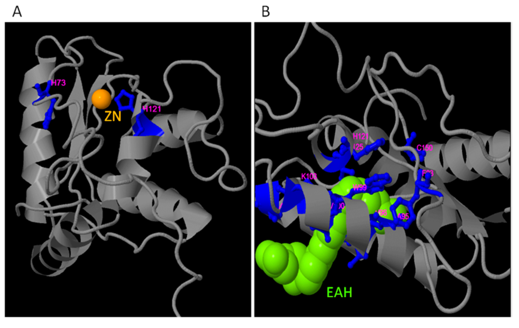
2.3. Identification of Putative Zn2+ and CaM Binding Domains

We have recently identified a CaM binding domain located at the C-terminal part of the protein that ensures the interaction of the CaM/Ca2+ complex with the TdPR1 protein [31]. To investigate whether other identified TdPR1s harbor such a domain, we analyzed the structure of the 12 TdPR1 using the calmodulin target database. As revealed in Figure 5, only TdPR1.2, TdPR1.3, and TdPR1.5 harbor calmodulin binding domains at the C-terminal portion of the protein, whereas all the other TdPR1 proteins do not have such domains in their structures (Figure 5).
On the other hand, COACH analysis revealed the presence of three putative zinc ligand binding sites in the TdPR1.2 protein. In fact, ion (Zn2+) and a zinc molecule EAH ((5S, 7E, 9E, 11Z, 14Z)-5-hydroxyicosa-7, 9, 11,14-tetraenoic acid) were predicted. Different amino acids are implicated in this binding as revealed by the server which are 73 and 121 aa for the Zn ion (PDB Hit: 3mz8A) (Figure 6A) and 35,39,42,83,95,96,99,100,103,121,125,150 amino acids implicated in a possible interaction with the molecule EAH (PDB Hit: 3u3uC; Figure 6B).

2.4. Physicochemical Properties

Several analyses showed the different characteristics of the TdPR1 proteins listed in Table 3. In fact, the amino acid lengths of different proteins were between 164 and 207 aa with a molecular weight (MW) for all proteins ranging from 17 to 19 kDa. Among these proteins, 50% are acidic (pI < 7) and the others are basic (pI > 7). The negative GRAVY index (Grand average of hydropathicity) indicates that all TdPR1 proteins are non-polar (hydrophilic) proteins [50]. In addition, the thermostability parameter of proteins is measured by the aliphatic index (AI). The high aliphatic index of TdPR1 proteins, ranging from 64.46 to 76.76, indicates that they are thermo-stable under a wide temperature range [51].

2.5. Prediction of Transmembrane Helices on TdPR1

The topological analysis shows the existence of the transmembrane helixes in all the TdPR1 proteins, except TdPR1.7. In fact, the three online tools indicated the presence of transmembrane domains in TdPR1.2; TdPR1.5; TdPR1.9, and TdPR1.10. The results also showed that 66.66% of the online servers predicted that TdPR1.1, TdPR1.3, TdPR1.4, TdPR1.6, TdPR1.8, and TdPR1.12 have the transmembrane domain, whereas one out of three online tools indicated that this transmembrane helix is present in TdPR1.11 with a percentage of 33.33 (Table 4).

2.6. Predicted Secondary and 3D Structures of TdPR-1 Proteins

Secondary structures of proteins were predicted using the SOPMA server. The percentage of α-helices, extended strand, β turn, and random coil were 30.92–40, 14.59–18.08, 2.44–6.78, and 40.36–48.17, respectively. Structural conformation of a protein can occurs via protein phosphorylation by adding the phosphate groups to serine, threonine, or tyrosine residues [52]. Using the NetPhos 3.1 server, phosphorylation sites were predicted in TdPR1 proteins and ranged between 11 and 22 (Figure 7). The predicted channel number found in the studied TdPR1 proteins were registered between 0 and 16 (Figure 7).
The predicted 3D structures of the TdPR1 proteins were constructed using the alpha fold online server. The 3D TdPR-1 proteins models presented four α-helices and several antiparallel β-sheets. According to the CASTp 3.0 analysis, molecular pockets were identified in all candidates. The top three predicted pockets, with the largest volume, are indicated as red, blue, and yellow, respectively (Figure 8).

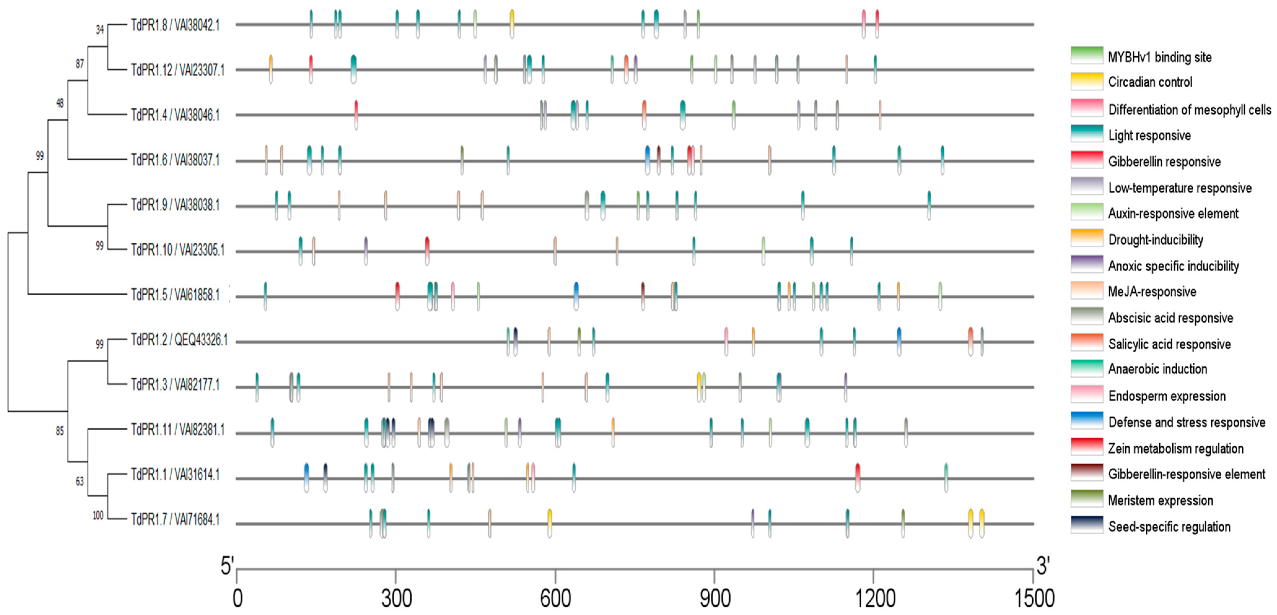
2.7. In Silico Analysis of Cis-Elements

The cis-acting element in the TdPR1 promoters could be divided into four categories, such as development-related elements, environmental stress-related elements, hormone-responsive elements, and transcription factor binding sites, based on the biological function analyzed by Plantcare (Figure 9 and Figure 10). Stress responsive cis-elements were found in most of the PR-1 promoters except for TdPR1.9. Cis-regulatory elements belonging to this class are associated with drought, anaerobic anoxic specific inducible element, and low-temperature response. All TdPR1 regulatory regions have more than four cis-element hormone responses. The MeJA- and abscisic acid responsive motifs are the most abundant in most of TdPR1. Thus, ABRE (abscisic acid response elements) are the elements which had the highest motif number in the TdPR1 promoter region (12 motifs in TdPR1.8). Salicylic acid, auxin, and gibberellin responsive elements were found in some TdPR1 promoters. Moreover, different cis-acting regulatory elements involved in growth and development are present. The most abundant motifs are related to light response as all the identified TdPR1 proteins presented a diversity of light response elements. G-box is the only motif present in all the identified promoters with a maximum number registered for TdPR1.8 (11 G-boxes). In addition, endosperm expression, seed-specific regulation, meristem expression, and circadian control are found in the majority of PR-1 in Triticum durum. The binding domains of a single TF family, MYB, were identified in four, eight, nine, and twelve TdPR1 promoters.

2.8. Predicting TdPR1 Pproteins Subcellular Localization

The subcellular localization of TdPR1 proteins performed by the online tool WoLF PSORT and represented by a heatmap graphic using Tbtools v1.108 are shown in Figure 11. Indeed, with a high level of prediction, 10 TdPR1 proteins were found to be located in the extracellular compartment. This subcellular location of PR1 is not only identified in the extracellular space but could also be in the extracellular, Vacuole, endoplasmic reticulum, endoplasmic reticulum plasma membrane, cytoplasm, and mitochondria.

2.9. Gene Ontology (GO) Term Distribution of Triticum durum PR-1

Gene ontology (GO) analyses performed by three different servers showed variable results on biological process, molecular function, and cellular component terms for 12 TdPR1 proteins (Figure 12). Based on CELLO2GO data, three molecular functions were identified: enzyme regulator activity, ion binding, and lipid binding. The PredictProtein tool predicted that 8% of the deduced PR1 had only a chitinase activity and 11 out of 12 (91.6%) had both functions: chitinase activity and chitin binding (Figure 12A). No result was obtained with PANNZER2. In terms of cellular components, CELLO2GO, PANNZER2, and PredictProtein identified that TdPR1 proteins could be located in the extracellular region with a percentage of 20, 100, and 46, respectively (Figure 12B). The biological processes regulated by all TdPR1 proteins (91.6–100%) were the response to biotic stimulus (GO:0009607) identified by the different web servers. Based on the GO enrichment analysis by PANNZER2 and PredictProtein servers, most of the PR-1 genes are implicated in defense responses (GO:0006952) (33.3%, 91.6%, respectively). Four other biological processes were found in CELLO2GO, namely transport (100%), extracellular matrix organization 100%), reproduction (66.6%), and immune system process (8.3%). Moreover, three other Gene Ontology terms were identified by the PredictProtein tool in one out of twelve TdPR1 proteins (8.3%): systemic acquired resistance (GO:0009627), response to water deprivation (GO:0009414), and response to vitamin B1 (GO:0010266) (Figure 12C).

2.10. Differential Expression of TdPR1.2 Gene under Various Stress Conditions

The full-length cDNA sequence of TdPR1.2 (GenBank accession no. MK570869.1) was previously identified by our group [31]. To investigate the possible biological functions of the TdPR1.2 gene, we assessed the expression patterns of TdPR1.2 genes in wheat under various abiotic stress conditions using qRT–PCR (Figure 13 and Figure 14).
In response to salt stress (150 mM NaCl), TdPR1.2 was significantly upregulated (Figure 13A). The same result was observed when plants were subjected to mannitol and PEG stresses (Figure 13B,C). When heat stress was applied to wheat plants (42 °C for 30 min), there was a significant increase in TdPR1.2 expression level in the roots and shoots, suggesting that this protein could have a putative protective role in controlling wheat heat tolerance (Figure 13D).
The hormonal response of TdPR1.2 gene was investigated by treating plants with salicylic acid (SA), indole acetic acid (IAA), jasmonic acid (JA), and abscisic acid (ABA). As shown in Figure 14, TdPR1.2 was upregulated in response to all hormones used in this work. Overall, these results demonstrate that TdPR1.2 is implicated in plant response to many abiotic and hormonal stresses.

3. Discussion

Different studies have shown that PR1 proteins play a crucial role in plants’ responses to different diseases that affect common wheat. In fact, the overexpression of TaPR1-7 enhanced plant resistance to infection by Puccinia striiformis f. sp. tritici (Pst) (Stripe (yellow) rust) [53], while TcLr19PR1 [54] and TaLr35PR1 genes [55] were induced after plant infection with Puccinia triticina (leaf rust) attack. Meanwhile, TaPR1a was highly expressed and caused plant resistance to both diseases (stripe (yellow) rust and leaf rust) [56]. Previously, we have demonstrated that the newly isolated gene, TdPR1.2, presented an antibacterial and antifungal activity in vitro. Interestingly, TdPR1.2 presented a positive effect in inhibiting Septoria tritici growth in vitro [31]. Moreover, pathogenesis-related protein 1 could positively interact with other PR families. Wang et al. [57] found that TaTLP1 and TaPR1 interacted physically to protect wheat plant from leaf rust.
Furthermore, PR protein families were activated by Septoria tritici blotch disease. For instance, PR1 and PR3 were upregulated in Sevin cultivar after plant infection with Septoria tritici [58], whereas in Wangshubai, it has been demonstrated that such infection causes the upregulation of PR1 and peroxidase genes [59]. Finally, Septoria tritici infection induced the upregulation of PR-1 in Seri 82 and Frontana cultivars, respectively [60].
While different studies have investigated the role of TaPR1 proteins in plants, little is known about its homologue in durum wheat, TdPR1.2. Therefore, the genetic richness of durum wheat PR-1 genes, its molecular functions in response to abiotic and biotic stresses, is a crucial topic to understand and resolve, not only for the agronomic problems of wheat, but also for the economics and marketing of the country.
Several researchers are emphasising the identification and characterization of PR-1 proteins in different plants. They state that most PR-1s are often encoded by multi-gene families as identified in many plant species such as black pepper [19], tomato [7], and the two genomes of the banana plant [24]. In this work, we identified 12 PR-encoding genes in durum wheat genome which is less than the number of genes identified in common wheat [18], soybean [20], rice, and Arabidopsis thaliana [16] suggesting that there is no correlation between the plant genome size and the identified PR-1 members [24]. As revealed in this work, the twelve identified TdPR1 genes were distributed on five chromosomes. Two genes were present on Chr5A, Chr7A, and Chr7B, whereas only one gene was located on chromosome 6B. The rest (about a third of the selected genes) were clustered in chromosome 5B (Figure 2, Table 1). Liu and Xue (2006) [61] explained the clustering of genes in one chromosome by the tandem gene duplication events which could occur during evolution.
A phylogenetic tree was performed by the MEGA11 software. The TdPR1 genes were subdivided into three phylogenetic groups: Group 1 (six acidic TdPR1), Group 2 (one basic TdPR1) and Group 3 (five basic TdPR1). The division of pathogenesis-related protein-1 genes into three groups based on protein isoforms (acidic/basic) have also been shown in previous studies, such as in rice [61]. The same result was found in common wheat, which has one of the biggest genomes (>16,000 megabases) among monocot agricultural plants and presented 23 PR-1 genes. The 23 TaPr-1 genes all have intron-free open reading frames that express a signal peptide at the N-terminus and a conserved PR-1-like domain, according to a sequence study of the genes. According to phylogenetic analysis, TaPr-1 genes, along with their counterparts in other monocots, form three major monophyletic groups; each group contains genes that encode basic, basic with a C-terminal extension, and acidic PR-1 proteins, respectively. This suggests that the functions of PR-1 genes in monocot plants are diverse and conserved [18]. In contrast, in sugarcane and tomato plants, pathogenesis-related proteins were clustered in the same group independently of their type (basic/acid) [7,23]. The gene structure analyses identified the presence of a single gene and a lack of intron in all TdPR1 except TdPR1.12 which presented two exons. In accordance with other plants, such as sugarcane (Saccharum spontaneum) [23], tea plant (Camellia sinensis (L.) O. Kuntze) [17], and banana (Musa balbisiana (DH-PKW)) [24], the majority of genes had only one exon and lacked introns (13 out of 19 ScPR1, 15 out of 17 CsPR1, and 10 out of 11 MaPR1, respectively). Previously, scientists have claimed that, during evolution, genes which were activated rapidly to respond to stresses were likely to decrease their intron density [62]. Zhang and his colleagues found that the expression of the CsPR1 gene, which does not have introns, were expressed more rapidly than genes with three introns in plants subjected to blight disease stress [17].
The majority of motifs are conserved among all TdPR1 proteins. The sequence alignment of the deduced protein presents two conserved domains belonging to the CRISP family. Previous studies have demonstrated that CRISPs may be involved in the plant to acquire resistance to biotic and abiotic stresses [63]. Moreover, the first isolated TdPR1.2 was aligned with eight PR-1 sequences from different species, demonstrating that the cysteine residues and the CRISP_1 domain are highly conserved in both monocotyledonous and dicotyledonous plants, whereas CRISP_2 is less conserved [11,31]. Moreover, all PR-1 proteins presented six well conserved cysteines residues [11,31,61]. These results are consistent with our findings and all the identified TdPR1 genes presented the conserved residues (Figure 1), suggesting a common molecular function of all identified PR-1 genes. In addition, the analysis of the TdPR1 gene structure and the conserved motifs indicates that TdPR1 genes are highly conserved, as demonstrated by the number of α and β helices and by the presence of signaling peptides. Such findings suggest that all the encoding proteins present the same function under environmental stress [19,24,64].
Numerous cis-elements were identified in TdPR1 genes (Figure 10) involved in stress, development, and hormone response. Additionally, the MYB transcription factors (TFs), who had a detectable binding domain in four TdPR1 genes, have a different role, such as plant growth and development, physiological activity, primary and secondary metabolic reactions, and responses to biotic and abiotic stresses [65], suggesting the implication of these proteins in plant metabolism and development. Since our promoter regions possess a myriad of cis-elements, we suggest that the identified genes could be implicated in different molecular and metabolic pathways.
The signal peptide plays an important role in the guiding of proteins into subcellular spaces [66]. Based on the results obtained by the WoLF PSORT web server, the two proteins which do not contain the signal peptide (TdPR1.3 and TdPR1.4) are more probably localized in chloroplast and vacuole, respectively. The PR-1 gene family identified in other plants may prove our suggestion. In fact, scientists have stated that pathogenesis-related proteins in grape and wheat containing the signal peptide could be secreted into the extracellular compartment [17,18,21]. However, in tea (Camellia sinensis (L.) O. Kuntze) and black pepper (Piper nigrum), PR-1 proteins without SPs were found in the intracellular compartment [17,19]. The acquisition of SP at the N terminal sequences might be affecting the subcellular localization of proteins. Therefore, TdPR1 proteins containing the signal peptide at the N terminal assume their guidance into the extracellular compartment in order to guarantee their cellular functions.
Putative ligand prediction shows that TdPR1.2 possess binding sites to Zn2+ and EAH, similarly to the HbPR-1 protein from the rubber tree (Hevea brasiliensis), which had imbibed in its structure two binding sites of those ligands plus a glycerol binding domain [67]. Interestingly, no glycerol binding sites were detected in TdPR1.2 proteins.
Recently, a CaM binding domain was identified by our group and in the C-terminal part of the TdPR1.2 [31]. TdPR1.2 interacts with CaMs in a calcium-dependent manner and this interaction enhances TdPR1.2 activity, especially in the presence of Mn2+ cations [31]. To further identify whether the other identified TdPR1 genes harbors a CaMBD in their structures, we performed in silico analysis using calmodulin target databases. In the sequence of three different PR proteins (PR1.2; PR1.3; and PR1.50), putative calmodulin-binding domains were identified. In these proteins, this domain is located at the C-terminal portion of the protein sequence. It was noted that this domain is necessary for calmodulin binding and the calcium-dependent activation of various plant proteins such as catalases [68,69], PRs [31], and mitogen-activated protein kinase phosphatases [70,71].
Calmodulins (CaMs) are omnipresent, tiny proteins with only four Ef-Hand motifs, which are common components of Ca2+ binding proteins [68]. The main intracellular Ca2+ signaling pathways are mediated by CaMs, and an increase in the amount of Ca2+ in the nucleus or cytosol leads to the formation of Ca2+/CaM complexes, which interact with a wide range of targets, including ion transporters, protein kinases, pathogen-related proteins, transcription factors, and protein phosphatases, and control cellular functions [9,70,72,73].
In this study, TdPR1.2 was upregulated in the leaf and root tissues of durum wheat subjected to NaCl (150 mM) and PEG (10% PEG 6000) treatments as previously shown in other studies such as in Zea mays (ZmPR-1; [74]), banana [24], tomato (13 SlPR-1 genes; [7]), Vitis vinifera (VvPR-1; [75]), and rice (OsPR1a; [76]), suggesting that this genes could have a dual role depending on the tissue expression in the plant.
It has been described in the literature that the SA and JA signaling pathways are stimulated after biotrophic/hemibiotrophic (under the control of SA) and necrotrophic (under the control of JA) pathogen infection [5]. Thus, we investigated the effect of SA and JA application on TdPR1.2 gene expression in wheat. Our results showed that TdPR1.2 was upregulated after application of those phytohormones in the roots and shoots of wheat. Other PR-1 proteins were reported to be upregulated after plant treatment with SA and JA [7,77]. In banana, MaPR1-1 was upregulated after plant treatment with SA and JA stresses due to the presence of cis-elements and binding sites for transcription factors [24]. Thus, the identification of the stress-responsive elements involved in the up/downregulation of PR-1 will help in understanding plants’ resistance mechanisms toward various stresses. These findings strongly suggest that the TdPR1.2 gene plays a crucial role in plant defense against environmental stresses. It has been suggested that PR-1 genes can serve as molecular markers associated with resistance to different biotic and abiotic stresses [5,19,78]. Thus, our findings could be useful for breeding programs aimed at increasing the resistance of wheat crops to salt, drought, and hormonal stresses as well as plant infection with pathogens.

4. Materials and Methods

4.1. Identification of PR-1 Genes from the T. durum Genome

In order to identify the genomic family members of the PR-1 genes, the Pfam’s CAP domain (PF00188), downloaded from the Pfam database version 35.0 (https://pfam.xfam.org/, accessed on 5 November 2022) [79], was used as a query for search against the Triticum durum genome (taxid:4567) by using Blastp program v 2.13.0. After the realisation of the homology search, only predicted PR-1 sequences with an e-value <10–10 were selected. The redundancy in the resulted sequence’s collection was removed by the decrease in redundancy (https://web.expasy.org/decrease_redundancy/; accessed on 8 November 2022) program. Selected sequences were then analysed using SMART database (http://smart.embl-heidelberg.de/, accessed on 8 November 2022) [80], PfamScan (https://www.ebi.ac.uk/Tools/pfa/pfamscan/; accessed on 8 November 2022) [81] and NCBI’s Conserved Domain Search v3.20 (http://www.ncbi.nlm.nih.gov/cdd/; accessed on 8 November 2022) [82]. The selected TdPR1 sequence possession of the conserved CAP domain was drawn by Tbtools v1.108 (Graphi [83]).

4.2. Sequence Analysis

Physiochemical properties of TdPR1 proteins were calculated with ProtParam program (https://web.expasy.org/protparam/; accessed on 12 November 2022) [84]. The possession of the signal peptide for the protein sequences was identified by SignalP 5.0 Server (https://services.healthtech.dtu.dk/service.php?SignalP-5.0; accessed on 13 November 2022) [85]. The presence of Zinc binding domain was revealed by COACH web server to determine the putative ligands abled to interact in the binding site of TdPR1.2 protein [86,87]. The presence of putative calmodulin binding sites was also revealed by Calmodulin target database (http://calcium.uhnres.utoronto.ca/ctdb/pub_pages/general/index.htm; accessed on 21 December 2022) [88].

4.3. Topological Analysis

The numbers of the transmembrane helices of each PR proteins were predicted by using various online servers such as TMHMM-2.0 (https://services.healthtech.dtu.dk/service.php?TMHMM-2.0; accessed on 5 January 2023) [89], SOSUI version 1.11 (http://harrier.nagahama-i-bio.ac.jp/sosui; accessed on 2 January 2023) [90], and TMDET version 2.0 (http://tmdet.enzim.hu/; accessed on 3 January 2023) [91].

4.4. Secondary and Tertiary Structure Prediction

By using SOPMA online server (Self-Optimized Prediction Method with Alignment) (https://npsa-prabi.ibcp.fr/cgi-bin/npsa_automat.pl?page=/NPSA/npsa_sopma.html; accessed on 26 December 2022), the secondary structure of the TdPR-1 proteins was predicted [92]. The putative number of the predicted phosphorylation sites in TdPR1 were identified by NetPhos 3.1 server (http://www.cbs.dtu.dk/services/NetPhos/; accessed on 5 January 2023) [93,94]. The 3D structures of the TdPR-1 proteins were predicted using the Alphafold online server v2. 0 (https://alphafold.ebi.ac.uk/; accessed on 27 December 2022) [95,96]. The BetaCavityWeb server (http://voronoi.hanyang.ac.kr/betacavityweb; accessed on 10 January 2023) was used to predict the number of channel structural [97]. The CASTp 3.0 (Computed Atlas of Surface Topography of proteins) online server (http://sts.bioe.uic.edu/castp/calculation.html; accessed on 27 December 2022) was used to predict the active site pockets of the TdPR-1 protein [98].

4.5. Conserved Motif, Multiple Alignment, and Phylogenetic Tree

ScanProsite online server were used to scan Protein domains and functional sites (http://prosite.expasy.org/prosite.html; accessed on 10 November 2022) [99] and the logo of the motifs signature was drawn by WebLogo version 2.8.2 (https://weblogo.berkeley.edu/logo.cgi; accessed on 14 January 2023) [100,101]. MEME server v5.1.1 (http://meme-suite.org/tools/meme; accessed on 12 November 2022) online tool were used to predict the conserved motif in TdPR1 [102], then the motifs were visualized by TBtools software v1.108 (http://github.com/CJ-Chen/TBtools) [83]. TdPR1 protein sequences were aligned by a multiple sequence alignment using MUSCLE program (accessed on 12 November 2022) [103], and the phylogenetic tree of the aligned TdPR-1s sequences was constructed with the Maximum-Likelihood and 1000 bootstrap values using the MEGA11 software (accessed on 12 November 2022) [104].

4.6. Cis-Elements, Chromosomal Locations, and Gene Structure Analyses

Promoters were identified as the 1.5 kb upstream the start codon region, and were downloaded from NCBI database (http://www.ncbi.nlm.nih.gov/; accessed on 23 January 2023). Analysis of cis-acting regulatory elements of promoter sequences were performed by PlantCARE online tools (http://bioinformatics.psb.ugent.be/webtools/plantcare/html/; accessed on 23 January 2023) [105] and visualized by TBtool software v1.108 (accessed on 23 January 2023) [83]. The chromosome length data were collected from the NCBI and used to visualize the genomic distribution of PR1 genes in durum wheat by using the online software MG2C v2.1 (http://mg2c.iask.in/mg2c_v2.1/; accessed on 19 November 2022) [106,107]. The identified PR-1 genes and coding sequences (CDS) features were obtained from NCBI, then their exon-intron structures were retrieved using the Gene Structure Display Server program 2.0 (http://gsds.gao-lab.org/; accessed on 19 November 2022) [108].

4.7. Subcellular Localization and Gene Ontology Analysis

Subcellular location was predicted using WoLFPSORT online tool (http://wolfpsort.seq.cbrc.jp/; accessed on 27 January 2023) [109]. Gene ontology (GO) analysis was performed by three different online tools, such as PANNZER2 (http://ekhidna2.biocenter.helsinki.fi/sanspanz/; accessed on 27 January 2023) [110], CELLO2GO (http://cello.life.nctu.edu.tw/cello2go/; accessed on 27 January 2023) [111], and PredictProtein (http://ppopen.rostlab.org; accessed on 27 January 2023) [112].

4.8. Plant Material and Stress Treatments

In this work, seeds of wheat (Triticum durum Desf.) (cv. Om Rabiaa) were sterilized in each box containing 30 mL of 0.6% NaClO solution for 15 min, then washed five times with 50 mL sterile water. For each treatment, 45 seeds were placed in each Petri dish (11 cm long, 2.5 cm high, and 11 cm wide) in the presence of a sponge and filter paper placed below to maintain moisture at 25 ± 2 °C. Seeds were then transferred to a greenhouse at 24 ± 2 °C, with photosynthetically active radiation of 280 μmol m−2 s−1, a 16 h photoperiod, and 60 ± 10% relative humidity. After 10 days, seedlings were subjected to stresses. In this study, nine treatments were used including the control (distilled water), 150 mM NaCl, 10% PEG, 200 mM mannitol, 5 mM of each phytohormone (SA, JA, IAA, and ABA), and heat (42 °C). Each treatment was replicated three times. Finally, shoots were harvested and immediately frozen in liquid nitrogen and stored at −80 °C.

4.9. RNA Extraction and Quantitative Real-Time Reverse Transcription PCR (qRT-PCR)

Total RNA was extracted from individual roots and leaves (0.5 g of each tissue) using the RNeasy Plant Mini Kit (QIAGEN, Hilden, Germany). Extracted RNA was then purified from genomic DNA (RNase free DNase set; QIAGEN), qualified by gel electrophoresis, and used for first-strand cDNA synthesis (GoScript Reverse Transcription System; Promega, Madison, USA) with an oligo-dT primer. PCR reactions were achieved in a 10 μL final volume tube in the presence of 3 μL cDNA (obtained from 40 ng of DNase-treated RNA), 0.5 μL of each primer of the TdPR1.2 gene at 10 μM (TdPR_Fw: 5′-ATGGCATCT TCCAAGAGT-3′ and TdPR_Rv: 5′-TCA AGG GTG AGG ACG CGA A-3), 5 μL 2 × SYBR Green I master mix, and 1 μL of RNase-free water (Sigma). The reaction consisted of an initial denaturation at 95 °C for 5 min followed by 40 cycles composed of 10 s at 95 °C, 20 s at 60 °C, and 30 s at 72 °C, then a melting curve (5 s at 95 °C, 1 min at 65 °C, and 5 min with the temperature increasing from 65 to 97 °C). Three biological repetitions were performed for each experimental condition, with three technical repetitions for each sample. Melting curve analysis at the end of cycling was used to verify whether there was single amplification. At the end of the reaction, the threshold cycle (CT) values of the triplicate PCRs were averaged and used for transcript quantification. The relative expression ratio of the TdPR1.2 gene was calculated by using the comparative CT method with the actin gene designed from the T. aestivum genome (actin_Fw: 5′-TCC CTC AGC ACA TTC CAG CAGAT-3 and actin_Rv: 5′-AAC GAT TCC TGG ACC TGC CTC ATC-3′) as an internal expression standard [113]. The relative expression level was calculated from triplicate measurements based on the 2−ΔΔCT, where ΔΔCT = (CT, target gene−CT, actin) stressed − (CT, target gene−CT, actin) control. Relative expression ratios from three independent experiments (three biological repetitions) are reported.

4.10. Statistical Analysis

Data are reported as mean ± S.E. The results were compared statistically by using Student’s t test, and differences were considered significant at p < 0.01.

5. Conclusions

PR 1 proteins play important roles in growth regulation, development, and plant response to biotic and abiotic stress. Here, plenty of in silico tools were used to enhance our comprehensive understanding of the PR-1 family in Triticum durum plants. In fact, twelve TdPR1 were identified and clustered into three phylogenetic groups. Gene structure, conserved motifs, and physicochemical properties showed that TdPR1 proteins present highly conserved structures. Moreover, a myriad of cis-elements in the up/down stream of TdPR1 genes were found and may act in the gene expression to stress responses, hormones, and growth in durum wheat. Following cis-regulatory elements of the promoter regions, TdPR1 are clustered on chromosomes Chr5A, Chr5B, Chr6B, Chr7A, and Chr7B. All the predicted PR1 proteins identified in this work were reported to be extracellular proteins. Collectively, the findings of this study will contribute to a better understanding of molecular mechanisms and provide useful and essential information for the further functional characterization of TdPR-1 genes in future research.

Supplementary Materials

The following supporting information can be downloaded at: https://www.mdpi.com/article/10.3390/plants12101998/s1, Figure S1: The two CRISP domains conserved region signature generated by WebLogo version 2.8.2. (A) CRISP-1 logo and (B) CRISP-2 logo signature.

Author Contributions

Conceptualization, I.Z. and F.B.; funding acquisition, M.G.; investigation, I.Z. and M.G.; methodology, I.Z., M.G. and F.B.; software, M.G., N.H. and M.B.; supervision, F.B.; validation, F.B.; writing—original draft, I.Z.; writing—review and editing, M.G. and F.B. All authors have read and agreed to the published version of the manuscript.

Funding

This research has been funded by Scientific Research Deanship at the University of Ha’il, Saudi Arabia through project number RG-20203.

Institutional Review Board Statement

Not applicable.

Informed Consent Statement

Not applicable.

Data Availability Statement

The data generated and analyzed during this study are included in this article.

Acknowledgments

This research has been funded by the Scientific Research Deanship at the University of Ha’il, Saudi Arabia through project number RG-20203.

Conflicts of Interest

The authors declare no conflict of interest.

References

  1. Ahmad, B.; Raina, A.; Khan, S. Biotic and Abiotic Stresses, Impact on Plants and Their Response. In Disease Resistance in Crop Plants; Springer: Berlin/Heidelberg, Germany, 2019. [Google Scholar]
  2. Gull, A.; Lone, A.A.; Wani, N.U.I. Biotic and Abiotic Stresses in Plants. Abiotic Biot. Stress Plants 2019, 1–19. [Google Scholar] [CrossRef]
  3. Jwa, N.-S.; Agrawal, G.K.; Rakwal, R.; Park, C.-H.; Agrawal, V.P. Molecular Cloning and Characterization of a Novel Jasmonate Inducible Pathogenesis-Related Class 10 Protein Gene, JIOsPR10, from Rice (Oryza sativa L.) Seedling Leaves. Biochem. Biophys. Res. Commun. 2001, 286, 973–983. [Google Scholar] [CrossRef]
  4. Breen, S.; Williams, S.J.; Outram, M.; Kobe, B.; Solomon, P.S. Emerging Insights into the Functions of Pathogenesis-Related Protein 1. Trends Plant Sci. 2017, 22, 871–879. [Google Scholar] [CrossRef] [PubMed]
  5. Zribi, I.; Ghorbel, M.; Brini, F. Pathogenesis Related Proteins (PRs): From Cellular Mechanisms to Plant Defense. Curr. Protein Pept. Sci. 2021, 22, 396–412. [Google Scholar] [CrossRef]
  6. Punja, Z.K. Genetic Engineering of Plants to Enhance Resistance to Fungal Pathogens—A Review of Progress and Future Prospects. Can. J. Plant Pathol. 2001, 23, 216–235. [Google Scholar] [CrossRef]
  7. Akbudak, M.A.; Yildiz, S.; Filiz, E. Pathogenesis Related Protein-1 (PR-1) Genes in Tomato (Solanum lycopersicum L.): Bioinformatics Analyses and Expression Profiles in Response to Drought Stress. Genomics 2020, 112, 4089–4099. [Google Scholar] [CrossRef]
  8. Agrawal, G.K.; Jwa, N.-S.; Rakwal, R. A Novel Rice (Oryza sativa L.) Acidic PR1 Gene Highly Responsive to Cut, Phytohormones, and Protein Phosphatase Inhibitors. Biochem. Biophys. Res. Commun. 2000, 274, 157–165. [Google Scholar] [CrossRef]
  9. Ghorbel, M.; Zribi, I.; Haddaji, N.; Besbes, M.; Bouali, N.; Brini, F. The Wheat Pathogenesis Related Protein (TdPR1. 2) Ensures Contrasting Behaviors to E. coli Transformant Cells under Stress Conditions. Adv. Microbiol. 2021, 11, 453–468. [Google Scholar] [CrossRef]
  10. Alhudaib, K.; Alanazi, N.; Ghorbel, M.; El-Ganainy, S.; Brini, F. Isolation and Characterization of a Novel Pathogenesis-Related Protein-1 Gene (AvPR-1) with Induced Expression in Oat (Avena sativa L.) during Abiotic and Hormonal Stresses. Plants 2022, 11, 2284. [Google Scholar] [CrossRef]
  11. Mou, Z.; Fan, W.; Dong, X. Inducers of Plant Systemic Acquired Resistance Regulate NPR1 Function through Redox Changes. Cell 2003, 113, 935–944. [Google Scholar] [CrossRef]
  12. Van Loon, L.; Pierpoint, W.; Boller, T.; Conejero, V. Recommendations for Naming Plant Pathogenesis-Related Proteins. Plant Mol. Biol. Rep. 1994, 12, 245–264. [Google Scholar] [CrossRef]
  13. Kaur, A.; Kaur, S.; Kaur, A.; Sarao, N.; Sharma, D. Pathogenesis-Related Proteins and Their Transgenic Expression for Developing Disease-Resistant Crops: Strategies Progress and Challenges; IntechOpen: London, UK, 2022. [Google Scholar] [CrossRef]
  14. Gibbs, G.M.; Roelants, K.; O’bryan, M.K. The CAP Superfamily: Cysteine-Rich Secretory Proteins, Antigen 5, and Pathogenesis-Related 1 Proteins—Roles in Reproduction, Cancer, and Immune Defense. Endocr. Rev. 2008, 29, 865–897. [Google Scholar] [CrossRef] [PubMed]
  15. Van Loon, L.; Van Kammen, A. Polyacrylamide Disc Electrophoresis of the Soluble Leaf Proteins from Nicotiana Tabacum Var.‘Samsun’ and ‘Samsun NN’: II. Changes in Protein Constitution after Infection with Tobacco Mosaic Virus. Virology 1970, 40, 199–211. [Google Scholar] [CrossRef]
  16. Van Loon, L.; Rep, M.; Pieterse, C.M.J. Significance of Inducible Defense-Related Proteins in Infected Plants. Annu. Rev. Phytopathol. 2006, 44, 135162. [Google Scholar] [CrossRef]
  17. Zhang, Q.; Guo, N.; Zhang, Y.; Yu, Y.; Liu, S. Genome-Wide Characterization and Expression Analysis of Pathogenesis-Related 1 (PR-1) Gene Family in Tea Plant (Camellia sinensis (L.) O. Kuntze) in Response to Blister-Blight Disease Stress. Int. J. Mol. Sci. 2022, 23, 1292. [Google Scholar] [CrossRef] [PubMed]
  18. Lu, S.; Friesen, T.L.; Faris, J.D. Molecular Characterization and Genomic Mapping of the Pathogenesis-Related Protein 1 (PR-1) Gene Family in Hexaploid Wheat (Triticum aestivum L.). Mol. Genet. Genom. 2011, 285, 485–503. [Google Scholar] [CrossRef]
  19. Kattupalli, D.; Srinivasan, A.; Soniya, E.V. A Genome-Wide Analysis of Pathogenesis-Related Protein-1 (PR-1) Genes from Piper Nigrum Reveals Its Critical Role during Phytophthora Capsici Infection. Genes 2021, 12, 1007. [Google Scholar] [CrossRef] [PubMed]
  20. Almeida-Silva, F.; Venancio, T.M. Pathogenesis-Related Protein 1 (PR-1) Genes in Soybean: Genome-Wide Identification, Structural Analysis and Expression Profiling under Multiple Biotic and Abiotic Stresses. Gene 2022, 809, 146013. [Google Scholar] [CrossRef]
  21. Li, Z.T.; Dhekney, S.A.; Gray, D.J. PR-1 Gene Family of Grapevine: A Uniquely Duplicated PR-1 Gene from a Vitis Interspecific Hybrid Confers High Level Resistance to Bacterial Disease in Transgenic Tobacco. Plant Cell Rep. 2011, 30, 1–11. [Google Scholar] [CrossRef]
  22. Edreva, A. Pathogenesis-Related Proteins: Research Progress in the Last 15 Years. Gen. Appl. Plant Physiol. 2005, 31, 105–124. [Google Scholar]
  23. Chu, N.; Zhou, J.-R.; Rott, P.C.; Li, J.; Fu, H.-Y.; Huang, M.-T.; Zhang, H.-L.; Gao, S.-J. ScPR1 Plays a Positive Role in the Regulation of Resistance to Diverse Stresses in Sugarcane (Saccharum spp.) and Arabidopsis thaliana. Ind. Crops Prod. 2022, 180, 114736. [Google Scholar] [CrossRef]
  24. Anuradha, C.; Arumugam, C.; Suthanthiram, B.; Raman, T.; Giribabu, P.; Uma, S. Genome-Wide Analysis of Pathogenesis-Related Protein 1 (PR-1) Gene Family from Musa spp. and Its Role in Defense Response during Stresses. Gene 2022, 821, 146334. [Google Scholar] [CrossRef] [PubMed]
  25. Chassot, C.; Nawrath, C.; Métraux, J. Cuticular Defects Lead to Full Immunity to a Major Plant Pathogen. Plant J. 2007, 49, 972–980. [Google Scholar] [CrossRef]
  26. Shin, S.H.; Pak, J.-H.; Kim, M.J.; Kim, H.J.; Oh, J.S.; Choi, H.K.; Jung, H.W.; Chung, Y.S. An Acidic Pathogenesis-Related1 Gene of Oryza Grandiglumis Is Involved in Disease Resistance Response against Bacterial Infection. Plant Pathol. J. 2014, 30, 208. [Google Scholar] [CrossRef]
  27. Schneiter, R.; Di Pietro, A. The CAP Protein Superfamily: Function in Sterol Export and Fungal Virulence. Biomol. Concepts 2013, 4, 519–525. [Google Scholar] [CrossRef]
  28. Chen, Y.-L.; Lee, C.-Y.; Cheng, K.-T.; Chang, W.-H.; Huang, R.-N.; Nam, H.G.; Chen, Y.-R. Quantitative Peptidomics Study Reveals That a Wound-Induced Peptide from PR-1 Regulates Immune Signaling in Tomato. Plant Cell 2014, 26, 4135–4148. [Google Scholar] [CrossRef]
  29. Fernández, C.; Szyperski, T.; Bruyere, T.; Ramage, P.; Mösinger, E.; Wüthrich, K. NMR Solution Structure of the Pathogenesis-Related Protein P14a. J. Mol. Biol. 1997, 266, 576–593. [Google Scholar] [CrossRef] [PubMed]
  30. Van Loon, L.C.; Van Strien, E. The Families of Pathogenesis-Related Proteins, Their Activities, and Comparative Analysis of PR-1 Type Proteins. Physiol. Mol. Plant Pathol. 1999, 55, 85–97. [Google Scholar] [CrossRef]
  31. Ghorbel, M.; Zribi, I.; Missaoui, K.; Drira-Fakhfekh, M.; Azzouzi, B.; Brini, F. Differential Regulation of the Durum Wheat Pathogenesis-Related Protein (PR1) by Calmodulin TdCaM1. 3 Protein. Mol. Biol. Rep. 2021, 48, 347–362. [Google Scholar] [CrossRef]
  32. Seo, J.S.; Diloknawarit, P.; Park, B.S.; Chua, N. ELF18-INDUCED LONG NONCODING RNA 1 Evicts Fibrillarin from Mediator Subunit to Enhance PATHOGENESIS-RELATED GENE 1 (PR1) Expression. New Phytol. 2019, 221, 2067–2079. [Google Scholar] [CrossRef]
  33. Goyal, R.K.; Fatima, T.; Topuz, M.; Bernadec, A.; Sicher, R.; Handa, A.K.; Mattoo, A.K. Pathogenesis-Related Protein 1b1 (PR1b1) Is a Major Tomato Fruit Protein Responsive to Chilling Temperature and Upregulated in High Polyamine Transgenic Genotypes. Front. Plant Sci. 2016, 7, 901. [Google Scholar] [CrossRef] [PubMed]
  34. Kiba, A.; Nishihara, M.; Nakatsuka, T.; Yamamura, S. Pathogenesis-Related Protein 1 Homologue Is an Antifungal Protein in Wasabia Japonica Leaves and Confers Resistance to Botrytis Cinerea in Transgenic Tobacco. Plant Biotechnol. 2007, 24, 247–253. [Google Scholar] [CrossRef]
  35. Sarowar, S.; Kim, Y.J.; Kim, E.N.; Kim, K.D.; Hwang, B.K.; Islam, R.; Shin, J.S. Overexpression of a Pepper Basic Pathogenesis-Related Protein 1 Gene in Tobacco Plants Enhances Resistance to Heavy Metal and Pathogen Stresses. Plant Cell Rep. 2005, 24, 216–224. [Google Scholar] [CrossRef] [PubMed]
  36. Wang, J.; Mao, X.; Wang, R.; Li, A.; Zhao, G.; Zhao, J.; Jing, R. Identification of Wheat Stress-Responding Genes and TaPR-1-1 Function by Screening a CDNA Yeast Library Prepared Following Abiotic Stress. Sci. Rep. 2019, 9, 141. [Google Scholar] [CrossRef]
  37. Hussain, R.M.F.; Sheikh, A.H.; Haider, I.; Quareshy, M.; Linthorst, H.J.M. Arabidopsis WRKY50 and TGA Transcription Factors Synergistically Activate Expression of PR1. Front. Plant Sci. 2018, 9, 930. [Google Scholar] [CrossRef] [PubMed]
  38. van Verk, M.; Neeleman, L.; Bol, J.; Linthorst, H. Tobacco Transcription Factor NtWRKY12 Interacts with TGA2.2 in Vitro and in Vivo. Front. Plant Sci. 2011, 2, 32. [Google Scholar] [CrossRef]
  39. Sall, A.T.; Chiari, T.; Legesse, W.; Seid-Ahmed, K.; Ortiz, R.; van Ginkel, M.; Bassi, F.M. Durum Wheat (Triticum durum Desf.): Origin, Cultivation and Potential Expansion in Sub-Saharan Africa. Agronomy 2019, 9, 263. [Google Scholar] [CrossRef]
  40. Feldman, M. Origin of Cultivated Wheat. In The World Wheat Book: A History of Wheat Breeding; Intercept Ltd.: Bambous, Mauritius, 2001; pp. 3–53. [Google Scholar]
  41. Ben Krima, S.; Slim, A.; Gelisse, S.; Kouki, H.; Nadaud, I.; Sourdille, P.; Yahyoui, A.; Ben M’Barek, S.; Suffert, F.; Marcel, T. Life Story of Tunisian Durum Wheat Landraces Revealed by Their Genetic and Phenotypic Diversity. bioRxiv 2020. [Google Scholar] [CrossRef]
  42. Robbana, C.; Kehel, Z.; Ammar, K.; Guzmán, C.; Naceur, M.B.; Amri, A. Unlocking the Patterns of the Tunisian Durum Wheat Landraces Genetic Structure Based on Phenotypic Characterization in Relation to Farmer’s Vernacular Name. Agronomy 2021, 11, 634. [Google Scholar] [CrossRef]
  43. Huhn, M.R.; Elias, E.M.; Ghavami, F.; Kianian, S.F.; Chao, S.; Zhong, S.; Alamri, M.S.; Yahyaoui, A.; Mergoum, M. Tetraploid Tunisian Wheat Germplasm as a New Source of Fusarium Head Blight Resistance. Crop Sci. 2012, 52, 136–145. [Google Scholar] [CrossRef]
  44. Ben M’Barek, S.; Laribi, M.; Kouki, H.; Castillo, D.; Araar, C.; Nefzaoui, M.; Ammar, K.; Saint-Pierre, C.; Yahyaoui, A.H. Phenotyping Mediterranean Durum Wheat Landraces for Resistance to Zymoseptoria Tritici in Tunisia. Genes 2022, 13, 355. [Google Scholar] [CrossRef] [PubMed]
  45. Fakhfakh, M.M.; Yahyaoui, A.; Rezgui, S.; Elias, E.M.; Daaloul, A. Inheritances of Fusarium Head Blight Resistance in a Cross Involving Local and Exotic Durum Wheat Cultivars. Crop Sci. 2011, 51, 2517–2524. [Google Scholar] [CrossRef]
  46. Abdedayem, W.; Patpour, M.; Laribi, M.; Justesen, A.F.; Kouki, H.; Fakhfakh, M.; Hovmøller, M.S.; Yahyaoui, A.H.; Hamza, S.; Ben M’Barek, S. Wheat Stem Rust Detection and Race Characterization in Tunisia. Plants 2023, 12, 552. [Google Scholar] [CrossRef] [PubMed]
  47. Miazzi, M.M.; Babay, E.; De Vita, P.; Montemurro, C.; Chaabane, R.; Taranto, F.; Mangini, G. Comparative Genetic Analysis of Durum Wheat Landraces and Cultivars Widespread in Tunisia. Front. Plant Sci. 2022, 13, 939609. [Google Scholar] [CrossRef] [PubMed]
  48. Villa, T.C.C.; Maxted, N.; Scholten, M.; Ford-Lloyd, B. Defining and Identifying Crop Landraces. Plant Genet. Resour. 2005, 3, 373–384. [Google Scholar] [CrossRef]
  49. Rachon, L.; Dziamba, S.; Obuchowski, W.; Kolodziejczyk, P. The usefulness of durum wheat (Triticum durum) and common wheat (Triticum aestivum ssp. vulgare) cultivars for pasta production. Ann. Univ. Mariae Curie-Sklodowska Sect. E Agric. 2002, 57, 77–86. [Google Scholar]
  50. Kyte, J.; Doolittle, R.F. A simple method for displaying the hydropathic character of a protein. J. Mol. Biol. 1982, 157, 105–132. [Google Scholar] [CrossRef]
  51. Ikai, A. Thermostability and Aliphatic Index of Globular Proteins. J. Biochem. 1980, 88, 1895–1898. [Google Scholar] [CrossRef]
  52. Cutillas, P.R. Chapter Fourteen-Targeted In-Depth Quantification of Signaling Using Label-Free Mass Spectrometry. In Methods in Enzymology; Shukla, A.K., Ed.; Proteomics in Biology, Part A; Academic Press: Cambridge, MA, USA, 2017; Volume 585, pp. 245–268. [Google Scholar] [CrossRef]
  53. Liu, R.; Lu, J.; Xing, J.; Xue, L.; Wu, Y.; Zhang, L. Characterization and Functional Analyses of Wheat TaPR1 Genes in Response to Stripe Rust Fungal Infection. Sci. Rep. 2023, 13, 3362. [Google Scholar] [CrossRef]
  54. Gao, L.; Wang, S.; Li, X.-Y.; Wei, X.-J.; Zhang, Y.-J.; Wang, H.-Y.; Liu, D.-Q. Expression and Functional Analysis of a Pathogenesis-Related Protein 1 Gene, TcLr19PR1, Involved in Wheat Resistance Against Leaf Rust Fungus. Plant Mol. Biol. Rep. 2015, 33, 797–805. [Google Scholar] [CrossRef]
  55. Li, X.; Zhang, Y.; Zhang, W.; Zhang, J.; Wang, H.; Liu, D. Expression Profiles of Pathogenesis-Related Gene, TaLr35PR1, as It Relate to Lr35-Mediated Adult Plant Leaf Rust Resistance. Plant Mol. Biol. Rep. 2016, 34, 1127–1135. [Google Scholar] [CrossRef]
  56. Bi, W.; Zhao, S.; Zhao, J.; Su, J.; Yu, X.; Liu, D.; Kang, Z.; Wang, X.; Wang, X. Rust Effector PNPi Interacting with Wheat TaPR1a Attenuates Plant Defense Response. Phytopathol. Res. 2020, 2, 34. [Google Scholar] [CrossRef]
  57. Wang, F.; Yuan, S.; Wu, W.; Yang, Y.; Cui, Z.; Wang, H.; Liu, D. TaTLP1 Interacts with TaPR1 to Contribute to Wheat Defense Responses to Leaf Rust Fungus. PLoS Genet. 2020, 16, e1008713. [Google Scholar] [CrossRef]
  58. Yang, F.; Melo-Braga, M.N.; Larsen, M.R.; Jørgensen, H.J.L.; Palmisano, G. Battle through Signaling between Wheat and the Fungal Pathogen Septoria Tritici Revealed by Proteomics and Phosphoproteomics. Mol. Cell. Proteom. 2013, 12, 2497–2508. [Google Scholar] [CrossRef]
  59. Sadeghi, M.; Reza, H.-H.; Ghasem, A.; Mohsen, M. Quantitative Gene Expression of Candidate Genes for Septoria Tritici Blotch (STB) Resistance in Wheat Infected by Mycosphaerella Graminicola. Crop Biotechnol. 2018, 7, 75–85. [Google Scholar]
  60. Farsad, L.K.; Mohsen, M.; Mohammad Ali, E. Quantitative Expression Analysis of Candidate Genes for Septoria Tritici Blotch Resistance in Wheat (Triticum aestivum L.). Prog. Biol. Sci. 2013, 3, 72–78. [Google Scholar]
  61. Liu, Q.; Xue, Q. Computational Identification of NovelPR-1-Type Genes Inoryza sativa. J. Genet. 2006, 85, 193–198. [Google Scholar] [CrossRef]
  62. Chen, J.; Gao, T.; Wan, S.; Zhang, Y.; Yang, J.; Yu, Y.; Wang, W. Genome-Wide Identification, Classification and Expression Analysis of the HSP Gene Superfamily in Tea Plant (Camellia sinensis). Int. J. Mol. Sci. 2018, 19, 2633. [Google Scholar] [CrossRef]
  63. Shikamoto, Y.; Suto, K.; Yamazaki, Y.; Morita, T.; Mizuno, H. Crystal Structure of a CRISP Family Ca2+-Channel Blocker Derived from Snake Venom. J. Mol. Biol. 2005, 350, 735–743. [Google Scholar] [CrossRef]
  64. Lincoln, J.E.; Sanchez, J.P.; Zumstein, K.; Gilchrist, D.G. Plant and Animal PR1 Family Members Inhibit Programmed Cell Death and Suppress Bacterial Pathogens in Plant Tissues. Mol. Plant Pathol. 2018, 19, 2111–2123. [Google Scholar] [CrossRef]
  65. Cao, Y.; Li, K.; Li, Y.; Zhao, X.; Wang, L. MYB Transcription Factors as Regulators of Secondary Metabolism in Plants. Biology 2020, 9, 61. [Google Scholar] [CrossRef] [PubMed]
  66. Xie, S.; Wang, Y.; Wei, W.; Lin, Y.; Yin, W.; Luo, C. Development of Novel Methods for Functional Evaluation of the Signal Peptide of Secreted Protein. Physiol. Mol. Plant Pathol. 2019, 106, 182–186. [Google Scholar] [CrossRef]
  67. Khunjan, U.; Ekchaweng, K.; Panrat, T.; Tian, M.; Churngchow, N. Molecular Cloning of HbPR-1 Gene from Rubber Tree, Expression of HbPR-1 Gene in Nicotiana Benthamiana and Its Inhibition of Phytophthora Palmivora. PLoS ONE 2016, 11, e0157591. [Google Scholar] [CrossRef] [PubMed]
  68. Yang, T.; Poovaiah, B.W. Hydrogen Peroxide Homeostasis: Activation of Plant Catalase by Calcium/Calmodulin. Proc. Natl. Acad. Sci. USA 2002, 99, 4097–4102. [Google Scholar] [CrossRef]
  69. Ghorbel, M.; Feki, K.; Tounsi, S.; Haddaji, N.; Hanin, M.; Brini, F. The Activity of the Durum Wheat (Triticum durum L.) Catalase 1 (TdCAT1) Is Modulated by Calmodulin. Antioxidants 2022, 11, 1483. [Google Scholar] [CrossRef]
  70. Ghorbel, M.; Zaidi, I.; Robe, E.; Ranty, B.; Mazars, C.; Galaud, J.-P.; Hanin, M. The Activity of the Wheat MAP Kinase Phosphatase 1 Is Regulated by Manganese and by Calmodulin. Biochimie 2015, 108, 13–19. [Google Scholar] [CrossRef]
  71. Ghorbel, M.; Zaidi, I.; Ebel, C.; Hanin, M. Differential regulation of the durum wheat MAPK phosphatase 1 by calmodulin, bivalent cations and possibly mitogen activated protein kinase 3. Plant Physiol. Biochem. 2019, 135, 242–252. [Google Scholar] [CrossRef]
  72. Ranty, B.; Aldon, D.; Galaud, J.-P. Plant Calmodulins and Calmodulin-Related Proteins: Multifaceted Relays to Decode Calcium Signals. Plant Signal. Behav. 2006, 1, 96–104. [Google Scholar] [CrossRef]
  73. Poovaiah, B.W.; Du, L.; Wang, H.; Yang, T. Recent Advances in Calcium/Calmodulin-Mediated Signaling with an Emphasis on Plant-Microbe Interactions. Plant Physiol. 2013, 163, 531–542. [Google Scholar] [CrossRef]
  74. Shi, F. Cloning and Function Study of Pathogenesis-Related Protein Genes ZmPR-1 and ZmPR-4. Ph.D. Thesis, Northeast Forestry University, Harbin, China, 2019. [Google Scholar]
  75. LiXia, H.; Chao, G.; YongMei, C.; FangGui, Z.; Xin, L. Gene cloning and expression analysis of pathogenesis-related protein 1 in Vitis vinifera. Plant Physiol. Commun. 2012, 48, 57–62. [Google Scholar]
  76. Kothari, K.S.; Dansana, P.K.; Giri, J.; Tyagi, A.K. Rice Stress Associated Protein 1 (OsSAP1) Interacts with Aminotransferase (OsAMTR1) and Pathogenesis-Related 1a Protein (OsSCP) and Regulates Abiotic Stress Responses. Front. Plant Sci. 2016, 7, 1057. [Google Scholar] [CrossRef] [PubMed]
  77. Tang, Y.; Kuang, J.; Wang, F.; Chen, L.; Hong, K.; Xiao, Y.; Xie, H.; Lu, W.; Chen, J. Molecular Characterization of PR and WRKY Genes during SA- and MeJA-Induced Resistance against Colletotrichum Musae in Banana Fruit. Postharvest Biol. Technol. 2013, 79, 62–68. [Google Scholar] [CrossRef]
  78. Anisimova, O.K.; Shchennikova, A.V.; Kochieva, E.Z.; Filyushin, M.A. Pathogenesis-Related Genes of PR1, PR2, PR4, and PR5 Families Are Involved in the Response to Fusarium Infection in Garlic (Allium sativum L.). Int. J. Mol. Sci. 2021, 22, 6688. [Google Scholar] [CrossRef] [PubMed]
  79. Mistry, J.; Chuguransky, S.; Williams, L.; Qureshi, M.; Salazar, G.A.; Sonnhammer, E.L.L.; Tosatto, S.C.E.; Paladin, L.; Raj, S.; Richardson, L.J.; et al. Pfam: The Protein Families Database in 2021. Nucleic Acids Res. 2021, 49, D412–D419. [Google Scholar] [CrossRef]
  80. Letunic, I.; Bork, P. 20 Years of the SMART Protein Domain Annotation Resource. Nucleic Acids Res. 2018, 46, D493–D496. [Google Scholar] [CrossRef]
  81. Madeira, F.; Pearce, M.; Tivey, A.R.N.; Basutkar, P.; Lee, J.; Edbali, O.; Madhusoodanan, N.; Kolesnikov, A.; Lopez, R. Search and Sequence Analysis Tools Services from EMBL-EBI in 2022. Nucleic Acids Res. 2022, 50, W276–W279. [Google Scholar] [CrossRef]
  82. Marchler-Bauer, A.; Derbyshire, M.K.; Gonzales, N.R.; Lu, S.; Chitsaz, F.; Geer, L.Y.; Geer, R.C.; He, J.; Gwadz, M.; Hurwitz, D.I.; et al. CDD: NCBI’s Conserved Domain Database. Nucleic Acids Res. 2015, 43, D222–D226. [Google Scholar] [CrossRef]
  83. Chen, C.; Chen, H.; Zhang, Y.; Thomas, H.R.; Frank, M.H.; He, Y.; Xia, R. TBtools: An Integrative Toolkit Developed for Interactive Analyses of Big Biological Data. Mol. Plant 2020, 13, 1194–1202. [Google Scholar] [CrossRef]
  84. Gasteiger, E.; Hoogland, C.; Gattiker, A.; Duvaud, S.; Wilkins, M.R.; Appel, R.D.; Bairoch, A. Protein Identification and Analysis Tools on the ExPASy Server. In The Proteomics Protocols Handbook; Walker, J.M., Ed.; Springer Protocols Handbooks; Humana Press: Totowa, NJ, USA, 2005; pp. 571–607. [Google Scholar] [CrossRef]
  85. Almagro Armenteros, J.J.; Tsirigos, K.D.; Sønderby, C.K.; Petersen, T.N.; Winther, O.; Brunak, S.; von Heijne, G.; Nielsen, H. SignalP 5.0 Improves Signal Peptide Predictions Using Deep Neural Networks. Nat. Biotechnol. 2019, 37, 420–423. [Google Scholar] [CrossRef]
  86. Yang, J.; Roy, A.; Zhang, Y. BioLiP: A Semi-Manually Curated Database for Biologically Relevant Ligand–Protein Interactions. Nucleic Acids Res. 2013, 41, D1096–D1103. [Google Scholar] [CrossRef]
  87. Yang, J.; Roy, A.; Zhang, Y. Protein–Ligand Binding Site Recognition Using Complementary Binding-Specific Substructure Comparison and Sequence Profile Alignment. Bioinformatics 2013, 29, 2588–2595. [Google Scholar] [CrossRef] [PubMed]
  88. Yap, K.L.; Kim, J.; Truong, K.; Sherman, M.; Yuan, T.; Ikura, M. Calmodulin Target Database. J. Struct. Func. Genom. 2000, 1, 8–14. [Google Scholar] [CrossRef] [PubMed]
  89. Krogh, A.; Larsson, B.; von Heijne, G.; Sonnhammer, E.L.L. Predicting Transmembrane Protein Topology with a Hidden Markov Model: Application to Complete Genomes. J. Mol. Biol. 2001, 305, 567–580. [Google Scholar] [CrossRef]
  90. Hirokawa, T.; Boon-Chieng, S.; Mitaku, S. SOSUI: Classification and Secondary Structure Prediction System for Membrane Proteins. Bioinformatics 1998, 14, 378–379. [Google Scholar] [CrossRef]
  91. Tusnády, G.E.; Dosztányi, Z.; Simon, I. TMDET: Web Server for Detecting Transmembrane Regions of Proteins by Using Their 3D Coordinates. Bioinformatics 2005, 21, 1276–1277. [Google Scholar] [CrossRef]
  92. Geourjon, C.; Deléage, G. SOPMA: Significant Improvements in Protein Secondary Structure Prediction by Consensus Prediction from Multiple Alignments. Bioinformatics 1995, 11, 681–684. [Google Scholar] [CrossRef]
  93. Blom, N.; Gammeltoft, S.; Brunak, S. Sequence and Structure-Based Prediction of Eukaryotic Protein Phosphorylation Sites. J. Mol. Biol. 1999, 294, 1351–1362. [Google Scholar] [CrossRef] [PubMed]
  94. Blom, N.; Sicheritz-Pontén, T.; Gupta, R.; Gammeltoft, S.; Brunak, S. Prediction of Post-Translational Glycosylation and Phosphorylation of Proteins from the Amino Acid Sequence. Proteomics 2004, 4, 1633–1649. [Google Scholar] [CrossRef] [PubMed]
  95. Jumper, J.; Evans, R.; Pritzel, A.; Green, T.; Figurnov, M.; Ronneberger, O.; Tunyasuvunakool, K.; Bates, R.; Žídek, A.; Potapenko, A.; et al. Highly Accurate Protein Structure Prediction with AlphaFold. Nature 2021, 596, 583–589. [Google Scholar] [CrossRef]
  96. Varadi, M.; Anyango, S.; Deshpande, M.; Nair, S.; Natassia, C.; Yordanova, G.; Yuan, D.; Stroe, O.; Wood, G.; Laydon, A.; et al. AlphaFold Protein Structure Database: Massively Expanding the Structural Coverage of Protein-Sequence Space with High-Accuracy Models. Nucleic Acids Res. 2022, 50, D439–D444. [Google Scholar] [CrossRef]
  97. Kim, J.-K.; Cho, Y.; Lee, M.; Laskowski, R.A.; Ryu, S.E.; Sugihara, K.; Kim, D.-S. BetaCavityWeb: A Webserver for Molecular Voids and Channels. Nucleic Acids Res. 2015, 43, W413–W418. [Google Scholar] [CrossRef] [PubMed]
  98. Tian, W.; Chen, C.; Lei, X.; Zhao, J.; Liang, J. CASTp 3.0: Computed Atlas of Surface Topography of Proteins. Nucleic Acids Res. 2018, 46, W363–W367. [Google Scholar] [CrossRef] [PubMed]
  99. de Castro, E.; Sigrist, C.J.A.; Gattiker, A.; Bulliard, V.; Langendijk-Genevaux, P.S.; Gasteiger, E.; Bairoch, A.; Hulo, N. ScanProsite: Detection of PROSITE Signature Matches and ProRule-Associated Functional and Structural Residues in Proteins. Nucleic Acids Res. 2006, 34, W362–W365. [Google Scholar] [CrossRef]
  100. Schneider, T.D.; Stephens, R.M. Sequence Logos: A New Way to Display Consensus Sequences. Nucleic Acids Res. 1990, 18, 6097–6100. [Google Scholar] [CrossRef]
  101. Crooks, G.E.; Hon, G.; Chandonia, J.-M.; Brenner, S.E. WebLogo: A Sequence Logo Generator. Genome Res. 2004, 14, 1188–1190. [Google Scholar] [CrossRef] [PubMed]
  102. Bailey, T.L.; Johnson, J.; Grant, C.E.; Noble, W.S. The MEME Suite. Nucleic Acids Res. 2015, 43, W39–W49. [Google Scholar] [CrossRef] [PubMed]
  103. Edgar, R.C. MUSCLE: Multiple Sequence Alignment with High Accuracy and High Throughput. Nucleic Acids Res. 2004, 32, 1792–1797. [Google Scholar] [CrossRef]
  104. Tamura, K.; Stecher, G.; Kumar, S. MEGA11: Molecular Evolutionary Genetics Analysis Version 11. Mol. Biol. Evol. 2021, 38, 3022–3027. [Google Scholar] [CrossRef]
  105. Lescot, M.; Déhais, P.; Thijs, G.; Marchal, K.; Moreau, Y.; Van de Peer, Y.; Rouzé, P.; Rombauts, S. PlantCARE, a Database of Plant Cis-Acting Regulatory Elements and a Portal to Tools for in Silico Analysis of Promoter Sequences. Nucleic Acids Res. 2002, 30, 325–327. [Google Scholar] [CrossRef]
  106. Jiangtao, C.; Yingzhen, K.; Qian, W.; Yuhe, S.; Daping, G.; Jing, L.; Guanshan, L. MapGene2Chrom, a Tool to Draw Gene Physical Map Based on Perl and SVG Languages. Yi Chuan 2015, 37, 91–97. [Google Scholar] [CrossRef]
  107. Chao, J.; Li, Z.; Sun, Y.; Aluko, O.O.; Wu, X.; Wang, Q.; Liu, G. MG2C: A User-Friendly Online Tool for Drawing Genetic Maps. Mol. Hortic. 2021, 1, 16. [Google Scholar] [CrossRef]
  108. Hu, B.; Jin, J.; Guo, A.-Y.; Zhang, H.; Luo, J.; Gao, G. GSDS 2.0: An Upgraded Gene Feature Visualization Server. Bioinformatics 2015, 31, 1296–1297. [Google Scholar] [CrossRef] [PubMed]
  109. Horton, P.; Park, K.-J.; Obayashi, T.; Fujita, N.; Harada, H.; Adams-Collier, C.J.; Nakai, K. WoLF PSORT: Protein Localization Predictor. Nucleic Acids Res. 2007, 35 (Suppl. S2), W585–W587. [Google Scholar] [CrossRef]
  110. Törönen, P.; Medlar, A.; Holm, L. PANNZER2: A Rapid Functional Annotation Web Server. Nucleic Acids Res. 2018, 46, W84–W88. [Google Scholar] [CrossRef] [PubMed]
  111. Yu, C.-S.; Cheng, C.-W.; Su, W.-C.; Chang, K.-C.; Huang, S.-W.; Hwang, J.-K.; Lu, C.-H. CELLO2GO: A Web Server for Protein SubCELlular LOcalization Prediction with Functional Gene Ontology Annotation. PLoS ONE 2014, 9, e99368. [Google Scholar] [CrossRef]
  112. Yachdav, G.; Kloppmann, E.; Kajan, L.; Hecht, M.; Goldberg, T.; Hamp, T.; Hönigschmid, P.; Schafferhans, A.; Roos, M.; Bernhofer, M.; et al. PredictProtein—An Open Resource for Online Prediction of Protein Structural and Functional Features. Nucleic Acids Res. 2014, 42, W337–W343. [Google Scholar] [CrossRef]
  113. Livak, K.J.; Schmittgen, T.D. Analysis of Relative Gene Expression Data Using Real-Time Quantitative PCR and the 2−ΔΔCT Method. Methods 2001, 25, 402–408. [Google Scholar] [CrossRef]
Figure 1. (A) Phylogenetic clustering performed by maximum-likelihood with MEGA 11, (B) CAP conserved domain represented by yellow color by using Tbtools v1.108, (C) Five conserved motifs represented by Tbtools v1.108, and (D) exon–intron structure of PR-1 genes in Triticum durum genome by using gsds2.0.
Plants 12 01998 g001
Figure 2. Chromosomal distribution of PR-1 genes in Triticum durum genome, based on their groups I, II, and III. Gene ID are colored in orange, green, and blue, respectively, by using MG2C server v2.1.
Plants 12 01998 g002
Figure 3. Phylogenetic reconstruction of PR-1 proteins from durum wheat. The phylogenetic tree was inferred to test maximum likelihood with 1000 bootstraps by MEGA 11. Group I present the acidic TdPR1 members in orange, Group II presents the basic TdPR1 members in green, and Group III presents the 2nd basic TdPR1 members in blue.
Plants 12 01998 g003
Figure 4. Multiple alignments of theTdPR-1 protein sequences. (blue rectangle: signal peptide; red line: CAP domain; orange star: conserved cysteine; black box: CRISP_1; and pink box: CRISP_2).
Plants 12 01998 g004
Figure 5. Identification of putative calmodulin binding domains in PR-1 proteins isolated from durum wheat using calmodulin target database.
Plants 12 01998 g005
Figure 6. Localization of putative Zn (A) and EAH (B) binding sites in TdPR1.2 structure.
Plants 12 01998 g006
Figure 7. Percentage occurrence of the secondary structure analyses of TdPR-1 proteins. The different conformations of TdPR-1 proteins: α-helices, extended strands, β turns, and random coils were provided by SOPMA tool. BetaCavityWeb server was used to identify the putative numbers of channels. Predicted phosphorylation sites (PPS) were identified by NetPhos 3.1 server. PR-1 proteins were identified by different colors.
Plants 12 01998 g007
Figure 8. Predicted 3D structure of the TdPR1 proteins using the CASTp 3.0 server. The top three predicted pockets are indicated as red, blue, and yellow, respectively.
Plants 12 01998 g008
Figure 9. The number of cis-elements identified in the PR-1 promoter regions in Triticum durum. The cis-element numbers from each function response to stress, hormone, growth and development, regulation and transcription factors were represented by blue, yellow, pink, green and red, respectively.
Plants 12 01998 g009
Figure 10. Representation of the cis-elements in the TdPR1 promoters using TBtools software v1.108. Each cis-element was identified by a unique color.
Plants 12 01998 g010
Figure 11. Heatmap of the subcellular localization prediction of the TdPR1 through WoLF PSORT using Tbtools v1.108. This graphic represents the prediction of subcellular localization in the different compartments (Extr: extracellular, Vacu: vacuole, Chlo: chloroplast E.R: endoplasmic reticulum, E.R. plas: endoplasmic reticulum plasma membrane, Cyto: cytoplasm, Mito: mitochondria). The intensity of color correlates with the subcellular localization prediction.
Plants 12 01998 g011
Figure 12. Comparative analysis of gene ontology (GO) by using three different online tools: CELLO2GO, PredictProtein, and PANNZER2. (A) Prediction of molecular function of TdPR1, (B) Cellular Component of PR1, and (C) Biological process predicted for the identified proteins.
Plants 12 01998 g012
Figure 13. qRT–PCR expression analysis of TdPR1.2 gene under different abiotic stresses, (A) salt, (B) mannitol, (C) PEG 10%, and (D) heat. (***) indicates value significantly different from the control. Statistical significance was assessed by applying the Student t-test at p < 0.01.
Plants 12 01998 g013
Figure 14. qRT–PCR expression analysis of TdPR1.2 gene under different hormonal stresses, (A) salicylic acid (SA), (B) jasmonic acid (JA), (C) abscisic acid (ABA), and (D) IAA. (***) indicates value significantly different from the control. Statistical significance was assessed by applying the Student t-test at p < 0.01.
Plants 12 01998 g014
Table 1. Features of PR-1 genes identified in Triticum durum.
Gene NameLocus/Protein IdGene Identifier/
ORF Names/Locus Tag
ChrStrandEMBL IDStartEndN° Exon
TdPR1.1VAI31614.1TRITD5Bv1G112400Chr5BLT934120.1330,646,842330,647,3361
TdPR1.2QEQ43326.1TRITD_7Bv1G043610Chr7B+MK570869.1120,612,592120,613,1161
TdPR1.3VAI82177.1TRITD_7Av1G281080ChrALT934123.1727,376,831727,377,4541
TdPR1.4VAI38046.1TRITD_5Bv1G217120Chr5B+LT934120.1613,827,887613,828,4531
TdPR1.5VAI61858.1TRITD6Bv1G202080Chr6B+LT934122.1630,324,650630,325,1831
TdPR1.6VAI38037.1TRITD_5Bv1G216890Chr5B+LT934120.1613,290,494613,290,9941
TdPR1.7VAI71684.1TRITD7Av1G051340Chr7A+LT934123.1113,022,659113,023,1531
TdPR1.8VAI38042.1TRITD5Bv1G217000Chr5BLT934120.1613,580,189613,580,6951
TdPR1.9VAI38038.1TRITD_5Bv1G216900Chr5B+LT934120.1613,295,645613,296,2021
TdPR1.10VAI23305.1TRITD_5Av1G219320Chr5A+LT934119.1582,431,569582,432,0721
TdPR1.11VAI82381.1TRITD_7Bv1G001260Chr7BLT934124.12,277,0122,277,5241
TdPR1.12VAI23307.1TRITD_5Av1G219360Chr5A+LT934119.1582,698,401
582,703,182
582,698,778
582,703,304
2
Table 2. Signal peptide region detected from the TdPR-1 proteins.
Gene NameLocus/Protein IdCleavage Site PositionSequence
Position
ProbabilitySignal Peptide (Sec/SPI)Other
TdPR1.1VAI31614.124–25SQA-QN0.86260.9968960.003104
TdPR1.2QEQ43326.125–26VSA-QN0.93740.9974160.002584
TdPR1.3VAI82177.1---0.0891980.910802
TdPR1.4VAI38046.1---0.2604820.739518
TdPR1.5VAI61858.123–24CNA-AF0.53840.7912960.208704
TdPR1.6VAI38037.123–24ATA-QN0.47660.99640.0036
TdPR1.7VAI71684.124–25SEA-QN0.84190.9987600.001240
TdPR1.8VAI38042.123–24VTA-QN0.53920.9930400.006960
TdPR1.9VAI38038.124–25VTA-QN0.81740.9940690.005931
TdPR1.10VAI23305.124–25VTA-QN0.68730.9952000.004800
TdPR1.11VAI82381.130–31CAA-QN0.58260.9754370.024563
TdPR1.12VAI23307.123–24VTA-QN0.55270.9913000.008700
Table 3. Physio-chemical characteristics of TdPR1 using Protparam ProtParam software v 3.1 (https://web.expasy.org/protparam/).
Gene NameLocus/Protein IdLengthMWpIAliphatic IndexGravy
TdPR1.1VAI31614.116417,634.898.7472.07−0.236
TdPR-1.2QEQ43326.117418,836.129.0265.11−0.238
TdPR1.3VAI82177.120722,622.689.4172.66−0.229
TdPR1.4VAI38046.118820,172.304.2376.76−0.043
TdPR1.5VAI61858.117719,162.688.8365.20−0.106
TdPR1.6VAI38037.116617,702.374.4764.10−0.330
TdPR1.7VAI71684.116417,592.728.2170.30−0.230
TdPR1.8VAI38042.116817,921.574.2864.46−0.320
TdPR1.9VAI38038.118519,868.145.1873.84−0.186
TdPR1.10VAI23305.116717,801.644.8270.12−0.265
TdPR1.11VAI82381.117018,449.727.5371.24−0.062
TdPR1.12VAI23307.116617,657.444.5868.19−0.184
Table 4. Topological analysis of PR proteins using different online tools. (0—absent; 1—Present).
Gene NameLocus/Protein IdTMHMMSOSUITMDETPercentage (%)
TdPR1.1VAI31614.101166.66
TdPR-1.2QEQ43326.1111100
TdPR1.3VAI82177.101166.66
TdPR1.4VAI38046.101166.66
TdPR1.5VAI61858.1111100
TdPR1.6VAI38037.101166.66
TdPR1.7VAI71684.10000
TdPR1.8VAI38042.101166.66
TdPR1.9VAI38038.1111100
TdPR1.10VAI23305.1111100
TdPR1.11VAI82381.101033.33
TdPR1.12VAI23307.101166.66
Disclaimer/Publisher’s Note: The statements, opinions and data contained in all publications are solely those of the individual author(s) and contributor(s) and not of MDPI and/or the editor(s). MDPI and/or the editor(s) disclaim responsibility for any injury to people or property resulting from any ideas, methods, instructions or products referred to in the content.

