
Biomaterials Based on Organic Polymers and Layered Double Hydroxides Nanocomposites: Drug Delivery and Tissue Engineering
 , , , , , and
Abstract
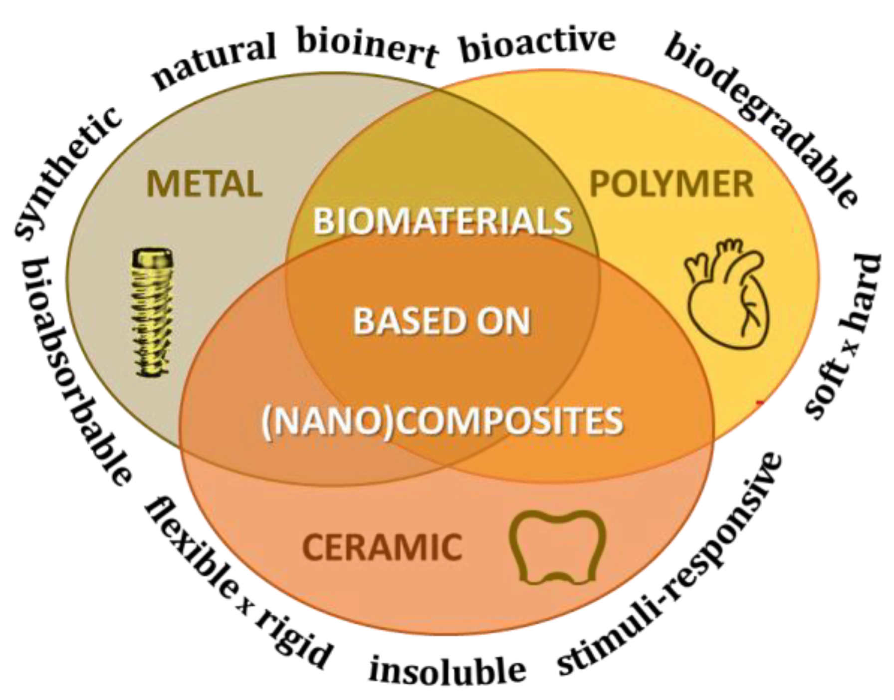
:1. Introduction
2. Properties of Organic Polymers to Attend Pharmaceutical and Medicinal Applications
3. Properties of Nanoparticles to Attend Pharmaceutical and Medicinal Applications
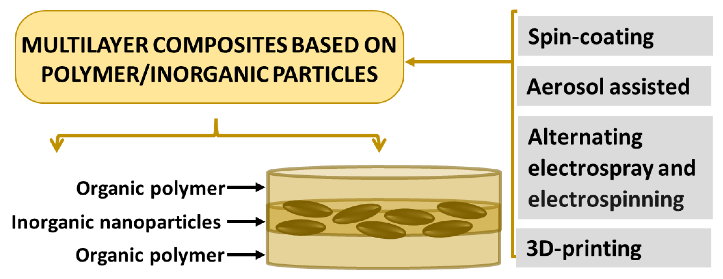
4. Preparation of Polymer/Nanoparticles (Nano)Composites to Attend Pharmaceutical and Medicinal Applications
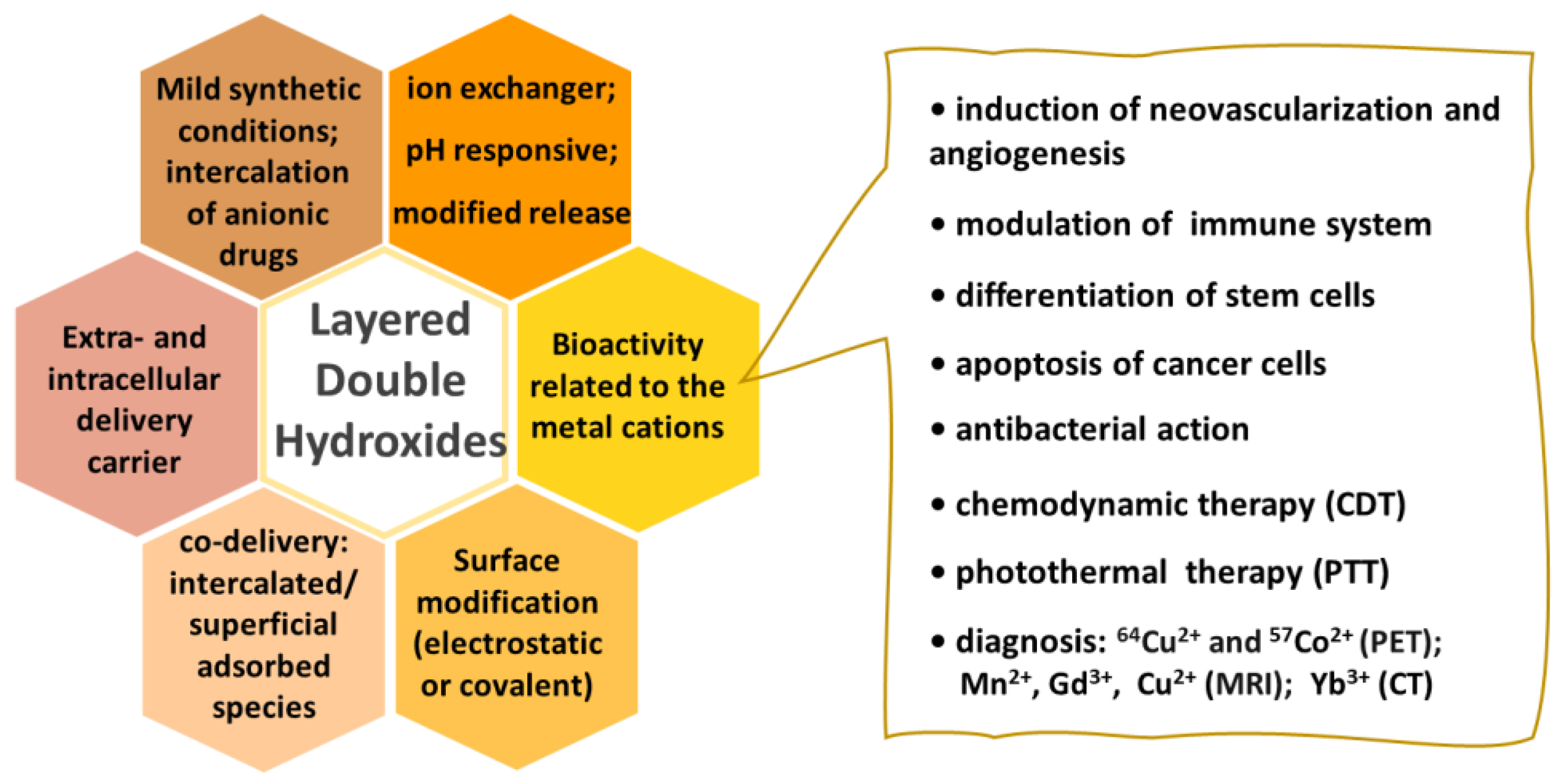
5. Layered Double Hydroxides: Structure, Physicochemical Properties, Biocompatibility
6. Drug Delivery Systems Based on Organic Polymers and Layered Double Hydroxides Nanocomposites
6.1. Polymer/LDH Nanocomposites for Oral Drug Delivery
6.2. Polymer/LDH Nanocomposites for Transdermal Drug Delivery
6.3. Polymer/LDH Nanocomposites for Ocular Drug Delivery
7. Organic Polymers and Layered Double Hydroxides Nanocomposites for Tissue Engineering
7.1. Polymer Composites and Skin Tissue Engineering
7.2. Polymer Composites and Bone Tissue Engineering
8. Conclusions
Author Contributions
Funding
Institutional Review Board Statement
Informed Consent Statement
Data Availability Statement
Conflicts of Interest
References
- Available online: https://sustainabledevelopment.un.org/outcomedocuments/agenda21 (accessed on 18 November 2022).
- Ghasemi-Mobarakeh, L.; Kolahreez, D.; Ramakrishna, S.; Williams, D. Key Terminology in Biomaterials and Biocompatibility. Curr. Opin. Biomed. Eng. 2019, 10, 45–50. [Google Scholar] [CrossRef]
- Williams, D.F. There Is No Such Thing as a Biocompatible Material. Biomaterials 2014, 35, 10009–10014. [Google Scholar] [CrossRef] [PubMed]
- IUPAC. Compendium of Chemical Terminology, 2nd ed.; the “Gold Book”; McNaught, A.D., Wilkinson, A., Eds.; Blackwell Scientific Publications: Oxford, UK, 1997; ISBN 0-9678550-9-8. [Google Scholar]
- Han, X.; Alu, A.; Liu, H.; Shi, Y.; Wei, X.; Cai, L.; Wei, Y. Biomaterial-Assisted Biotherapy: A Brief Review of Biomaterials Used in Drug Delivery, Vaccine Development, Gene Therapy, and Stem Cell Therapy. Bioact. Mater. 2022, 17, 29–48. [Google Scholar] [CrossRef] [PubMed]
- Plocher, J.; Mencattelli, L.; Narducci, F.; Pinho, S. Learning from Nature: Bio-Inspiration for Damage-Tolerant High-Performance Fibre-Reinforced Composites. Compos. Sci. Technol. 2021, 208, 108669. [Google Scholar] [CrossRef]
- Egbo, M.K. A Fundamental Review on Composite Materials and Some of Their Applications in Biomedical Engineering. J. King Saud Univ. Eng. Sci. 2021, 33, 557–568. [Google Scholar] [CrossRef]
- Basu, B.; Gowtham, N.H.; Xiao, Y.; Kalidindi, S.R.; Leong, K.W. Biomaterialomics: Data Science-Driven Pathways to Develop Fourth-Generation Biomaterials. Acta Biomater. Biomater. 2022, 143, 1–25. [Google Scholar] [CrossRef]
- Hench, L.L.; Thompson, I. Twenty-First Century Challenges for Biomaterials. J. R. Soc. Interface 2010, 7, S379–S391. [Google Scholar] [CrossRef] [Green Version]
- Kowalski, P.S.; Bhattacharya, C.; Afewerki, S.; Langer, R. Smart Biomaterials: Recent Advances and Future Directions. ACS Biomater. Sci. Eng. 2018, 4, 3809–3817. [Google Scholar] [CrossRef]
- Montoya, C.; Du, Y.; Gianforcaro, A.L.; Orrego, S.; Yang, M.; Lelkes, P.I. On the Road to Smart Biomaterials for Bone Research: Definitions, Concepts, Advances, and Outlook. Bone Res. 2021, 9, 12. [Google Scholar] [CrossRef]
- Di Silvio, L. Bone Tissue Engineering and Biomineralization. In Tissue Engineering Using Ceramics and Polymers; Elsevier: Amsterdam, The Netherlands, 2007; pp. 319–331. [Google Scholar]
- Evans, D.G.; Slade, R.C.T.S. Structural Aspects of Layered Double Hydroxides. In Structure & Bonding; Duan, X., Evans, D.G., Eds.; Springer: Berlin/Heidelberg, Germany, 2006; pp. 1–87. ISBN 3540282793. [Google Scholar]
- Liu, J.; Zhao, C.; Chen, W.R.; Zhou, B. Recent Progress in Two-Dimensional Nanomaterials for Cancer Theranostics. Coord. Chem. Rev. 2022, 469, 214654. [Google Scholar] [CrossRef]
- Mishra, G.; Dash, B.; Pandey, S. Layered Double Hydroxides: A Brief Review from Fundamentals to Application as Evolving Biomaterials. Appl. Clay Sci. 2018, 153, 172–186. [Google Scholar] [CrossRef]
- Cunha, V.R.R.; Ferreira, A.M.d.C.; Constantino, V.R.L.; Tronto, J.; Valim, J.B. Layered Double Hydroxides: Inorganic Nanoparticles for Storage and Release of Species of Biological and Therapeutic Interest. Quim. Nova 2010, 33, 159–171. [Google Scholar] [CrossRef] [Green Version]
- Del Hoyo, C. Layered Double Hydroxides and Human Health: An Overview. Appl. Clay Sci. 2007, 36, 103–121. [Google Scholar] [CrossRef]
- Allou, N.B.; Saikia, P.; Borah, A.; Goswamee, R.L. Hybrid Nanocomposites of Layered Double Hydroxides: An Update of Their Biological Applications and Future Prospects. Colloid Polym. Sci. 2017, 295, 725–747. [Google Scholar] [CrossRef]
- Kuthati, Y.; Kankala, R.K.; Lee, C.H. Layered Double Hydroxide Nanoparticles for Biomedical Applications: Current Status and Recent Prospects. Appl. Clay Sci. 2015, 112–113, 100–116. [Google Scholar] [CrossRef]
- Saifullah, B.; Hussein, M.Z.B. Inorganic Nanolayers: Structure, Preparation, and Biomedical Applications. Int. J. Nanomed. 2015, 10, 5609–5633. [Google Scholar] [CrossRef] [Green Version]
- Bi, X.; Zhang, H.; Dou, L. Layered Double Hydroxide-Based Nanocarriers for Drug Delivery. Pharmaceutics 2014, 6, 298–332. [Google Scholar] [CrossRef]
- Rives, V.; del Arco, M.; Martín, C. Intercalation of Drugs in Layered Double Hydroxides and Their Controlled Release: A Review. Appl. Clay Sci. 2014, 88–89, 239–269. [Google Scholar] [CrossRef]
- Liang, R.; Wei, M.; Evans, D.G.; Duan, X. Inorganic Nanomaterials for Bioimaging, Targeted Drug Delivery and Therapeutics. Chem. Commun. 2014, 50, 14071–14081. [Google Scholar] [CrossRef]
- Rives, V.; Del Arco, M.; Martín, C. Layered Double Hydroxides as Drug Carriers and for Controlled Release of Non-Steroidal Antiinflammatory Drugs (NSAIDs): A Review. J. Control. Release 2013, 169, 28–39. [Google Scholar] [CrossRef]
- Oh, J.-M.; Park, D.-H.; Choi, S.-J.; Choy, J.-H. LDH Nanocontainers as Bio-Reservoirs and Drug Delivery Carriers. Recent Pat. Nanotechnol. 2012, 6, 200–217. [Google Scholar] [CrossRef] [PubMed]
- Benoit, D.S.W.; Overby, C.T.; Sims, K.R., Jr.; Ackun-Farmmer, M.A. Drug Delivery Systems. In Biomaterials Science; Elsevier: Amsterdam, The Netherlands, 2020; pp. 1237–1266. ISBN 9780128161371. [Google Scholar]
- Armentano, I.; Puglia, D.; Luzi, F.; Arciola, C.; Morena, F.; Martino, S.; Torre, L. Nanocomposites Based on Biodegradable Polymers. Materials 2018, 11, 795. [Google Scholar] [CrossRef] [PubMed] [Green Version]
- Lee, J.H.; Yeo, Y. Controlled Drug Release from Pharmaceutical Nanocarriers. Chem. Eng. Sci. 2015, 125, 75–84. [Google Scholar] [CrossRef] [PubMed] [Green Version]
- Vasile, C.; Pamfil, D.; Stoleru, E.; Baican, M. New Developments in Medical Applications of Hybrid Hydrogels Containing Natural Polymers. Molecules 2020, 25, 1539. [Google Scholar] [CrossRef] [PubMed] [Green Version]
- Tran, H.D.N.; Park, K.D.; Ching, Y.C.; Huynh, C.; Nguyen, D.H. A Comprehensive Review on Polymeric Hydrogel and Its Composite: Matrices of Choice for Bone and Cartilage Tissue Engineering. J. Ind. Eng. Chem. 2020, 89, 58–82. [Google Scholar] [CrossRef]
- Kou, S.G.; Peters, L.; Mucalo, M. Chitosan: A Review of Molecular Structure, Bioactivities and Interactions with the Human Body and Micro-Organisms. Carbohydr. Polym. 2022, 282, 119132. [Google Scholar] [CrossRef]
- Minzanova, S.; Mironov, V.; Arkhipova, D.; Khabibullina, A.; Mironova, L.; Zakirova, Y.; Milyukov, V. Biological Activity and Pharmacological Application of Pectic Polysaccharides: A Review. Polymers 2018, 10, 1407. [Google Scholar] [CrossRef] [Green Version]
- Freitas, C.M.P.; Coimbra, J.S.R.; Souza, V.G.L.; Sousa, R.C.S. Structure and Applications of Pectin in Food, Biomedical, and Pharmaceutical Industry: A Review. Coatings 2021, 11, 922. [Google Scholar] [CrossRef]
- D’souza, A.A.; Shegokar, R. Polyethylene Glycol (PEG): A Versatile Polymer for Pharmaceutical Applications. Expert Opin. Drug Deliv. 2016, 13, 1257–1275. [Google Scholar] [CrossRef]
- Hong, L.; Wang, Z.; Wei, X.; Shi, J.; Li, C. Antibodies against Polyethylene Glycol in Human Blood: A Literature Review. J. Pharmacol. Toxicol. Methods 2020, 102, 106678. [Google Scholar] [CrossRef]
- Haq, H.N.; Khan, H.; Chaudhry, H.; Nimmala, S.; Demidovich, J.; Papudesi, B.N.; Potluri, S.D. Pfizer-BioNTech (BNT162b2), Moderna (MRNA-1273) COVID-19 MRNA Vaccines and Hypersensitivity Reactions. J. Natl. Med. Assoc. 2022, 114, 601–612. [Google Scholar] [CrossRef]
- dos Santos, J.; da Silva, G.S.; Velho, M.C.; Beck, R.C.R. Eudragit®: A Versatile Family of Polymers for Hot Melt Extrusion and 3D Printing Processes in Pharmaceutics. Pharmaceutics 2021, 13, 1424. [Google Scholar] [CrossRef]
- Crago, M.; Lee, A.; Farajikhah, S.; Oveissi, F.; Fletcher, D.F.; Dehghani, F.; Winlaw, D.S.; Naficy, S. The Evolution of Polyurethane Heart Valve Replacements: How Chemistry Translates to the Clinic. Mater. Today Commun. 2022, 33, 104916. [Google Scholar] [CrossRef]
- Claeys, B.; Vervaeck, A.; Hillewaere, X.K.D.; Possemiers, S.; Hansen, L.; De Beer, T.; Remon, J.P.; Vervaet, C. Thermoplastic Polyurethanes for the Manufacturing of Highly Dosed Oral Sustained Release Matrices via Hot Melt Extrusion and Injection Molding. Eur. J. Pharm. Biopharm. 2015, 90, 44–52. [Google Scholar] [CrossRef] [Green Version]
- Rusu, L.-C.; Ardelean, L.C.; Jitariu, A.-A.; Miu, C.A.; Streian, C.G. An Insight into the Structural Diversity and Clinical Applicability of Polyurethanes in Biomedicine. Polymers 2020, 12, 1197. [Google Scholar] [CrossRef]
- Makvandi, P.; Iftekhar, S.; Pizzetti, F.; Zarepour, A.; Zare, E.N.; Ashrafizadeh, M.; Agarwal, T.; Padil, V.V.T.; Mohammadinejad, R.; Sillanpaa, M.; et al. Functionalization of Polymers and Nanomaterials for Water Treatment, Food Packaging, Textile and Biomedical Applications: A Review. Environ. Chem. Lett. 2021, 19, 583–611. [Google Scholar] [CrossRef]
- Narayanan, K.B.; Bhaskar, R.; Han, S.S. Recent Advances in the Biomedical Applications of Functionalized Nanogels. Pharmaceutics 2022, 14, 2832. [Google Scholar] [CrossRef]
- Rivera Gil, P.; Hühn, D.; del Mercato, L.L.; Sasse, D.; Parak, W.J. Nanopharmacy: Inorganic Nanoscale Devices as Vectors and Active Compounds. Pharmacol. Res. 2010, 62, 115–125. [Google Scholar] [CrossRef]
- Gonzalez Solveyra, E.; Szleifer, I. What Is the Role of Curvature on the Properties of Nanomaterials for Biomedical Applications? WIREs Nanomed. Nanobiotechnology 2016, 8, 334–354. [Google Scholar] [CrossRef] [Green Version]
- Oliva, N.; Unterman, S.; Zhang, Y.; Conde, J.; Song, H.S.; Artzi, N. Personalizing Biomaterials for Precision Nanomedicine Considering the Local Tissue Microenvironment. Adv. Healthc. Mater. 2015, 4, 1584–1599. [Google Scholar] [CrossRef] [Green Version]
- Min, Y.; Caster, J.M.; Eblan, M.J.; Wang, A.Z. Clinical Translation of Nanomedicine. Chem. Rev. 2015, 115, 11147–11190. [Google Scholar] [CrossRef] [PubMed] [Green Version]
- Nguyen, V.H.; Lee, B.-J. Protein Corona: A New Approach for Nanomedicine Design. Int. J. Nanomed. 2017, 12, 3137–3151. [Google Scholar] [CrossRef] [PubMed] [Green Version]
- Gu, Z.; Zuo, H.; Li, L.; Wu, A.; Xu, Z.P. Pre-Coating Layered Double Hydroxide Nanoparticles with Albumin to Improve Colloidal Stability and Cellular Uptake. J. Mater. Chem. B 2015, 3, 3331–3339. [Google Scholar] [CrossRef] [PubMed]
- Zuo, H.; Gu, Z.; Cooper, H.; Xu, Z.P. Crosslinking to Enhance Colloidal Stability and Redispersity of Layered Double Hydroxide Nanoparticles. J. Colloid Interface Sci. 2015, 459, 10–16. [Google Scholar] [CrossRef] [PubMed] [Green Version]
- Rojas, R.; Giacomelli, C.E. Size-Tunable LDH–protein Hybrids toward the Optimization of Drug Nanocarriers. J. Mater. Chem. B 2015, 3, 2778–2785. [Google Scholar] [CrossRef]
- Fu, S.; Sun, Z.; Huang, P.; Li, Y.; Hu, N. Some Basic Aspects of Polymer Nanocomposites: A Critical Review. Nano Mater. Sci. 2019, 1, 2–30. [Google Scholar] [CrossRef]
- Kumar, S.; Nehra, M.; Dilbaghi, N.; Tankeshwar, K.; Kim, K.-H. Recent Advances and Remaining Challenges for Polymeric Nanocomposites in Healthcare Applications. Prog. Polym. Sci. 2018, 80, 1–38. [Google Scholar] [CrossRef]
- do Nascimento, G.M.; Constantino, V.R.L.; Landers, R.; Temperini, M.L.A. Spectroscopic Characterization of Polyaniline Formed in the Presence of Montmorillonite Clay. Polymer 2006, 47, 6131–6139. [Google Scholar] [CrossRef]
- Karim, A.V.; Hassani, A.; Eghbali, P.; Nidheesh, P.V. Nanostructured Modified Layered Double Hydroxides (LDHs)-Based Catalysts: A Review on Synthesis, Characterization, and Applications in Water Remediation by Advanced Oxidation Processes. Curr. Opin. Solid State Mater. Sci. 2022, 26, 100965. [Google Scholar] [CrossRef]
- Perotti, G.F.; Barud, H.S.; Messaddeq, Y.; Ribeiro, S.J.L.; Constantino, V.R.L. Bacterial Cellulose–laponite Clay Nanocomposites. Polymer 2011, 52, 157–163. [Google Scholar] [CrossRef]
- Gabryś, T.; Fryczkowska, B.; Fabia, J.; Biniaś, D. Preparation of an Active Dressing by In Situ Biosynthesis of a Bacterial Cellulose–Graphene Oxide Composite. Polymers 2022, 14, 2864. [Google Scholar] [CrossRef]
- Drioli, E.; Giorno, L. Encyclopedia of Membranes; Springer: Berlin/Heidelberg, Germany, 2016; ISBN 978-3-662-44323-1. [Google Scholar]
- Drioli, E.; Giorno, L. Membranes Operation—Innovative Separations and Transformations; Wiley-VCH: Weinheim, Germany, 2009; ISBN 978-3-527-32038-7. [Google Scholar]
- Liang, N.; Liu, W.; Zuo, D.; Peng, P.; Qu, R.; Chen, D.; Zhang, H. Quaternized Polysulfone-Based Nanocomposite Membranes and Improved Properties by Intercalated Layered Double Hydroxide. Polym. Eng. Sci. 2018, 58, 767–774. [Google Scholar] [CrossRef]
- Martos, A.M.; Herrero, M.; Várez, A.; Levenfeld, B. Synthesis and Characterization of New Membranes Based on Sulfonated Polysulfone/Zn,Al-Heptamolibdate LDH. Mater. Lett. 2015, 152, 125–127. [Google Scholar] [CrossRef] [Green Version]
- Mohapi, M.; Sefadi, J.S.; Mochane, M.J.; Magagula, S.I.; Lebelo, K. Effect of LDHs and Other Clays on Polymer Composite in Adsorptive Removal of Contaminants: A Review. Crystals 2020, 10, 957. [Google Scholar] [CrossRef]
- Oyarzabal, A.; Mugica, A.; Müller, A.J.; Zubitur, M. Hydrolytic Degradation of Nanocomposites Based on Poly(L-Lactic Acid) and Layered Double Hydroxides Modified with a Model Drug. J. Appl. Polym. Sci. 2016, 133, 43648. [Google Scholar] [CrossRef]
- Yadollahi, M.; Namazi, H.; Barkhordari, S. Preparation and Properties of Carboxymethyl Cellulose/Layered Double Hydroxide Bionanocomposite Films. Carbohydr. Polym. 2014, 108, 83–90. [Google Scholar] [CrossRef]
- Figueiredo, M.P.; Lini, B.; García-Villén, F.; Borrego-Sánchez, A.; Rossi, A.; Viseras, C.; Constantino, V.R.L. Innovative Membrane Containing Iron-Based Layered Double Hydroxide Intercalated with Phyto Therapeutic Diterpenoid. Appl. Clay Sci. 2022, 216, 106358. [Google Scholar] [CrossRef]
- Figueiredo, M.P.; Borrego-Sánchez, A.; García-Villén, F.; Miele, D.; Rossi, S.; Sandri, G.; Viseras, C.; Constantino, V.R.L. Polymer/Iron-Based Layered Double Hydroxides as Multifunctional Wound Dressings. Pharmaceutics 2020, 12, 1130. [Google Scholar] [CrossRef]
- Ghosal, K.; Agatemor, C.; Tucker, N.; Kny, E.; Thomas, S. CHAPTER 1 Electrical Spinning to Electrospinning: A Brief History. In Electrospinning: From Basic Research to Commercialization; The Royal Society of Chemistry: London, UK, 2018; pp. 1–23. ISBN 978-1-78801-100-6. [Google Scholar]
- Bhardwaj, N.; Kundu, S.C. Electrospinning: A Fascinating Fiber Fabrication Technique. Biotechnol. Adv. 2010, 28, 325–347. [Google Scholar] [CrossRef]
- Boda, S.K.; Li, X.; Xie, J. Electrospraying an Enabling Technology for Pharmaceutical and Biomedical Applications: A Review. J. Aerosol Sci. 2018, 125, 164–181. [Google Scholar] [CrossRef]
- Noruzi, M. Electrospun Nanofibres in Agriculture and the Food Industry: A Review. J. Sci. Food Agric. 2016, 96, 4663–4678. [Google Scholar] [CrossRef] [PubMed]
- Li, Z.; Zhang, J.; Yu, L.; Zhang, J. Electrospun Porous Nanofibers for Electrochemical Energy Storage. J. Mater. Sci. 2017, 52, 6173–6195. [Google Scholar] [CrossRef]
- Liu, M.; Duan, X.-P.; Li, Y.-M.; Yang, D.-P.; Long, Y.-Z. Electrospun Nanofibers for Wound Healing. Mater. Sci. Eng. C 2017, 76, 1413–1423. [Google Scholar] [CrossRef] [PubMed]
- Valarezo, E.; Tammaro, L.; González, S.; Malagón, O.; Vittoria, V. Fabrication and Sustained Release Properties of Poly(ε-Caprolactone) Electrospun Fibers Loaded with Layered Double Hydroxide Nanoparticles Intercalated with Amoxicillin. Appl. Clay Sci. 2013, 72, 104–109. [Google Scholar] [CrossRef]
- Figueiredo, M.P.; Layrac, G.; Hébraud, A.; Limousy, L.; Brendle, J.; Schlatter, G.; Constantino, V.R.L. Design of 3D Multi-Layered Electrospun Membranes Embedding Iron-Based Layered Double Hydroxide for Drug Storage and Control of Sustained Release. Eur. Polym. J. 2020, 131, 109675. [Google Scholar] [CrossRef]
- Rogalski, J.J.; Bastiaansen, C.W.M.; Peijs, T. Rotary Jet Spinning Review—A Potential High Yield Future for Polymer Nanofibers. Nanocomposites 2017, 3, 97–121. [Google Scholar] [CrossRef] [Green Version]
- Lang, B.; McGinity, J.W.; Williams, R.O. Hot-Melt Extrusion—Basic Principles and Pharmaceutical Applications. Drug Dev. Ind. Pharm. 2014, 40, 1133–1155. [Google Scholar] [CrossRef]
- Geng, Z.; Zhen, W. Preparation, Performance, and Kinetics of Poly(Lactic-Acid)/Amidated Benzoic Acid Intercalated Layered Double Hydroxides Nanocomposites by Reactive Extrusion Process. Polym. Compos. 2019, 40, 2668–2680. [Google Scholar] [CrossRef]
- Ha, J.U.; Xanthos, M. Drug Release Characteristics from Nanoclay Hybrids and Their Dispersions in Organic Polymers. Int. J. Pharm. 2011, 414, 321–331. [Google Scholar] [CrossRef]
- Haldorai, Y.; Shim, J.-J.; Lim, K.T. Synthesis of Polymer–inorganic Filler Nanocomposites in Supercritical CO2. J. Supercrit. Fluids 2012, 71, 45–63. [Google Scholar] [CrossRef]
- Ogunsona, E.O.; Dagnon, K.L.; D’Souza, N.A. Multi-Fold Enhancement in Compressive Properties of Polystyrene Foam Using Pre-Delaminated Stearate Functionalized Layer Double Hydroxides. Polymers 2019, 12, 8. [Google Scholar] [CrossRef] [Green Version]
- Nandiyanto, A.B.D.; Okuyama, K. Progress in Developing Spray-Drying Methods for the Production of Controlled Morphology Particles: From the Nanometer to Submicrometer Size Ranges. Adv. Powder Technol. 2011, 22, 1–19. [Google Scholar] [CrossRef]
- Wang, Y.; Zhang, F.; Xu, S.; Wang, X.; Evans, D.G.; Duan, X. Preparation of Layered Double Hydroxide Microspheres by Spray Drying. Ind. Eng. Chem. Res. 2008, 47, 5746–5750. [Google Scholar] [CrossRef]
- Jin, H.; Yuan, D.; Zhu, S.; Zhu, X.; Zhu, J. Ni–Co Layered Double Hydroxide on Carbon Nanorods and Graphene Nanoribbons Derived from MOFs for Supercapacitors. Dalton Trans. 2018, 47, 8706–8715. [Google Scholar] [CrossRef]
- Smalenskaite, A.; Kaba, M.M.; Grigoraviciute-Puroniene, I.; Mikoliunaite, L.; Zarkov, A.; Ramanauskas, R.; Morkan, I.A.; Kareiva, A. Sol–Gel Synthesis and Characterization of Coatings of Mg-Al Layered Double Hydroxides. Materials 2019, 12, 3738. [Google Scholar] [CrossRef] [Green Version]
- Oliveira, M.; Machado, A. Preparation of Polymer-Based Nanocomposites by Different Routes. In Nanocomposites: Synthesis, Characterization and Applications; Wang, X., Ed.; NOVA Publishers: New York, NY, USA, 2013; p. 22. ISBN 978-1-62948-227-9. [Google Scholar]
- Pedroso-Santana, S.; Fleitas-Salazar, N. Ionotropic Gelation Method in the Synthesis of Nanoparticles/Microparticles for Biomedical Purposes. Polym. Int. 2020, 69, 443–447. [Google Scholar] [CrossRef]
- Viscusi, G.; Gorrasi, G. Facile Preparation of Layered Double Hydroxide (LDH)-Alginate Beads as Sustainable System for the Triggered Release of Diclofenac: Effect of PH and Temperature on Release Rate. Int. J. Biol. Macromol. 2021, 184, 271–281. [Google Scholar] [CrossRef]
- Wang, C.; Huang, W.; Zhou, Y.; He, L.; He, Z.; Chen, Z.; He, X.; Tian, S.; Liao, J.; Lu, B.; et al. 3D Printing of Bone Tissue Engineering Scaffolds. Bioact. Mater. 2020, 5, 82–91. [Google Scholar] [CrossRef]
- Ahn, J.; Park, Y.S.; Lee, S.; Yang, J.; Pyo, J.; Lee, J.; Kim, G.H.; Choi, S.M.; Seol, S.K. 3D-Printed NiFe-Layered Double Hydroxide Pyramid Electrodes for Enhanced Electrocatalytic Oxygen Evolution Reaction. Sci. Rep. 2022, 12, 346. [Google Scholar] [CrossRef]
- Ren, Y.; Feng, J. Skin-Inspired Multifunctional Luminescent Hydrogel Containing Layered Rare-Earth Hydroxide with 3D Printability for Human Motion Sensing. ACS Appl. Mater. Interfaces 2020, 12, 6797–6805. [Google Scholar] [CrossRef]
- Qian, L.; Zhang, H. Controlled Freezing and Freeze Drying: A Versatile Route for Porous and Micro-/Nano-Structured Materials. J. Chem. Technol. Biotechnol. 2011, 86, 172–184. [Google Scholar] [CrossRef]
- Liang, Q.; Pan, Y.; Zhang, D.; Lü, T.; Zhao, H.; Zhang, Y. Preparation of Bichar/Layered Double Hydroxide@alginate Aerogel as a Highly Efficient Adsorbent for Cu2+ and Cd2+. J. Appl. Polym. Sci. 2023, 140, e53361. [Google Scholar] [CrossRef]
- Hibino, T. New Nanocomposite Hydrogels Containing Layered Double Hydroxide. Appl. Clay Sci. 2010, 50, 282–287. [Google Scholar] [CrossRef]
- Cao, Y.; Zheng, D.; Zhang, F.; Pan, J.; Lin, C. Layered Double Hydroxide (LDH) for Multi-Functionalized Corrosion Protection of Metals: A Review. J. Mater. Sci. Technol. 2022, 102, 232–263. [Google Scholar] [CrossRef]
- Guan, Q.-F.; Han, Z.-M.; Luo, T.-T.; Yang, H.-B.; Liang, H.-W.; Chen, S.-M.; Wang, G.-S.; Yu, S.-H. A General Aerosol-Assisted Biosynthesis of Functional Bulk Nanocomposites. Natl. Sci. Rev. 2019, 6, 64–73. [Google Scholar] [CrossRef]
- Sangian, D.; Ide, Y.; Bando, Y.; Rowan, A.E.; Yamauchi, Y. Materials Nanoarchitectonics Using 2D Layered Materials: Recent Developments in the Intercalation Process. Small 2018, 14, 1800551. [Google Scholar] [CrossRef]
- Cavani, F.; Trifirò, F.; Vaccari, A. Hydrotalcite-Type Anionic Clays: Preperation, Properties and Applications. Catal. Today 1991, 11, 173–301. [Google Scholar] [CrossRef]
- Mills, S.J.; Christy, A.G.; Génin, J.-M.R.; Kameda, T.; Colombo, F. Nomenclature of the Hydrotalcite Supergroup: Natural Layered Double Hydroxides. Mineral. Mag. 2012, 76, 1289–1336. [Google Scholar] [CrossRef] [Green Version]
- Yu, J.; Wang, Q.; O’Hare, D.; Sun, L. Preparation of Two Dimensional Layered Double Hydroxide Nanosheets and Their Applications. Chem. Soc. Rev. 2017, 46, 5950–5974. [Google Scholar] [CrossRef]
- Qu, J.; Zhang, Q.; Li, X.; He, X.; Song, S. Mechanochemical Approaches to Synthesize Layered Double Hydroxides: A Review. Appl. Clay Sci. 2016, 119, 185–192. [Google Scholar] [CrossRef]
- Zhang, C.; Gui, R.; Wang, Z.; Chen, S.; Wang, Q. Recent Advances in the Synthesis of Layered Double Hydroxides. In Progress in Layered Double Hydroxides: From Synthesis to New Applications; Nocchetti, M., Costantino, U., Eds.; World Scientific: Singapore, 2022; pp. 41–131. [Google Scholar]
- Radha, S.; Kamath, P.V. Structural Synthon Approach to Predict the Possible Polytypes of Layered Double Hydroxides. Z. Anorg. Allg. Chem. 2012, 638, 2317–2323. [Google Scholar] [CrossRef] [Green Version]
- Available online: https://ec.europa.eu/environment/chemicals/nanotech/faq/definition_en.htm (accessed on 27 July 2019).
- Gardner, E.; Pinnavaia, T.J. On the Nature of Selective Olefin Oxidation Catalysts Derived from Molybdate- and Tungstate-Intercalated Layered Double Hydroxides. Appl. Catal. A Gen. 1998, 167, 65–74. [Google Scholar] [CrossRef]
- Crepaldi, E.L.; Pavan, P.C.; Valim, J.B. A New Method of Intercalation by Anion Exchange in Layered Double Hydroxides. Chem. Commun. 1999, 2, 155–156. [Google Scholar] [CrossRef]
- Perotti, G.F.; Bortotti, J.R.; Lima, F.S.; Michels, L.; dos Santos, E.C.; Altoé, M.A.S.; Grassi, G.; Silva, G.J.; Droppa-Jr, R.; Fossum, J.O.; et al. Exfoliation of Carboxymethylcellulose-Intercalated Layered Double Hydroxide in Water. Appl. Clay Sci. 2021, 205, 106005. [Google Scholar] [CrossRef]
- Alcântara, A.C.S.; Aranda, P.; Darder, M.; Ruiz-Hitzky, E. Bionanocomposites Based on Alginate–zein/Layered Double Hydroxide Materials as Drug Delivery Systems. J. Mater. Chem. 2010, 20, 9495–9504. [Google Scholar] [CrossRef]
- Aisawa, S.; Higashiyama, N.; Takahashi, S.; Hirahara, H.; Ikematsu, D.; Kondo, H.; Nakayama, H.; Narita, E. Intercalation Behavior of L-Ascorbic Acid into Layered Double Hydroxides. Appl. Clay Sci. 2007, 35, 146–154. [Google Scholar] [CrossRef]
- Choy, J.H.; Jung, J.S.; Oh, J.M.; Park, M.; Jeong, J.; Kang, Y.K.; Han, O.J. Layered Double Hydroxide as an Efficient Drug Reservoir for Folate Derivatives. Biomaterials 2004, 25, 3059–3064. [Google Scholar] [CrossRef]
- Madhusha, C.; Rajapaksha, K.; Munaweera, I.; de Silva, M.; Perera, C.; Wijesinghe, G.; Weerasekera, M.; Attygalle, D.; Sandaruwan, C.; Kottegoda, N. A Novel Green Approach to Synthesize Curcuminoid-Layered Double Hydroxide Nanohybrids: Adroit Biomaterials for Future Antimicrobial Applications. ACS Omega 2021, 6, 9600–9608. [Google Scholar] [CrossRef]
- Guilherme, V.A.; Cunha, V.R.R.; de Paula, E.; de Araujo, D.R.; Constantino, V.R.L. Anti-Inflammatory and Analgesic Evaluation of a Phytochemical Intercalated into Layered Double Hydroxide. Pharmaceutics 2022, 14, 934. [Google Scholar] [CrossRef]
- Figueiredo, M.P.; Diaz Suarez, E.M.; Petrilli, H.; Leroux, F.; Taviot-Guého, C.; Constantino, V.R.L. Limiting Content of Trivalent Iron to Form Organic-Inorganic Single-Phase Layered Double Hydroxides Hybrids by Coprecipitation. Appl. Clay Sci. 2022, 228, 106642. [Google Scholar] [CrossRef]
- Perotti, G.F.; Silva, F.F.; de Couto, R.A.A.; Lima, F.C.D.A.; Petrilli, H.M.; Leroux, F.; Ferreira, A.M.C.; Constantino, V.R.L. Intercalation of Apocarotenoids from Annatto (Bixa Orellana L.) into Layered Double Hydroxides. J. Braz. Chem. Soc. 2020, 31, 2211–2223. [Google Scholar] [CrossRef]
- Rocha, M.A.; Petersen, P.A.D.; Teixeira-Neto, E.; Petrilli, H.M.; Leroux, F.; Taviot-Gueho, C.; Constantino, V.R.L. Layered Double Hydroxide and Sulindac Coiled and Scrolled Nanoassemblies for Storage and Drug Release. RSC Adv. 2016, 6, 16419–16436. [Google Scholar] [CrossRef]
- Troutier-Thuilliez, A.-L.L.; Taviot-Guého, C.; Cellier, J.; Hintze-Bruening, H.; Leroux, F. Layered Particle-Based Polymer Composites for Coatings: Part I. Evaluation of Layered Double Hydroxides. Prog. Org. Coat. 2009, 64, 182–192. [Google Scholar] [CrossRef]
- Mao, N.; Zhou, C.H.; Tong, D.S.; Yu, W.H.; Cynthia Lin, C.X. Exfoliation of Layered Double Hydroxide Solids into Functional Nanosheets. Appl. Clay Sci. 2017, 144, 60–78. [Google Scholar] [CrossRef]
- Zhang, Y.; Xu, H.; Lu, S. Preparation and Application of Layered Double Hydroxide Nanosheets. RSC Adv. 2021, 11, 24254–24281. [Google Scholar] [CrossRef]
- Liang, J.; Si, H.; Cui, S.; Ma, R.; Sasaki, T. Unilamellar Crystalline Sheets of Layered Double Hydroxides via Soft Chemistry Delamination: A Unique Class of 2D Functional Materials. In Progress in Layered Double Hydroxides: From Synthesis to New Applications; Nocchetti, M., Costantino, U., Eds.; World Scientific: Singapore, 2022; pp. 133–187. [Google Scholar]
- Mallakpour, S.; Dinari, M.; Talebi, M. Exfoliation and Dispersion of LDH Modified with N-Tetrabromophthaloyl-Glutamic in Poly(Vinyl Alcohol): Morphological and Thermal Studies. J. Chem. Sci. 2015, 127, 519–525. [Google Scholar] [CrossRef] [Green Version]
- Costantino, U.; Bugatti, V.; Gorrasi, G.; Montanari, F.; Nocchetti, M.; Tammaro, L.; Vittoria, V. New Polymeric Composites Based on Poly(ϵ-Caprolactone) and Layered Double Hydroxides Containing Antimicrobial Species. ACS Appl. Mater. Interfaces 2009, 1, 668–677. [Google Scholar] [CrossRef]
- Hu, Z.; Chen, G. Novel Nanocomposite Hydrogels Consisting of Layered Double Hydroxide with Ultrahigh Tensibility and Hierarchical Porous Structure at Low Inorganic Content. Adv. Mater. 2014, 26, 5950–5956. [Google Scholar] [CrossRef]
- Cao, Z.; Adnan, N.N.M.; Wang, G.; Rawal, A.; Shi, B.; Liu, R.; Liang, K.; Zhao, L.; Gooding, J.J.; Boyer, C.; et al. Enhanced Colloidal Stability and Protein Resistance of Layered Double Hydroxide Nanoparticles with Phosphonic Acid-Terminated PEG Coating for Drug Delivery. J. Colloid Interface Sci. 2018, 521, 242–251. [Google Scholar] [CrossRef]
- Yu, J.; Martin, B.R.; Clearfield, A.; Luo, Z.; Sun, L. One-Step Direct Synthesis of Layered Double Hydroxide Single-Layer Nanosheets. Nanoscale 2015, 7, 9448–9451. [Google Scholar] [CrossRef]
- Guan, S.; Yang, D.; Weng, Y.; Lu, H.; Meng, X.; Qu, X.; Zhou, S. Excitation-Dependent Theranostic Nanosheet for Cancer Treatment. Adv. Healthc. Mater. 2018, 7, e1701123. [Google Scholar] [CrossRef]
- Weng, Y.; Guan, S.; Lu, H.; Meng, X.; Kaassis, A.Y.; Ren, X.; Qu, X.; Sun, C.; Xie, Z.; Zhou, S. Confinement of Carbon Dots Localizing to the Ultrathin Layered Double Hydroxides toward Simultaneous Triple-Mode Bioimaging and Photothermal Therapy. Talanta 2018, 184, 50–57. [Google Scholar] [CrossRef]
- Mei, X.; Wang, W.; Yan, L.; Hu, T.; Liang, R.; Yan, D.; Wei, M.; Evans, D.G.; Duan, X. Hydrotalcite Monolayer toward High Performance Synergistic Dual-Modal Imaging and Cancer Therapy. Biomaterials 2018, 165, 14–24. [Google Scholar] [CrossRef]
- Peng, L.; Mei, X.; He, J.; Xu, J.; Zhang, W.; Liang, R.; Wei, M.; Evans, D.G.; Duan, X. Monolayer Nanosheets with an Extremely High Drug Loading toward Controlled Delivery and Cancer Theranostics. Adv. Mater. 2018, 30, e1707389. [Google Scholar] [CrossRef]
- Mei, X.; Ma, J.; Bai, X.; Zhang, X.; Zhang, S.; Liang, R.; Wei, M.; Evans, D.G.; Duan, X. A Bottom-up Synthesis of Rare-Earth-Hydrotalcite Monolayer Nanosheets toward Multimode Imaging and Synergetic Therapy. Chem. Sci. 2018, 9, 5630–5639. [Google Scholar] [CrossRef] [Green Version]
- Xu, Z.P.; Stevenson, G.S.; Lu, C.-Q.; Lu, G.Q.M.; Bartlett, P.F.; Gray, P.P. Stable Suspension of Layered Double Hydroxide Nanoparticles in Aqueous Solution. J. Am. Chem. Soc. 2006, 128, 36–37. [Google Scholar] [CrossRef]
- Shi, S.; Fliss, B.C.; Gu, Z.; Zhu, Y.; Hong, H.; Valdovinos, H.F.; Hernandez, R.; Goel, S.; Luo, H.; Chen, F.; et al. Chelator-Free Labeling of Layered Double Hydroxide Nanoparticles for in Vivo PET Imaging. Sci. Rep. 2015, 5, 16930. [Google Scholar] [CrossRef] [Green Version]
- Li, B.; Gu, Z.; Kurniawan, N.; Chen, W.; Xu, Z.P. Manganese-Based Layered Double Hydroxide Nanoparticles as a T1-MRI Contrast Agent with Ultrasensitive PH Response and High Relaxivity. Adv. Mater. 2017, 29, 1700373. [Google Scholar] [CrossRef]
- Li, B.; Tang, J.; Chen, W.; Hao, G.; Kurniawan, N.; Gu, Z.; Xu, Z.P. Novel Theranostic Nanoplatform for Complete Mice Tumor Elimination via MR Imaging-Guided Acid-Enhanced Photothermo-/Chemo-Therapy. Biomaterials 2018, 177, 40–51. [Google Scholar] [CrossRef]
- Chen, W.; Zuo, H.; Zhang, E.; Li, L.; Henrich-Noack, P.; Cooper, H.; Qian, Y.; Xu, Z.P. Brain Targeting Delivery Facilitated by Ligand-Functionalized Layered Double Hydroxide Nanoparticles. ACS Appl. Mater. Interfaces 2018, 10, 20326–20333. [Google Scholar] [CrossRef]
- Cao, Z.; Zhang, L.; Liang, K.; Cheong, S.; Boyer, C.; Gooding, J.J.; Chen, Y.; Gu, Z. Biodegradable 2D Fe-Al Hydroxide for Nanocatalytic Tumor-Dynamic Therapy with Tumor Specificity. Adv. Sci. 2018, 5, 1801155. [Google Scholar] [CrossRef] [PubMed] [Green Version]
- Regi, M.V.; Esbrit, P.; Salinas, A.J. Degradative Effects of the Biological Environment on Ceramic Biomaterials. In Biomaterials Science; Elsevier: Amsterdam, The Netherlands, 2020; pp. 955–971. [Google Scholar]
- Chen, Z.; Zhang, W.; Wang, M.; Backman, L.J.; Chen, J. Effects of Zinc, Magnesium, and Iron Ions on Bone Tissue Engineering. ACS Biomater. Sci. Eng. 2022, 8, 2321–2335. [Google Scholar] [CrossRef] [PubMed]
- Available online: https://www.drugs.com/international/hydrotalcite.htm (accessed on 15 December 2022).
- Tarnawski, A.S.; Tomikawa, M.; Ohta, M.; Sarfeh, I.J. Antacid Talcid Activates in Gastric Mucosa Genes Encoding for EGF and Its Receptor. The Molecular Basis for Its Ulcer Healing Action. J. Physiol. 2000, 94, 93–98. [Google Scholar] [CrossRef] [PubMed]
- Tarnawski, A.; Ahluwalia, A.; Jones, M.K. Gastric Cytoprotection Beyond Prostaglandins: Cellular and Molecular Mechanisms of Gastroprotective and Ulcer Healing Actions of Antacids. Curr. Pharm. Des. 2013, 19, 126–132. [Google Scholar] [CrossRef]
- Bhattacharjee, A.; Hasanur, S.; Saha, S.; Chakraborty, M. Determination of Half Maximal Inhibitory Concentration of CaAl Layered Double Hydroxide on Cancer Cells and Its Role in the Apoptotic Pathway. Appl. Clay Sci. 2019, 168, 31–35. [Google Scholar] [CrossRef]
- Kang, H.; Kim, M.; Feng, Q.; Lin, S.; Wei, K.; Li, R.; Choi, C.J.; Kim, T.-H.; Li, G.; Oh, J.-M.; et al. Nanolayered Hybrid Mediates Synergistic Co-Delivery of Ligand and Ligation Activator for Inducing Stem Cell Differentiation and Tissue Healing. Biomaterials 2017, 149, 12–28. [Google Scholar] [CrossRef]
- Kang, H.R.; da Costa Fernandes, C.J.; da Silva, R.A.; Constantino, V.R.L.; Koh, I.H.J.; Zambuzzi, W.F. Mg–Al and Zn–Al Layered Double Hydroxides Promote Dynamic Expression of Marker Genes in Osteogenic Differentiation by Modulating Mitogen-Activated Protein Kinases. Adv. Healthc. Mater. 2018, 7, 1700693. [Google Scholar] [CrossRef]
- Cunha, V.R.R.; de Souza, R.B.; da Fonseca Martins, A.M.C.R.P.; Koh, I.H.J.; Constantino, V.R.L. Accessing the Biocompatibility of Layered Double Hydroxide by Intramuscular Implantation: Histological and Microcirculation Evaluation. Sci. Rep. 2016, 6, 30547. [Google Scholar] [CrossRef] [Green Version]
- Figueiredo, M.P.; Cunha, V.R.R.; Leroux, F.; Taviot-Gueho, C.; Nakamae, M.N.; Kang, Y.R.; Souza, R.B.; Martins, A.M.C.R.P.F.; Koh, I.H.J.; Constantino, V.R.L. Iron-Based Layered Double Hydroxide Implants: Potential Drug Delivery Carriers with Tissue Biointegration Promotion and Blood Microcirculation Preservation. ACS Omega 2018, 3, 18263–18274. [Google Scholar] [CrossRef] [Green Version]
- Kim, T.H.; Lee, J.Y.; Kim, M.K.; Park, J.H.; Oh, J.M. Radioisotope Co-57 Incorporated Layered Double Hydroxide Nanoparticles as a Cancer Imaging Agent. RSC Adv. 2016, 6, 48415–48419. [Google Scholar] [CrossRef]
- Shen, S.; Wu, Y.; Liu, Y.; Wu, D. High Drug-Loading Nanomedicines: Progress, Current Status, and Prospects. Int. J. Nanomed. 2017, 12, 4085–4109. [Google Scholar] [CrossRef] [Green Version]
- Cunha, V.R.R.; Petersen, P.A.D.; Gonçalves, M.B.; Petrilli, H.M.; Taviot-Gueho, C.; Leroux, F.; Temperini, M.L.A.; Constantino, V.R.L. Structural, Spectroscopic (NMR, IR, and Raman), and DFT Investigation of the Self-Assembled Nanostructure of Pravastatin-LDH (Layered Double Hydroxides) Systems. Chem. Mater. 2012, 24, 1415–1425. [Google Scholar] [CrossRef]
- Figueiredo, M.P.; Cunha, V.R.R.; Cellier, J.; Taviot-Gueho, C.; Constantino, V.R.L. Fe(III)-Based Layered Double Hydroxides Carrying Model Naproxenate Anions: Compositional and Structural Aspects. ChemistrySelect 2022, 7, e202103880. [Google Scholar] [CrossRef]
- Spirescu, V.A.; Chircov, C.; Grumezescu, A.M.; Vasile, B.Ș.; Andronescu, E. Inorganic Nanoparticles and Composite Films for Antimicrobial Therapies. Int. J. Mol. Sci. 2021, 22, 4595. [Google Scholar] [CrossRef]
- Forano, C.; Bruna, F.; Mousty, C.; Prevot, V. Interactions between Biological Cells and Layered Double Hydroxides: Towards Functional Materials. Chem. Rec. 2018, 18, 1150–1166. [Google Scholar] [CrossRef]
- Li, A.; Qin, L.; Zhu, D.; Zhu, R.; Sun, J.; Wang, S. Signalling Pathways Involved in the Activation of Dendritic Cells by Layered Double Hydroxide Nanoparticles. Biomaterials 2010, 31, 748–756. [Google Scholar] [CrossRef]
- Huang, H.; Wang, Z.; Chen, L.; Yu, H.; Chen, Y. Catalytic Biomaterials and Nanomedicines with Exogenous and Endogenous Activations. Adv. Healthc. Mater. 2022. Early View. [Google Scholar] [CrossRef]
- Constantino, V.R.L.; Cunha, V.R.R.; Rocha, M.A.; Figueiredo, M.P.; Magri, V.R.; Eulálio, D.; Perotti, G.F.; Bizeto, M.A.; Zambuzzi, W.F.; Koh, I.H.J. Layered Double Hydroxides: Characterization, Therapeutic Activities and Biocompatibility. In Progress in Layered Double Hydroxides: From Synthesis to New Applications; Nocchetti, M., Costantino, U., Eds.; Series on Chemistry, Energy and the Environment; World Scientific: Singapore, 2022; Volume 8, pp. 413–482. ISBN 978-981-12-4060-7. [Google Scholar]
- Oh, J.-M.; Choi, S.-J.; Kim, S.-T.; Choy, J.-H. Cellular Uptake Mechanism of an Inorganic Nanovehicle and Its Drug Conjugates: Enhanced Efficacy Due To Clathrin-Mediated Endocytosis. Bioconjugate Chem. 2006, 17, 1411–1417. [Google Scholar] [CrossRef]
- Choi, G.; Choy, J. Recent Progress in Layered Double Hydroxides as a Cancer Theranostic Nanoplatform. WIREs Nanomed. Nanobiotechnology 2020, 13, e1679. [Google Scholar] [CrossRef]
- Xu, Z.P.; Niebert, M.; Porazik, K.; Walker, T.L.; Cooper, H.M.; Middelberg, A.P.J.; Gray, P.P.; Bartlett, P.F.; Lu, G.Q.M. Subcellular Compartment Targeting of Layered Double Hydroxide Nanoparticles. J. Control. Release 2008, 130, 86–94. [Google Scholar] [CrossRef]
- Li, S.; Li, J.; Wang, C.J.; Wang, Q.; Cader, M.Z.; Lu, J.; Evans, D.G.; Duan, X.; O’Hare, D. Cellular Uptake and Gene Delivery Using Layered Double Hydroxide Nanoparticles. J. Mater. Chem. B 2013, 1, 61–68. [Google Scholar] [CrossRef] [PubMed]
- Yan, L.; Gonca, S.; Zhu, G.; Zhang, W.; Chen, X. Layered Double Hydroxide Nanostructures and Nanocomposites for Biomedical Applications. J. Mater. Chem. B 2019, 7, 5583–5601. [Google Scholar] [CrossRef] [PubMed] [Green Version]
- Dang, Y.; Guan, J. Nanoparticle-Based Drug Delivery Systems for Cancer Therapy. Smart Mater. Med. 2020, 1, 10–19. [Google Scholar] [CrossRef] [PubMed]
- Pavlovic, M.; Szerlauth, A.; Muráth, S.; Varga, G.; Szilagyi, I. Surface Modification of Two-Dimensional Layered Double Hydroxide Nanoparticles with Biopolymers for Biomedical Applications. Adv. Drug Deliv. Rev. 2022, 191, 114590. [Google Scholar] [CrossRef] [PubMed]
- Li, D.; Xu, X.; Xu, J.; Hou, W. Poly(Ethylene Glycol) Haired Layered Double Hydroxides as Biocompatible Nanovehicles: Morphology and Dispersity Study. Colloids Surf. A Physicochem. Eng. Asp. 2011, 384, 585–591. [Google Scholar] [CrossRef]
- Mallakpour, S.; Hatami, M.; Hussain, C.M. Recent Innovations in Functionalized Layered Double Hydroxides: Fabrication, Characterization, and Industrial Applications. Adv. Colloid Interface Sci. 2020, 283, 102216. [Google Scholar] [CrossRef]
- Manzi-Nshuti, C.; Songtipya, P.; Manias, E.; Jimenez-Gasco, M.d.M.; Hossenlopp, J.M.; Wilkie, C.A. Polymer Nanocomposites Using Zinc Aluminum and Magnesium Aluminum Oleate Layered Double Hydroxides: Effects of the Polymeric Compatibilizer and of Composition on the Thermal and Fire Properties of PP/LDH Nanocomposites. Polym. Degrad. Stab. 2009, 94, 2042–2054. [Google Scholar] [CrossRef]
- Kim, T.-H.; Young Lee, J.; Xie, J.; Hoon Park, J.; Oh, J.-M. Topology Dependent Modification of Layered Double Hydroxide for Therapeutic and Diagnostic Platform. Adv. Drug Deliv. Rev. 2022, 188, 114459. [Google Scholar] [CrossRef]
- Li, L.; Zhang, R.; Gu, W.; Xu, Z.P. Mannose-Conjugated Layered Double Hydroxide Nanocomposite for Targeted SiRNA Delivery to Enhance Cancer Therapy. Nanomed. Nanotechnol. Biol. Med. 2018, 14, 2355–2364. [Google Scholar] [CrossRef]
- Mochane, M.J.; Magagula, S.I.; Sefadi, J.S.; Sadiku, E.R.; Mokhena, T.C. Morphology, Thermal Stability, and Flammability Properties of Polymer-Layered Double Hydroxide (LDH) Nanocomposites: A Review. Crystals 2020, 10, 612. [Google Scholar] [CrossRef]
- Chen, C.; Buffet, J.-C.; O’Hare, D. Surface Modification of Aqueous Miscible Organic Layered Double Hydroxides (AMO-LDHs). Dalton Trans. 2020, 49, 8498–8503. [Google Scholar] [CrossRef]
- Kankala, R.K. Nanoarchitectured Two-Dimensional Layered Double Hydroxides-Based Nanocomposites for Biomedical Applications. Adv. Drug Deliv. Rev. 2022, 186, 114270. [Google Scholar] [CrossRef]
- Rojas, R.; Mosconi, G.; Pablo, J.; Gil, G.A. Layered Double Hydroxide Applications in Biomedical Implants. Appl. Clay Sci. 2022, 224, 106514. [Google Scholar] [CrossRef]
- Izbudak, B.; Cecen, B.; Anaya, I.; Miri, A.K.; Bal-Ozturk, A.; Karaoz, E. Layered Double Hydroxide-Based Nanocomposite Scaffolds in Tissue Engineering Applications. RSC Adv. 2021, 11, 30237–30252. [Google Scholar] [CrossRef]
- Ameena Shirin, V.K.; Sankar, R.; Johnson, A.P.; Gangadharappa, H.V.; Pramod, K. Advanced Drug Delivery Applications of Layered Double Hydroxide. J. Control. Release 2021, 330, 398–426. [Google Scholar] [CrossRef]
- Mallakpour, S.; Azadi, E.; Hussain, C.M. Recent Advancements in Synthesis and Drug Delivery Utilization of Polysaccharides-Based Nanocomposites: The Important Role of Nanoparticles and Layered Double Hydroxides. Int. J. Biol. Macromol. 2021, 193, 183–204. [Google Scholar] [CrossRef]
- Cao, Z.; Li, B.; Sun, L.; Li, L.; Xu, Z.P.; Gu, Z. 2D Layered Double Hydroxide Nanoparticles: Recent Progress toward Preclinical/Clinical Nanomedicine. Small Methods 2020, 4, 1900343. [Google Scholar] [CrossRef]
- Chatterjee, A.; Bharadiya, P.; Hansora, D. Layered Double Hydroxide Based Bionanocomposites. Appl. Clay Sci. 2019, 177, 19–36. [Google Scholar] [CrossRef]
- Kelkar, S.S.; Reineke, T.M. Theranostics: Combining Imaging and Therapy. Bioconjugate Chem. 2011, 22, 1879–1903. [Google Scholar] [CrossRef]
- Lim, E.-K.; Kim, T.; Paik, S.; Haam, S.; Huh, Y.-M.; Lee, K. Nanomaterials for Theranostics: Recent Advances and Future Challenges. Chem. Rev. 2015, 115, 327–394. [Google Scholar] [CrossRef]
- Gao, R.; Mei, X.; Yan, D.; Liang, R.; Wei, M. Nano-Photosensitizer Based on Layered Double Hydroxide and Isophthalic Acid for Singlet Oxygenation and Photodynamic Therapy. Nat. Commun. 2018, 9, 2798. [Google Scholar] [CrossRef] [Green Version]
- Kang, H.; Mintri, S.; Menon, A.V.; Lee, H.Y.; Choi, H.S.; Kim, J. Pharmacokinetics, Pharmacodynamics and Toxicology of Theranostic Nanoparticles. Nanoscale 2015, 7, 18848–18862. [Google Scholar] [CrossRef] [Green Version]
- Moghimi, S.M.; Hunter, A.C.; Andresen, T.L. Factors Controlling Nanoparticle Pharmacokinetics: An Integrated Analysis and Perspective. Annu. Rev. Pharmacol. Toxicol. 2012, 52, 481–503. [Google Scholar] [CrossRef] [PubMed]
- Zylberberg, C.; Gaskill, K.; Pasley, S.; Matosevic, S. Engineering Liposomal Nanoparticles for Targeted Gene Therapy. Gene Ther. 2017, 24, 441–452. [Google Scholar] [CrossRef] [PubMed]
- Ryu, J.H.; Lee, S.; Son, S.; Kim, S.H.; Leary, J.F.; Choi, K.; Kwon, I.C. Theranostic Nanoparticles for Future Personalized Medicine. J. Control. Release 2014, 190, 477–484. [Google Scholar] [CrossRef] [PubMed]
- Raza, K.; Kumar, P.; Kumar, N.; Malik, R. Pharmacokinetics and Biodistribution of the Nanoparticles. In Advances in Nanomedicine for the Delivery of Therapeutic Nucleic Acids; Elsevier: Amsterdam, The Netherlands, 2017; pp. 165–186. [Google Scholar]
- Liang, R.; Tian, R.; Ma, L.; Zhang, L.; Hu, Y.; Wang, J.; Wei, M.; Yan, D.; Evans, D.G.; Duan, X. A Supermolecular Photosensitizer with Excellent Anticancer Performance in Photodynamic Therapy. Adv. Funct. Mater. 2014, 24, 3144–3151. [Google Scholar] [CrossRef]
- Guan, S.; Weng, Y.; Li, M.; Liang, R.; Sun, C.; Qu, X.; Zhou, S. An NIR-Sensitive Layered Supramolecular Nanovehicle for Combined Dual-Modal Imaging and Synergistic Therapy. Nanoscale 2017, 9, 10367–10374. [Google Scholar] [CrossRef] [Green Version]
- Moore, T.L.; Rodriguez-Lorenzo, L.; Hirsch, V.; Balog, S.; Urban, D.; Jud, C.; Rothen-Rutishauser, B.; Lattuada, M.; Petri-Fink, A. Nanoparticle Colloidal Stability in Cell Culture Media and Impact on Cellular Interactions. Chem. Soc. Rev. 2015, 44, 6287–6305. [Google Scholar] [CrossRef] [Green Version]
- Yang, G.; Phua, S.Z.F.; Bindra, A.K.; Zhao, Y. Degradability and Clearance of Inorganic Nanoparticles for Biomedical Applications. Adv. Mater. 2019, 31, 1805730. [Google Scholar] [CrossRef]
- Vasti, C.; Bedoya, D.A.; Rojas, R.; Giacomelli, C.E. Effect of the Protein Corona on the Colloidal Stability and Reactivity of LDH-Based Nanocarriers. J. Mater. Chem. B 2016, 4, 2008–2016. [Google Scholar] [CrossRef]
- Hakeem, A.; Zhan, G.; Xu, Q.; Yong, T.; Yang, X.; Gan, L. Facile Synthesis of PH-Responsive Doxorubicin-Loaded Layered Double Hydroxide for Efficient Cancer Therapy. J. Mater. Chem. B 2018, 6, 5768–5774. [Google Scholar] [CrossRef]
- Yan, L.; Zhou, M.; Zhang, X.; Huang, L.; Chen, W.; Roy, V.A.L.; Zhang, W.; Chen, X. A Novel Type of Aqueous Dispersible Ultrathin-Layered Double Hydroxide Nanosheets for in Vivo Bioimaging and Drug Delivery. ACS Appl. Mater. Interfaces 2017, 9, 34185–34193. [Google Scholar] [CrossRef] [Green Version]
- Xu, T.; Liu, J.; Sun, L.; Zhang, R.; Xu, Z.P.; Sun, Q. Enhancing Tumor Accumulation and Cellular Uptake of Layered Double Hydroxide Nanoparticles by Coating/Detaching PH-Triggered Charge-Convertible Polymers. ACS Omega 2021, 6, 3822–3830. [Google Scholar] [CrossRef]
- Park, D.-H.; Cho, J.; Kwon, O.-J.; Yun, C.-O.; Choy, J.-H. Biodegradable Inorganic Nanovector: Passive versus Active Tumor Targeting in SiRNA Transportation. Angew. Chem. Int. Ed. 2016, 55, 4582–4586. [Google Scholar] [CrossRef]
- Wen, J.; Lv, Y.; Xu, Y.; Zhang, P.; Li, H.; Chen, X.; Li, X.; Zhang, L.; Liu, F.; Zeng, W.; et al. Construction of a Biodegradable, Versatile Nanocarrier for Optional Combination Cancer Therapy. Acta Biomater. 2019, 83, 359–371. [Google Scholar] [CrossRef]
- Choi, G.; Eom, S.; Vinu, A.; Choy, J.H. 2D Nanostructured Metal Hydroxides with Gene Delivery and Theranostic Functions; A Comprehensive Review. Chem. Rec. 2018, 18, 1033–1053. [Google Scholar] [CrossRef]
- Park, D.-H.; Choi, G.; Choy, J.-H. Bio-Layered Double Hydroxides Nanohybrids for Theranostics Applications. In Photofunctional Layered Materials. Structure and Bonding; Yan, D., Wei, M., Eds.; Springer: Berlin/Heidelberg, Germany, 2015; pp. 137–175. ISBN 978-3-319-16991-0. [Google Scholar]
- Wen, J.; Yang, K.; Huang, J.; Sun, S. Recent Advances in LDH-Based Nanosystems for Cancer Therapy. Mater. Des. 2021, 198, 109298. [Google Scholar] [CrossRef]
- Yun, Y.H.; Lee, B.K.; Park, K. Controlled Drug Delivery: Historical Perspective for the next Generation. J. Control. Release 2015, 219, 2–7. [Google Scholar] [CrossRef] [Green Version]
- Lee, P.I.; Li, J.-X. Evolution of Oral Controlled Release Dosage Forms. In Oral Controlled Release Formulation Design and Drug Delivery; John Wiley & Sons, Inc.: Hoboken, NJ, USA, 2010; pp. 21–31. [Google Scholar]
- Park, K. Controlled Drug Delivery Systems: Past Forward and Future Back. J. Control. Release 2014, 190, 3–8. [Google Scholar] [CrossRef] [Green Version]
- Heinemann, L. The Failure of Exubera: Are We Beating a Dead Horse? J. Diabetes Sci. Technol. 2008, 2, 518–529. [Google Scholar] [CrossRef] [Green Version]
- Vargason, A.M.; Anselmo, A.C.; Mitragotri, S. The Evolution of Commercial Drug Delivery Technologies. Nat. Biomed. Eng. 2021, 5, 951–967. [Google Scholar] [CrossRef] [PubMed]
- Wong, P.T.; Choi, S.K. Mechanisms of Drug Release in Nanotherapeutic Delivery Systems. Chem. Rev. 2015, 115, 3388–3432. [Google Scholar] [CrossRef] [PubMed]
- Ding, C.; Li, Z. A Review of Drug Release Mechanisms from Nanocarrier Systems. Mater. Sci. Eng. C 2017, 76, 1440–1453. [Google Scholar] [CrossRef] [PubMed]
- Luengo, C.V.; Crescitelli, M.C.; Lopez, N.A.; Avena, M.J. Synthesis of Layered Double Hydroxides Intercalated With Drugs for Controlled Release: Successful Intercalation of Ibuprofen and Failed Intercalation of Paracetamol. J. Pharm. Sci. 2021, 110, 1779–1787. [Google Scholar] [CrossRef]
- Maggi, L.; Bruni, G.; Ferrara, C.; Puscalau, C.; Quinzeni, I.; Friuli, V.; Monteforte, F.; Capsoni, D. Zaltoprofen-Layered Double Hydroxide Hybrids to Enhance Zaltoprofen Solubility and Dissolution Rate. Appl. Clay Sci. 2023, 231, 106723. [Google Scholar] [CrossRef]
- Guagliano, M.; Monteforte, F.; Bruni, G.; Friuli, V.; Maggi, L.; Quinzeni, I.; Bini, M. The Peculiar Dissolution Behaviour of Piretanide Hosted in Layered Double Hydroxides. Appl. Clay Sci. 2020, 198, 105826. [Google Scholar] [CrossRef]
- Kleyi, P.E.; Mudaly, P.; Kesavan Pillai, S.; de Beer, M. Zn/Al Layered Double Hydroxides Nanostructure as Effective Controlled Release Vehicle of Nicotinic Acid for Topical Applications. Appl. Clay Sci. 2021, 215, 106304. [Google Scholar] [CrossRef]
- Mehta, S.; Suresh, A.; Nayak, Y.; Narayan, R.; Nayak, U.Y. Hybrid Nanostructures: Versatile Systems for Biomedical Applications. Coord. Chem. Rev. 2022, 460, 214482. [Google Scholar] [CrossRef]
- Wang, L.; Zhang, B.; Yang, X.; Guo, S.; Waterhouse, G.I.N.; Song, G.; Guan, S.; Liu, A.; Cheng, L.; Zhou, S. Targeted Alleviation of Ischemic Stroke Reperfusion via Atorvastatin-Ferritin Gd-Layered Double Hydroxide. Bioact. Mater. 2023, 20, 126–136. [Google Scholar] [CrossRef]
- Adepu, S.; Ramakrishna, S. Controlled Drug Delivery Systems: Current Status and Future Directions. Molecules 2021, 26, 5905. [Google Scholar] [CrossRef]
- Kim, H.-J.; Jeung, D.-G.; Oh, J.-M. Boosting the Anticancer Activity of Doxorubicin with a Layered Double Hydroxide Nanocarrier. Appl. Clay Sci. 2021, 203, 106000. [Google Scholar] [CrossRef]
- Ashrafizadeh, M.; Nazarzadeh Zare, E.; Rossi, F.; Rabiee, N.; Sharifi, E.; Makvandi, P. Photoactive Polymers-Decorated Cu-Al Layered Double Hydroxide Hexagonal Architectures: A Potential Non-Viral Vector for Photothermal Therapy and Co-Delivery of DOX/PCRISPR. Chem. Eng. J. 2022, 448, 137747. [Google Scholar] [CrossRef]
- Gu, D.; Pan, H.; Xu, S.; Chen, W.; Zhu, R.; Jiang, W.; Pan, W. Construction and Evaluation of Hyaluronic Acid–Coated Flurbiprofen-Layered Double Hydroxide Ocular Drug Delivery System. AAPS PharmSciTech 2022, 23, 287. [Google Scholar] [CrossRef]
- Rejinold, N.S.; Piao, H.; Choi, G.; Jin, G.-W.; Choy, J.-H. Niclosamide-Exfoliated Anionic Clay Nanohybrid Repurposed as an Antiviral Drug for Tackling Covid-19; Oral Formulation with Tween 60/Eudragit S100. Clays Clay Miner. 2021, 69, 533–546. [Google Scholar] [CrossRef]
- Liu, J.; Liu, K.; Zhang, L.; Zhong, M.; Hong, T.; Zhang, R.; Gao, Y.; Li, R.; Xu, T.; Xu, Z.P. Heat/PH-Boosted Release of 5-Fluorouracil and Albumin-Bound Paclitaxel from Cu-Doped Layered Double Hydroxide Nanomedicine for Synergistical Chemo-Photo-Therapy of Breast Cancer. J. Control. Release 2021, 335, 49–58. [Google Scholar] [CrossRef]
- Zhang, H.; Zhang, L.; Cao, Z.; Cheong, S.; Boyer, C.; Wang, Z.; Yun, S.L.J.; Amal, R.; Gu, Z. Two-Dimensional Ultra-Thin Nanosheets with Extraordinarily High Drug Loading and Long Blood Circulation for Cancer Therapy. Small 2022, 18, 2200299. [Google Scholar] [CrossRef]
- Li, L.; Qian, Y.; Sun, L.; Han, F.Y.; Zhang, R.; Wang, P.-Y.; Xu, Z.P. Albumin-Stabilized Layered Double Hydroxide Nanoparticles Synergized Combination Chemotherapy for Colorectal Cancer Treatment. Nanomed. Nanotechnol. Biol. Med. 2021, 34, 102369. [Google Scholar] [CrossRef]
- Rayment, P.; Wright, P.; Hoad, C.; Ciampi, E.; Haydock, D.; Gowland, P.; Butler, M.F. Investigation of Alginate Beads for Gastro-Intestinal Functionality, Part 1: In Vitro Characterisation. Food Hydrocoll. 2009, 23, 816–822. [Google Scholar] [CrossRef]
- Costantino, U.; Ambrogi, V.; Nocchetti, M.; Perioli, L. Hydrotalcite-like Compounds: Versatile Layered Hosts of Molecular Anions with Biological Activity. Microporous Mesoporous Mater. 2008, 107, 149–160. [Google Scholar] [CrossRef]
- Alcântara, A.C.S.; Darder, M. Building Up Functional Bionanocomposites from the Assembly of Clays and Biopolymers. Chem. Rec. 2018, 18, 696–712. [Google Scholar] [CrossRef]
- Ribeiro, C.; Arizaga, G.G.C.; Wypych, F.; Sierakowski, M.-R. Nanocomposites Coated with Xyloglucan for Drug Delivery: In Vitro Studies. Int. J. Pharm. 2009, 367, 204–210. [Google Scholar] [CrossRef] [PubMed]
- San Román, M.S.; Holgado, M.J.; Salinas, B.; Rives, V. Drug Release from Layered Double Hydroxides and from Their Polylactic Acid (PLA) Nanocomposites. Appl. Clay Sci. 2013, 71, 1–7. [Google Scholar] [CrossRef]
- Li, B.; He, J.; Evans, D.G.; Duan, X. Enteric-Coated Layered Double Hydroxides as a Controlled Release Drug Delivery System. Int. J. Pharm. 2004, 287, 89–95. [Google Scholar] [CrossRef] [PubMed]
- Zhang, J.-P.; Wang, Q.; Xie, X.-L.; Li, X.; Wang, A.-Q. Preparation and Swelling Properties of pH-Sensitive Sodium Alginate/Layered Double Hydroxides Hybrid Beads for Controlled Release of Diclofenac Sodium. J. Biomed. Mater. Res. Part B Appl. Biomater. 2010, 92B, 205–214. [Google Scholar] [CrossRef]
- Mahkam, M.; Davatgar, M.; Rezvani, Z.; Nejati, K. Preparation of PH-Sensitive Polymers/Layered Double Hydroxide Hybrid Beads for Controlled Release of Insulin. Int. J. Polym. Mater. 2013, 62, 57–60. [Google Scholar] [CrossRef]
- Barkhordari, S.; Yadollahi, M.; Namazi, H. PH Sensitive Nanocomposite Hydrogel Beads Based on Carboxymethyl Cellulose/Layered Double Hydroxide as Drug Delivery Systems. J. Polym. Res. 2014, 21, 454. [Google Scholar] [CrossRef]
- Barkhordari, S.; Yadollahi, M. Carboxymethyl Cellulose Capsulated Layered Double Hydroxides/Drug Nanohybrids for Cephalexin Oral Delivery. Appl. Clay Sci. 2016, 121–122, 77–85. [Google Scholar] [CrossRef]
- Nejati, K.; Keypour, H.; Nezhad, P.D.K.; Rezvani, Z.; Asadpour-Zeynali, K. Preparation and Characterization of Cetirizine Intercalated Layered Double Hydroxide and Chitosan Nanocomposites. J. Taiwan Inst. Chem. Eng. 2015, 53, 168–175. [Google Scholar] [CrossRef]
- Rezvani, Z.; Shahbaei, M. Bionanocomposites Based on Alginate and Chitosan/Layered Double Hydroxide with Ciprofloxacin Drug: Investigation of Structure and Controlled Release Properties. Polym. Compos. 2015, 36, 1819–1825. [Google Scholar] [CrossRef]
- Xu, Y.; Zhan, C.; Fan, L.; Wang, L.; Zheng, H. Preparation of Dual Crosslinked Alginate–chitosan Blend Gel Beads and in Vitro Controlled Release in Oral Site-Specific Drug Delivery System. Int. J. Pharm. 2007, 336, 329–337. [Google Scholar] [CrossRef]
- Rebitski, E.P.; Souza, G.P.; Santana, S.A.A.; Pergher, S.B.C.; Alcântara, A.C.S. Bionanocomposites Based on Cationic and Anionic Layered Clays as Controlled Release Devices of Amoxicillin. Appl. Clay Sci. 2019, 173, 35–45. [Google Scholar] [CrossRef]
- Anirudhan, T.S.; Sekhar, V.C. Fabrication of Functionalized Layered Double Hydroxide/Chitosan Nanocomposite with Dual Responsive Drug Release for the Targeted Therapy of Breast Cancer. Eur. Polym. J. 2020, 139, 109993. [Google Scholar] [CrossRef]
- Ribeiro, L.N.M.; Alcântara, A.C.S.; Darder, M.; Aranda, P.; Herrmann, P.S.P.; Araújo-Moreira, F.M.; García-Hernández, M.; Ruiz-Hitzky, E. Bionanocomposites Containing Magnetic Graphite as Potential Systems for Drug Delivery. Int. J. Pharm. 2014, 477, 553–563. [Google Scholar] [CrossRef]
- Abniki, M.; Moghimi, A.; Azizinejad, F. Fabrication of Bionanocomposite Based on LDH Using Biopolymer of Gum Arabic and Chitosan-Coating for Sustained Drug-Release. J. Serbian Chem. Soc. 2020, 85, 1223–1235. [Google Scholar] [CrossRef] [Green Version]
- Pu, L.; Yu, H.; Du, J.; Zhang, Y.; Chen, S. Hydrotalcite–PLGA Composite Nanoparticles for Loading and Delivery of Danshensu. RSC Adv. 2020, 10, 22010–22018. [Google Scholar] [CrossRef]
- Kankala, R.K.; Kuthati, Y.; Sie, H.-W.; Shih, H.-Y.; Lue, S.-I.; Kankala, S.; Jeng, C.-C.; Deng, J.-P.; Weng, C.-F.; Liu, C.-L.; et al. Multi-Laminated Metal Hydroxide Nanocontainers for Oral-Specific Delivery for Bioavailability Improvement and Treatment of Inflammatory Paw Edema in Mice. J. Colloid Interface Sci. 2015, 458, 217–228. [Google Scholar] [CrossRef]
- Barkhordari, S.; Alizadeh, A. Fabrication of PH-Sensitive Chitosan/Layered Double Hydroxide (LDH)/Fe3O4 Nanocomposite Hydrogel Beads for Controlled Release of Diclofenac. Polym. Bull. 2022, 79, 5533–5548. [Google Scholar] [CrossRef]
- Lee, J.; Seo, H.S.; Park, W.; Park, C.G.; Jeon, Y.; Park, D.-H. Biofunctional Layered Double Hydroxide Nanohybrids for Cancer Therapy. Materials 2022, 15, 7977. [Google Scholar] [CrossRef]
- Li, L.; Li, B.; Gu, W.; Xu, Z.P. Modifying Layered Double Hydroxide Nanoparticles for Tumor Imaging and Therapy. Clays Clay Miner. 2019, 67, 72–80. [Google Scholar] [CrossRef]
- Ebadi, M.; Buskaran, K.; Bullo, S.; Hussein, M.Z.; Fakurazi, S.; Pastorin, G. Drug Delivery System Based on Magnetic Iron Oxide Nanoparticles Coated with (Polyvinyl Alcohol-Zinc/Aluminium-Layered Double Hydroxide-Sorafenib). Alex. Eng. J. 2021, 60, 733–747. [Google Scholar] [CrossRef]
- Pooresmaeil, M.; Behzadi Nia, S.; Namazi, H. Green Encapsulation of LDH(Zn/Al)-5-Fu with Carboxymethyl Cellulose Biopolymer; New Nanovehicle for Oral Colorectal Cancer Treatment. Int. J. Biol. Macromol. 2019, 139, 994–1001. [Google Scholar] [CrossRef] [PubMed]
- Pooresmaeil, M.; Namazi, H. Facile Coating of the Methotrexate-Layered Double Hydroxide Nanohybrid via Carboxymethyl Starch as a PH-Responsive Biopolymer to Improve Its Performance for Colon-Specific Therapy. Eur. Polym. J. 2022, 165, 111026. [Google Scholar] [CrossRef]
- Ranjbar, E.; Namazi, H.; Pooresmaeil, M. Carboxymethyl Starch Encapsulated 5-FU and DOX Co-Loaded Layered Double Hydroxide for Evaluation of Its in Vitro Performance as a Drug Delivery Agent. Int. J. Biol. Macromol. 2022, 201, 193–202. [Google Scholar] [CrossRef] [PubMed]
- Li, X.; Takashima, M.; Yuba, E.; Harada, A.; Kono, K. PEGylated PAMAM Dendrimer–doxorubicin Conjugate-Hybridized Gold Nanorod for Combined Photothermal-Chemotherapy. Biomaterials 2014, 35, 6576–6584. [Google Scholar] [CrossRef]
- Wu, L.; Shan, W.; Zhang, Z.; Huang, Y. Engineering Nanomaterials to Overcome the Mucosal Barrier by Modulating Surface Properties. Adv. Drug Deliv. Rev. 2018, 124, 150–163. [Google Scholar] [CrossRef]
- Lefebvre, D.E.; Venema, K.; Gombau, L.; Valerio, L.G.; Raju, J.; Bondy, G.S.; Bouwmeester, H.; Singh, R.P.; Clippinger, A.J.; Collnot, E.-M.; et al. Utility of Models of the Gastrointestinal Tract for Assessment of the Digestion and Absorption of Engineered Nanomaterials Released from Food Matrices. Nanotoxicology 2015, 9, 523–542. [Google Scholar] [CrossRef]
- Yu, X.; Wen, T.; Cao, P.; Shan, L.; Li, L. Alginate-Chitosan Coated Layered Double Hydroxide Nanocomposites for Enhanced Oral Vaccine Delivery. J. Colloid Interface Sci. 2019, 556, 258–265. [Google Scholar] [CrossRef]
- Kim, M.H.; Park, D.-H.; Yang, J.-H.; Choy, Y.B.; Choy, J.-H. Drug-Inorganic-Polymer Nanohybrid for Transdermal Delivery. Int. J. Pharm. 2013, 444, 120–127. [Google Scholar] [CrossRef]
- Yan, L.; Raphael, A.P.; Zhu, X.; Wang, B.; Chen, W.; Tang, T.; Deng, Y.; Sant, H.J.; Zhu, G.; Choy, K.W.; et al. Nanocomposite-Strengthened Dissolving Microneedles for Improved Transdermal Delivery to Human Skin. Adv. Healthc. Mater. 2014, 3, 555–564. [Google Scholar] [CrossRef] [Green Version]
- Wang, Y.; Xu, X.; Gu, Y.; Cheng, Y.; Cao, F. Recent Advance of Nanoparticle-Based Topical Drug Delivery to the Posterior Segment of the Eye. Expert Opin. Drug Deliv. 2018, 15, 687–701. [Google Scholar] [CrossRef]
- Xu, T.; Zhang, J.; Chi, H.; Cao, F. Multifunctional Properties of Organic-Inorganic Hybrid Nanocomposites Based on Chitosan Derivatives and Layered Double Hydroxides for Ocular Drug Delivery. Acta Biomater. 2016, 36, 152–163. [Google Scholar] [CrossRef]
- Qin, Z.; Zhang, J.; Chi, H.; Cao, F. Organic–inorganic Hybrid Nanocomposites Based on Chitosan Derivatives and Layered Double Hydroxides with Intercalated Phacolysin as Ocular Delivery System. J. Nanoparticle Res. 2015, 17, 468. [Google Scholar] [CrossRef]
- Xu, T.; Xu, X.; Gu, Y.; Fang, L.; Cao, F. Functional Intercalated Nanocomposites with Chitosan-Glutathione-Glycylsarcosine and Layered Double Hydroxides for Topical Ocular Drug Delivery. Int. J. Nanomed. 2018, 13, 917–937. [Google Scholar] [CrossRef] [Green Version]
- Chi, H.; Gu, Y.; Xu, T.; Cao, F. Multifunctional Organic–Inorganic Hybrid Nanoparticles and Nanosheets Based on Chitosan Derivative and Layered Double Hydroxide: Cellular Uptake Mechanism and Application for Topical Ocular Drug Delivery. Int. J. Nanomed. 2017, 12, 1607–1620. [Google Scholar] [CrossRef] [Green Version]
- Wang, Y.; Zhou, L.; Fang, L.; Cao, F. Multifunctional Carboxymethyl Chitosan Derivatives-Layered Double Hydroxide Hybrid Nanocomposites for Efficient Drug Delivery to the Posterior Segment of the Eye. Acta Biomater. 2020, 104, 104–114. [Google Scholar] [CrossRef]
- Sun, J.; Lei, Y.; Dai, Z.; Liu, X.; Huang, T.; Wu, J.; Xu, Z.P.; Sun, X. Sustained Release of Brimonidine from a New Composite Drug Delivery System for Treatment of Glaucoma. ACS Appl. Mater. Interfaces 2017, 9, 7990–7999. [Google Scholar] [CrossRef]
- Aslam Khan, M.U.; Abd Razak, S.I.; Al Arjan, W.S.; Nazir, S.; Sahaya Anand, T.J.; Mehboob, H.; Amin, R. Recent Advances in Biopolymeric Composite Materials for Tissue Engineering and Regenerative Medicines: A Review. Molecules 2021, 26, 619. [Google Scholar] [CrossRef]
- Gonzalez, A.C.d.O.; Costa, T.F.; Andrade, Z.d.A.; Medrado, A.R.A.P. Wound Healing—A Literature Review. An. Bras. Dermatol. 2016, 91, 614–620. [Google Scholar] [CrossRef] [Green Version]
- Hamdan, S.; Pastar, I.; Drakulich, S.; Dikici, E.; Tomic-Canic, M.; Deo, S.; Daunert, S. Nanotechnology-Driven Therapeutic Interventions in Wound Healing: Potential Uses and Applications. ACS Cent. Sci. 2017, 3, 163–175. [Google Scholar] [CrossRef]
- Coger, V.; Million, N.; Rehbock, C.; Sures, B.; Nachev, M.; Barcikowski, S.; Wistuba, N.; Strauß, S.; Vogt, P.M. Tissue Concentrations of Zinc, Iron, Copper, and Magnesium During the Phases of Full Thickness Wound Healing in a Rodent Model. Biol. Trace Elem. Res. 2019, 191, 167–176. [Google Scholar] [CrossRef] [Green Version]
- Inoue, Y.; Hasegawa, S.; Ban, S.; Yamada, T.; Date, Y.; Mizutani, H.; Nakata, S.; Tanaka, M.; Hirashima, N. ZIP2 Protein, a Zinc Transporter, Is Associated with Keratinocyte Differentiation. J. Biol. Chem. 2014, 289, 21451–21462. [Google Scholar] [CrossRef] [PubMed] [Green Version]
- Ellis, S.; Lin, E.; Tartar, D. Immunology of Wound Healing. Curr. Dermatol. Rep. 2018, 7, 350–358. [Google Scholar] [CrossRef] [PubMed] [Green Version]
- Barakullah, A.; Gulzar, A.; Arshad, M.; Ali, M.N.; Mir, M. Polymeric Biomaterials for Wound Healing—A Review. Prog. Polym. Sci. 2018, 7, 1–21. [Google Scholar] [CrossRef] [Green Version]
- Guo, S.; DiPietro, L.A. Factors Affecting Wound Healing. J. Dent. Res. 2010, 89, 219–229. [Google Scholar] [CrossRef] [PubMed]
- UN DESA. World Population Ageing 2020: Highlights (ST/ESA/SER.A/451); UN DESA: New York, NY, USA, 2020. [Google Scholar]
- Available online: https://www.fortunebusinessinsights.com/industry-reports/chronic-wound-care-market-100222 (accessed on 16 December 2022).
- Negut, I.; Dorcioman, G.; Grumezescu, V. Scaffolds for Wound Healing Applications. Polymers 2020, 12, 2010. [Google Scholar] [CrossRef]
- Verma, K.D.; Lewis, F.; Mejia, M.; Chalasani, M.; Marcus, K.A. Food and Drug Administration Perspective: Advancing Product Development for Non-Healing Chronic Wounds. Wound Repair Regen. 2022, 30, 299–302. [Google Scholar] [CrossRef]
- Wright, J.A.; Richards, T.; Srai, S.K.S. The Role of Iron in the Skin and Cutaneous Wound Healing. Front. Pharmacol. 2014, 5, 156. [Google Scholar] [CrossRef] [Green Version]
- Mohammadi, A.; Abdolvand, H.; Ayati Najafabadi, S.A.; Nejaddehbashi, F.; Beigi-Boroujeni, S.; Makvandi, P.; Daemi, H. Antibacterial Host–Guest Intercalated LDH-Adorned Polyurethane for Accelerated Dermal Wound Healing. ACS Appl. Bio Mater. 2022, 5, 5800–5815. [Google Scholar] [CrossRef]
- Yoosefi, S.; Rakhshani, A.; Montazeri, V.; Tavakoli, M.; Aliabadi, A.; Fatahi, Y.; Behrouzfar, H.; Keihankhadiv, S.; Darbasizadeh, B.; Motasadizadeh, H.; et al. Dual Drug Delivery System Based on Layered Double Hydroxides/Carboxymethyl Cellulose-Poly Ethylene Oxide Bionanocomposite Electrospun Fibrous Mats: Fabrication, Characterization, in-Vitro and in-Vivo Studies. Int. J. Biol. Macromol. 2022, 222, 3142–3154. [Google Scholar] [CrossRef]
- Cámara-Torres, M.; Duarte, S.; Sinha, R.; Egizabal, A.; Álvarez, N.; Bastianini, M.; Sisani, M.; Scopece, P.; Scatto, M.; Bonetto, A.; et al. 3D Additive Manufactured Composite Scaffolds with Antibiotic-Loaded Lamellar Fillers for Bone Infection Prevention and Tissue Regeneration. Bioact. Mater. 2021, 6, 1073–1082. [Google Scholar] [CrossRef]
- Enderami, S.E.; Shafiei, S.S.; Shamsara, M.; Enderami, S.E.; Rostamian Tabari, A. Evaluation of Osteogenic Differentiation of Bone Marrow-Derived Mesenchymal Stem Cell on Highly Porous Polycaprolactone Scaffold Reinforced with Layered Double Hydroxides Nanoclay. Front. Bioeng. Biotechnol. 2022, 10, 1–17. [Google Scholar] [CrossRef]
- Wang, Y.; Shen, S.; Hu, T.; Williams, G.R.; Bian, Y.; Feng, B.; Liang, R.; Weng, X. Layered Double Hydroxide Modified Bone Cement Promoting Osseointegration via Multiple Osteogenic Signal Pathways. ACS Nano 2021, 15, 9732–9745. [Google Scholar] [CrossRef]
- Gemini-Piperni, S.; Milani, R.; Bertazzo, S.; Peppelenbosch, M.; Takamori, E.R.; Granjeiro, J.M.; Ferreira, C.V.; Teti, A.; Zambuzzi, W. Kinome Profiling of Osteoblasts on Hydroxyapatite Opens New Avenues on Biomaterial Cell Signaling. Biotechnol. Bioeng. 2014, 111, 1900–1905. [Google Scholar] [CrossRef]
- Bahraminasab, M.; Janmohammadi, M.; Arab, S.; Talebi, A.; Nooshabadi, V.T.; Koohsarian, P.; Nourbakhsh, M.S. Bone Scaffolds: An Incorporation of Biomaterials, Cells, and Biofactors. ACS Biomater. Sci. Eng. 2021, 7, 5397–5431. [Google Scholar] [CrossRef]
- Guerrieri, A.N.; Montesi, M.; Sprio, S.; Laranga, R.; Mercatali, L.; Tampieri, A.; Donati, D.M.; Lucarelli, E. Innovative Options for Bone Metastasis Treatment: An Extensive Analysis on Biomaterials-Based Strategies for Orthopedic Surgeons. Front. Bioeng. Biotechnol. 2020, 8, 589964. [Google Scholar] [CrossRef]
- Arora, M. Polymethylmethacrylate Bone Cements and Additives: A Review of the Literature. World J. Orthop. 2013, 4, 67–74. [Google Scholar] [CrossRef]
- Kapusetti, G.; Mishra, R.R.; Srivastava, S.; Misra, N.; Singh, V.; Roy, P.; Singh, S.K.; Chakraborty, C.; Malik, S.; Maiti, P. Layered Double Hydroxide Induced Advancement in Joint Prosthesis Using Bone Cement: The Effect of Metal Substitution. J. Mater. Chem. B 2013, 1, 2275–2288. [Google Scholar] [CrossRef]
- Alarçin, E.; İzbudak, B.; Yüce Erarslan, E.; Domingo, S.; Tutar, R.; Titi, K.; Kocaaga, B.; Guner, F.S.; Bal-Öztürk, A. Optimization of Methacrylated Gelatin /Layered Double Hydroxides Nanocomposite Cell-Laden Hydrogel Bioinks with High Printability for 3D Extrusion Bioprinting. J. Biomed. Mater. Res. Part A 2023, 111, 209–223. [Google Scholar] [CrossRef]
- Sinha, R.; Sanchez, A.; Camara-Torres, M.; Uriszar-Aldaca, I.C.; Calore, A.R.; Harings, J.; Gambardella, A.; Ciccarelli, L.; Vanzanella, V.; Sisani, M.; et al. Additive Manufactured Scaffolds for Bone Tissue Engineering: Physical Characterization of Thermoplastic Composites with Functional Fillers. ACS Appl. Polym. Mater. 2021, 3, 3788–3799. [Google Scholar] [CrossRef]
- Belgheisi, G.; Haghbin Nazarpak, M.; Solati-Hashjin, M. Fabrication and Evaluation of Combined 3D Printed/Pamidronate-Layered Double Hydroxides Enriched Electrospun Scaffolds for Bone Tissue Engineering Applications. Appl. Clay Sci. 2022, 225, 106538. [Google Scholar] [CrossRef]
- Gandolfi, M.G.; Zamparini, F.; Degli Esposti, M.; Chiellini, F.; Fava, F.; Fabbri, P.; Taddei, P.; Prati, C. Highly Porous Polycaprolactone Scaffolds Doped with Calcium Silicate and Dicalcium Phosphate Dihydrate Designed for Bone Regeneration. Mater. Sci. Eng. C 2019, 102, 341–361. [Google Scholar] [CrossRef] [PubMed]
- Chen, Y.X.; Zhu, R.; Ke, Q.F.; Gao, Y.S.; Zhang, C.Q.; Guo, Y.P. MgAl Layered Double Hydroxide/Chitosan Porous Scaffolds Loaded with PFTα to Promote Bone Regeneration. Nanoscale 2017, 9, 6765–6776. [Google Scholar] [CrossRef] [PubMed]


| Title | Year | Ref. |
|---|---|---|
| Nanoarchitectured two-dimensional layered double hydroxides-based nanocomposites for biomedical applications | 2022 | [167] |
| Layered double hydroxide applications in biomedical implants | 2022 | [168] |
| Surface modification of two-dimensional layered double hydroxide nanoparticles with biopolymers for biomedical application | 2022 | [159] |
| Topology dependent modification of layered double hydroxide for therapeutic and diagnostic platform | 2022 | [163] |
| Layered double hydroxide-based nanocomposite scaffolds in tissue engineering applications | 2021 | [169] |
| Advanced drug delivery applications of layered double hydroxide | 2021 | [170] |
| Recent advancements in synthesis and drug delivery utilization of polysaccharides-based nanocomposites: The important role of nanoparticles and layered double hydroxides | 2021 | [171] |
| 2D Layered Double Hydroxide Nanoparticles: Recent Progress toward Preclinical/Clinical Nanomedicine | 2019 | [172] |
| Layered double hydroxide nanostructures and nanocomposites for biomedical applications | 2019 | [157] |
| Layered double hydroxide based bionanocomposites | 2019 | [173] |
| Layered double hydroxides: A brief review from fundamentals to application as evolving biomaterials | 2018 | [15] |
| Hybrid nanocomposites of layered double hydroxides: an update of their biological applications and future prospects | 2017 | [18] |
Disclaimer/Publisher’s Note: The statements, opinions and data contained in all publications are solely those of the individual author(s) and contributor(s) and not of MDPI and/or the editor(s). MDPI and/or the editor(s) disclaim responsibility for any injury to people or property resulting from any ideas, methods, instructions or products referred to in the content. |
© 2023 by the authors. Licensee MDPI, Basel, Switzerland. This article is an open access article distributed under the terms and conditions of the Creative Commons Attribution (CC BY) license (https://creativecommons.org/licenses/by/4.0/).
Share and Cite
Constantino, V.R.L.; Figueiredo, M.P.; Magri, V.R.; Eulálio, D.; Cunha, V.R.R.; Alcântara, A.C.S.; Perotti, G.F. Biomaterials Based on Organic Polymers and Layered Double Hydroxides Nanocomposites: Drug Delivery and Tissue Engineering. Pharmaceutics 2023, 15, 413. https://doi.org/10.3390/pharmaceutics15020413
Constantino VRL, Figueiredo MP, Magri VR, Eulálio D, Cunha VRR, Alcântara ACS, Perotti GF. Biomaterials Based on Organic Polymers and Layered Double Hydroxides Nanocomposites: Drug Delivery and Tissue Engineering. Pharmaceutics. 2023; 15(2):413. https://doi.org/10.3390/pharmaceutics15020413
Chicago/Turabian StyleConstantino, Vera Regina Leopoldo, Mariana Pires Figueiredo, Vagner Roberto Magri, Denise Eulálio, Vanessa Roberta Rodrigues Cunha, Ana Clecia Santos Alcântara, and Gustavo Frigi Perotti. 2023. "Biomaterials Based on Organic Polymers and Layered Double Hydroxides Nanocomposites: Drug Delivery and Tissue Engineering" Pharmaceutics 15, no. 2: 413. https://doi.org/10.3390/pharmaceutics15020413