Review

Redox-Active Selenium Compounds—From Toxicity and Cell Death to Cancer Treatment

1
Division of Pathology F46, Department of Laboratory Medicine, Karolinska Institutet, Karolinska University Hospital Huddinge, Stockholm 141 86, Sweden
2
Department of Nutritional Sciences, College of Human Sciences, Texas Tech University, P.O. Box 41270, Lubbock, TX 79409-1270, USA
*
Author to whom correspondence should be addressed.
Nutrients 2015, 7(5), 3536-3556; https://doi.org/10.3390/nu7053536
Submission received: 28 March 2015 / Revised: 24 April 2015 / Accepted: 5 May 2015 / Published: 13 May 2015
(This article belongs to the Special Issue Dietary Selenium and Human Health)

Abstract

:
Selenium is generally known as an antioxidant due to its presence in selenoproteins as selenocysteine, but it is also toxic. The toxic effects of selenium are, however, strictly concentration and chemical species dependent. One class of selenium compounds is a potent inhibitor of cell growth with remarkable tumor specificity. These redox active compounds are pro-oxidative and highly cytotoxic to tumor cells and are promising candidates to be used in chemotherapy against cancer. Herein we elaborate upon the major forms of dietary selenium compounds, their metabolic pathways, and their antioxidant and pro-oxidant potentials with emphasis on cytotoxic mechanisms. Relative cytotoxicity of inorganic selenite and organic selenocystine compounds to different cancer cells are presented as evidence to our perspective. Furthermore, new novel classes of selenium compounds specifically designed to target tumor cells are presented and the potential of selenium in modern oncology is extensively discussed.

1. Introduction

The common feature among selenium compounds inhibiting the growth of neoplastic cells is their pronounced and unique redox activity either by the compound itself or by metabolites, where some selenium compounds, as exemplified by Se-methylselenocysteine, are to be considered as prodrugs. This redox property is essential for selenium’s antineoplastic effects and the application of selenium compounds in cancer therapeutics. Naturally occurring selenium compounds in possession of these properties are selenite, selenocysteine, other selenolates (RSe¯) and the methylselenol metabolite (CH3Se¯) of Se-methylselenocysteine and selenomethionine. There is also a new generation of compounds available and under development that will efficiently deliver redox active cytotoxic selenium to tumor cells. These new compounds, together with the well-known natural dietary supplemental compounds, will be discussed in this review. The cytotoxic effects of all selenium compounds with effects against tumor cells are dependent on the appearance and regeneration of free selenols and selenolates, either chemically by the oxidation of any accessible thiols, protein bound or free (e.g., glutathione), or enzymatically generated by reductases as exemplified by the thioredoxin and glutaredoxin systems [1,2,3]. The selenolates and hydrogen selenide (HSe¯) efficiently react with oxygen and thiols leading to a non-stoichiometric consumption of thiols and NADPH, massive oxidative stress and eventually cell death due to apoptosis, necrosis or necroptosis [2,3,4].
The usual physiological and nutritional properties and functions of selenium are commonly recognized. There is, though, a major difference between selenium compounds and other trace elements, vitamins and antioxidants, since selenium compounds can be extremely toxic, a property that will be further elaborated upon in this review. This inherent, concentration and redox dependent, toxicity is the basis for the unique anti-proliferative and anti-neoplastic effects of selenium compounds.
Several selenium compounds are perfect candidates as chemotherapeutics since they are, to a certain extent, tumor selective, both in terms of their uptake and also in terms of their interactions with the unique pro-survival environment within or in the microenvironment of tumor cells. There is, thus, a true “selenium paradox in cancer” as features of the drug resistant cancer phenotype facilitate the cytotoxic action of the redox active selenium compounds.
This review aims at clarifying the importance of the chemical selenium species selected since the physiological and pharmacological effects are completely dependent on the relative redox reactivity often ignored by investigators. We further aim at explaining why several selenium compounds do have a marked pharmacological potential. Although these remarkable effects have been known for more than a century, there are very few clinical trials and until now, to our knowledge, no systematic phase I trial that would make systematic selenium therapeutic evaluation trials possible.

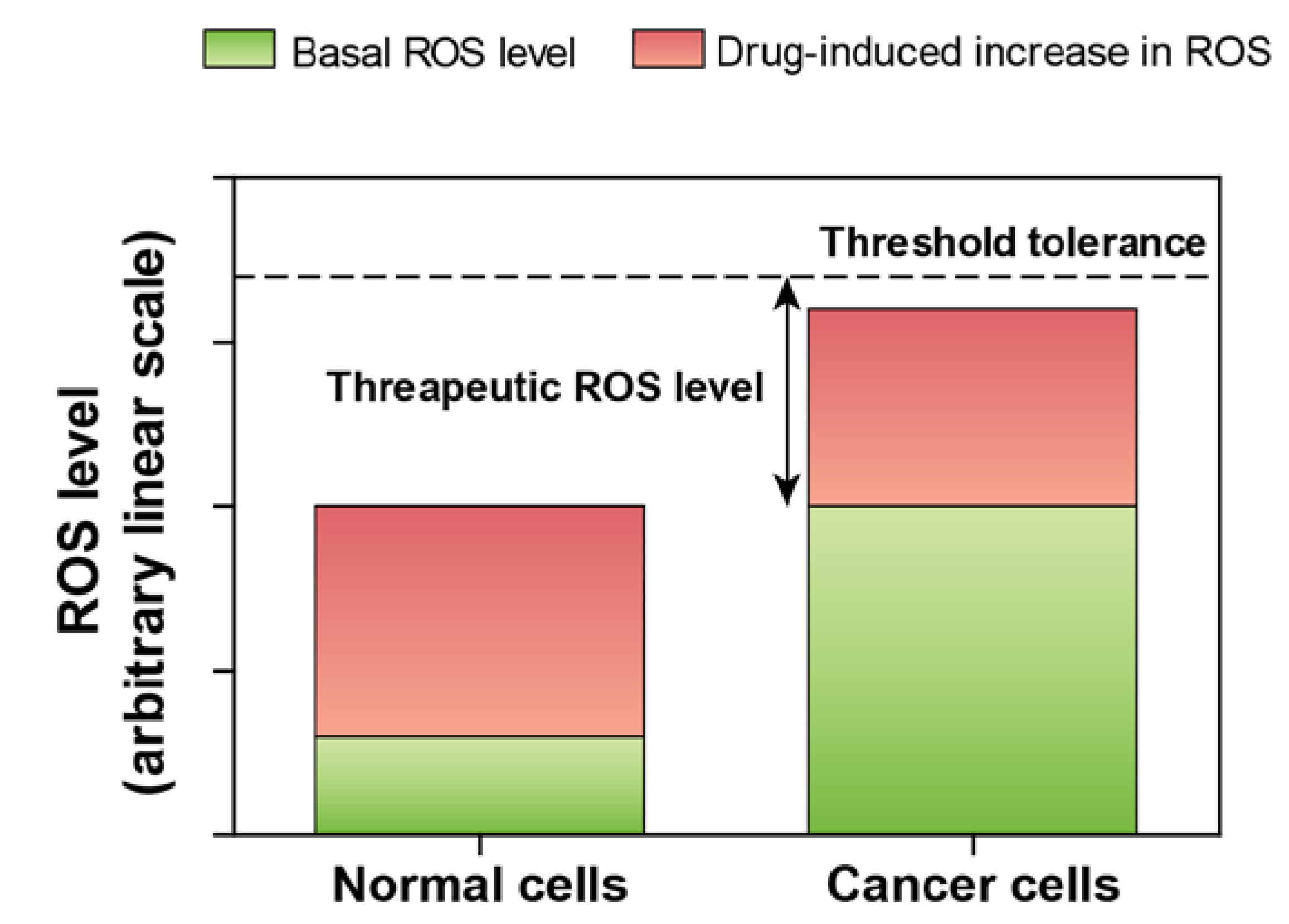
2. Selenium—An Antioxidant with Strong Pro-Oxidant Properties

Oxidation-reduction reactions are critical for the maintenance of the physiological homeostasis both in unicellular and multicellular organisms. The term “oxidative stress” is defined as the perturbations of the physiological redox homeostasis when the rate of cellular reduction is overwhelmed by the rate of cellular oxidation [5]. Both the enzymatic and non-enzymatic antioxidants can reverse this balance when the cellular damage is not beyond repair. In the context of selenium, the antioxidant properties are predominantly exerted by its incorporation into selenoproteins that can catalyze the reduction of disulfide bonds in proteins and peptides [6,7]. Such reductive properties are important for protecting against indiscriminate oxidation of intracellular and extracellular constituents by intrinsic and extrinsic oxidants, spanning the spectrum of disease conditions to harmful chemicals. Therefore, deciphering the physiological roles of selenium as an antioxidant is one of the key research areas that have been investigated and reinvestigated ever since the discovery of selenium as an essential trace element by Schwarz and Foltz in 1957 [8].
There is a broad diversity in nature of chemical species of selenium due to its four different oxidation states (−2, 0, +4 and +6). Interestingly, all selenium compounds with their different oxidation states are implicated in increased selenoprotein expression in in vitro and in vivo investigations; hence all oxidation states are potentially bioavailable for selenoprotein biosynthesis. These chemically diverse selenium compounds are utilized for physiological functions by unique metabolic pathways for each compound with variable degrees of overlapping intermediate metabolites. While hydrogen selenide is one of the major intermediary metabolites of inorganic selenium compounds, methylated selenium compounds (described in details in later section) are the major intermediary metabolites of organic selenium compounds [9,10]. On the basis of an increased capacity of selenoprotein synthesis by both of these classes of selenium compounds in eukaryotes, it is plausible that they are metabolized into a common pool of hydrogen selenide. Hydrogen selenide is subsequently converted into seleno-phosphate by seleno-phosphate synthetase 2 [11], a key step for the biosynthesis of selenocysteine and selenoproteins [12].
There is a stringent physiological regulation on the extensive capacity of selenoproteins biosynthesis in eukaryotes upon selenium supplementation in any chemical form. An enzymatic and protein saturation effect is found at optimum supplementation levels. Under this condition, any excess supply of selenium results in increased metabolism, but marginal or no further increases in selenoprotein biosynthesis. Many of these metabolites including hydrogen selenide and monomethylselenol are highly redox reactive and generate reactive oxygen species (ROS) upon reaction with and oxidation of thiols [13]. These compounds are therefore termed as redox-active selenium compounds (e.g., selenite, selenocystine, methylseleninic acid, Se-methylselenocysteine, etc.) that are known to exert oxidative stress [13]. Such pro-oxidative properties reflect the opposite spectrum of a common consensus that selenium is just an antioxidant [14]. It may, therefore, be clarified by selenium researchers, when so ever applicable, that redox-active selenium compounds are not antioxidants by themselves, but only when incorporated into selenoproteins with oxidoreductase functions and supplied at dietary dose levels corresponding to physiological optima.
From the above discussion, it is obvious that supplementation of supra-physiological levels of redox-active selenium compounds is necessary for induction of oxidative stress. An interesting question pertaining to such unfavorable biological effect is what implications this might have within the context of human health and disease? Although no obvious benefits may be conceived of by the damaging nature of ROS in a healthy condition, a potential application in the treatment of cancer can be harnessed, as there is a significant and emerging interest in the recent development of cancer therapeutics based on ROS-mediated mechanisms of action [15,16,17]. The underlying concept relies on the observations that cancer cells usually exhibit high basal levels of ROS compared to normal cells. Increased basal ROS levels and concomitant up regulation of antioxidant defense systems are pathophysiological adaptations of cancer cells favoring malignancy [16]. However, cancer cells have a lower tolerance to increased levels of ROS compared to normal cells [15,18], thereby providing a plausible window for therapeutic interventions by redox-active selenium compounds (Figure 1) [19,20].
Figure 1. Effect of elevated level of ROS in normal and cancer cells.
Nutrients 07 03536 g001

3. Acute and Chronic Selenium Toxicity

To understand the anti-neoplastic mechanisms of redox active selenium compounds, its toxicity needs to be clarified. There are several reports of chronic and acute toxicity in man and animals, of natural, accidental and suicidal causes. A recent incident with fatal consequences happened in April 2009 where probably the largest test of acute selenium toxicity was unknowingly carried out in Venezuelan Polo Horses. The prized Lechuza Caracas horses were given an IV injection of Biodyl, a complex of vitamins and minerals used in Europe to increase stamina and reduce recovery time from fatigue in horses. The Biodyl formulation, unapproved in the United States by the Food and Drug Administration, was formulated in a pharmacy in Ocala, Florida. Several hours before the polo event, the horses were given an IV injection of Biodyl. Within a short period of time the horses began to collapse and within a few hours 21 of the prized Polo horses were dead.
The cause of death of the horses was reported to be owing to a misformulation of the Biodyl administered to the horses prior to the Polo match. The ensuing investigation focused upon the compounding pharmacy and the discovery that a mistake had been made in the amount of the sodium selenite component of the Biodyl formulation. The formulary chemist had mistakenly added ten times the amount of selenium that should have been contained in the Biodyl later injected into the horses. The Biodyl formula normally called for 500 µg of sodium selenite per mL of formulary but the amount of selenium from the pharmacy contained 5000 µg of sodium selenite per mL in the Biodyl formulation. News agencies reported that the fatal oral dose of sodium selenite to horses is 3300 µg or 3.3 mg/kg per body weight.
The amount of Biodyl injected into these horses by volume remains uncertain from the news reports. However, amounts of sodium selenite can be estimated from the treatment guidance for horses given from the Biodyl website. For horses, the French recommendation is for from 4–5 Biodyl IV administrations of 20 mL of Biodyl; each 4–5 h prior to the tournament or event. Thus these horses, by assumption, may potentially have received IV up to 20–25 g of selenium as sodium selenite within a relatively short period of time. While this is almost equivalent to the dietary toxicity of sodium selenite reported for horses, the fact that it was likely administered IV would make the sodium selenite much more toxic. The likely cause of death would have been massive erythrocyte hemolysis and suffocation consistent with the reported symptoms of the events at the time.
While the Polo horse deaths were indeed acute and tragic, chronic selenium toxicity in horses was also likely described by Marco Polo, circa 1265, while traveling the “Silk Road” in China [21]. Chronic selenium toxicity among horses and other range animals was reported and identified as such in the western United States in the 1930s. The differences systemically between acute and chronic selenium toxicity is dependent upon the dose, time and chemical species of the selenium compound either injected, as in the Polo horses, or as ingested by horses in the open range. It does seem that a common underlying effect of selenium toxicity, based upon a great deal of in vivo research with cells and in animals, is selenium’s ability as discussed below to catalytically oxidize thiols generating oxidative stress [22,23]. Selenium compounds of sufficient concentration to generate oxidative stress from thiols (RSH) must it seems have to be always present as a selenide or selenolate anion (RSe¯), as in sulfides (RS¯). Such anions of selenium are formed from the reaction of selenite with glutathione or other thiols. They initially generate a selenotrisulfide (RSSeSR), as initially proposed by Ganther [24] and upon further reduction form an unstable reduced selenopersulfide; RSSeH, ionizing at physiological pH forming the selenopersulfide anion, RSSe¯ [25]. This selenide catalyst oxidizes non-stociometrically additional thiols present cycling electrons through the selenium anion from thiols to oxygen forming superoxide, as originally proposed and shown below by Seko [26], for selenite (Figure 2) and later by Chaudière et al. for organic diselenides (Figure 3) [27]. It is from such reactions that the oxidative stress causing toxicity and the death of the Polo horses from selenite likely involved extensive hemolysis. The selenopersulfide anion catalyst is seen in reactions with selenite and GSH; and as the selenide or selenoate anion upon reduction in many diselenides experimentally using luminometry that measures superoxide [28]. Toxicity of selenium compounds was seen in early oral comparisons of selenium toxicity in mice, from a large library of mono- and diselenides as reported by Schwarz [29]. Almost without exception, organic diselenides were more toxic to mice than the corresponding monoselenides, due to the relative ease of diselenides being reduced to two catalytic organic selenides not readily occurring with monoselenides, (RSeR).
Figure 2. Reaction of selenite with glutathione and subsequent generation of superoxide anion, adapted from [2].
Nutrients 07 03536 g002
Figure 3. Redox cycling of selenium intermediates in the presence of GSH. Adapted from [27] and later republished [30]. Presented with permission from Taylor and Francis Group.
Nutrients 07 03536 g003

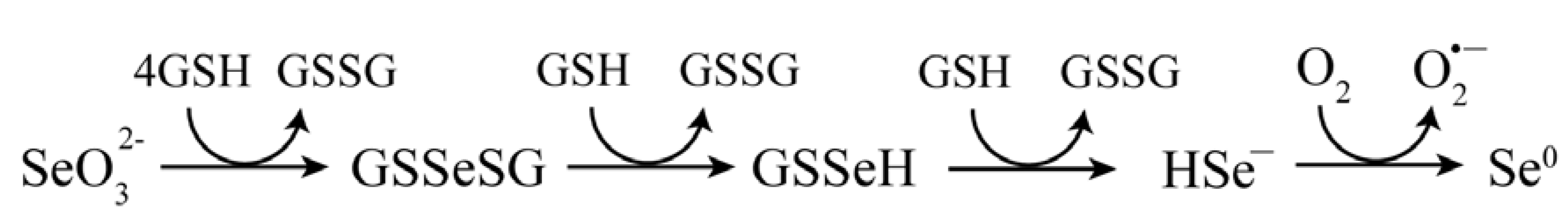
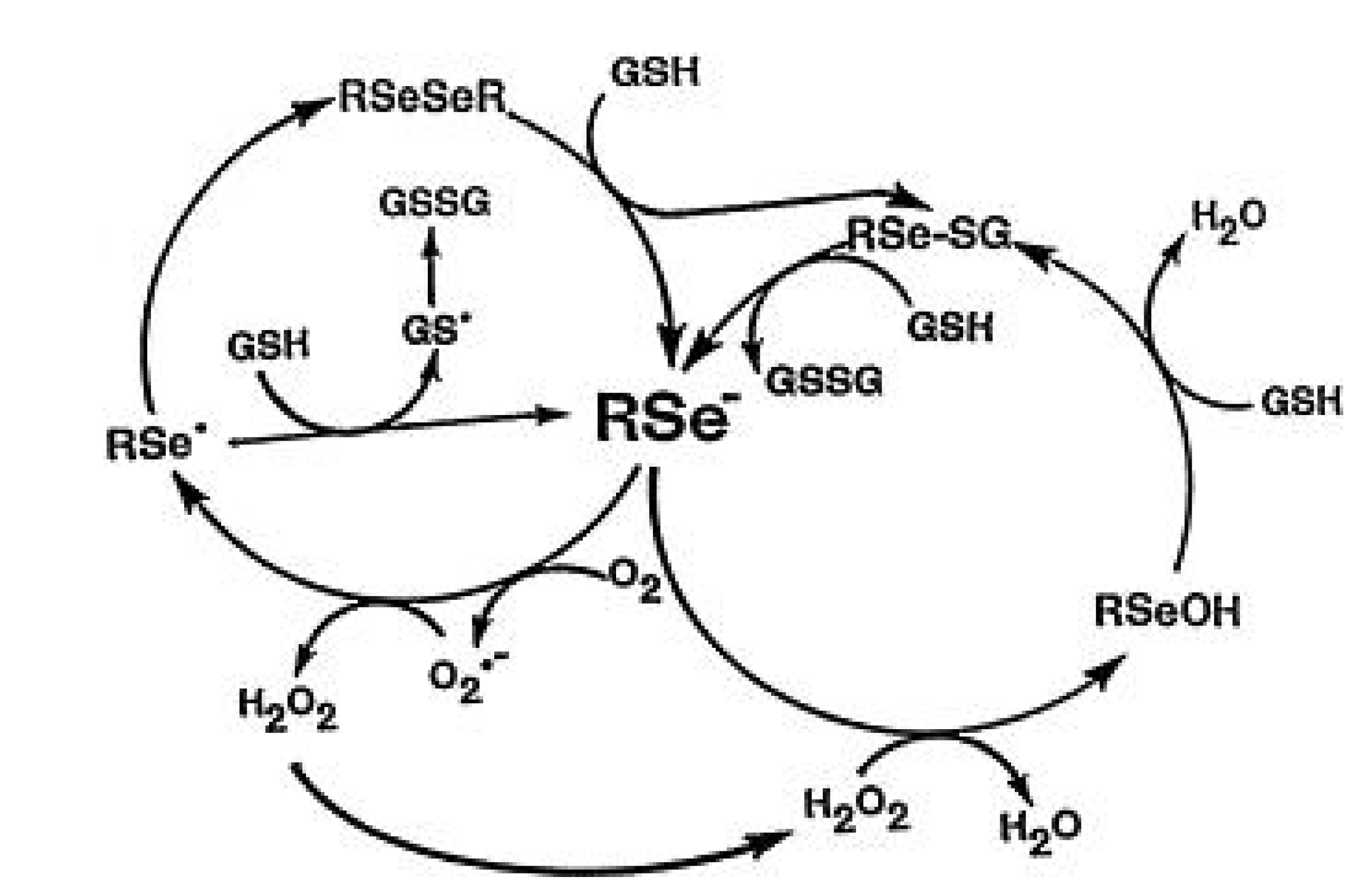
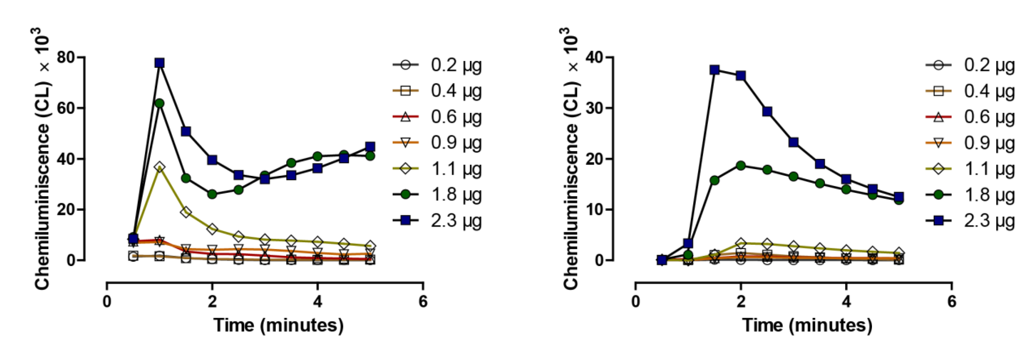
4. Redox Active Selenium Compounds and Detection of Superoxide

As noted above, selenium compounds are toxic based upon their chemical form, the dose and the biological exposure time. At nutritional levels of selenium intake and with a TUL of 400 µg Se/day, dietary selenium or supplemental selenium is not toxic to humans. In 1989, Seko et al. [31] were the first to suggest that the catalytic activity of selenite having been observed earlier by Feigal and West [32], by Tsen and Tappel, [33] by Rhead and Schrauzer [34] and later confirmed by Spallholz and Whittam [35], was likely toxic because it oxidized thiols and generated superoxide from GSH as shown in Figure 2 above by Seko and Imura [26].
The chemistry above is easily quantitated by the measurement of superoxide, the actively generated end product of the reaction. The superoxide so generated can be quantitated by a number of assays including cytochrome c [36] and methylene blue reduction [34] using visual spectroscopy. The superoxide electron may also be “trapped” and measured by ESR, Electron Spin Resonance Spectroscopy. We use a chemiluminescent cocktail of GSH and Lucigenin (bis-N-methylacridinium nitrate) and a water-jacketed luminometer to routinely measure superoxide. Additionally, quantitatively superoxide dismutase (SOD) [28,37] as well as iodoacetate, quenches the above and similar selenide or selenoate reactions with thiols confirming the generation of superoxide. In vitro and/or ex vivo a probe for superoxide, dihydroethidium (DHE) and its florescence, comes from the reaction of DHE with superoxide; Lambda Ex, 380 nm; 500 nm, Lambda Em, 580 nm. Cells treated with DHE from the reaction with superoxide will appear red in color when intercalating with DNA. The reaction can be semi-quantitated visually and fully quantitated using a fluorescence plate reader.
In 1992, Chaudière et al. [27] showed that for the diselenide selenocystamine, reduction of selenocystamine by GSH produced two molecules of redox active selenides that redox cycled in the presence of GSH. The organic diselenides, as shown in Figure 3 from Spallholz et al. [30] below, like selenite, generates superoxide while consuming oxygen, and these authors likewise concluded that this redox chemistry of organic diselenides, as proposed for selenite by Seko et al. [31], likely accounts for selenium’s toxicity.
The reaction of selenocystamine as shown in the above equation in Figure 3 has also been extended to other aliphatic and aromatic diselenides. The catalytic selenium species in Figure 2 is likely to be GSSe-, a selenopersulfide, as selenodiglutathione (on left), the intermediate of his equation is more catalytic in a comparative chemiluminescent assay with selenite (on right) when equivalent amounts of selenium are added to equal amounts of GSH, (Figure 4) [38].
Figure 4. Quantitative analysis of generation of ROS, superoxide, by selenodiglutathione (left) and by selenite (right) in a lucigenin chemiluminescence-based assay, adapted from [38].
Nutrients 07 03536 g004
Superoxide is being measured in both panels of Figure 4 under conditions as in the Seko equation (Figure 2). On the left is the reaction with selenodiglutathione, and on the right, with selenite, the chemiluminescence is being shown over 5 min (mean of n = 3). The selenium, GSH concentration and all other parameters are the same in both assays. Note the chemiluminescent maxima are reached with GSSeSG nearly a full minute prior to being reached with addition of selenite. Total chemiluminescence generated by GSSeSG is also greater at all thiol concentrations in comparison to selenite. Our explanation for these differences is the time lag required for selenite to react with GSH forming GSSeSG, and the GSSe¯ catalytic anion, already preformed when GSSeSG is added to the reaction, requiring only a single reduction to the GSSe¯ anion, which we purpose as the catalytic species of the Seko equation. Both reactions with either GSSeSG or selenite eventually terminate with the formation of elemental selenium as shown by Seko (Figure 2) [26].
In addition to almost all selenium diselenides, selenium isoselenocyanates, (RNCSe) as well as isothiocyanates (RNCS) may continuously redox cycle in the presence of thiols and generate superoxide, as shown by Crampsie et al. [39] (Figure 5) without generating elemental selenium.
Figure 5. Comparative catalytic oxidation of GSH by isothiocyanates and isoselenocyanates, adapted from [39].
Nutrients 07 03536 g005
The redox activity of the reduced diselenides depends upon the R group moiety as it affects the oxidizing ability of the Se anion. The same is likely true for the isoselenocyanates (RNCSe) as well as the isothiocyanates (RNCS). In essence, both classes of the latter compounds likely have resonant features depending upon R that makes their analogs redox cycle. In any case, the selenium compounds, as shown with their sulfur analogs above are usually more redox active in generating superoxide than the corresponding sulfur analogs. A long held chemoprevention compound, sulforaphane, found in various cruciferous and other plant foods, is known to induce oxidative stress and upregulate Phase 1 and 2 enzymes, as does supplemental selenium [40]. It would be interesting to test sulforaphane in vitro in the chemiluminescent assay to see if, like the other isothiocyanates, it redox cycles, generating superoxide, as it is reported to do in human bronchial epithelial cells [41]. Many other authors have described either the toxicity and/or redox cycling of other selenium compounds and only some of these reports have been referenced here. The compounds include a number of modified organic compounds with selenium and most recently the emergence of seleno-nano particles containing selenite [42]. The authors believe that it can be concluded that those selenium compounds that generally redox cycle in vitro in any chemical spectral, fluorescence and/or chemiluminescense assay are very likely to be the more toxic selenium compounds to cells in culture and to animals in vivo.
As originally reported by Misra in 1974, the autoxidation of GSH spontaneously generates superoxide [43]. Therefore, across experimental time, researchers have consciously or unconsciously shown that selenium toxicity is simply manifested by the accelerated oxidation of GSH and other thiols generating superoxide and other reactive oxygen species. The actual death of the Polo Horses, so noted above, likely came from a combination of selenium and Fenton chemistry within the erythrocytes. A best guess scenario is selenite rapidly entered the horse erythrocytes generating superoxide and hydrogen peroxide. The iron being disturbed from hemoglobin induced Fenton chemistry, forming the highly toxic hydroxyl radical (OH˙) [44]. The addition of selenite to rat erythrocytes in vitro is known to cause hemolysis and cell destruction, as can be seen in scanning electron micrographs of the erythrocytes [45]. This redox chemistry can also be seen on polymer matrixes, where selenides are covalently or hydrophobically attached to cellulose, poly(methyl) methacrylate, or silicon. Such polymer surfaces coated with redox selenium become catalytic, generate superoxide in the presence of GSH, and visibly “oxidize” bacteria in their prevention of a biofilm formation on the polymer surface [46].
In addition to GSH and other low molecular weight thiols, the thioredoxin and glutaredoxin systems efficiently redox cycle with selenide or selenolates in the presence of oxygen [1,2,3]. These redox systems also reduce selenite and GSSeSG to selenide and to a limited extent reduce selenium from its highest oxidation state, selenate. Thus, selenium compounds that have a free selenolate or selenide, where a free selenolate might be formed, react widely within the cell with all accessible thiols and several redox systems, resulting in a non-stoichiometric consumption of thiols, NADPH and oxygen forming ROS and oxidative stress.

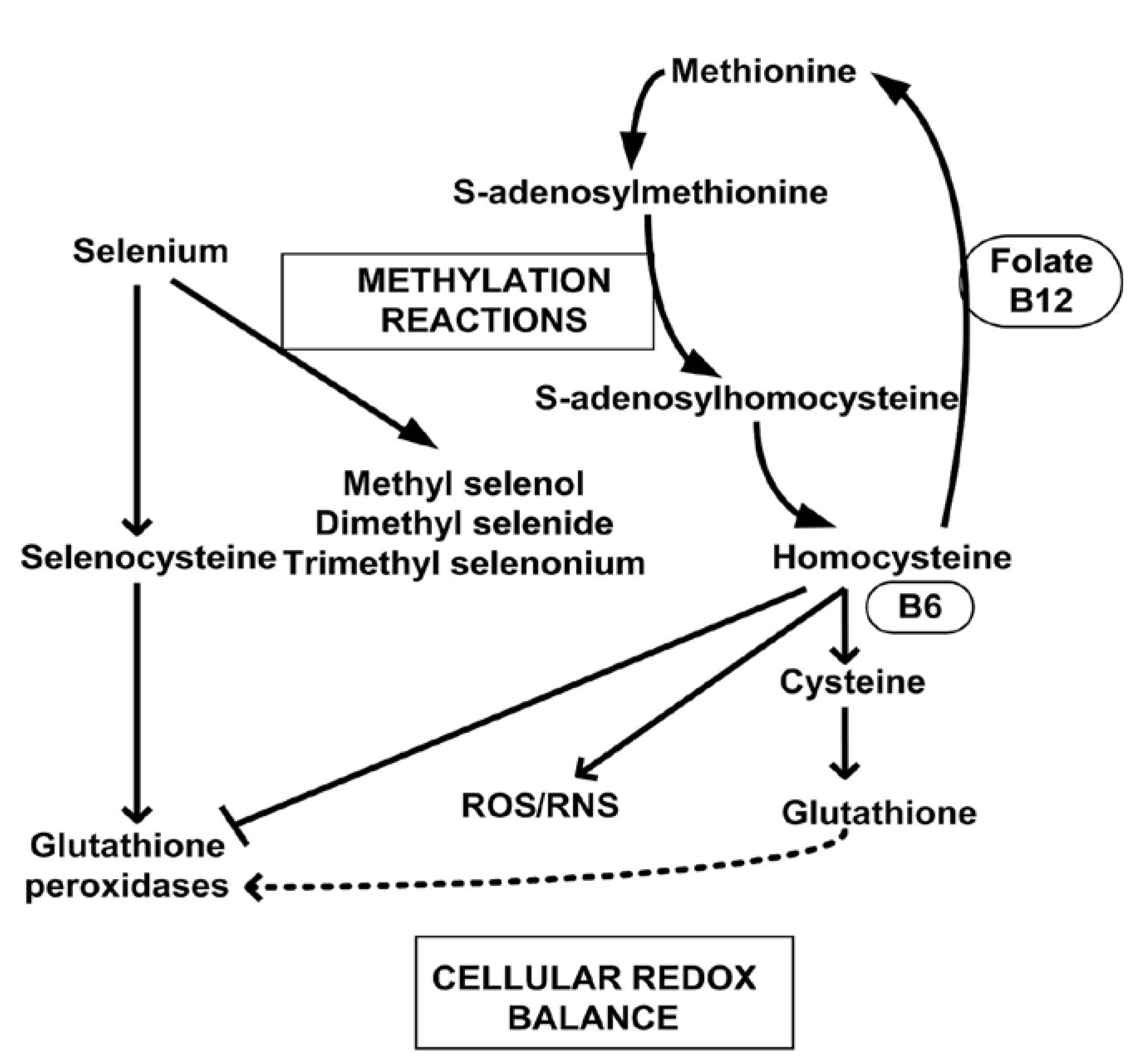
5. Methylation Reactions and Methylated Selenium Species

In order to prevent selenium toxicity by selenoate or selenide formation in animals and cells, methylated selenides are synthesized forming mono- and dimethylselenide and a trimethylselenonium ion [47]. Complete or dimethylation renders any monoselenide non-catalytic, non-toxic, and, like other nutritional mono- and diselenides, they are ultimately and mostly excreted through the lungs or urine, following metabolism. The trimethylselenonium ion and Se-methylselenol-N-acetyl-galactosamine are major urinary metabolites of selenium [48]. Methylation of selenium is why the major dietary monoselenides, selenomethionine and Se-methylselenocysteine, do not generate superoxide in vitro and are not very toxic in vivo. Methylselenol, really methylselenide (CH3Se¯), is toxic and redox cycles when formed from reduced dimethyldiselenide, or when these two dietary amino acids are metabolically acted upon by gamma- and beta-lyases generating methylselenol; methylselenide. There is some consensus within the literature that the monomethylselenide is likely the chemoprevention/anti-cancer selenium metabolite [49] formed from the metabolism of selenomethionine, Se-methylselenocysteine or H2Se, which may also redox cycle as HSe¯, prior to being methylated as methylselenide. The metabolic pathway of selenium methylation and methylation reactions are shown in Figure 6 [50].
Methylation of some metals, non-metals and metalloids, which induce oxidative stress and are toxic, seems to be a general means of biological detoxification [51]. Mercury maybe the exception as inorganic mercury is less toxic than mono- or dimethyl mercury. Epigenetic control of genes is perhaps also influenced by the methylated dietary selenoamino acids and metabolic selenium intermediates [52].
Figure 6. Important selenium methylation reactions and redox species in the maintenance of cell’s redox balance, adapted from [50].
Nutrients 07 03536 g006

6. Potent Antitumor Effects of Redox-Active Selenium Compounds

The redox reactivity of different selenium compounds has been described in the previous sections above. It seems obvious that the pro-oxidant functions of many of these redox-active forms of selenium are the key to their anticancer effects. We have recently documented the anticancer effects of selenite in detail (to be published soon). Herein we present the toxicity of two other important redox-active selenium compounds, Se-methylselenocysteine and selenocystine, and the rationale for their use as potential cancer chemotherapeutics.

7. Se-Methylselenocysteine
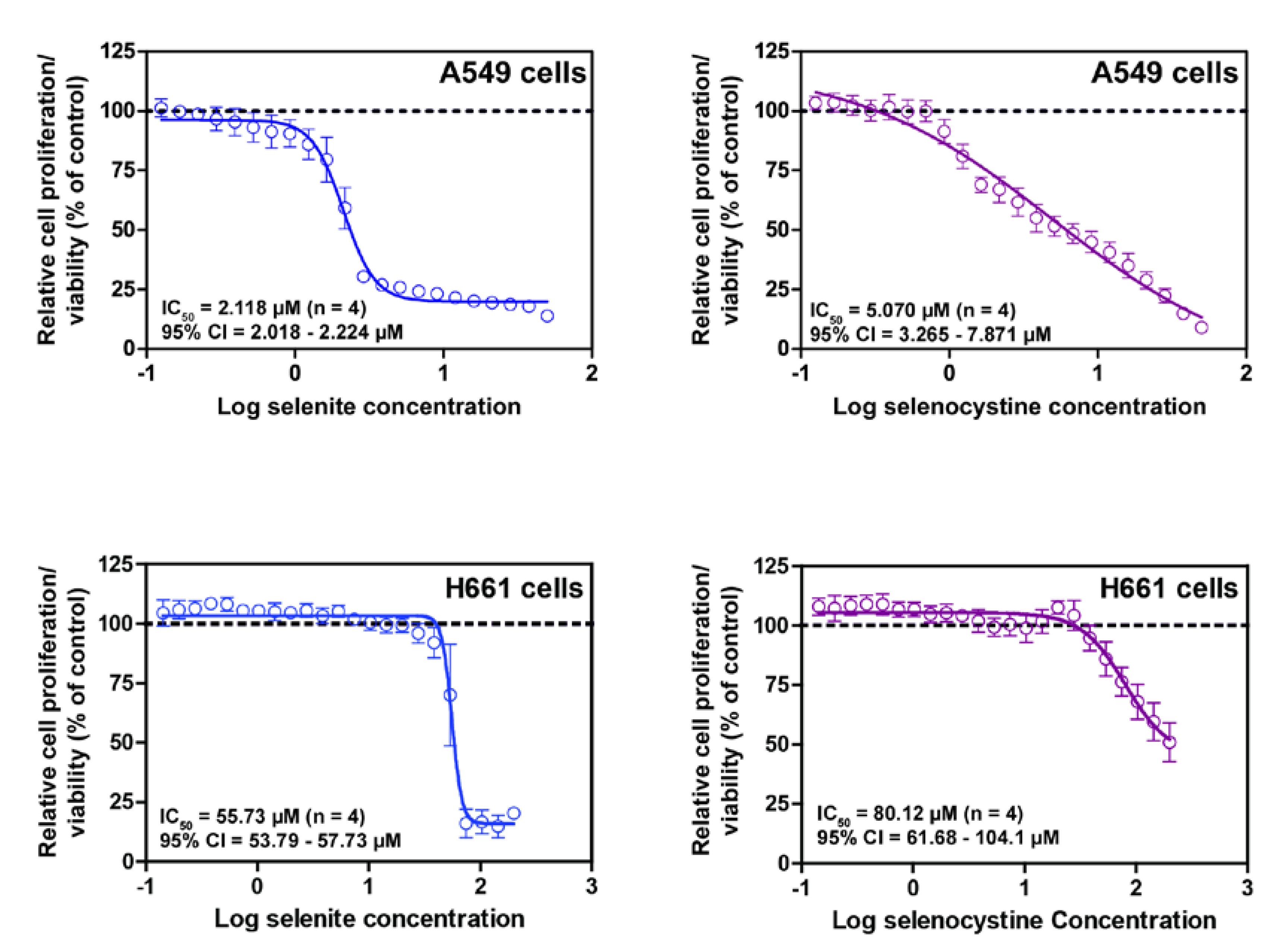
Se-methylselenocysteine (SMC) is not a standalone redox-active compound by itself. It is metabolized by glutamine transaminase K into methylselenol, which is highly redox reactive (described in earlier section). SMC is highly bioavailable, as indicated by increased SEPP biosynthesis and GPX1 activity upon supplementation, but relatively less toxic to cancer cells compared to other selenium compounds [53]. The reported 72 h IC50 values are 177 ± 32.2 µM and 100 ± 20 µM for HepG2 [53] and A549 cells [54], respectively. However, SMC is not very toxic to normal human cells (our own unpublished data). The significance of such observations indicates that it is possible to achieve a therapeutic selenium window. However, the in vivo therapeutic efficacy of SMC as an anticancer treatment in humans remains as an important question to be addressed.
Emerging data from animal studies indicate encouraging anticancer properties of SMC. In an orthotropic model of human colon carcinoma in athymic nude mice, it was shown that SMC reduces blood volume and micro vessel density in transplanted tumors [55]. Such inhibitory effects on angiogenesis were believed to be mediated via down-regulation of VEGF, COX2, HIF-1α and iNOS, as reported in an earlier study [56]. SMC not only inhibited angiogenesis, but also increased vascular maturation and reduced interstitial fluid pressure (reviewed in [57]). These effects were shown to be specific for tumor but not for normal organs, like the liver [58]. Such observations are suggestive of potentially increased chemotherapeutic drug delivery to the tumor without further increase in side effects. In fact, a recent study clearly demonstrates the remarkable antitumor capacity of SMC in combination with four different cytostatic drugs along with reduced side effects [59]. Certain combinations of SMC and cytostatic drugs resulted in complete remissions in an advanced xenograft model of Ward colorectal carcinoma in rats and human head and neck tumors in nude mice. Most importantly, SMC completely protected against Oxaliplatin-induced severe myelosuppresion in Fisher rats. Henceforth, the unique potential of this selenium compound strongly encourages further investigations on its application in the treatment of human cancer.

8. Selenocystine

Selenocystine (CysSeSeCys), is one of the key redox-active selenium compounds with dual functionalities—acting both as an antioxidant and a pro-oxidant. The parent compound itself does not exhibit any redox activity unless reduced by low molecular thiols and disulfide reductases to selenocysteine (Sec), which is highly redox active. Thus selenocystine is not significantly different than the reduction of selenocystamine, as shown in Figure 3. Under reducing condition, glutathione peroxidase-like activity of selenocystine has long been known [60]. Selenocystine is efficiently reduced to Sec by mammalian thioredoxin reductase with an apparent Km and kcat values of 6.0 µM and 3200 min−1, respectively [61]. Due to the lower pKa value of Sec, compared to cysteine, it is an efficient nucleophile. An excellent review on this subject has been published elsewhere [62]. The nucleophilic nature of Sec makes it very unstable, hence a potent reducing agent in its free form. The involvement of Sec insertion machinery in selenoprotein biosynthesis is probably one of the classical examples of evolutionary adaptation to harness Sec via aminoacylation of tRNA[Ser]Sec thereby protecting the native form of Sec from its own nucleophilic activity prior to incorporation into selenoproteins.
The redox potential of the intracellular milieu is conducive for efficient reduction of selenocystine. At nutritional levels, it may be presumed that selenocystine functions as an antioxidant with high bioavailability [53]. In spite of a potent electrophile inactivation capacity of Sec, selenocysteine is a potent toxic agent when present in excess. One of the key redox-active intermediate metabolites of Sec is hydrogen selenide that is generated by the action of the pyridoxal phosphate-dependent enzyme, selenocysteine lyase. Since this is a common metabolite of sodium selenite, one would expect a similar cytotoxicity profile of selenocystine and selenite when their uptake is similar. To address this, we performed cytotoxicity assays with selenite and selenocystine in A549 and H661 lung cancer cells. As shown in Figure 7, we found a remarkably similar cytotoxicity profile for these compounds. In another study, the 72 h IC50 data for these two compounds [63] corroborate very well with such assumptions. In this study, the authors noted higher sensitivity of selenite to normal cells compared to selenocystine (IC50 > 400 µM). Similar cytotoxic effects of selenocystine have been reported in mice thymic lymphoma cells, but not in isolated primary spleen lymphocytes [64]. This is somewhat suggestive of the cancer cell-specific effects of this compound. In a recent study, we have shown that selenocystine induces an unfolded protein response (UPR), ER-stress and large cytoplasmic vacuolization in HeLa cells [9]. The involvement of UPR as one of the key stress-associated pathway provides a possible mechanistic link between misfolded proteins and the disulfide reductase activity of Sec. It has been shown that selenocystine is less toxic compared to selenite at comparable doses in early postnatal male Wister rats [65]. Orally administered selenocystine is well tolerated in ICR mice when given up to 10 mg/kg body weight with no increase in plasma AST and ALT levels [66].
Figure 7. Cytotoxicity of selenite and selenocystine, as measured by a WST-1 assay kit (Roche, Mannheim, Germany), to A549 and H661 cells following 24 h exposure to 22 different selenium concentrations. The highest concentration of selenite and selenocystine was 50 and 200 µM for A549 and H661 cells, respectively. Note that due to low toxicity of selenocystine in H661 cells, the IC50 value should be interpreted carefully.
Nutrients 07 03536 g007
The pharmacokinetics data suggest that intravenous administration of this compound is very efficient in maintaining plasma selenium levels in comparison to oral administration in mice [67]. Early studies on DMBA-induced mammary carcinogenesis in BALB/c female mice showed limited protective effects of selenocystine on mammary adenocarcinoma [68]. The same study reported a significant lesser effect than selenite treatment. Comparative analysis indicates that this compound induces less DNA damage compared to selenite [69].
The most remarkable effects of selenocystine in humans were shown in the treatment of acute and chronic myeloid leukemia [70]. The authors reported greater effects of this compound on immature leucocytes than in mature leucocytes without any notable effect on bone marrow. Another interesting finding from the same study indicated increased response to selenocystine treatment in patients’ refractory to other chemotherapeutic agents then available. This recapitulates the findings from our own clinical trial study in which many patients who did not respond to cytostatic drugs, responded to subsequent selenite treatment (unpublished data, [71]). All the above studies clearly indicate that selenocystine may be a potential cancer chemotherapeutic candidate, especially for leukemias.

9. Selenium in the Treatment of Cancer—A New Era in Oncology

The applied dose and chemical species of selenium are the important aspects in the context of the development of tumor and neoplastic growth. Undoubtedly, selenium at low doses incorporated into enzymes and proteins function as an antioxidant for both normal and cancer cells. For the latter, optimum selenium anti-oxidant activity renders survival advantages to these genetically damaged cells during the progression and promotion stages of tumorigenesis. Nevertheless, selenium as an antioxidant protects against DNA damage that accumulates during the initiation phase of tumor development in a similar dose regimen. The augment of an antioxidant-based protective effect and improved immune functions by selenium are critical aspects for minimizing DNA damage and elimination of damaged cells by immune surveillance. Hence, the cancer chemopreventive effects of selenium as a single agent can only be predicted at physiologically relevant doses to evade DNA damage associated with carcinogenesis. In summary, it is the context that dictates the biological effects of nutritional levels of selenium at different stages of tumor development.
A purely cancer chemotherapeutic application of selenium requires higher doses of redox-active selenium compounds. Numerous studies have reported growth inhibitory and anti-neoplastic effects of selenium both in vitro and in animal models. Prior to the understanding of the pro-oxidative activity of selenite and diselenides, Greeder and Milner were much more successful in treating Ehrlich Ascites Tumor cells in mice with the later selenium compounds than with selenomethionine on a dose related basis [72]. Nevertheless, there are very few reports on the application of high doses of selenium for cancer treatment in humans. In the first half of the 20th century, colloidal selenium was successfully used for cancer treatment [73]. The evaluation of treatment efficacy is difficult to interpret from these studies, since the standard RECIST criteria were not available and the radiological methods were very primitive. A case series in 1956 reported favorable effects of selenocystine in the treatment of leukemia [70]. In two recent studies, moderate doses of selenite 0.2 mg/kg/day for 7 and 30 days, respectively, were administered together with standard cytostatic therapy with the primary aim to decrease side effects in patients with non-Hodgkin Lymphoma. The result was encouraging since the administration of selenite lead to a better response to therapy, apoptosis in lymphoma cells and a down-regulation of Bcl-2 [74,75]. Despite these promising reports, there has instead of a selenium treatment focus, there has been predominately only a focus of the possible preventive effects of selenium within the selenium research field. However, due to the failure of the SELECT trial [76] to prevent prostate cancer based on the use of selenomethionine and vitamin E, there has become confusion about the role and effects of selenium in the prevention of cancer. Several studies report beneficial effects by decreased side effects of the combination of low doses of selenium with cytostatic drugs and radiotherapy [77,78]. However, this possible clinical use of selenium and the preventive effects of cryotherapy are beyond the scope of the present review and will not be treated herein.
The major challenge in the use of selenium as a cancer therapeutic is to deliver redox active selenium, preferentially and directly to the tumor and/or metastatic cells. The most studied and simplest selenium compound, selenite must be administrated intravenously since no or very limited amounts of it will be detected in plasma after oral administration. We have recently completed a unique phase I trial in which the maximum tolerated dose, pharmacokinetics and safety of selenite was established (Unpublished data, [71]). The findings from this study will facilitate a long anticipated effect of selenite evaluation studies possible in humans. Selenite requires reduction prior to uptake and a reducing extracellular environment would facilitate its uptake [79]. Selenite might also be reduced by plasma thiols and there is possibly a complex kinetic mechanism where part of the administrated selenite will be reduced to elemental selenium [80]. Elemental selenium could serve as a storage form for further reduction to selenide. A similar metabolic transformation of selenite into elemental selenium has been shown in cancer cells [81] and in other animal species [82]. Selenomethionine is not suitable as a cytostatic drug since it is not redox active (rather a scavenger of ROS by auto-oxidation) and has a long biological half-life, due to its random incorporation in proteins in place of methionine, particularly albumin. Furthermore, although selenomethionine is a potential source of methylselenol (methylselenide) it is not applicable in humans due to a very poor activity of gamma-lyase.
Se-Methylselenocysteine (SMC) (see section of methylated selenium compounds) is a promising per oral candidate drug due to a very good bioavailability and the presence of active beta-lyases in human cells [83] (our unpublished data). This selenium compound is also one of the active organic constituents in selenized yeast. The anti-neoplastic action of SMC depends on the expression of beta-lyases. Only limited information is present regarding the expression of these highly interesting classes of enzymes in tumors. Further studies are ongoing to determine which tumor types SMC would be an efficient chemotherapeutic due to possible elevated expression of beta-lyases.

10. Novel Selenium Compounds and Future Perspectives for Therapeutic Selenium Drugs

The use of selenium in attempts to treat disease, particularly cancer, has a relatively long history dating back to at least 1912 when selenite was used to reportedly cure a tongue cancer. The literature contains many reports of selenium compounds, initially selenite in particular, but constantly being expanded to newer organic selenium compounds, arresting cancer cell growth in vitro. As noted above, some reports even suggest that selenium is more toxic to cancer cell lines than the more normal parent cells. It seems, on this basis, that the first reported successful prevention of prostate cancer in men was made by Clark et al. [84] using supplements of selenium-fortified yeast. When the major component of the selenium yeast, selenomethionine, was used in a larger human prostate cancer trial, the reported Clark outcome of the earlier trial was not, as noted, substantiated in the later larger human trial, SELECT [76]. Such prospective human trials, although few in number, and past animal cancer trials suggest to us that pure oral selenium supplementation of a normal selenium dietary intake will not prove to be broadly practical nor significantly beneficial to people with cancer ingesting adequate dietary selenium.
The authors do believe that it should be possible to target selenium’s toxicity for a more selective approach to treat diseases than by way of purely systemic supplementation. This approach today seems fully rational, as the toxicity of selenium compounds can be tailor-made and demonstrated to generate superoxide in cells in vitro. As in Figure 5 with the isothiocyanates and isoselenocyanates, it is empirically possible to design selenium compounds with organic functional groups that highly redox cycle and that can be covalently attached to improve anticancer drugs as in the case of Temozolomide [85], or attach redox selenium to monoclonal antibodies similar to T-DM-1, Kadcyla® (ado-Trastuzumab Emtansine), in which Herceptin® (Trastuzumab) carries the cytotoxic cytoskeletal drug, Emtansine. Over the course of the last decade, the authors and colleagues with whom we have collaborated have attached redox selenium to two vitamers, folic acid and biotin, both shown to redox cycle and are time and concentration dependently toxic to cancer cells in vitro. Together with graduate students, we have attached redox selenium to transferrin proteins [86] and monoclonal antibodies; Transferrin, Herceptin® (Trastuzumab), and Avastin® (Bevacizumab), all shown to be dose and time dependently more cytotoxic against cancer cells [87]. Such targeting of redox selenium offers the future prospect of using selenium toxicity in a more broadly beneficial therapeutic way.

11. Summary and Conclusions

Many selenium compounds are toxic, generating initially superoxide in vitro and thereafter other ROS ex vivo and in vivo. Toxicity is pH, selenium and thiol concentration dependent as measured for superoxide using chemiluminescense. Toxicity of selenite and organic diselenides, such as selenocystine, isoselenocyanates as well as aliphatic and aromatic selenocyanates are all positively or negatively affected by the R substitutions upon the selenide/selenonate anion. This information permits the synthesis of new selenium redox active compounds that may be added to existing drugs, or targeted with specific molecules, such as monoclonal antibodies. The modification of existing molecules with redox selenium, including nano-selenium containing particles is only limited by one’s imagination. One might even imagine a new era of selenium drug therapies [88].

Acknowledgments

We thank Cancerfonden (The Swedish Cancer Society), Cancer-och Allergifonden, Stockholm County Council (ALF), Stichting AF Jochnick Foundation and Radiumhemmets Forskningsfonder for grant support. We also acknowledge with appreciation support to the students of Nutritional Sciences at Texas Tech University participating in some of this research cited above over past years.

Author Contributions

All authors contributed in the writing of the manuscript; Mikael Björnstedt and Sougat Misra were mainly responsible for introduction, first, fifth and sixth sections. Julian Spallholz and Mallory Boylan were mainly responsible for the second, third, fourth and seventh sections. Arun Selvam performed the experiments illustrated in the Figure 7, wrote the abstract and selenocystine section and was responsible for the final editing of the entire manuscript.

Conflicts of Interest

MB is listed as inventor in a patent application that has been filed for the use of Selenite in cancer treatment. JS is listed as inventor of a US Patent for the attachment of redox selenium to targeting molecules and insoluble matrix substrates.

References

  1. Björnstedt, M.; Kumar, S.; Holmgren, A. Selenodiglutathione is a highly efficient oxidant of reduced thioredoxin and a substrate for mammalian thioredoxin reductase. J. Biol. Chem. 1992, 267, 8030–8034. [Google Scholar] [PubMed]
  2. Kumar, S.; Björnstedt, M.; Holmgren, A. Selenite is a substrate for calf thymus thioredoxin reductase and thioredoxin and elicits a large non-stoichiometric oxidation of NADPH in the presence of oxygen. Eur. J. Biochem. 1992, 207, 435–439. [Google Scholar] [CrossRef] [PubMed]
  3. Wallenberg, M.; Olm, E.; Hebert, C.; Björnstedt, M.; Fernandes, A.P. Selenium compounds are substrates for glutaredoxins: A novel pathway for selenium metabolism and a potential mechanism for selenium-mediated cytotoxicity. Biochem. J. 2010, 429, 85–93. [Google Scholar] [CrossRef] [PubMed]
  4. Selenius, M.; Rundlof, A.K.; Olm, E.; Fernandes, A.P.; Björnstedt, M. Selenium and the selenoprotein thioredoxin reductase in the prevention, treatment and diagnostics of cancer. Antioxid. Redox Signal. 2010, 12, 867–880. [Google Scholar] [CrossRef] [PubMed]
  5. Halliwell, B. Biochemistry of oxidative stress. Biochem. Soc. Trans. 2007, 35, 1147–1150. [Google Scholar] [CrossRef] [PubMed]
  6. Labunskyy, V.M.; Hatfield, D.L.; Gladyshev, V.N. Selenoproteins: Molecular pathways and physiological roles. Physiol. Rev. 2014, 94, 739–777. [Google Scholar] [CrossRef] [PubMed]
  7. Moghadaszadeh, B.; Beggs, A.H. Selenoproteins and their impact on human health through diverse physiological pathways. Physiology (Bethesda) 2006, 21, 307–315. [Google Scholar] [CrossRef]
  8. Schwarz, K.; Foltz, C.M. Selenium as an integral part of factor 3 against dietary necrotic liver degeneration. J. Am. Chem. Soc. 1957, 79, 3292–3293. [Google Scholar] [CrossRef]
  9. Wallenberg, M.; Misra, S.; Wasik, A.M.; Marzano, C.; Björnstedt, M.; Gandin, V.; Fernandes, A.P. Selenium induces a multi-targeted cell death process in addition to ROS formation. J. Cell. Mol. Med. 2014, 18, 671–684. [Google Scholar] [CrossRef] [PubMed]
  10. Weekley, C.M.; Harris, H.H. Which form is that? The importance of selenium speciation and metabolism in the prevention and treatment of disease. Chem. Soc. Rev. 2013, 42, 8870–8894. [Google Scholar] [CrossRef] [PubMed]
  11. Guimaraes, M.J.; Peterson, D.; Vicari, A.; Cocks, B.G.; Copeland, N.G.; Gilbert, D.J.; Jenkins, N.A.; Ferrick, D.A.; Kastelein, R.A.; Bazan, J.F.; et al. Identification of a novel seld homolog from eukaryotes, bacteria, and archaea: Is there an autoregulatory mechanism in selenocysteine metabolism? Proc. Natl. Acad. Sci. USA 1996, 93, 15086–15091. [Google Scholar] [CrossRef] [PubMed]
  12. Forstrom, J.W.; Zakowski, J.J.; Tappel, A.L. Identification of the catalytic site of rat liver glutathione peroxidase as selenocysteine. Biochemistry 1978, 17, 2639–2644. [Google Scholar] [CrossRef] [PubMed]
  13. Spallholz, J.E. On the nature of selenium toxicity and carcinostatic activity. Free Radic. Biol. Med. 1994, 17, 45–64. [Google Scholar] [CrossRef] [PubMed]
  14. Jukes, T.H. Selenium, an “essential poison”. J. Appl. Biochem. 1983, 5, 233–234. [Google Scholar] [PubMed]
  15. Gorrini, C.; Harris, I.S.; Mak, T.W. Modulation of oxidative stress as an anticancer strategy. Nat. Rev. Drug Discov. 2013, 12, 931–947. [Google Scholar] [CrossRef] [PubMed]
  16. Trachootham, D.; Alexandre, J.; Huang, P. Targeting cancer cells by ros-mediated mechanisms: A radical therapeutic approach? Nat. Rev. Drug Discov. 2009, 8, 579–591. [Google Scholar] [CrossRef] [PubMed]
  17. Wondrak, G.T. Redox-directed cancer therapeutics: Molecular mechanisms and opportunities. Antioxid. Redox Signal. 2009, 11, 3013–3069. [Google Scholar] [CrossRef] [PubMed]
  18. Kong, Q.; Beel, J.A.; Lillehei, K.O. A threshold concept for cancer therapy. Med. Hypotheses 2000, 55, 29–35. [Google Scholar] [CrossRef] [PubMed]
  19. Abdullaev, F.I.; MacVicar, C.; Frenkel, G.D. Inhibition by selenium of DNA and RNA synthesis in normal and malignant human cells in vitro. Cancer Lett. 1992, 65, 43–49. [Google Scholar] [CrossRef] [PubMed]
  20. Caffrey, P.B.; Frenkel, G.D. Selenite cytotoxicity in drug resistant and nonresistant human ovarian tumor cells. Cancer Res. 1992, 52, 4812–4816. [Google Scholar] [PubMed]
  21. Polo, M.; Marsden, W.; Corbino, J. The travels of marco polo the venetian; J.M. DENT SONS, Ltd.: London, UK, 1908; p. 439. [Google Scholar]
  22. Anundi, I.; Hogberg, J.; Stahl, A. Involvement of glutathione reductase in selenite metabolism and toxicity, studied in isolated rat hepatocytes. Arch. Toxicol. 1982, 50, 113–123. [Google Scholar] [CrossRef] [PubMed]
  23. Frenkel, G.D.; Falvey, D. Evidence for the involvement of sulfhydryl compounds in the inhibition of cellular DNA synthesis by selenite. Mol. Pharmacol. 1988, 34, 573–577. [Google Scholar] [PubMed]
  24. Ganther, H.E. Selenotrisulfides. Formation by the reaction of thiols with selenious acid. Biochemistry 1968, 7, 2898–2905. [Google Scholar] [CrossRef] [PubMed]
  25. Ganther, H.E. Reduction of the selenotrisulfide derivative of glutathione to a persulfide analog by glutathione reductase. Biochemistry 1971, 10, 4089–4098. [Google Scholar] [CrossRef] [PubMed]
  26. Seko, Y.; Imura, N. Active oxygen generation as a possible mechanism of selenium toxicity. Biomed. Environ. Sci. 1997, 10, 333–339. [Google Scholar] [PubMed]
  27. Chaudiere, J.; Courtin, O.; Leclaire, J. Glutathione oxidase activity of selenocystamine: A mechanistic study. Arch. Biochem. Biophys. 1992, 296, 328–336. [Google Scholar] [CrossRef] [PubMed]
  28. Chen, J.J.; Boylan, L.M.; Wu, C.K.; Spallholz, J.E. Oxidation of glutathione and superoxide generation by inorganic and organic selenium compounds. Biofactors 2007, 31, 55–66. [Google Scholar] [CrossRef] [PubMed]
  29. Schwarz, K. Biological potency of organic selenium compounds. I. Aliphatic monoseleno- and diseleno-dicarboxylic acids. J. Biol. Chem. 1969, 244, 2103–2110. [Google Scholar] [PubMed]
  30. Spallholz, J.E.; Shriver, B.J.; Reid, T.W. Dimethyldiselenide and methylseleninic acid generate superoxide in an in vitro chemiluminescence assay in the presence of glutathione: Implications for the anticarcinogenic activity of l-selenomethionine and l-se-methylselenocysteine. Nutr. Cancer 2001, 40, 34–41. [Google Scholar] [CrossRef] [PubMed]
  31. Seko, Y.; Saito, Y.; Kitahara, J.; Imura, N. Active oxygen generation by the reaction of selenite with reduced glutathione in vitro. In Selenium in Biology and Medicine; Wendel, A., Ed.; Springer Berlin Heidelberg: Tübingen, Germany, 1989; pp. 70–73. [Google Scholar]
  32. Feigl, F.; West, P.W. Test for selenium based on a catalytic effect. Anal. Chem. 1947, 19, 351–353. [Google Scholar] [CrossRef]
  33. Tsen, C.C.; Tappel, A.L. Catalytic oxidation of glutathione and other sulfhydryl compounds by selenite. J. Biol. Chem. 1958, 233, 1230–1232. [Google Scholar] [PubMed]
  34. Rhead, W.J.; Schrauzer, G.N. The selenium catalyzed reduction of methylene blue by thiols. Bioinorg. Chem. 1974, 3, 225–242. [Google Scholar] [CrossRef] [PubMed]
  35. Spallholz, J.E.; Whittam, J.H. Seleniurn toxicity interpreted from biological, catalytic, chemilurninescent and scanning electron microscopic data. In Proceedings of the Fifth International Symposium on Selenium in Biology and Medicine, Nashville, TN, USA, 20–23 July 1992.
  36. Levander, O.A.; Morris, V.C.; Higgs, D.J. Selenium as a catalyst for the reduction of cytochrome C by glutathione. Biochemistry 1973, 12, 4591–4595. [Google Scholar] [CrossRef] [PubMed]
  37. McCord, J.M.; Fridovich, I. The utility of superoxide dismutase in studying free radical reactions. I. Radicals generated by the interaction of sulfite, dimethyl sulfoxide, and oxygen. J. Biol. Chem. 1969, 244, 6056–6063. [Google Scholar] [PubMed]
  38. Goswami, D. Cytotoxic Effects of Selenium Conjugated Transferrins on Leukemia Cell Lines. Ph.D Thesis, Texas Tech University, Lubbock, TX, USA, 2014. [Google Scholar]
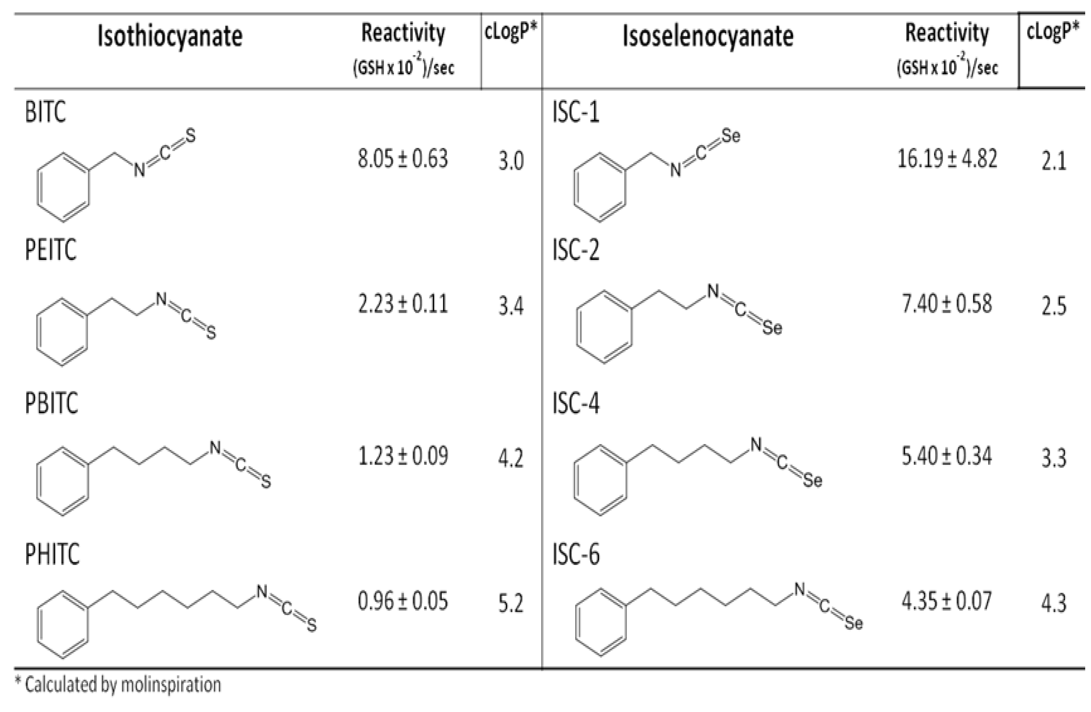
  39. Crampsie, M.A.; Pandey, M.K.; Desai, D.; Spallholz, J.; Amin, S.; Sharma, A.K. Phenylalkyl isoselenocyanates vs phenylalkyl isothiocyanates: Thiol reactivity and its implications. Chemico-Biol. Interact. 2012, 200, 28–37. [Google Scholar] [CrossRef]
  40. Lenzi, M.; Fimognari, C.; Hrelia, P. Sulforaphane as a promising molecule for fighting cancer. Cancer Treat. Res. 2014, 159, 207–223. [Google Scholar] [PubMed]
  41. Lee, Y.J.; Lee, S.H. Sulforaphane induces antioxidative and antiproliferative responses by generating reactive oxygen species in human bronchial epithelial beas-2b cells. J. Korean Med. Sci. 2011, 26, 1474–1482. [Google Scholar] [CrossRef] [PubMed]
  42. Liu, T.; Zeng, L.; Jiang, W.; Fu, Y.; Zheng, W.; Chen, T. Rational design of cancer-targeted selenium nanoparticles to antagonize multidrug resistance in cancer cells. Nanomedicine 2015. [Google Scholar] [CrossRef]
  43. Misra, H.P. Generation of superoxide free radical during the autoxidation of thiols. J. Biol. Chem. 1974, 249, 2151–2155. [Google Scholar] [PubMed]
  44. Gutteridge, J.M.; Beard, A.P.; Quinlan, G.J. Superoxide-dependent lipid peroxidation. Problems with the use of catalase as a specific probe for fenton-derived hydroxyl radicals. Biochem. Biophys. Res. Commun. 1983, 117, 901–907. [Google Scholar] [CrossRef] [PubMed]
  45. Hu, M.L.; Spallholz, J.E. In vitro hemolysis of rat erythrocytes by selenium compounds. Biochem. Pharmacol. 1983, 32, 957–961. [Google Scholar] [CrossRef] [PubMed]
  46. Tran, P.L.; Hammond, A.A.; Mosley, T.; Cortez, J.; Gray, T.; Colmer-Hamood, J.A.; Shashtri, M.; Spallholz, J.E.; Hamood, A.N.; Reid, T.W. Organoselenium coating on cellulose inhibits the formation of biofilms by pseudomonas aeruginosa and staphylococcus aureus. Appl. Environ. Microbiol. 2009, 75, 3586–3592. [Google Scholar] [CrossRef] [PubMed]
  47. Ganther, H.E. Enzymic synthesis of dimethyl selenide from sodium selenite in mouse liver extracts. Biochemistry 1966, 5, 1089–1098. [Google Scholar] [CrossRef] [PubMed]
  48. Loeschner, K.; Hadrup, N.; Hansen, M.; Pereira, S.A.; Gammelgaard, B.; Moller, L.H.; Mortensen, A.; Lam, H.R.; Larsen, E.H. Absorption, distribution, metabolism and excretion of selenium following oral administration of elemental selenium nanoparticles or selenite in rats. Metallomics 2014, 6, 330–337. [Google Scholar] [CrossRef] [PubMed]
  49. Ip, C.; Thompson, H.J.; Zhu, Z.; Ganther, H.E. In vitro and in vivo studies of methylseleninic acid: Evidence that a monomethylated selenium metabolite is critical for cancer chemoprevention. Cancer Res. 2000, 60, 2882–2886. [Google Scholar] [PubMed]
  50. Metes-Kosik, N.; Luptak, I.; Dibello, P.M.; Handy, D.E.; Tang, S.S.; Zhi, H.; Qin, F.; Jacobsen, D.W.; Loscalzo, J.; Joseph, J. Both selenium deficiency and modest selenium supplementation lead to myocardial fibrosis in mice via effects on redox-methylation balance. Mol. Nutr. Food Res. 2012, 56, 1812–1824. [Google Scholar] [CrossRef] [PubMed]
  51. Stohs, S.J.; Bagchi, D. Oxidative mechanisms in the toxicity of metal ions. Free Radic. Biol. Med. 1995, 18, 321–336. [Google Scholar] [CrossRef] [PubMed]
  52. Ong, T.P.; Moreno, F.S.; Ross, S.A. Targeting the epigenome with bioactive food components for cancer prevention. J. Nutrigenet. Nutrigenomics 2011, 4, 275–292. [Google Scholar] [CrossRef] [PubMed]
  53. Hoefig, C.S.; Renko, K.; Kohrle, J.; Birringer, M.; Schomburg, L. Comparison of different selenocompounds with respect to nutritional value vs. Toxicity using liver cells in culture. J. Nutr. Biochem. 2011, 22, 945–955. [Google Scholar] [CrossRef] [PubMed]
  54. Weekley, C.M.; Aitken, J.B.; Vogt, S.; Finney, L.A.; Paterson, D.J.; de Jonge, M.D.; Howard, D.L.; Musgrave, I.F.; Harris, H.H. Uptake, distribution, and speciation of selenoamino acids by human cancer cells: X-ray absorption and fluorescence methods. Biochemistry 2011, 50, 1641–1650. [Google Scholar] [CrossRef] [PubMed]
  55. Bhattacharya, A.; Turowski, S.G.; San Martin, I.D.; Rajput, A.; Rustum, Y.M.; Hoffman, R.M.; Seshadri, M. Magnetic resonance and fluorescence-protein imaging of the anti-angiogenic and anti-tumor efficacy of selenium in an orthotopic model of human colon cancer. Anticancer Res. 2011, 31, 387–393. [Google Scholar] [PubMed]
  56. Yin, M.B.; Li, Z.R.; Toth, K.; Cao, S.; Durrani, F.A.; Hapke, G.; Bhattacharya, A.; Azrak, R.G.; Frank, C.; Rustum, Y.M. Potentiation of irinotecan sensitivity by se-methylselenocysteine in an in vivo tumor model is associated with downregulation of cyclooxygenase-2, inducible nitric oxide synthase, and hypoxia-inducible factor 1alpha expression, resulting in reduced angiogenesis. Oncogene 2006, 25, 2509–2519. [Google Scholar] [CrossRef] [PubMed]
  57. Bhattacharya, A. Methylselenocysteine: A promising antiangiogenic agent for overcoming drug delivery barriers in solid malignancies for therapeutic synergy with anticancer drugs. Expert Opin. Drug Deliv. 2011, 8, 749–763. [Google Scholar] [CrossRef] [PubMed]
  58. Bhattacharya, A.; Seshadri, M.; Oven, S.D.; Toth, K.; Vaughan, M.M.; Rustum, Y.M. Tumor vascular maturation and improved drug delivery induced by methylselenocysteine leads to therapeutic synergy with anticancer drugs. Clin. Cancer Res. 2008, 14, 3926–3932. [Google Scholar] [CrossRef] [PubMed]
  59. Cao, S.; Durrani, F.A.; Toth, K.; Rustum, Y.M. Se-methylselenocysteine offers selective protection against toxicity and potentiates the antitumour activity of anticancer drugs in preclinical animal models. Br. J. Cancer 2014, 110, 1733–1743. [Google Scholar] [CrossRef] [PubMed]
  60. Yasuda, K.; Watanabe, H.; Yamazaki, S.; Toda, S. Glutathione peroxidase activity of d,l-selenocystine and selenocystamine. Biochem. Biophys. Res. Commun. 1980, 96, 243–249. [Google Scholar] [CrossRef] [PubMed]
  61. Björnstedt, M.; Kumar, S.; Bjorkhem, L.; Spyrou, G.; Holmgren, A. Selenium and the thioredoxin and glutaredoxin systems. Biomed. Environ. Sci. 1997, 10, 271–279. [Google Scholar] [PubMed]
  62. Hondal, R.J.; Marino, S.M.; Gladyshev, V.N. Selenocysteine in thiol/disulfide-like exchange reactions. Antioxid. Redox Signal. 2013, 18, 1675–1689. [Google Scholar] [CrossRef] [PubMed]
  63. Chen, T.; Wong, Y.S. Selenocystine induces reactive oxygen species-mediated apoptosis in human cancer cells. Biomed. Pharmacother. 2009, 63, 105–113. [Google Scholar] [CrossRef] [PubMed]
  64. Kumar, B.S.; Kunwar, A.; Ahmad, A.; Kumbhare, L.B.; Jain, V.K.; Priyadarsini, K.I. In vitro radioprotection studies of organoselenium compounds: Differences between mono- and diselenides. Radiat. Environ. Biophys. 2009, 48, 379–384. [Google Scholar] [CrossRef] [PubMed]
  65. Ošťádalová, I.; Babický, A. Toxic effect of various selenium compounds on the rat in the early postnatal period. Arch. Toxicol. 1980, 45, 207–211. [Google Scholar] [CrossRef] [PubMed]
  66. Hasegawa, T.; Taniguchi, S.; Mihara, M.; Nakamuro, K.; Sayato, Y. Toxicity and chemical form of selenium in the liver of mice orally administered selenocystine for 90 days. Arch. Toxicol. 1994, 68, 91–95. [Google Scholar] [CrossRef] [PubMed]
  67. Hasegawa, T.; Mihara, M.; Nakamuro, K.; Sayato, Y. Distribution and chemical form of selenium in mice after administration of selenocystine. Biol. Pharm Bull. 1994, 17, 1215–1219. [Google Scholar] [CrossRef] [PubMed]
  68. Lane, H.W.; Teer, P.; Dukes, J.; Johnson, J.; White, M.T. The effect of four chemical forms of selenium on mammary tumor incidence in balb/c female mice treated with 7–12-dimethylbenz[a]anthracene. Cancer Lett. 1990, 50, 39–44. [Google Scholar] [CrossRef] [PubMed]
  69. Whiting, R.F.; Wei, L.; Stich, H.F. Unscheduled DNA synthesis and chromosome aberrations induced by inorganic and organic selenium compounds in the presence of glutathione. Mutat. Res. 1980, 78, 159–169. [Google Scholar] [CrossRef] [PubMed]
  70. Weisberger, A.S.; Suhrland, L.G. Studies on analogues of l-cysteine and l-cystine. III. The effect of selenium cystine on leukemia. Blood 1956, 11, 19–30. [Google Scholar] [PubMed]
  71. Brodin, O.; Eksborg, S.; Wallenberg, M.; Asker-Hagelberg, C.; Huusfeldt Larsen, E.; Mohlkert, D.; Lenneby-Helleday, C.; Jacobsson, H.; Linder, S.; Misra, S.; et al. Pharmacokinetics and toxicity of sodium selenite in the treatment of patients with carcinoma in a phase I clinical trial: The SECAR study. Nutrients 2015. submitted for publication. [Google Scholar]
  72. Greeder, G.A.; Milner, J.A. Factors influencing the inhibitory effect of selenium on mice inoculated with ehrlich ascites tumor cells. Science 1980, 209, 825–827. [Google Scholar] [CrossRef] [PubMed]
  73. Watson-Williams, E. A preliminary note on the treatment of inoperable carcinoma with selenium. Br. Med. J. 1919, 2, 463–464. [Google Scholar] [CrossRef] [PubMed]
  74. Asfour, I.A.; El-Tehewi, M.M.; Ahmed, M.H.; Abdel-Sattar, M.A.; Moustafa, N.N.; Hegab, H.M.; Fathey, O.M. High-dose sodium selenite can induce apoptosis of lymphoma cells in adult patients with non-hodgkin’s lymphoma. Biol. Trace Elem. Res. 2009, 127, 200–210. [Google Scholar] [CrossRef] [PubMed]
  75. Asfour, I.A.; Fayek, M.; Raouf, S.; Soliman, M.; Hegab, H.M.; El-Desoky, H.; Saleh, R.; Moussa, M.A.R. The impact of high-dose sodium selenite therapy on bcl-2 expression in adult non-hodgkin’s lymphoma patients: Correlation with response and survival. Biol. Trace Elem. Res. 2007, 120, 1–10. [Google Scholar] [CrossRef] [PubMed]
  76. Lippman, S.M.; Klein, E.A.; Goodman, P.J.; Lucia, M.S.; Thompson, I.M.; Ford, L.G.; Parnes, H.L.; Minasian, L.M.; Gaziano, J.M.; Hartline, J.A.; et al. Effect of selenium and vitamin E on risk of prostate cancer and other cancers: The selenium and vitamin E cancer prevention trial (SELECT). JAMA 2009, 301, 39–51. [Google Scholar] [CrossRef] [PubMed]
  77. Hu, Y.-J.; Chen, Y.; Zhang, Y.-Q.; Zhou, M.-Z.; Song, X.-M.; Zhang, B.-Z.; Luo, L.; Xu, P.-M.; Zhao, Y.-N.; Zhao, Y.-B.; et al. The protective role of selenium on the toxicity of cisplatin-contained chemotherapy regimen in cancer patients. Biol. Trace Elem. Res. 1997, 56, 331–341. [Google Scholar] [CrossRef] [PubMed]
  78. Sieja, K.; Talerczyk, M. Selenium as an element in the treatment of ovarian cancer in women receiving chemotherapy. Gynecol. Oncol. 2004, 93, 320–327. [Google Scholar] [CrossRef] [PubMed]
  79. Olm, E.; Fernandes, A.P.; Hebert, C.; Rundlof, A.K.; Larsen, E.H.; Danielsson, O.; Björnstedt, M. Extracellular thiol-assisted selenium uptake dependent on the x(c)- cystine transporter explains the cancer-specific cytotoxicity of selenite. Proc. Natl. Acad. Sci. USA 2009, 106, 11400–11405. [Google Scholar] [CrossRef] [PubMed]
  80. Liu, D. Speciation of Arsenic and Selenium in Rabbit Using X-Ray Absorption Spectroscopy. Master’s Thesis, University of Saskatchewan, Saskatoon, Canada, 2011. [Google Scholar]
  81. Weekley, C.M.; Aitken, J.B.; Vogt, S.; Finney, L.A.; Paterson, D.J.; de Jonge, M.D.; Howard, D.L.; Witting, P.K.; Musgrave, I.F.; Harris, H.H. Metabolism of selenite in human lung cancer cells: X-ray absorption and fluorescence studies. J. Am. Chem. Soc. 2011, 133, 18272–18279. [Google Scholar] [CrossRef] [PubMed]
  82. Misra, S.; Peak, D.; Niyogi, S. Application of XANES spectroscopy in understanding the metabolism of selenium in isolated rainbow trout hepatocytes: Insights into selenium toxicity. Metallomics 2010, 2, 710–717. [Google Scholar] [CrossRef] [PubMed]
  83. Wallenberg, M.; Jawad, R.; Arodin, L.; Björnstedt, M.; Karolinska Institute, Stockholm, Sweden. 2015.
  84. Clark, L.C.; Dalkin, B.; Krongrad, A.; Combs, G.F., Jr.; Turnbull, B.W.; Slate, E.H.; Witherington, R.; Herlong, J.H.; Janosko, E.; Carpenter, D.; et al. Decreased incidence of prostate cancer with selenium supplementation: Results of a double-blind cancer prevention trial. Br. J. Urol. 1998, 81, 730–734. [Google Scholar] [CrossRef] [PubMed]
  85. Cheng, Y.; Sk, U.H.; Zhang, Y.; Ren, X.; Zhang, L.; Huber-Keener, K.J.; Sun, Y.W.; Liao, J.; Amin, S.; Sharma, A.K.; et al. Rational incorporation of selenium into temozolomide elicits superior antitumor activity associated with both apoptotic and autophagic cell death. PLoS ONE 2012, 7, e35104. [Google Scholar] [CrossRef] [PubMed]
  86. Goswami, D.; Bapat, P.; Shastri, A.; Boylan, L.M.; Harris, E.; Spallholz, J. Seleno-transferrin for treatment of leukemia. In Proceedings of the International Meeting on Selenium in Biology and Medicine, Berlin, Germany, 14–18 September 2013; p. 75.
  87. Bapat, P.; Goswami, D.; Shastri, A.; Boylan, M.; Zapada, K.; Cobos, E.; Plano, D.; Sharma, A.; Spallholz, J.E. A photographic comparison of seleno-trastuzumab, trastuzumab, and selenite on inhibition of the HER2+ human breast cancer cell line BT-474. In Proceedings of the International Meeting on Selenium in Biology and Medicine, Berlin, Germany, 14–18 September 2013; p. S15.
  88. Sanmartin, C.; Plano, D.; Sharma, A.K.; Palop, J.A. Selenium compounds, apoptosis and other types of cell death: An overview for cancer therapy. Int. J. Mol. Sci. 2012, 13, 9649–9672. [Google Scholar] [CrossRef] [PubMed]

