Radiation Induced Surface Modification of Nanoparticles and Their Dispersion in the Polymer Matrix


1
CAS Center for Excellence on TMSR Energy System, Shanghai Institute of Applied Physics, Chinese Academy of Sciences, No. 2019, Jialuo Road, Jiading District, Shanghai 201800, China
2
College of Material, Chemistry and Chemical Engineering, Hangzhou Normal University, No. 16 Xuelin Rd., Hangzhou 310036, China
3
University of Chinese Academy of Sciences, Beijing 100049, China
*
Authors to whom correspondence should be addressed.
Nanomaterials 2020, 10(11), 2237; https://doi.org/10.3390/nano10112237
Submission received: 20 October 2020
/
Revised: 30 October 2020
/
Accepted: 6 November 2020
/
Published: 11 November 2020
(This article belongs to the Special Issue Advanced Radiation Technology for Nanomaterials: Fabrication, Effects and Applications)
Abstract
:Polymer grafted inorganic nanoparticles attract significant attention, but pose challenges because of the complexity. In this work, a facile strategy to the graft polymer onto the surface of nanoparticles have been introduced. The vinyl functionalized SiO2 nanoparticles (NPs) were first prepared by the surface modification of the unmodified SiO2 using γ-methacryloxy propyl-trimethoxylsilane. The NPs were then mixed with polyvinylidene fluoride (PVDF), which was followed by the Co-60 Gamma radiation at room temperature. PVDF molecular chains were chemically grafted onto the surface of SiO2 nanoparticles by the linking of the double bond on the NPs. The graft ratio of PVDF on SiO2 NPs surface can be precisely controlled by adjusting the absorbed dose and reactant feed ratio (maximum graft ratio was 31.3 wt%). The strategy is simple and it should be applied to the surface modification of many other nanoparticles. The prepared PVDF-grafted SiO2 NPs were then dispersed in the PVDF matrix to make the nanocomposites. It was found that the modified NPs can be precisely dispersed into the PVDF matrix, as compared with pristine silica. The filling content of modifications SiO2 NPs on the PVDF nanocomposites is almost doubled than the pristine SiO2 counterpart. Accordingly, the mechanical property of the nanocomposites is significantly improved.
1. Introduction
Organic–inorganic hybrid nanocomposites have attracted extensive interest because of their excellent comprehensive properties through the synergism of an inorganic and organic counterpart [1,2,3]. Unfortunately, simply dispersing inorganic nanoparticles in an organic (polymer) matrix is usually prone to phase segregation [4]. In order to achieve more efficient overall performance between inorganic and organic moieties, physical or chemical surface modification of the nanoparticles with organic ligands is necessary [2,5]. For physical methods, the physisorption of polymer chains onto particles surfaces with van der Waals interactions or hydrogen bonds were improved [6,7,8,9] while, for traditional chemical modifications, the enhancement of the synergistic effect was to create a covalent bond between macromolecules and the particles’ surface by various grafting reactions, including “grafting through,” “grafting from,” and “grafting onto” [10,11,12,13,14]. However, the most common grafting reactions are generally focused on the reactive polymers and nanoparticles, which required both the polymer and nanoparticle surface to have good reactivity groups [15,16]. For weak reactivity components, various reaction groups have to have an additional modification, which needs a delicate operation, severe pre-treatment, and a costly/complex synthetic technique [17,18,19,20]. It is necessary to explore a more simple strategy to circumvent these problems to construct the nanohybrid effectively.
Recently, radiation grafting methods have been found to be one of the versatile means for the preparation of a range of nanohybrids with different functionalities [21,22]. This makes it possible to combine a functional component with polymers available in various physical forms. Radicals can be created on the polymer by radiation and react with the reactive group (typically vinyl group) of the desired counterpart. Therefore, the nanohybrid obtained by this method are advantageous in terms of preparation, tailored composition, and tuned the desired characteristics [23,24]. For instance, Wu et al. [25] have proposed an idea that produced titanium dioxide coatings on ultra-high molecular weight polyethylene (UHMWPE) fabric by a radiation-induced graft polymerization process. The thermal and ultraviolet resistance of UHMWPE fabric is improved. However, the current application of the radiation grafting strategy is mainly focused on the polymer functionalization. Few investigations have been carried out to surface modification of nanoparticles with the polymer by this process.
In the present work, we propose a new strategy to surface modify the silica (SiO2) nanoparticles with polyvinylidene fluoride (PVDF) based on the “radiation grafting” process. As shown in Figure 1, the vinyl functionalized SiO2 NPs were first prepared by the surface modification of the unmodified SiO2 using γ-methacryloxy propyl-trimethoxylsilane. The PVDF moiety was then covalently bonded to the NPs via radicals reacting with vinyl groups on the silica surface under Co-60 Gamma radiation. The structure of functionalized NPs was extensively characterized, and the effect of the reaction parameters under radiation on silica binding PVDF was investigated. It should be noted that, due to the radiation grafting method, all reactions above were conducted under extremely mild conditions. PVDF can be grafted onto the surface of NP directly rather than traditional chemical modification that involves additional complex procedures. Meanwhile, the PVDF-grafted SiO2 NPs (F-SiO2) can be well dispersed into the PVDF matrix, as compared with pristine silica, since the PVDF chain on the silica surface improved the compatibility between nanoparticles and the matrix.
2. Materials and Methods
2.1. Materials
The poly(vinylidene fluoride) (PVDF, KF850) was purchased from Kureha Chemicals Japan (Ibaraki, Japan) and dried in vacuum at 80 °C for 24 h before use. Nanosilica with a mean size of 10 nm was purchased from Carbot bluestar Co. China (Jiujiang, China) and dried in vacuum at 200 °C for 12 h before use. Acetone (≥98.0%), Dimethyl sulfoxide (DMSO, ≥98.0%), N, N-Dimethylformamide (DMF, ≥99.5), Dimethylsulfoxide (DMSO, ≥99.5), and Ethanol absolute (≥99.8%) andγ-methacryloxy propyl-trimethoxyl silane (γ-MPS, ≥98.0%) were all purchased from a Sinopharm Chemical Reagent (Shanghai, China). All reactions were carried out under a nitrogen condition unless otherwise stated.
2.2. General Procedure for Exterior Functionalization of SiO2 with Vinyl Groups (SiO2-Vinyl)
SiO2-vinyl was prepared similarly to the method demonstrated in our previous work [26,27,28]. In addition, 5 g of silica NPs was charged into 250 mL DMF and the mixture was ultrasonicated for 15 min. Then, the suspension and γ-MPS (7.5 mL, 3 vol%) were transferred into a three-necked flask equipped with an N2 inlet and refluxed at 110 °C for 8 h. The reaction mixture was centrifuged and washed thoroughly with acetone and ethanol to remove the unreacted γ-MPS, respectively, and SiO2-vinyl was dried under vacuum at 65 °C for 24 h.
2.3. General Procedure for Grafting PVDF Chains onto the Exterior Surface of SiO2-Vinyl by the “Radiation Grafting” Method (F-SiO2)
F-SiO2 was available by grafting the PVDF chain onto SiO2-vinyl through the radiation grafting method in the presence of Co-60 gamma-ray irradiation. A typical procedure was as follows: SiO2-vinyl (1 g) was first dissolved into 50 mL of DMF and ultrasonicated for 5 min, respectively. Then, 0.1–2 g PVDF was incorporated to SiO2-vinyl by solution blending, respectively. The PVDF was grafted onto SiO2-vinyl by exposing the PVDF/SiO2-vinyl mixture to gamma-ray at 30–70 kGy for 17 h under room temperature. All samples were centrifuged and washed thoroughly with DMSO and DMF to remove the engrafted PVDF. The washing procedure was repeated three times and F-SiO2 was dried under vacuum at 100 °C for 24 h.
2.4. Preparation of Blending Materials
PVDF was dried in a vacuum oven at 80 °C for a minimum of 24 h prior to compounding. The various surface modified silica was incorporated into the PVDF matrix through a simple melt-blending method directly in a Haake Polylab QC (Thermo Fisher Scientific, Germany) mixer at 190 °C for 10 min. The screw speed of the mixing was 50 rpm/min. The nanocomposites were then compression-molded by hot pressing at 20 MPa and 190 °C for 15 min and followed by quenching to room temperature.
2.5. Characterizations
Fourier Transform Infrared Spectroscopy (FTIR). FTIR spectra were acquired using a VERTEX 70 V spectrometer (Bruker, Germany) at room temperature under vacuum. Samples were ginned and compressed into KBr flake over the range of 4000–400 cm−1 at a resolution of 2 cm−1 with a minimum of 64 scans added to obtain each spectrum.
Nuclear magnetic resonance (NMR). The chemical structure of the modified silica was analyzed by 1H-NMR and 13C-NMR using an AVANCE III 500 MHz spectrometer (Bruker, Germany). Samples were dissolved in DMSO. The operating frequency was 500 MHz.
Thermal analysis. Thermogravimetric analysis (TGA) curves were carried out on a TGA Q500 (TA Instrument, New Castle, DE, USA-) in nitrogen and air atmosphere from 40 °C to 650 °C. The heating rate was 10 °C/min. The content of grafted PVDF and vinyl groups on the silica surface was calculated by the equation in Formula S1 in the support information. The differential scanning calorimeter (DSC) curves were carried out on a DSC Q2000 (TA Instrument, New Castle, DE, USA) in nitrogen atmosphere from −50 °C to 220 °C. The heating rate was 10 °C/min. Dynamic mechanical analysis (DMA) curves were performed on a DMA—Q800 (TA Instruments, New Castle, DE, USA) in the tension mode and under a nitrogen atmosphere from −50 to 220 °C. The frequency was 3 Hz and the heating rate was 10 °C/min. The samples were tailored to dimensions of 8 mm × 6.30 mm × 0.50 mm in length, width, and thickness, respectively.
Rheological testing. Oscillatory rheological characterizations were carried out on a physical rheometer MCR301 (Anton Paar Instrument, Graz, Austria) in nitrogen at 200 °C. The diameters of the parallel plates were 25 mm and the gap between the two plates was 1 mm. The strain amplitude was set to be 1%. The frequencies used in this system ranged from 0.01 to 25 rad/s.
Mechanical testing. Tensile tests were performed with an Instron 5966 universal testing machine (Instron, USA) at a crosshead speed of 10 mm/min. Samples were punched into tensile specimens with a standard dumbbell shape and aged for 24 h at room temperature prior to measurements.
Electron microscopy. Transmission electron microscopy (TEM, Hitachi HT-7700, Tokyo, Japan) was used to observe the microstructure of various surface modified silica operating at an accelerating voltage of 100 kV. The specimens were first dissolved in DMF and dropped on the formvar stabilized with carbon support films, which dried under vacuum at 60 °C for 8 h. The dispersion of various surface modified silica in the PVDF matrix were analyzed by scanning electron microscopy (SEM, Hitachi, S-4800, Tokyo, Japan) and Atomic Force Microscope (AFM, Nanocute, E-Sweep, Tokyo, Japan). Energy dispersive X-ray (EDX) spectra and elemental mapping of the samples were performed on a Zeiss-4800 (ZEISS, Oberkochen, Germany). The specimens were fractured by immersion in liquid nitrogen for 5 min and then sputter-coated with Au prior to analysis.
3. Results and Discussion
3.1. Preparation of F-SiO2: Functionalization of SiO2
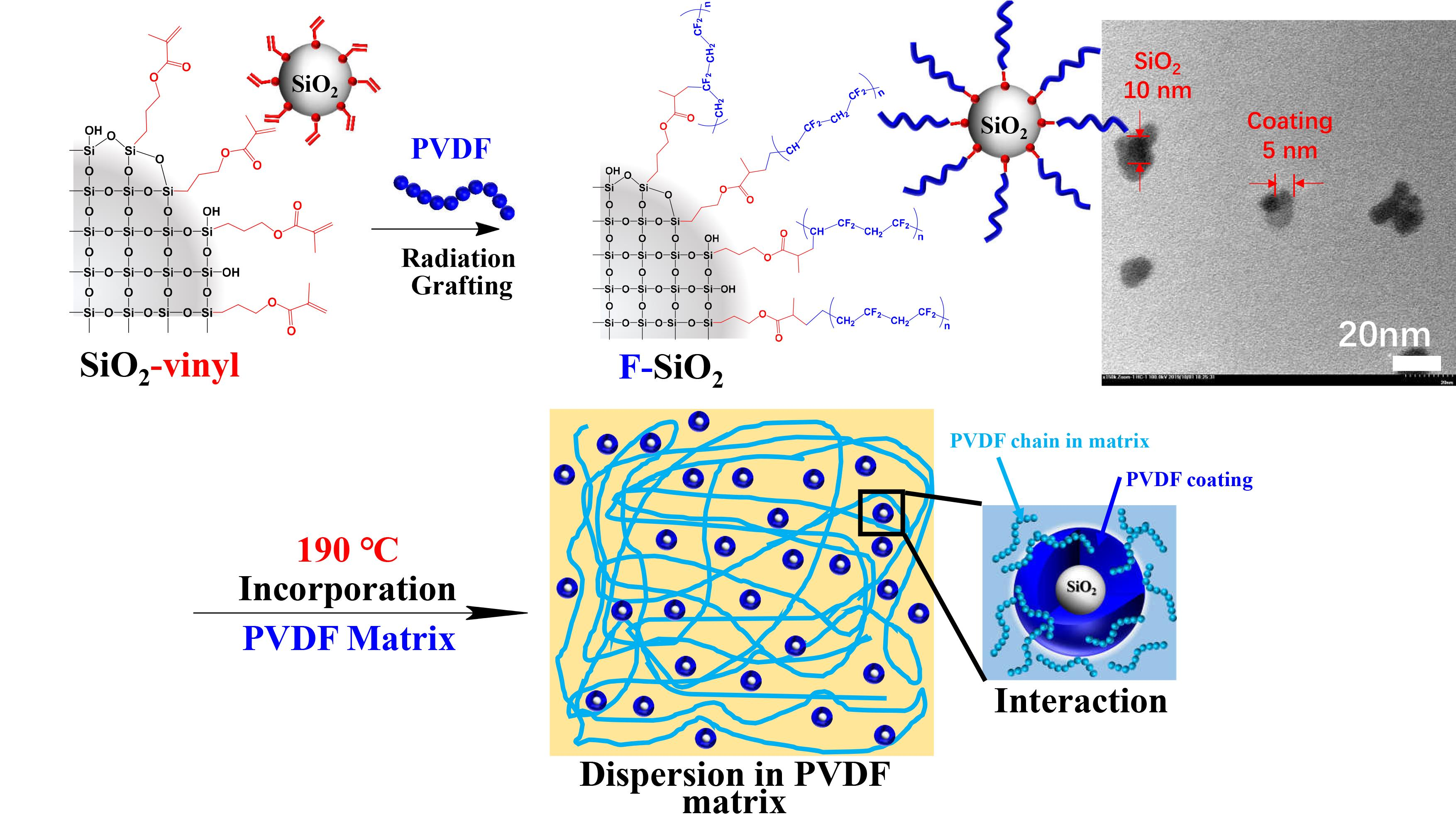
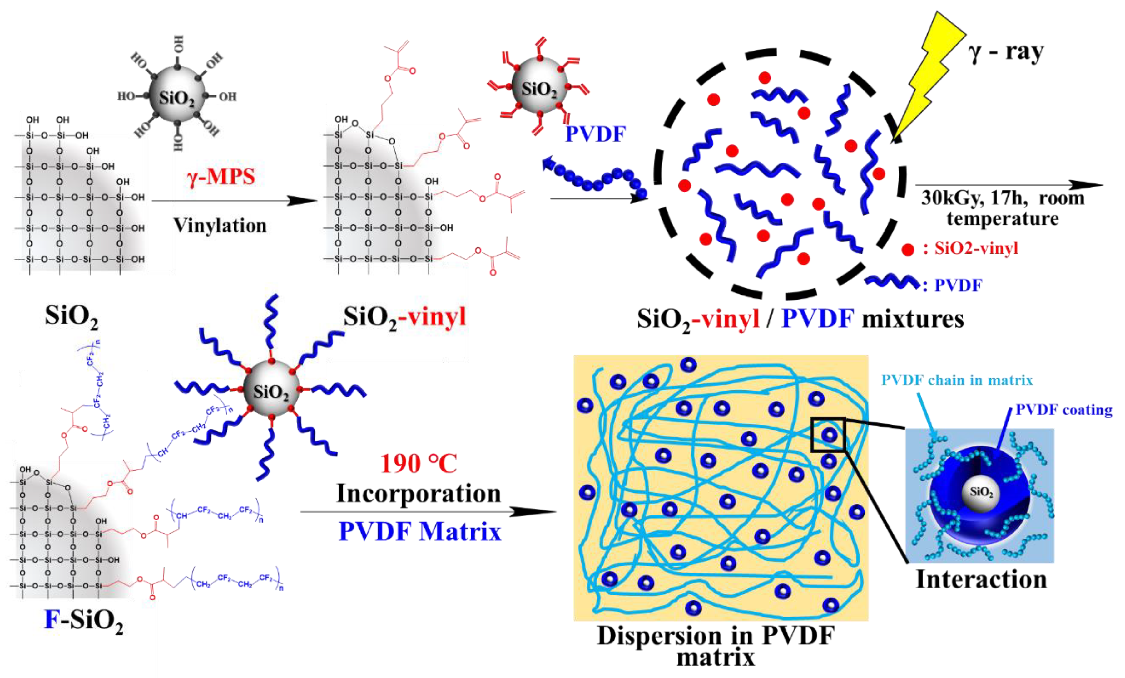
The fabrication procedure of the surface modified silica NPs is illustrated in Figure 2. The functionalization of SiO2 NPs with a PVDF chain (F-SiO2) was synthesized by sequential immobilization of reactive vinyl groups and PVDF chains onto the exterior surface of SiO2 using a silane coupling agent and commercially available PVDF, respectively. First, the pristine SiO2 was functionalized with vinyl groups by chemical modification using γ-methacryloxy propyl-trimethoxylsilane to yield reactive SiO2 (Vinyl-SiO2). Then, a small amount of PVDF was added and directly rooted on the SiO2 surface by gamma-radiation, which created radicals to initiate the efficient reaction with the vinyl group during the conditions and formed side chain grafts [23]. The newly developed process has the following advantages: the graft reactions between SiO2-vinyl and PVDF can be initiated over a wide temperature range including sub-ambient levels, making it a reproducible preparation of hybrid nanoparticles for industrial production.
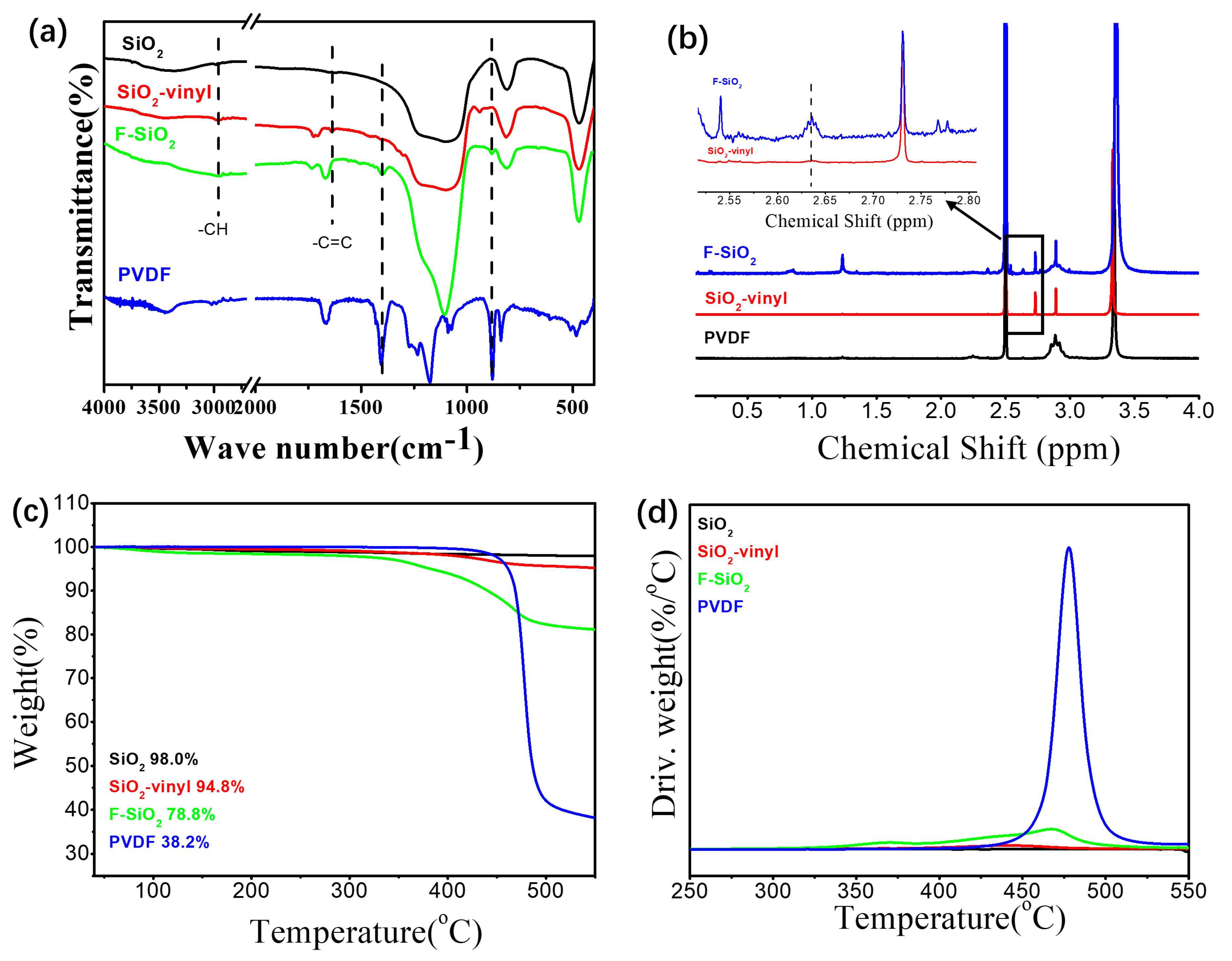
Figure 3 showed the FTIR, H1-NMR, TGA, and DTG curves of the silica NPs with various surface natures. As shown in Figure 3a, the strong and wide bands at 817 cm−1 and 1080 cm−1 was the absorption due to the asymmetric bending vibration of Si–OH and asymmetric stretching vibration of Si–O–Si, respectively, which can be observed in all the samples of modification silica, confirming the existence of SiO2 in the hybrid nanoparticles. Compared with the pristine SiO2, the peak of Si–OH became weaker and the new peaks at 2810–3050 cm−1, 1715 cm−1, and 1640 cm−1 can be observed for the Vinyl-SiO2 samples. These peaks were assigned to the stretching vibration of –CH, C=O, and C=C from γ-MPS, respectively, indicating the occurrence of a reaction between silica hydroxyl of pristine SiO2 and methoxyl of γ-MPS. In addition, the characteristic absorption peaks of PVDF at 1408 cm−1 and 880 cm−1 were also observed in the F–SiO2 samples. Since the unreacted PVDF was removed by extensive washing with organic solvents, the spectrum of F-SiO2 means PVDF chains successfully grafted onto the silica surface. Furthermore, 1H-NMR (Figure 3b) also demonstrated the appearance of γ-MPS (2.63 ppm) and PVDF (2.85 ppm, 2.89 ppm, and 2.91 ppm) for F-SiO2. The similar results can be also observed in the 13C-NMR (Figure S1) [29]. It implied that the reaction of SiO2 with γ-MPS occurred and PVDF was successfully grafted onto the surface of silica NPs, confirming further the PVDF was successfully grafted onto the surface of silica NPs.
TGA was used to measure the effect of modification on thermal stability of silica NPs. The TGA and derivative thermogravimetric analysis (DTG) curves was given in Figure 3c,d. It is clear that the thermal degradation of pristine silica nearly did not occur and the degradation of silica was almost negligible. With the modification of γ-MPS on the silica surface, the thermal weight loss was reduced (from 98.0% to 94.8%) and shown a thermal degradation temperature at 440 °C. In comparison, the further incorporation of PVDF leaded to a significantly decrease in the thermal weight loss (about 35.9 wt% of the PVDF grafting ratio) and improved the thermal stability of silica NPs (from 440 °C to 467 °C), which was close to 477 °C of pristine PVDF. The detailed molecular parameters of the surface modified silica NPs were shown in Table S1. These data confirmed again that F-SiO2 was successfully prepared via the radiation grafting process.
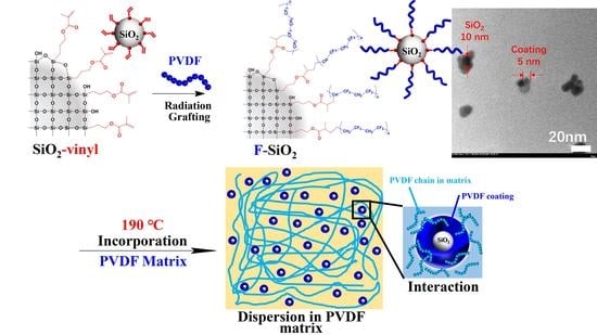
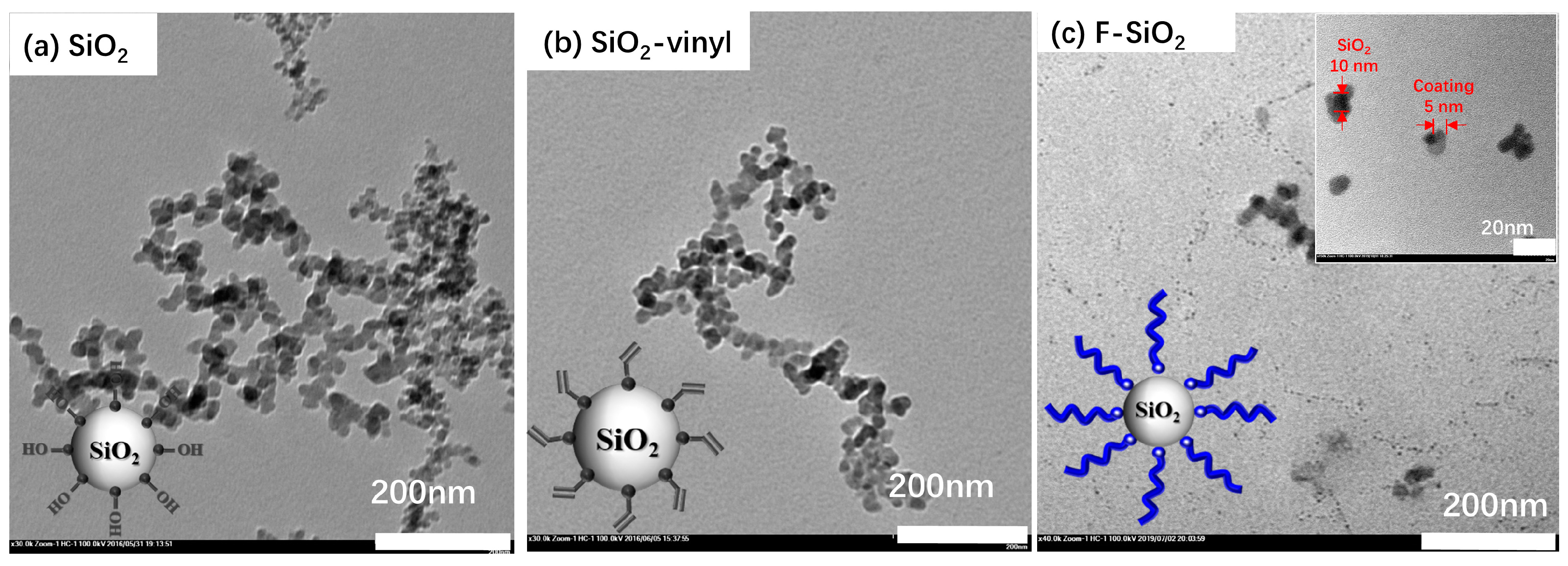
The same result was also clearly confirmed by TEM measurements. All the silica with an average diameter of 10 nm were used as model nanoparticles (Figure 4). Pristine SiO2 showed a typical agglomeration dropping cast onto a TEM grid (Figure 4a), which was contributed to the intense original characteristic of NPs. The modification silica with the vinyl group by silane coupling agents can remarkably improve the dispersibility of silica, while the NPs still show slight aggregation. While the gray corona of the γ-MPS on silica NPs surface was inconspicuous due to the small size and low molecular weight of silane coupling agent (Figure 3b). Compared with the pristine SiO2 and SiO2-vinyl, an explicit polymer coating layer (about 5-nm thickness) can be observed on the surface of the F-SiO2 after the radiation grafting of PVDF and the dispersion of the F-SiO2 was enhanced significantly. The size of F-SiO2 nanoparticles is about 5 nm based on observing TEM. The similar result was demonstrated in the X-ray diffraction (XRD) of the surface modification nanoparticles (Figure S2a,b). The original SiO2 showed a dominant XRD peak from amorphous structures of silica at 21.5°. With the increase of a modification degree, the peak was shifted to the lower wave number and the half-peak breadth was decreased due to the increase in nanoparticles size. To determine the crystal structures of F-SiO2 hybrid nanoparticles more accurately, the XRD Rietveld refinements [30] taken at room temperature were investigated by using the GSAS program (Figure S2c). It was found that the characteristic peaks of the α-phase crystal in the PVDF chain can be observed at 2θ = 19.4° in the F-SiO2 samples. This again means that the F-SiO2 was successfully prepared via the radiation grafting process and the effects of structure evolution of hybrid nanoparticles on crystallization behavior is still being studied.
3.2. Interface Modification Control of the PVDF Graft Ratio on Silica
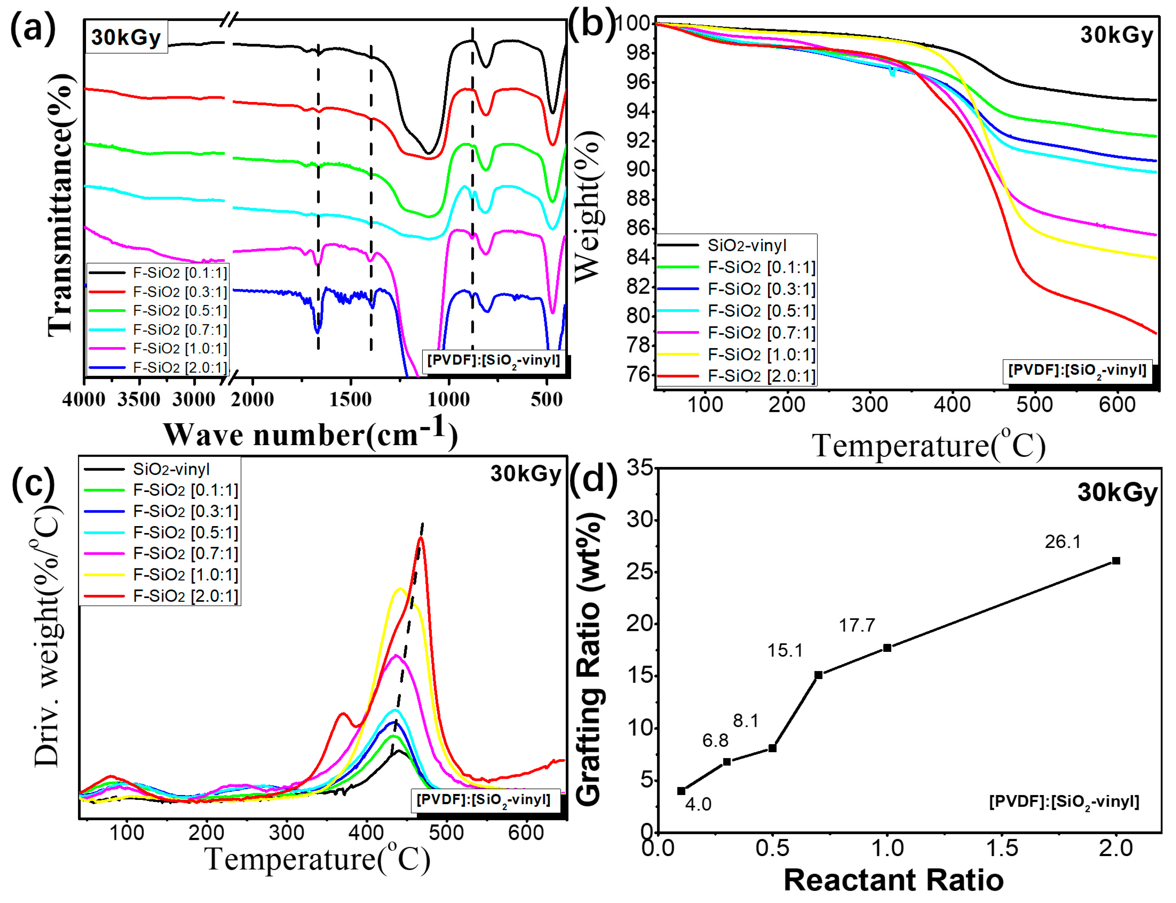
To get further information on the control of PVDF graft ratio on silica by this strategy, FTIR and TGA of the F-SiO2 NPs with different reactant ratio was performed, and the results were shown in Figure 5. It can be clearly seen that, with the increasing feed ratio of the PVDF, the intensity of the PVDF characteristic peak (1408 cm−1 and 880 cm−1, Figure 5a) and the thermal weight loss of the organic component (Figure 5b) in the F-SiO2 samples were significantly enhanced under the same absorption dose and reaction time (30 kGy, 17 h). The maximum weight loss temperature of the F-SiO2 was gradually improved in the same time frame (from 433 °C to 467 °C, Figure 4c), suggesting that the thermal stability increased with the rise of the grafting degree of PVDF. These results were mainly attributed to the increase of the PVDF feed ratio, which can significantly improve the reaction opportunities between the vinyl group on the surface of nanoparticles and the PVDF radicals generated during a gamma-ray radiation process, leading to the grafting degree of PVDF on the surface of nanoparticles being controlled.
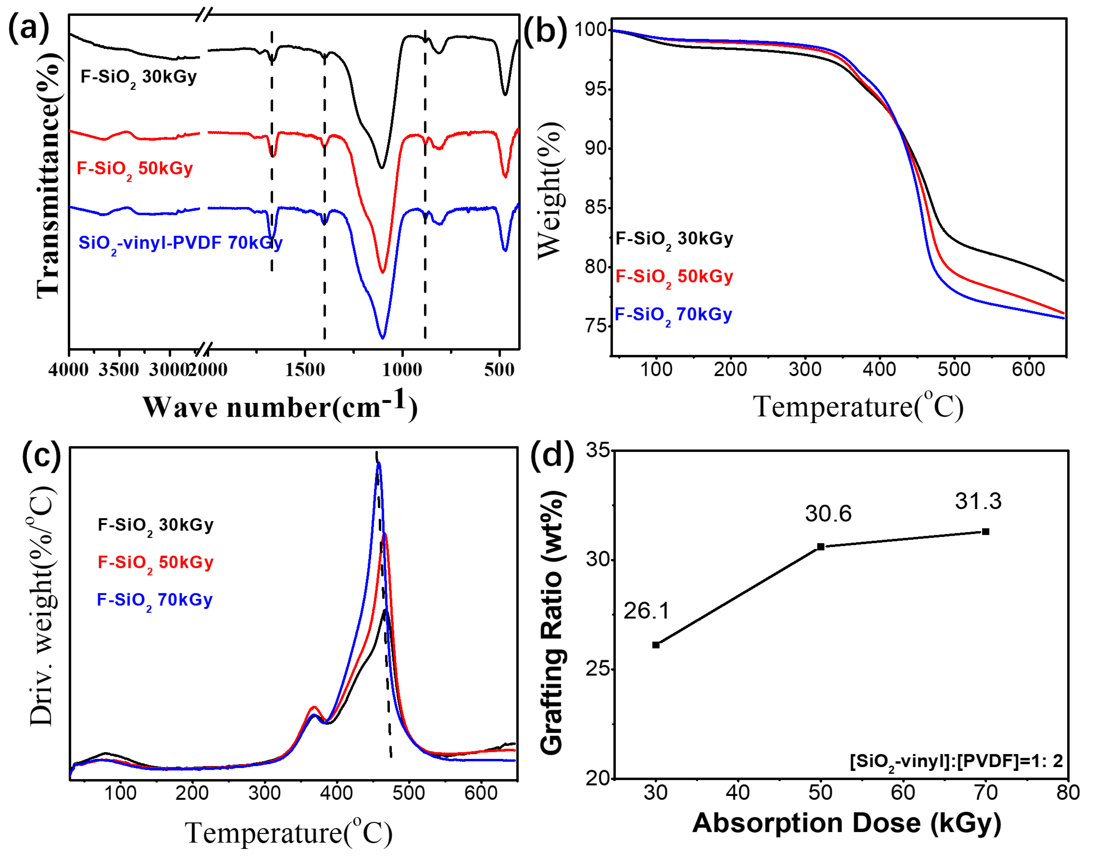
On the other hand, increasing the absorption dose of the grafting reaction can achieve the same effect as the strategy of change in the feed ratio (Figure 6). The higher radiation dose can improve the production of radicals in PVDF chain to react with SiO2-vinyl, leading to the grafting degree of PVDF on the silica surface increasing (Figure 6a,b). However, compared with the 50 kGy radiation samples, the graft ratio of F–SiO2 with the 70 kGy absorbed dose was not significantly improved and a clear reduction in the thermal stability of the F-SiO2 was observed with the increase of the absorbed dose (Figure 4c). It indicated that the improvement of regulating the grafting degree by the absorption dose was limited and the degradation of PVDF will be dominant depending on the radiation conditions. A higher radiation dose not only leads to high-energy consumption but also impairs the molecular weight of PVDF, which influenced the thermal stability of the F–SiO2. Therefore, the maximum graft ratio of PVDF on silica NPs we used in the present work were prepared under the 30 kGy adsorption dose. Free radical formation together with little chain scission are the main concern. The detailed molecular parameters of the surface modified silica NPs with PVDF were shown in Table S2.
3.3. Dispersion Property of Modification Silica in the PVDF Matrix
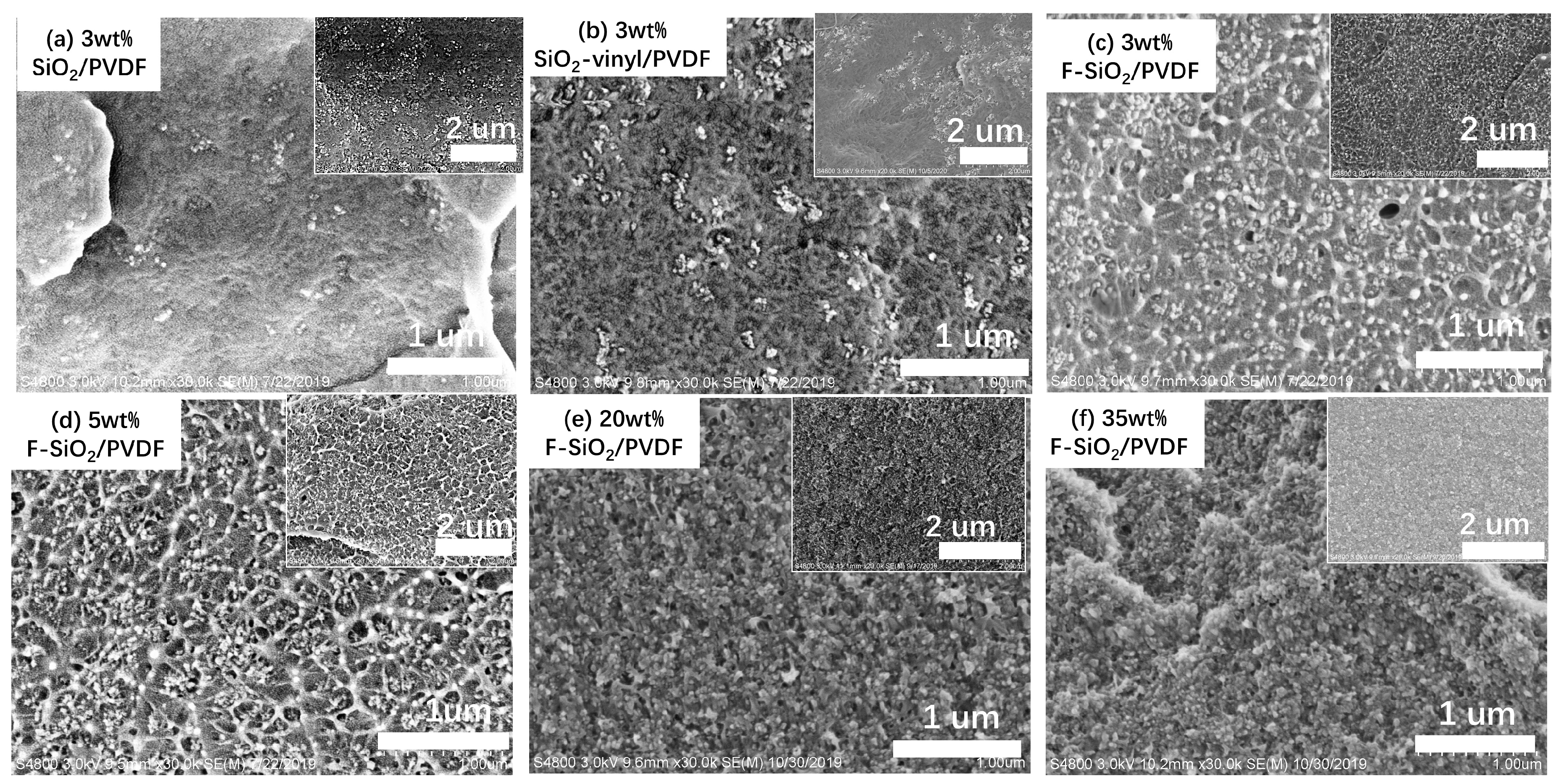
This allows a meaningful appraisal of the performance of surface modification silica by the radiation grafting technique. The silica NPs have been incorporated into the PVDF matrix by a simple melt-blending. Figure 7 shows the SEM morphologies of the silica NPs, which have different surface modifications and content, blending with PVDF matrix. It is observed that both the pristine SiO2 and SiO2-vinyl shown a significantly agglomeration in the PVDF matrix results in severe phase separation in the nanocomposites (Figure 7a,b). However, the modification of SiO2 by Gamma-radiation (F–SiO2) can remarkably improve the dispersibility of SiO2 particles in the nanocomposites (Figure 7c). To further differentiate the component of nanoparticles in the PVDF phase, the EDX and AFM was used to analyse the different parts of the nanocomposite [31,32,33], as shown in Figures S3 and S4. It was similar to the result of SEM where only the F–SiO2 nanoparticles can be uniformly dispersed in the PVDF matrix. We proposed that it was directly related to the immobilization effect of PVDF onto the solid inorganic core. Pristine SiO2 and SiO2-vinyl have a highly specific surface energy with the PVDF matrix due to the weak interaction by the inherent propensity. However, grafting of PVDF onto the SiO2 surface greatly improved the compatibilization between the NPs and polymer matrix, leading to the SiO2 particles dispersed in the PVDF uniformly.
The surface modification silica by the radiation grafting strategy can also improve the nanoparticle content in the PVDF matrix. When increasing the indicated amount of silica NPs from 3 wt% to 35 wt%, the aggregation of pristine SiO2 in the SiO2/PVDF samples was significantly enhanced (Figure S5). The macroscopic phase separation could be observed when the filling amount of silica exceeds 20 wt% (Figure S6), indicating the immiscible between silica with the PVDF matrix. However, for the addition of F–SiO2, the dispersion of nanoparticles was a remarkable improvement and the filling rate could reach 35 wt% without phase separation (Figure 7d–f). This again means that the coating of PVDF on silica enhanced the compatibilization of nanoparticles with the PVDF matrix and the radiation grafting was an effective strategy to surface modification PVDF on silica nanoparticles.
3.4. Modification Silica Loading Effect on the Physical Properties of the PVDF Matrix
3.4.1. Mechanical Properties
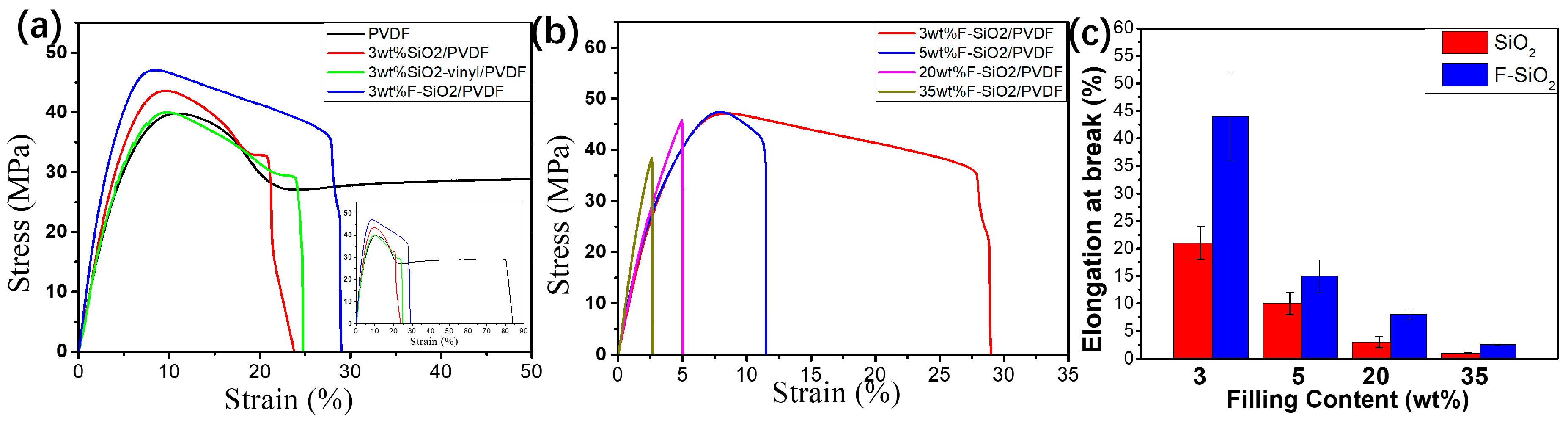
Tensile tests were carried out on an Instron universal material testing instrument at 25 °C with a tensile speed of 5 mm/min. The corresponding tensile properties of PVDF filling with the different content of pristine silica and F-SiO2 were shown in Figure 8, respectively. Neat PVDF shown a typical low yield strength (41.6 ± 1.4 MPa) and low modulus (782 ± 80 MPa) tensile behavior due to the low surface energy of the fluorine element in the main chain. With 3 wt% amount of pristine silica, both the yield strength (43.2 ± 0.8 MPa) and modulus (877 ± 118 MPa) of PVDF nanocomposites get enhanced. However, the improvement of the mechanical performance remained at a low efficiency. Similarly, the addition of SiO2-vinyl did not improve either the yield strength or the modulus. The mechanical property was even lower than that of the pristine SiO2 samples. These results were due to the bad dispersion properties and poor compatibility of silica with the PVDF matrix. On the other hand, the composites incorporation of nanoparticles in which grafting of PVDF onto the silica surface underwent an observable enhancement in mechanical strength compared with the same content of pristine silica. The yield strength and modulus of 3 wt% F-SiO2/PVDF are 45.8 ± 1.0 MPa and 1024 ± 40 MPa, respectively. In addition, both the yield strength and modulus of the F-SiO2/PVDF can be significantly improved, and the decrease of the elongation at break by the inherent propensity of SiO2 was suppressed with the increase of F–SiO2 content (Figure 8b,c). Thus, we were reasonable to deduce that the self-entanglements of a grafted PVDF chain on the SiO2 surface with the PVDF matrix reinforced the interactions between the nanoparticles and polymer, which accounted for the improvement of yield strength and modulus in nanocomposites.
3.4.2. The Crystallization Behavior
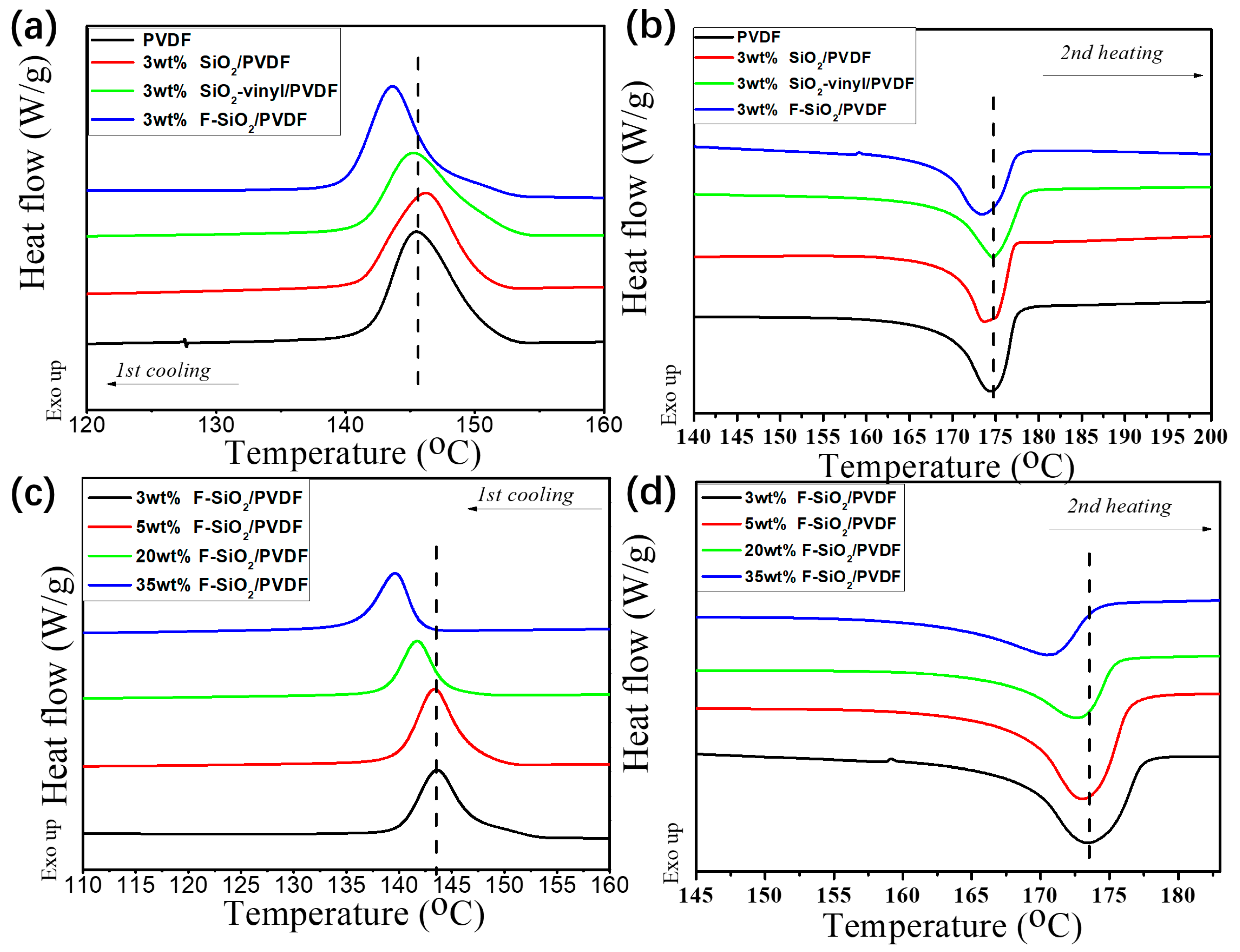
The crystallization behavior had strong affinities to the mechanical properties of materials [34,35]. The influence of surface modification silica nanoparticles contents on the crystallization behavior of PVDF in the matrix was examined in Figure 9. It was clear that PVDF exhibited strong crystallization ability with high crystallization temperature (Tc) and narrow half-peak breadth as a semi-crystalline polymer. For pristine SiO2 and SiO2-vinyl, the crystallization and melting behaviors did not vary significantly in the PVDF matrix. However, the melt (Tm) and crystallization (Tc) temperature of F-SiO2/PVDF nanocomposites were clearly obstructed (Figure 9a,b). This was because the grafted PVDF on the SiO2 surface have a strong interaction with the PVDF matrix, which broke the regularity of the molecular chain in the PVDF matrix and increased the hindrance during migration and arrangement when cooling down from the melt.
On the other hand, the change of nanoparticle content in the PVDF matrix can also impact the crystallization behavior of the nanocomposites. For pristine SiO2, the crystallization and melting behaviours did not vary significantly in PVDF/SiO2 nanocomposites with the change of NPs’ filling content (Figure S7). However, the higher content of F–SiO2 was, the lower Tm and Tc of nanocomposites became. The half-peak breadth for F–SiO2/PVDF samples were also broadened by the increasing addition of F–SiO2 (Figure 9c,d). It should be noted that the relative location of the PVDF diffraction peak did not change and show a significant structure of PVDF-αphases with various modification nanoparticles addition (Figure S8). Thus, we ascribed the improvements in the mechanical properties of the nanocomposites, which originate from the addition of F–SiO2, leading a strong interaction between the F–SiO2 surface PVDF coating with the PVDF matrix.
3.4.3. The Thermomechanical Behavior
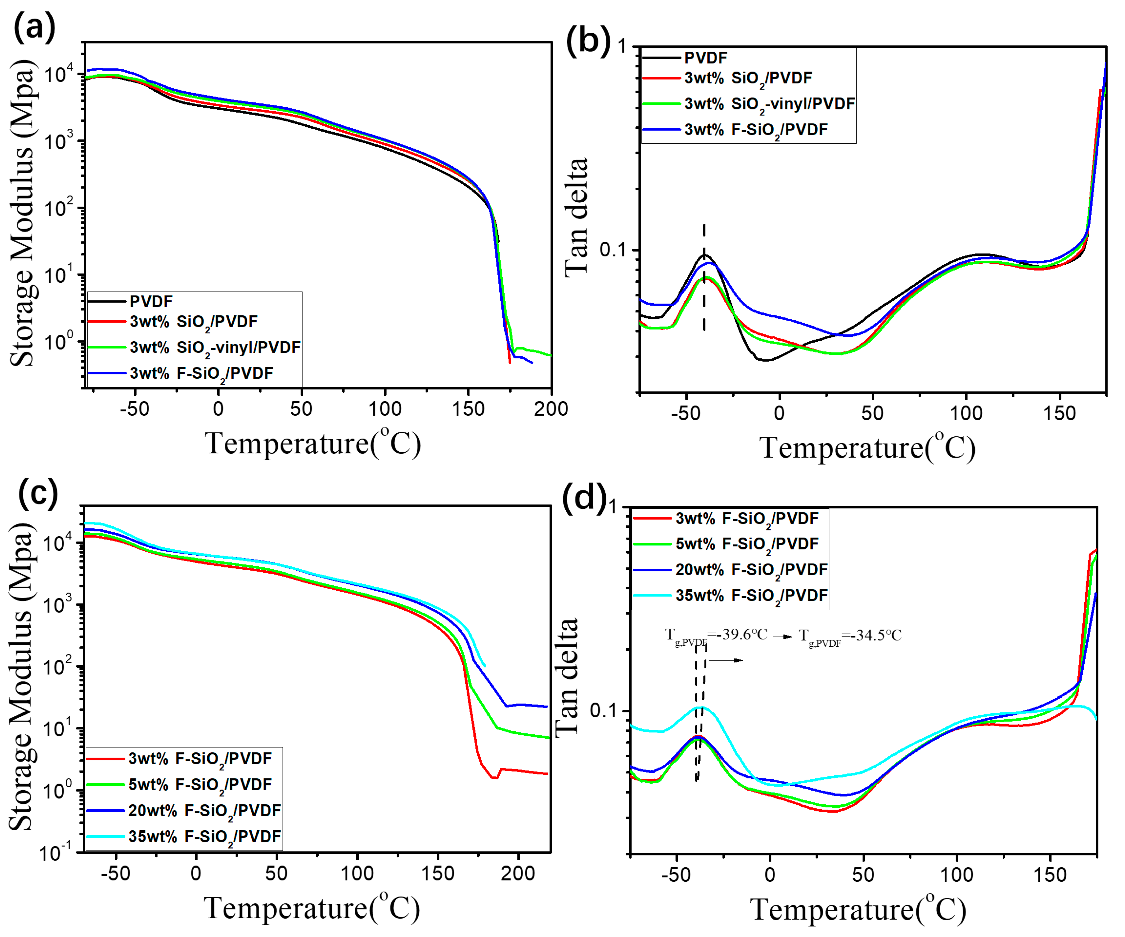
Generally speaking, the compatibilization of nanoparticles with polymers determined the glass transition behaviors of nanocomposites and can be observed with the changing of thermomechanical properties [36,37]. To get further information on the effective modification of nanoparticles by radiation grafting, thermomechanical properties of SiO2/PVDF nanocomposites are studied by DMA analysis. Figure 10a,b show the storage modulus and loss factor (tanδ) of PVDF nanocomposites with various modified SiO2. It can be seen that, with the degree of the surface modification of SiO2, the storage modulus of the SiO2/PVDF blends increased gradually (Figure 10a). However, the nanocomposites, which have 3 wt% pristine SiO2 and SiO2-vinyl content nanoparticles, only had the same distinct mechanical relaxation (glass transition temperature, Tg) with the neat PVDF at −42.0 °C. The PVDF incorporation with 3 wt% F-SiO2 shows a higher Tg at −39.0 °C (Figure 10b). In addition, with the increase of the filling amount (from 3 wt% to 35 wt%), the Tg and storage modulus of F-SiO2/PVDF was further improved, which can be contributed to the enhancement of physical network density and entangled state by limiting the movement of molecular chains in the PVDF matrix (Figure 10c,d). These results are in good accordance with the previous analysis. The increase in DMA indicates that there are strong interactions between F–SiO2 and the PVDF matrix, leading to the amelioration in the mechanical properties and dispersity of silica NPs in the PVDF matrix.
3.4.4. The Rheological Properties
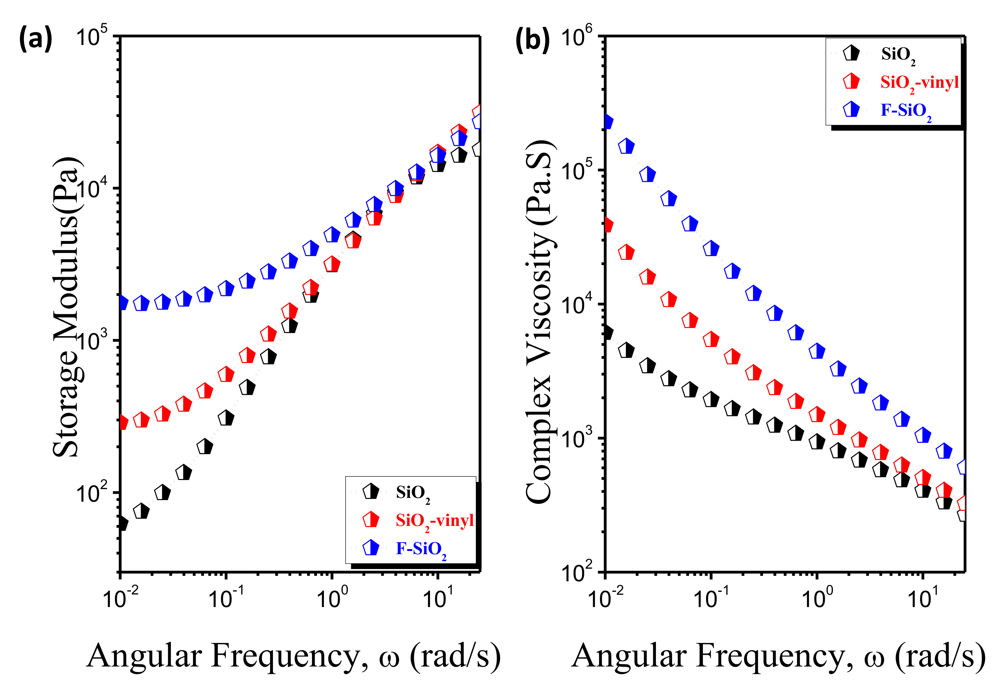
The dynamic rheological response is believed to be an effective method for providing information regarding the structure/morphology of materials under small amplitude oscillatory shear (SAOS). The rheological behaviors of the PVDF matrix with different surface modification SiO2 was shown in Figure 11. It is clear that F–SiO2 exhibited a completely different response of viscosity and elasticity in the SAOS tests, as compared with the pristine SiO2 and SiO2-vinyl, which show a sensitive frequency-independent and pseudo solid-like responses at the terminal region. It can be inferred that the self-entanglements of grafted PVDF chain on the SiO2 surface with the PVDF matrix reinforced the interactions between nanoparticles and polymer, leading to the effective dispersal in the PVDF matrix and enhancement of interfacial adhesion.
4. Conclusions
We demonstrated a facile strategy to graft the polymer onto the surface of nanoparticles through a radiation technique. SiO2 NPs were vinyl functionalized (SiO2-vinyl) by using γ-methacryloxy propyl-trimethoxylsilane. The SiO2-vinyl were then physically mixed with PVDF, which was followed by radiation from the Co60 gamma ray at room temperature. PVDF radicals were generated directly and then chemically grafted onto the surface of SiO2 nanoparticles by the linking of the double bond on the NPs. The graft ratio of PVDF on SiO2 NPs surface can be simply controlled by adjusting the absorbed dose and feed ratio of PVDF to SiO2 NPs. The prepared F–SiO2 were then dispersed in the PVDF matrix to make the nanocomposites. It was found that the F–SiO2 dispersed uniformly in the PVDF matrix, as compared with SiO2 and SiO2-vinyl. Besides, the filling content of F–SiO2 in the PVDF matrix is almost two times higher than the pristine SiO2 counterpart. Accordingly, the mechanical property of the F-SiO2/PVDF nanocomposites is significantly improved. Therefore, it is a promising way to combine the nanoparticles with polymers efficiently using a simple radiation-induced grafting method. Considering the increasing demand for the hybrid nanoparticles, this approach could be applied to various combinations of nanoparticles and polymers, providing new possibilities for surface modification nanoparticles in industrial production.
Supplementary Materials
The following are available online at https://www.mdpi.com/2079-4991/10/11/2237/s1. Formula S1: Formula for calculating the grafting ratio of PVDF on F-SiO2 nanoparticles surface. Figure S1: 13C-NMR spectra of pristine SiO2, SiO2-vinyl and F-SiO2 nanoparticles, respectively. Figure S2: (a) XRD patterns of pristine SiO2, SiO2-vinyl, F-SiO2, and PVDF. (b) Enlarged XRD patterns of pristine SiO2, SiO2-vinyl, F-SiO2 in the 2θ range of 15–30°. (c) Enlarged Rietveld refinement of the pristine SiO2, SiO2-vinyl, and F-SiO2 XRD data in the 2θ range of 15–30°. Figure S3: SEM and Elemental Mapping image (EMI) of PVDF matrix incorporated with 3 wt% (a) pristine SiO2, (b) SiO2-vinyl, and (c) F-SiO2 nanoparticles, respectively. The subscript of 1 and 2 correspond to the signal of silicon and fluoride element in EMI, respectively. Figure S4: AFM of PVDF matrix incorporated with three wt% (a) pristine SiO2, (b) SiO2-vinyl, and (c) F-SiO2 nanoparticles, respectively. Figure S5: SEM image of PVDF matrix incorporated 5 wt% (a), 20wt% (b), 35 wt%, and (c) SiO2 nanoparticles, respectively. Figure S6: Digital photograph of the PVDF matrix incorporated 3 wt% (a)—35 wt% (g) SiO2 nanoparticles, respectively. Digital photograph of the PVDF matrix incorporated 3 wt% (h)—35 wt% (n) F-SiO2 nanoparticles, respectively. Figure S7: DSC spectra of 1st cooling (c) and 2nd heating (d) of the PVDF matrix incorporated with 3 wt%, 5 wt%, 20 wt%, and 35 wt% SiO2 nanoparticles, respectively. Figure S8: XRD spectra of the PVDF matrix incorporated with 3 wt% SiO2, SiO2-vinyl, and F-SiO2 nanoparticles, respectively. Table S1: Molecular parameter of the surface modified silica NPs. Table S2: The graft content of PVDF grafted onto silica nanoparticles under a different reactant ratio (wt%/wt%) and radiation dose.
Author Contributions
Conceptualization, Y.L. and J.L. Methodology, Z.F., X.G., and L.H. Software, Z.F. Formal analysis, Z.F. and X.G. Writing—original draft preparation, Z.F. Writing—review and editing, Z.F., Y.L., and J.L. Supervision, Y.L. and J.L. All authors have read and agreed to the published version of the manuscript.
Funding
This work was funded by the National Natural Science Foundation of China (21674033, 21374027).
Conflicts of Interest
The authors declare no conflict of interest.
References
- Pastore, V.J.; Cook, T.R. Coordination-Driven Self-Assembly in Polymer–Inorganic Hybrid Materials. Chem. Mater. 2020, 32, 3680–3700. [Google Scholar] [CrossRef]
- Dolbecq, A.; Dumas, E.; Mayer, C.R.; Mialane, P. Hybrid organic-inorganic polyoxometalate compounds: From structural diversity to applications. Chem. Rev. 2010, 110, 6009–6048. [Google Scholar] [CrossRef] [PubMed]
- Sanchez, C.; Julián, B.; Belleville, P.; Popall, M. Applications of Hybrid Organic–Inorganic Nanocomposites. J. Mater. Chem. 2005, 15, 3559–3592. [Google Scholar] [CrossRef]
- Zhang, Y.; Zhao, H.Y. Surfactant Behavior of Amphiphilic Polymer-Tethered Nanoparticles. Langmuir 2016, 32, 3567–3579. [Google Scholar] [CrossRef]
- Gonzalez-Burgos, M.; Latorre-Sanchez, A.; Pomposo, J.A. Advances in single chain technology. Chem. Soc. Rev. 2015, 44, 6122–6142. [Google Scholar] [CrossRef] [Green Version]
- Zhang, X.; Yang, Y.; Tian, J.; Zhao, H. Vesicles fabricated by hybrid nanoparticles. Chem. Commun. 2009, 25, 3807–3809. [Google Scholar] [CrossRef]
- Li, K.; Liang, S.; Lu, Y.; Wang, Q. Synthesis of Telechelic Fluoropolymers with Well-Defined Functional End Groups for Cross-Linked Networks and Nanocomposites. Macromolecules 2007, 40, 4121–4123. [Google Scholar] [CrossRef]
- Amiinu, I.S.; Liang, X.; Tu, Z.; Zhang, H.; Feng, J.; Wan, Z.; Pan, M. Anhydrous Proton Conducting Materials Based on Sulfonated Dimethylphenethylchlorosilane Grafted Mesoporous Silica/Ionic Liquid Composite. ACS Appl. Mater. Interfaces 2013, 5, 11535–11543. [Google Scholar] [CrossRef]
- Tanahashi, M.; Hirose, M.; Watanabe, Y.; Lee, J.-C.; Takeda, K.J. Silica/Perfluoropolymer Nanocomposites Fabricated by Direct Melt-Compounding: A Novel Method without Surface Modification on Nano-Silica. J. Nanosci. Nanotechnol. 2007, 7, 1–10. [Google Scholar] [CrossRef]
- Darr, J.A.; Zhang, J.; Makwana, N.M.; Weng, X. Continuous hydrothermal synthesis of inorganic nanoparticles: Applications and future directions. Chem. Rev. 2017, 117, 11125–11238. [Google Scholar] [CrossRef] [Green Version]
- Gravano, S.M.; Dumas, R.; Liu, K.; Patten, T.E. Methods for the surface functionalization of γ-Fe2O3 nanoparticles with initiators for atom transfer radical polymerization and the formation of core–shell inorganic–polymer structures. J. Polym. Sci. Part A Polym. Chem. 2005, 43, 3675–3688. [Google Scholar] [CrossRef]
- Pankhurst, Q.A.; Thanh, N.T.K.; Jones, S.K.; Dobson, J. Progress in applications of magnetic nanoparticles in biomedicine. J. Phys. D Appl. Phys. 2009, 42, 224001. [Google Scholar] [CrossRef] [Green Version]
- Wen, J.; Yuan, L.; Yang, Y.; Liu, L.; Zhao, H. Self-assembly of monotethered single-chain nanoparticle shape amphiphiles. ACS Macro Lett. 2013, 2, 100–106. [Google Scholar] [CrossRef]
- Kayser, M.J.; Reinholdt, M.X.; Kaliaguine, S. Amine grafted silica/SPEEK nanocomposites as proton exchange membranes. J. Phys. Chem. B 2010, 114, 8387–8395. [Google Scholar] [CrossRef]
- Liao, Z.; Wu, G.; Lee, D.; Yang, S. Ultrastable Underwater Anti-Oil Fouling Coatings from Spray Assemblies of Polyelectrolyte Grafted Silica Nanochains. ACS Appl. Mater. Interfaces 2019, 11, 13642–13651. [Google Scholar] [CrossRef]
- Xu, J.; Zhang, Y.; Zhu, W.; Cui, Y. Synthesis of Polymeric Nanocomposite Hydrogels Containing the Pendant ZnS Nanoparticles: Approach to Higher Refractive Index Optical Polymeric Nanocomposites. Macromolecules 2018, 51, 2672–2681. [Google Scholar] [CrossRef]
- Durand, N.; Boutevin, B.; Silly, G.; Ameduri, B. “Grafting From” Polymerization of Vinylidene Fluoride (VDF) from Silica to Achieve Original Silica–PVDF Core–Shells. Macromolecules 2011, 44, 8487–8493. [Google Scholar] [CrossRef]
- Durand, N.; Gaveau, P.; Silly, G.; Ameduri, B.; Boutevin, B. Radical Grafting of Tetrafluoroethylene and Vinylidene Fluoride Telomers onto Silica Bearing Vinyl Groups. Macromolecules 2011, 44, 6249–6257. [Google Scholar] [CrossRef]
- Pribyl, J.; Benicewicz, B.; Bell, M.; Wagener, K.; Ning, X.; Schadler, L.; Jimenez, A.; Kumar, S. Polyethylene Grafted Silica Nanoparticles Prepared via Surface-Initiated ROMP. ACS Macro Lett. 2019, 8, 228–232. [Google Scholar] [CrossRef]
- Vukicevic, R.; Beuermann, S. Fullerenes Decorated with Poly (vinylidene fluoride). Macromolecules 2011, 44, 2597–2603. [Google Scholar] [CrossRef]
- Nasef, M.M. Radiation-grafted membranes for polymer electrolyte fuel cells: Current trends and future directions. Chem. Rev. 2014, 114, 12278–12329. [Google Scholar] [CrossRef] [PubMed]
- Mohammad, F. High-energy radiation induced sustainable coloration and functional finishing of textile materials. Ind. Eng. Chem. Res. 2015, 54, 3727–3745. [Google Scholar]
- Nasef, M.M.; Hegazy, E.S.A. Preparation and applications of ion exchange membranes by radiation-induced graft copolymerization of polar monomers onto non-polar films. Prog. Polym. Sci. 2004, 29, 499–561. [Google Scholar] [CrossRef]
- Dargaville, T.R.; George, G.A.; Hill, D.J.; Whittaker, A.K. High energy radiation grafting of fluoropolymers. Prog. Polym. Sci. 2003, 28, 1355–1376. [Google Scholar] [CrossRef]
- Hu, J.; Gao, Q.; Xu, L.; Zhang, M.; Xing, Z.; Guo, X.; Zhang, K.; Wu, G. Significant improvement in thermal and UV resistances of UHMWPE fabric through in situ formation of polysiloxane–TiO2 hybrid layers. ACS Appl. Mater. Interfaces 2016, 8, 23311–23320. [Google Scholar] [CrossRef]
- Wang, H.; Fu, Z.; Zhao, X.; Li, Y.; Li, J. Reactive Nanoparticles Compatibilized Immiscible Polymer Blends: Synthesis of Reactive SiO2 with Long Poly(methyl methacrylate) Chains and the in Situ Formation of Janus SiO2 Nanoparticles Anchored Exclusively at the Interface. ACS Appl. Mater. Interfaces 2017, 9, 14358–14370. [Google Scholar] [CrossRef]
- Zhao, X.; Wang, H.; Fu, Z.; Li, Y. Enhanced Interfacial Adhesion by Reactive Carbon Nanotubes: New Route to High-Performance Immiscible Polymer Blend Nanocomposites with Simultaneously Enhanced Toughness, Tensile Strength, and Electrical Conductivity. ACS Appl. Mater. Interfaces 2018, 10, 8411–8416. [Google Scholar] [CrossRef]
- Fu, Z.; Wang, H.; Zhao, X.; Li, X.; Gu, X.; Li, Y. Flame-retarding nanoparticles as the compatibilizers for immiscible polymer blends: Simultaneously enhanced mechanical performance and flame retardancy. J. Mater. Chem. A 2019, 7, 4903–4912. [Google Scholar] [CrossRef]
- Constantin, L.V.; Iconaru, S.; Ciobanu, C.S. Europium doped hydroxyapatite for applications in environmental field. Rom. Rep. Phys. 2012, 64, 788–794. [Google Scholar]
- Ciobanu, C.S.; Andronescu, E.; Prodan, A.M.; Pall, L.; Iconaru, S.L. Physico-chemical and antibacterial studies on silver doped nano-hydroxyapatite. J. Optoelectron. Adv. Mater. 2013, 15, 918–922. [Google Scholar]
- Iconaru, S.L.; Prodan, A.M.; Turculet, C.S.; Beuran, M.; Ghita, R.V.; Costescu, A.; Groza, A.; Chifiriuc, M.C.; Chapon, P.; Gaiaschi, S.; et al. Enamel based composite layers deposited on titanium substrate with antifungal activity. J. Spectrosc. 2016, 2016, 1–13. [Google Scholar] [CrossRef]
- Ghita, R.; Iconaru, S.L.; Popa, C.; Costescu, A.; Coustumer, P.L.; Motelicaheino, M.; Ciobanu, C.S. Tetraethyl orthosilicate coated hydroxyapatite powders for lead ions removal from aqueous solutions. J. Nanomater. 2014, 2014, 2. [Google Scholar] [CrossRef] [Green Version]
- Iconaru, S.L.; Turculet, C.; Coustumer, P.L.; Bleotu, C.; Prodan, A.M. Biological studies on dextrin coated iron oxide nanoparticles. Rom. Rep. Phys. 2016, 68, 1536–1544. [Google Scholar]
- Li, H.; Yan, S. Surface-induced polymer crystallization and the resultant structures and morphologies. Macromolecules 2011, 44, 417–428. [Google Scholar] [CrossRef]
- Lotz, B.; Miyoshi, T.; Cheng, S.Z. 50th anniversary perspective: Polymer crystals and crystallization: Personal journeys in a challenging research field. Macromolecules 2017, 50, 5995–6025. [Google Scholar] [CrossRef]
- Huang, Y.; Zheng, Y.; Sarkar, A.; Xu, Y.; Stefik, M.; Benicewicz, B.C. Matrix-Free Polymer Nanocomposite Thermoplastic Elastomers. Macromolecules 2017, 50, 4742–4753. [Google Scholar] [CrossRef]
- Frantisek, O.; Petr, L.; Marek, Z.; Klara, Z.; Leon, E.G.; Josef, J. Effect of Nanoparticle Organization on Molecular Mobility and Mechanical Properties of Polymer Nanocomposites. Macromolecules 2019, 52, 6250–6259. [Google Scholar]
Figure 1.
Schematic diagram of the surface modified silica nanoparticles with polyvinylidene fluoride (PVDF) coating by using radiation-induced graft polymerization under a wide temperature range and the distribution of F-SiO2 in the PVDF matrix.
Figure 1.
Schematic diagram of the surface modified silica nanoparticles with polyvinylidene fluoride (PVDF) coating by using radiation-induced graft polymerization under a wide temperature range and the distribution of F-SiO2 in the PVDF matrix.

Figure 2.
Illustrative synthesis of F-SiO2 through a “radiation-induced graft polymerization” method.
Figure 2.
Illustrative synthesis of F-SiO2 through a “radiation-induced graft polymerization” method.

Figure 3.
FT-IR (a), 1H-NMR (b), TGA (c), and DTG (d) curves of the SiO2, SiO2-vinyl, F-SiO2, and polyvinylidene fluoride (PVDF).
Figure 3.
FT-IR (a), 1H-NMR (b), TGA (c), and DTG (d) curves of the SiO2, SiO2-vinyl, F-SiO2, and polyvinylidene fluoride (PVDF).

Figure 4.
TEM image of the SiO2 (a), SiO2-vinyl (b), and F-SiO2 (c).

Figure 5.
FT-IR (a), TGA (b), DTG (c) and the grafting ratio curves (d) of the F-SiO2 with a different [PVDF]/[SiO2-vinyl] reactant ratio under 30 kGy, 17 h Gamma-radiation condition.
Figure 5.
FT-IR (a), TGA (b), DTG (c) and the grafting ratio curves (d) of the F-SiO2 with a different [PVDF]/[SiO2-vinyl] reactant ratio under 30 kGy, 17 h Gamma-radiation condition.

Figure 6.
FT-IR (a), TGA (b), DTG (c) and the grafting ratio (d) curves of the F-SiO2 under a different absorbed dose and a 17-h Gamma-radiation condition. The reactant ratio of [SiO2-vinyl]:[PVDF] = 1:2.
Figure 6.
FT-IR (a), TGA (b), DTG (c) and the grafting ratio (d) curves of the F-SiO2 under a different absorbed dose and a 17-h Gamma-radiation condition. The reactant ratio of [SiO2-vinyl]:[PVDF] = 1:2.

Figure 7.
SEM images of the PVDF matrix incorporated with 3wt%, (a) pristine SiO2, (b) SiO2-vinyl, and (c) F-SiO2 nanoparticles, respectively. SEM images of PVDF matrix incorporated 5 wt% (d), 20 wt% (e), 35 wt%, and (f) F-SiO2 nanoparticles, respectively.
Figure 7.
SEM images of the PVDF matrix incorporated with 3wt%, (a) pristine SiO2, (b) SiO2-vinyl, and (c) F-SiO2 nanoparticles, respectively. SEM images of PVDF matrix incorporated 5 wt% (d), 20 wt% (e), 35 wt%, and (f) F-SiO2 nanoparticles, respectively.

Figure 8.
(a) Stress-strain curves of the PVDF matrix incorporated with 3 wt% SiO2, SiO2-vinyl, and F-SiO2 nanoparticles, respectively. (b) Stress-strain curves of the PVDF matrix incorporated with 3 wt%, 5 wt%, 20 wt%, and 35 wt% F-SiO2 nanoparticles, respectively. (c) The elongation at break of the F–SiO2/PVDF with the addition of a different content of pristine SiO2 and F–SiO2, respectively.

Figure 9.
DSC spectra of 1st cooling (a) and 2nd heating (b) of the PVDF matrix incorporated with 3 wt% SiO2, SiO2-vinyl, and F-SiO2 nanoparticles, respectively. DSC spectra of first cooling (c) and second heating (d) of the PVDF matrix incorporated with 3 wt%, 5 wt%, 20 wt%, and 35 wt% F-SiO2 nanoparticles, respectively.

Figure 10.
Storage modulus (a) and Loss tangent (b) as a function of temperature with a frequency of 5 Hz for the PVDF matrix with 3 wt% of SiO2, SiO2-vinyl, and F-SiO2, respectively. Storage modulus (c) and Loss tangent (d) as a function of temperature with a frequency of 5 Hz for the PVDF matrix with 3 wt%, 5 wt%, 20 wt%, and 35 wt% of F-SiO2, respectively. These results were obtained by dynamic thermomechanical analysis (DMA).

Figure 11.
The storage modulus (a) and complex viscosity (b) of the PVDF matrix incorporated with 3 wt% pristine SiO2, SiO2-vinyl, and F-SiO2 nanoparticles, respectively.
Figure 11.
The storage modulus (a) and complex viscosity (b) of the PVDF matrix incorporated with 3 wt% pristine SiO2, SiO2-vinyl, and F-SiO2 nanoparticles, respectively.

Publisher’s Note: MDPI stays neutral with regard to jurisdictional claims in published maps and institutional affiliations. |
© 2020 by the authors. Licensee MDPI, Basel, Switzerland. This article is an open access article distributed under the terms and conditions of the Creative Commons Attribution (CC BY) license (http://creativecommons.org/licenses/by/4.0/).
Share and Cite
MDPI and ACS Style
Fu, Z.; Gu, X.; Hu, L.; Li, Y.; Li, J. Radiation Induced Surface Modification of Nanoparticles and Their Dispersion in the Polymer Matrix. Nanomaterials 2020, 10, 2237. https://doi.org/10.3390/nano10112237
AMA Style
Fu Z, Gu X, Hu L, Li Y, Li J. Radiation Induced Surface Modification of Nanoparticles and Their Dispersion in the Polymer Matrix. Nanomaterials. 2020; 10(11):2237. https://doi.org/10.3390/nano10112237
Chicago/Turabian StyleFu, Zhiang, Xiaoying Gu, Lingmin Hu, Yongjin Li, and Jingye Li. 2020. "Radiation Induced Surface Modification of Nanoparticles and Their Dispersion in the Polymer Matrix" Nanomaterials 10, no. 11: 2237. https://doi.org/10.3390/nano10112237
Note that from the first issue of 2016, this journal uses article numbers instead of page numbers. See further details here.