Potential Applications of Magnesium-Based Polymeric Nanocomposites Obtained by Electrospinning Technique

 ,
,
Abstract
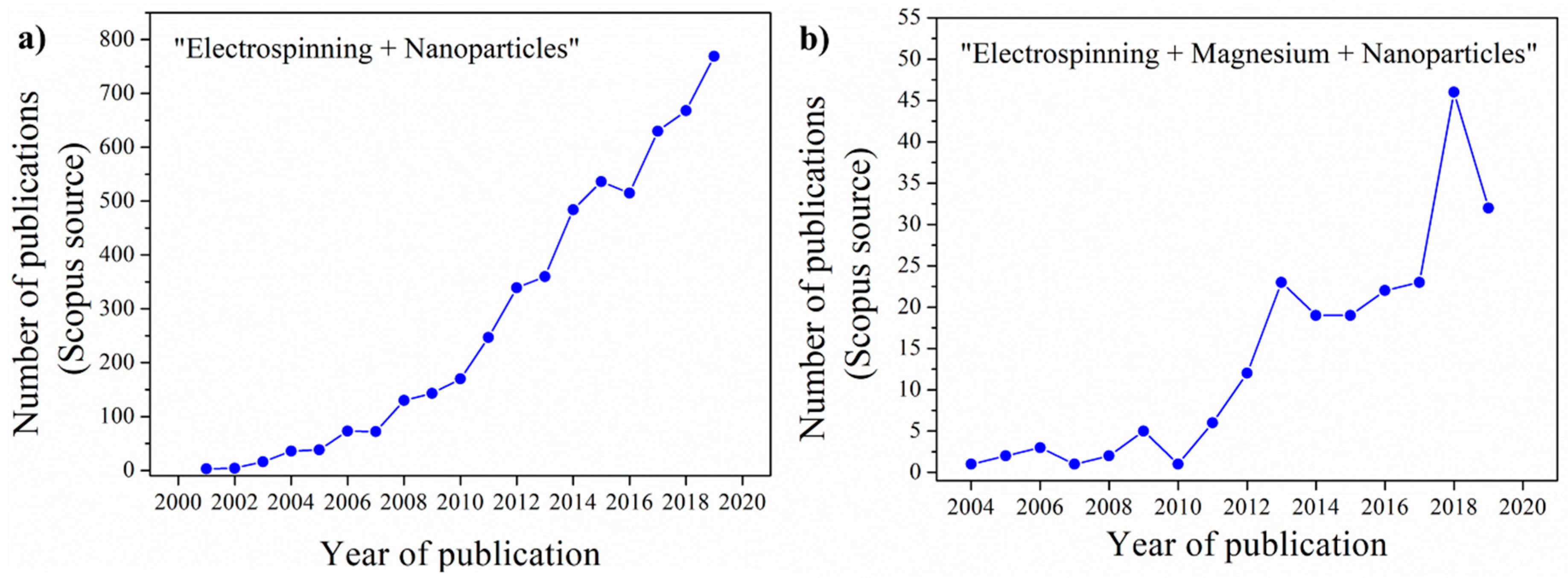
:1. Introduction
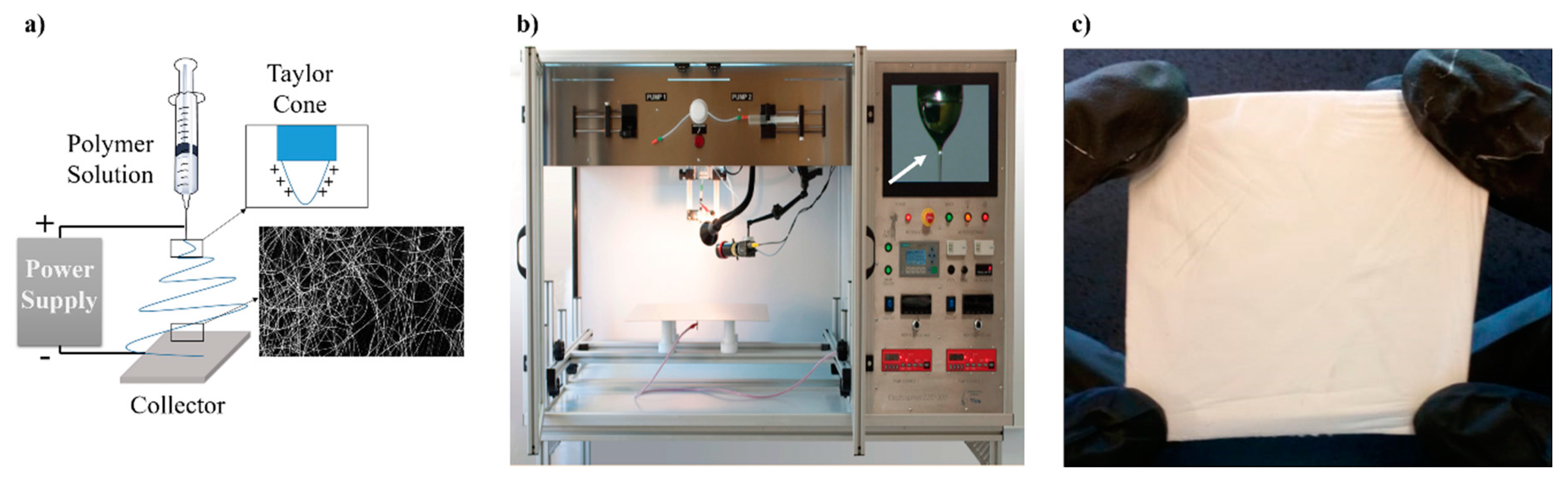
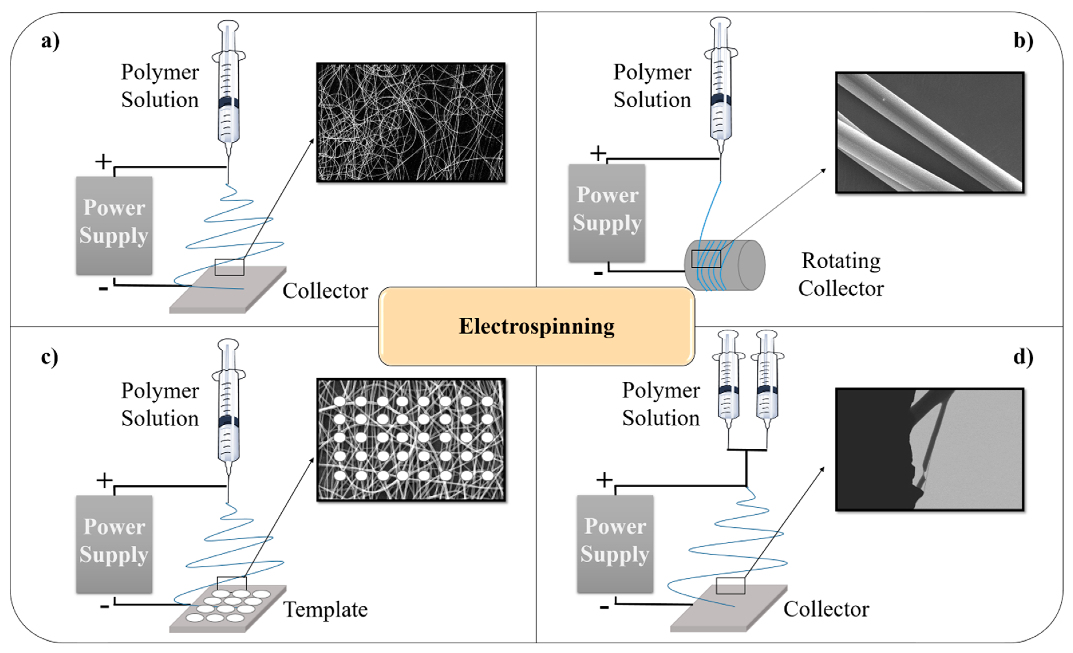
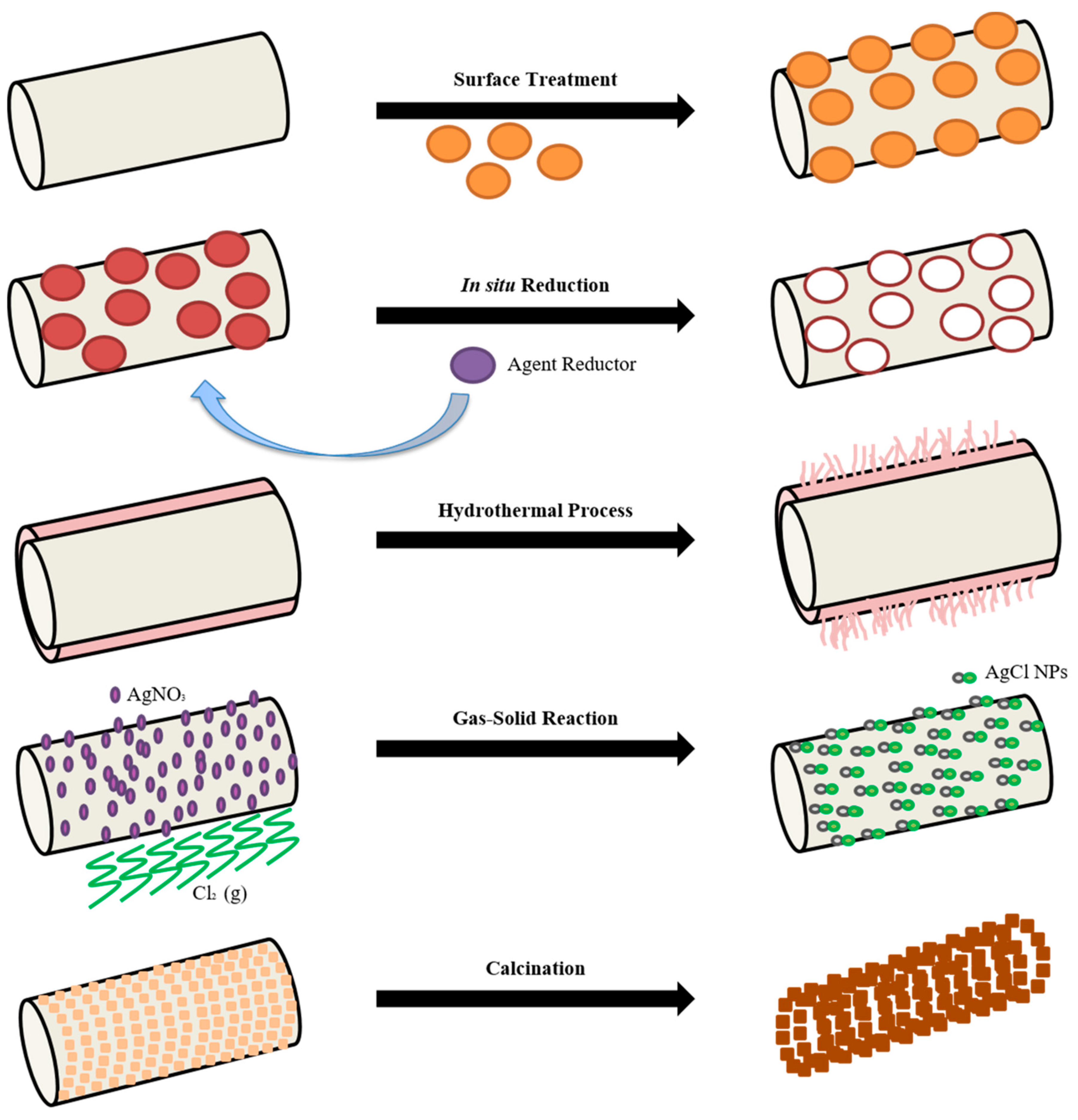
2. Electrospinning Technique and Post-Processing Methods
3. Magnesium Nanoparticles (NPs) Role as Reinforcement of Nanofibers in Environmental Remediation, Energetic Devices and Industrial Catalysis Fields
3.1. Environmental Applications
3.2. Energetic Devices
3.3. Industrial Catalysis
3.4. High-Temperature Applications
4. Development of Electrospun Polymers Reinforced with Mg-Based NPs in Biomedical Applications

| Living Tissue | Young Modulus (MPa) | Ultimate Tensile Strength (MPa) | Elongation At Break (%) | Ref. |
| Aorta valve, human | 2–15 | 0.4–2.6 | 0.22–0.30 | [165,166] |
| Mitral valve anterior leaflet | 3.6 ± 1.8 | 0.05–0.45 Anterior 0.10–0.20 Posterior | - | [167,168] |
| Mitral valve chordae tendinese, human | 330 ± 228 to 388 ± 290 | 36.8 ± 22.5 to 40.8 ± 24.6 | 0.20 ± 0.09 to 0.21 ± 0.12 | [169] |
| Skin, human | 3–54 | 1–20 | 30–70 | [170] |
| Wound Dressing Material | Young Modulus (MPa) | Ultimate Tensile Strength (MPa) | Elongation At Break (%) | Ref. |
| Omiderm | 60.4 ± 4.5 | - | 56.3 ± 3.0 | [171] |
| Chitosan-Alginate + Alphasan® + Silpuran® | - | 43.5 ± 5.5 | 3.9 ± 0.5 | [172] |
| Dental Membranes | Young Modulus (MPa) | Ultimate Tensile Strength (MPa) | Elongation At Break (%) | Ref. |
| Bio-Guide® | 15.7 | 4.8 | - | [173] |
| Collprotect® | 158.5 | 13.1 | - | [173] |
| Jason® | 178.9 | 13.0 | - | [173] |
5. Conclusions
Funding
Acknowledgments
Conflicts of Interest
References
- Patzke, G.R.; Krumeich, F.; Nesper, R. Oxidic nanotubes and nanorods-Anisotropic modules for a future nanotechnology. Angew. Chem. Int. Ed. 2002, 41, 2446–2461. [Google Scholar] [CrossRef]
- Zhao, Q.Q.; Boxman, A.; Chowdhry, U. Nanotechnology in the chemical industry–opportunities and challenges. J. Nanoparticle Res. 2003, 5, 567–572. [Google Scholar] [CrossRef]
- Reynolds, J.G.; Hart, B.R. Nanomaterials and Their Application to Defense and Homeland Security. JOM 2004, 56, 36–39. [Google Scholar] [CrossRef]
- Peponi, L.; Puglia, D.; Torre, L.; Valentini, L.; Kenny, J.M. Processing of nanostructured polymers and advanced polymeric based nanocomposites. Mater. Sci. Eng. R Rep. 2014, 85, 1–46. [Google Scholar] [CrossRef] [Green Version]
- Cushen, M.; Kerry, J.; Morris, M.; Cruz-Romero, M.; Cummins, E. Nanotechnologies in the food industry-Recent developments, risks and regulation. Trends Food Sci. Technol. 2012, 24, 30–46. [Google Scholar] [CrossRef]
- Kim, D.-H.; Ghaffari, R.; Lu, N.; Rogers, J.A. Flexible and Stretchable Electronics for Biointegrated Devices. Annu. Rev. Biomed. Eng. 2012, 14, 113–128. [Google Scholar] [CrossRef] [PubMed] [Green Version]
- Khin, M.M.; Nair, A.S.; Babu, V.J.; Murugan, R.; Ramakrishna, S. A review on nanomaterials for environmental remediation. Energy Environ. Sci. 2012, 5, 8075–8109. [Google Scholar] [CrossRef]
- Glotzer, S.C.; Solomon, M.J. Anisotropy of building blocks and their assembly into complex structures. Nat. Mater. 2007, 6, 557–562. [Google Scholar] [CrossRef]
- Mujica-Garcia, A.; Navarro-Baena, I.; Kenny, J.M.; Peponi, L. Influence of the Processing Parameters on the Electrospinning of Biopolymeric Fibers. J. Renew. Mater. 2014, 2, 23–34. [Google Scholar] [CrossRef]
- Zhang, C.L.; Yu, S.H. Nanoparticles meet electrospinning: Recent advances and future prospects. Chem. Soc. Rev. 2014, 43, 4423–4448. [Google Scholar] [CrossRef]
- Yuan, J.; Xu, Y.; Müller, A.H.E. One-dimensional magnetic inorganic–organic hybrid nanomaterials. Chem. Soc. Rev. 2011, 40, 640–655. [Google Scholar] [CrossRef] [PubMed]
- Echegoyen, Y.; Fabra, M.J.; Castro-Mayorga, J.L.; Cherpinski, A.; Lagaron, J.M. High throughput electro-hydrodynamic processing in food encapsulation and food packaging applications: Viewpoint. Trends Food Sci. Technol. 2017, 60, 71–79. [Google Scholar] [CrossRef]
- Yoo, H.S.; Kim, T.G.; Park, T.G. Surface-functionalized electrospun nanofibers for tissue engineering and drug delivery. Adv. Drug Deliv. Rev. 2009, 61, 1033–1042. [Google Scholar] [CrossRef] [PubMed]
- Reneker, D.H.; Yarin, A.L. Electrospinning jets and polymer nanofibers. Polymer 2008, 49, 2387–2425. [Google Scholar] [CrossRef] [Green Version]
- Pankhurst, Q.A.; Connolly, J.; Jones, S.K.; Dobson, J. Applications of magnetic nanoparticles in biomedicine. J. Phys. D Appl. Phys. 2003, 36, R167–R181. [Google Scholar] [CrossRef] [Green Version]
- Davis, M.E.; Chen, Z.; Shin, D.M. Nanoparticle therapeutics: An emerging treatment modality for cancer. Nat. Rev. Drug Discov. 2008, 7, 771–782. [Google Scholar] [CrossRef]
- Shipway, A.N.; Katz, E.; Willner, I. Nanoparticle arrays on surfaces for electronic, optical, and sensor applications. ChemPhysChem 2000, 1, 18–52. [Google Scholar] [CrossRef]
- Czerwinski, F. Magnesium and Its Alloys. In Magnesium Injection Molding; Springer: New York, NY, USA, 2008; pp. 1–79. [Google Scholar]
- Beyth, N.; Houri-Haddad, Y.; Domb, A.; Khan, W.; Hazan, R. Alternative Antimicrobial Approach: Nano-Antimicrobial Materials. Evid. Based Complementary Altern. Med. 2015, 2015, 1–16. [Google Scholar] [CrossRef] [Green Version]
- Dhivya, S.; Ajita, J.; Selvamurugan, N. Metallic nanomaterials for bone tissue engineering. J. Biomed. Nanotechnol. 2015, 11, 1675–1700. [Google Scholar] [CrossRef]
- Formhals, A. Process and Apparatus for Preparing Artificial Threads. U.S. Patent 1975,504 10 February 1934. [Google Scholar]
- Zeleny, J. The Electrical Discharge from Liquid Points, and a Hydrostatic Method of Measuring the Electric Intensity at Their Surfaces. Phys. Rev. 1914, 3, 69–91. [Google Scholar] [CrossRef] [Green Version]
- Larrondo, L.; St. John Manley, R. Electrostatic fiber spinning from polymer melts. II. Examination of the flow field in an electrically driven jet. J. Polym. Sci. Polym. Phys. Ed. 1981, 19, 921–932. [Google Scholar] [CrossRef]
- Reneker, D.H.; Chun, I. Nanometre diameter fibres of polymer, produced by electrospinning. Nanotechnology 1996, 7, 216–223. [Google Scholar] [CrossRef] [Green Version]
- Noruzi, M. Electrospun nanofibres in agriculture and the food industry: A review. J. Sci. Food Agric. 2016, 96, 4663–4678. [Google Scholar] [CrossRef] [PubMed]
- Jaworek, A.; Krupa, A.; Lackowski, M.; Sobczyk, A.T.; Czech, T.; Ramakrishna, S.; Sundarrajan, S.; Pliszka, D. Nanocomposite fabric formation by electrospinning and electrospraying technologies. J. Electrostat. 2009, 67, 435–438. [Google Scholar] [CrossRef]
- Xie, X.; Chen, Y.; Wang, X.; Xu, X.; Shen, Y.; Khan, A.u.R.; Aldalbahi, A.; Fetz, A.E.; Bowlin, G.L.; El-Newehy, M.; et al. Electrospinning nanofiber scaffolds for soft and hard tissue regeneration. J. Mater. Sci. Technol. 2020, 59, 243–261. [Google Scholar] [CrossRef]
- Arrieta, M.P.; García, A.D.; López, D.; Fiori, S.; Peponi, L. Antioxidant bilayers based on PHBV and plasticized electrospun PLA-PHB fibers encapsulating catechin. Nanomaterials 2019, 9, 346. [Google Scholar] [CrossRef] [Green Version]
- Shenoy, S.L.; Bates, W.D.; Frisch, H.L.; Wnek, G.E. Role of chain entanglements on fiber formation during electrospinning of polymer solutions: Good solvent, non-specific polymer-polymer interaction limit. Polymer 2005, 46, 3372–3384. [Google Scholar] [CrossRef]
- Bhardwaj, N.; Kundu, S.C. Electrospinning: A fascinating fiber fabrication technique. Biotechnol. Adv. 2010, 28, 325–347. [Google Scholar] [CrossRef]
- Deitzel, J.M.; Kleinmeyer, J.; Harris, D.; Beck Tan, N.C. The effect of processing variables on the morphology of electrospun nanofibers and textiles. Polymer 2001, 42, 261–272. [Google Scholar] [CrossRef]
- Demir, M.M.; Yilgor, I.; Yilgor, E.; Erman, B. Electrospinning of polyurethane fibers. Polymer 2002, 43, 3303–3309. [Google Scholar] [CrossRef]
- Araújo, E.; Araújo, E.; Nascimento, M.; de Oliveira, H. Electrospinning of Polymeric Fibres: An Unconventional View on the Influence of Surface Tension on Fibre Diameter. Fibres Text. East. Eur. 2016, 24, 22–29. [Google Scholar] [CrossRef]
- Arrieta, M.P.; López, J.; López, D.; Kenny, J.M.; Peponi, L. Biodegradable electrospun bionanocomposite fibers based on plasticized PLA–PHB blends reinforced with cellulose nanocrystals. Ind. Crops Prod. 2016, 93, 290–301. [Google Scholar] [CrossRef]
- Huang, Z.M.; Zhang, Y.Z.; Kotaki, M.; Ramakrishna, S. A review on polymer nanofibers by electrospinning and their applications in nanocomposites. Compos. Sci. Technol. 2003, 63, 2223–2253. [Google Scholar] [CrossRef]
- Hsu, C.-M.; Shivkumar, S. N,N-Dimethylformamide Additions to the Solution for the Electrospinning of Poly(ε-caprolactone) Nanofibers. Macromol. Mater. Eng. 2004, 289, 334–340. [Google Scholar] [CrossRef]
- Sohrabi, A.; Shaibani, P.M.; Thundat, T. The effect of applied electric field on the diameter and size distribution of electrospun Nylon6 nanofibers. Scanning 2013, 35, 183–188. [Google Scholar] [CrossRef] [PubMed]
- Buchko, C.J.; Chen, L.C.; Shen, Y.; Martina, D.C. Processing and microstructural characterization of porous biocompatible protein polymer thin films. Polymer 1999, 40, 7397–7407. [Google Scholar] [CrossRef]
- Hohman, M.M.; Shin, M.; Rutledge, G.; Brenner, M.P. Electrospinning and electrically forced jets. II. Applications. Phys. Fluids 2001, 13, 2221–2236. [Google Scholar] [CrossRef] [Green Version]
- Xiao, W.; Li, Q.; He, H.; Li, W.; Cao, X.; Dong, H. Patterning Multi-Nanostructured Poly( L-lactic acid) Fibrous Matrices to Manipulate Biomolecule Distribution and Functions. ACS Appl. Mater. Interfaces 2018, 10, 8465–8473. [Google Scholar] [CrossRef]
- Wannatong, L.; Sirivat, A.; Supaphol, P. Effects of solvents on electrospun polymeric fibers: Preliminary study on polystyrene. Polym. Int. 2004, 53, 1851–1859. [Google Scholar] [CrossRef]
- Huang, C.; Thomas, N.L. Fabricating porous poly(lactic acid) fibres via electrospinning. Eur. Polym. J. 2018, 99, 464–476. [Google Scholar] [CrossRef] [Green Version]
- Baumgarten, P.K. Electrostatic spinning of acrylic microfibers. J. Colloid Interface Sci. 1971, 36, 71–79. [Google Scholar] [CrossRef]
- Pu, Y.; Yang, X.; Zhang, Y.; Li, L.; Xie, Y.; He, B.; Yuan, D.; Ning, X. Fabrication and Characterization of Highly Oriented Composite Nanofibers with Excellent Mechanical Strength and Thermal Stability. Macromol. Mater. Eng. 2020, 305, 1900691. [Google Scholar] [CrossRef]
- Hussein, A.; Ramasundaram, S.; Kim, B. A novel method for fabricating bioinspired layered nanocomposites using aligned graphene oxide/PVDF and their micromechanical modeling. Mater. Today Commun. 2020, 24, 101050. [Google Scholar] [CrossRef]
- Fernández-Pérez, J.; Kador, K.E.; Lynch, A.P.; Ahearne, M. Characterization of extracellular matrix modified poly(ε-caprolactone) electrospun scaffolds with differing fiber orientations for corneal stroma regeneration. Mater. Sci. Eng. C 2020, 108, 110415. [Google Scholar] [CrossRef] [PubMed]
- Pathalamuthu, P.; Thameem Shahana, S.; Nivedha, U.; Siddharthan, A.; Giridev, V.R. Analysis of the inhomogeneity of an electrospun mat produced using static and dynamic collectors of different template design, including its mitigation. Fibres Text. East. Eur. 2017, 25, 53–61. [Google Scholar] [CrossRef]
- Rojas, A.; Velásquez, E.; Garrido, L.; Galotto, M.J.; López de Dicastillo, C. Design of active electrospun mats with single and core-shell structures to achieve different curcumin release kinetics. J. Food Eng. 2020, 273, 109900. [Google Scholar] [CrossRef]
- Farboudi, A.; Nouri, A.; Shirinzad, S.; Sojoudi, P.; Davaran, S.; Akrami, M.; Irani, M. Synthesis of magnetic gold coated poly (ε-caprolactonediol) based polyurethane/poly(N-isopropylacrylamide)-grafted-chitosan core-shell nanofibers for controlled release of paclitaxel and 5-FU. Int. J. Biol. Macromol. 2020, 150, 1130–1140. [Google Scholar] [CrossRef]
- Li, W.; Cicek, N.; Levin, D.B.; Logsetty, S.; Liu, S. Bacteria-triggered release of a potent biocide from core-shell polyhydroxyalkanoate (PHA)-based nanofibers for wound dressing applications. J. Biomater. Sci. Polym. Ed. 2020, 31, 394–406. [Google Scholar] [CrossRef]
- Leonés, A.; Sonseca, A.; López, D.; Fiori, S.; Peponi, L. Shape memory effect on electrospun PLA-based fibers tailoring their thermal response. Eur. Polym. J. 2019, 117, 217–226. [Google Scholar] [CrossRef]
- Sessini, V.; López Galisteo, A.J.; Leonés, A.; Ureña, A.; Peponi, L. Sandwich-Type Composites Based on Smart Ionomeric Polymer and Electrospun Microfibers. Front. Mater. 2019, 6, 1–15. [Google Scholar] [CrossRef] [Green Version]
- Arpatappeh, F.A.; Papila, M.; Aydemir, B.E. Multiscale Reinforcing Interlayers of Self-same P(St-co-GMA) Nanofibers Loaded with MCF for Polymer Composites and Nanocomposites. In Proceedings of the AIAA Scitech 2019 Forum, San Diego, CA, USA, 7–11 January 2019. [Google Scholar] [CrossRef] [Green Version]
- Sagitha, P.; Reshmi, C.R.; Sundaran, S.P.; Sujith, A. Recent advances in post-modification strategies of polymeric electrospun membranes. Eur. Polym. J. 2018, 105, 227–249. [Google Scholar] [CrossRef]
- Boyraz, E.; Yalcinkaya, F.; Hruza, J.; Maryska, J. Surface-Modified Nanofibrous PVDF Membranes for Liquid Separation Technology. Materials 2019, 12, 2702. [Google Scholar] [CrossRef] [PubMed] [Green Version]
- Lu, X.; Wang, C.; Wei, Y. One-dimensional composite nanomaterials: Synthesis by electrospinning and their applications. Small 2009, 5, 2349–2370. [Google Scholar] [CrossRef]
- Burda, C.; Chen, X.; Narayanan, R.; El-Sayed, M.A. Chemistry and Proerties of Nanocrystals of Different Shapes. Chem. Rev. 2005, 105, 1025–1102. [Google Scholar] [CrossRef] [PubMed]
- Zhu, W.; Chi, M.; Gao, M.; Wang, C.; Lu, X. Controlled synthesis of titanium dioxide/molybdenum disulfide core-shell hybrid nanofibers with enhanced peroxidase-like activity for colorimetric detection of glutathione. J. Colloid Interface Sci. 2018, 528, 410–418. [Google Scholar] [CrossRef] [PubMed]
- Broujeni, B.R.; Nilchi, A. Preparation and Characterization of Polyacrylonitrile/Aluminum Oxide Nanofiber Adsorbent Modified with 2-Amino-3-Methyl-1-Hexanethiol for the Adsorption of Thorium (IV) Ion from Aqueous Solution. J. Environ. Eng. 2018, 144, 04018099. [Google Scholar] [CrossRef]
- Liu, K.; Zhang, Z.; Lu, N.; Dong, B. In Situ Generation of Copper Species Nanocrystals in TiO2 Electrospun Nanofibers: A Multi-hetero-junction Photocatalyst for Highly Efficient Water Reduction. ACS Sustain. Chem. Eng. 2018, 6, 1934–1940. [Google Scholar] [CrossRef]
- Da Silva, E.P.; Rubira, A.F.; Ferreira, O.P.; Silva, R.; Muniz, E.C. In situ growth of manganese oxide nanosheets over titanium dioxide nanofibers and their performance as active material for supercapacitor. J. Colloid Interface Sci. 2019, 555, 373–382. [Google Scholar] [CrossRef]
- Zhang, C.; Liu, Q.; Zhan, N.; Yang, Q.; Song, Y.; Sun, L.; Wang, H.; Li, Y. A novel approach to prepare silver chloride nanoparticles grown on the surface of PAN nanofibre via electrospinning combined with gas-solid reaction. Colloids Surf. A Physicochem. Eng. Asp. 2010, 353, 64–68. [Google Scholar] [CrossRef]
- Ding, Y.; Wang, Y.; Su, L.; Bellagamba, M.; Zhang, H.; Lei, Y. Electrospun Co3O4 nanofibers for sensitive and selective glucose detection. Biosens. Bioelectron. 2010, 26, 542–548. [Google Scholar] [CrossRef]
- Patel, A.C.; Li, S.; Wang, C.; Zhang, W.; Wei, Y. Electrospinning of porous silica nanofibers containing silver nanoparticles for catalytic applications. Chem. Mater. 2007, 19, 1231–1238. [Google Scholar] [CrossRef]
- Madhugiri, S.; Sun, B.; Smirniotis, P.G.; Ferraris, J.P.; Balkus, K.J. Electrospun mesoporous titanium dioxide fibers. Microporous Mesoporous Mater. 2004, 69, 77–83. [Google Scholar] [CrossRef]
- Formo, E.; Lee, E.; Campbell, D.; Xia, Y. Functionalization of Electrospun TiO2 Nanofibers with Pt Nanoparticles and Nanowires for Catalytic Applications. Nano Lett. 2008, 8, 668–672. [Google Scholar] [CrossRef]
- Arrieta, M.P.; Perdiguero, M.; Fiori, S.; Kenny, J.M.; Peponi, L. Biodegradable electrospun PLA-PHB fibers plasticized with oligomeric lactic acid. Polym. Degrad. Stabil. 2020, 179, 109226. [Google Scholar] [CrossRef]
- Akinalan Balik, B.; Argin, S.; Lagaron, J.M.; Torres-Giner, S. Preparation and Characterization of Electrospun Pectin-Based Films and Their Application in Sustainable Aroma Barrier Multilayer Packaging. Appl. Sci. 2019, 9, 5136. [Google Scholar] [CrossRef] [Green Version]
- Frohbergh, M.E.; Katsman, A.; Botta, G.P.; Lazarovici, P.; Schauer, C.L.; Wegst, U.G.K.; Lelkes, P.I. Electrospun hydroxyapatite-containing chitosan nanofibers crosslinked with genipin for bone tissue engineering. Biomaterials 2012, 33, 9167–9178. [Google Scholar] [CrossRef] [PubMed] [Green Version]
- Zhang, Y.; Venugopal, J.R.; El-Turki, A.; Ramakrishna, S.; Su, B.; Lim, C.T. Electrospun biomimetic nanocomposite nanofibers of hydroxyapatite/chitosan for bone tissue engineering. Biomaterials 2008, 29, 4314–4322. [Google Scholar] [CrossRef] [PubMed]
- Shao, C.; Guan, H.; Liu, Y.; Mu, R. MgO nanofibres via an electrospinning technique. J. Mater. Sci. 2006, 41, 3821–3824. [Google Scholar] [CrossRef]
- Jaworek, A.; Krupa, A.; Lackowski, M.; Sobczyk, A.T.; Czech, T.; Ramakrishna, S.; Sundarrajan, S.; Pliszka, D. Electrostatic method for the production of polymer nanofibers blended with metal-oxide nanoparticles. J. Phys. Conf. Ser. 2009, 146, 012006. [Google Scholar] [CrossRef]
- Dadvar, S.; Tavanai, H.; Morshed, M. Fabrication of nanocomposite PAN nanofibers containing MgO and Al2O3 nanoparticles. Polym. Sci. Ser. A 2014, 56, 358–365. [Google Scholar] [CrossRef]
- Jaworek, A.; Krupa, A.; Lackowski, M.; Sobczyk, A.T.; Czech, T.; Ramakrishna, S.; Sundarrajan, S.; Pliszka, D. Electrospinning and electrospraying techniques for nanocomposite non-woven fabric production. Fibres Text. East. Eur. 2009, 75, 77–81. [Google Scholar]
- Kalia, S.; Kango, S.; Kumar, A.; Haldorai, Y.; Kumari, B.; Kumar, R. Magnetic polymer nanocomposites for environmental and biomedical applications. Colloid Polym. Sci. 2014, 292, 2025–2052. [Google Scholar] [CrossRef]
- Ali, A.; Gul, A.; Mannan, A.; Zia, M. Efficient metal adsorption and microbial reduction from Rawal Lake wastewater using metal nanoparticle coated cotton. Sci. Total Environ. 2018, 639, 26–39. [Google Scholar] [CrossRef] [PubMed]
- Wan, S.; Ding, W.; Wang, Y.; Wu, J.; Gu, Y.; He, F. Manganese oxide nanoparticles impregnated graphene oxide aggregates for cadmium and copper remediation. Chem. Eng. J. 2018, 350, 1135–1143. [Google Scholar] [CrossRef]
- Gao, C.; Zhang, W.; Li, H.; Lang, L.; Xu, Z. Controllable fabrication of mesoporous MgO with various morphologies and their absorption performance for toxic pollutants in water. Cryst. Growth Des. 2008, 8, 3785–3790. [Google Scholar] [CrossRef]
- Lange, L.E.; Obendorf, S.K. Effect of plasma etching on destructive adsorption properties of polypropylene fibers containing magnesium oxide nanoparticles. Arch. Environ. Contam. Toxicol. 2012, 62, 185–194. [Google Scholar] [CrossRef]
- Rajagopalan, S.; Koper, O.; Decker, S.; Klabunde, K.J. Nanocrystalline metal oxides as destructive adsorbents for organophosphorus compounds at ambient temperatures. Chemistry 2002, 8, 2602–2607. [Google Scholar] [CrossRef]
- Sharma, L.; Kakkar, R. Hierarchically structured magnesium based oxides: Synthesis strategies and applications in organic pollutant remediation. CrystEngComm 2017, 19, 6913–6926. [Google Scholar] [CrossRef]
- Woo, D.J.; Obendorf, S.K. MgO-embedded fibre-based substrate as an effective sorbent for toxic organophosphates. RSC Adv. 2014, 4, 15727–15735. [Google Scholar] [CrossRef] [Green Version]
- Wei, X.-H.; Li, C.; Wang, C.; Lin, S.; Wu, J.; Guo, M.-J. Rapid and destructive adsorption of paraoxon-ethyl toxin via a self-detoxifying hybrid electrospun nanofibrous membrane. Chem. Eng. J. 2018, 351, 31–39. [Google Scholar] [CrossRef]
- Yu, Z.; Xu, C.; Yuan, K.; Gan, X.; Zhou, H.; Wang, X.; Zhu, L.; Zhang, G.; Xu, D. Template-free synthesis of MgO mesoporous nanofibers with superior adsorption for fluoride and Congo red. Ceram. Int. 2018, 44, 9454–9462. [Google Scholar] [CrossRef]
- Mantilaka, M.M.M.G.P.G.; De Silva, R.T.; Ratnayake, S.P.; Amaratunga, G.; de Silva, K.M.N. Photocatalytic activity of electrospun MgO nanofibres: Synthesis, characterization and applications. Mater. Res. Bull. 2018, 99, 204–210. [Google Scholar] [CrossRef]
- Ren, B. Electrospinning Synthesis of MgO Nanofibers and Properties of Uranium (VI)-sorption. J. Chem. Soc. Pakistan 2014, 36, 250–254. [Google Scholar]
- Othman, F.E.C.; Yusof, N.; Jaafar, J.; Ismail, A.F.; Abdullah, N.; Hasbullah, H. Adsorption of cadmium (II) ions by polyacrylonitrile-based activated carbon nanofibers/magnesium oxide as its adsorbents. Malays. J. Anal. Sci. 2016, 20, 1467–1473. [Google Scholar]
- Almasian, A.; Najafi, F.; Maleknia, L.; Giahi, M. Mesoporous MgO/PPG hybrid nanofibers: Synthesis, optimization, characterization and heavy metal removal property. New J. Chem. 2018, 42, 2013–2029. [Google Scholar] [CrossRef]
- Xu, C.; Shi, S.; Dong, Q.; Zhu, S.; Wang, Y.; Zhou, H.; Wang, X.; Zhu, L.; Zhang, G.; Xu, D. Citric-acid-assisted sol-gel synthesis of mesoporous silicon-magnesium oxide ceramic fibers and their adsorption characteristics. Ceram. Int. 2020, 46, 10105–10114. [Google Scholar] [CrossRef]
- Xu, C.; Shi, S.; Wang, X.; Zhou, H.; Wang, L.; Zhu, L.; Zhang, G.; Xu, D. Electrospun SiO2-MgO hybrid fibers for heavy metal removal: Characterization and adsorption study of Pb(II) and Cu(II). J. Hazard. Mater. 2020, 381, 120974. [Google Scholar] [CrossRef]
- Jia, B.-B.; Wang, J.-N.; Wu, J.; Li, C.-J. “Flower-Like” PA6@Mg(OH)2 electrospun nanofibers with Cr (VI)-removal capacity. Chem. Eng. J. 2014, 254, 98–105. [Google Scholar] [CrossRef]
- Chen, H.; Lin, J.; Zhang, N.; Chen, L.; Zhong, S.; Wang, Y.; Zhang, W.; Ling, Q. Preparation of MgAl-EDTA-LDH based electrospun nanofiber membrane and its adsorption properties of copper(II) from wastewater. J. Hazard. Mater. 2018, 345, 1–9. [Google Scholar] [CrossRef]
- Dehghan, S.F.; Golbabaei, F.; Maddah, B.; Latifi, M.; Pezeshk, H.; Hasanzadeh, M.; Akbar-Khanzadeh, F. Optimization of electrospinning parameters for polyacrylonitrile-MgO nanofibers applied in air filtration. J. Air Waste Manag. Assoc. 2016, 66, 912–921. [Google Scholar] [CrossRef] [Green Version]
- Dehghan, S.F.; Golbabaei, F.; Mousavi, T.; Mohammadi, H.; Kohneshahri, M.H.; Bakhtiari, R. Production of nanofibers containing magnesium oxide nanoparticles for the purpose of bioaerosol removal. Pollution 2020, 6, 185–196. [Google Scholar]
- Gu, Y.; Chen, D.; Jiao, X. Synthesis and Electrochemical Properties of Nanostructured LiCoO2 Fibers as Cathode Materials for Lithium-Ion Batteries. J. Phys. Chem. B 2005, 109, 17901–17906. [Google Scholar] [CrossRef] [PubMed]
- Gu, Y.; Chen, D.; Jiao, X.; Liu, F. LiCoO2-MgO coaxial fibers: Co-electrospun fabrication, characterization and electrochemical properties. J. Mater. Chem. 2007, 17, 1769–1776. [Google Scholar] [CrossRef]
- Ma, D.; Zhu, R.; Ruan, F.; Sonamuthu, J.; Zhang, Q.; Sun, M.; Li, H.; Cai, Y. Rational design of hierarchical C–MgO@ZnO nanofiber as sulfur host for high-performance lithium–sulfur batteries. J. Mater. Sci. 2020, 55, 5534–5544. [Google Scholar] [CrossRef]
- Darbar, D.; Reddy, M.V.; Sundarrajan, S.; Pattabiraman, R.; Ramakrishna, S.; Chowdari, B.V.R. Anodic electrochemical performances of MgCo2O4 synthesized by oxalate decomposition method and electrospinning technique for Li-ion battery application. Mater. Res. Bull. 2016, 73, 369–376. [Google Scholar] [CrossRef]
- Gong, C.; Bai, Y.J.; Qi, Y.X.; Lun, N.; Feng, J. Preparation of carbon-coated MgFe2O4 with excellent cycling and rate performance. Electrochim. Acta 2013, 90, 119–127. [Google Scholar] [CrossRef]
- Yin, Y.; Zhang, B.; Zhang, X.; Xu, J.; Yang, S. Nano MgFe2O4 synthesized by sol–gel auto-combustion method as anode materials for lithium ion batteries. J. Sol. Gel Sci. Technol. 2013, 66, 540–543. [Google Scholar] [CrossRef]
- Qiao, H.; Luo, L.; Chen, K.; Fei, Y.; Cui, R.; Wei, Q. Electrospun synthesis and lithium storage properties of magnesium ferrite nanofibers. Electrochim. Acta 2015, 160, 43–49. [Google Scholar] [CrossRef]
- Avellaneda, C.O.; Gonçalves, A.D.; Benedetti, J.E.; Nogueira, A.F. Preparation and characterization of core-shell electrodes for application in gel electrolyte-based dye-sensitized solar cells. Electrochim. Acta 2010, 55, 1468–1474. [Google Scholar] [CrossRef]
- Du, P.; Song, L.; Xiong, J.; Wang, L.; Li, N. A photovoltaic smart textile and a photocatalytic functional textile based on co-electrospun TiO2/MgO core-sheath nanorods: Novel textiles of integrating energy and environmental science with textile research. Text. Res. J. 2013, 83, 1690–1702. [Google Scholar] [CrossRef]
- Sainudeen, S.S.; Asok, L.B.; Varghese, A.; Nair, A.S.; Krishnan, G. Surfactant-driven direct synthesis of a hierarchical hollow MgO nanofiber–nanoparticle composite by electrospinning. RSC Adv. 2017, 7, 35160–35168. [Google Scholar] [CrossRef] [Green Version]
- Makino, T.; Chia, C.H.; Tuan, N.T.; Sun, H.D.; Segawa, Y.; Kawasaki, M.; Ohtomo, A.; Tamura, K.; Koinuma, H. Room-temperature luminescence of excitons in ZnO/(Mg, Zn)O multiple quantum wells on lattice-matched substrates. Appl. Phys. Lett. 2000, 77, 975. [Google Scholar] [CrossRef]
- Sharma, A.K.; Narayan, J.; Muth, J.F.; Teng, C.W.; Jin, C.; Kvit, A.; Kolbas, R.M.; Holland, O.W. Optical and structural properties of epitaxial MgxZn1-xO alloys. Appl. Phys. Lett. 1999, 75, 3327–3329. [Google Scholar] [CrossRef]
- Makino, T.; Tuan, N.T.; Sun, H.D.; Chia, C.H.; Segawa, Y.; Kawasaki, M.; Ohtomo, A.; Tamura, K.; Suemoto, T.; Akiyama, H.; et al. Temperature dependence of near ultraviolet photoluminescence in ZnO/(Mg, Zn)O multiple quantum wells. Appl. Phys. Lett. 2001, 78, 1979–1981. [Google Scholar] [CrossRef] [Green Version]
- Cetin, S.S.; Uslu, I.; Aytimur, A.; Ozcelik, S. Characterization of Mg doped ZnO nanocrystallites prepared via electrospinning. Ceram. Int. 2012, 38, 4201–4208. [Google Scholar] [CrossRef]
- Orujalipoor, I.; Aytimur, A.; Tükel, C.; Ide, S.; Uslu, I. SAXS and WAXS analysis of MgO doped ZnO nanostructured ceramics grown on Si and glass substrate. J. Sol. Gel Sci. Technol. 2014, 70, 125–132. [Google Scholar] [CrossRef]
- Choi, S.H.; Jang, B.H.; Park, J.S.; Demadrille, R.; Tuller, H.L.; Kim, I.D. Low voltage operating field effect transistors with composite In2O3-ZnO-ZnGa2O4 nanofiber network as active channel layer. ACS Nano 2014, 8, 2318–2327. [Google Scholar] [CrossRef]
- Aykut, Y. Electrospun MgO-loaded carbon nanofibers: Enhanced field electron emission from the fibers in vacuum. J. Phys. Chem. Solids 2013, 74, 328–337. [Google Scholar] [CrossRef]
- Tan, J.; Han, Y.; He, L.; Dong, Y.; Xu, X.; Liu, D.; Yan, H.; Yu, Q.; Huang, C.; Mai, L. In situ nitrogen-doped mesoporous carbon nanofibers as flexible freestanding electrodes for high-performance supercapacitors. J. Mater. Chem. A 2017, 5, 23620–23627. [Google Scholar] [CrossRef]
- Zhang, W.; Guo, Z.; Chen, Y.; Cao, Y. Nanomaterial Based Biosensors for Detection of Biomarkers of Exposure to OP Pesticides and Nerve Agents: A Review. Electroanalysis 2017, 29, 1206–1213. [Google Scholar] [CrossRef]
- Fan, Y.; Moon, J.J. Particulate delivery systems for vaccination against bioterrorism agents and emerging infectious pathogens. Wiley Interdiscip. Rev. Nanomed. Nanobiotechnol. 2017, 9, 1–26. [Google Scholar] [CrossRef] [PubMed] [Green Version]
- Koper, O.; Lucas, E.; Klabunde, K.J. Development of reactive topical skin protectants against sulfur mustard and nerve agents. J. Appl. Toxicol. 1999, 19 (Suppl. 1), S59–S70. [Google Scholar] [CrossRef]
- Stoimenov, P.K.; Klinger, R.L.; Marchin, G.L.; Klabunde, K.J. Metal oxide nanoparticles as bactericidal agents. Langmuir 2002, 18, 6679–6686. [Google Scholar] [CrossRef]
- Koper, O.B.; Klabunde, J.S.; Marchin, G.L.; Klabunde, K.J.; Stoimenov, P.; Bohra, L. Nanoscale powders and formulations with biocidal activity toward spores and vegetative cells of Bacillus species, viruses, and toxins. Curr. Microbiol. 2002, 44, 49–55. [Google Scholar] [CrossRef]
- Sundarrajan, S.; Ramakrishna, S. Fabrication of nanocomposite membranes from nanofibers and nanoparticles for protection against chemical warfare stimulants. J. Mater. Sci. 2007, 42, 8400–8407. [Google Scholar] [CrossRef]
- Ryu, S.Y.; Chung, J.W.; Kwak, S.Y. Tunable multilayer assemblies of nanofibrous composite mats as permeable protective materials against chemical warfare agents. RSC Adv. 2017, 7, 9964–9974. [Google Scholar] [CrossRef] [Green Version]
- Xin, J.H.; Daoud, W.A.; Kong, Y.Y. A New Approach to UV-Blocking Treatment for Cotton Fabrics. Text. Res. J. 2004, 74, 97–100. [Google Scholar] [CrossRef]
- Qien, L.; Hinestroza, J.P. Application of Nanotechnology for High Performance Textiles. J. Text. Appar. Technol. Manag. 2004, 4, 1–7. [Google Scholar]
- Lee, S. Developing UV-protective textiles based on electrospun zinc oxide nanocomposite fibers. Fibers Polym. 2009, 10, 295–301. [Google Scholar] [CrossRef]
- Dadvar, S.; Tavanai, H.; Morshed, M. UV-protection properties of electrospun polyacrylonitrile nanofibrous mats embedded with MgO and Al2O3 nanoparticles. J. Nanoparticle Res. 2011, 13, 5163–5169. [Google Scholar] [CrossRef]
- Liu, H.; Yue, J.; Lei, Q.; Gou, X.; Chen, S.Y.; He, Y.Y.; Wu, X. Ultraviolet B Inhibits Skin Wound Healing by Affecting Focal Adhesion Dynamics. Photochem. Photobiol. 2015, 91, 909–916. [Google Scholar] [CrossRef] [PubMed] [Green Version]
- Jan, N.A.M.; Sahar, M.R.; Sulhadi, S.; El-Mallawany, R. Thermal, structural and magnetic properties of TeO2-MgO-Na2O-Nd2O3 glass system with NiO nanoparticles. J. Non. Cryst. Solids 2019, 522, 119566. [Google Scholar] [CrossRef]
- Vesali-Kermani, E.; Habibi-Yangjeh, A.; Ghosh, S. Visible-light-induced nitrogen photofixation ability of g-C3N4 nanosheets decorated with MgO nanoparticles. J. Ind. Eng. Chem. 2020, 84, 185–195. [Google Scholar] [CrossRef]
- Wang, Y.; Cen, C.; Chen, J.; Fu, L. MgO/carboxymethyl chitosan nanocomposite improves thermal stability, waterproof and antibacterial performance for food packaging. Carbohydr. Polym. 2020, 236, 116078. [Google Scholar] [CrossRef] [PubMed]
- Rios, R.B.; Bastos-Neto, M.; Amora, M.R.; Torres, A.E.B.; Azevedo, D.C.S.; Cavalcante, C.L. Experimental analysis of the efficiency on charge/discharge cycles in natural gas storage by adsorption. Fuel 2011, 90, 113–119. [Google Scholar] [CrossRef]
- Yeoh, W.M.; Lee, K.Y.; Chai, S.P.; Lee, K.T.; Mohamed, A.R. Effective synthesis of carbon nanotubes via catalytic decomposition of methane: Influence of calcination temperature on metal-support interaction of Co-Mo/MgO catalyst. J. Phys. Chem. Solids 2013, 74, 1553–1559. [Google Scholar] [CrossRef]
- Che Othman, F.E.; Yusof, N.; Hasbullah, H.; Jaafar, J.; Ismail, A.F.; Abdullah, N.; Md Nordin, N.A.H.; Aziz, F.; Wan Salleh, W.N. Polyacrylonitrile/magnesium oxide-based activated carbon nanofibers with well-developed microporous structure and their adsorption performance for methane. J. Ind. Eng. Chem. 2017, 51, 281–287. [Google Scholar] [CrossRef]
- Ma, Y.; Ma, Y.; Long, G.; Li, J.; Hu, X.; Ye, Z.; Wang, Z.; Buckley, C.E.; Dong, D. Synergistic promotion effect of MgO and CeO2 on nanofibrous Ni/Al2O3 catalysts for methane partial oxidation. Fuel 2019, 258, 116103. [Google Scholar] [CrossRef]
- Khalim Khafidz, N.Z.A.; Yaakob, Z.; Timmiati, S.N.; Lin, K.S.; Lim, K.L. Hydrogen sorption of magnesium oxide carbon nanofibre composite. Malays. J. Anal. Sci. 2019, 23, 60–70. [Google Scholar]
- Triviño, M.L.T.; Jeon, H.; Lim, A.C.S.; Hiremath, V.; Sekine, Y.; Seo, J.G. Encapsulation of Phase-Changing Eutectic Salts in Magnesium Oxide Fibers for High-Temperature Carbon Dioxide Capture: Beyond the Capacity-Stability Tradeoff. ACS Appl. Mater. Interfaces 2020, 12, 518–526. [Google Scholar] [CrossRef]
- Liu, H.; Bai, J.; Li, C.; Xu, W.; Sun, W.; Xu, T.; Huang, Y.; Li, H. An effective approach to preparing MgO-Ag NPs-CNFs and Al2O3-Ag NPs-CNFs for styrene epoxidation action. RSC Adv. 2014, 4, 3195–3200. [Google Scholar] [CrossRef]
- Kordas, G. Sol-gel preparation of MgO fibers. J. Mater. Chem. 2000, 10, 1157–1160. [Google Scholar] [CrossRef]
- Xu, C.; Yuan, K.; Jin, X.; Yu, Z.; Zheng, L.; Lü, Y.; Wang, X.; Zhu, L.; Zhang, G.; Xu, D. High-temperature stable electrospun MgO nanofibers, formation mechanism and thermal properties. Ceram. Int. 2017, 43, 16210–16216. [Google Scholar] [CrossRef]
- Vacanti, J.P.; Vacanti, C.A. Chapter 1—The History and Scope of Tissue Engineering. In Principles of Tissue Engineering, 2nd ed.; Lanza, R.P., Langer, R., Vacanti, J., Eds.; Tissue Engineering Intelligence Unit; Academic Press: San Diego, CA, USA, 2000; pp. 3–7. [Google Scholar]
- Jagur-Grodzinski, J. Polymers for tissue engineering, medical devices, and regenerative medicine. Concise general review of recent studies. Polym. Adv. Technol. 2006, 17, 395–418. [Google Scholar] [CrossRef]
- Langer, R.; Vacanti, J. Tissue engineering. Science 1993, 260, 920–926. [Google Scholar] [CrossRef] [Green Version]
- Mikos, A.G.; Temenoff, J.S. Formation of highly porous biodegradable scaffolds for tissue engineering. Electron. J. Biotechnol. 2000, 3, 1995–2000. [Google Scholar] [CrossRef]
- Pereira, H.F.; Cengiz, I.F.; Silva, F.S.; Reis, R.L.; Oliveira, J.M. Scaffolds and coatings for bone regeneration. J. Mater. Sci. Mater. Med. 2020, 31, 27. [Google Scholar] [CrossRef] [Green Version]
- Wang, X.; Xu, S.; Zhou, S.; Xu, W.; Leary, M.; Choong, P.; Qian, M.; Brandt, M.; Xie, Y.M. Topological design and additive manufacturing of porous metals for bone scaffolds and orthopaedic implants: A review. Biomaterials 2016, 83, 127–141. [Google Scholar] [CrossRef]
- Rahaman, M.N.; Day, D.E.; Sonny Bal, B.; Fu, Q.; Jung, S.B.; Bonewald, L.F.; Tomsia, A.P. Bioactive glass in tissue engineering. Acta Biomater. 2011, 7, 2355–2373. [Google Scholar] [CrossRef] [Green Version]
- Eliaz, N.; Metoki, N. Calcium Phosphate Bioceramics: A Review of Their History, Structure, Properties, Coating Technologies and Biomedical Applications. Materials 2017, 10, 334. [Google Scholar] [CrossRef] [Green Version]
- Li, Y.; Zhang, F.; Liu, Y.; Leng, J. 4D printed shape memory polymers and their structures for biomedical applications. Sci. China Technol. Sci. 2020, 63, 545–560. [Google Scholar] [CrossRef] [Green Version]
- Zaszczynska, A.; Sajkiewicz, P.; Gradys, A. Piezoelectric Scaffolds as Smart Materials for Neural Tissue Engineering. Polymers 2020, 12, 161. [Google Scholar] [CrossRef] [PubMed] [Green Version]
- Polo-Corrales, L.; Latorre-Esteves, M.; Ramirez-Vick, J.E. Scaffold Design for Bone Regeneration. J. Nanosci. Nanotechnol. 2014, 14, 15–56. [Google Scholar] [CrossRef] [PubMed] [Green Version]
- Williams, D.F. On the mechanisms of biocompatibility. Biomaterials 2008, 29, 2941–2953. [Google Scholar] [CrossRef] [PubMed]
- Martău, G.A.; Mihai, M.; Vodnar, D.C. The Use of Chitosan, Alginate, and Pectin in the Biomedical and Food Sector—Biocompatibility, Bioadhesiveness, and Biodegradability. Polymers 2019, 11, 1837. [Google Scholar] [CrossRef] [Green Version]
- Constantin, C.P.; Aflori, M.; Damian, R.F.; Rusu, R.D. Biocompatibility of Polyimides: A Mini-Review. Materials 2019, 12, 3166. [Google Scholar] [CrossRef] [Green Version]
- Elmowafy, E.M.; Tiboni, M.; Soliman, M.E. Biocompatibility, biodegradation and biomedical applications of poly(lactic acid)/poly(lactic-co-glycolic acid) micro and nanoparticles. J. Pharm. Investig. 2019, 49, 347–380. [Google Scholar] [CrossRef]
- Tao, F.; Cheng, Y.; Shi, X.; Zheng, H.; Du, Y.; Xiang, W.; Deng, H. Applications of chitin and chitosan nanofibers in bone regenerative engineering. Carbohydr. Polym. 2020, 230, 115658. [Google Scholar] [CrossRef]
- Donnely, E.; Griffin, M.; Butler, P.E. Breast Reconstruction with a Tissue Engineering and Regenerative Medicine Approach (Systematic Review). Ann. Biomed. Eng. 2020, 48, 9–25. [Google Scholar] [CrossRef] [Green Version]
- Xiao, S.; Zhao, T.; Wang, J.; Wang, C.; Du, J.; Ying, L.; Lin, J.; Zhang, C.; Hu, W.; Wang, L.; et al. Gelatin Methacrylate (GelMA)-Based Hydrogels for Cell Transplantation: An Effective Strategy for Tissue Engineering. Stem Cell Rev. Rep. 2019, 15, 664–679. [Google Scholar] [CrossRef]
- Pang, M.; Huang, Y.; Meng, F.; Zhuang, Y.; Liu, H.; Du, M.; Ma, Q.; Wang, Q.; Chen, Z.; Chen, L.; et al. Application of bacterial cellulose in skin and bone tissue engineering. Eur. Polym. J. 2020, 122, 109365. [Google Scholar] [CrossRef]
- Abbasian, M.; Massoumi, B.; Mohammad-Rezaei, R.; Samadian, H.; Jaymand, M. Scaffolding polymeric biomaterials: Are naturally occurring biological macromolecules more appropriate for tissue engineering? Int. J. Biol. Macromol. 2019, 134, 673–694. [Google Scholar] [CrossRef] [PubMed]
- Leonés, A.; Mujica-Garcia, A.; Arrieta, M.P.; Salaris, V.; Lopez, D.; Kenny, J.M.; Peponi, L. Organic and Inorganic PCL-Based Electrospun Fibers. Polymers 2020, 12, 1325. [Google Scholar] [CrossRef] [PubMed]
- Mariani, E.; Lisignoli, G.; Borzì, R.M.; Pulsatelli, L. Biomaterials: Foreign Bodies or Tuners for the Immune Response? Int. J. Mol. Sci. 2019, 20, 636. [Google Scholar] [CrossRef] [PubMed] [Green Version]
- Przekora, A.; Palka, K.; Ginalska, G. Biomedical potential of chitosan/HA and chitosan/β-1,3-glucan/HA biomaterials as scaffolds for bone regeneration—A comparative study. Mater. Sci. Eng. C 2016, 58, 891–899. [Google Scholar] [CrossRef]
- Alvarez, K.; Nakajima, H. Metallic scaffolds for bone regeneration. Materials. 2009, 2, 790–832. [Google Scholar] [CrossRef]
- Przekora, A. Current Trends in Fabrication of Biomaterials for Bone and Cartilage Regeneration: Materials Modifications and Biophysical Stimulations. Int. J. Mol. Sci. 2019, 20, 435. [Google Scholar] [CrossRef] [Green Version]
- Okuma, T. Magnesium and bone strength. Nutrition 2001, 17, 679–680. [Google Scholar] [CrossRef]
- Vormann, J. Magnesium: Nutrition and metabolism. Mol. Aspects Med. 2003, 24, 27–37. [Google Scholar] [CrossRef]
- de Baaij, J.H.F.; Hoenderop, J.G.J.; Bindels, R.J.M. Magnesium in man: Implications for health and disease. Physiol. Rev. 2015, 95, 1–46. [Google Scholar] [CrossRef]
- Hasan, A.; Ragaert, K.; Swieszkowski, W.; Selimović, Š.; Paul, A.; Camci-Unal, G.; Mofrad, M.R.K.; Khademhosseini, A. Biomechanical properties of native and tissue engineered heart valve constructs. J. Biomech. 2014, 47, 1949–1963. [Google Scholar] [CrossRef] [PubMed]
- Balguid, A.; Rubbens, M.P.; Mol, A.; Bank, R.A.; Bogers, A.J.J.C.; van Kats, J.P.; de Mol, B.A.J.M.; Baaijens, F.P.T.; Bouten, C.V.C. The Role of Collagen Cross-Links in Biomechanical Behavior of Human Aortic Heart Valve Leaflets—Relevance for Tissue Engineering. Tissue Eng. 2007, 13, 1501–1511. [Google Scholar] [CrossRef] [PubMed]
- Liao, J.; Yang, L.; Grashow, J.; Sacks, M.S. The Relation Between Collagen Fibril Kinematics and Mechanical Properties in the Mitral Valve Anterior Leaflet. J. Biomech. Eng. 2007, 129, 78. [Google Scholar] [CrossRef]
- Wenk, J.F.; Ratcliffe, M.B.; Guccione, J.M. Finite element modeling of mitral leaflet tissue using a layered shell approximation. Med. Biol. Eng. Comput. 2012, 50, 1071–1079. [Google Scholar] [CrossRef] [PubMed] [Green Version]
- Zuo, K.; Pham, T.; Li, K.; Martin, C.; He, Z.; Sun, W. Characterization of biomechanical properties of aged human and ovine mitral valve chordae tendineae. J. Mech. Behav. Biomed. Mater. 2016, 62, 607–618. [Google Scholar] [CrossRef] [PubMed] [Green Version]
- Ni Annaidh, A.; Bruyere, K.; Destrade, M.; Gilchrist, M.D.; Ottenio, M. Characterization of the anisotropic mechanical properties of excised human skin. J. Mech. Behav. Biomed. Mater. 2012, 5, 139–148. [Google Scholar] [CrossRef] [Green Version]
- Peh, K.; Khan, T.; Ch’ng, H. Mechanical, bioadhesive strength and biological evaluations of chitosan films for wound dressing. J. Pharm. Pharm. Sci. 2000, 3, 303–311. [Google Scholar]
- Pires, A.L.R.; Moraes, Â.M. Improvement of the mechanical properties of chitosan-alginate wound dressings containing silver through the addition of a biocompatible silicone rubber. J. Appl. Polym. Sci. 2015, 132, 1–9. [Google Scholar] [CrossRef]
- Bozzuto, G.; Ruggieri, P.; Molinari, A. Molecular aspects of tumor cell migration and invasion. Ann. Ist. Super. Sanità 2010, 46, 66–80. [Google Scholar] [CrossRef]
- Cifuentes, S.C.; Bensiamar, F.; Gallardo-Moreno, A.M.; Osswald, T.A.; González-Carrasco, J.L.; Benavente, R.; González-Martín, M.L.; García-Rey, E.; Vilaboa, N.; Saldaña, L. Incorporation of Mg particles into PDLLA regulates mesenchymal stem cell and macrophage responses. J. Biomed. Mater. Res. Part A 2016, 104, 866–878. [Google Scholar] [CrossRef] [Green Version]
- Witte, F.; Kaese, V.; Haferkamp, H.; Switzer, E.; Meyer-Lindenberg, A.; Wirth, C.J.; Windhagen, H. In vivo corrosion of four magnesium alloys and the associated bone response. Biomaterials 2005, 26, 3557–3563. [Google Scholar] [CrossRef] [PubMed]
- Salimi, M.H.; Heughebaert, J.C.; Nancollas, G.H. Crystal Growth of Calcium Phosphates in the Presence of Magnesium Ions. Langmuir 1985, 1, 119–122. [Google Scholar] [CrossRef]
- Yun, Y.; Dong, Z.; Lee, N.; Liu, Y.; Xue, D.; Guo, X.; Kuhlmann, J.; Doepke, A.; Halsall, H.B.; Heineman, W.; et al. Revolutionizing biodegradable metals. Mater. Today 2009, 12, 22–32. [Google Scholar] [CrossRef]
- Sankara Narayanan, T.S.N.; Park, I.S.; Lee, M.H. Strategies to improve the corrosion resistance of microarc oxidation (MAO) coated magnesium alloys for degradable implants: Prospects and challenges. Prog. Mater. Sci. 2014, 60, 1–71. [Google Scholar] [CrossRef]
- Cifuentes, S.C.; Frutos, E.; González-Carrasco, J.L.; Muñoz, M.; Multigner, M.; Chao, J.; Benavente, R.; Lieblich, M. Novel PLLA/magnesium composite for orthopedic applications: A proof of concept. Mater. Lett. 2012, 74, 239–242. [Google Scholar] [CrossRef] [Green Version]
- Cifuentes, S.C.; Lieblich, M.; Saldaña, L.; González-Carrasco, J.L.; Benavente, R. In vitro degradation of biodegradable polylactic acid/Mg composites: Influence of nature and crystalline degree of the polymeric matrix. Materialia 2019, 6, 100270. [Google Scholar] [CrossRef]
- Ferrández-Montero, A.; Lieblich, M.; González-Carrasco, J.L.; Benavente, R.; Lorenzo, V.; Detsch, R.; Boccaccini, A.R.; Ferrari, B. Development of biocompatible and fully bioabsorbable PLA/Mg films for tissue regeneration applications. Acta Biomater. 2019, 98, 114–124. [Google Scholar] [CrossRef]
- Asgharnia, S.; Alizadeh, P. Synthesis and characterization of SiO2-CaO-P2O5-MgO based bioactive glass and glass-ceramic nanofibres by electrospinning. Mater. Lett. 2013, 101, 107–110. [Google Scholar] [CrossRef]
- Boakye, M.; Rijal, N.; Adhikari, U.; Bhattarai, N. Fabrication and Characterization of Electrospun PCL-MgO-Keratin-Based Composite Nanofibers for Biomedical Applications. Materials 2015, 8, 4080–4095. [Google Scholar] [CrossRef]
- Souza, M.T.; Peitl, O.; Zanotto, E.D.; Boccaccini, A.R. Novel Double-Layered Conduit Containing Highly Bioactive Glass Fibers for Potential Nerve Guide Application. Int. J. Appl. Glas. Sci. 2016, 7, 183–194. [Google Scholar] [CrossRef]
- De Silva, R.T.; Mantilaka, M.M.M.G.P.G.; Goh, K.L.; Ratnayake, S.P.; Amaratunga, G.A.J.; de Silva, K.M.N. Magnesium Oxide Nanoparticles Reinforced Electrospun Alginate-Based Nanofibrous Scaffolds with Improved Physical Properties. Int. J. Biomater. 2017, 2017, 1–9. [Google Scholar] [CrossRef] [PubMed]
- Suryavanshi, A.; Khanna, K.; Sindhu, K.R.; Bellare, J.; Srivastava, R. Magnesium oxide nanoparticle-loaded polycaprolactone composite electrospun fiber scaffolds for bone–soft tissue engineering applications: In-vitro and in-vivo evaluation. Biomed. Mater. 2017, 12, 055011. [Google Scholar] [CrossRef] [PubMed]
- Rijal, N.P.; Adhikari, U.; Khanal, S.; Pai, D.; Sankar, J.; Bhattarai, N. Magnesium oxide-poly(ε-caprolactone)-chitosan-based composite nanofiber for tissue engineering applications. Mater. Sci. Eng. B Solid State Mater. Adv. Technol. 2018, 228, 18–27. [Google Scholar] [CrossRef]
- Lee, S.; Matsugaki, A.; Kasuga, T.; Nakano, T. Development of bifunctional oriented bioactive glass/poly(lactic acid) composite scaffolds to control osteoblast alignment and proliferation. J. Biomed. Mater. Res. Part A 2019, 107, 1031–1041. [Google Scholar] [CrossRef]
- Mani, M.P.; Jaganathan, S.K.; Khudzari, A.Z.M.; Prabhakaran, P. Development of advanced nanostructured polyurethane composites comprising hybrid fillers with enhanced properties for regenerative medicine. Polym. Test. 2019, 73, 12–20. [Google Scholar] [CrossRef]
- Romeo, V.; Gorrasi, G.; Vittoria, V.; Chronakis, I.S. Encapsulation and exfoliation of inorganic lamellar fillers into polycaprolactone by electrospinning. Biomacromolecules 2007, 8, 3147–3152. [Google Scholar] [CrossRef]
- Hoseinzadeh, E.; Makhdoumi, P.; Taha, P.; Hossini, H.; Stelling, J.; Amjad Kamal, M.; Md Ashraf, G. A Review on Nano-Antimicrobials: Metal Nanoparticles, Methods and Mechanisms. Curr. Drug Metab. 2017, 18, 120–128. [Google Scholar] [CrossRef]
- Dizaj, S.M.; Lotfipour, F.; Barzegar-Jalali, M.; Zarrintan, M.H.; Adibkia, K. Antimicrobial activity of the metals and metal oxide nanoparticles. Mater. Sci. Eng. C 2014, 44, 278–284. [Google Scholar] [CrossRef]
- Jin, T.; He, Y. Antibacterial activities of magnesium oxide (MgO) nanoparticles against foodborne pathogens. J. Nanoparticle Res. 2011, 13, 6877–6885. [Google Scholar] [CrossRef]
- Wahid, F.; Zhong, C.; Wang, H.-S.; Hu, X.-H.; Chu, L.-Q. Recent Advances in Antimicrobial Hydrogels Containing Metal Ions and Metals/Metal Oxide Nanoparticles. Polymers 2017, 9, 636. [Google Scholar] [CrossRef] [Green Version]
- Besinis, A.; De Peralta, T.; Handy, R.D. The antibacterial effects of silver, titanium dioxide and silica dioxide nanoparticles compared to the dental disinfectant chlorhexidine on Streptococcus mutans using a suite of bioassays. Nanotoxicology 2014, 8, 1–16. [Google Scholar] [CrossRef] [Green Version]
- Leung, Y.H.; Ng, A.M.C.; Xu, X.; Shen, Z.; Gethings, L.A.; Wong, M.T.; Chan, C.M.N.; Guo, M.Y.; Ng, Y.H.; Djurišić, A.B.; et al. Mechanisms of antibacterial activity of MgO: Non-ros mediated toxicity of MgO nanoparticles towards escherichia coli. Small 2014, 10, 1171–1183. [Google Scholar] [CrossRef] [PubMed]
- Dhineshbabu, N.R.; Karunakaran, G.; Suriyaprabha, R.; Manivasakan, P.; Rajendran, V. Electrospun MgO/Nylon 6 Hybrid Nanofibers for Protective Clothing. Nano Micro Lett. 2014, 6, 46–54. [Google Scholar] [CrossRef]
- Dhineshbabu, N.R.; Manivasakan, P.; Karthik, A.; Rajendran, V. Hydrophobicity, flame retardancy and antibacterial properties of cotton fabrics functionalised with MgO/methyl silicate nanocomposites. RSC Adv. 2014, 4, 32161. [Google Scholar] [CrossRef]
- Vidic, J.; Stankic, S.; Haque, F.; Ciric, D.; Le Goffic, R.; Vidy, A.; Jupille, J.; Delmas, B. Selective antibacterial effects of mixed ZnMgO nanoparticles. J. Nanoparticle Res. 2013, 15, 1595. [Google Scholar] [CrossRef] [Green Version]
- Venkatram, M.; Narasimha Murthy, H.N.R.; Gaikwad, A.; Mankunipoyil, S.A.; Ramakrishna, S.; Ayalasomayajula Ratna, P. Antibacterial and Flame Retardant Properties of Ag-MgO/Nylon 6 Electrospun Nanofibers for Protective Applications. Cloth. Text. Res. J. 2018, 36, 296–309. [Google Scholar] [CrossRef]
- Zheng, Y.; Miao, J.; Maeda, N.; Frey, D.; Linhardt, R.J.; Simmons, T.J. Uniform nanoparticle coating of cellulose fibers during wet electrospinning. J. Mater. Chem. A 2014, 2, 15029–15034. [Google Scholar] [CrossRef]
- Bakhsheshi-Rad, H.R.; Ismail, A.F.; Aziz, M.; Hadisi, Z.; Omidi, M.; Chen, X. Antibacterial activity and corrosion resistance of Ta2O5 thin film and electrospun PCL/MgO-Ag nanofiber coatings on biodegradable Mg alloy implants. Ceram. Int. 2019, 45, 11883–11892. [Google Scholar] [CrossRef]
- Hussein, A.A.; Sabr, O.H. Preparation and facilitation of antibacterial activity, hydrophilicity of piezo-PVDF/n-MgO film by electro-spinning and spin coated for wound dressing: A comparative study. J. Mech. Eng. Res. Dev. 2019, 42, 23–31. [Google Scholar] [CrossRef]
- Pelaz, B.; Charron, G.; Pfeiffer, C.; Zhao, Y.; de la Fuente, J.M.; Liang, X.-J.; Parak, W.J.; Del Pino, P. Interfacing engineered nanoparticles with biological systems: Anticipating adverse nano-bio interactions. Small 2013, 9, 1573–1584. [Google Scholar] [CrossRef]
- Salvati, A.; Pitek, A.S.; Monopoli, M.P.; Prapainop, K.; Bombelli, F.B.; Hristov, D.R.; Kelly, P.M.; Åberg, C.; Mahon, E.; Dawson, K.A. Transferrin-functionalized nanoparticles lose their targeting capabilities when a biomolecule corona adsorbs on the surface. Nat. Nanotechnol. 2013, 8, 137–143. [Google Scholar] [PubMed] [Green Version]
- Jokerst, J.V.; Lobovkina, T.; Zare, R.N.; Gambhir, S.S. Nanoparticle PEGylation for imaging and therapy. Nanomedicine 2011, 6, 715–728. [Google Scholar] [CrossRef] [PubMed] [Green Version]
- Steichen, S.D.; Caldorera-Moore, M.; Peppas, N.A. A review of current nanoparticle and targeting moieties for the delivery of cancer therapeutics. Eur. J. Pharm. Sci. 2013, 48, 416–427. [Google Scholar] [CrossRef] [PubMed] [Green Version]


| Polymer Parameters | Effect on the Fibers |
| Molecular weight | With an increase in molecular weight, the number of drops and beads decrease. |
| Polymer concentration | With an increase in polymer concentration, the average diameter of fibers increases. |
| Viscosity | With an increase in viscosity, the number of drops and beads decrease and the average diameter of fibers increases. |
| Processing Conditions | Effect on the Fibers |
| Voltage | With an increase in voltage, the average diameter of fibers increases. |
| Flow rate | With an increase in flow rate, the average diameter of fibers increases. |
| Collector | Changing the collector type, aligned or random fibers can be obtained. |
| Polymer | NPs | Electrospinning Parameters | Post-Electrospinning Process | Fibers Diameter (nm) | Adsorbate | Performance * | pH | Ref. |
|---|---|---|---|---|---|---|---|---|
| Cellulose acetate-Poly ethylene oxide | MgO | V = 16–18 kV, D = 15 cm, Fr = 0.04–0.08 mL/h. | Direct | 1350 ± 390 | Methyl parathion | 33% | – | [82] |
| Poly ethersulfone | MgO | V = 18 kV, D = 15 cm, Fr = 2 mL/h. | Surface treatment | – | Ethyl Paraoxon | 92% | – | [83] |
| Magnesium Acetate–Magnesium Citrate | – | V = 20 kV, D = 20 cm, Fr = 0.6 mL/h. | Calcination | 250–500 | Fluoride Congo Red | 237.49 mg/g 4802.27 mg/g | – | [84] |
| Poly vinyl alcohol | MgO | V = 26–28 kV, D = 10 cm, Fr = 0.5 mL/h. | Calcination | 35–200 | Remazol Yellow | 98% | – | [85] |
| Poly vinylpyrrolidone | MgO | – | Calcination | 180–260 | Uranium (VI) | 90 mg/g | 6 | [86] |
| Poly acrylonitrile | MgO | V = 12 kV, D = 20 cm, Fr = 1 mL/h. | Calcination | 316.6 ± 41.72 | Cadmium (II) | 3 mg/g | – | [87] |
| Poly propylene glycol | MgO | V = 21 kV, D = 15 cm, Fr = 0.5 mL/h. | Calcination | 60–76 | Lead (II) Copper (II) Cadmium (II) | 1900.05 mg/g 1919.28 mg/g 1922.79 mg/g | 7.5 7.5 7.5 | [88] |
| Poly vinyl alcohol | MgO | V = 15 kV, D = 20 cm, Fr = 1.0 mL/h | Calcination | 1230 ± 340 | Lead (II) Copper (II) Methylene Blue Fulvic acid | 753.1 mg/g 481.0 mg/g 315.6 mg/g 24.0 mg/g | 6–7 | [89] |
| Poly amide 6 | Mg(OH)2 | V = 20 kV, D = 15 cm, Fr = 0.5 mL/h | Hydrothermal process | – | Chromium (VI) | 296.4 mg/g | 2 | [91] |
| Poly acrylonitrile | MgO | V = 15 kV, D = 13 cm, | Direct | 31.33–116.94 | Dioctyl phthalate particles | 99.97% for collecting the 0.3 µm particles | – | [93] |
| Indispensable Requirements | Importance of the Requirement |
| Biocompatibility | Capacity to be in a host tissue without initiating an inflammatory response. |
| Osteoinductivity | Ability to recruit and differentiate mesenchymal cells. |
| Suitable chemistry | To allow protein adsorption between implanted scaffold and surrounding tissue. |
| Suitable surface topography | Influence cellular behavior such as adhesion, proliferation, and differentiation. |
| 3D structure | To host the newly formed tissue. |
| Mechanical properties | To support the defect area. Influence cell behavior. |
| Porosity and pore shape | To allow tissue ingrowth, nutrient and oxygen change; neovascularization and influence cell behavior. |
| Wettability | A proper wettability enhances the adhesion of proteins and thus the cell attachment. |
| Desired Additional Requirements for the Design of Scaffold in Bone Tissue Engineering | Importance of the Requirement |
| Antibacterial activity | To inhibit the growth of bacteria and prevents infections. |
| Advanced smart properties | To respond to an external stimulus (Shape Memory Polymers, piezoelectric capacity, drug reléase, …). |
| Characteristics | Synthetic | Natural |
|---|---|---|
| Polymer Types | Poly(anhydride), Poly(propylene fumarate) (PPF), Poly(caprolactone) (PCL), Poly(phosphazene), Poly(lactic acid) (PLA), Poly(ether ether ketone) (PEEK) poly(glycolic acid) (PGA) poly(lactic-co-glycolic acid) (PLGA) | agarose alginate collagen fibrin, glycosaminoglycans hyaluronic acid, chitosan silk |
| Advantages | inert, high reproducibility, availability on demand, reduced costs, constant quality supporting industrial scale production, possibility to design or tune, mechanical properties, composition adaptable to needs, possibility to fabricate complex shapes, controlled degradation rate, long shelf life, cell attachment improvement, potential to deliver soluble molecules | readily available, mass producible, large quantities constantly available, cost, low immunogenicity, bioactive properties, binding sites for cells and adhesion molecules |
| Drawbacks | immune response, lower ability to interact with cells, strong inflammasome reaction | sterilization cost, in vivo source natural variability, lot-to-lot variability, limited mechanical properties, degradation rate difficult to control, unwanted immune reactions due to impurities |
| Host Innate Immune response | high | low |
| Host Adaptive Immune response | not applicable | low |
| Polymer | NPs | Electrospinning Parameters | Post-Processing | Fibers Diameter (nm) | Properties | Applications | Ref. |
|---|---|---|---|---|---|---|---|
| PCL-Keratin | MgO | V = 11 kV, D = 10 cm, Fr = 1–2 mL/h. | Direct | 200–2200 | E: 1–10.5 MPa UTS: 0.5–3.5 MPa | Tissue engineering, drug delivery, wound healing | [183] |
| Alginate-PVA | MgO | V = 26–28 kV, D = 10 cm, Fr = 8–10 µl/h. | Direct | 60–250 | UTS: 4.5 MPa EB: 6.73% | Regeneration of tissues, extracelular matrix substitutes | [185] |
| PCL | MgO | V = 19 kV, Fr = 1.9 mL/h. | Direct | 200–600 | E: 16.8 MPa UTS: 7.3 MPa | Bone–soft tissue engineering scaffold | [186] |
| PCL-CS | MgO | V = 25–27 kV, D = 7 cm, Fr = 2.5 mL/h. | Direct | 60–250 | UTS: 2.3–2.6 MPa E: 6.8–8.6 MPa | Tissue engineering, extracelular matrix substitutes | [187] |
| Poly (vinyl pyrrolidone) | SiO2-CaO-MgO-P2O5 | D = 6 cm | Calcination | 156 | - | Bond formation, bone substitutes | [182] |
| PCL | SiO2-Na2O-K2O-MgO-CaO-P2O5 | V = 17 kV, Fr = 1 mL/h, | Deposition of polymer fibers on bioactive glass fibers. | 20000 ± 2300glass 750 ± 540PCL | UTS: 60 ± 16 MPa EB: 10 ± 2% | Nerve growth, nerve guide | [184] |
| PLA | Bioglass with MgO | V = 16 kV, D = 20 cm, Fr = 0.15 mL/min. | Direct | 3300–6900 | - | Bone tissue regeneration | [188] |
| Poly urethane | MgO | V = 10 kV, D = 20 cm, Fr = 0.2 mL/h. | Direct | From 622 ± 174.75 to 522 ± 159.10 | UTS: 8.18–10.15 MPa EB: 320–400% | Regenerative medical applications | [189] |
| PCL | LDHs | V = 20 kV, D = 20 cm | Direct | 600 ± 50PCL-LDH 350 ± 50PCL-LDH organically modified | - | Host of active molecules, drug molecules exchange | [190] |
© 2020 by the authors. Licensee MDPI, Basel, Switzerland. This article is an open access article distributed under the terms and conditions of the Creative Commons Attribution (CC BY) license (http://creativecommons.org/licenses/by/4.0/).
Share and Cite
Leonés, A.; Lieblich, M.; Benavente, R.; Gonzalez, J.L.; Peponi, L. Potential Applications of Magnesium-Based Polymeric Nanocomposites Obtained by Electrospinning Technique. Nanomaterials 2020, 10, 1524. https://doi.org/10.3390/nano10081524
Leonés A, Lieblich M, Benavente R, Gonzalez JL, Peponi L. Potential Applications of Magnesium-Based Polymeric Nanocomposites Obtained by Electrospinning Technique. Nanomaterials. 2020; 10(8):1524. https://doi.org/10.3390/nano10081524
Chicago/Turabian StyleLeonés, Adrián, Marcela Lieblich, Rosario Benavente, José Luis Gonzalez, and Laura Peponi. 2020. "Potential Applications of Magnesium-Based Polymeric Nanocomposites Obtained by Electrospinning Technique" Nanomaterials 10, no. 8: 1524. https://doi.org/10.3390/nano10081524