
Advances in Medical Wearable Biosensors: Design, Fabrication and Materials Strategies in Healthcare Monitoring


Abstract
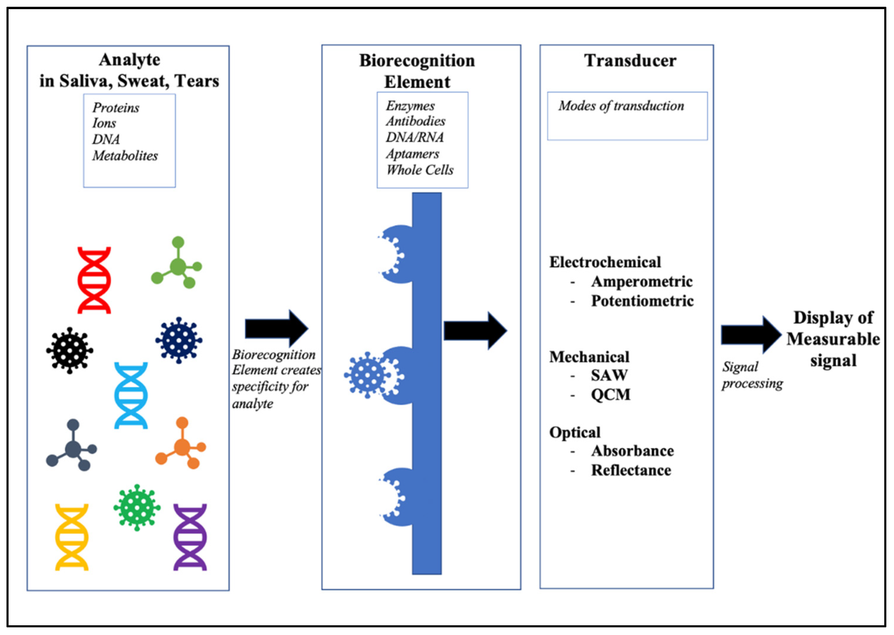
:1. Introduction
2. Design, Ideal Requirements, and Types of Wearable Biosensors
2.1. Ideal Requirements for Use as Wearable Biosensors
2.2. Types of Wearable Biosensors
3. Electrochemical Biosensors
4. Electromechanical Biosensors
5. Optoelectronic Biosensors
5.1. SPR-Based Optical Sensors
5.2. Optical Fiber-Based Biosensors

6. Material Considerations in Wearable Biosensors
6.1. Carbon-Based Sensors
6.1.1. Graphene-Based Sensor Materials
6.1.2. Carbon Dots
6.1.3. Carbon Nanotubes
6.2. Non-Carbon-Based Sensor Materials
Metal-Based Ceramics
6.3. Organic Materials

6.3.1. Natural Polymers
6.3.2. Synthetic Polymers
7. Conclusions and Future Perspectives
Author Contributions
Funding
Acknowledgments
Conflicts of Interest
References
- Nikhil, B.; Pawan, J.; Nello, F.; Pedro, E. Introduction to biosensors. Essays Biochem. 2016, 60, 1–8. [Google Scholar]
- Heineman, W.R.; Jensen, W.B. Leland c. clark jr. (1918–2005). Biosens. Bioelectron. 2006, 8, 1403–1404. [Google Scholar] [CrossRef]
- Vestergaard, M. Nanobiosensors and Nanobioanalyses; Springer: Tokyo, Japan, 2015. [Google Scholar]
- Yoo, E.-H.; Lee, S.-Y. Glucose biosensors: An overview of use in clinical practice. Sensors 2010, 10, 4558–4576. [Google Scholar] [CrossRef] [Green Version]
- Liedberg, B.; Nylander, C.; Lunström, I. Surface plasmon resonance for gas detection and biosensing. Sens. Actuators 1983, 4, 299–304. [Google Scholar] [CrossRef]
- Narita, F.; Wang, Z.; Kurita, H.; Li, Z.; Shi, Y.; Jia, Y.; Soutis, C. A review of piezoelectric and magnetostrictive biosensor materials for detection of COVID-19 and other viruses. Adv. Mater. 2021, 33, 2005448. [Google Scholar] [CrossRef]
- CBINSIGHTS. 24 Industries & Technologies That Will Shape the Post-Virus World. Available online: https://www.cbinsights.com/research/report/industries-tech-shaping-world-post-covid/ (accessed on 27 January 2021).
- Lee, E.K.; Yoo, H.; Lee, C.H. Advanced Materials and Assembly Strategies for Wearable Biosensors: A Review. In Biosensors—Current and Novel Strategies for Biosensing; Intech Open Limited: London, UK, 2020. [Google Scholar]
- Mohankumar, P.; Ajayan, J.; Mohanraj, T.; Yasodharan, R. Recent developments in biosensors for healthcare and biomedical applications: A review. Measurement 2021, 167, 108293. [Google Scholar] [CrossRef]
- Cesewski, E.; Johnson, B.N. Electrochemical biosensors for pathogen detection. Biosens. Bioelectron. 2020, 159, 112214. [Google Scholar] [CrossRef] [PubMed]
- Liu, Q.; Zhao, C.; Chen, M.; Liu, Y.; Zhao, Z.; Wu, F.; Li, Z.; Weiss, P.S.; Andrews, A.M.; Zhou, C. Flexible multiplexed In2O3 nanoribbon aptamer-field-effect transistors for biosensing. iScience 2020, 23, 101469. [Google Scholar] [CrossRef] [PubMed]
- Choi, J.H.; Choi, M.; Kang, T.; Ho, T.S.; Choi, S.H.; Byun, K.M. Combination of Porous Silk Fibroin Substrate and Gold Nanocracks as a Novel SERS Platform for a High-Sensitivity Biosensor. Biosensors 2021, 11, 441. [Google Scholar] [CrossRef] [PubMed]
- Luka, G.; Ahmadi, A.; Najjaran, H.; Alocilja, E.; DeRosa, M.; Wolthers, K.; Malki, A.; Aziz, H.; Althani, A.; Hoorfar, M. Microfluidics integrated biosensors: A leading technology towards lab-on-a-chip and sensing applications. Sensors 2015, 15, 30011–30031. [Google Scholar] [CrossRef] [PubMed] [Green Version]
- Pei, Z.; Yu, Z.; Li, M.; Bai, L.; Wang, W.; Chen, H.; Yang, H.; Wei, D.; Yang, L. Self-healing and toughness cellulose nanocrystals nanocomposite hydrogels for strain-sensitive wearable flexible sensor. Int. J. Biol. Macromol. 2021, 179, 324–332. [Google Scholar] [CrossRef] [PubMed]
- Ashley, B.K.; Brown, M.S.; Park, Y.; Kuan, S.; Koh, A. Skin-inspired, open mesh electrochemical sensors for lactate and oxygen monitoring. Biosens. Bioelectron. 2019, 132, 343–351. [Google Scholar] [CrossRef] [PubMed]
- Saldanha, D.J.; Abdali, Z.; Modafferi, D.; Janfeshan, B.; Courchesne, N.-M.D. Fabrication of fluorescent pH-responsive protein-textile composites. Sci. Rep. 2020, 10, 1–12. [Google Scholar]
- Zhao, C.; Li, X.; Wu, Q.; Liu, X. A thread-based wearable sweat nanobiosensor. Biosens. Bioelectron. 2021, 188, 113270. [Google Scholar] [CrossRef]
- Vaquer, A.; Barón, E.; de la Rica, R. Wearable Analytical Platform with Enzyme-Modulated Dynamic Range for the Simultaneous Colorimetric Detection of Sweat Volume and Sweat Biomarkers. ACS Sens. 2020, 6, 130–136. [Google Scholar] [CrossRef]
- Ding, J.; Qiao, Z.; Zhang, Y.; Wei, D.; Chen, S.; Tang, J.; Chen, L.; Wei, D.; Sun, J.; Fan, H. NIR-responsive multi-healing HMPAM/dextran/AgNWs hydrogel sensor with recoverable mechanics and conductivity for human-machine interaction. Carbohydr. Polym. 2020, 247, 116686. [Google Scholar] [CrossRef]
- Vinoth, R.; Nakagawa, T.; Mathiyarasu, J.; Mohan, A.V. Fully Printed Wearable Microfluidic Devices for High-Throughput Sweat Sampling and Multiplexed Electrochemical Analysis. ACS Sens. 2021, 6, 1174–1186. [Google Scholar] [CrossRef]
- Contacts vs. Glasses: The Pros and Cons. Available online: https://ca.zennioptical.com/blog/contacts-vs-glasses/ (accessed on 16 November 2021).
- PARC Mouthguard Helps Track Athlete Fatigue through Saliva. Available online: https://www.sporttechie.com/xerox-parc-nextflex-ucsd-saliva-mouthguard-athlete-fatigue/ (accessed on 16 November 2021).
- Bioengineering, N. Wearable Patch Sends Simultaneous Signals of Health. Available online: https://www.nibib.nih.gov/news-events/newsroom/wearable-patch-sends-simultaneous-signals-health (accessed on 16 November 2021).
- Move over Fitbit: Sweat-Sensing Bracelet Could Be Next Wearable Tech. Available online: https://www.statnews.com/2016/01/27/sweat-wearable-tech/ (accessed on 16 November 2021).
- Textile Pressure Sensors Can Be Washed. Available online: https://www.electronicsweekly.com/news/research-news/textile-pressure-sensors-can-be-washed-2015-10/ (accessed on 16 November 2021).
- Romeo, A.; Moya, A.; Leung, T.S.; Gabriel, G.; Villa, R.; Sanchez, S. Inkjet printed flexible non-enzymatic glucose sensor for tear fluid analysis. Appl. Mater. Today 2018, 10, 133–141. [Google Scholar] [CrossRef]
- Biological Sensor Can Detect Glucose Levels in Saliva More Accurately and Cost-Efficiently than Blood Test. Available online: https://phys.org/news/2017-05-biological-sensor-glucose-saliva-accurately.html (accessed on 16 November 2021).
- Berkeley, U. Wearable Sensors Detect What’s in Your Sweat. Available online: https://news.berkeley.edu/2019/08/16/wearable-sensors-detect-whats-in-your-sweat/ (accessed on 16 November 2021).
- Owyeung, R.E.; Terse-Thakoor, T.; Rezaei Nejad, H.; Panzer, M.J.; Sonkusale, S.R. Highly flexible transistor threads for all-thread based integrated circuits and multiplexed diagnostics. ACS Appl. Mater. Interfaces 2019, 11, 31096–31104. [Google Scholar] [CrossRef]
- Wireless, Implantable Sensors the Size of a Grain of Sand Could Have Wide Use in Body Monitoring. Available online: https://medicalxpress.com/news/2016-08-wireless-implantable-sensors-size-grain.html (accessed on 16 November 2021).
- Grieshaber, D.; MacKenzie, R.; Vörös, J.; Reimhult, E. Electrochemical biosensors-sensor principles and architectures. Sensors 2008, 8, 1400–1458. [Google Scholar] [CrossRef]
- Chaubey, A.; Malhotra, B. Mediated biosensors. Biosens. Bioelectron. 2002, 17, 441–456. [Google Scholar] [CrossRef]
- Guiseppi-Elie, A.; Lingerfelt, L. Impedimetric detection of DNA hybridization: Towards near-patient DNA diagnostics. In Immobilisation of DNA on Chips I; Springer: Berlin, Germany, 2005. [Google Scholar]
- Thévenot, D.R.; Toth, K.; Durst, R.A.; Wilson, G.S. Electrochemical biosensors: Recommended definitions and classification. Biosens. Bioelectron. 2001, 16, 121–131. [Google Scholar] [CrossRef]
- Cho, I.-H.; Kim, D.H.; Park, S. Electrochemical biosensors: Perspective on functional nanomaterials for on-site analysis. Biomater. Res. 2020, 24, 1–12. [Google Scholar] [CrossRef] [Green Version]
- Lei, Y.; Zhao, W.; Zhang, Y.; Jiang, Q.; He, J.H.; Baeumner, A.J.; Wolfbeis, O.S.; Wang, Z.L.; Salama, K.N.; Alshareef, H.N. A MXene-based wearable biosensor system for high-performance in vitro perspiration analysis. Small 2019, 15, 1901190. [Google Scholar] [CrossRef] [Green Version]
- Wang, Y.; Wang, X.; Lu, W.; Yuan, Q.; Zheng, Y.; Yao, B. A thin film polyethylene terephthalate (PET) electrochemical sensor for detection of glucose in sweat. Talanta 2019, 198, 86–92. [Google Scholar] [CrossRef] [PubMed]
- Lee, H.; Hong, Y.J.; Baik, S.; Hyeon, T.; Kim, D.H. Enzyme-based glucose sensor: From invasive to wearable device. Adv. Healthc. Mater. 2018, 7, 1701150. [Google Scholar] [CrossRef] [Green Version]
- Zhao, J.; Lin, Y.; Wu, J.; Nyein, H.Y.Y.; Bariya, M.; Tai, L.-C.; Chao, M.; Ji, W.; Zhang, G.; Fan, Z. A fully integrated and self-powered smartwatch for continuous sweat glucose monitoring. ACS Sens. 2019, 4, 1925–1933. [Google Scholar] [CrossRef] [Green Version]
- Lin, K.-C.; Muthukumar, S.; Prasad, S. Flex-GO (Flexible graphene oxide) sensor for electrochemical monitoring lactate in low-volume passive perspired human sweat. Talanta 2020, 214, 120810. [Google Scholar] [CrossRef]
- Goud, K.Y.; Moonla, C.; Mishra, R.K.; Yu, C.; Narayan, R.; Litvan, I.; Wang, J. Wearable electrochemical microneedle sensor for continuous monitoring of levodopa: Toward Parkinson management. ACS Sens. 2019, 4, 2196–2204. [Google Scholar] [CrossRef] [PubMed]
- Wang, R.; Zhai, Q.; An, T.; Gong, S.; Cheng, W. Stretchable gold fiber-based wearable textile electrochemical biosensor for lactate monitoring in sweat. Talanta 2021, 222, 121484. [Google Scholar] [CrossRef] [PubMed]
- Zhang, Q.; Jiang, D.; Xu, C.; Ge, Y.; Liu, X.; Wei, Q.; Huang, L.; Ren, X.; Wang, C.; Wang, Y. Wearable electrochemical biosensor based on molecularly imprinted Ag nanowires for noninvasive monitoring lactate in human sweat. Sens. Actuators B 2020, 320, 128325. [Google Scholar] [CrossRef]
- Barfidokht, A.; Mishra, R.K.; Seenivasan, R.; Liu, S.; Hubble, L.; Wang, J.; Hall, D.A. Wearable electrochemical glove-based sensor for rapid and on-site detection of fentanyl. Sens. Actuators B 2019, 296, 126422. [Google Scholar] [CrossRef] [PubMed]
- Zanfrognini, B.; Pigani, L.; Zanardi, C. Recent advances in the direct electrochemical detection of drugs of abuse. J. Solid State Electrochem. 2020, 24, 2603–2616. [Google Scholar] [CrossRef]
- Rocha, R.G.; Stefano, J.S.; Arantes, I.V.; Ribeiro, M.M.; Santana, M.H.; Richter, E.M.; Munoz, R.A. Simple Strategy for Selective Determination of Levamisole in Seized Cocaine and Pharmaceutical Samples Using Disposable Screen-printed Electrodes. Electroanalysis 2019, 31, 153–159. [Google Scholar] [CrossRef] [Green Version]
- Florea, A.; Cowen, T.; Piletsky, S.; De Wael, K. Electrochemical sensing of cocaine in real samples based on electrodeposited biomimetic affinity ligands. Analyst 2019, 144, 4639–4646. [Google Scholar] [CrossRef]
- Tavakkoli, N.; Soltani, N.; Mohammadi, F. A nanoporous gold-based electrochemical aptasensor for sensitive detection of cocaine. RSC Adv. 2019, 9, 14296–14301. [Google Scholar] [CrossRef] [Green Version]
- Renaud-Young, M.; Mayall, R.M.; Salehi, V.; Goledzinowski, M.; Comeau, F.J.; MacCallum, J.L.; Birss, V.I. Development of an ultra-sensitive electrochemical sensor for Δ9-tetrahydrocannabinol (THC) and its metabolites using carbon paper electrodes. Electrochim. Acta 2019, 307, 351–359. [Google Scholar] [CrossRef]
- Zhang, Q.; Berg, D.; Mugo, S.M. Molecularly imprinted carbon based electrodes for tetrahydrocannabinol sensing. Inorg. Chem. Commun. 2019, 107, 107459. [Google Scholar] [CrossRef]
- Takeda, Y.; Kanai, M.; Hatano, A.; Yoshimi, Y.; Kida, M. A “single-use” ceramic-based electrochemical sensor chip using molecularly imprinted carbon paste electrode. Sensors 2020, 20, 5847. [Google Scholar]
- Rawson, T.M.; Gowers, S.A.; Freeman, D.M.; Wilson, R.C.; Sharma, S.; Gilchrist, M.; MacGowan, A.; Lovering, A.; Bayliss, M.; Kyriakides, M. Microneedle biosensors for real-time, minimally invasive drug monitoring of phenoxymethylpenicillin: A first-in-human evaluation in healthy volunteers. Lancet Digit. Health 2019, 1, e335–e343. [Google Scholar] [CrossRef] [Green Version]
- Ahmad-Tarar, A.; Mohammad, U.; Srivastava, S.K. Wearable skin sensors and their challenges: A review of transdermal, optical, and Mechanical Sensors. Biosensors 2020, 10, 56. [Google Scholar] [CrossRef] [PubMed]
- Busch-Vishniac, I. Trends in electromechanical transduction. J. Acoust. Soc. Am. 1998, 103, 2860. [Google Scholar] [CrossRef]
- Rim, Y.S.; Bae, S.H.; Chen, H.; De Marco, N.; Yang, Y. Recent progress in materials and devices toward printable and flexible sensors. Adv. Mater. 2016, 28, 4415–4440. [Google Scholar] [CrossRef]
- Heikenfeld, J.; Jajack, A.; Rogers, J.; Gutruf, P.; Tian, L.; Pan, T.; Li, R.; Khine, M.; Kim, J.; Wang, J. Wearable sensors: Modalities, challenges, and prospects. Lab Chip 2018, 18, 217–248. [Google Scholar] [CrossRef] [PubMed] [Green Version]
- Fan, F.-R.; Tian, Z.-Q.; Wang, Z.L. Flexible triboelectric generator. Nano Energy 2012, 1, 328–334. [Google Scholar] [CrossRef]
- Chen, H.; Xu, Y.; Zhang, J.; Wu, W.; Song, G. Enhanced stretchable graphene-based triboelectric nanogenerator via control of surface nanostructure. Nano Energy 2019, 58, 304–311. [Google Scholar] [CrossRef]
- Tang, N.; Zhou, C.; Qu, D.; Fang, Y.; Zheng, Y.; Hu, W.; Jin, K.; Wu, W.; Duan, X.; Haick, H. A Highly Aligned Nanowire-Based Strain Sensor for Ultrasensitive Monitoring of Subtle Human Motion. Small 2020, 16, 2001363. [Google Scholar] [CrossRef]
- Wang, S.; Bai, Y.; Yang, X.; Liu, L.; Li, L.; Lu, Q.; Li, T.; Zhang, T. Highly stretchable potentiometric ion sensor based on surface strain redistributed fiber for sweat monitoring. Talanta 2020, 214, 120869. [Google Scholar] [CrossRef]
- Wang, C.; Li, X.; Gao, E.; Jian, M.; Xia, K.; Wang, Q.; Xu, Z.; Ren, T.; Zhang, Y. Carbonized silk fabric for ultrastretchable, highly sensitive, and wearable strain sensors. Adv. Mater. 2016, 28, 6640–6648. [Google Scholar] [CrossRef]
- Yang, M.; Pan, J.; Xu, A.; Luo, L.; Cheng, D.; Cai, G.; Wang, J.; Tang, B.; Wang, X. Conductive cotton fabrics for motion sensing and heating applications. Polymers 2018, 10, 568. [Google Scholar] [CrossRef] [Green Version]
- Bi, S.; Hou, L.; Zhao, H.; Zhu, L.; Lu, Y. Ultrasensitive and highly repeatable pen ink decorated cuprammonium rayon (cupra) fabrics for multifunctional sensors. J. Mater. Chem. A 2018, 6, 16556–16565. [Google Scholar] [CrossRef]
- Ren, J.; Wang, C.; Zhang, X.; Carey, T.; Chen, K.; Yin, Y.; Torrisi, F. Environmentally-friendly conductive cotton fabric as flexible strain sensor based on hot press reduced graphene oxide. Carbon 2017, 111, 622–630. [Google Scholar] [CrossRef] [Green Version]
- Liu, S.; Hu, M.; Yang, J. A facile way of fabricating a flexible and conductive cotton fabric. J. Mater. Chem. C 2016, 4, 1320–1325. [Google Scholar] [CrossRef]
- Yang, S.; Li, C.; Chen, X.; Zhao, Y.; Zhang, H.; Wen, N.; Fan, Z.; Pan, L. Facile fabrication of high-performance pen ink-decorated textile strain sensors for human motion detection. ACS Appl. Mater. Interfaces 2020, 12, 19874–19881. [Google Scholar] [CrossRef] [PubMed]
- Senthil, K.; Chen, P.-Y.; Ren, H. A review of printable flexible and stretchable tactile sensors. Research 2019, 2019, 3018568. [Google Scholar] [CrossRef] [PubMed] [Green Version]
- Lee, K.; Ni, X.; Lee, J.Y.; Arafa, H.; David, J.P.; Xu, S.; Avila, R.; Irie, M.; Lee, J.H.; Easterlin, R.L. Mechano-acoustic sensing of physiological processes and body motions via a soft wireless device placed at the suprasternal notch. Nat. Biomed. Eng. 2020, 4, 148–158. [Google Scholar] [CrossRef] [PubMed]
- Mehta, D.D.; Zanartu, M.; Feng, S.W.; Cheyne II, H.A.; Hillman, R.E. Mobile voice health monitoring using a wearable accelerometer sensor and a smartphone platform. IEEE Trans. Biomed. Eng. 2012, 59, 3090–3096. [Google Scholar] [CrossRef] [Green Version]
- Liu, Y.; Norton, J.J.; Qazi, R.; Zou, Z.; Ammann, K.R.; Liu, H.; Yan, L.; Tran, P.L.; Jang, K.-I.; Lee, J.W. Epidermal mechano-acoustic sensing electronics for cardiovascular diagnostics and human-machine interfaces. Sci. Adv. 2016, 2, e1601185. [Google Scholar] [CrossRef] [Green Version]
- Jang, J.; Jun, Y.S.; Seo, H.; Kim, M.; Park, J.-U. Motion detection using tactile sensors based on pressure-sensitive transistor arrays. Sensors 2020, 20, 3624. [Google Scholar] [CrossRef]
- Ozioko, O.; Karipoth, P.; Hersh, M.; Dahiya, R. Wearable assistive tactile communication interface based on integrated touch sensors and actuators. IEEE Trans. Neural Syst. Rehabil. Eng. 2020, 28, 1344–1352. [Google Scholar] [CrossRef]
- Qi, K.; Wang, H.; You, X.; Tao, X.; Li, M.; Zhou, Y.; Zhang, Y.; He, J.; Shao, W.; Cui, S. Core-sheath nanofiber yarn for textile pressure sensor with high pressure sensitivity and spatial tactile acuity. J. Colloid Interface Sci. 2020, 561, 93–103. [Google Scholar] [CrossRef] [PubMed]
- Yi, Q.; Najafikhoshnoo, S.; Das, P.; Noh, S.; Hoang, E.; Kim, T.; Esfandyarpour, R. All-3D-Printed, Flexible, and Hybrid Wearable Bioelectronic Tactile Sensors Using Biocompatible Nanocomposites for Health Monitoring. Adv. Mater. Technol. 2021, 2021, 2101034. [Google Scholar] [CrossRef]
- Borisov, S.M.; Wolfbeis, O.S. Optical biosensors. Chem. Rev. 2008, 108, 423–461. [Google Scholar] [CrossRef]
- Chen, C.; Wang, J. Optical biosensors: An exhaustive and comprehensive review. Analyst 2020, 145, 1605–1628. [Google Scholar] [CrossRef] [PubMed]
- Zanchetta, G.; Lanfranco, R.; Giavazzi, F.; Bellini, T.; Buscaglia, M. Emerging applications of label-free optical biosensors. Nanophotonics 2017, 6, 627–645. [Google Scholar] [CrossRef]
- Cooper, M.A. Optical biosensors in drug discovery. Nat. Rev. Drug Discov. 2002, 1, 515–528. [Google Scholar] [CrossRef] [PubMed]
- Foubert, A.; Beloglazova, N.V.; Hedström, M.; De Saeger, S. Antibody immobilization strategy for the development of a capacitive immunosensor detecting zearalenone. Talanta 2019, 191, 202–208. [Google Scholar] [CrossRef] [PubMed]
- Fernandes, E.; Cabral, P.D.; Campos, R.; Machado, G., Jr.; Cerqueira, M.F.; Sousa, C.; Freitas, P.P.; Borme, J.; Petrovykh, D.Y.; Alpuim, P. Functionalization of single-layer graphene for immunoassays. Appl. Surf. Sci. 2019, 480, 709–716. [Google Scholar] [CrossRef]
- Estevez, M.C.; Alvarez, M.; Lechuga, L.M. Integrated optical devices for lab-on-a-chip biosensing applications. Laser Photonics Rev. 2012, 6, 463–487. [Google Scholar] [CrossRef] [Green Version]
- Wong, L.S.; Khan, F.; Micklefield, J. Selective covalent protein immobilization: Strategies and applications. Chem. Rev. 2009, 109, 4025–4053. [Google Scholar] [CrossRef]
- Mayer, K.M.; Lee, S.; Liao, H.; Rostro, B.C.; Fuentes, A.; Scully, P.T.; Nehl, C.L.; Hafner, J.H. A label-free immunoassay based upon localized surface plasmon resonance of gold nanorods. ACS Nano 2008, 2, 687–692. [Google Scholar] [CrossRef]
- Dhanabalan, S.S.; Sriram, S.; Walia, S.; Avaninathan, S.R.; Carrasco, M.F.; Bhaskaran, M. Wearable Label-Free Optical Biodetectors: Progress and Perspectives. Adv. Photonics Res. 2021, 2, 2000076. [Google Scholar] [CrossRef]
- Homola, J. Surface plasmon resonance sensors for detection of chemical and biological species. Chem. Rev. 2008, 108, 462–493. [Google Scholar] [CrossRef]
- Dey, B.; Islam, M.S.; Park, J. Numerical design of high-performance WS2/metal/WS2/graphene heterostructure based surface plasmon resonance refractive index sensor. Results Phys. 2021, 23, 104021. [Google Scholar] [CrossRef]
- Rahman, M.M.; Rana, M.M.; Rahman, M.S.; Anower, M.; Mollah, M.A.; Paul, A.K. Sensitivity enhancement of SPR biosensors employing heterostructure of PtSe2 and 2D materials. Opt. Mater. 2020, 107, 110123. [Google Scholar] [CrossRef]
- Jia, Y.; Li, Z.; Wang, H.; Saeed, M.; Cai, H. Sensitivity enhancement of a surface plasmon resonance sensor with platinum diselenide. Sensors 2020, 20, 131. [Google Scholar] [CrossRef] [PubMed] [Green Version]
- Bijalwan, A.; Singh, B.K.; Rastogi, V. Surface plasmon resonance-based sensors using nano-ribbons of graphene and wse 2. Plasmonics 2020, 15, 1–9. [Google Scholar] [CrossRef]
- Zhang, Y.; Sun, J.; Liu, L.; Qiao, H. A review of biosensor technology and algorithms for glucose monitoring. J. Diabetes Compl. 2021, 35, 107929. [Google Scholar] [CrossRef] [PubMed]
- Koushki, E.; Mohammadabadi, F.M.; Baedi, J.; Ghasedi, A. The effects of glucose and glucose oxidase on the Uv-vis spectrum of gold nanoparticles: A study on optical biosensor for saliva glucose monitoring. Photodiagn. Photodynam. Ther. 2020, 30, 101771. [Google Scholar] [CrossRef]
- Yadav, A.; Sharan, P.; Kumar, A. Surface plasmonic resonance based five layered structure-biosensor for sugar level measurement in human. Results Opt. 2020, 1, 100002. [Google Scholar] [CrossRef]
- Mostufa, S.; Paul, A.K.; Chakrabarti, K. Detection of hemoglobin in blood and urine glucose level samples using a graphene-coated SPR based biosensor. OSA Contin. 2021, 4, 2164–2176. [Google Scholar] [CrossRef]
- Mitu, S.A.; Ahmed, K.; Al Zahrani, F.A.; Grover, A.; Rajan, M.S.M.; Moni, M.A. Development and analysis of surface plasmon resonance based refractive index sensor for pregnancy testing. Opt. Lasers Eng. 2021, 140, 106551. [Google Scholar] [CrossRef]
- Alwis, L.; Sun, T.; Grattan, K. Developments in optical fibre sensors for industrial applications. Opt. Laser Technol. 2016, 78, 62–66. [Google Scholar] [CrossRef]
- Leal-Junior, A.G.; Diaz, C.A.; Avellar, L.M.; Pontes, M.J.; Marques, C.; Frizera, A. Polymer optical fiber sensors in healthcare applications: A comprehensive review. Sensors 2019, 19, 3156. [Google Scholar] [CrossRef] [Green Version]
- Broadway, C.; Min, R.; Leal-Junior, A.G.; Marques, C.; Caucheteur, C. Toward commercial polymer fiber Bragg grating sensors: Review and applications. J. Lightwave Technol. 2019, 37, 2605–2615. [Google Scholar] [CrossRef]
- Massaroni, C.; Zaltieri, M.; Presti, L.; Nicolò, A.; Tosi, D.; Schena, E. Fiber Bragg grating sensors for cardiorespiratory monitoring: A review. IEEE Sens. J. 2020, 13, 14069–14080. [Google Scholar] [CrossRef]
- Li, L.; Li, Y.; Yang, L.; Fang, F.; Yan, Z.; Sun, Q. Continuous and Accurate Blood Pressure Monitoring Based on Wearable Optical Fiber Wristband. IEEE Sens. J. 2020, 21, 3049–3057. [Google Scholar] [CrossRef]
- Shrestha, P.; Kim, J.-H.; Park, Y.; Kim, C.-G. Impact localization on composite structure using FBG sensors and novel impact localization technique based on error outliers. Compos. Struct. 2016, 142, 263–271. [Google Scholar] [CrossRef]
- Ding, Z.; Wang, C.; Liu, K.; Jiang, J.; Yang, D.; Pan, G.; Pu, Z.; Liu, T. Distributed optical fiber sensors based on optical frequency domain reflectometry: A review. Sensors 2018, 18, 1072. [Google Scholar] [CrossRef] [Green Version]
- Leal-Junior, A.; Avellar, L.; Frizera, A.; Marques, C. Smart textiles for multimodal wearable sensing using highly stretchable multiplexed optical fiber system. Sci. Rep. 2020, 10, 13867. [Google Scholar]
- Safaee, M.M.; Gravely, M.; Roxbury, D. A Wearable Optical Microfibrous Biomaterial with Encapsulated Nanosensors Enables Wireless Monitoring of Oxidative Stress. Adv. Funct. Mater. 2021, 31, 2006254. [Google Scholar] [CrossRef]
- Leal-Junior, A.G.; Ribeiro, D.; Avellar, L.M.; Silveira, M.; Díaz, C.A.R.; Frizera-Neto, A.; Blanc, W.; Rocon, E.; Marques, C. Wearable and fully-portable smart garment for mechanical perturbation detection with nanoparticles optical fibers. IEEE Sens. J. 2020, 21, 2995–3003. [Google Scholar] [CrossRef]
- Zhu, H.T.; Zhan, L.W.; Dai, Q.; Xu, B.; Chen, Y.; Lu, Y.Q.; Xu, F. Self-Assembled Wavy Optical Microfiber for Stretchable Wearable Sensor. Adv. Opt. Mater. 2021, 9, 2002206. [Google Scholar] [CrossRef]
- Pan, J.; Zhang, Z.; Jiang, C.; Zhang, L.; Tong, L. A multifunctional skin-like wearable optical sensor based on an optical micro-/nanofibre. Nanoscale 2020, 12, 17538–17544. [Google Scholar] [CrossRef]
- Pang, Y.-N.; Liu, B.; Liu, J.; Wan, S.P.; Wu, T.; He, X.; Yuan, J.; Zhou, X.; Long, K.; Wu, Q. Wearable optical fiber sensor based on a bend singlemode-multimode-Singlemode fiber structure for respiration monitoring. IEEE Sens. J. 2020, 21, 4610–4617. [Google Scholar] [CrossRef]
- Boukhayma, A.; Barison, A.; Haddad, S.; Caizzone, A. Ring-embedded micro-power mm-sized optical sensor for accurate heart beat monitoring. IEEE Access 2021, 9, 127217–127225. [Google Scholar] [CrossRef]
- Cheng, R.; Shang, Y.; Hayes, D., Jr.; Saha, S.P.; Yu, G. Noninvasive optical evaluation of spontaneous low frequency oscillations in cerebral hemodynamics. Neuroimage 2012, 62, 1445–1454. [Google Scholar] [CrossRef]
- Shang, Y.; Zhao, Y.; Cheng, R.; Dong, L.; Irwin, D.; Yu, G. Portable optical tissue flow oximeter based on diffuse correlation spectroscopy. Opt. Lett. 2009, 34, 3556–3558. [Google Scholar] [CrossRef]
- Gurley, K.; Shang, Y.; Yu, G. Noninvasive optical quantification of absolute blood flow, blood oxygenation, and oxygen consumption rate in exercising skeletal muscle. J. Biomed. Opt. 2012, 17, 075010. [Google Scholar]
- Mendelson, Y.; Ochs, B.D. Noninvasive pulse oximetry utilizing skin reflectance photoplethysmography. IEEE Trans. Biomed. Eng. 1988, 35, 798–805. [Google Scholar] [CrossRef]
- Boukhayma, A.; Barison, A.; Haddad, S.; Caizzone, A. Earbud-embedded micro-power mm-sized optical sensor for accurate heart beat monitoring. IEEE Sens. J. 2021, 21, 19967–19977. [Google Scholar] [CrossRef]
- Anichini, C.; Samori, P. Graphene-Based Hybrid Functional Materials. Small 2021, 17, e2100514. [Google Scholar] [CrossRef] [PubMed]
- Xu, G.; Abbott, J.; Qin, L.; Yeung, K.Y.; Song, Y.; Yoon, H.; Kong, J.; Ham, D. Electrophoretic and field-effect graphene for all-electrical DNA array technology. Nat. Commun. 2014, 5, 4866. [Google Scholar] [CrossRef] [Green Version]
- Fu, W.; Feng, L.; Panaitov, G.; Kireev, D.; Mayer, D.; Offenhausser, A.; Krause, H.J. Biosensing near the neutrality point of graphene. Sci. Adv. 2017, 3, e1701247. [Google Scholar] [CrossRef] [PubMed] [Green Version]
- Dong, H.; Gao, W.; Yan, F.; Ji, H.; Ju, H. Fluorescence resonance energy transfer between quantum dots and graphene oxide for sensing biomolecules. Anal. Chem. 2010, 82, 5511–5517. [Google Scholar] [CrossRef] [PubMed]
- Duan, B.; Zhou, J.; Fang, Z.; Wang, C.; Wang, X.; Hemond, H.F.; Chan-Park, M.B.; Duan, H. Surface enhanced Raman scattering by graphene-nanosheet-gapped plasmonic nanoparticle arrays for multiplexed DNA detection. Nanoscale 2015, 7, 12606–12613. [Google Scholar] [CrossRef] [PubMed] [Green Version]
- Gao, X.G.; Cheng, L.X.; Jiang, W.S.; Li, X.K.; Xing, F. Graphene and its Derivatives-Based Optical Sensors. Front. Chem. 2021, 9, 615164. [Google Scholar] [CrossRef]
- Xu, X.; Ray, R.; Gu, Y.; Ploehn, H.J.; Gearheart, L.; Raker, K.; Scrivens, W.A. Electrophoretic analysis and purification of fluorescent single-walled carbon nanotube fragments. J. Am. Chem. Soc. 2004, 126, 12736–12737. [Google Scholar] [CrossRef] [PubMed]
- Wan, J.; Zhang, X.; Fu, K.; Zhang, X.; Shang, L.; Su, Z. Highly fluorescent carbon dots as novel theranostic agents for biomedical applications. Nanoscale 2021, 13, 17236–17253. [Google Scholar] [CrossRef]
- Ji, C.; Zhou, Y.; Leblanc, R.M.; Peng, Z. Recent Developments of Carbon Dots in Biosensing: A Review. ACS Sens. 2020, 5, 2724–2741. [Google Scholar] [CrossRef]
- Campuzano, S.; Yanez-Sedeno, P.; Pingarron, J.M. Carbon Dots and Graphene Quantum Dots in Electrochemical Biosensing. Nanomaterials 2019, 9, 634. [Google Scholar] [CrossRef] [PubMed] [Green Version]
- Jiang, Y.; Wang, B.; Meng, F.; Cheng, Y.; Zhu, C. Microwave-assisted preparation of N-doped carbon dots as a biosensor for electrochemical dopamine detection. J. Colloid Interface Sci. 2015, 452, 199–202. [Google Scholar] [CrossRef]
- Buk, V.; Pemble, M.E. A highly sensitive glucose biosensor based on a micro disk array electrode design modified with carbon quantum dots and gold nanoparticles. Electrochim. Acta 2019, 298, 97–105. [Google Scholar] [CrossRef]
- Shen, P.; Xia, Y. Synthesis-modification integration: One-step fabrication of boronic acid functionalized carbon dots for fluorescent blood sugar sensing. Anal. Chem. 2014, 86, 5323–5329. [Google Scholar] [CrossRef] [PubMed]
- Wang, H.; Lu, Q.; Hou, Y.; Liu, Y.; Zhang, Y. High fluorescence S, N co-doped carbon dots as an ultra-sensitive fluorescent probe for the determination of uric acid. Talanta 2016, 155, 62–69. [Google Scholar] [CrossRef]
- Sharma, S.K.; Micic, M.; Li, S.; Hoar, B.; Paudyal, S.; Zahran, E.M.; Leblanc, R.M. Conjugation of Carbon Dots with beta-Galactosidase Enzyme: Surface Chemistry and Use in Biosensing. Molecules 2019, 24, 3275. [Google Scholar] [CrossRef] [Green Version]
- Wang, Y.; Wang, Z.; Rui, Y.; Li, M. Horseradish peroxidase immobilization on carbon nanodots/CoFe layered double hydroxides: Direct electrochemistry and hydrogen peroxide sensing. Biosens. Bioelectron. 2015, 64, 57–62. [Google Scholar] [CrossRef]
- Mohammadi, S.; Salimi, A.; Hamd-Ghadareh, S.; Fathi, F.; Soleimani, F. A FRET immunosensor for sensitive detection of CA 15-3 tumor marker in human serum sample and breast cancer cells using antibody functionalized luminescent carbon-dots and AuNPs-dendrimer aptamer as donor-acceptor pair. Anal. Biochem. 2018, 557, 18–26. [Google Scholar] [CrossRef]
- Sarkar, T.; Bohidar, H.B.; Solanki, P.R. Carbon dots-modified chitosan based electrochemical biosensing platform for detection of vitamin D. Int. J. Biol. Macromol. 2018, 109, 687–697. [Google Scholar] [CrossRef]
- Qaddare, S.H.; Salimi, A. Amplified fluorescent sensing of DNA using luminescent carbon dots and AuNPs/GO as a sensing platform: A novel coupling of FRET and DNA hybridization for homogeneous HIV-1 gene detection at femtomolar level. Biosens. Bioelectron. 2017, 89, 773–780. [Google Scholar] [CrossRef]
- Rivas, G.A.; Rubianes, M.D.; Rodriguez, M.C.; Ferreyra, N.F.; Luque, G.L.; Pedano, M.L.; Miscoria, S.A.; Parrado, C. Carbon nanotubes for electrochemical biosensing. Talanta 2007, 74, 291–307. [Google Scholar] [CrossRef]
- Sireesha, M.; Jagadeesh Babu, V.; Kranthi Kiran, A.S.; Ramakrishna, S. A review on carbon nanotubes in biosensor devices and their applications in medicine. Nanocomposites 2018, 4, 36–57. [Google Scholar] [CrossRef]
- Meng, L.; Wu, P.; Chen, G.; Cai, C.; Sun, Y.; Yuan, Z. Low potential detection of glutamate based on the electrocatalytic oxidation of NADH at thionine/single-walled carbon nanotubes composite modified electrode. Biosens. Bioelectron. 2009, 24, 1751–1756. [Google Scholar] [CrossRef] [PubMed]
- Boussema, F.; Gross, A.J.; Hmida, F.; Ayed, B.; Majdoub, H.; Cosnier, S.; Maaref, A.; Holzinger, M. Dawson-type polyoxometalate nanoclusters confined in a carbon nanotube matrix as efficient redox mediators for enzymatic glucose biofuel cell anodes and glucose biosensors. Biosens. Bioelectron. 2018, 109, 20–26. [Google Scholar] [CrossRef]
- Zhu, L.; Xu, L.; Tan, L.; Tan, H.; Yang, S.; Yao, S. Direct electrochemistry of cholesterol oxidase immobilized on gold nanoparticles-decorated multiwalled carbon nanotubes and cholesterol sensing. Talanta 2013, 106, 192–199. [Google Scholar] [CrossRef] [PubMed]
- Hu, F.; Chen, S.; Wang, C.; Yuan, R.; Xiang, Y.; Wang, C. Multi-wall carbon nanotube-polyaniline biosensor based on lectin-carbohydrate affinity for ultrasensitive detection of Con A. Biosens. Bioelectron. 2012, 34, 202–207. [Google Scholar] [CrossRef]
- Dagar, K.; Pundir, C.S. An improved amperometric L-lactate biosensor based on covalent immobilization of microbial lactate oxidase onto carboxylated multiwalled carbon nanotubes/copper nanoparticles/polyaniline modified pencil graphite electrode. Enzym. Microb. Technol. 2017, 96, 177–186. [Google Scholar] [CrossRef] [PubMed]
- Muhulet, A.; Miculescu, F.; Voicu, S.I.; Schütt, F.; Thakur, V.K.; Mishra, Y.K. Fundamentals and scopes of doped carbon nanotubes towards energy and biosensing applications. Mater. Today Energy 2018, 9, 154–186. [Google Scholar] [CrossRef]
- Alim, S.; Vejayan, J.; Yusoff, M.M.; Kafi, A. Recent uses of carbon nanotubes & gold nanoparticles in electrochemistry with application in biosensing: A review. Biosens. Bioelectron. 2018, 121, 125–136. [Google Scholar]
- Yun, D.; Song, M.-J.; Hwang, S.; Hong, S.-I. Fabrication and electrochemical characterization of nanoporous silicon electrode for Amperometric urea biosensor. Jpn. J. Appl. Phys. 2012, 51, 06FG02. [Google Scholar] [CrossRef]
- Leonardi, A.A.; Lo Faro, M.J.; Irrera, A. Biosensing platforms based on silicon nanostructures: A critical review. Anal. Chim. Acta 2021, 1160, 338393. [Google Scholar] [CrossRef]
- Song, M.J.; Yun, D.H.; Min, N.K.; Hong, S.I. Electrochemical biosensor array for liver diagnosis using silanization technique on nanoporous silicon electrode. J. Biosci. Bioeng. 2007, 103, 32–37. [Google Scholar] [CrossRef] [PubMed]
- Hemmilä, S.; Gao, A.; Lu, N.; Li, T.; Wang, Y.; Kallio, P. Integration of microfluidic sample delivery system on silicon nanowire-based biosensor. Microsyst. Techol. 2015, 21, 571–580. [Google Scholar] [CrossRef]
- Syshchyk, O.; Skryshevsky, V.A.; Soldatkin, O.O.; Soldatkin, A.P. Enzyme biosensor systems based on porous silicon photoluminescence for detection of glucose, urea and heavy metals. Biosens. Bioelectron. 2015, 66, 89–94. [Google Scholar] [CrossRef]
- Krismastuti, F.S.; Cavallaro, A.; Prieto-Simon, B.; Voelcker, N.H. Toward Multiplexing Detection of Wound Healing Biomarkers on Porous Silicon Resonant Microcavities. Adv. Sci. 2016, 3, 1500383. [Google Scholar] [CrossRef] [PubMed] [Green Version]
- Tereshchenko, A.; Bechelany, M.; Viter, R.; Khranovskyy, V.; Smyntyna, V.; Starodub, N.; Yakimova, R. Optical biosensors based on ZnO nanostructures: Advantages and perspectives. A review. Sens. Actuators B 2016, 229, 664–677. [Google Scholar] [CrossRef] [Green Version]
- Zhao, L.; Li, H.; Meng, J.; Wang, A.C.; Tan, P.; Zou, Y.; Yuan, Z.; Lu, J.; Pan, C.; Fan, Y. Reversible conversion between schottky and ohmic contacts for highly sensitive, multifunctional biosensors. Adv. Funct. Mater. 2020, 30, 1907999. [Google Scholar] [CrossRef]
- Alam, M.; Rahman, M.M.; Uddin, M.; Asiri, A.M.; Chani, M.T.S.; Islam, M. Development of l-glutamic acid biosensor with ternary ZnO/NiO/Al2O3 nanoparticles. J. Luminesc. 2020, 227, 117528. [Google Scholar] [CrossRef]
- Zhao, M.; Shang, J.; Qu, H.; Gao, R.; Li, H.; Chen, S. Fabrication of the Ni/ZnO/BiOI foam for the improved electrochemical biosensing performance to glucose. Anal. Chim. Acta 2020, 1095, 93–98. [Google Scholar] [CrossRef]
- Pal, S.; Prajapati, Y.K.; Saini, J. Influence of graphene’s chemical potential on SPR biosensor using ZnO for DNA hybridization. Opt. Rev. 2020, 27, 57–64. [Google Scholar] [CrossRef]
- Subbaiah, G.B.; Ratnam, K.V.; Janardhan, S.; Shiprath, K.; Manjunatha, H.; Ramesha, M.; Prasad, N.V.K.; Ramesh, S.; Babu, T.A. Metal and Metal Oxide Based Advanced Ceramics for Electrochemical Biosensors-A Short Review. Front. Mater. 2021, 8, 682025. [Google Scholar] [CrossRef]
- Wu, W.; Liu, C.; Huang, F.; Li, H.; Wang, Y.; Hong, D.; Wang, S.; Zeng, X. A simple and Novel Electroanalytical Method for Determination of Brain Serotonin Based on the MWNTs/Al2O3/chitosan SPE. Int. J. Electrochem. Sci. 2018, 13, 7129–7140. [Google Scholar] [CrossRef]
- Mekawy, M.M.; Hassan, R.Y.; Ramnani, P.; Yu, X.; Mulchandani, A. Electrochemical detection of dihydronicotinamide adenine dinucleotide using Al2O3-GO nanocomposite modified electrode. Arab. J. Chem. 2018, 11, 942–949. [Google Scholar] [CrossRef]
- Parvin, M.H.; Arjomandi, J.; Lee, J.Y. γ-Al2O3 nanoparticle catalyst mediated polyaniline gold electrode biosensor for vitamin E. Catal. Commun. 2018, 110, 59–63. [Google Scholar] [CrossRef]
- Wang, Y.; Xiong, Y.; Tian, H.; Wang, S.; Zhang, Q.; Zhang, M.; Ding, H.; Gu, Y.; Peng, A. Application of Sensitive Sensor Modified with MWNTs/Al2O3/poly-L-lysine Composite for 17β–estradiol Determination in Pregnant Woman Blood. Int. J. Electrochem. Sci. 2018, 13, 147–158. [Google Scholar] [CrossRef]
- Rakib, R.H.; Hasnat, M.A.; Uddin, M.N.; Alam, M.; Asiri, A.M.; Rahman, M.M.; Siddiquey, I.A. Fabrication of a 3, 4-Diaminotoluene Sensor Based on a TiO2-Al2O3 Nanocomposite Synthesized by a Fast and Facile Microwave Irradiation Method. Chem. Sel. 2019, 4, 12592–12600. [Google Scholar]
- Spychalska, K.; Zając, D.; Baluta, S.; Halicka, K.; Cabaj, J. Functional polymers structures for (Bio) sensing application—A review. Polymers 2020, 12, 1154. [Google Scholar] [CrossRef]
- Zhang, M.; Liao, C.; Mak, C.H.; You, P.; Mak, C.L.; Yan, F. Highly sensitive glucose sensors based on enzyme-modified whole-graphene solution-gated transistors. Sci. Rep. 2015, 5, 8311. [Google Scholar]
- Hussain, K.K.; Gurudatt, N.; Akhtar, M.H.; Seo, K.-D.; Park, D.-S.; Shim, Y.-B. Nano-biosensor for the in vitro lactate detection using bi-functionalized conducting polymer/N, S-doped carbon; the effect of αCHC inhibitor on lactate level in cancer cell lines. Biosens. Bioelectron. 2020, 155, 112094. [Google Scholar] [CrossRef] [PubMed]
- Kuralay, F.; Vural, T.; Bayram, C.; Denkbas, E.B.; Abaci, S. Carbon nanotube-chitosan modified disposable pencil graphite electrode for vitamin B12 analysis. Colloids Surf. B Biointerfaces 2011, 87, 18–22. [Google Scholar] [CrossRef] [PubMed]
- Xia, J.; Cao, X.; Wang, Z.; Yang, M.; Zhang, F.; Lu, B.; Li, F.; Xia, L.; Li, Y.; Xia, Y. Molecularly imprinted electrochemical biosensor based on chitosan/ionic liquid–graphene composites modified electrode for determination of bovine serum albumin. Sens. Actuators B 2016, 225, 305–311. [Google Scholar] [CrossRef]
- Salvo-Comino, C.; Gonzalez-Gil, A.; Rodriguez-Valentin, J.; Garcia-Hernandez, C.; Martin-Pedrosa, F.; Garcia-Cabezon, C.; Rodriguez-Mendez, M.L. Biosensors Platform Based on Chitosan/AuNPs/Phthalocyanine Composite Films for the Electrochemical Detection of Catechol. The Role of the Surface structure. Sensors 2020, 20, 2152. [Google Scholar] [CrossRef] [Green Version]
- Hroncekova, S.; Bertok, T.; Hires, M.; Jane, E.; Lorencova, L.; Vikartovska, A.; Tanvir, A.; Kasak, P.; Tkac, J. Ultrasensitive Ti3C2TX MXene/Chitosan Nanocomposite-Based Amperometric Biosensor for Detection of Potential Prostate Cancer Marker in Urine Samples. Processes 2020, 8, 580. [Google Scholar] [CrossRef] [PubMed]
- Fu, X.; Liang, Y.; Wu, R.; Shen, J.; Chen, Z.; Chen, Y.; Wang, Y.; Xia, Y. Conductive core-sheath calcium alginate/graphene composite fibers with polymeric ionic liquids as an intermediate. Carbohydr. Polym. 2019, 206, 328–335. [Google Scholar] [CrossRef] [PubMed]
- Kerdjoudj, H.; Boulmedais, F.; Berthelemy, N.; Mjahed, H.; Louis, H.; Schaaf, P.; Voegel, J.; Menu, P. Cellularized alginate sheets for blood vessel reconstruction. Soft Matter 2011, 7, 3621–3626. [Google Scholar] [CrossRef]
- Gilshteyn, E.P.; Lin, S.; Kondrashov, V.A.; Kopylova, D.S.; Tsapenko, A.P.; Anisimov, A.S.; Hart, A.J.; Zhao, X.; Nasibulin, A.G. A One-Step Method of Hydrogel Modification by Single-Walled Carbon Nanotubes for Highly Stretchable and Transparent Electronics. ACS Appl. Mater. Interfaces 2018, 10, 28069–28075. [Google Scholar] [CrossRef] [PubMed]
- Wang, F.; Jiang, J.; Sun, F.; Sun, L.; Wang, T.; Liu, Y.; Li, M. Flexible wearable graphene/alginate composite non-woven fabric temperature sensor with high sensitivity and anti-interference. Cellulose 2020, 27, 2369–2380. [Google Scholar] [CrossRef]
- Yuk, H.; Zhang, T.; Parada, G.A.; Liu, X.; Zhao, X. Skin-inspired hydrogel-elastomer hybrids with robust interfaces and functional microstructures. Nat. Commun. 2016, 7, 12028. [Google Scholar] [CrossRef] [Green Version]
- Cai, P.; Wan, C.; Pan, L.; Matsuhisa, N.; He, K.; Cui, Z.; Zhang, W.; Li, C.; Wang, J.; Yu, J.; et al. Locally coupled electromechanical interfaces based on cytoadhesion-inspired hybrids to identify muscular excitation-contraction signatures. Nat. Commun. 2020, 11, 2183. [Google Scholar] [CrossRef]
- Pan, S.; Zhang, F.; Cai, P.; Wang, M.; He, K.; Luo, Y.; Li, Z.; Chen, G.; Ji, S.; Liu, Z. Mechanically Interlocked Hydrogel–Elastomer Hybrids for On-Skin Electronics. Adv. Funct. Mater. 2020, 30, 1909540. [Google Scholar] [CrossRef]
- Yavuz, B.; Chambre, L.; Harrington, K.; Kluge, J.; Valenti, L.; Kaplan, D.L. Silk Fibroin Microneedle Patches for the Sustained Release of Levonorgestrel. ACS Appl. Bio Mater. 2020, 3, 5375–5382. [Google Scholar] [CrossRef]
- Liang, B.; Fang, L.; Hu, Y.; Yang, G.; Zhu, Q.; Ye, X. Fabrication and application of flexible graphene silk composite film electrodes decorated with spiky Pt nanospheres. Nanoscale 2014, 6, 4264–4274. [Google Scholar] [CrossRef]
- Ye, C.; Ren, J.; Wang, Y.; Zhang, W.; Qian, C.; Han, J.; Zhang, C.; Jin, K.; Buehler, M.J.; Kaplan, D.L. Design and fabrication of silk templated electronic yarns and applications in multifunctional textiles. Matter 2019, 1, 1411–1425. [Google Scholar] [CrossRef] [Green Version]
- Ryan, J.D.; Mengistie, D.A.; Gabrielsson, R.; Lund, A.; Muller, C. Machine-Washable PEDOT:PSS Dyed Silk Yarns for Electronic Textiles. ACS Appl. Mater. Interfaces 2017, 9, 9045–9050. [Google Scholar] [CrossRef] [PubMed]
- Baik, S.; Lee, H.J.; Kim, D.W.; Kim, J.W.; Lee, Y.; Pang, C. Bioinspired adhesive architectures: From skin patch to integrated bioelectronics. Adv. Mater. 2019, 31, 1803309. [Google Scholar] [CrossRef] [PubMed]
- Song, Y.; Wang, H.; Yue, F.; Lv, Q.; Cai, B.; Dong, N.; Wang, Z.; Wang, L. Silk-Based Biomaterials for Cardiac Tissue Engineering. Adv. Healthc. Mater. 2020, 9, e2000735. [Google Scholar] [CrossRef]
- Shin, S.R.; Farzad, R.; Tamayol, A.; Manoharan, V.; Mostafalu, P.; Zhang, Y.S.; Akbari, M.; Jung, S.M.; Kim, D.; Comotto, M.; et al. A Bioactive Carbon Nanotube-Based Ink for Printing 2D and 3D Flexible Electronics. Adv. Mater. 2016, 28, 3280–3289. [Google Scholar] [CrossRef] [PubMed]
- Chen, H.; Ren, X.; Gao, G. Skin-Inspired Gels with Toughness, Antifreezing, Conductivity, and Remoldability. ACS Appl. Mater. Interfaces 2019, 11, 28336–28344. [Google Scholar] [CrossRef]
- Regazzoli, D.; Leone, P.P.; Colombo, A.; Latib, A. New generation bioresorbable scaffold technologies: An update on novel devices and clinical results. J. Thorac. Dis. 2017, 9, S979–S985. [Google Scholar] [CrossRef] [Green Version]
- Saberi, A.; Jabbari, F.; Zarrintaj, P.; Saeb, M.R.; Mozafari, M. Electrically conductive materials: Opportunities and challenges in tissue engineering. Biomolecules 2019, 9, 448. [Google Scholar] [CrossRef] [Green Version]
- Lanzalaco, S.; Molina, B.G. Polymers and Plastics Modified Electrodes for Biosensors: A Review. Molecules 2020, 25, 2446. [Google Scholar] [CrossRef] [PubMed]


| Analyte, Parameter | Wearable Platform | Mode of Transduction | Materials | Application | Challenges | Ref. |
|---|---|---|---|---|---|---|
| Glucose, lactate, pH | Patch | Electrochemical | MXene (Ti3C2Tx)—Prussian blue | Sweat monitoring | Fabrication and assembly due to its multiphase–multifunctional nature | [36] |
| Glucose | Mountable chip | Electrochemical | Polyethylene terephthalate (PET) | Sweat monitoring | Proof-of-concept study, needs optimization and validation to integrate into wearables | [37] |
| Glucose | Smartwatch | Electrochemical | PET | Sweat monitoring | Complex design and high cost of fabricating smartwatch components | [39] |
| Lactate | Mountable chip | Electrochemical | Graphene oxide | Sweat monitoring | Modulation of pH, dielectric strength, and conductivity of electrolyte and integration of wearables need validation | [40] |
| Lactate | Textile-based | Electrochemical | Gold fibers | Sweat monitoring | Textile shelf-life, fiber displacement, cleaning difficulties | [42] |
| Lactate | Skin-mountable chip | Electrochemical | Ag nanowires (AgNWs) and molecularly imprinted polymers (MIPs) | Sweat monitoring | Still a proof-of-concept study, needs validation studies for commercialization | [43] |
| Fentanyl | Gloves | Electrochemical | Printed carbon electrodes with an ionic liquid carbon nanotube composite film | Drug monitoring | Main focus on drug sensitivity, translation to wearable design at infancy | [44] |
| Vancomycin, meropenem, theophylline, phenobarbital | Bedside monitor | Electrochemical | Ceramic-MIP, carbon paste electrodes | Therapeutic drug monitoring | Partially invasive due to drug monitoring in blood | [51] |
| Levodopa | Microneedle patch | Electrochemical | Tyrosinase modified carbon-paste microneedle electrodes | Drug monitoring | Lack clinical validation and human skin biocompatibility tests | [41] |
| Beta-lactam | Microneedle patch | Electrochemical | Polycarbonate microneedles | Therapeutic drug monitoring | Proof-of-concept study, minimally invasive | [52] |
| Surface deformation | Mountable sensor | Electromechanical | Aligned nanowires | Motion detection | Proof-of-concept sensor, needs integration into wearable device | [59] |
| Sodium | Textile-based | Electrochemical–mechanical | Ion-based SSRE-fiber | Sweat monitoring | Lack of on-body trials, needs optimization for integration to textiles | [60] |
| Strain and conductivity | Textile-based | Electromechanical | Commercial spandex and carbon ink pigment-coated polyamides | Pulse rate, motion detection, and breathing | Textile/coated ink shelf life, cleaning, and multistep fabrication process | [66] |
| Vibro-tactile feedback | Finger–hand-based | Electromechanical | Velostat-polymer impregnated with carbon black | Tactile communication | Lacks longitudinal study to predict the interface success | [72] |
| Pressure sensations | Textile-based | Electromechanical | Ni-coated core-sheath nanofiber yarn with CNT-embedded polyurethane | Motion, pulse detection | Proof-of-concept design, needs optimization and validation for textile integration | [73] |
| Tactile sensations | Skin-mounted | Electromechanical | 3D-printed nanocomposites | Motion detection | Proof-of-concept study; needs miniaturization to develop skin-compatible, compressible devices | [74] |
| Glucose, glucose oxidase | In-vitro model | Optical (SPR) | Au nanoparticles | Saliva monitoring | Proof-of-concept study, lacks integration into wearable device | [91] |
| Glucose | Bedside monitoring | Optical (SPR) | Metamaterial and SiO2-based SPR | Urine monitoring | High reliance on reflective dip angles, competing assays already in market | [92] |
| Hemoglobin and glucose | In-vitro model | Optical (SPR) | Prism (BK7), gold, PtSe2, and graphene | Blood and urine monitoring | Invasiveness, proof-of-concept studies, lacks integration into wearable devices | [93] |
| Reflectance due to pulse deformation | Wristband | Optical fiber | Polydimethylsiloxane (PDMS) + Ag composite diaphragm | Blood pressure monitoring | Optimization and display integration into wrist devices could be expensive | [99] |
| Perturbation and fiber displacement by external forces | Textile-based | Optical fiber | Magnesium- and erbium-coated nanoparticle optical fiber (NPF) | Motion and movement detection | Textile shelf-life, cleaning challenges | [104] |
| Temperature, transverse force, and angular displacements | Textile-based | Optical fiber | Polymer optic fiber (POF) using light polymerization spinning (LPS) | Motion and movement detection | Textile shelf-life, fiber disturbance during usage, and cleaning challenges | [102] |
| Hydrogen peroxide and ROS (reactive oxygen species) | Textile-based | Optical fiber | Optical core-shell microfibrous textile with SWCNTs | Wound monitoring | Novel design but patient comfort and wearable design considerations for site of wound | [103] |
| PPG-based tissue oxygen/blood saturation | Earbud | Optical fiber free | CMOS image sessor (CIS) with Bluetooth, power unit, and microcontroller | Heart rate monitoring | Commercialization and cost factors due to competing tech, and communication/power drawbacks needs to be addressed | [113] |
| PPG-based tissue oxygen/blood saturation | Ring | Optical fiber free | CMOS image sensor with Bluetooth, power unit, and microcontroller | Heart rate monitoring | Communication/power drawbacks and cost needs to be addressed for large-scale commercial applications | [108] |
Publisher’s Note: MDPI stays neutral with regard to jurisdictional claims in published maps and institutional affiliations. |
© 2021 by the authors. Licensee MDPI, Basel, Switzerland. This article is an open access article distributed under the terms and conditions of the Creative Commons Attribution (CC BY) license (https://creativecommons.org/licenses/by/4.0/).
Share and Cite
Pillai, S.; Upadhyay, A.; Sayson, D.; Nguyen, B.H.; Tran, S.D. Advances in Medical Wearable Biosensors: Design, Fabrication and Materials Strategies in Healthcare Monitoring. Molecules 2022, 27, 165. https://doi.org/10.3390/molecules27010165
Pillai S, Upadhyay A, Sayson D, Nguyen BH, Tran SD. Advances in Medical Wearable Biosensors: Design, Fabrication and Materials Strategies in Healthcare Monitoring. Molecules. 2022; 27(1):165. https://doi.org/10.3390/molecules27010165
Chicago/Turabian StylePillai, Sangeeth, Akshaya Upadhyay, Darren Sayson, Bich Hong Nguyen, and Simon D. Tran. 2022. "Advances in Medical Wearable Biosensors: Design, Fabrication and Materials Strategies in Healthcare Monitoring" Molecules 27, no. 1: 165. https://doi.org/10.3390/molecules27010165