
Assessment of the In Vivo Release and Biocompatibility of Novel Vesicles Containing Zinc in Rats

 , ,
, ,
Abstract
:1. Introduction
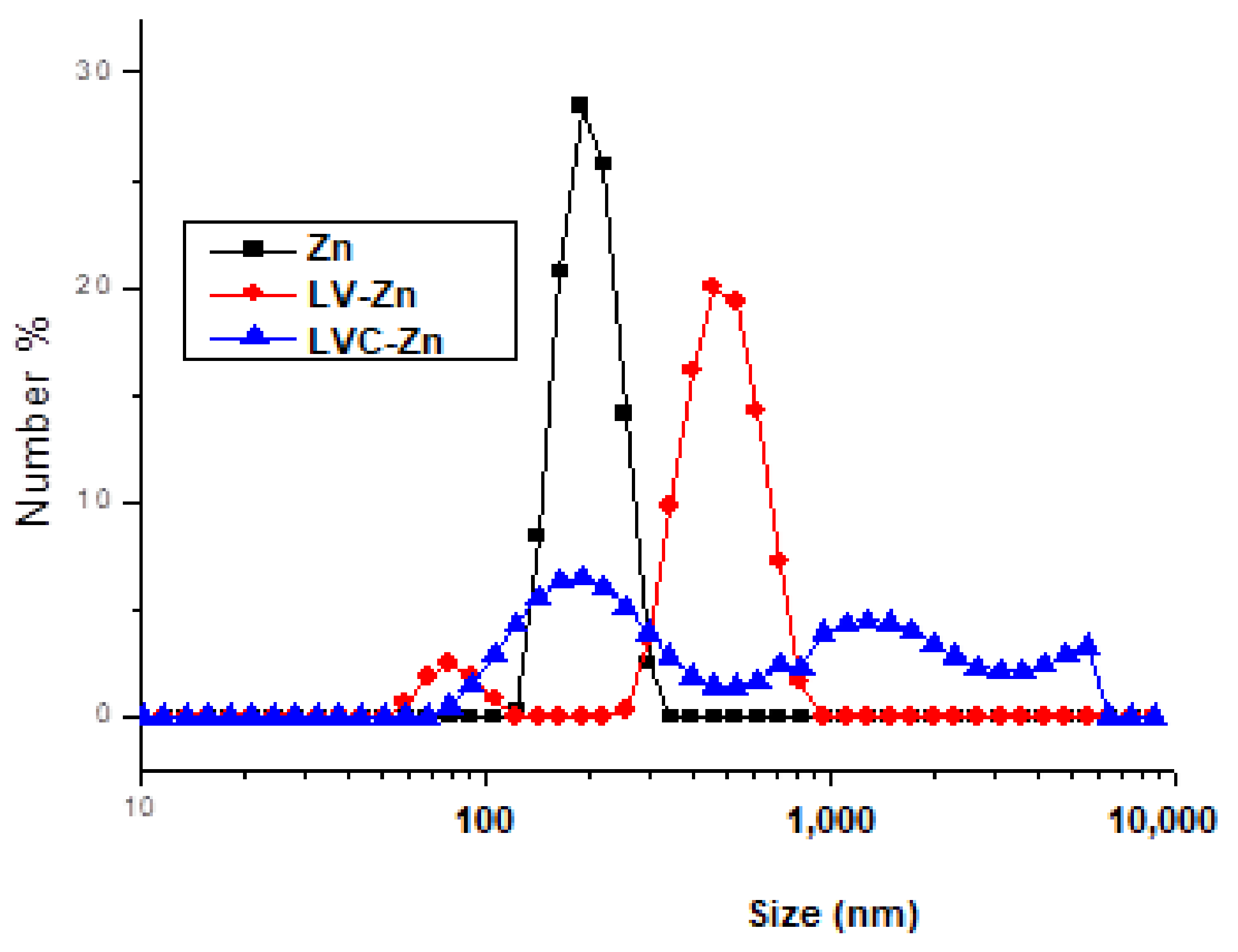
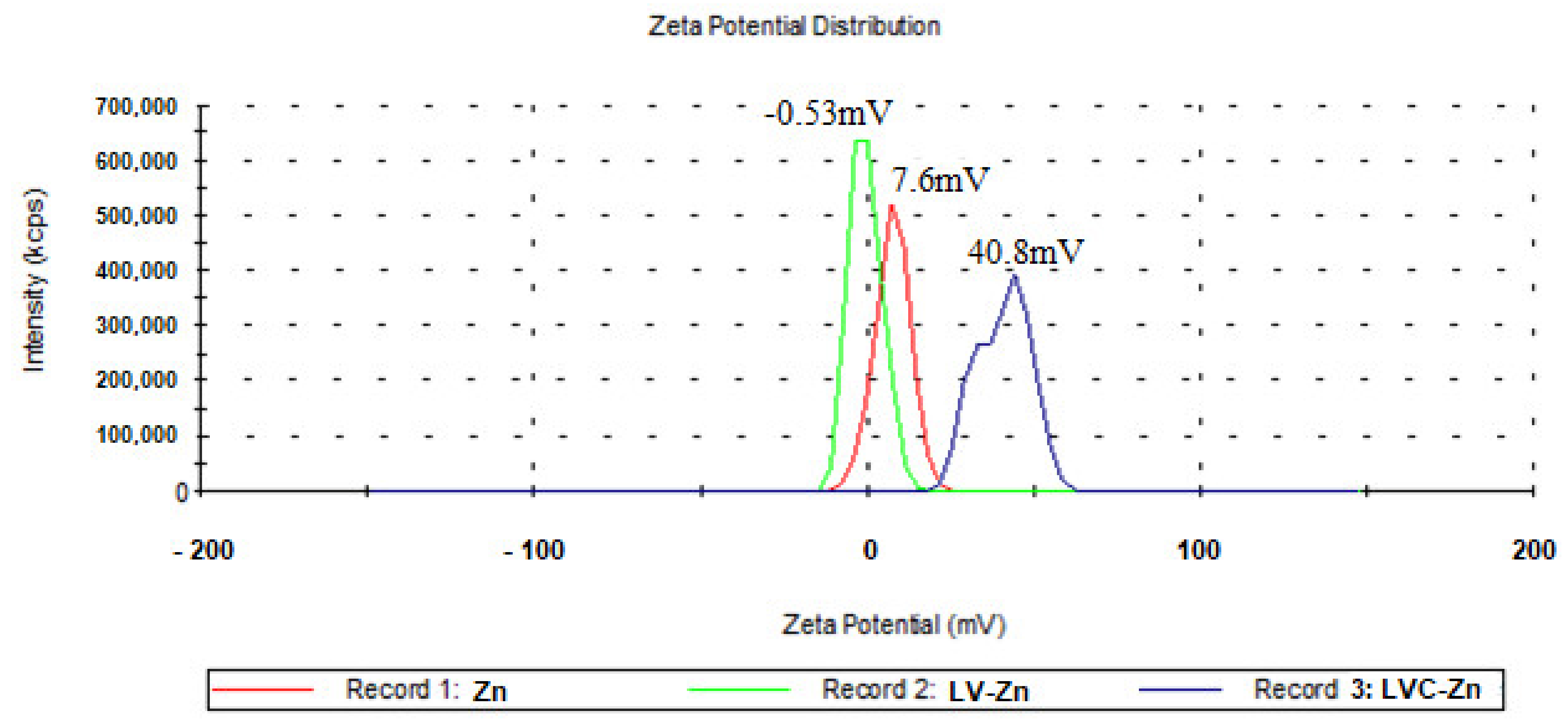
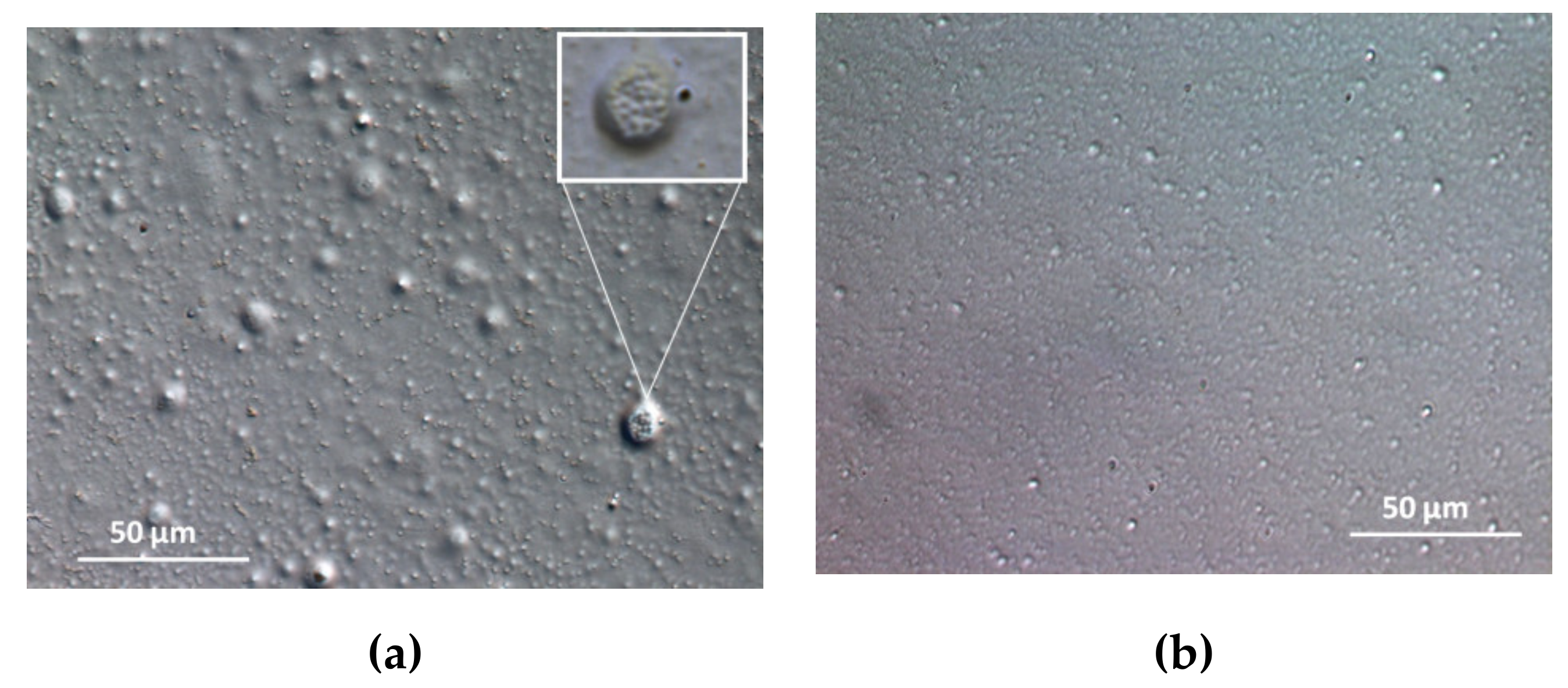
2. Results
3. Discussion
4. Materials and Methods
4.1. Substances
4.2. The Obtaining of Zn Submicrometric Vesicles
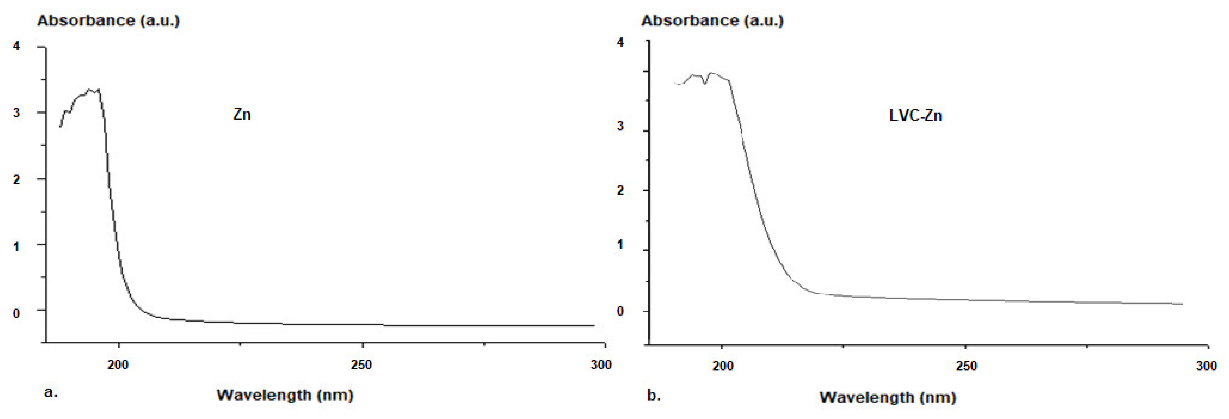
4.3. The Characterization of Zn Submicrometric Vesicles
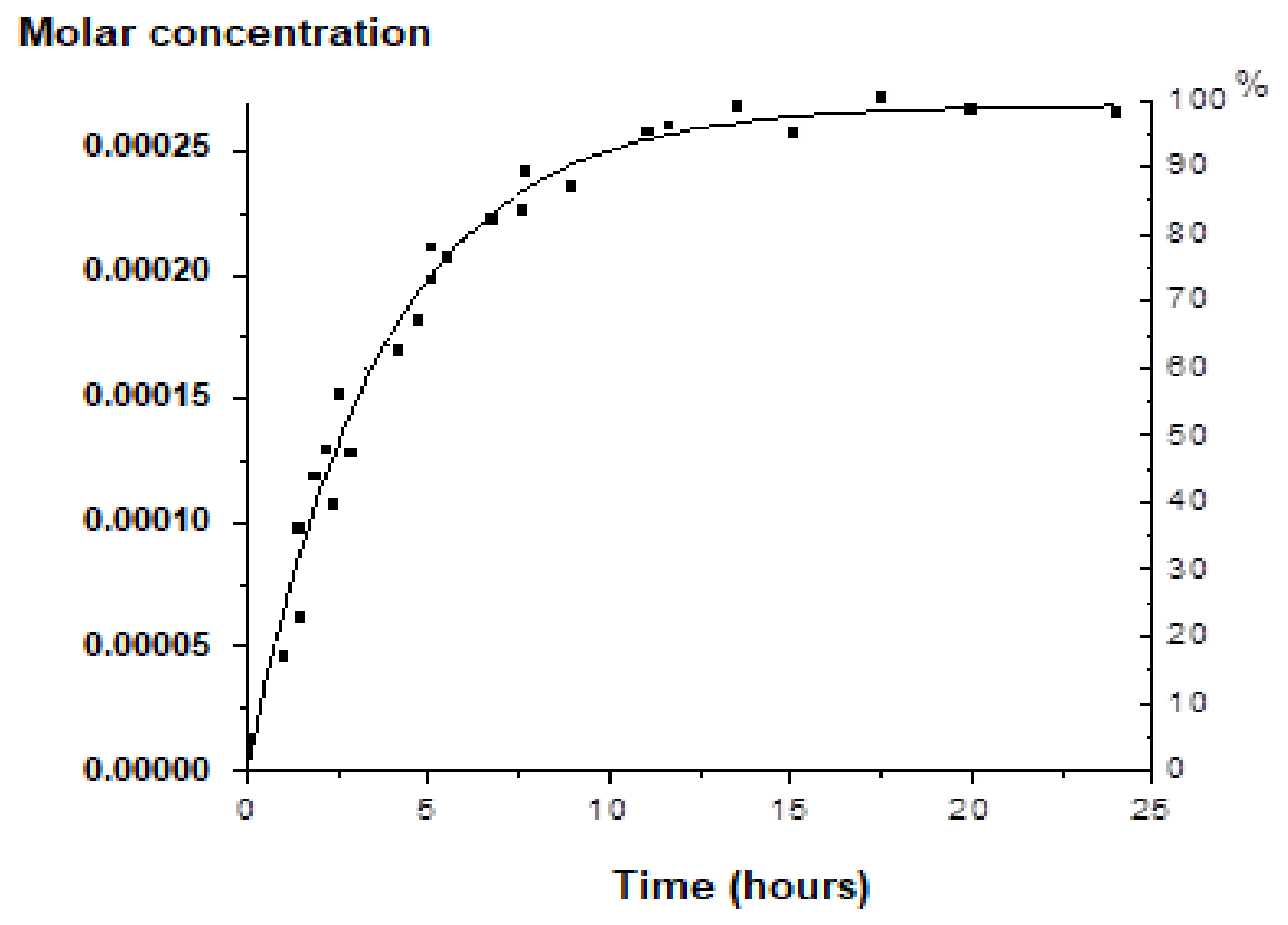
4.4. The In Vitro Release of Zn from the Submicrometric Vesicles
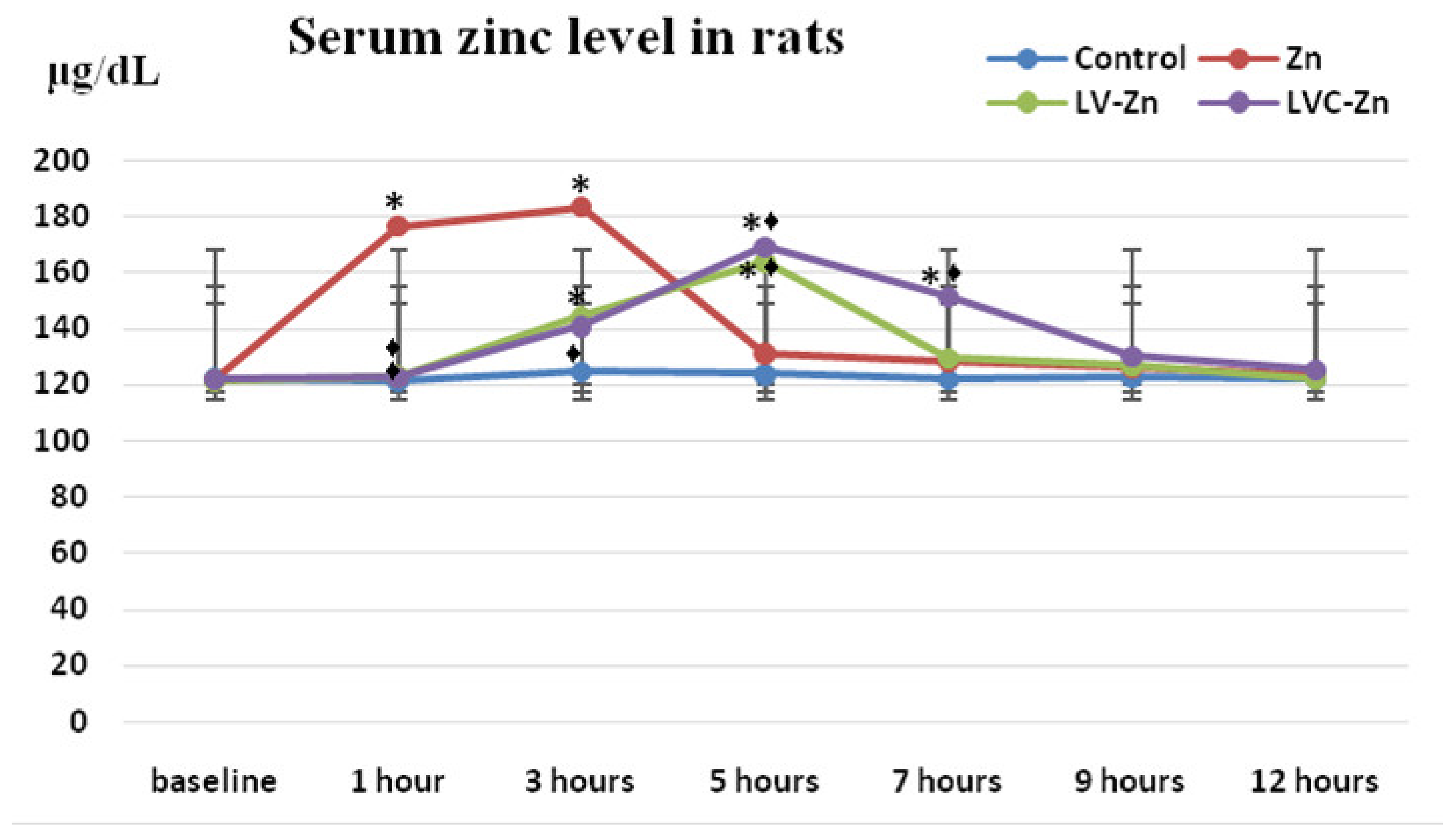
4.5. The In Vivo Release of Zn fromSubmicrometric Vesicles
- ▪
- Group I (control): deionized water 0.3 mL/100 g body weight;
- ▪
- Group II (Zn): 2 mg/kg body weight (kbw) Zn chloride;
- ▪
- Group III (LV-Zn): 2 mg/kbw Zn chloride entrapped in soft vesicles;
- ▪
- Group IV (LVC-Zn): 2 mg/kbw Zn chloride entrapped in soft vesicles stabilized with chitosan.
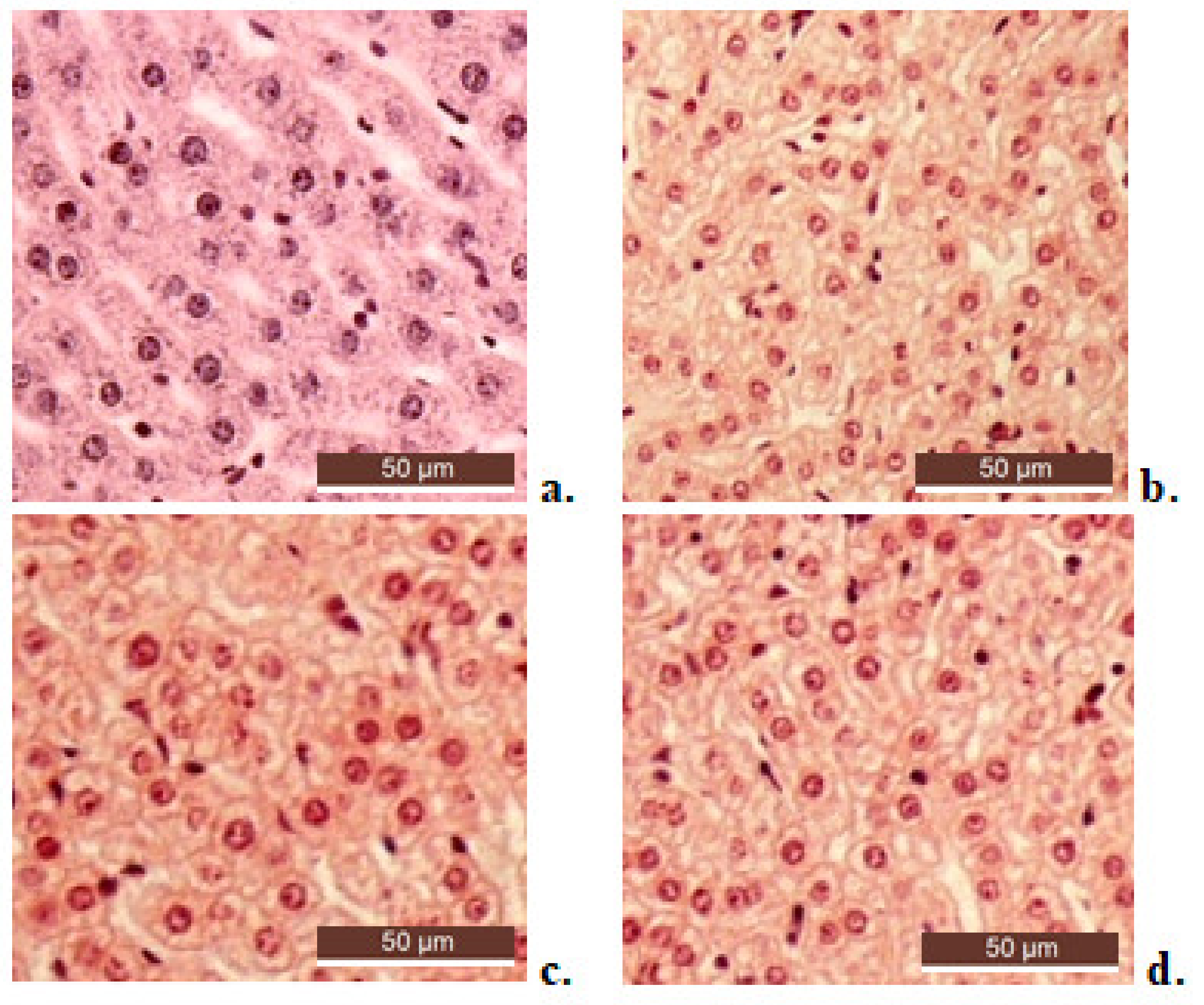
4.6. Biocompatibility Evaluation of Submicrometric Vesicles Entrapping Zn
4.7. Statistical Analysis
5. Conclusions
Author Contributions
Funding
Institutional Review Board Statement
Informed Consent Statement
Data Availability Statement
Conflicts of Interest
Sample Availability
References
- Teslariu, O.; Pasca, A.S.; Mititelu-Tartau, L.; Schiriac, C.E.; Gales, C.; Saftencu, P.M.; Nechifor, M. The protective effects of zinc in experimental gentamicin induced acute renal failure in rats. J. Physiol. Pharmacol. 2016, 67, 751–757. [Google Scholar]
- Kaur, K.; Gupta, R.; Saraf, S.A.; Sharaf, S.K. Zinc: The Metal of Life. Compr. Rev. Food Sci. Food Saf. 2014, 13, 358–376. [Google Scholar] [CrossRef]
- Oteiza, P.I. Zinc and the modulation of redox homeostasis. Free Radic. Biol. Med. 2012, 53, 1748–1759. [Google Scholar] [CrossRef] [Green Version]
- Dardenne, M. Zinc and immune function. Eur. J. Clin. Nutr. 2002, 56, S20–S23. [Google Scholar] [CrossRef] [Green Version]
- Mills, D.A.; Schmidt, B.; Hiser, C.; Westley, E.; Ferguson-Miller, S. Membrane potential-controlled inhibition of cytochrome c oxidase by zinc. J. Biol. Chem. 2002, 277, 14894–14901. [Google Scholar] [CrossRef] [PubMed] [Green Version]
- Sharif, R.; Thomas, P.; Zalewski, P.; Fenech, M. The role of zinc in genomic stability. Mutat. Res. Fundam. Mol. Mech. Mutagenesis 2012, 733, 111–121. [Google Scholar] [CrossRef]
- Andreini, C.; Banci, L.; Bertini, I.; Rosato, A. Zinc through the three domains of life. J. Proteome Res. 2006, 5, 3173–3178. [Google Scholar] [CrossRef] [PubMed]
- Frederickson, C.J.; Bush, A.I. Synaptically released zinc: Physiological functions and pathological effects. Biometals 2001, 14, 353–366. [Google Scholar] [CrossRef]
- Frieke, C. Function and mechanism of zinc. J. Nutr. 2000, 130, 1437S–1446S. [Google Scholar]
- Prasad, A.S. Zinc: Role in immunity, oxydative stress and chronic inflammation. Curr. Opin. Clin. Nutr. Metab. Care 2009, 12, 646–652. [Google Scholar] [CrossRef]
- Haase, H.; Rink, L. Functional significance of zinc-related signaling pathways in immune cells. Annu. Rev. Nutr. 2009, 29, 133–152. [Google Scholar] [CrossRef]
- Lee, S.R. Critical role of zinc as either an antioxidant or a prooxidant in cellular systems. Oxid. Med. Cell Longev. 2018, 20, 9156285. [Google Scholar] [CrossRef] [Green Version]
- Frassinneti, S.; Bronzetti, G.; Calravuturo, L.; Cini, M.; Croce, C.D. The role of zinc in life: A review. J. Environ. Pathol. Toxicol. Oncol. 2006, 25, 597–610. [Google Scholar] [CrossRef] [PubMed]
- Marreiro, D.D.; Cruz, K.J.; Morais, J.B.; Beserra, J.B.; Severo, J.S.; De Oliveira, A.R. Zinc and oxidative stress: Current mechanisms. Antioxidants 2017, 6, 24. [Google Scholar] [CrossRef] [PubMed]
- Squitti, R.; Pal, A.; Picozza, M.; Avan, A.; Ventriglia, M.; Rongioletti, M.C.; Hoogenraad, T. Zinc Therapy in Early Alzheimer’s Disease: Safety and Potential Therapeutic Efficacy. Biomolecules 2020, 10, 1164. [Google Scholar] [CrossRef] [PubMed]
- Kondaiah, P.; Yaduvanshi, P.S.; Sharp, P.A.; Pullakhandam, R. Iron and Zinc Homeostasis and Interactions: Does Enteric Zinc Excretion Cross-Talk with Intestinal Iron Absorption? Nutrients 2019, 11, 1885. [Google Scholar] [CrossRef] [Green Version]
- Read, S.A.; Obeid, S.; Ahlenstiel, C.; Ahlenstiel, G. The Role of Zinc in Antiviral Immunity. Adv. Nutr. 2019, 10, 696–710. [Google Scholar] [CrossRef] [PubMed] [Green Version]
- Kodama, H.; Tanaka, M.; Naito, Y.; Katayama, K.; Moriyama, M. Japan’s Practical Guidelines for Zinc Deficiency with a Particular Focus on Taste Disorders, Inflammatory Bowel Disease, and Liver Cirrhosis. Int. J. Mol. Sci. 2020, 21, 2941. [Google Scholar] [CrossRef]
- Blasiak, J.; Pawlowska, E.; Chojnacki, J.; Szczepanska, J.; Chojnacki, C.; Kaarniranta, K. Zinc and Autophagy in Age-Related Macular Degeneration. Int. J. Mol. Sci. 2020, 21, 4994. [Google Scholar] [CrossRef]
- Uwitonze, A.M.; Ojeh, N.; Murererehe, J.; Atfi, A.; Razzaque, M.S. Zinc Adequacy Is Essential for the Maintenance of Optimal Oral Health. Nutrients 2020, 12, 949. [Google Scholar] [CrossRef] [Green Version]
- Gammoh, N.Z.; Rink, L. Zinc in Infection and Inflammation. Nutrients 2017, 9, 624. [Google Scholar] [CrossRef] [Green Version]
- Maares, M.; Haase, H. A Guide to Human Zinc Absorption: General Overview and Recent Advances of In Vitro Intestinal Models. Nutrients 2020, 12, 762. [Google Scholar] [CrossRef] [Green Version]
- Pompano, L.M.; Boy, E. Effects of Dose and Duration of Zinc Interventions on Risk Factors for Type 2 Diabetes and Cardiovascular Disease: A Systematic Review and Meta-Analysis. Adv. Nutr. 2021, 12, 141–160. [Google Scholar] [CrossRef] [PubMed]
- Anwekar, H.; Patel, S.; Singhai, A.K. Liposome—As drug carriers. Int. J. Pharm. Life Sci. 2011, 2, 945–951. [Google Scholar]
- Barba, A.A.; Bochicchio, S.; Bertoncin, P.; Lamberti, G.; Dalmoro, A. Coating of Nanolipid Structures by a Novel Simil-Microfluidic Technique: Experimental and Theoretical Approaches. Coatings 2019, 9, 491. [Google Scholar] [CrossRef] [Green Version]
- Sebaaly, C.; Trifan, A.; Sieniawska, E.; Greige-Gerges, H. Chitosan-Coating Effect on the Characteristics of Liposomes: A Focus on Bioactive Compounds and Essential Oils: A Review. Processes 2021, 9, 445. [Google Scholar] [CrossRef]
- Yetisgin, A.A.; Cetinel, S.; Zuvin, M.; Kosar, A.; Kutlu, O. Therapeutic nanoparticles and their targeted delivery applications. Molecules 2020, 25, 2193. [Google Scholar] [CrossRef]
- Sharma, A.; Garg, T.; Aman, A.; Panchal, K.; Sharma, R.; Kumar, S.; Markandeywar, T. Nanogel—An advanced drug delivery tool: Current and future. Artif. Cells Nanomed. Biotechnol. 2016, 44, 165–177. [Google Scholar] [CrossRef]
- Bhatia, S. Nanoparticles types, classification, characterization, fabrication methods and drug delivery applications. In Natural Polymer Drug Delivery Systems: Nanoparticles, Plants, and Algae; Bhatia, S., Ed.; Springer International Publishing: Cham, Switzerland, 2016; pp. 33–93. [Google Scholar]
- Neamtu, I.; Rusu, A.G.; Diaconu, A.; Nita, L.E.; Chiriac, A.P. Basic concepts and recent advances in nanogels as carriers for medical applications. Drug Deliv. 2017, 24, 539–557. [Google Scholar] [CrossRef] [Green Version]
- Chenthamara, D.; Subramaniam, S.; Ramakrishnan, S.G.; Krishnaswamy, S.; Essa, M.M.; Lin, F.-H.; Qoronfleh, M.W. Therapeutic efficacy of nanoparticles and routes of administration. Biomater. Res. 2019, 23, 1–29. [Google Scholar] [CrossRef]
- López-Laguna, H.; Sánchez, J.; Unzueta, U.; Mangues, R.; Vázquez, E.; Villaverde, A. Divalent Cations: A Molecular Glue for Protein Materials. Trends Biochem. Sci. 2020, 45, 992–1003. [Google Scholar] [CrossRef]
- Nanja, A.F.; Focke, W.W.; Musee, N. Aggregation and dissolution of aluminium oxide and copper oxide nanoparticles in natural aqueous matrixes. SN Appl. Sci. 2020, 2, 1–16. [Google Scholar] [CrossRef]
- Zhang, Y.; Chen, Y.; Westerhoff, P.; Crittenden, J. Impact of natural organic matter and divalent cations on the stability of aqueous nanoparticles. Water Res. 2009, 43, 4249–4257. [Google Scholar] [CrossRef]
- Czyżowska, A.; Barbasz, A. A review: Zinc oxide nanoparticles—friends or enemies? Int. J. Environ. Health Res. 2020, 10, 1–17. [Google Scholar] [CrossRef] [PubMed]
- Vimercati, L.; Cavone, D.; Caputi, A.; De Maria, L.; Tria, M.; Prato, E.; Ferri, G.M. Nanoparticles: An experimental study of zinc nanoparticles toxicity on marine crustaceans. General overview on the health implications in humans. Front. Public Health 2020, 8, 192. [Google Scholar] [CrossRef] [PubMed]
- Wiesmann, N.; Tremel, W.; Brieger, J. Zinc oxide nanoparticles for therapeutic purposes in cancer medicine. J. Mater. Chem. B 2020, 8, 4973–4989. [Google Scholar] [CrossRef] [PubMed]
- Basuthakur, P.; Patra, C.R. Zinc oxide nanoparticles: Future therapy for cerebral ischemia. Nanomedicine 2020, 15, 2729–2732. [Google Scholar] [CrossRef]
- Cruz, D.M.; Mostafavi, E.; Vernet-Crua, A.; Barabadi, H.; Shah, V.; Cholula-Díaz, J.-L.; Guisbiers, G.; Webster, T.J. Green nanotechnology-based zinc oxide (ZnO) nanomaterials for biomedical applications: A review. J. Phys. Mater. 2020, 3, 034005. [Google Scholar]
- Le, T.D.H.; Trinh, K.S. Synthesis of zinc oxide nanoparticles and their antibacterial activity. In Proceedings of the 2020 5th International Conference on Green Technology and Sustainable Development (GTSD), Ho Chi Minh City, Vietnam, 27–28 November 2020; pp. 119–123. [Google Scholar]
- Jiang, S.; Lin, K.; Cai, M. ZnO Nanomaterials: Current Advancements in Antibacterial Mechanisms and Applications. Front. Chem. 2020, 8, 1–5. [Google Scholar] [CrossRef]
- Barreto, M.S.R.; Andrade, C.T.; Azero, E.G.; Paschoalin, V.M.F.; Del Aguila, E.M. Production of chitosan/zinc oxide complex by ultrasonic treatment with antibacterial activity. J. Bacteriol. Parasitol. 2017, 8, 1–7. [Google Scholar] [CrossRef]
- Yusof, N.A.A.; Zain, N.M.; Pauzi, N. Synthesis of chitosan/zinc oxide nanoparticles stabilized by chitosan via microwave heating. Bull. Chem. React. Eng. Catal. 2019, 14, 450–458. [Google Scholar] [CrossRef] [Green Version]
- Ailincai, D.; TartauMititelu, L.; Marin, L. Drug delivery systems based on biocompatible imino-chitosan hydrogels for local anticancer therapy. Drug Deliv. 2018, 25, 1080–1090. [Google Scholar] [CrossRef] [Green Version]
- Tartau, L.; Cazacu, A.; Melnig, V. Ketoprofen-liposomes formulation for clinical therapy. J. Mater. Sci. Mater. Med. 2012, 23, 2499–2507. [Google Scholar] [CrossRef] [PubMed]
- Vasile, C.; Stoleru, E.; Darie-Nita, R.N.; Dumitriu, R.P.; Pamfil, P.; Tartau, L. Biocompatible materials based on plasticized poly(lactic acid), chitosan and Rosemary ethanolic extract I. Effect of chitosan on the properties of plasticized poly (lactic acid) materials. Polymers 2019, 11, 941. [Google Scholar] [CrossRef] [PubMed] [Green Version]
- Trung, T.S.; Thein-Han, W.W.; Qui, N.T.; Ng, C.H.; Stevens, W.F. Functional characteristics of shrimp chitosan and its membranes as affected by the degree of deacetylation. Bioresour. Technol. 2006, 97, 659–663. [Google Scholar] [CrossRef] [PubMed]
- Kumar, A.; Dixit, C.K. Methods for characterization of nanoparticles. In Advances in Nanomedicine for the Delivery of Therapeutic Nucleic Acids; Woodhead Publishing: Sawston, UK, 2017; Chapter 3; pp. 43–58. [Google Scholar]
- Siba, S.; Sanjeet, K.; Sanjibani, M.; Padan, K.J.; Satish, K.V.; Purnendu, P. Evaluation of antibacterial and antioxidant potential of the zinc oxide nanoparticles synthesized by aqueous and polyol method. Microb. Pathog. 2018, 119, 145–151. [Google Scholar]
- Hajinezhad, M.R. The effects of ZnO nanoparticles and ZnO/chitosan NCs on liver histology and serum parameters in rats. J. Environ. Treat. Tech. 2019, 7, 364–369. [Google Scholar]
- Zulfia, H.; Junaid, A.K.; Hafeez, A.; Naila, A.; Sehrish, M.; Ambreen, A.; Iram, A. Synthesis, characterization, and pharmacological evaluation of zinc oxide nanoparticles formulation. Toxicol. Ind. Health 2018, 34, 753–763. [Google Scholar]
- Yan, G.; Huang, Y.; Bu, Q.; Lv, L.; Deng, P.; Zhou, J.; Wang, Y.; Yang, Y.; Liu, Q.; Cen, X.; et al. Zinc oxide nanoparticles cause nephrotoxicity and kidney metabolism alterations in rats. J. Environ. Sci. Health Part A 2012, 47, 577–588. [Google Scholar] [CrossRef]
- Ansari, M.M.; Ahmad, A.; Mishra, R.K.; Raza, S.S.; Khan, R. Zinc gluconate-loaded chitosan nanoparticles reduce the severity of collagen-induced arthritis in Wistar rats. ACS Biomater. Sci. Eng. 2019, 5, 3380–3397. [Google Scholar] [CrossRef]
- Gârlea, A.; Popa, M.I.; Pohoaţă, V.; Melnig, V. Ibuprofen/ketoprofen entrapment in chitosan based vesicle carrier. Rom. J. Biophys. 2007, 17, 157–168. [Google Scholar]
- Boanca, M.; Mititelu-Tartau, L.; Lupusoru, R.V.; Poroch, V.; Bibire, N.; Lupusoru, C.E. The effects of soft matter vesicles entrapping magnesium chloride in nociceptive reactivity in mice. Farmacia 2015, 63, 362–365. [Google Scholar]
- Li, X.; Wang, L.; Fan, Y.; Feng, Q.; Cui, F.Z. Biocompatibility and toxicity of nanoparticles and nanotubes. J. Nanomater. 2012, 2012. [Google Scholar] [CrossRef] [Green Version]
- Wolf, M.F.; Anderson, J.M. Practical approach to blood compatibility assessments: General considerations and standards in Boutrand J-P edition. In Biocompatibility and Performance of Medical Devices; Woodhead Publishing: Sawston, UK, 2012; Volume 159–201, pp. 201e–206e. [Google Scholar]
- Lindstrom, N.M.; Moore, D.M.; Zimmerman, K.; Smith, S.A. Hematologic assessment in pet rats, mice, hamsters, and gerbils: Blood sample collection and blood cell identification. Vet. Clin. N. Am. Exot. Anim. Pract. 2015, 18, 21–32. [Google Scholar] [CrossRef] [PubMed]
- Toft, M.F.; Petersen, M.H.; Dragstead, N.; Hansen, A.K. The impact of different sampling methods on laboratory rats under different types of anaesthesia. Lab. Anim. 2006, 40, 261–274. [Google Scholar] [CrossRef]
- Zou, W.; Yang, Y.; Gu, Y.; Zhu, P.; Zhang, M.; Cheng, Z.; Liu, X.; Yu, Y.; Peng, X. Repeated blood collection from tail vein of non-anesthetized rats with a vacuum blood collection system. J. Vis. Exp. 2017, 130, e55852. [Google Scholar] [CrossRef] [Green Version]
- Lee, G.; Goosens, K.A. Sampling blood from the lateral tail vein of the rat. J. Vis. Exp. 2015, 99, 52766. [Google Scholar] [CrossRef] [PubMed] [Green Version]
- European Union. DIRECTIVE 2010/63/EU of the European Parliament and of the Council of 22 September 2010 on the Protection of Animals Used for Scientific Purposes; European Union: Brussels, Belgium, 2010.


| Groups | Leucocyte Formula (% Values) | |||||
|---|---|---|---|---|---|---|
| PMN | Ly | E | M | B | ||
| Control | 24 h | 29.3 ± 5.45 | 66.5 ± 9.47 | 0.6 ± 0.1 | 3.4 ± 0.05 | 0.2 ± 0.1 |
| 7 days | 29.8 ± 4.37 | 65.8 ± 10.29 | 0.7 ± 0.1 | 3.5 ± 0.1 | 0.2 ± 0.05 | |
| Zn | 24 h | 29.4 ± 4.83 | 66.0 ± 10.45 | 0.8 ± 0.05 | 3.6 ± 0.05 | 0.2 ± 0.1 |
| 7 days | 29.6 ± 5.29 | 66.1 ± 9.63 | 0.6 ± 0.05 | 3.5 ± 0.05 | 0.2 ± 0.05 | |
| LV-Zn | 24 h | 29.5 ± 5.17 | 66.2 ± 11.17 | 0.7 ± 0.1 | 3.4 ± 0.1 | 0.2 ± 0.05 |
| 7 days | 29.7 ± 5.33 | 65.7 ± 10.33 | 0.8 ± 0.05 | 3.6 ± 0.1 | 0.2 ± 0.05 | |
| LVC-Zn | 24 h | 29.4 ± 4.65 | 66.0 ± 9.81 | 0.8 ± 0.1 | 3.6 ± 0.1 | 0.2 ± 0.1 |
| 7 days | 29.8 ± 5.52 | 66.3 ± 10.55 | 0.6 ± 0.1 | 3.5 ± 0.05 | 0.2 ± 0.1 | |
| Groups | AST (U/mL) | ALT (U/mL) | LDH (U/mL) | |
|---|---|---|---|---|
| Control | 24 h | 43.5 ± 7.55 | 96.5 ± 14.27 | 342.38 ± 61.55 |
| 7 days | 44.8 ± 7.39 | 97.2 ± 15.19 | 344.29 ± 57.67 | |
| Zn | 24 h | 42.9 ± 8.83 | 97.4 ± 14.33 | 343.67 ± 59.83 |
| 7 days | 43.3 ± 7.17 | 98.1 ± 13.64 | 344.45 ± 60.64 | |
| LV-Zn | 24 h | 43.6 ± 8.72 | 96.7 ± 15.39 | 343.52 ± 62.33 |
| 7 days | 45.1 ± 6.33 | 97.3 ± 15.72 | 347.43 ± 61.45 | |
| LVC-Zn | 24 h | 42.9 ± 6.64 | 97.9 ± 14.45 | 344.21 ± 59.72 |
| 7 days | 44.2 ± 7.43 | 98.5 ± 13.27 | 346.17 ± 58.83 | |
| Groups | Urea (mg/dL) | Creatinine (mg/dL) | |
|---|---|---|---|
| Control | 24 h | 36.5 ± 5.29 | 0.05 |
| 7 days | 38.7 ± 4.65 | 0.13 | |
| Zn | 24 h | 37.1 ± 5.33 | 0.11 |
| 7 days | 39.3 ± 6.17 | 0.15 | |
| LV-Zn | 24 h | 37.4 ± 5.83 | 0.07 |
| 7 days | 38.5 ± 5.37 | 0.15 | |
| LVC-Zn | 24 h | 36.4 ± 4.19 | 0.07 |
| 7 days | 37.2 ± 5.43 | 0.09 | |
| Groups | Complement | NBT Test | |
|---|---|---|---|
| Control | 24 h | 16.33 ± 3.29 | 52.19 ± 8.64 |
| 7 days | 17.45 ± 3.33 | 54.25 ± 7.17 | |
| Zn | 24 h | 17.67 ± 4.17 | 52.64 ± 8.55 |
| 7 days | 17.55 ± 3.64 | 55.33 ± 6.83 | |
| LV-Zn | 24 h | 17.29 ± 3.83 | 53.42 ± 7.29 |
| 7 days | 17.74 ± 4.45 | 54.83 ± 6.67 | |
| LVC-Zn | 24 h | 16.17 ± 3.67 | 52.67 ± 7.33 |
| 7 days | 16.83 ± 3.37 | 53.34 ± 7.64 | |
Publisher’s Note: MDPI stays neutral with regard to jurisdictional claims in published maps and institutional affiliations. |
© 2021 by the authors. Licensee MDPI, Basel, Switzerland. This article is an open access article distributed under the terms and conditions of the Creative Commons Attribution (CC BY) license (https://creativecommons.org/licenses/by/4.0/).
Share and Cite
Mititelu-Tartau, L.; Bogdan, M.; Pricop, D.A.; Buca, B.R.; Pauna, A.-M.; Dijmarescu, L.A.; Pelin, A.-M.; Pavel, L.L.; Popa, G.E. Assessment of the In Vivo Release and Biocompatibility of Novel Vesicles Containing Zinc in Rats. Molecules 2021, 26, 4101. https://doi.org/10.3390/molecules26134101
Mititelu-Tartau L, Bogdan M, Pricop DA, Buca BR, Pauna A-M, Dijmarescu LA, Pelin A-M, Pavel LL, Popa GE. Assessment of the In Vivo Release and Biocompatibility of Novel Vesicles Containing Zinc in Rats. Molecules. 2021; 26(13):4101. https://doi.org/10.3390/molecules26134101
Chicago/Turabian StyleMititelu-Tartau, Liliana, Maria Bogdan, Daniela Angelica Pricop, Beatrice Rozalina Buca, Ana-Maria Pauna, Lorena Anda Dijmarescu, Ana-Maria Pelin, Liliana Lacramioara Pavel, and Gratiela Eliza Popa. 2021. "Assessment of the In Vivo Release and Biocompatibility of Novel Vesicles Containing Zinc in Rats" Molecules 26, no. 13: 4101. https://doi.org/10.3390/molecules26134101