Napthrene Compounds from Mycelial Fermentation Products of Marasmius berteroi
by
Ning Ning Yang
1,2,†, 
Qing Yun Ma
1,3,†,
Fan Dong Kong
1,3,
Qing Yi Xie
1,3,
Hao Fu Dai
1,3,
Li Man Zhou
1,
Zhi Fang Yu
4,* and
You Xing Zhao
1,3,* 1
Hainan Key Laboratory for Research and Development of Natural Product from Li Folk Medicine, Institute of Tropical Bioscience and Biotechnology, Chinese Academy of Tropical Agricultural Sciences, Haikou 571101, China
2
College of Food and Bioengineering, Bengbu University, Bengbu 233030, China
3
Hainan Institute for Tropical Agricultural Resources, Chinese Academy of Tropical Agricultural Sciences, Haikou 571101, China
4
College of Food Science and Technology, Nanjing Agricultural University, Nanjing 210095, China
*
Authors to whom correspondence should be addressed.
†
These authors contributed equally to this work.
Molecules 2020, 25(17), 3898; https://doi.org/10.3390/molecules25173898
Submission received: 15 July 2020
/
Revised: 21 August 2020
/
Accepted: 24 August 2020
/
Published: 26 August 2020
(This article belongs to the Special Issue Natural Products Discovery and Development: A Themed Issue Dedicated to Professor Leslie Gunatilaka)
Abstract
:The metabolites of the genus Marasmius are diverse, showing good research prospects for finding new bioactive molecules. In order to explore the active metabolites of the fungi Marasmius berteroi, the deep chemical investigation on the bioactive compounds from its cultures was undertaken, which led to the isolation of three new naphthalene compounds dipolynaphthalenes A–B (1,2) and naphthone C (3), as well as 12 known compounds (4–15). Compounds 1, 2, and 4 are dimeric naphthalene compounds. Their structures were elucidated by MS, 1D and 2D NMR spectroscopic data, as well as ECD calculations. Compounds 2–4 and 7 exhibited acetylcholinesterase (AChE) inhibitory activities at the concentration of 50 μg/mL with inhibition ratios of 42.74%, 44.63%, 39.50% and 51.49%, respectively. Compounds 5 and 7,8 showed weak inhibitory activities towards two tumor cell lines, with IC50 of 0.10, 0.076 and 0.058 mM (K562) and 0.13, 0.18, and 0.15 mM (SGC-7901), respectively.
1. Introduction
Natural products are an important source of innovative chemical drugs [1]. Finding natural active ingredients for nerve protection and tumor suppression are also hot topics in the current research [2]. The genus Marasmius is a common basidiomycete in tropical and subtropical areas, which belongs to the family Marasmiaceae [3]. A few common species of Marasmius have been studied for searching of bioactive metabolites, and some active compounds such as terpenoids [4,5,6,7,8], steroids [9], cyclic peptides [10], isocoumarins [11] and piperidones [12] have been isolated. Many of these compounds showed antibacterial, cytotoxic and antihypertensive activities [9,10,11,12]. Marasmius berteroi is a small orange mushroom belonging to the genus Marasmius, which is widely distributed in the southeast of mainland China, mainly in Hainan, Guangdong, and Taiwan provinces. So far, few researches on chemical constituents of this fungus were reported [13]. To seek for new active small molecules from Marasmius berteroi, the deep chemical investigation on its cultures was thus undertaken, which led to the isolation of three new naphthalene ring compounds dipolynaphthalenes A–B (1,2) and naphthone C (3) along with twelve known analogues (4–15). The isolation process and structural elucidation of three new compounds, as well as their AChE inhibitory and cytotoxic activity, are described in this paper.
2. Results and Discussion
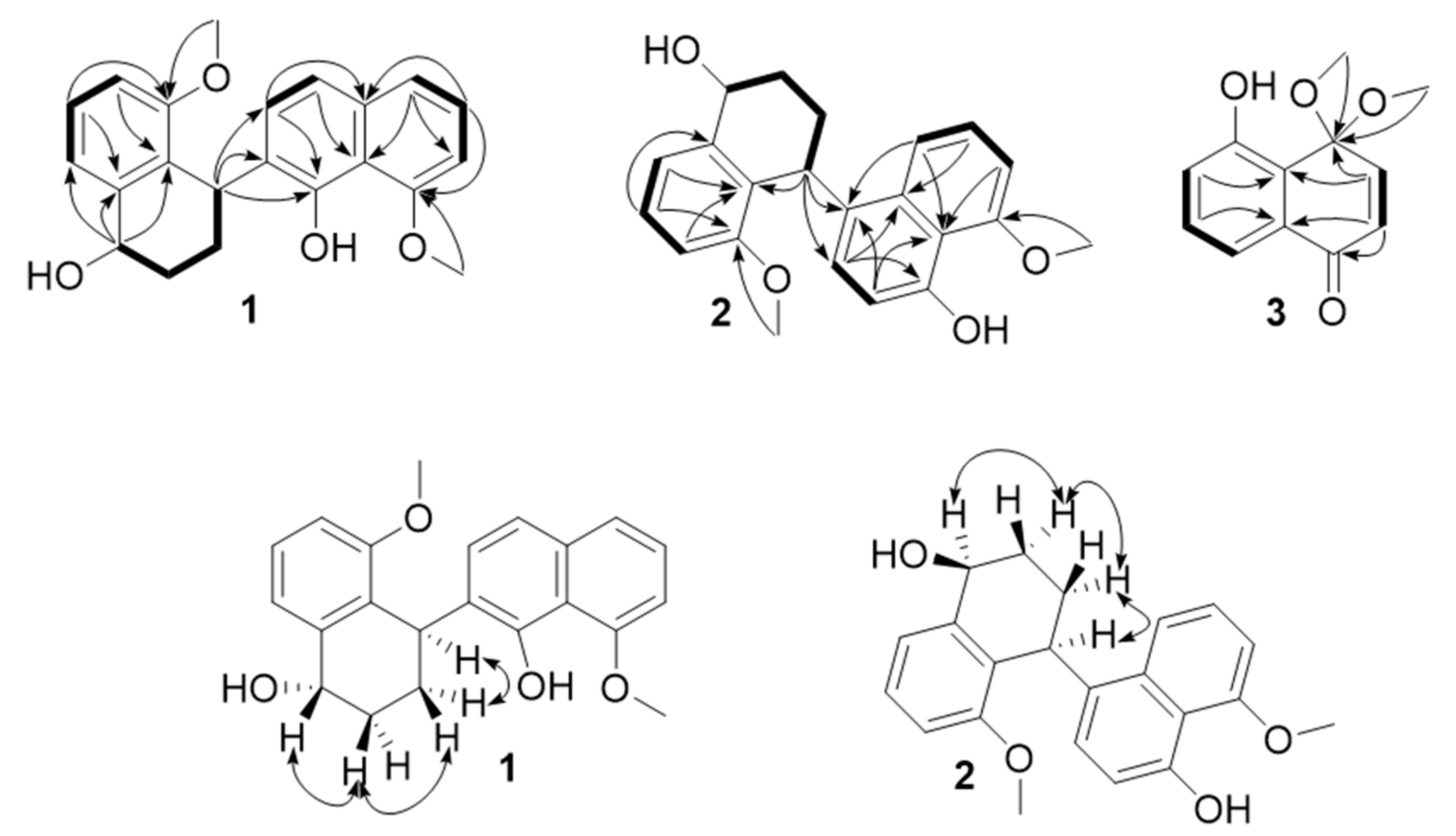
Dipolynaphthalene A (compound 1) was isolated as pale-yellow oil. Its molecular formula was assigned to be C22H22O4 with twelve degrees of unsaturation according to its positive HR-ESI-MS (m/z 373.1412 [M + Na]+, calcd. 373.1410 for C22H22O4Na) and NMR spectroscopic data (Table 1). The IR (Supplementary Materials) spectrum revealed the presence of hydroxyl (3305 cm−1) and benzene ring (1581cm−1) absorptions. The 13C-NMR and DEPT spectroscopic data (Table 1) showed 22 carbon resonances, including two methoxyls, two sp3 methylenes, ten methines (eight olefinic and one oxygenated), and eight quaternary carbons (three oxygenated). The 1H-NMR (Table 1) showed the presence of signals for two singlet methoxys (δ 4.06 (3H, s, H-5), 4.08 (3H, s, H-8′)). According to a comparison of the corresponding NMR data, compound 1 was similar to nodulisporin B [14], a dimer of naphthalene ring compound, except for the two naphthalene rings have different binding sites. Compound 1 was 4,2′-binaphthalene, while nodulisporin B was 2,2′-binaphthalene, which was evidenced by the key HMBC correlations from H-3 (δ 2.12 m, 2.21 m) to quaternary carbon C-2′ (δC 128.1), H-4 (δ 4.74 m) to quaternary carbon C-1′ (δC 150.4). Other correlations in the HMBC (C-5/H-6/H-7/OCH3, C-1/H-2, H-1/C-8a/C-2, C-1′/H-4, C-8′/H-7′/H-6′) and 1H–1H COSY spectra further supported the atom connectivity in compound 1 (Figure 1 and Figure 2). The relative configuration of compound 1 was determined by ROESY cross-peaks H-4[δH 4.74 (1H, m)]/H-3α[δH 2.12 (1H, m)]/H-2α[δH 1.75 (1H, m)]/H-3′[δH 7.23 (1H, d, J = 8.1)], H-3β[δH 2.21 (1H, m)]/H-2β[δH 1.95 (1H, m)]/H-1 [δH 4.85 (1H, dd, J = 5.4, 8.8 Hz)]. Thus, compound 1 was assigned as shown in Figure 1 and it was named dipolynaphthalene A.
Dipolynaphthalene B (compound 2) was purified as pale yellow oil, and possessed the molecular formula C22H22O4 based on HR-ESI-MS (m/z 373.1410 [M + Na]+, calcd for C22H22O4Na, 373.1410) with twelve degrees of unsaturation. The IR spectrum showed the presence of hydroxyl (3399 cm−1) and benzene ring (1583 cm−1) absorptions. The 13C-NMR spectrum of 2 displayed 22 carbon resonances extremely similar to those of compound 1, suggesting that both of them were dimeric naphthalene rings. The only difference was that compound 2 was 4,4′-binaphthalene instead of 4,2′-binaphthalene in 1, which was further supported by the HMBC correlations (Figure 2) from H-4 [δ 5.03 d (5.4)] to C-3′ (δC 126.8) and C-4′a (δC 131.6), H-3 (δ 1.85 m) to C-4′ (δC 128.3). Other correlations in the HMBC and 1H–1H COSY spectra (Figure 2) further supported the atom connectivity in compound 2. The configurations of 1-α OH and 4-β H in 2 were deduced from its ROESY cross-peaks (Figure 2) H-1α/H-2α [δH 1.81 (1H, m)]/H-3α[δH 2.37 (1H, m)]/H-4 [δH 5.03 (1H, d, J = 5.4 Hz)]. Thus, compound 2 was assigned as shown in Figure 1, and named dipolynaphthalene B.
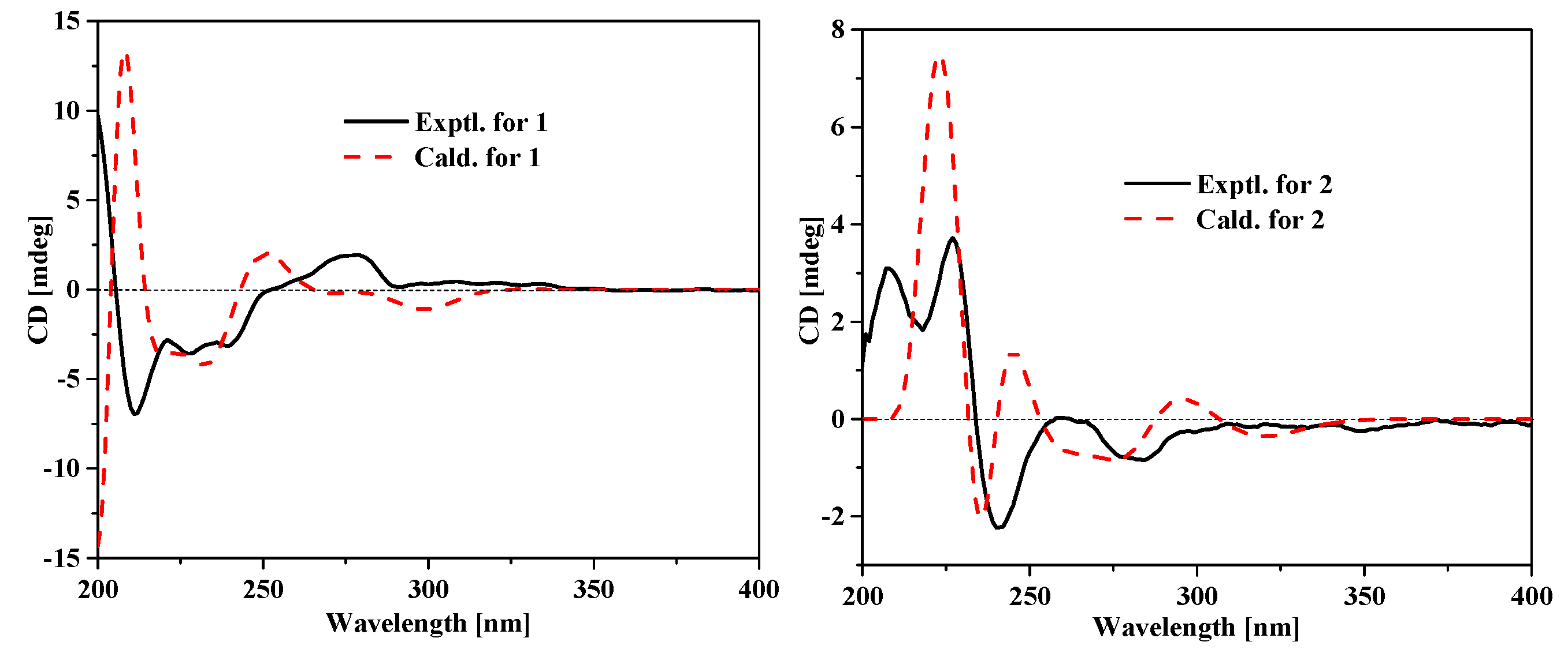
In order to determine the absolute configurations of 1 and 2, the ECD spectra were calculated by the TDDFT method at the apfd/6-311+g (2d, p) level. The calculated ECD spectra of 1 and 2 are generally consistent with their measured spectra (Figure 3), implying the (1S,4R)- configuration for 1 and the (1S,4S)- configuration for 2 (Figure 3). However, much more work was needed to confirm the absolute configurations of 1 and 2, for there are also some differences present between the calculated and measured ECD spectra.
Naphthone C (3) was isolated as pale yellow oil, which molecular formula was assigned to be C12H12O4 with seven degrees of unsaturation according to its positive HR-ESI-MS (m/z 243.0622 [M + Na]+, calcd. 243.0628 for C12H12O4Na) and NMR spectroscopic data (Table 2). The IR spectrum revealed the presence of hydroxyl (3323 cm−1), carbonyl (1714 cm−1) and double bond (1677cm−1) absorptions. The 13C-NMR and DEPT spectroscopic data (Table 2) showed 12 carbon resonances, including two methoxyls, five methines, and five quaternary carbons (two oxygenated and one carbonyl carbon), which was inferred that the basic skeleton of compound 3 was naphthone. The 1H-NMR (Table 2) showed the presence of signals for singlet methoxy (δ 3.35 (3H, s, H-4)). According to a comparison of the corresponding NMR data, compound 3 was similar to 4,4,5-trimethoxy-1(4H)-naphthone [15], a derivative of naphthalene ring compound, except for the hydroxyl (C-5 δC157.9) in 3 instead of methoxy in 4,4,5-trimethoxy-1(4H)-naphthone, which was further supported by the HMBC correlations (Figure 2) from H-6 [δH 6.94 dd (8.3,1.06)] to C-5 (δC 157.9) and H-7 [δH 7.27 t (7.5)] to C-5. Other correlations in the HMBC further supported the atom connectivity in compound. Thus, compound 3 was assigned as shown in Figure 1, and named naphthone C.
The twelve known compounds (4–15) was determined to be daldinone C (4) [16], 1,8-dimethoxynaphthalene (5) [17], 8-methoxy-1-naphthol (6) [18], 8-methoxynaphthalen e-1,7 -diol (7) [19], 5-methoxy-1,4-naphthoquinone (8) [20], isosclerone (9) [21], 5-O-methylsclerone (10) [22] 4,6,8-trihydroxy-3,4-dihydronaphthalen-1(2H)-one(6-hydroxy-isosclerone) (11) [23], trans-3,4-dihydro-3,4,8-trihydroxynaphthalene-1-(2H)-one(12) [24], scytalone (13) [25], xylarenone (14) [26] and cis-4-hydroxyscytalone (15) [27] by comparison of its spectroscopic data with those of literature.
The acetylcholinesterase inhibitory activity of compounds 1–15 were tested by previous method as described in the literature [28]. The results showed that compounds 2–4 and compound 7 exhibited anti-acetylcholinesterase activities at a concentration of 50 μg/mL with inhibition ratios of 42.74%, 44.63%, 9.50%, and 51.49%, respectively (Table 3). In the screening test of cytotoxicity [29], compounds 5, 7, 8 showed inhibitory activity against two tumor cell lines, with IC50 of 0.10, 0.076 and 0.058 mM (K562) and 0.13, 0.18, and 0.15 mM (SGC-7901), respectively (Table 4). Binaphthalenes are dimeric compounds of naphthalene ring, mainly derivatives of DHN and juglone formed by different linking modes [30]. The acetylcholinesterase inhibitory activity evaluation results showed that compounds 2 and 4 exhibited anti-acetylcholinesterase activities, while compound 1 was inactive. Structurally, the active compounds 2 and 4 were naphthalene ring dimers with para-para ligation, while compound 1 was ortho-para ligation dimer, which indicated that the polymeric sites in binaphthalene compounds were very important for their biological activity. In the cytotoxic assay, compound 8 were active, while 9–15 were inactive, which may suggest that the 1,4-dione moiety is essential for their cytotoxicity.
3. Materials and Methods
3.1. General Information
Optical rotations were measured with a Rudolph Autopol III polarimeter (Rudolph Research Analytical, Hackettstown, NJ, USA). Shimadzu UV-2550 spectrometer (Beckman, Brea, CA, USA) was used for scanning UV spectroscopy. IR spectra were obtained on a Tensor 27 spectrometer, as KBr pellets (Thermo, Pittsburgh, PA, USA). NMR spectra were recorded on an AV-500 spectrometer (Bruker, Bremen, Germany) with TMS (Tetramethylsilane) as an internal standard. HR-ESI-MS were performed on an API QSTAR Pulsar mass spectrometer (Billerica, MA, USA). Silica gel (200–300 mesh, Qingdao Marine Chemical Inc., Qingdao, China), RP-18 (40–70 mm, Fuji Silysia Chemical Ltd., Kasugai Aichi, Japan) and Sephadex LH-20 (GE Healthcare, Uppsala, Sweden) were used for column chromatography (CC). Semipreparative HPLC (Agilent 1100, Agilent Technologies Inc., Santa Clara, CA, USA) was performed on an Agilent 1100 liquid chromatograph with a Zorbax SB-C18, 9.4 mm × 25 cm, column. Fractions were monitored by TLC and spots were visualized by heating after spraying with 5% H2SO4 in ethanol.
3.2. Fungal Material
The fungus Marasmius berteroi was collected in the valley of Yinggeling, Hainan province of China, in November 2015, and identified by Associate Professor Sheng-zhuo Huang, the Institute of Tropical Bioscience and Biotechnology, Chinese Academy of Tropical Agricultural Sciences. The mycelium was isolated from the Marasmius berteroi and its strain was maintained on potato dextrose agar (PDA) slant at 4 °C. A voucher specimen (YGL-18) was deposited at the Institute of Tropical Bioscience and Biotechnology, Chinese Academy of Tropical Agricultural Sciences, Haikou, China.
3.3. Fermentation, Extraction and Isolation
The fungus was cultured on slants of PDA medium at 28 °C for 5 days. Plugs of agar supporting mycelium growth were cut and transferred aseptically to 200 × 1000 mL Erlenmeyer flasks each containing 300 mL medium. The flask was incubated at room temperature under static conditions for 30 days. The culture broth (60 L) was filtered to give the filtrate and mycelia. The filtrate was evaporated in vacuo to a small volume and then suspended in H2O and partitioned successively with EtOAc and n-BuOH. The EtOAc solution was evaporated under reduced pressure to give a crude extract (32.0 g), which was separated into fractions 1–6 on silica gel CC using a gradient eluent of petroleum ether-EtOAc (15:1–1:1, v/v, each 3 L). Fr. 2 (7.5 g) was subjected to repeated RP-18 CC (eluted with MeOH/H2O from 2:8 to 10:0, v/v, each 500 mL) and silica gel CC (eluted with petroleum ether–EtOAc from 5:1 to 1:1, v/v, each 600 mL) to afford compounds 1 (7.0 mg), 2 (5.0 mg) and 4 (3.0 mg). Fr. 3 (6.0 g) was applied to octadecyl silane (ODS) gel with gradient elution of MeOH–H2O (1:5, 2:3, 3:2, 4:1, 1:0) to yield compounds 3 (4.0 mg), 6 (8.0 mg) and 7 (2.0 mg). Fr. 4 (5.0 g) was purified by HPLC over an ODS column (30–85% MeOH/H2O, v/v) to give compounds 5 (4 mg), 8 (8.0 mg), 9 (2.0 mg) and 11 (2.0 mg). Fr.5 (3.5 g) was purified by HPLC over an ODS column (30–75% MeOH/H2O, v/v) to give compound 10 (4 mg) and 12 (2 mg). The compounds 13 (3 mg), 14 (9.0 mg), 15 (4.0 mg) were purified from Fr.6 (5 g) by HPLC over an ODS column (30–75% MeOH/H2O, v/v).
3.3.1. Dipolynaphthalene A (1)
Pale yellow oil; [α]25D − 14 (c 0.10, MeOH); IR (KBr) νmax 3305, 2947, 2346, 2301, 1735, 1661, 1581, 1459, 1258, 1015 cm−1; 1H- and 13C-NMR data see Table 1; ESI-MS positive m/z [M + Na]+ 373; HR-ESI-MS m/z [M + Na]+ 373.1412 (calcd for C22H22O4Na, 373.1410).
3.3.2. Dipolynaphthalene B (2)
Pale yellow oil; [α]25D − 8 (c 0.10, MeOH); IR (KBr) νmax 3399, 2943, 2366, 2344, 1657, 1618, 1583, 1467, 1407, 1258 cm−1; 1H- and 13C-NMR data see Table 1; ESI-MS positive m/z [M + Na]+ 373; HR-ESI-MS m/z [M + Na]+ 373.1410 (calcd for C22H22O4Na, 373.1410).
3.3.3. Naphthone C (3)
Pale yellow oil; [α]25D − 42.9 (c 0.15, MeOH); IR (KBr) νmax 3323, 2354, 2319, 1714, 1677, 1577, 1461, 1273 cm−1; 1H- and 13C-NMR data see Table 2; ESI-MS positive m/z [M + Na]+ 243; HR-ESI-MS m/z [M + Na]+ 243.0622 (calcd for C15H24O5Na, 243.0628).
3.4. Bioassay of AChE Inhibitory Activity and Cytotoxic Activity
AChE inhibitory activity of these compounds was assayed by the spectrophotometric method developed by Ellman [28]. Acetylthiocholine iodide (Sigma, St. Louis, MO, USA) was used as substrate in the assay. Na2HPO4 (94.7 mL, 0.1 M) and NaH2PO4 (5.3 mL, 0.1 M) were mixed to get phosphate buffer (PB, pH 8.0). Compounds were dissolved in DMSO (2% in PB). The reaction mixture contained PB (110 μL), test compound solution (10 μL, 2000 μM) and acetyl cholinesterase solution (40 μL, 0.1 U/mL), which were mixed and incubated for 20 min (30 °C). The reaction was initiated by the addition of DTNB (5,5-dithiobis-2-nitrobenzoic acid, 20 μL, 6.25 mM) and acetylthiocholine iodide (20 μL, 6.25 mM). The hydrolysis of acetylthiocholine was monitored at 405 nm every 30 s. Tacrine (Sigma-Aldrich 99%) was used as positive control (final concentration 0.33 μM), while 2% DMSO in PB was set as negative control (NC). All reactions were performed in triplicate. The percentage inhibition was calculated as follows: % age inhibition = (E − S)/E × 100 (E is activity of the enzyme without test compound and S is the activity of enzyme with test compound).
The cytotoxicities of compounds 1–15 (purity of tested compounds over 95%) were assessed using the MTT [3-4,5-dimethylthiazol-2-yl)-2,5-diphenyltetrazolium bromide] assay against human cell lines K562, BEL7702, andSGC7901 according to the method described in previous literature [29] Taxol (Kyowa Hakko Kogyo Co., Tokyo, Japan; purity 99%) was used as a positive control.
4. Conclusions
The secondary metabolites of fungi were various, which were good sources of active substances. The special compounds isolated from Marasmius berteroi also showed the diversity of the genus Marasmius. In the present study, three new naphthalene compounds dipolynaphthalenes A–B (1–2) and naphthone C (3), as well as 12 known compounds were isolated from the mycelial fermentation products of Marasmius berteroi. The bioactivity evaluation assays showed that dipolynaphthalene B (2), naphthone C (3), daldinone C (4) and 8-methoxynaphthalene-1,7-diol (7) showed anti-acetylcholinesterase activities.
Supplementary Materials
The following are available online. Figure S1: 1H-NMR spectrum of dipolynaphthalene A (1); Figure S2: 13C-NMR spectrum of dipolynaphthalene A (1); Figure S3: HSQC spectrum ofdipolynaphthalene A (1); Figure S4: HMBC spectrum ofdipolynaphthalene A (1); Figure S5: 1H-1H COSY spectrum of dipolynaphthalene A (1); Figure S6: ROESY spectrum of dipolynaphthalene A (1); Figure S7: HR-ESI-MS of dipolynaphthalene A (1); Figure S8: IR spectrum of dipolynaphthalene A (1); Figure S9: 1H-NMR spectrum of dipolynaphthalene B (2); Figure S10: 13C-NMR spectrum of dipolynaphthalene B (2); Figure S11: HSQC spectrum ofxylariaine B (2); Figure S12: HMBC spectrum of dipolynaphthalene B (2); Figure S13: 1H-1H COSY spectrum of dipolynaphthalene B (2); Figure S14: ROESY spectrum of dipolynaphthalene B (2); Figure S15: HR-ESI-MS of dipolynaphthalene B (2); Figure S16: IR spectrum of dipolynaphthalene B (2); Figure S17: 1H-NMR spectrum ofnaphthone C (3); Figure S18: 13C-NMR spectrumof naphthone C (3); Figure S19: HSQC spectrum of naphthone C (3); Figure S20: HMBC spectrum of naphthone C (3); Figure S21: 1H-1H COSY spectrum ofnaphthone C (3); Figure S22: HR-ESI-MS of naphthone C (3); Figure S23: IR spectrum of naphthone C (3).
Author Contributions
N.N.Y.: wrote the manuscript and isolate compounds; Q.Y.M.: activity test; F.D.K.: structural elucidation; Q.Y.X.: method adviser; H.F.D.: data analysi; L.M.Z.: data analysis; Z.F.Y.: revised the manuscript; Y.X.Z.: theory and orientation adviser. All authors have read and agree to the published version of the manuscript.
Funding
This research received no external funding.
Acknowledgments
This work was supported by Key Research & Development Program of Hainan Province (ZDYF2019127), Financial Fund of the Ministry of Agriculture and Rural Affairs, P. R. of China (NFZX2018), China Agriculture Research System (CARS-21) and Central Public-interest Scientific Institution Basal Research Fund for Chinese Academy of Tropical Agricultural Sciences (17CXTD-15, 1630052016008).
Conflicts of Interest
The authors declare no conflict of interest.
References
- Liu, R.X.; Chang, P.; Jiang, Z.F. Research Progress of Bioactive Polysaccharides in Neuroprotection and in the Prevention and Treatment of Alzheimer’s Disease. Nat. Prod. Res. Dev. 2014, 26, 2076–2081. [Google Scholar]
- Hiremathad, A.; Piemontese, L. Heterocyclic compounds as key structures for the interaction with old and new targets in Alzheimer’s disease therapy. Neural Regen. Res. 2017, 12, 1256–1261. [Google Scholar] [PubMed]
- Song, B.; Deng, C.Y.; Wu, X.L.; Li, T.H. Known Species of Marasmius from China and Their Distribution. Guizhou Sci. 2009, 27, 1–18. [Google Scholar]
- Ayer, W.A.; Craw, P.A.; Stout, T.J.; Clardy, J. Novel sesquiterpenoids from the fairy ring fungus, Marasmius oreades. Can. J. Chem. 1989, 67, 773–778. [Google Scholar] [CrossRef]
- Evans, L.; Hedger, J.; O’Donnell, G.; Skelton, B.W.; White, A.H.; Williamson, R.T.; Gibbons, S. Structure elucidation of some highly unusual tricyclic cis-caryophyllane sesquiterpenes from Marasmiellus troyanus. Tetrahedron Lett. 2010, 51, 5493–5496. [Google Scholar] [CrossRef]
- Meng, J.; Li, Y.Y.; Ou, Y.X.; Song, L.F.; Lu, C.H.; Shen, Y.M. New sesquiterpenes from Marasmius cladophyllus. Mycology 2011, 2, 30–36. [Google Scholar] [CrossRef]
- Liermann, J.C.; Thines, E.; Opatz, T.; Anke, H. Drimane sesquiterpenoids from Marasmius sp. inhibiting the conidial germination of plant-pathogenic fungi. J. Nat. Prod. 2012, 75, 1983–1986. [Google Scholar] [CrossRef]
- Isaka, M.; Palasarn, S.; Sappan, M.; Supothina, S.; Boonpratuang, T. Hirsutane sesquiterpenes from cultures of the basidiomycete Marasmiellus sp. BCC 22389. Nat. Prod. Bioprospect. 2016, 6, 257–260. [Google Scholar] [CrossRef] [Green Version]
- Fattorusso, E.; Giovannitti, B.; Lanzotti, V.; Magno, S.; Violante, U. 4,4-Dimethyl-5 alpha -ergosta -8,24(28)-dien-3 beta-ol from the fungus Marasmius oreades. Steroids 1992, 57, 119–121. [Google Scholar] [CrossRef]
- Ványolós, A.; Dékány, M.; Kovács, B.; Krámos, B.; Bérdi, P.; Zupkó, I.; Hohmann, J.; Béni, Z. Gymnopeptides A and B, cyclic octadecapeptides from the mushroom Gymnopus fusipes. Org. Lett. 2016, 18, 2688–2691. [Google Scholar] [CrossRef]
- Thongbai, B.; Surup, F.; Mohr, K.; Kuhnert, E.; Hyde, K.D.; Stadler, M. Gymnopalynes A and B, chloropropynyl-isocoumarin antibiotics from cultures of the basidiomycete Gymnopus sp. J. Nat. Prod. 2013, 76, 2141–2144. [Google Scholar] [CrossRef] [PubMed]
- Zhang, L.; Yang, M.; Song, Y.; Sun, Z.; Peng, Y.; Qu, K.; Zhu, H. Antihypertensive effect of 3,3,5,5-tetramethyl-4-piperidone, a new compound extracted from Marasmius androsaceus. J. Ethnopharmacol. 2009, 123, 34–39. [Google Scholar] [CrossRef] [PubMed]
- Mao, X.L. Macromycetes of China, 1st ed.; Science Press: Beijing, China, 2009. [Google Scholar]
- Dai, J.; Karsten, K.; Ulrich, F.; Siegfried, D.; Barbara, S.; Attila, K.S.; Sándor, A.; Tibor, K.; Teunis van, R. Metabolites from the endophytic fungus Nodulisporium sp. from Juniperus cedre. Eur. J. Org. Chem. 2006, 15, 3498–3506. [Google Scholar] [CrossRef]
- Crouse, D.J.; Wheeler, D.M.S. Preparation of a 4-monoketal of juglone methyl ether. Tetrahedron Lett. 1979, 20, 4797–4798. [Google Scholar] [CrossRef]
- Gu, W.; Ge, H.M.; Song, Y.C.; Ding, H.; Zhu, H.L.; Zhao, X.A.; Tan, R.X. Cytotoxic benzo[j]fluoranthene metabolites from Hypoxylon truncatum IFB-18, an endophyte of Artemisia annua. J. Nat. Prod. 2007, 70, 114–117. [Google Scholar] [CrossRef]
- Li, D.L.; Wu, Z.C.; Chen, Y.C.; Tao, M.H.; Zhang, W.M. Chemical constituents of endophytic fungus Nodulisporium sp. A4 from Aquilaria sinensis. China J. Chin. Mater. Med. 2011, 36, 3276–3280. [Google Scholar]
- Nadeau, A.K.; Sorensen, J.L. Polyketides produced by Daldinia loculata cultured from Northern Manitoba. Tetrahedron Lett. 2011, 52, 1697–1699. [Google Scholar] [CrossRef]
- Chang, C.; Chang, H.; Cheng, M. Inhibitory effects of constituents of an endophytic fungus Hypoxylon investiens on nitric oxide and interleukin-6 production in RAW264.7 macrophages. Chem. Biodivers. 2014, 11, 949–961. [Google Scholar] [CrossRef]
- Tietze, L.F.; Güntner, C.; Gericke, K.M. A Diels-Alder reaction for the total synthesis of the novel antibiotic antitumor agent mensacarcin. Eur. J. Org. Chem. 2005, 2005, 2459–2467. [Google Scholar] [CrossRef]
- William, A.A.; Latchezar, S.T.; Leonard, J.H. Metabolites from a wood-inhabiting cup fungus, Urnula craterium. Nat. Prod. Lett. 2000, 14, 405–410. [Google Scholar]
- Arai, M.; Yamamoto, K.; Namatame, I. New monordens produced by amidepsine-producing fungus Humicola sp. FO-2942. J. Antibiot. 2003, 56, 526–532. [Google Scholar] [CrossRef] [PubMed]
- Dong, J.Y.; Song, H.C.; Li, J.H. Ymf 1029A-E, preussomerin analogues from the fresh-water-derived fungus YMF 1.01029. J. Nat. Prod. 2008, 71, 952–956. [Google Scholar] [CrossRef] [PubMed]
- Couché, E.; Fkyerat, A.; Tabacchi, R. Stereoselective synthesis of cis- and trans-3,4-dihydro-3, 4,8-trihydroxynaphthalen-1(2H)-one. Helv. Chim. Acta 2010, 92, 903–917. [Google Scholar] [CrossRef]
- Li, X.J.; Gao, J.M.; Zhang, A.L. Toxins from a symbiotic fungus, Leptographium qinlingensis associated with Dendroctonus armandi and their in vitro toxicities to Pinus armandi seedlings. Eur. J. Plant Pathol. 2012, 134, 239–247. [Google Scholar] [CrossRef]
- Rukachaisirikul, V.; Sommart, U.; Phongpaichit, S. Metabolites from the xylariaceous fungus PSU-A80. Chem. Pharm. Bull. 2007, 55, 1316–1318. [Google Scholar] [CrossRef] [PubMed] [Green Version]
- Huang, R.; Wang, T.; Xie, X.S. Secondary metabolites from an endophytic fungus Nigrospora sp. Chem. Nat. Compd. 2016, 52, 697–699. [Google Scholar] [CrossRef]
- Ellman, G.L.; Courtney, K.D.; Andres, V.; Featherstone, R.M. A new and rapid colorimetric determination of acetylcholinesterase activity. Biochem. Pharmacol. 1961, 7, 88–95. [Google Scholar] [CrossRef]
- Mosmann, T. Rapid colorimetric assay for cellular growth and survival: Application to proliferation and cytotoxicity assays. J. Immunol. Methods 1983, 65, 55–63. [Google Scholar] [CrossRef]
- Barnes, E.C.; Jumpathong, J.; Lumyong, S.; Voigt, K.; Hertweck, C. Daldionin, an unprecedented binaphthyl derivative, and diverse polyketide congeners from a fungal orchid endophyte. Chem. Eur. J. 2016, 22, 4551–4555. [Google Scholar] [CrossRef]
Figure 1.
The structures of compounds 1–15.

Figure 2.
Key 1H-1H COSY (▬), HMBC (H→C), and ROESY (↔) correlations of 1–3.

Figure 3.
Experimental and calculated ECD spectra for compounds 1 and 2.


Table 1.
1H (500 MHz) and 13C-NMR (125 MHz) Data of Compounds 1–2 (in CDCl3).
| No. | 1 | 2 | ||
|---|---|---|---|---|
| δC | δH (J in Hz) | δC | δH (J in Hz) | |
| 1 | 69.7 | 4.85 dd (5.4, 8.8) | 67.5 | 4.87 t (2.7) |
| 2α | 29.2 | 1.75 m | 26.8 | 1.81 m |
| 2β | 1.95 m | 1.75 m | ||
| 3α | 26.2 | 2.12 m | 23.6 | 2.37 m |
| 3β | 2.21 m | 1.85 m | ||
| 4 | 33.6 | 4.74 m | 33.9 | 5.03 d (5.4) |
| 4a | 127.8 | 134.2 | ||
| 5 | 156.2 | 157.3 | ||
| 6 | 109.9 | 6.74 d (8.0) | 103.9 | 6.85 d (8.0) |
| 7 | 127.4 | 7.27 t (8.0) | 125.6 | 7.45 t (8.0) |
| 8 | 119.5 | 7.26 d (8.0) | 118.0 | 7.9 d (8.0) |
| 8a | 141.9 | 140.5 | ||
| 5-OCH3 | 56.2 | 4.06 s | 55.7 | 3.49 s |
| 1′ | 150.4 | 152.9 | ||
| 2′ | 128.1 | 110.4 | 6.79 d (8.0) | |
| 3′ | 124.7 | 7.23 d (8.1) | 126.8 | 6.37 d (8.0) |
| 4′ | 117.8 | 7.11 d (8.1) | 128.3 | |
| 4′a | 135.3 | 131.6 | ||
| 5′ | 109.8 | 6.74 d (7.8) | 121.7 | 7.11 d (7.8) |
| 6′ | 121.9 | 7.34 d (7.8) | 127.9 | 7.32 t (7.8) |
| 7′ | 103.8 | 6.76 d (7.8) | 109.5 | 6.60 d (7.8) |
| 8′ | 157.4 | 157.0 | ||
| 8′a | 115.0 | 115.7 | ||
| 8′-OCH3 | 55.7 | 4.08 s | 56.3 | 3.49 s |
Table 2.
1H (500 MHz) and 13C-NMR (125 MHz) Data of Compounds 3 (in CDCl3).
| No. | 3 | |
|---|---|---|
| δC | δH (J in Hz) | |
| 1 | 193.3 (s) | |
| 2 | 145.3 (d) | 7.24 d (9.9) |
| 3 | 124.6 (d) | 6.07 d (9.9) |
| 4 | 97.5 (s) | |
| 4a | 119.4 (s) | |
| 5 | 157.9 (s) | |
| 6 | 120.2 (d) | 6.94 dd (8.3, 1.0) |
| 7 | 131.7 (d) | 7.27 t (7.5) |
| 8 | 122.6 (d) | 6.83 dd (8.3, 1.0) |
| 8a | 132.6 (s) | |
| OCH3 | 52.3 (q) | 3.35 s |
Table 3.
The inhibitory activity of compounds 1–15 against AChE.
| Compound | Inhibition Rate (%) | Initial Screening Concentration (Final Concentration)/μM |
|---|---|---|
| 21.35 ± 0.57 | 143 | |
| 2 | 42.74 ± 0.93 | 143 |
| 3 | 44.63 ± 0.52 | 227 |
| 4 | 39.50 ± 2.14 | 149 |
| 5 | 12.40 ± 0.60 | 266 |
| 6 | 24.74 ± 1.70 | 287 |
| 7 | 51.49 ± 0.32 | 263 |
| 8 | 13.72 ± 1.52 | 266 |
| 9 | 14.51 ± 5.20 | 281 |
| 10 | 17.69 ± 0.89 | 260 |
| 11 | 14.87 ± 3.14 | 258 |
| 12 | 13.33 ± 1.46 | 258 |
| 13 | 15.18 ± 2.91 | 258 |
| 14 | 10.59 ± 3.97 | 260 |
| 15 | 11.80 ± 4.10 | 238 |
| Tacrine | 71.79 ± 1.11 | 0.33 |
Table 4.
Cytotoxic activities of compounds 1–15 against K562 and SGC-7901 cell lines (IC50, mM).
| Compound | K562 | SGC-7901 |
|---|---|---|
| 1 | >0.25 | >0.25 |
| 2 | >0.25 | >0.25 |
| 3 | >0.25 | >0.25 |
| 4 | >0.25 | >0.25 |
| 5 | 0.10 | 0.13 |
| 6 | >0.25 | >0.25 |
| 7 | 0.076 | 0.18 |
| 8 | 0.058 | 0.15 |
| 9 | >0.25 | >0.25 |
| 10 | >0.25 | >0.25 |
| 11 | >0.25 | >0.25 |
| 12 | >0.25 | >0.25 |
| 13 | >0.25 | >0.25 |
| 14 | >0.25 | >0.25 |
| 15 | >0.25 | >0.25 |
| taxol | 0.00021 | 0.0010 |
© 2020 by the authors. Licensee MDPI, Basel, Switzerland. This article is an open access article distributed under the terms and conditions of the Creative Commons Attribution (CC BY) license (http://creativecommons.org/licenses/by/4.0/).
Share and Cite
MDPI and ACS Style
Yang, N.N.; Ma, Q.Y.; Kong, F.D.; Xie, Q.Y.; Dai, H.F.; Zhou, L.M.; Yu, Z.F.; Zhao, Y.X. Napthrene Compounds from Mycelial Fermentation Products of Marasmius berteroi. Molecules 2020, 25, 3898. https://doi.org/10.3390/molecules25173898
AMA Style
Yang NN, Ma QY, Kong FD, Xie QY, Dai HF, Zhou LM, Yu ZF, Zhao YX. Napthrene Compounds from Mycelial Fermentation Products of Marasmius berteroi. Molecules. 2020; 25(17):3898. https://doi.org/10.3390/molecules25173898
Chicago/Turabian StyleYang, Ning Ning, Qing Yun Ma, Fan Dong Kong, Qing Yi Xie, Hao Fu Dai, Li Man Zhou, Zhi Fang Yu, and You Xing Zhao. 2020. "Napthrene Compounds from Mycelial Fermentation Products of Marasmius berteroi" Molecules 25, no. 17: 3898. https://doi.org/10.3390/molecules25173898