Article

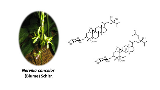
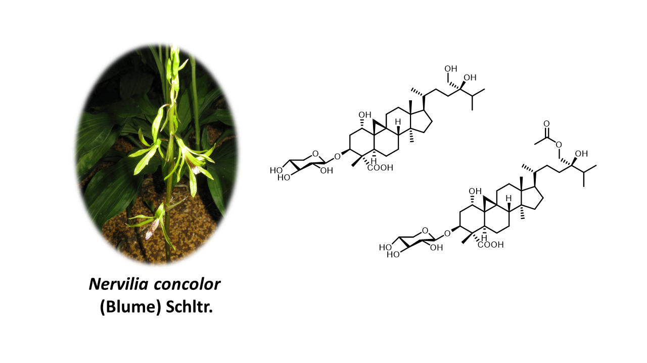
Nervisides I–J: Unconventional Side-Chain-Bearing Cycloartane Glycosides from Nervilia concolor

1
Graduate University of Science and Technology, Vietnam Academy of Science and Technology, 18 Hoang Quoc Viet, Cau Giay, Ha Noi 100803, Vietnam
2
Institute of Applied Sciences, Ho Chi Minh City University of Technology (HUTECH), 475 A Dien Bien Phu Street, Ward 25, Binh Thanh District, Ho Chi Minh City 758307, Viet Nam
3
Équipe “Pharmacognosie–Chimie des Substances Naturelles”, BioCIS, Univ. Paris-Sud, CNRS, Université Paris-Saclay, 5 Rue Jean-Baptiste Clément, 92290 Châtenay-Malabry, France
4
Institute of Chemical Technology, Vietnam Academy of Science and Technology, 01 Mac Dinh Chi, Ho Chi Minh City 758307, Vietnam
5
Center of Excellence in Natural Products Chemistry, Department of Chemistry, Faculty of Science, Chulalongkorn University, Pathumwan, Bangkok 10330, Thailand
6
Faculty of Science and Technology, Thammasat University Lampang Campus, Lampang 52190, Thailand
7
Mien Dong University of Technology, MUT, Dong Nai Provine 813847, Vietnam
8
Ho Chi Minh City University of Technology (HCMUT), Ho Chi Minh City 758307, Vietnam
9
Industrial University of Ho Chi Minh City, Ho Chi Minh City 758307, Vietnam
10
Faculty of Biotechnology, Nguyen Tat Thanh University, 300A Nguyen Tat Thanh Str., Dist. 4, Ho Chi Minh City 758307, Vietnam
11
Department for Management of Science and Technology Development, Ton Duc Thang University, Ho Chi Minh City 758307, Vietnam
12
Faculty of Applied Sciences, Ton Duc Thang University, Ho Chi Minh City 758307, Vietnam
*
Authors to whom correspondence should be addressed.
These authors contributed equally to this work.
Molecules 2019, 24(14), 2599; https://doi.org/10.3390/molecules24142599
Submission received: 4 June 2019 / Revised: 5 July 2019 / Accepted: 16 July 2019 / Published: 17 July 2019
(This article belongs to the Section Natural Products Chemistry)

Abstract

:
Two new cycloartane glycosides, nervisides I–J, were isolated from Nervilia concolor whole plants. Their structures were unambiguously established by interpretation of their HRESIMS and 1D and 2D NMR data. These cycloartanes comprised a stereogenic center at C-24, the R configuration of which was assigned based on DFT-NMR calculations and the subsequent DP4 probability score. These compounds were tested for cytotoxicity against K562 and MCF-7 tumor cell lines, revealing mild cytotoxic activity.

Graphical Abstract

1. Introduction

The terrestrial orchid genus Nervilia contains approximately 65 species which are mostly found in tropical and subtropical Africa, Asia, Australia, and the Southwest Pacific Islands [1]. The herbal plant Nervilia concolor (Blume) Schltr. (Orchidaceae) (syn. N. aragoana) is regionally distributed in Dak Lak, KonTum, An Giang, and Dong Nai provinces of Vietnam. This plant is widely used in traditional Chinese medicine for a variety of diseases, such as bronchitis, stomatitis, acute pneumonia, and laryngitis [2,3,4,5,6]. As of 2019, phytochemical studies undertaken on Nervilia species have led to the identification of ca. 60 compounds, mostly including flavonoids (>20), a dozen terpenes, and some sterols and amino acids. Nevertheless, as far as can be ascertained, N. concolor (syn. N. aragoana) has not been studied from a chemical perspective so far. This article describes the isolation and structural elucidation of two new cycloartane glycosides, namely, nervisides I–J (12), from this plant source. Consistent with previously reported structures of Nervilia species, these natural products reveal an unconventional side chain bearing an alkyl substituent at C-24, the absolute configuration of which has been difficult to assign, often leading to an undetermined configuration of this stereogenic center in structurally related compounds, including nervisides A–H isolated from Nervilia fordii [7,8], which were not defined as to this carbon. Such C-24 hydroxymethylated cycloartanes were also repeatedly reported to occur within Passiflora species [9,10,11]. In this study, besides benefitting from an extensive set of NMR and HRESIMS analyses, C-24 configuration was determined based on GIAO NMR shift calculation of the two possible epimers and the subsequent DP4 probability score, leading to the assignment of a 24R configuration with a quantifiable confidence of 99.2%.

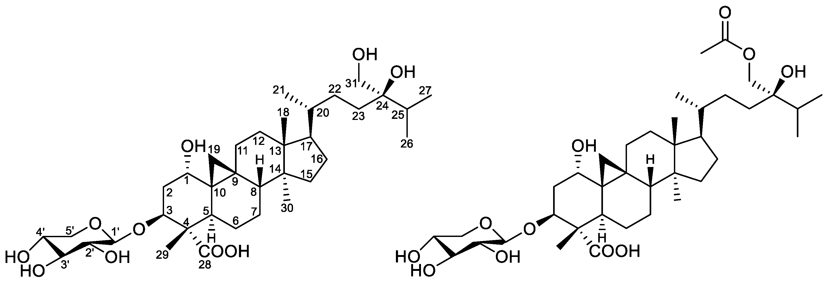
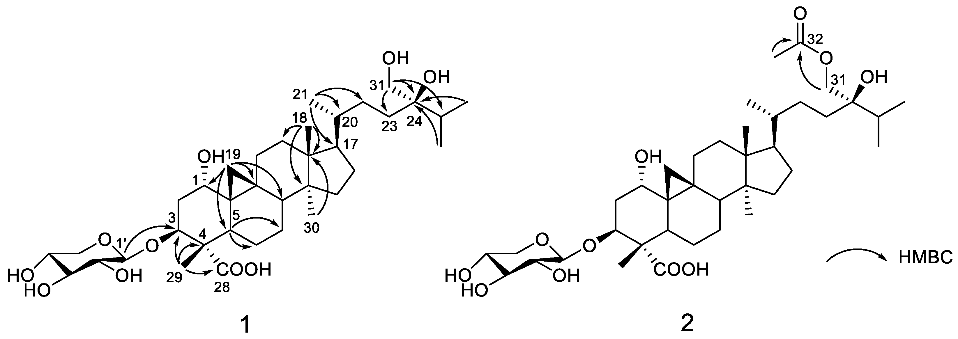
2. Results and Discussion

Compound 1 was obtained as a white gum. Its molecular formula was determined to be C36H60O10 based on the deprotonated molecular ion at m/z 651.4103 (calcd for C36H59O10, 651.4114). The 13C NMR spectrum, in conjunction with the HSQC spectrum, revealed 36 carbon signals, of which 31 could be assigned to a triterpenoid sapogenol core and 5 belonged to a monosaccharide unit. The 31 carbon resonances of the aglycone part consisted of 6 methyl carbons; 11 methylene carbons, 1 of which was oxygenated; 8 methine carbons, 3 of which bore oxygen functionalities; and 7 quaternary carbons comprising 1 carbonyl and an oxygenated carbon. Both the 1H upfield methylenic protons at δH 0.36 and 0.56 (each 1H, doublet with J = 4.0 Hz) and the six unsaturation degrees of the aglycone moiety led to define a carboxylic-acid-substituted cycloartane scaffold [12,13] (Figure 1 and Supplementary Materials). The thorough analysis of the COSY, HSQC, and HMBC spectra led to fully assign the 1H and 13C signals for compound 1 (Table 1). In the A-ring, a methyl and a carboxylic acid group could be assigned at C-4 (δC 53.0) based on HMBC correlations of both oxymethine H-3 (δH 4.40) and methyl H3-29 (δH 0.97) to carbons C-4 and C-28 (δC 178.5) as well as the HMBC cross-peak of H3-29 and C-3 (δC 78.8) (Figure 2). A hydroxy group could be anchored at C-1 based on HMBC cross-peaks of H2-19, H-3, and H-5 to C-1 and of H-1 (δH 3.34) to C-2 (δC 36.1), C-3 (δC 78.8), and C-5 (δC 36.6). A deshielded signal assigned to H-1 eq., partly overlapped with the water signal, resonated at 3.34 ppm as a broad singlet, diagnostic of the occurrence of a α-hydroxy group owing to the lack of a trans-diaxial coupling constant [14]. The H-3 signal appeared as a double doublet owing to diaxial (J = 12.0 Hz) and axial–equatorial coupling (J = 4.5 Hz) defining its axial orientation [9,15,16]. The glycosylation shift at C-3 (δC 78.8) of the aglycone indicated that the monosaccharide was linked at this specific position, as further backed up by the long-range heteronuclear correlation from H-1′ to C-3. The δ 2.5–4.5 ppm region of the 1H NMR spectrum validated the occurrence of a single saccharide, which could be directly identified as a xylopyranose unit based on the diagnostic triplet signal for the H-5′ α-proton at 2.97 ppm [14]. The COSY spectrum revealed the correlations of all the protons in the xylopyranose ring, and the magnitude of the vicinal coupling constant values were in excellent agreement with formerly reported J values for β-d-xylopyranose residues [17,18,19]. The NOE cross-peaks between H-1, H2-19, and H3-29 defined their β-orientation, thereby determining the α-position of the 4-COOH group (Figure 3). The canonical stereochemistry of the ABCD rings [20] was supported by the nearly identical 1H and 13C NMR data of 1 with cycloartane triterpenes [21], nervisides A–C [7], nervisides D–H [8], and cyclopassifloic acid series [10], as supported by the key NOE correlations outlined in Figure 3. This only left the relative configuration of the side chain pending assignment. The HMBC experiment revealed correlations between the oxygenated methylene protons resonating at δH 3.25 to C-23, C-24, and C-25 that defined the occurrence of a hydroxymethyl group at C-24 consistently with the side chain of formerly reported nervisides. Accordingly, nonconventional side chain triterpenes and sterols were repeatedly described from Nervilia species. [22,23]. Defining the absolute configuration of C-24 alkyl sterols and triterpenes is a vexing problem in NMR spectroscopy that has tentatively been overcome through tailored chromatographic procedures [24,25]. These difficulties result in some authors not defining C-24 absolute configuration on such related scaffolds including cyclotricuspidosides A–C [26] and nervisides A–H [7,8], even though derivatization-based NMR spectroscopy affords reliable outcomes as to this specific point [9,10,27]. To assign the absolute configuration at C-24, 13C NMR chemical shift calculations of simplified bicyclic models only including cycles C and D were performed using electronic structure methods of the lowest-energy conformer of both C-24 epimers. The spectral position of triterpene carbon side-chain bands does not vary over extensive sets of derivatives involving the central ring system [28]. In particular, chemical shifts of the side chain could be used to determine the absolute configuration of C-24 in several sterols owing to their chemical shifts being insensitive to structural changes remote from the asymmetric carbon [29]. The lowest energy conformation of the core ring system was more quickly located by seeding the potential energy surface scan with initial coordinates available in X-ray crystallographic CIF files associated with formerly reported cycloartanes [30,31,32]. Subsequent 13C NMR data comparison of the two possible epimers against the experimental dataset resulted in the prediction of the 24R configuration with a quantifiable confidence of 99.2%. Accordingly, compound 1, namely nerviside I, was identified as 3β-O-d-xylopyranosyl-1α,24R,31-trihydroxylcycloartan-28-oic acid.
Compound 2, obtained as a white amorphous solid, gave a molecular formula of C38H62O11 based on its negative-ion mode HRESIMS data, which displayed a [M–H] peak at m/z 693.4215 (calcd for C38H61O11, 693.4219). This hinted that 2 differed from 1 by a supplementary acetyl group. Accordingly, both the 1H and 13C NMR spectroscopic data were very similar between the two compounds, but the 1H NMR spectrum of 2 revealed one more methyl group resonating at δH 1.99 (3H, s), while the 13C NMR spectrum exhibited one more carbonyl carbon at δC 170.3. The occurence of an acetyl group was deduced from the HMBC correlation originating from the methyl proton at δH 1.99 to carbonyl carbon C-32 at δC 170.3. The thorough analysis of the 2D NMR spectra determined a similar cycloartane glycoside core as in compound 1 except for the acetylation of the hydroxy group at C-31, further backed up by the key HMBC correlation from H2-31 (3.88 and 3.84) to C-32. An identical C-24 R configuration was assigned based on the good agreement between the carbon signals due to C-23, C-24, and C-25 and biogenetic considerations. From the above evidence, compound 2, namely nerviside J, was established as 3β-O-d-xylopyranosyl-31-O-acetyl-1α,24R-dihydroxycycloartan-28-oic acid.
In this study, compounds 12 were evaluated for their cytotoxicity against K562 (chronic myelogenous leukemia) and MCF-7 (breast cancer) cell lines. Both compounds 1 and 2 exerted moderate activity against these two cancer cell lines, with respective IC50 values of 20.5 (±0.2) and 20.6 (±0.1) µg/mL for 1 and 40.1 (±0.6) and 90.5 (±3.5) µg/mL for 2.

3. Materials and Methods

3.1. General

NMR spectra were performed on a Bruker AM500 FT-NMR spectrometer (500 MHz for 1H NMR and 125 MHz for 13C NMR). The ESI-HRMS data were generated with a Bruker MicroTOF-QII spectrometer (Bremen, Germany). Open-column chromatography was performed on silica gel 40–63 µm phase (Merck, Darmstadt, Germany) and reversed-phase C18 (Merck, Darmstadt, Germany). TLC analyses were carried out on precoated silica gel 60 F254 (Merck, Darmstadt, Germany), and spots were visualized by spraying the plates with 10% H2SO4 solution followed by heating.

3.2. Plant Material

N. concolor whole plants were collected in the Cu M’gar district, Dak Lak province, from August to November 2017 and authenticated by Dr. Cong-Luan Tran, Research Center of Ginseng and Medicinal Materials of Ho Chi Minh City National Institute of Medicinal Materials. A voucher specimen (no. NA-0621) was deposited in the Bioactive Compounds Laboratory, Institute of Chemical Technology.

3.3. Extraction and Isolation

The dried whole plants (4.0 kg) were milled prior to being extracted with 96% EtOH three times (3 × 30 L, each 8 h) at room temperature. The filtered solution was concentrated in vacuo to afford a crude extract (280 g). This dried residue was successively re-extracted using solvents of increasing polarities: n-hexane (H, 110 g), CHCl3 (C, 25 g), EtOAc (EA, 90 g), and H2O (W, 45 g). Extract EA was subjected to silica gel column chromatography and eluted with a chloroform/MeOH solvent system (stepwise, 1:0 to 1:0) to afford seven fractions: E1–E7. Fraction E5 (20 g) was selected for further purification using column chromatography based on a CHCl3–MeOH solvent system gradient (20:1 to 1:1) to yield five subfractions (E5.1–5.5). Subfraction E5.1 (1.1 g) was subjected to silica gel column chromatography using an isocratic mobile phase consisting of a CHCl3/MeOH/H2O solvent system (10:1:0.1) to afford 1 (10 mg) and 2 (14 mg).
Nerviside I (1). White gum. 1H- and 13C NMR see Table 1; HRESIMS m/z 651.4103 [M–H] (calcd for C36H59O10, 651.4114).
Nerviside J (2). White amorphous solid 1H- and 13C NMR see Table 1; HRESIMS m/z 693.4215 [M–H] (calcd for C38H61O11, 693.4219).

3.4. Computational Chemistry

Truncated models of 1 (without the sugar moiety) and its epimer were assembled and the cycloartane skeleton was arranged in both with a conformation identical to that found in [30,31,32]. A conformation search was then performed on the overall structure using the basin hopping method [33] with the MMFF94 force field [34] as implemented in the scan program from the Tinker v8.6.1 software package [35,36,37]. Coordinates of the lowest energy minimum for both epimers were then further truncated by keeping only rings C and D and optimized at the B3LYP/6-31G(d) level [38,39,40] using the Gaussian 16 software package [41]. Vibrational analysis within the harmonic approximation was performed at the same level of theory upon geometrical optimization convergence prior to characterizing local minima by the absence of imaginary frequency. Chemical shifts were deduced from NMR shielding tensors calculated using the GIAO method [42,43] and corrected against values for the corresponding nucleus in TMS, both at the same level of theory. DP4 probability values were calculated using online implementation available from http://www-jmg.ch.cam.ac.uk/tools/nmr/DP4/ [44].

3.5. Biological Assays

Cytotoxic activities of the formerly unreported metabolites were evaluated against the MCF-7 (breast cancer) and K562 (chronic myelogenous leukemia) tumor cell lines. These two cell lines were cultured in RPMI 1640 medium or in DMEM medium, respectively; supplemented with 10% fetal bovine serum (FBS), 100 IU/mL penicillin, and 100 μg/mL streptomycin; and maintained at 37 °C and 5% CO2 with 95% humidity. Viable cells were counted and inoculated in a 96-well plate with a density of 104 cells/100 μL/well for MCF-7 and 105 cells/100 μL/well for K562. After 24 h, the cells were treated with the compounds and doxorubicin (positive control) diluted in culture media at 100, 50, 25, 12.5, 6.25, 3.125, and 0 µg/mL concentration containing 1%, 0.5%, 0.25%, 0.125%, 0.0625%, 0.03125%, and 0% dimethyl sulfoxide (DMSO), respectively. DMSO in culture media was used as a negative control. In addition, culture medium without cells was used as a blank. All experiments were done in triplicate. The plates were incubated in 5% CO2 with 95% humidity at 37 °C for 72 h. Ten microliters of 3-(4,5-dimethylthiazol-2-yl)-2,5-diphenyltetrazolium bromide (MTT, 5 mg/mL stock solution) were added to each well and incubated in 37 °C in 5% CO2 for 3.5 h. Seventy microliters of detergent reagent (10% SDS) were added to each well and the plate was maintained in 37 °C for 16 h. The optical density of each well was read by using a scanning multiwall spectrophotometer (Sunrise) at a wavelength of 595 nm. Cell survival was measured as the percentage absorbance compared to the negative control (DMSO-treated cells).

4. Conclusions

The ethnopharmacological relevance of N. concolor paved the way for the currently reported phytochemical investigation that resulted in the isolation and structural elucidation of two new C-24 alkyl-substituted cycloartane glycosides, namely, nervisides I–J. While unconventional side-chain-bearing triterpenes and sterols have regularly been reported from Nervilia species, these compounds are the first nervisides having a defined C-24 absolute configuration, deduced from a DP4-based computational chemistry approach.

Supplementary Materials

The following are available online. 1H, 13C NMR, HMBC, and HRMS spectra for 12, DFT calculations results for 24R and 24S epimers of 1 and 13C NMR spectroscopic data for 1, Atomic coordinates of nerviside I (1) and its 24S epimer.

Author Contributions

D.-T.M., C.-L.T. and T.-H.D. conceived and designed the experiments; T.-N.-M.T. and T.-P.N. performed the isolation work; T.-H.D., J.S., V.-K.N. and P.L.P. analyzed NMR data; M.A.B., G.B. and P.L.P. designed and performed DFT-NMR calculations; H.-H.N., P.-V.D. and N.-M.-A.T. performed the biological activity; and D.-T.M., P.L.P. and T.-H.D. wrote the manuscript. All the authors reviewed and validated the present manuscript prior to its being submitted.

Funding

This research received no external funding.

Acknowledgments

Leo Gohrs from Alionis is thanked for the donation of computing hardware.

Conflicts of Interest

The authors declare no conflict of interests.

References

  1. Gale, S.; Yukawa, T.; Kuroiwa, N. Studies in Asian Nervilia (Orchidaceae) I: Neotypification and Circumscription of N. nipponica in Japan. Kew Bull. 2007, 62, 85–94. [Google Scholar]
  2. Zhou, G.-X.; Lu, C.-L.; Wang, H.-S.; Yao, X.-S. An acetyl flavonol from Nervilia fordii (Hance) Schltr. J. Asian Nat. Prod. Res. 2009, 11, 498–502. [Google Scholar] [CrossRef] [PubMed]
  3. Chen, J.-M.; Wei, L.-B.; Lu, C.-L.; Zhou, G.-X. A flavonoid 8- C -glycoside and a triterpenoid cinnamate from Nervilia fordii. J. Asian Nat. Prod. Res. 2013, 15, 1088–1093. [Google Scholar] [CrossRef] [PubMed]
  4. Tian, L.-W.; Pei, Y.; Zhang, Y.-J.; Wang, Y.-F.; Yang, C.-R. 7-O-Methylkaempferol and quercetin Glycosides from the Whole Plant of Nervilia fordii. J. Nat. Prod. 2009, 72, 1057–1060. [Google Scholar] [CrossRef] [PubMed]
  5. Zhang, L.; Zhao, Z.-X.; Lin, C.-Z.; Zhu, C.-C.; Gao, L. Three new flavonol glycosides from Nervilia fordii. Phytochem. Lett. 2012, 5, 104–107. [Google Scholar] [CrossRef]
  6. Qiu, L.; Jiao, Y.; Xie, J.-Z.; Huang, G.-K.; Qiu, S.-L.; Miao, J.-H.; Yao, X.-S. Five new flavonoid glycosides from Nervilia fordii. J. Asian Nat. Prod. Res. 2013, 15, 589–599. [Google Scholar] [CrossRef] [PubMed]
  7. Wei, L.-B.; Chen, J.-M.; Ye, W.-C.; Yao, X.-S.; Zhou, G.-X. Three new cycloartane glycosides from Nervilia fordii. J. Asian Nat. Prod. Res. 2012, 14, 521–527. [Google Scholar] [CrossRef] [PubMed]
  8. Wei, L.-B.; Yuan, H.-E.; Chen, J.-M.; Wang, Q.-Q.; Wang, H.; Ye, W.-C.; Zhou, G.-X. Five new Cycloartane-type triterpenoid saponins from Nervilia fordii. Helv. Chim. Acta 2013, 96, 150–157. [Google Scholar] [CrossRef]
  9. Yoshikawa, K.; Katsuta, S.; Mizumori, J.; Arihara, S. New cycloartane triterpenoids from Passiflora edulis. J. Nat. Prod. 2000, 63, 1377–1380. [Google Scholar] [CrossRef]
  10. Yoshikawa, K.; Katsuta, S.; Mizumori, J.; Arihara, S. Four cycloartane triterpenoids and six related saponins from Passiflora edulis. J. Nat. Prod. 2000, 63, 1229–1234. [Google Scholar] [CrossRef]
  11. Xu, F.-Q.; Wang, N.; Fan, W.-W.; Zi, C.-T.; Zhao, H.-S.; Hu, J.-M.; Zhou, J. Protective effects of cycloartane triterpenoids from Passiflora edulis Sims against glutamate-induced neurotoxicity in PC12 cell. Fitoterapia 2016, 115, 122–127. [Google Scholar] [CrossRef] [PubMed]
  12. Della Greca, M.; Fiorention, A.; Monaco, P.; Previtera, L. Cycloartane triterpenes from Juncus effusus. Phytochemistry 1994, 35, 1017–1022. [Google Scholar] [CrossRef]
  13. Linnek, J.; Mitaine-Offer, A.-C.; Miyamoto, T.; Tanaka, C.; Paululat, T.; Avunduk, S.; Alankuş-Çalişkan, Ö.; Lacaille-Dubois, M.-A. Cycloartane glycosides from three species of Astragalus (Fabaceae). Helv. Chim. Acta 2011, 94, 230–237. [Google Scholar] [CrossRef]
  14. Imai, A.; Lankin, D.C.; Nikolić, D.; Ahn, S.; van Breemen, R.B.; Farnsworth, N.R.; McAlpine, J.B.; Chen, S.-N.; Pauli, G.F. Cycloartane triterpenes from the aerial parts of Actaea racemosa. J. Nat. Prod. 2016, 79, 541–554. [Google Scholar] [CrossRef] [PubMed]
  15. Banskota, A.H.; Tezuka, Y.; Tran, K.Q.; Tanaka, K.; Saiki, I.; Kadota, S. Thirteen novel cycloartane-type triterpenes from Combretum quadrangulare. J. Nat. Prod. 2000, 63, 57–64. [Google Scholar] [CrossRef] [PubMed]
  16. Gutierrez-Lugo, M.-T.; Singh, M.P.; Maiese, W.M.; Timmermann, B.N. New antimicrobial cycloartane triterpenes from Acalypha communis. J. Nat. Prod. 2002, 65, 872–875. [Google Scholar] [CrossRef] [PubMed]
  17. Ahmed, A.A. A diterpene xyloside from Conyza steudellii. Phytochemistry 1991, 30, 611–612. [Google Scholar] [CrossRef]
  18. Piacente, S.; Balderrama, L.; Tommasi, N.D.; Morales, L.; Vargas, L.; Pizza, C. Anadanthoside: A flavanol-3-O-b-d-xylopyranoside from Anadenanthera macrocarpa. Phytochemistry 1999, 51, 709–711. [Google Scholar] [CrossRef]
  19. Ali, Z.; Khan, S.; Khan, I. Phytochemical study of Actaea rubra and biological screenings of isolates. Planta Med. 2006, 72, 1350–1352. [Google Scholar] [CrossRef] [PubMed]
  20. Thimmappa, R.; Geisler, K.; Louveau, T.; O’Maille, P.; Osbourn, A. Triterpene biosynthesis in plants. Annu. Rev. Plant Biol. 2014, 65, 225–257. [Google Scholar] [CrossRef]
  21. Escobedo-Martínez, C.; Concepción Lozada, M.; Hernández-Ortega, S.; Villarreal, M.L.; Gnecco, D.; Enríquez, R.G.; Reynolds, W. 1H and 13C NMR characterization of new cycloartane triterpenes from Mangifera indica. Magn. Reson. Chem. 2012, 50, 52–57. [Google Scholar] [CrossRef] [PubMed]
  22. Kikuchi, T.; Kadota, S.; Suehara, H.; Namba, T. Occurrence of non-conventional side chain sterols in an orchidaceous plant, Nervilia purpurea Schlechter and structure of nervisterol. Chem. Pharm. Bull. 1982, 30, 370–373. [Google Scholar] [CrossRef]
  23. Kadota, S.; Shima, T.; Kikuchi, T. Studies on the constituents of orchidaceous plants. VII. The C-24 stereochemistry of cyclohomonervilol and 24-isopropenylcholesterol non-conventional side chain triterpene and sterol from Nervilia purpurea Schlechter. Chem. Pharm. Bull. 1987, 35, 11. [Google Scholar] [CrossRef]
  24. Thompson, M.J.; Patterson, G.W.; Dutky, S.R.; Svoboda, J.A.; Kaplanis, J.N. Techniques for the isolation and identification of steroids in insects and algae. Lipids 1980, 15, 719–733. [Google Scholar] [CrossRef] [PubMed]
  25. Ikekawa, N.; Fujimoto, Y.; Kadota, S.; Kikuchi, T. Effective separation of sterol C-24 epimers. J. Chromatogr. A 1989, 468, 91–98. [Google Scholar] [CrossRef]
  26. Kasai, R.; Sasaki, A.; Hashimoto, T.; Kaneko, T.; Ohtani, K.; Yamasaki, K. Cycloartane glycosides from Trichosanthes tricuspidata. Phytochemistry 1999, 51, 803–808. [Google Scholar] [CrossRef]
  27. Wang, W.; Jang, H.; Hong, J.; Lee, C.-O.; Im, K.S.; Bae, S.-J.; Jung, J.H. Additional cytotoxic sterols and saponins from the starfish Certonardoa semiregularis. J. Nat. Prod. 2004, 67, 1654–1660. [Google Scholar] [CrossRef]
  28. Knight, S.A. Carbon-13 NMR spectra of some tetra- and pentacyclic triterpenoids. Org. Magn. Reson. 1974, 6, 603–611. [Google Scholar] [CrossRef]
  29. Wright, J.L.C.; McInnes, A.G.; Shimizu, S.; Smith, D.G.; Walter, J.A.; Idler, D.; Khalil, W. Identification of C-24 alkyl epimers of marine sterols by 13C nuclear magnetic resonance spectroscopy. Can. J. Chem. 1978, 56, 1898–1903. [Google Scholar]
  30. Herz, W.; Watanabe, K.; Kulanthaivel, P.; Blount, J.F. Cycloartanes from Lindheimera texana. Phytochemistry 1985, 24, 2645–2654. [Google Scholar] [CrossRef]
  31. Ju, J.; Liu, D.; Lin, G.; Zhang, Y.; Yang, J.; Lu, Y.; Gong, N.; Zheng, Q. Beesiosides G, H, and J−N, seven new cycloartane triterpene glycosides from Beesia calthifolia. J. Nat. Prod. 2002, 65, 147–152. [Google Scholar] [CrossRef] [PubMed]
  32. Fu, X.; Li, X.-C.; Smillie, T.J.; Carvalho, P.; Mabusela, W.; Syce, J.; Johnson, Q.; Folk, W.; Avery, M.A.; Khan, I.A. Cycloartane glycosides from Sutherlandia frutescens. J. Nat. Prod. 2008, 71, 1749–1753. [Google Scholar] [CrossRef] [PubMed]
  33. Kolossváry, I.; Guida, W.C. Low-mode conformational search elucidated: Application to C39H80 and flexible docking of 9-deazaguanine inhibitors into PNP. J. Comput. Chem. 1999, 20, 14. [Google Scholar] [CrossRef]
  34. Halgren, T.A. Merck molecular force field. I. Basis, form, scope, parameterization, and performance of MMFF94. J. Comput. Chem. 1995, 17, 490–519. [Google Scholar] [CrossRef]
  35. Kundrot, C.E.; Ponder, J.W.; Richards, F.M. Algorithms for calculating excluded volume and its derivatives as a function of molecular conformation and their use in energy minimization. J. Comput. Chem. 1991, 12, 402–409. [Google Scholar] [CrossRef] [Green Version]
  36. Dudek, M.J.; Ponder, J.W. Accurate modeling of the intramolecular electrostatic energy of proteins. J. Comput. Chem. 1995, 16, 791–816. [Google Scholar] [CrossRef] [Green Version]
  37. Kong, Y.; Ponder, J.W. Calculation of the reaction field due to off-center point multipoles. J. Chem. Phys. 1997, 107, 481–492. [Google Scholar] [CrossRef] [Green Version]
  38. Hehre, W.J.; Radom, L.; Schleyer, P.V.R.; Pople, J.A. Ab initio Molecular Orbital Theory; John Wiley & Sons, Inc.: New York, NY, USA, 1986. [Google Scholar]
  39. Lee, C.; Yang, W.; Parr, R.G. Development of the Colle-Salvetti correlation-energy formula into a functional of the electron density. Phys. Rev. B 1988, 37, 785–789. [Google Scholar] [CrossRef] [Green Version]
  40. Becke, A.D. Becke’s three parameter hybrid method using the LYP correlation functional. J. Chem. Phys. 1993, 98, 5648–5652. [Google Scholar] [CrossRef]
  41. Frisch, M.J.; Trucks, H.B.; Schlegel, G.W.; Scuseria, G.E.; Robb, M.A.; Cheeseman, J.R.; Scalmani, G.; Barone, V.; Petersson, G.A.; Nakatsuji, H.; et al. Gaussian 16 Revision B.01; Gaussian Inc.: Wallingford, CT, USA, 2016. [Google Scholar]
  42. Ditchfield, R. Self-consistent perturbation theory of diamagnetism: I. A gauge-invariant LCAO method for NMR chemical shifts. Mol. Phys. 1974, 27, 789–807. [Google Scholar] [CrossRef]
  43. Wolinski, K.; Hinton, J.F.; Pulay, P. Efficient implementation of the gauge-independent atomic orbital method for NMR chemical shift calculations. J. Am. Chem. Soc. 1990, 112, 8251–8260. [Google Scholar] [CrossRef]
  44. Smith, S.G.; Goodman, J.M. Assigning stereochemistry to single diastereoisomers by GIAO NMR calculation: The DP4 probability. J. Am. Chem. Soc. 2010, 132, 12946–12959. [Google Scholar] [CrossRef] [PubMed]
Sample Availability: Sample of compounds 1 and 2 are not available from the authors.
Figure 1. Structures of compounds 1–2.
Molecules 24 02599 g001
Figure 2. Key HMBC correlations of 1–2.
Molecules 24 02599 g002
Figure 3. Key NOE correlations of 1.
Molecules 24 02599 g003
Table 1. 13C and 1H NMR spectroscopic data (125/500 MHz) for 12 in dimethyl sulfoxide (DMSO)-d6 (δ in ppm).
12
δCδH (J, Hz)δCδH (J, Hz)
170.83.34, 1H, br s 70.73.34, 1H, br s
236.11.84, 1H, m
1.60, 1H, m
36.01.84, 1H, m
1.59, 1H, m
378.84.40, 1H, dd, 12.0, 4.578.84.40, 1H, dd, 12.0, 4.5
453.0_52.9_
536.62.41, 1H, dd, 13.0, 4.536.52.42,1H, dd, 12.5, 3.5
622.01.14, 1H, m
0.84, 1H, m
21.91.16, 1H, m
0.83, 1H, m
725.11.02, 2H, m25.01.02, 2H, m
847.51.43, 1H, m47.31.46, 1H, m
920.1-19.9-
1028.9-28.7
1125.02.27, 1H, m
1.17, 1H, m
24.92.27, 1H, m
1.19, 1H, m
1232.61.55–1.57, 2H, m32.81.56–1.60, 2H, m
1344.8-44.7-
1448.7-48.6-
1535.31.21–1.22, 2H, m35.21.26–1.29, 2H, m
1627.81.84, 1H, m
1.22, 1H, m
27.61.84, 1H, m
1.23, 1H, m
1751.91.54, 1H, m51.61.57, 1H, m
1818.00.91, 3H, s17.80.90, 3H, m
1928.80.36, 1H, d, 4.0
0.56, 1H, d, 4.0
28.70.35, 1H, d, 4.0
0.57, 1H, d, 4.0
2036.41.26, 1H, m36.01.29, 1H, m
2118.30.82, 3H, s18.20.85, 3H, s
2229.01.43, 1H, m
0.92, 1H, m
29.01.45, 1H, m
1.00, 1H, m
2330.81.43, 1H, m
1.23, 1H, m
30.91.45, 1H, m
1.30, 1H, m
2474.6-73.4_
2532.31.70, 1H, m32.41.72, 1H, m
2617.10.80–0.82, 3H, m16.80.80–0.85, 3H, m
2717.10.80–0.82, 3H, m16.70.80–0.85, 3H, m
28178.5 178.6
299.50.97, 3H, s9.50.97, 3H, s
3019.10.89, 3H, s19.00.91, 3H, s
3164.73.25, 2H, m66.93.89, 1H, d, 11.0
3.85, 2H, d, 11.0
1’104.14.14, 1H, d, 7.5104.04.15, 1H, d, 7.5
2’73.72.88, 1H, dd, 9.0, 7.573.52.88, 1H, t, 8.5
3’76.43.05, 1H, t, 9.076.33.06, 1H, t, 8.5
4’69.63.23, 1H, m69.53.24, 1H, m
5’65.73.66, 1H, dd, 11.5, 5.065.63.66, 1H, dd, 11.0, 5.0
2.97, 1H, t, 11.5 2.97, 1H, t, 11.0
OAc 20.71.99, 3H, s
170.3

