Article

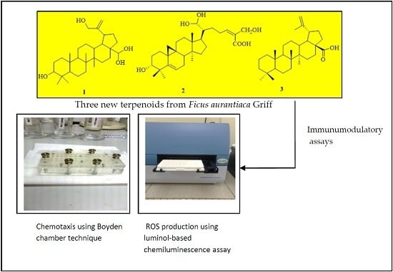
Isolation of Terpenoids from the Stem of Ficus aurantiaca Griff and their Effects on Reactive Oxygen Species Production and Chemotactic Activity of Neutrophils

Drug and Herbal Research Centre, Faculty of Pharmacy, Universiti Kebangsaan Malaysia (UKM), Jalan Raja Muda Abdul Aziz, Kuala Lumpur 50300, Malaysia
*
Author to whom correspondence should be addressed.
Molecules 2016, 21(1), 9; https://doi.org/10.3390/molecules21010009
Submission received: 9 November 2015 / Revised: 9 December 2015 / Accepted: 14 December 2015 / Published: 5 January 2016
(This article belongs to the Section Natural Products Chemistry)

Abstract

:
Three new triterpenoids; namely 28,28,30-trihydroxylupeol (1); 3,21,21,26-tetrahydroxy-lanostanoic acid (2) and dehydroxybetulinic acid (3) and seven known compounds; i.e., taraxerone (4); taraxerol (5); ethyl palmitate (6); herniarin (7); stigmasterol (8); ursolic acid (9) and acetyl ursolic acid (10) were isolated from the stem of Ficus aurantiaca Griff. The structures of the compounds were established by spectroscopic techniques. The compounds were evaluated for their inhibitory effects on polymorphonuclear leukocyte (PMN) chemotaxis by using the Boyden chamber technique and on human whole blood and neutrophil reactive oxygen species (ROS) production by using a luminol-based chemiluminescence assay. Among the compounds tested, compounds 14, 6 and 9 exhibited strong inhibition of PMN migration towards the chemoattractant N-formyl-methionyl-leucyl-phenylalanine (fMLP) with IC50 values of 6.8; 2.8; 2.5; 4.1; 3.7 and 3.6 μM, respectively, comparable to that of the positive control ibuprofen (6.7 μM). Compounds 24, 6, 7 and 9 exhibited strong inhibition of ROS production of PMNs with IC50 values of 0.9; 0.9; 1.3; 1.1; 0.5 and 0.8 μM, respectively, which were lower than that of aspirin (9.4 μM). The bioactive compounds might be potential lead molecules for the development of new immunomodulatory agents to modulate the innate immune response of phagocytes.

Graphical Abstract

1. Introduction

Medicinal plant products have long been used in traditional medicines for the treatment of many immunological disorders. Their therapeutic effects may be due to their effects on the immune system [1]. Many herbs such as Andrographis paniculata, Allium sativum, Trigonella foenum graecum, Pouteria cambodiana, Centella asiatica, Asparagus racemosus, Baliospermum montanum, Curcuma longa, Panax ginseng, Phyllanthus debilis, Tinospora cordifolia, and Picrorhiza scrophulariiflora have been reported to be able to modulate the immune system, including both adaptive and innate arms of the immune responses, exhibiting either immunostimulant or immunosuppressive effects [2,3,4]. Immunomodulation by these plant extracts and their active components provide new sources of lead molecules for development of natural immunomodulators for a variety of immunologic diseases. Assessment of the immunological effects of compounds is based on their selective activities on the different components of the immune system. In recent years, there was an increased interest in the search for natural immunomodulators from medicinal plants to substitute conventional therapy as they are considered to possess fewer side effects [5].
Ficus (family: Moraceae) is one of the largest genera of angiosperms, with more than 800 species of trees, shrubs, hemi-epiphytes and climbers in the tropics and sub-tropics worldwide [6]. Due to its high economic and nutritional values the genus is a vital hereditary source and also an important component of biodiversity of the rainforest ecosystem. It is also a good source of food for fruit eating animals in tropical area [7]. Chemical investigations of various Ficus species have shown the presence of flavonoids, coumarins, alkaloids, steroids, triterpenoids, simple phenols and salicylic acids from F. benghalensis, F. carica, F. hirta, F. hispida, F. microcarpa, F. nymphaeifolia, F. ruficaulis and F. septica [8,9]. Some of these plants exhibited a wide range of biological activities such as anti-inflammatory [10,11,12], antioxidant, hypolipidemic and hypoglycemic activities [13]. Some Ficus species are well known in Asia as medicinal plants and are widely used in folk medicines for the treatment of flu, malaria, tonsillitis, bronchitis and rheumatism [14]. One of the most biologically active species of Ficus is F. carica which has been reported to have some 21 traditional and current uses in various ethnopharmacological practices [15].
Ficus aurantiaca Griff is an evergreen tree that can grow up to 9 m. It is widespread in the lowland forests at Kelantan, Terengganu, Perak, Pahang, Selangor, Melaka and Johor in Malaysia, where it is locally known as “tengkuk biawak” or “akar tengkuk biawak hitam”. Traditionally, the plant parts have been used for the treatment of various ailments including headache, wound and toothache [16]. There is little investigation on the chemical composition and biological properties of F. aurantiaca. Our preliminary study on the stem extract of this plant had demonstrated that it inhibited in vitro chemotactic migration as well as suppressed the release of reactive oxygen species by granulocytes (unpublished data). The aims of the present study were to isolate and identify the constituents of the stem of F. aurantiaca and evaluate their effects on reactive oxygen species (ROS) production and chemotactic activity of neutrophils.

2. Results and Discussion

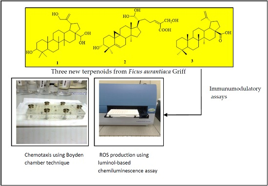
2.1. Characterization of the Isolated Compounds

Successive separations of n-hexane, ethyl acetate and methanol extracts using silica gel chromatography afforded three new triterpenoids, 28,28,30-trihydroxylupeol (1), 3,21,21,26-tetrahydroxylanostanoic acid (2) and dehydroxybetulinic acid (3) along with five known triterpenoids, taraxerone (4), taraxerol (5), stigmasterol (8), ursolic acid (9), acetyl ursolic acid (10), one sesquiterpenoid, herniarin (7) and one diterpenoid, ethyl palmitate (6). The structures of the new compounds are shown in Figure 1. The structures of the known compounds were elucidated by the combination of ESIMS, 1H- and 13C-NMR spectral data and comparison of their spectral data with literature values [17,18,19,20,21,22,23].
Figure 1. New terpenoids from F. aurantica.
Molecules 21 00009 g001
Compound 1 (20 mg) was isolated as a white powder with a melting point range of 222–224 °C. The IR spectrum showed a band at 3311 cm−1 due to the presence of hydroxyl groups (-OH). C-H stretching vibrations were observed at 2920 cm−1 while the methyl bending vibration was at 1411 cm−1. The HRESIMS displayed a molecular ion at m/z 474.1465 [M]+, corresponding to a molecular formula of C30H50O4 (M = 474.1465). The 1H-NMR spectrum exhibited singlets indicating the presence of five methyl groups, deshielded methine protons at δH 2.04 (H-18) and 2.38 (H-19) and two olefinic protons at δH 4.69 and 4.57. The corresponding 13C-NMR spectrum displayed 30 carbon signals, including five methyls, twelve methylenes, seven methines and six quaternary carbons. Two methine carbons at δC 80.1 (C-3) and 95.1 (C-28) were typical for C-OH and two sp2 carbons at δC 150.9 (C-20) and 109.3 (C-29), indicated the presence of one double bond. Another methylene carbon at δC 69.2 (C-30) was characteristic for hydroxyl (C-OH). The 1H- and 13C-NMR data of 1 were very similar to those reported for lupeol [24], the main difference being the presence of four hydroxyl groups instead of only one. Two methyl group protons at C-28, were substituted by two hydroxyl groups, and in the C-30 methyl group, a proton was also ubstituted by a hydroxyl group. The positions of the hydroxyl groups were established by the HMQC correlation of the hydroxyl (-OH) protons, δH 2.37, 2.04, 2.29 and 2.39 with the δC 80.1 (C-3) 69.2 (C-30) and 95.1 (C-28) carbon signals, respectively.
The connectivity between protons and carbons established by the HMQC and HMBC spectra indicated that the two olefinic protons were attached to carbon at δC 109.3 (C-29), the two adjacent methyl protons resonating as a singlet at δH 0.79 (s, 3H, H-23) and 0.83 (s, 3H, H-24) attached to a quaternary carbon at δC 39.6 (C-4) were attributed to methine carbon δC 55.6 (C-5) and hydroxyl methine carbon δC 80.1 (C-3) by 3J correlation, respectively. Meanwhile another methyl proton at δH 0.94 (s, 3H, H-25) displayed 3J correlations with methylene carbon at δC 38.1 (C-1) and methine carbon at δC 50.3 (C-9). Another two groups of methyl protons were observed at δH 1.01 (s, 3H, H-26) and 1.07 (s, 3H, H-27), showed 3J correlation with methylene carbon at δC 34.1(C-7) and δC 27.4 (C-15), respectively. One hydroxyl methine proton at δH 3.39 (dd, 1H, J = 10.8, 4.8 Hz, H-3) showed a 2J correlation with methylene carbon δC (C-2). Another three methine protons were visible at δH 1.34 (m, 1H, H-13), 2.04 (m, 1H, H-18) and 2.38 (d, 1H, J = 7.2 Hz, H-19) whereas at δH 1.34 (m, 1H, H-13) showed 2J correlation with methylene carbon at δC 25.1 (C-12) and methine carbon at δC 47.9 (C-18) and proton at δH 2.04 (m, 1H, H-18) and 2.38 (d, 1H, J = 7.2 Hz, H-19) showed 2J correlation with methine carbon at δC 48.2 (C-19) and quaternary carbon at δC 150.9 (C-20), respectively. Based on these 1H and 13C-NMR spectral data of 1, summarized in Table 1, and the comparison with the literature values of lupeol [24], 1 was identified as 28,28,30-trihydroxylupeol. Selected C-H correlations in 1 established by HMBC are shown in Figure 2. In the genus Ficus, lupeol was previously isolated from F. carica [25], F. micricarpa [26] and F. benjamina [9], while lupeol acetate was isolated from F. microcarpa [26]. This is the first report of a hydroxy derivative of lupeol in Ficus species.
Table 1. 1H- and 13C-NMR spectroscopic data (600 MHz, CDCl3) for triterpenoids 13.
PositionsCompound 1Compound 2Compound 3
δCδH (J in Hz)δCδH (J in Hz)δCδH (J in Hz)
138.11.28 (d, 4H, J = 6.6)29.80.94, (m, 2H)36.11.41 (m, 1H)
1.31 (m,1H)
227.31.68 (d, 4H, J = 3.0)25.71.01 (m, 2H)28.21.42 (m, 1H)
1.32 (m, 1H)
380.13.39 (dd, 1H, J = 10.8, 4.8)80.13.44 (1H, t, J = 18.0), 4.68 (OH)36.71.42 (m, 1H)
1.33 (m, 1H)
439.6-43.0-30.8-
555.61.32 (d, 1H, J = 7.2)151.0-59.61.34 (m, 1H)
617.71.28 (m, 4H, J = 6.6)109.34.79 (t, 1H, J = 6.0)18.61.65 (m, 2H)
734.11.28 (m, 4H)31.91.77 (m, 2H)37.41.46 (m, 1H)
840.9-50.31.48 (m, 1H)40.1-
950.30.88 (m, 10H)48.2-51.91.56 (m, 1H)
1037.3-39.9-36.7-
1121.01.61 (m, 4H)23.71.34 (m, 2H)20.61.54 (m, 1H)
1.29 (m, 1H)
1225.11.68 (m, 4H)22.71.31 (m, 2H)25.21.55 (m, 1H)
1.29 (m, 1H)
1337.91.34 (m, 1H)42.8-37.71.24 (m, 1H)
1442.8-40.9-42.9-
1527.41.26 (m, 4H)34.51.57 (m, 2H)39.71.49 (m, 1H)
1.00 (m, 1H)
1635.51.26 (m, 4H)29.41.63 (m, 2H)40.41.86 (m, 1H)
1.25 (m,1H)
1743.0-55.61.48 (m,1H)59.6-
1847.92.04 (m, 1H)27.41.03 (s, 2H)46.51.68 (m, 1H)
1948.22.38 (d, 1H, J = 7.2)17.71.04 s, 1H37.22.09 (m, 1H)
20150.9-35.31.68 (m, 1H)150.9-
2129.71.88–1.94 (m, 2H)95.14.80 (m, 1H)29.21.60 (m, 6H)
2239.91.34 (m, 4H)35.51.18 (m, 2H)23.01.75 (t, 2H, J = 5.4)
2314.50.79 (s, 3H)25.01.35 (m, 2H)22.10.83 (s, 6H)
2418.00.83 (s, 3H)150.94.57 (m, 1H)16.60.90 (s, 3H)
2515.90.94 (s, 3H)109.3-15.40.76 (s, 3H)
2616.11.01(s, 3H)69.24.03 (d, 1H, J = 12)17.60.89 (s, 3H)
2714.11.07 (s, 3H)173.8-13.90.73 (s, 3H)
2895.14.79 (t, 1H, J = 6 Hz)14.10.80 (s, 3H)183.5-
29109.34.69 (br s, 1H)15.90.85 (s, 1H)116.35.32 (brs, 1H)
4.57 (br s, 1H)
3069.25.14 (m, 1H, H-30)16.50.92 (q, 3H)20.11.10 (s, 3H)
5.19 (m, 1H, H-30)
Figure 2. Selected HMBC correlations (C→H) of compounds 13.
Molecules 21 00009 g002
Compound 2 was isolated as a white powder with a melting point range of 150–155 °C. The IR spectrum displayed absorption bands for a carboxylic acid group (3215 cm−1, 1678 cm−1) and a keto function at 1735 cm−1 and unsaturation at 1643 cm−1. The HRESIMS showed a molecular ion at m/z 502.3290 [M]+, 479.1023 [M − 23]+, 453.0885 [M − CH3OH − 17]+ and 311.1563 [M − side chain (C8H14O5) − 1]+. Based on HRESIMS data its molecular formula was determined to be C30H46O6 and the molecular weight was established as 502.3290 [M]+. The 1H-NMR spectrum displayed characteristic signals for methyl protons at δH 0.80 (s, 3H, H-28), 0.85 (s, 1H, H-29), 0.92 (q, 3H, H-30) and 1.04 (s, 1H, H-19). One proton downfield signal appeared at δH 4.79 (t, 1H, J = 6.0 Hz, H-6) and was attributed to the vinylic H-6 proton. Another proton resonating as a triplet at δH 3.44 (1H, t, J = 18.0 Hz) was ascribed to a β-oriented H-3 carbinol proton. The 13C-NMR spectrum showed thirty carbon signals, including four methyls, ten methylenes, six methines and eight quaternary carbon signals. One methylene carbon at δC 69.2 and two methine carbons at δC 80.1 and δC 95.1 were typical for C-OH moieties. Important signals were observed for a carboxylic acid carbon at δC 173.8 (C-27), vinylic carbons at δC 151.0 (C-5) and δC 109.3 (C-6) carbinol carbons at δC 80.1 (C-3) and methyl carbons between δC 17.7–14.1. The 1H- and 13C-NMR data of 2 were compared to the reported data of other lanostanoic acids [22,27].
Proton and carbon connectivity established by HMQC and HMBC spectral analysis indicated that four methyl protons at δH 0.80, 0.80, 0.92 and 1.04 were connected to the C-28, C-29, C-30 and C-19 tertiary methyl protons, respectively, all attached to the saturated carbons. Two methyl groups resonating as a singlet were observed at δH 0.80 (s, 3H, H-28), 0.85 (s, 1H, H-29) and were attached to quaternary carbon at δC 43.0 (C-4). Methyl protons at δH 0.80 (s, 3H, H-28) and 0.85 (s, 1H, H-29) displayed 3J correlations with quaternary carbon at δC 151.0 (C-5) and the carbinol carbon at δC 80.1 (C-3). Meanwhile two methylene protons observed at δH 0.94, (m, 2H, H-1) and 1.01 (m, 2H, H-2) showed 3J and 2J correlations, respectively, with the same hydroxyl carbinol carbon at δC 80.1 (C-3). Another two methyl groups positioned at δH 1.04 (s, 1H, H-19) and 0.92 (q, 3H, H-30) were attached to quaternary carbons at δC 42.8 (C-13) and δC 40.9 (C-14), respectively. Three methylene proton signals at δH 1.34 (m, 2H, H-11), 1.31 (m, 2H, H-12) and 1.03 (s, 2H, H-18) were observed to show 2J, 3J and 2J correlations, respectively, with the quaternary carbon at δC 48.2 (C-9). One carbinol proton appearing as multiplet at δH 4.80 was attributed to C-21 and showed a 2J correlation with the methine carbon at δC 35.3 (C-20). The COSY spectrum revealed the presence of two sets of methyl groups at δH 0.80 (s, 3H, H-28) and δH 0.85 (s, 1H, H-29) coupled with each other and attached to a quaternary carbon at C-4 (δC 43.0) and another set of methyl protons at δH 0.92 (q, 3H, H-30) coupled with a methylene proton at δH 1.57 (m, 2H, H-15). The 1H- and 13C-NMR spectral data are summarized in Table 1. The C-H correlations established by HMBC are shown in Figure 2. Based on these evidences and comparisons with the spectral data of lanostene type triterpenoic acids [22,27], the structure of 2 was identified as 3,21,21, 26-tetrahydroxylanostanoic acid.
Compound 3 was isolated as a white powder with a melting point range of 285–290 °C. The UV spectrum showed an absorption at νmax 254 nm. The IR spectrum displayed absorption bands for carboxylic acid hydroxyl groups at νmax 2924 cm−1 (C-OH, acid), carboxylic acid carbonyl carbon at 1732 cm−1 (C=O, acid) and olefins at 1648 (C=C). There is no absorption band for an alcoholic hydroxyl (-OH, alcohol). The ESIMS (positive mode) showed a molecular ion at m/z 441.3663 [M + H]+ and 463.3474 [M + Na]+ while ESIMS (negative mode) showed a molecular ion at m/z 439.3759 [M − H] and 879.7791 [2M − H]. HRESIMS (positive mode) displayed a molecular ion at m/z 457.2734 [M + OH]+. Its molecular formula was thus deduced to be C30H48O2 (calculated 440.3650 [M+] for C30H48O2). The 1H-NMR spectrum of 3 revealed signals for five tertiary methyls at δH 0.83 (s, 6H, H-23), 0.90 (s, 3H, H-24), 0.76 (s, 3H, H-25), 0.89 (s, 3H, H-26), 0.73 (s, 6H, H-27), a vinyl methyl at δH 1.10 ppm (s, 3H, H-30) and an exo-methylene proton at δH 5.32 ppm (brs, 1H, H-29).
The 13C-NMR spectrum showed six methyl groups at δC 22.1 (C-23), 16.6 (C-24), 15.4 (C-25), 17.6 (C-26), 13.9 (C-27), and 20.1 ppm (C-30), an exo-methylene group at δC 150.9 (C-20) and 116.3 (C-29) and a carboxylic acid group at δC 183.5 (C-28). In addition the 13C-NMR spectrum showed twelve methylene carbons, five methine carbons and six quaternary carbon signals. The 1H- and 13C-NMR data of 3 were very similar to those reported for betulinic acid [23]. The difference lies only the presence of a hydrogen instead a hydroxyl at C-3. These NMR spectral data indicated that compound 3 is a pentacyclic lupeol-type triterpenoic acid without a secondary hydroxyl-bearing carbinol carbon at the C-3 position. Proton and carbon connectivity was determined by HMQC and HMBC spectral analyses. Two methyl protons resonating as a singlet observed at δH 0.90 (H-24) and 0.83 (H-23) were attached to C-4 (δC 30.8) and correlated with carbons at δC 36.7 (C-3) and 59.6 (C-5), respectively, by a 3J correlation. One of these methyl protons also showed a 2J correlation with the quaternary carbon at C-4 (δC 30.8). Another methyl group at δH 0.76 (H-25) displayed 3J correlations to a methylene carbon at δC 36.1 (C-1) and two methyne carbons at δC 59.6 (C-5) and 51.9 (C-9). An exo-methylene proton at δH 5.32 (H-29) resonating as a broad singlet attached to a quaternary carbon at δC 150.9 (C-20) was correlated with a methine carbon at δC 37.2 (C-19). Two methyl groups at δH 0.73 (s, 3H, H-27) and 1.10 (s, 3H, H-30) resonating as a singlet were attached to a quaternary carbon at δC 42.9 (C-14) and 150.9 (C-20), respectively, whereas a methyl proton at δH 0.73 (s, 3H, H-27) showed 3J correlations with the methylene carbon at δC 39.7 (C-15), methine carbon δC 37.7 (C-13) and the proton at δH 1.10 (s, 3H, H-30) showed a 2J correlation with quaternary carbon at δC 150.9 (C-20). Two methylene protons at δH 1.86 (m, 1H, 16a) and 1.25 (m, 1H, 16b) displayed 2J correlations with δC 39.7 (C-15) and δC 59.6 (C-17). These methylene protons also showed 3J correlations with the methine carbon at δC 46.5 (C-18) and the quaternary carboxylic carbon at δC 183.5 (C-28). Similarly the carboxylic acid carbon at δC 183.5 (C-28) was correlated with methylene proton at δH 1.75 (t, 2H, J = 5.4 Hz, H-22) by 3J correlation. Two methine protons at δH 1.68 (m, 1H, H-18) and 2.09 (m, 1H, H-19) were observed to show 3J and 2J correlations with the quaternary carbon at δC 150.9 (C-20) and the proton at δH 2.09 (m, 1H, H-19) also showed a 2J correlation with δC 46.5 (C-18). In the same way the methine proton at δH 1.24 (m, 1H, H-13) was correlated with the methine carbon at δC 46.5 (C-18) and the quaternary carbon at δC 40.1 (C-8) by 2J and 3J correlations, respectively. Another methine proton at δH 1.56 (m, 1H, H-9) showed 2J and 3J correlations with the quaternary carbon at δC 36.7 (C-10) and 42.9 (C-14), respectively.
The COSY spectrum revealed the presence of one set of methylene protons at δH 5.32 (brs, 1H, H-29) attached to C-29 (δC 116.3) were coupled to each other and with one set of methyl proton δH 1.10 (s, 3H, H-30). Based on the above 1H-NMR, 13C-NMR information as well as the HMBC, HSQC and COSY data and comparison with the literature values of betulinic acid [23], the structure of 3 was identified as dehydroxybetulinic acid. The 1H- and 13C-NMR spectral data of 3 are summarized in Table 1. The positions of carbons and protons were established by 2D-NMR data (HMBC, HSQC and COSY) and the C-H correlations established by HMBC are shown in Figure 2. In the genus Ficus, betulinic acid and acetylbetulinic acid and betulonic acid were previously isolated from F. microcarpa [28]. This is the first report of dehydroxybetulinic acid from a Ficus species.

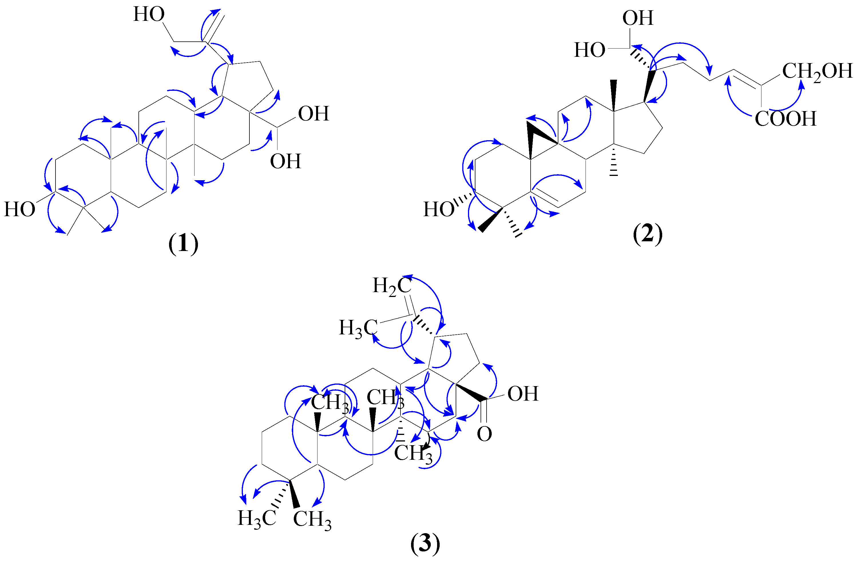
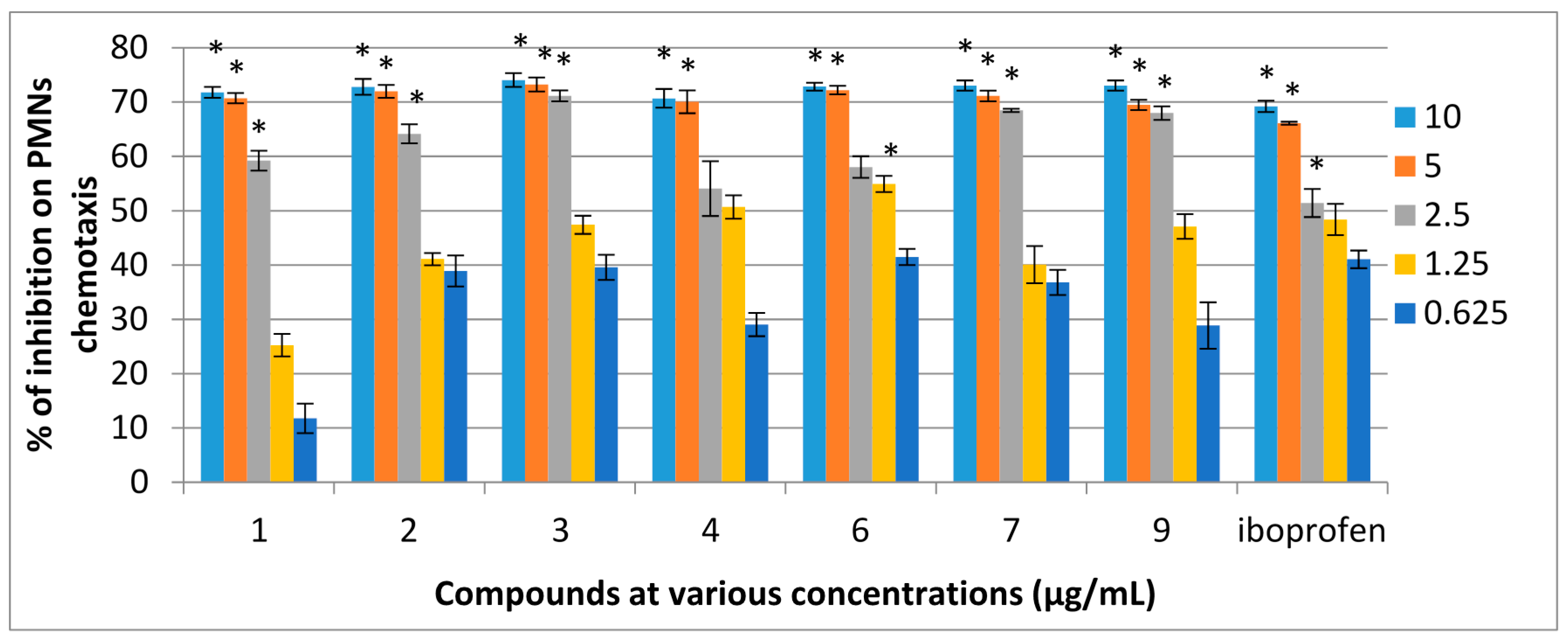
2.2. Chemotactic Activity

The cell viability test was performed using trypan blue to determine the toxicity of the compounds on immune cells at different concentrations. The elevated cell viability indicated that the compounds were nontoxic to immune cells and were able to modulate the cellular immune response of phagocytes. Cells were viable (>92%) at the concentrations of 6.25 and 100 μg/mL of the extracts after incubation for 2 h. All the compounds were tested for chemotaxis activity at five different concentrations (10, 5, 2.5, 1.25 and 0.625 μg/mL). Distance migrated by cell was specified in μm. The percentage of inhibition and IC50 values were calculated comparing the distance travelled by negative control and samples. The average distance travelled by the negative control (DMSO and HBSS, 1:1 ratio) was 15.8 μm while that for the positive control, ibuprofen, was 4.8 μm. Ibuprofen was used as a positive control as it was found to be the most effective drug in a study to determine the effect of selected nonsteroidal anti-inflammatory drug (NSAIDs) in blocking the migration of PMNs [29,30]. The results showed that DMSO did not affect the movement of cells. Seven compounds showed high percentage of inhibition (70% to 80%), significantly different to the control (p < 0.05). Among the ten compounds, seven showed strong inhibitory activities with dose-dependent effects on the migration of PMNs towards the chemo attractant (fMLP). Compounds 14, 6, 7 and 9 exhibited strong (more than 70%) inhibition and the IC50 values of these compounds were comparable to that of ibuprofen. The percentage of inhibitions of PMNs chemotaxis for the active compounds is shown in Figure 3. The IC50 values of active compounds with positive control are shown in Table 2.
Figure 3. Dose dependent percentage of inhibition of compounds of F. aurantiaca on PMNs chemotaxis. Data are mean ± SEM (n = 3). * p < 0.05 is significant difference compared with the respective control determined by one-way ANOVA followed by Tukey’s test.
Molecules 21 00009 g003
Table 2. IC50 values of compounds isolated from F. aurantiaca.
CompoundsIC50 Value (μM)
ROSChemotaxis
PMNsWB
28,28,30-Trihydroxylupeol (1)9.2 ± 0.511.5 ± 0.056.8 ± 0.1
3,21,21,26-Tetrahydroxylanostanoic acid (2)0.9 ± 0.030.1 ± 0.32.8 ± 0.1
Dehydroxybetulinic acid (3)0.9 ± 0.11.7 ± 0.12.5 ± 0.1
Taraxerone (4)1.3 ± 0.14.6 ± 0.084.1 ± 0.5
Taraxerol (5)---
Ethyl palmitate (6)1.1 ± 0.050.7 ± 0.053.7 ± 0.2
Herniarin (7)0.5 ± 0.51.6 ± 0.18.2 ± 0.2
Stigmasterol (8)---
Ursolic acid (9)0.8 ± 0.51.3 ± 0.83.6 ± 0.2
Acetylursolic acid (10)---
Aspirin (positive control)9.4 ± 0.511.6 ± 0.3-
Ibuprofen (positive control)--6.7 ± 0.5

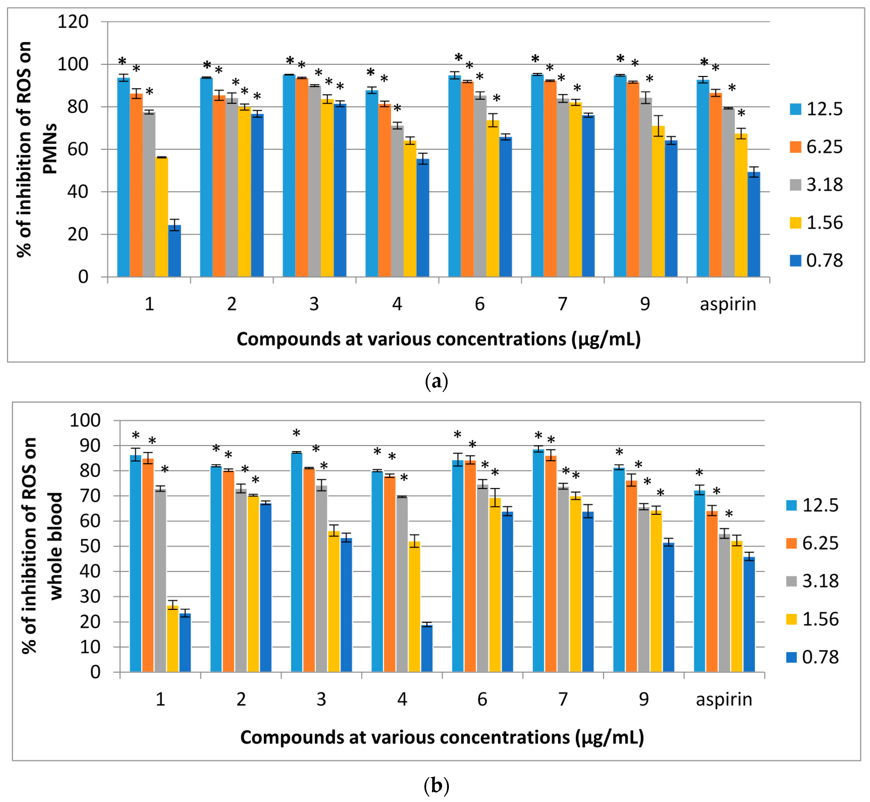
2.3. Reactive Oxygen Species (ROS) Inhibitory Activity of Isolated Pure Compounds on Human Whole Blood (WB) and PMNs

Compounds 110 were evaluated for their effects on the oxidative burst of PMNs. Of the ten compounds, 14, 6, 7 and 9 were strongly active against PMNs. The IC50 values of these compounds were lower than that of aspirin which was used as a positive control. The result showed that the compounds inhibited ROS generation during the metabolic phase of phagocytises in a dose-dependent manner as the concentrations of samples increased the percentage of inhibition was also increased. Further evaluation of compounds 110 for their effects on oxidative burst on human whole blood showed that compounds 14, 6, 7 and 9 exhibited remarkable activity for luminol-enhanced chemiluminescence. In addition, compounds 13, 6, 7 and 9 showed strong inhibition on both PMNs and whole blood whereas inhibition of PMNs was higher than that of whole blood. The dose dependent ROS inhibitory effects on the oxidative burst of human whole blood (b) and PMNs (a) for active compounds are shown in Figure 4 and their IC50 values are shown in Table 2.
Figure 4. (a) Dose dependent percentage of inhibition of ROS inhibitory activity of compounds isolated from F. aurantiaca on PMNs and (b) whole blood assayed by luminol amplified chemiluminescence. Data are mean ± SEM (n = 3).* p < 0.05 is significant difference compared with the respective control determined by one-way ANOVA followed by Tukey’s test.
Molecules 21 00009 g004
The potency of triterpenoids from F. aurantiaca as inhibitors of chemotaxis and ROS production of neutrophils is in agreement with many previous studies. Triterpenoids, especially pentacyclic triterpenes like compounds 1 and 3 were implicated in studies on the mechanisms of action and pharmacological effects of many medicinal plants used in folk medicine against diseases related to the immune system such as anti-inflammatory, antiviral, antimicrobial, antitumoral agents, as well as immunomodulating effects. Several of them are implicated in the resolution of immune diseases, although their effects have not always been clearly correlated [31]. Oleanolic acid and its related compounds such as ursolic acid and betulinic acid are known to be anti-inflammatory and immuno-suppressive through their reduction of relevant cytokines such as interleukin-1 (IL-1) interleukin-6 (IL-6), and tumor necrosis factor-α (TNF-α), as well as their effect on the classic pathway of complement activation though the inhibition of C3 convertase. Triterpenoic acid such as oleanolic acid and 3-epi-katonic acid also inhibited adenosine deaminase, an enzyme which is found at increased levels in various immune diseases [31,32]. The immunosuppressive effects of Tripterygium wilfordii might also involve triterpenoids, as well as compounds such as oleanolic acid, 3-epi-katonic acid, terpenoic acid. Eight cycloartanes isolated from Astragalus melanophrurius (Fabaceae) were found to show interesting immunomodulatory activity in an isolated human lymphocyte stimulation test [32]. The effects of a mixture of triterpenes from Quillaja saponaria (Rosaceae) on the production of IL-1 and IL-6, as well as their role in the activation of antigen-presenting cells (APC), a prerequisite for the development of immune responses have been reported [33].
Triterpenes of diverse structural types are widely distributed in prokaryotes and eukaryotes. The physiological function of these compounds is generally supposed to be a chemical defense against pathogens. Triterpenes act against certain pathogens causing human and animal diseases, such as imflammation. This may be primarily due to the hydrophobic nature of most of the compounds [34]. In this study, among the eight triterpenes, there are two lupine-type skeletons (compounds 1 and 3), two taraxerane-type (4 and 5), two ursane-type (9 and 10), one lanostane-type skeleton (2) and one stigmastane skeleton (8). A variety of terpenoids with oleanane-, ursane-, taraxerane-, lupine- and friedelane-type skeletons previously identified in many higher plants can act as precursors for many biomarkers found in biological screening. Oleanane, ursane and lupane triterpenes with carboxylic acid groups and alcoholic derivatives of oleanane, ursane and lupane are active against inflammation. However, it is difficult to identify exact molecular motifs, largely spread among these terpenes and implicated in their anti-inflammatory action. Some of the terpenoids act as plant hormones regulating different physiological roles, but some secondary metabolites is concerned in host defence and in the protection of the plant or animal from possible pathogens. Recent research in the field of the regulation of innate immunity from insects to mammals has established the existence of a previously unexpected protection in the pathways (receptors, kinases and effector molecules) that are involved in this process [35]. We suggest that the general analysis of the relationship between chemical structure and immunomodulatory activity of the isolated triterpenes may be related to the presence of an oxygenated group at C-3 and carboxyl group at C-28.

3. Experimental Section

3.1. General Information

Melting points were determined with an electrothermal apparatus (digital series) and were uncorrected. Ultraviolet (UV) spectra were recorded with a Shimadzu UV-1601 Spectrophotometer (Shimadzu Corp., Tokyo, Japan), for ethanol and methanol solutions in 1 cm Quartz cells. IR spectra were recorded on a GX FTIR instrument (Perkin-Elmer Corp., Waltham, MA, USA) using potassium bromide pellets and sodium chloride cells. NMR spectral analyses were carried out with a FT-NMR 600 MHz Cryoprobe spectrometer (Bruker, Basel, Switzerland). 1H-1H COSY, 1H-13C HSQC and 1H-13C HMBC were obtained with the usual pulse sequence and data processing was performed with the ACD lab software (Version 12, Advanced Chemistry Development, Toronto, ON, Canada). Mass spectra ESIMS and HRESIMS were measured on Micro TOF-Q mass spectrometer (Bruker, Basel, Switzerland). All experiments were carried out at Universiti Kebangsaan Malaysia (UKM). Analytical Thin Layer Chromatography (TLC) was carried out on pre-coated silica gel 60 GF 254 (20 × 20) TLC plate (Merck, art 5554, Darmstadt, Germany). The plates were visualized under ultraviolet light (λ254 nm, model UVGL-58) and by charring the compounds after spraying the plates with 10% sulphuric acid or Dragendorff reagent. Vacuum liquid chromatography was carried out over silica gel type H (10–0 μm, Sigma Chemical Co., St. Louis, MO, USA). The conventional column chromatography was done by using silica gel 60 (230–400 mesh ASTM, Merck, art 9385) or Sephadex LH20 (GE Healthcare Bio-Sciences AB, Uppsala, Sweden). Radial chromatography (centrifugal chromatography) was performed on a Chromatotron (Analtech Inc., Newark, Denmark) using silica gel 60 GF 254 containing gypsum (TLC grade) (Merck, art. 7749). Organic solvents such as hexane, ethyl acetate, chloroform, diethyl ether and methanol were purchased from Merck.

3.2. Plant Material

Ficus aurantiaca Griff was collected from Banting, Selangor, Malaysia and a voucher specimen (SM2109) was identified and deposited at the Herbarium of Universiti Kebangsaan Malaysia (UKM), Bangi, Malaysia.

3.3. Extraction and Isolation

Extraction and isolation of compounds from F. aurantiaca was carried out by modification of the method described by Rukachaisirikul [36]. The plant material was dried at room temperature and its air-dried stems were ground. The ground powder (196 g) was sequentially extracted thrice with n-hexane, ethyl acetate and methanol (500 mL each) by soaking at least for 48 h at room temperature for each time. After filtration each of the solvent extracts was evaporated to dryness under reduced pressure using a rotary evaporator to yield crude n-hexane (7.8 g), ethyl acetate (4.0 g) and methanol (10.0 g) extracts, respectively. The separation of hexane, ethyl acetate and methanol extracts was carried out using vacuum liquid chromatography (VLC), column chromatography and Chromatotron over silica gel, respectively.
The n-hexane crude extract (6.0 g) of F. aurantiaca was chromatographed using VLC on silica gel 60 GF254 (TLC grade) and hexane–ethyl acetate (10:0 to 0:10, v/v) as solvent system to give sixteen fractions (F1–F16) that were analyzed using TLC and combined into five fractions (Fa1–Fa5) based on their TLC profiles. For further isolation and purification, each fraction was subjected to column chromatography (CC) using silica gel 60 (230–400 mesh ASTM). Fa1 (1.234 g) was subjected to CC on silica gel as stationary phase and hexane–ethyl acetate (10:0 to 0:10, v/v) of increasing polarity as mobile phase to obtain seven subfractions (Fa1A–Fa1G). Subfractions Fa1A and Fa1B were subjected to silica gel CC eluted with hexane–ethyl acetate (9:1 to 5:5 v/v) and further recrystallized from 100% n-hexane to obtain compound 6. Fraction Fa3 (2.135 g) was purified by silica gel CC eluted with hexane–ethyl acetate (9:1 to 0:10 v/v) to obtain compound 7.
The ethyl acetate crude extract (3.0 g) of F. aurantiaca was subjected to silica gel CC eluted with hexane–ethyl acetate (5:5 to 0:10, v/v) and ethyl acetate–methanol (10:0 to 6:4, v/v) to obtain eighteen fractions (F1–F18). Based on the TLC profiles, these eighteen fractions were combined into six fractions (Faa–Faf). Fractions Faa (322.1 mg) and Fab (173.2 mg) were separated and purified using repeated CC to obtain compounds 1 and 4, respectively. In the same way 5, 2 and 8 were obtained from fractions Fad (321.7 mg), Fae (523.2 mg) and Faf (457.2 mg), respectively. Similarly the methanol extract of F. aurantiaca (8.0 g) was fractionated using VLC on silica gel 60 GF254 using ethyl acetate-methanol of increasing polarity as eluent and the fractions were purified by silica gel CC to obtain subfractions FF1–FF10. Subfractions FF2 (567.0 mg), FF5 (234.0 mg) and FF7 (638.0 mg) was run on a Chromatotron eluted with ethyl acetate–methanol (10:0 to 2:8, v/v) to yield compounds 9, 3 and 10, respectively. To summarize, compounds 6 and 7 were obtained from the n-hexane extract; compounds 1, 4, 5, 2 and 8 were isolated from ethyl acetate extract while compounds 9, 3 and 10 were isolated from the methanol extract. The structure elucidation of the isolated compounds was performed using UV, IR, ESIMS and NMR spectral data analyses and comparison with appropriate literature values.
28,28,30-Trihydroxylupeol (1): White powder (20.0 mg), melting point 222–224 °C, UV λmax (C2H5OH) nm: 272, 210 (log ε 3.4, 2.2). IR λmax (CHCl3) cm−1: 3310, 2920, 2158, 2042, 1976, 1611, 1411, 1021. 1H-NMR (CDCl3): δH 0.79 (s, 3H, H-23), 0.83 (s, 3H, H-24), 0.94 (s, 3H, H-25), 1.01 (s, 3H, H-26), 1.07 (s, 3H, H-27), 0.88 (m, 10H, H-9), 1.26 (m, 4H, H-15, H-16), 1.28 (m, 4H, J = 6.6 Hz, H-6, H-7), 1.32 (m, 1H, J = 7.2 Hz, H-5), 1.34 (m, 4H, H-22, H-13), 1.61 (m, 4H, H-11), 1.68 (m, 4H, J = 3.0 Hz, H-2, H-12), 1.88–1.94 (m, 2H, H-21), 2.04 (m, 1H, H-18), 2.38 (d, 1H, J = 7.2 Hz, H-19), 3.39 (dd, 1H, J = 10.8, 4.8 Hz, H-3), 4.57 (br s, 1H, H-29), 4.69 (br s, 1H, H-29), 4.79 (t, 1H, J = 6.0 Hz, H-28), 5.14 (m, 1H, H-30) 5.19 (m, 1H, H-30). 13C-NMR (CDCl3): δC 14.1(C-27), 14.5 (C-23), 15.9 (C-25), 16.1 (C-26), 17.7 (C-6), 18.0 (C-24), 21.0 (C-11), 25.1 (C-12), 27.3 (C-2), 27.4 (C-15), 29.7 (C-21), 34.1 (C-7), 35.5 (C-16), 37.3 (C-10), 37.9 (C-13), 38.1 (C-1), 39.6 (C-4), 39.9 (C-22), 40.9 (C-8), 42.8 (C-14), 43.0 (C-17), 47.9 (C-18), 48.2 (C-19), 50.3 (C-9), 55.6 (C-5), 69.2 (C-30), 80.1(C-3), 95.1 (C-28), 109.3 (C-29), 150.9 (C-20). ESIMS m/z: 463.3393 [M + H − 12]+, 931.7436 [2M + H − 18]+, HRESIMS m/z: 474.1465 [M]+, calculated 474.1465 for molecular formula C30H50O4.
3,21,21,26-Tetrahydroxylanostanoic acid (2): White powder (17.2 mg), melting point 150–155 °C, UV νmax (C2H5OH) nm: 272, 207 (log ε 2.3, 2.8). IR νmax (CHCl3) cm−1: 3215, 2915, 2850, 2134, 1985, 1735, 1678, 1642, 1472, 1120. ESIMS (negative mode) m/z: 502.3290 [M]+, 479.1023 [M − 23]+, 453.0885 [M − CH3OH − 17]+, and 311.1563 [M − side chain (C8H14O5) − 1]+, HRESIMS (positive mode) m/z: 503.3570 [M + H]+, calculated 502.3290 for molecular formula C30H50O. 1H-NMR (CDCl3): δH 0.80 (s, 3H, H-28), 0.85 (s, 1H, H-29), 0.92 (q, 3H, H-30), 0.94 (m, 2H), 0.99 (s, 2H, H-18), 1.01(m, 2H, H-2), 1.03 (s, 1H, H-19), 1.18 (m, 2H, H-22), 1.31 (m, 2H, H-12), 1.34 (m, 2H, H-11), 1.35 (m, 2H, H-23), 1.48 (m, 1H, H-8, H-17), 1.57 (m, 2H, H-15), 1.63 (m, 2H, H-16), 1.68 (m, 1H, H-20), 1.77 (m, 2H, H-7), 3.44 (t, 1H, H-3), 4.03 (d, 1H, J = 12.0 Hz, H-26), 4.57 (m,1H, H-24), 4.79 (t, 1H, J = 6.0 Hz, H-6), 4.80 (m, 1H, H-21), 2.25 (C-21), 2.30 (C-21), 4.68 (C-3), 4.69 (C-26). 13C-NMR (CDCl3): δC 29.8 (C-1), 25.7 (C-2), 80.1 (C-3), 43.0 (C-4), 151.0 (C-5), 109.3 (C-6), 31.9 (C-7), 50.3 (C-8), 48.2 (C-9), 39.9 (C-10), 23.7 (C-11), 22.7 (C-12), 42.8 (C-13), 40.9 (C-14), 34.5 (C-15), 29.4 (C-16), 55.6 (C-17), 27.4 (C-18), 17.7 (C-19), 35.3 (C-20), 95.1 (C-21), 35.5 (C-22), 25.0 (C-23), 150.9 (C-24), 109.3 (C-25), 69.2 (C-26), 173.8 (C-27), 14.1 (C-28), 15.9 (C-29), 16.5 (C-30).
Dehydroxybetulinic acid (3): White powder (17.4 mg) with a melting point 285–290 °C. UV λmax (C2H5OH) nm: 195. IR νmax(CHCl3) cm−1: 2924, 2238, 2158, 1732 1648, 1463, 1248 and 971 cm−1. ESIMS (positive mode) m/z: 441.3663 [M + H]+ 463.3474 [M + Na]+and ESIMS (negative mode) m/z: 439.3759 [M − H], 879.7791 [2M − H]. HRESIMS (positive mode) m/z: 457.2734 [M + OH]+, calculated 440.3787 for molecular formula C30H48O2. 1H-NMR (CDCl3): δH 1.41 (m, 1H, H-1), 1.31 (m, 1H, H-1), 1.42 (m, 1H, H-2), 1.32 (m, 1H, H-2), 1.42 (m, 1H, H-3), 1.33 (m, 1H, H-3), 1.34 (m, 1H, H-5), 1.65 (m, 2H, H-6), 1.46 (m, 1H, H-7), 1.56 (m, 1H, H-9), 1.54 (m, 1H, H-11), 1.29 (m, 1H, H-11), 1.55 (m, 1H, H-12), 1.29 (m, 1H, H-12), 1.24 (m, 1H, H-13), 1.49 (m, 1H, H-15), 1.00 (m, 1H, H-15), 1.80 (m, 1H, H-16), 1.25 (m, 1H, H-16), 1.68 (m, 1H, H-18), 2.09 (m, 1H, H-19), 1.60 (m, 6H, H-21), 1.75 (t, 2H, J = 5.4 Hz, H-22), 0.83 (s, 6H, H-23), 0.90 (s, 3H, H-24), 0.76 (s, 3H, H-25), 0.89 (s, 3H, H-26), 0.73 (s, 6H, H-27), 5.32 (br s, 1H, H-29), 1.10 (s, 3H, H-30). 13C-NMR (CDCl3): δC 36.1 (C-1), 28.2 (C-2), 36.7 (C-3), 30.8 (C-4), 59.6 (C-5), 18.6 (C-6), 37.4 (C-7), 40.1 (C-8), 51.9 (C-9), 37.7 (C-10), 20.6 (C-11), 25.2 (C-12), 37.7 (C-13), 42.9 (C-14), 39.7 (C-15), 40.4 (C-16), 59.6 (C-17), 46.5 (C-18), 37.2 (C-19), 150.9 (C-20), 29.2 (C-21), 23.0 (C-22), 22.1 (C-23), 16.6 (C-24), 15.4 (C-25), 17.6 (C-26), 13.9 (C-27), 183.5 (C-28), 116.3 (C-29), 20.1 (C-30).

3.4. Chemicals, Reagents and Equipment

Serum opsonized zymosan A (Saccharomyses cerevisiae suspensions and serum), luminol (3-aminophthalhydrazide), phosphate buffer saline (PBS), Hanks Balance Salt Solution (HBSS++), ficoll, N-formyl-methionylleucyl-phenilalanine (fMLP), tryphan blue, phorbol 12-myristate 13-acetate (PMA), dimethyl sulfoxide (DMSO), acetylsalicylic acid (purity 99%) and ibuprofen (purity 99%) were purchased from Sigma. Haematoxylin and xylene were obtained from BDH (London, UK). Chemiluminscence measurements were carried out on a Luminoscan Ascent luminometer (Thermo Scientific, Loughborough, UK). A Boyden chamber with a 3 and 5 μm polycarbonate membrane filter separating the upper and lower compartments was purchased from Neuro probe (Cabin John, Montgomery County, MD, USA).

3.5. Isolation of Polymorphonnuclear Leucocytes (PMNs)

Human blood was collected from a healthy volunteer fasted for at least 8 h. PMNs were isolated by Ficol-gradient separation following the published method [37]. The use of human blood in this study was approved by the Human Ethics Committee, Universiti Kebangsaan Malaysia Medical Centre (HUKM), Cheras with permission (number FF-220-2008). Cell counts were performed using a haemocytometer.

3.6. Cell Viability

Cell viability tests were performed using trypan blue dye exclusion method with the modification of a published procedure [38]. The neutrophils (1 × 10⁶ mL−1) were incubated with 6.25 or 100 μg/mL of pure compounds in triplicate at 37 °C for 1 to 2 h. The cell death was indicated by the blue dye uptake. The percentage of cell viability was considered from the total cell counts.

3.7. Chemiluminescence Assay

Luminol enhanced chemiluminescence assay was carried out using the modification of the published method [39]. In brief, 25 μL of human whole blood or 25 μL of isolated PMN cells were suspended in HBSS++ into each well of 96-well microplate. The plate was incubated with 25 μL of tested compounds and aspirin at five different concentrations (12.5, 6.25, 3.18, 1.56 and 0.78 μg/mL) of each sample for 50 min at 37 °C in a luminoscan while 25 μL of HBSS++ was used as the negative control. The cells were induced with 25 μL of serum opsonized zymosan (SOZ) followed by 25 μL of luminol into each well. Then HBSS++ solution was added into each well to make the final volume of 200 μL. Aspirin was used as a positive control while the negative control contained zymosan, luminol, DMSO (0.5%), HBSS++ and cells. The percentage of inhibition was calculated by the measurement of RLU (reading luminometer unit) of peak and total integral values with repeated scans.

3.8. Chemotaxis Assay

The assay was carried out using the modified 48-well Boyden chamber method [40]. The Boyden chamber contains 48 wells with a diameter of 8 μm and is divided into two compartments by a filter separation. In brief, 25 μL of chemo attractant, fMLP (10−8 M, diluted with chemo attractant buffer solution) was added to the lower compartment of the Boyden chamber. PMN cell suspension (45 μL) with 5 μL of test compounds and ibuprofen at five different concentrations (6.25, 12.5, 25, 50, and 100 μg/mL) were added to the upper compartment of the chamber whereas the negative control contained 45 μL of PMN cell suspension and 5 μL of chemo attractant buffer. Ibuprofen was used as a positive control. The final concentrations of test compounds and ibuprofen in the wells were 0.625, 1.25, 2.5, 5 and 10 μg/mL. The chamber was incubated in 5% carbon dioxide incubator for 1 h at 37 °C. After incubation the polycarbonate membrane (where the migrated cells were remained) was stained with PBS, methanol (99.5%), haematoxylin, distilled water, ethanol (70% and 95.8%) and xylene respectively. The distance of cell’s migration was measured using a low power microscope with magnifications 40×.

3.9. Statistical Analysis

Data were analyzed using One-way ANOVA, post hoc Tukey’s test. p < 0.05 was considered to be statistically significant using the SPSS 17.0 (SPSS Inc., Chicago, IL, USA) statistical software. All values were characterized as mean ± SEM. Probit programme was used to determine the IC50 values for active compounds.

4. Conclusions

In conclusion, a phytochemical investigation of the stem of F. aurantiaca afforded three new triterpenoids—28,28,30-trihydroxylupeol (1), 3,21,21,26-tetrahydroxylanostanoic acid (2) and dehydroxybetulinic acid (3) together with five known triterpenoids: taraxerone (4), taraxerol (5), stigmasterol (8), ursolic acid (9), acetyl ursolic acid (10), one coumarin, herniarin (7) and one unsaturated hydrocarbon, ethyl palmitate (6). This is the first report on the phytochemical investigation and biological effects of compounds from F. aurantiaca on PMN chemotaxis and ROS inhibitory activity of human whole blood and PMNs. Compounds 13, 6, 7 and 9 showed the strongest inhibitory activities for both chemiluminescence and chemotaxis whereas the IC50 values were lower than those of the standard drugs used. This study provided evidence that F. aurantiaca is a source of new immunomodulatory compounds to modulate the innate immune response of phagocytes and further studies are needed to investigate the mechanisms of their immuno-modulatory responses.

Acknowledgments

This work was supported by the grant UKM-GGPM-TKP-058-2010, UKM-Pharmacy-03-FRGS 0029-2010 and UKM-DIPM-006-2011. Special thanks to the Ministry of Higher Education Malaysia, for CSFP.

Author Contributions

S.M., I.J., and K.H. designed research; S.M. and K.H. performed research and analyzed the data; S.M., I.J. and K.H. wrote the paper, S.M. and I.J. performed on reactive oxygen species production and chemotactic activity of neutrophils. All authors read and approved the final manuscript.

Conflicts of Interest

The authors declare that they have no conflict of interests.

References

  1. Vander, N.J.M.; Klerx, J.P.; Vandijk, H.; de Silva, K.T.; Labadie, R.P. Immunomodulatory activity of an aqueous extract of Azadirachta indica stem bark. J. Ethnopharmacol. 1987, 19, 125–131. [Google Scholar]
  2. Diwanay, S.; Chitre, D.; Patwardhan, B. Immunoprotection by botanical drugs in cancer chemotherapy. J. Ethnopharmacol. 2004, 90, 49–55. [Google Scholar]
  3. Gautam, M.; Diwanay, S.; Gairola, S.; Shinde, Y.; Patki, P.; Patwardhan, B. Immuno adjuvantpotential of Asparagus racemosus aqueous extract in experimental system. J. Ethnopharmacol. 2004, 91, 251–255. [Google Scholar] [CrossRef] [PubMed]
  4. Jayathirtha, M.G.; Mishra, S.H. Preliminary immunomodulatory activities of methanol extracts of Eclipta alba and Centella asiatica. Phytomedicine 2004, 11, 361–365. [Google Scholar] [CrossRef] [PubMed]
  5. Diasio, R.B.; LoBuglio, A.F. Immunomodulators: Immunosuppressive agents and immunostimulants. In Goodman and Gilman’s The Pharmacological Basis of Therapeutics, 9th ed.; Hardman, J.G., Limbird, L.E., Molinoff, P.B., Ruddon, R.W., Eds.; McGraw-Hill: New York, NY, USA, 1996; pp. 1291–1307. [Google Scholar]
  6. Fordin, D.G. History and concepts of big plant genera. Taxon 2004, 53, 753–776. [Google Scholar] [CrossRef]
  7. Ronsted, N. Phylogeny, biogeography and ecology of Ficus section Malvanther (Moraceae). Mol. Phylogenet. Evol. 2008, 48, 12–22. [Google Scholar] [CrossRef] [PubMed]
  8. Lansky, E.P.; Paavilainen, H.M.; Pawlus, A.D.; Newman, R.A. Ficus spp. (fig): Ethnobotany and potential as anticancer and anti-inflammatory agents. J. Ethnopharmacol. 2008, 119, 195–213. [Google Scholar] [CrossRef] [PubMed]
  9. Simo, C.C.F.; Simeon, F.K.; Poumale, H.M.P.; Simo, I.K.; Ngadjui, B.T.; Green, I.R.; Krohn, K. Benjaminamide: A new ceramide and other compounds from the twigs of Ficus benjamin (Moraceae). Biochem. Syst. Ecol. 2008, 36, 238–243. [Google Scholar] [CrossRef]
  10. Li, R.W.; Leach, D.N.; Myers, S.P.; Lin, G.D.; Leach, G.J.; Waterman, P.G. A new anti-inflammatory glucoside from Ficus racemosa L. Planta. Med. 2004, 70, 421–426. [Google Scholar] [PubMed]
  11. Mandal, S.C.; Maity, T.K.; Das, J.; Das, B.P.; Pal, M. Anti-inflammatory evaluation of Ficus racemosa Linn. Leaf extract. J. Ethnopharmacol. 2000, 72, 87–92. [Google Scholar] [CrossRef]
  12. Sackyfio, A.C.; Lugeleka, O.M. The anti-inflammatory effect of a crude aqueous extract of the root bark of Ficus elastica in the rat. Arch. Int. Pharmacodyn. Ther. 1986, 281, 169–176. [Google Scholar]
  13. Shukla, R.; Gupta, S.; Gambhir, J.K.; Prabhu, K.M.; Murthy, P.S. Antioxidant effect of aqueous extract of the bark of Ficus benghalensis in hyper cholesterolaemic rabbits. J. Ethnopharmacol. 2004, 92, 47–51. [Google Scholar] [CrossRef] [PubMed]
  14. Burkill, I.H. A Dictioanary of the Economic Products of the Malays Panisular; Ministry of Agriculture and Co-Operatives: Kuala Lumpur, Malaysia, 1966. [Google Scholar]
  15. Mawa, S.; Husain, K.; Jantan, I. Ficus carica L. (Moraceae): Phytochemistry, traditional uses and biological activities. Evid. Based Complement. Altern. Med. 2013. [Google Scholar] [CrossRef] [PubMed]
  16. Kamaruddin, M.S.; Latif, A. Tumbuhan Ubatan Malaysia Selangor; Pusat Pengurusan Penyelidikan: Bangi, Malaysia, 2002; Volume 1. [Google Scholar]
  17. Himanshu, J.; Arun, B.J.; Hemlata, S.; Gururaja, M.P.; Prajwal, R.S.; Subrahmanyam, A.V.S.; Satyanarayana, D. Fatty acids from Memecylonum bellatum (Burm). Asian J. Res. Chem. 2009, 2, 178–180. [Google Scholar]
  18. Ibrahim, A.; Najmuldeen, A.; Hamid, A.H.; Mohamad, K.; Khalijah, A.; Mehran, F.N.; Kamal, A.K.; Mat Ropi, M.; Hiroshi, M. Steroids from Chisocheton tomentosus. Malays. J. Sci. 2011, 30, 144–153. [Google Scholar]
  19. Jamal, A.K.; Yacoob, W.A.; Din, L.B. Triterpenes from the root bark of Phyllanthus columnaris. Aust. J. Basic Appl. Sci. 2009, 3, 1428–1431. [Google Scholar]
  20. Mahdi, A.; Amirhossein, S.; Mehrda, I. Synthesis and purification of 7-prenyloxycoumarins and herniarin as bioactive natural coumarins. Iran. J. Basic Med. Sci. 2009, 1, 63–69. [Google Scholar]
  21. Mawa, S.; Ikram, M.S. Chemical constituents of Garcinia prainiana. J. Sains Malays. 2012, 41, 585–590. [Google Scholar]
  22. Shahid, H.A.; Ali, M.; Kamran, J.N. New manglanostanoic acid from the stem bark of Megifera indica var. “Fazli”. J. Soudi Chem. Soc. 2014, 18, 561–565. [Google Scholar]
  23. Uddin, G.; Waliullah, M.; Siddiqui, B.S.; Alam, M.; Sadat, A.; Ahmad, A.; Uddin, A. Chemical constituents and phytotoxicity of solvent extracted fractions of stem bark of Grew optiva Drummond ex Burret. Middle East J. Sci. Res. 2011, 8, 85–91. [Google Scholar]
  24. Supaluk, P.; Puttirat, S.; Rungrot, C.; Somsak, R.; Virapong, P. New bioactive triterpenoids and antimalarial activity of Diospyros rubra lec. EXCLI J. 2010, 9, 1–10. [Google Scholar]
  25. Ahmed, W.; Khan, A.Q.; Malik, A. Two triterpenes from the leaves of Ficus carica. Planta Med. 1988, 54. [Google Scholar] [CrossRef] [PubMed]
  26. Chiang, Y.M.; Kuo, Y.H. Novel triterpenoids from the arial parts of Ficus microcarpa. J. Org. Chem. 2002, 67, 7656–7661. [Google Scholar] [CrossRef] [PubMed]
  27. Ali, M.; Shuaib, M.; Naquvi, K.J. New lanostane type triterpenes from the oleo-gum resin of Commiphora myrrha (NEES) ENGL. Int. J. Pharm. Pharm. Sci. 2014, 6, 372–375. [Google Scholar]
  28. Chiang, Y.M.; Chang, J.Y.; Kuo, C.C.; Kuo, Y.H.; Chang, C.Y. Cytotoxic triterpenes from the arial roots of Ficus microcarpa. Phytochemistry 2005, 66, 495–501. [Google Scholar] [CrossRef] [PubMed]
  29. Spisani, S.; Vanzini, G.; Traniello, S. Inhibition of human leucocytes locomotion by anti-inflammatory drugs. Experientia 1979, 35, 803–804. [Google Scholar] [CrossRef] [PubMed]
  30. José-Luis, R. Effects of triterpenes on the immune system. J. Ethnopharmacol. 2010, 128, 1–14. [Google Scholar]
  31. Brinker, A.M.; Ma, J.; Lipsky, P.E.; Raskin, I. Medicinal chemistry and pharmacology of genus Tripterygium (Celastraceae). Phytochemistry 2007, 68, 732–766. [Google Scholar] [CrossRef] [PubMed]
  32. Calis, I.; Yürüker, A.; demir, D.; Wright, A.D.; Sticher, O.; Luo, Y.D.; Pezzuto, J.M. Cycloartane triterpene glycosides from the roots of Astragalus melanophrurius. Planta Med. 1997, 63, 183–186. [Google Scholar] [CrossRef] [PubMed]
  33. Behboudi, S.; Morein, B.; Villacres-Eriksson, M. In vitro activation of antigen presentin cells (APC) by defined composition of Quillaja saponaria molina triterpenoids. Clin. Exp. Immunol. 1996, 105, 26–30. [Google Scholar] [CrossRef] [PubMed]
  34. De las, H.B.; Rodríguez, B.; Boscá, L.; Villar, A.M. Terpenoids: Sources, structure elucidation and therapeutic potential in inflammation. Curr. Top. Med. Chem. 2003, 3, 53–67. [Google Scholar]
  35. Shashi, B.M.; Sucharita, S. Advances in triterpenoid research, 1990–1994. Phytochemistry 1997, 44, 1185–1236. [Google Scholar]
  36. Rukachaisirikul, V.; Ritthiwigrom, T.; Pinsa, A.; Sawangchote, P.; Taylor, W.C. Xanthones from the bark of Garcinia nigrolineata. Phytochemistry 2003, 64, 1149–1156. [Google Scholar] [CrossRef]
  37. Demirkiran, O.; Mesaik, M.A.; Beynek, H.; Abbaskhan, A.; Iqbal, C.M. Cellular reactive oxygen species inhibitory constituents of Hypericum thasium. Phytochemistry 2009, 70, 244–249. [Google Scholar] [CrossRef] [PubMed]
  38. Koko, W.S.; Mesaik, M.A.; Yousof, S.; Galal, M.; Choudary, M.I. In vitro immonumodulating properties of selected Sudanese medicinal plants. J. Ethnopharmacol. 2008, 118, 26–34. [Google Scholar] [CrossRef] [PubMed]
  39. Haklar, G.; Ozveri, E.S.; Yuksel, M.; Aktan, A.; Yalcin, A.S. Different kinds of reactive oxygen and nitrogen species were detected in colon and breast tumors. Cancer Lett. 2001, 165, 219–224. [Google Scholar] [CrossRef]
  40. Sacerdote, P.; Massi, P.; Panerai, A.E.; Parolaro, D. In vivo and in vitro treatment with the synthetic cannabinoid CP55,940 decreases the in vitro migration of macrophages in the rat: involvement of both CB1 and CB2 receptors. J. Neuroimmunol. 2000, 109, 155–163. [Google Scholar] [CrossRef]
  • Sample Availability: Samples of the compounds are available from the authors.

