G-Quadruplex Guanosine Gels and Single Walled Carbon Nanotubes

Abstract
:
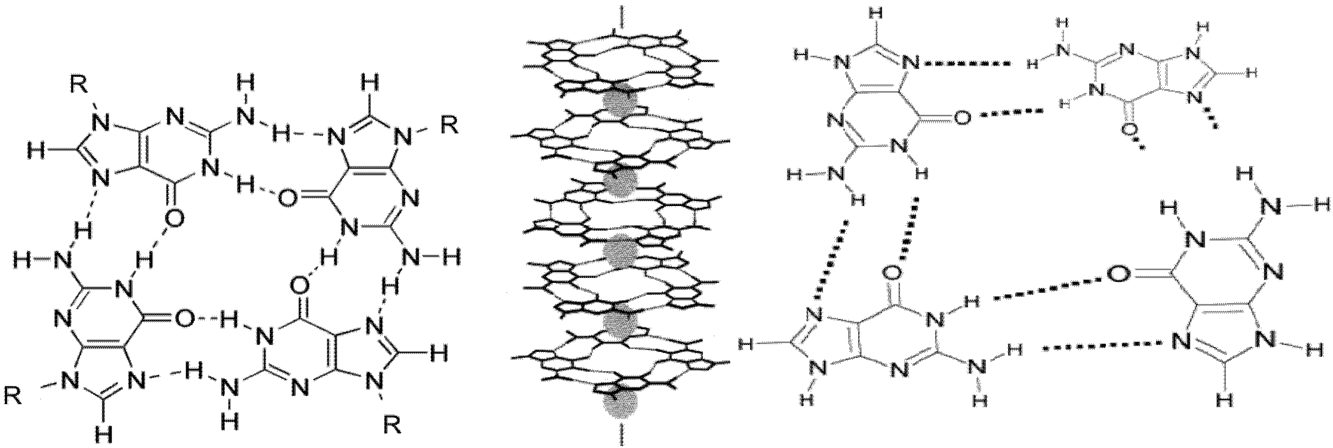
1. Introduction


2. Results and Discussion
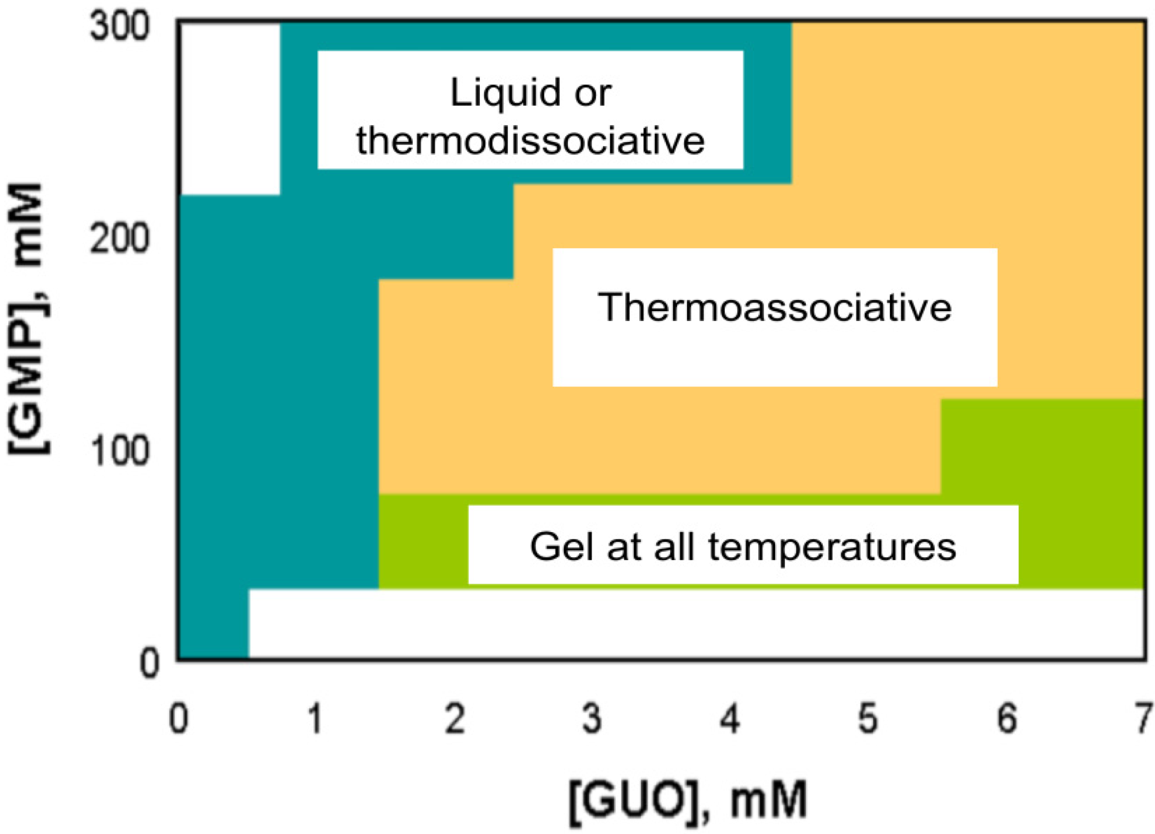
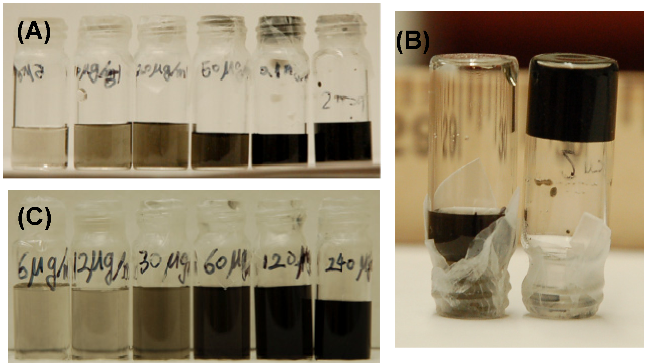
2.1. SWNT Solubilization


| Without SWNTs | With SWNTs | ||||
|---|---|---|---|---|---|
| KCl (M) | LT | RT | SWNT (mg/mL) | LT | RT |
| 0.01 | Liquid | Viscous | 1.5 | Gel | Gel |
| 0.03 | Liquid | Gel | 1.2 | Gel | Gel |
| 0.07 | Liquid | Viscous | 2.1 | Viscous | Gel |
| 0.09 | Liquid | Viscous | 2.4 | Liquid | Gel |
| 0.15 | Liquid | Liquid | 2.8 | Liquid | Viscous |
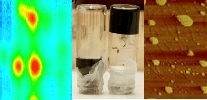
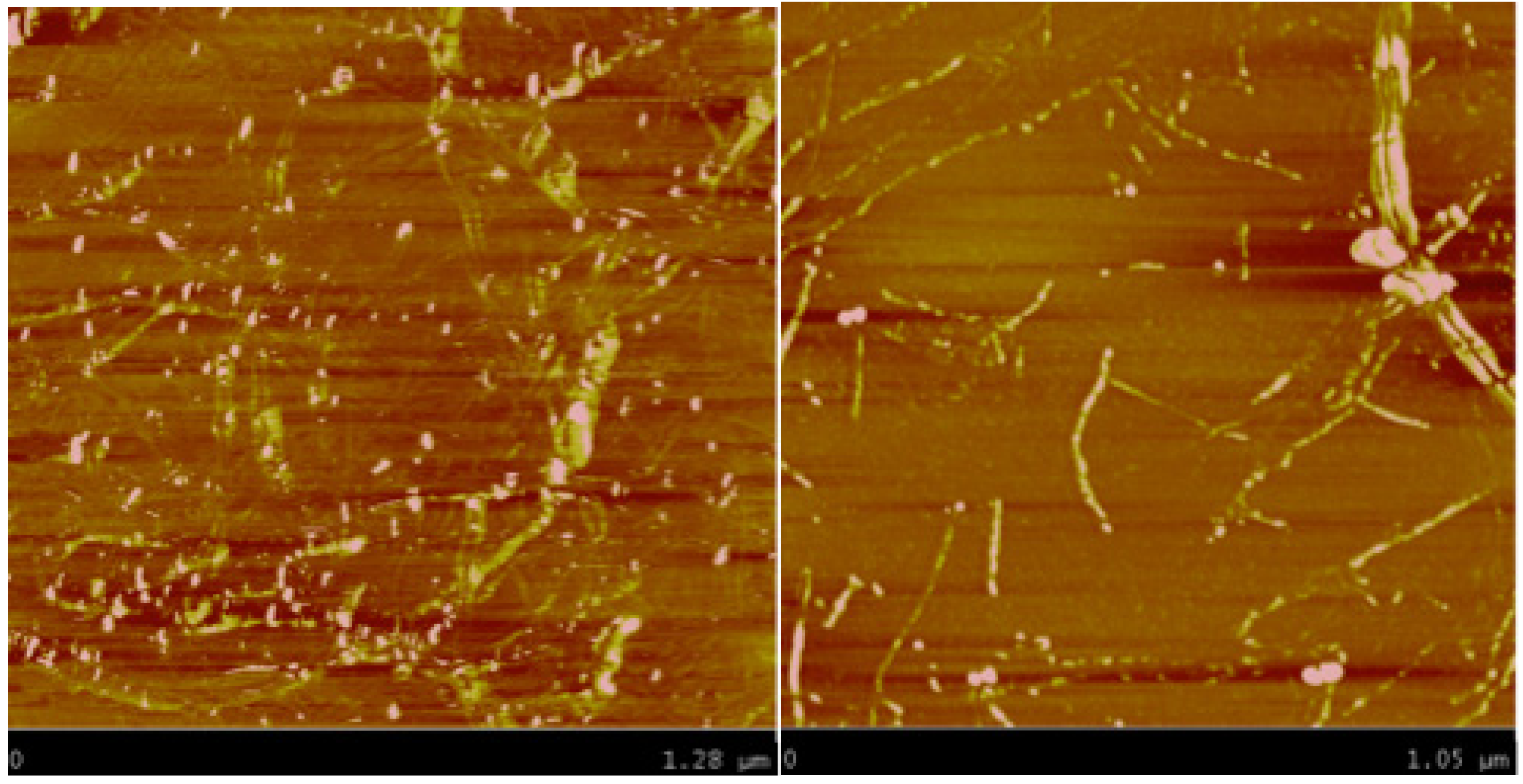
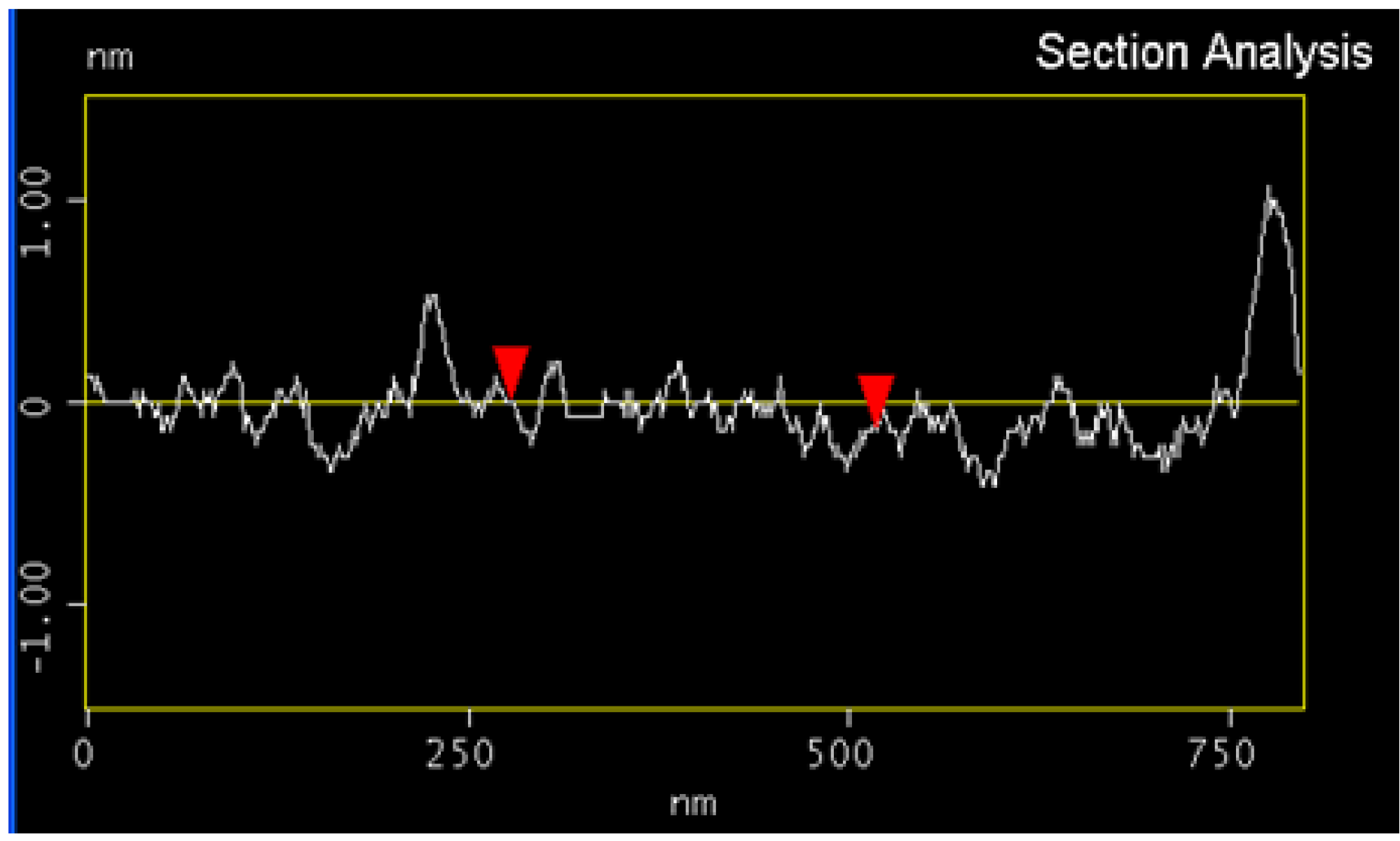
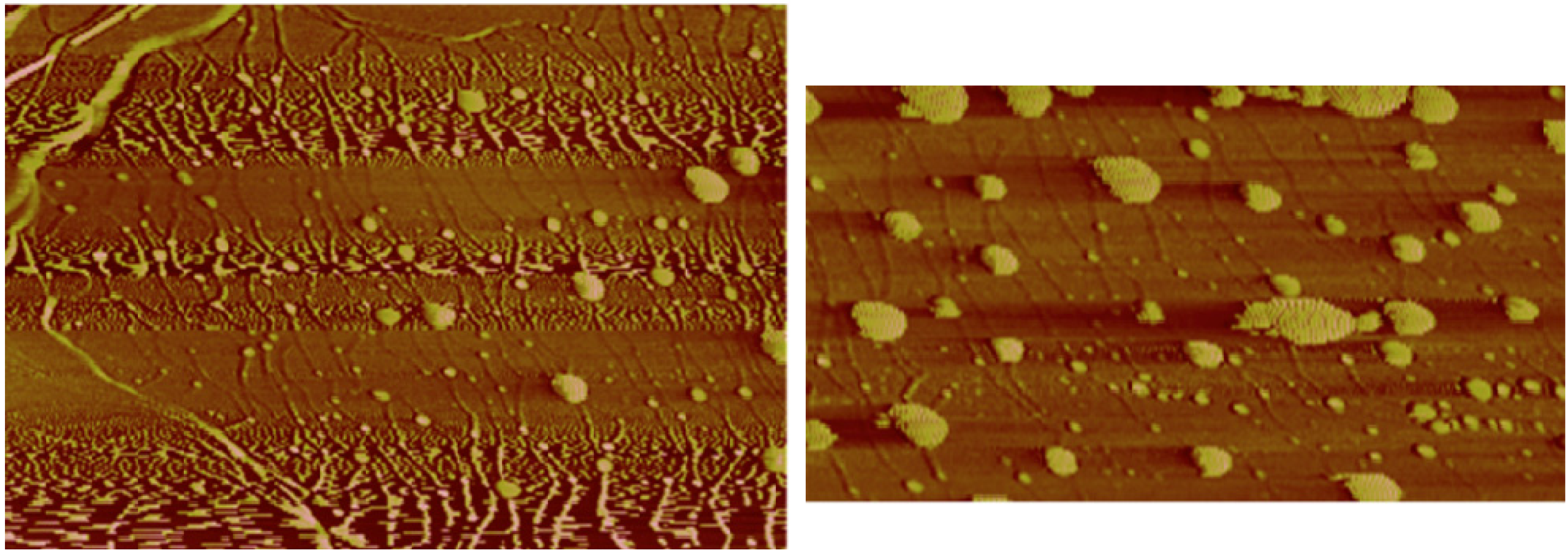
2.2. Imaging Results
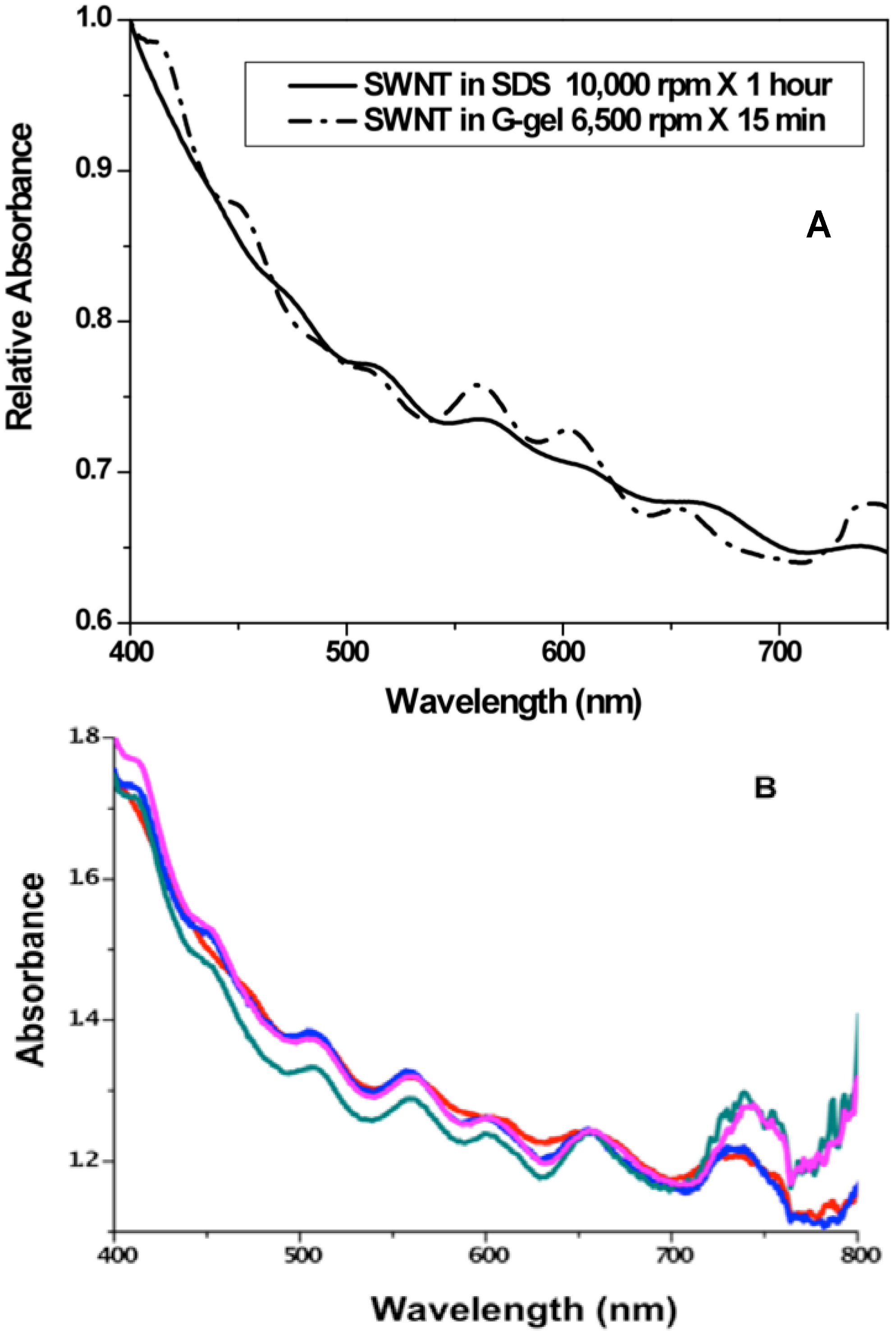
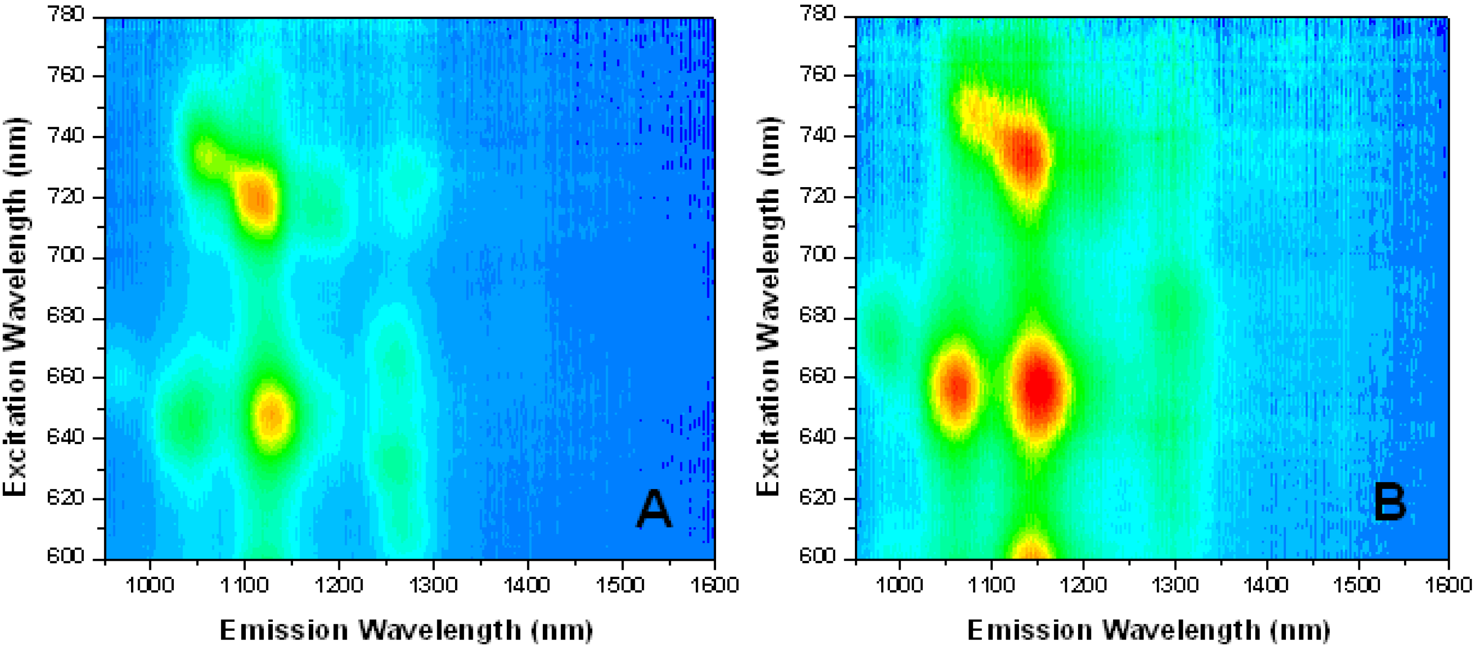
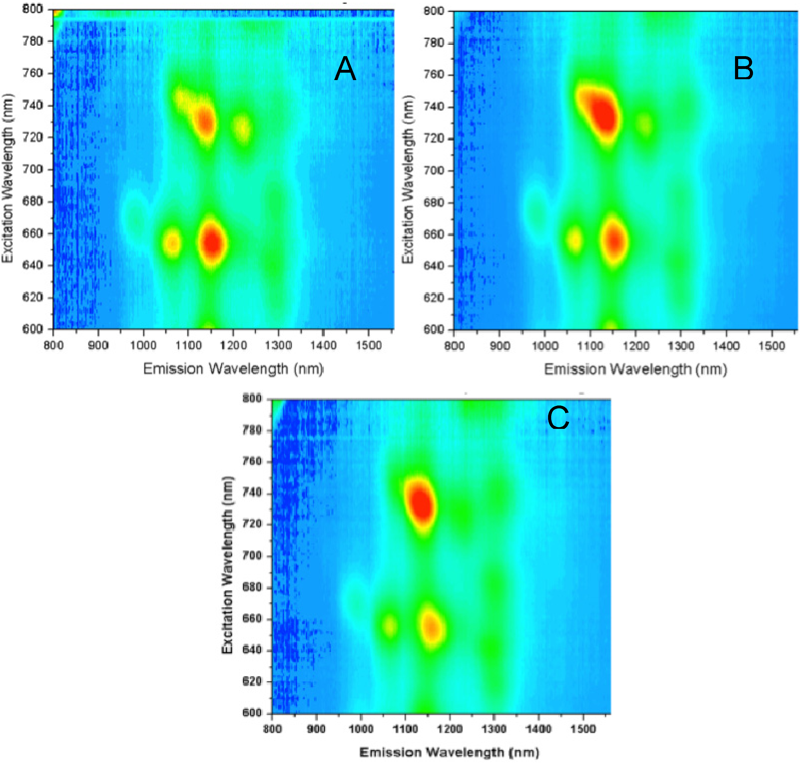
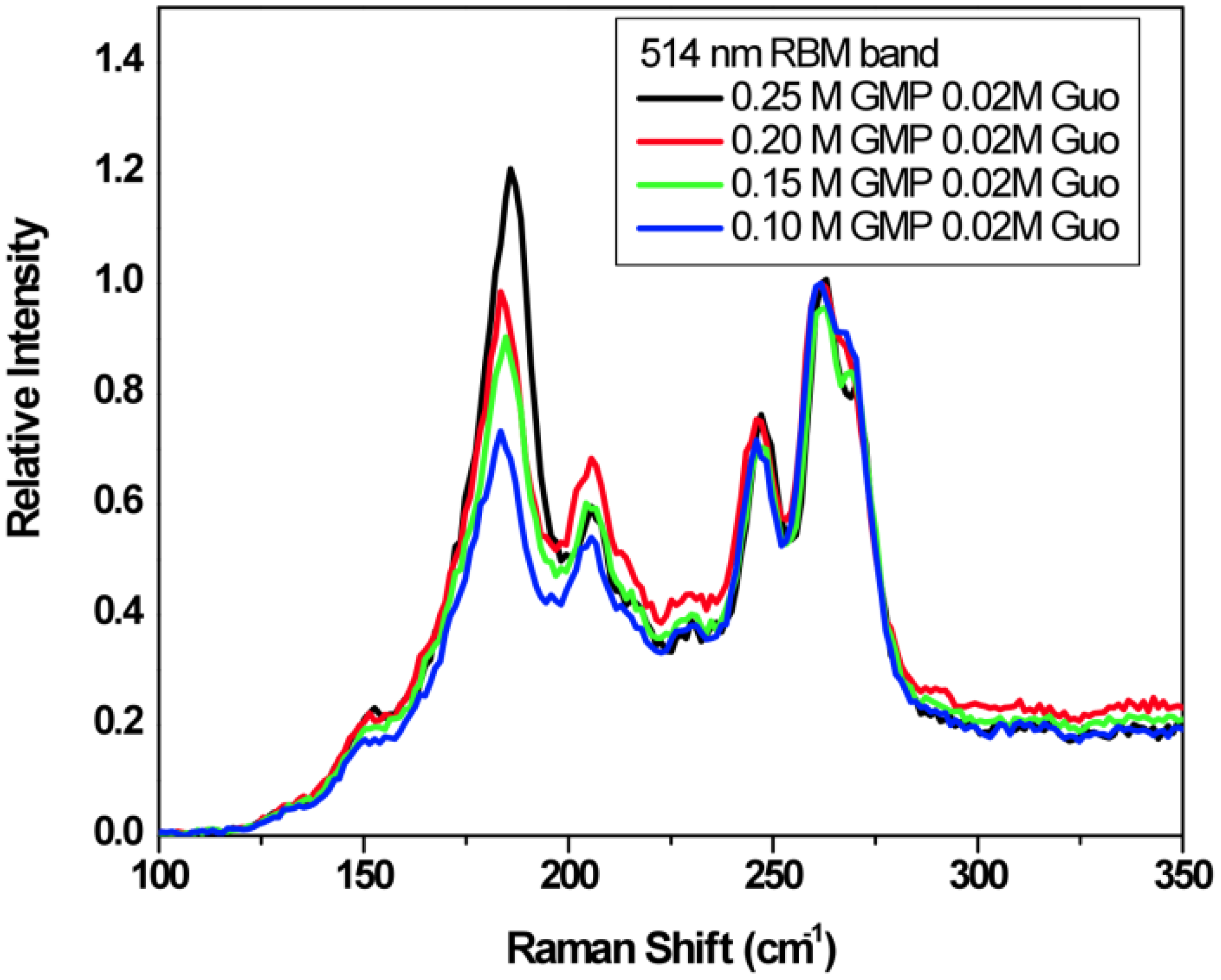
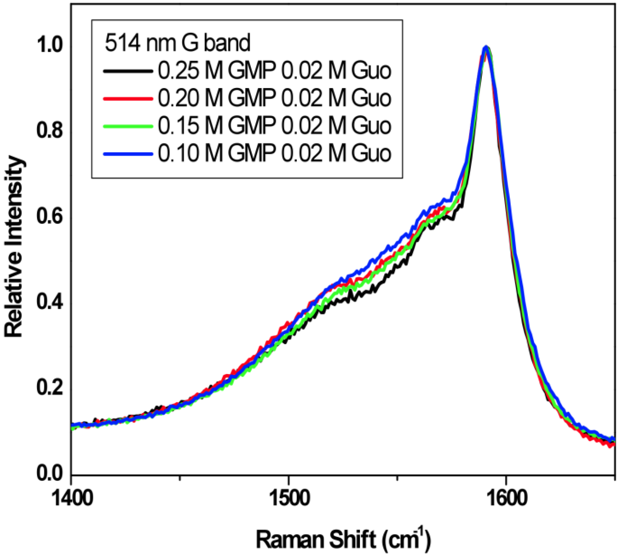
2.3. Spectroscopic Measurements


3. Experimental
3.1. Materials

3.2. Gel Preparation
3.3. Atomic Force Microscopy
3.4. UV-Visible Absorption Spectroscopy
3.5. Near-Infrared Fluorescence Spectroscopy
3.6. MicroRaman Spectroscopy
4. Conclusions
Acknowledgments
Conflicts of Interest
References
- Gellert, M.; Lipsett, M.N.; Davies, D.R. Helix formation by guanylic acid. Proc. Natl. Acad. Sci. USA 1962, 48, 2013–2018. [Google Scholar] [CrossRef]
- Chantot, J.F.; Sarocchi, M.T.; Guschlbauer, W. Physico-chemical properties of nucleosides. 4. Gel formation by guanosine and its analogues. Biochimie 1971, 53, 347–354. [Google Scholar] [CrossRef]
- Guschlbauer, W.; Chantot, J.-F.; Thiele, D. Four-stranded nucleic acid structures 25 years later: From guanosime gels to telomere dna. J. Biomol. Struct. Dyn. 1990, 8, 491–511. [Google Scholar] [CrossRef]
- Pieraccini, S.; Giorgi, T.; Masiero, S.; Spada, G.P.; Gottarelli, G. Guanosine derivatives: Self-assembly and lyotropic liquid crystal formation. Mol. Cryst. Liq. Cryst. 2003, 398, 57–73. [Google Scholar] [CrossRef]
- Davis, J.T. G-Quartets forty years later: From 5'GMP to molecular biology and supramolecular chemistry. Angew. Chem. Int. Ed. 2004, 43, 668–698. [Google Scholar] [CrossRef]
- Davis, J.T.; Spada, G.P. Supramolecular architectures generated by self-assembly of guanosine derivatives. Chem. Soc. Rev. 2007, 36, 296–313. [Google Scholar] [CrossRef]
- Marsh, T.C.; Vesenka, J.; Henderson, E. A new DNA nanostructure, the G-wire, imaged by scanning probe microscopy. Nucleic Acids Res. 1995, 23, 696–700. [Google Scholar] [CrossRef]
- Calzolari, A.; di Felice, R.; Molinari, E.; Garbesi, A. G-Quartet biomolecular nanowires. Appl. Phys. Lett. 2002, 80, 3331–3333. [Google Scholar] [CrossRef]
- Sasisekharan, V.; Zimmerman, S.; Davies, D.R. The Structure of Helical 5'-Guanosine Monophosphate. J. Mol. Biol. 1975, 92, 171–197. [Google Scholar] [CrossRef]
- Sreenivasachary, N.; Lehn, J.-M. Gelation-driven component selection in the generation of constitutional dynamic hydrogels based on guanine-quartet formation. Proc. Natl. Acad. Sci. USA 2005, 102, 5938–5943. [Google Scholar] [CrossRef]
- Walmsley, J.A.; Burnett, J.F. A new model for the K+-induced macromolecular structure of guanosine-5'-monophosphate in solution. Biochemistry 1999, 38, 14063–14068. [Google Scholar] [CrossRef]
- Spindler, L.; Olenik, I.D.; Copic, M.; Romih, R.; Cerar, J.; Skerjanc, J.; Mariani, P. Dynamic light scattering and 31P NMR spectroscopy study of the self-assembly of deoxyguanosine 5'-monophosphate. Eur. Phys. J. E 2002, 7, 95–102. [Google Scholar] [CrossRef]
- Yu, Y.; Nakamura, D.T.; DeBoyace, K.; Neisius, A.W.; McGown, L.B. Tunable thermoassociation of binary guanosine gels. J. Phys. Chem. B 2008, 112, 1130–1134. [Google Scholar] [CrossRef]
- Lokesh; Suryaparkash, N. Weakly ordered chiral alignment medium derived from 5'-GMP:guanosine. Chem. Commun. 2013, 49, 2049–2051. [Google Scholar] [CrossRef]
- Buerkle, L.E.; Li, Z.; Jamieson, A.M.; Rowan, S.J. Tailoring the properties of guanosine-based supramolecular hydrogels. Langmuir 2009, 25, 8833–8840. [Google Scholar] [CrossRef]
- Hersam, M.C. Progress towards monodisperse single-walled carbon nanotubes. Nat. Nanotechnol. 2008, 3, 387–394. [Google Scholar] [CrossRef]
- Hirsch, A. Functionalization of single-walled carbon nanotubes. Angew. Chem. 2002, 41, 1853–1859. [Google Scholar] [CrossRef]
- Ericson, L.M.; Fan, H.; Peng, H.; Davis, V.A.; Zhou, W.; Sulpizio, J.; Wang, Y.; Booker, R.; Vavro, J.; Guthy, C.; et al. Macroscopic, neat, single-walled carbon nanotube fibers. Science 2004, 305, 1447–1450. [Google Scholar] [CrossRef]
- Fujigaya, T.; Nakashima, N. Methodology for homogeneous dispersion of single-walled carbon nanotubes by physical modification. Polym. J. 2008, 40, 577–589. [Google Scholar] [CrossRef]
- Mitchell, C.A.; Bahr, J.L.; Arepalli, S.; Tour, J.M.; Krishnamoorti, R. Dispersion of functionalized carbon nanotubes in polystyrene. Macromolecules 2002, 35, 8825–8830. [Google Scholar]
- O’Connell, M.J.; Boul, P.; Ericson, L.M.; Huffman, C.; Wang, Y.; Haroz, E.; Kuper, C.; Tour, J.; Ausman, K.D.; Smalley, R.E. Reversible water- solubilization of single-walled carbon nanotubes by polymer wrapping. Chem. Phys. Lett. 2001, 342, 265–271. [Google Scholar] [CrossRef]
- Star, A.; Stoddart, J.F.; Steuerman, D.; Diehl, M.; Boukai, A.; Wong, E.W.; Yang, X.; Chung, S.W.; Choi, H.; Heath, J.R. Preparation and properties of polymer-wrapped single-walled carbon nanotubes. Angew. Chem. 2001, 40, 1721–1725. [Google Scholar] [CrossRef]
- Rice, N.A.; Soper, K.; Zhou, N.; Merschrod, E.; Zhao, Y. Dispersing as-prepared single-walled carbon nanotube powders with linear conjugated polymers. Chem. Commun. 2006, 47, 4937–4939. [Google Scholar]
- Simmons, T.J.; Bult, J.; Hashim, D.P.; Linhardt, R.J.; Ajayan, P.M. Noncovalent functionalization as an alternative to oxidative acid treatment of single wall carbon nanotubes with applications for polymer composites. ACS Nano 2009, 3, 865–870. [Google Scholar] [CrossRef]
- Matarredona, O.; Rhoads, H.; Li, Z.; Harwell, J.H.; Balzano, L.; Resasco, D.E. Dispersion of single-walled carbon nanotubes in aqueous solutions of the anionic surfactant NaDDBS. J. Phys. Chem. B 2003, 107, 13357–13367. [Google Scholar]
- Wang, D.; Ji, W.-X.; Li, Z.-C.; Chen, L. A biomimetic “Polysoap” for single-walled carbon nanotube dispersion. J. Am. Chem. Soc. 2006, 128, 6556–6557. [Google Scholar] [CrossRef]
- Zheng, M.; Jagota, A.; Semke, E.D.; Diner, B.A.; McLean, R.S.; Lustig, S.R.; Richardson, R.E.; Tassi, N.G. DNA-assisted dispersion and separation of carbon nanotubes. Nat. Mater. 2003, 2, 338–342. [Google Scholar] [CrossRef]
- Dieckmann, G.R.; Dalton, A.B.; Johnson, P.A.; Razal, J.; Chen, J.; Giordano, G.M.; Munoz, E.; Musselman, I.H.; Baughman, R.H.; Draper, R.K. Controlled assembly of carbon nanotubes by designed amphiphilic peptide helices. J. Am. Chem. Soc. 2003, 125, 1770–1777. [Google Scholar] [CrossRef]
- O’Connell, M.J.; Bachilo, S.M.; Huffman, C.B.; Moore, V.C.; Strano, M.S.; Haroz, E.H.; Rialon, K.L.; Boul, P.J.; Noon, W.H.; Kittrell, C.; et al. Band gap fluorescence from individual single-walled carbon nanotubes. Science 2002, 297, 593–596. [Google Scholar] [CrossRef]
- Moore, V.C.; Strano, M.S.; Haroz, E.H.; Hauge, R.H.; Smalley, R.E.; Schmidt, J.; Talmon, Y. Individually suspended single-walled carbon nanotubes in various surfactants. Nano Lett. 2003, 3, 1379–1382. [Google Scholar] [CrossRef]
- Gao, H.; Kong, Y. Simulation of DNA-nanotube interactions. Annu. Rev. Mater. Res. 2004, 34, 123–150. [Google Scholar] [CrossRef]
- Chattopadhayaz, D.; Galeska, I.; Papadimitrakopolaus, F. A route for bulk separation of semiconducting from metallic single-wall carbon nanotubes. J. Am. Chem. Soc. 2003, 125, 3370–3375. [Google Scholar] [CrossRef]
- Sample Availability: Samples of the GMP-Guanosine gel media and SWNT-gel constructs described in the text are available from the authors.
© 2013 by the authors; licensee MDPI, Basel, Switzerland. This article is an open access article distributed under the terms and conditions of the Creative Commons Attribution license (http://creativecommons.org/licenses/by/3.0/).
Share and Cite
Yu, Y.; Pushparaj, V.L.; Nalamasu, O.; McGown, L.B. G-Quadruplex Guanosine Gels and Single Walled Carbon Nanotubes. Molecules 2013, 18, 15434-15447. https://doi.org/10.3390/molecules181215434
Yu Y, Pushparaj VL, Nalamasu O, McGown LB. G-Quadruplex Guanosine Gels and Single Walled Carbon Nanotubes. Molecules. 2013; 18(12):15434-15447. https://doi.org/10.3390/molecules181215434
Chicago/Turabian StyleYu, Yuehua, Victor L. Pushparaj, Omkaram Nalamasu, and Linda B. McGown. 2013. "G-Quadruplex Guanosine Gels and Single Walled Carbon Nanotubes" Molecules 18, no. 12: 15434-15447. https://doi.org/10.3390/molecules181215434