Article

Genomic and Phenotypic Characteristics in Geographically Separated Clinical Campylobacter jejuni ST353CC Isolates

1
Clinical Microbiology, Department of Medical Sciences, Uppsala University, SE-75185 Uppsala, Sweden
2
Department of Animal Nutrition and Management, Swedish University of Agricultural Sciences, SE-75007 Uppsala, Sweden
3
Centre for Translational Microbiome Research, Department of Microbiology, Tumor and Cell Biology, Karolinska Institute, SE-17177 Stockholm, Sweden
*
Author to whom correspondence should be addressed.
Present address: Department of Ecology, Swedish University of Agricultural Sciences, SE-75007 Uppsala, Sweden.
Microorganisms 2021, 9(12), 2540; https://doi.org/10.3390/microorganisms9122540
Submission received: 6 October 2021 / Revised: 25 November 2021 / Accepted: 2 December 2021 / Published: 8 December 2021
(This article belongs to the Special Issue Recent Advances in Campylobacter jejuni and Helicobacter pylori)

Abstract

:
Campylobacter jejuni fecal isolates of eight international travelers, 5 of which had traveled to Ecuador and 3 to Bangladesh, were characterized, and the possible relationship between bacterial traits and clinical symptoms was further analyzed. All eight isolates belonged to the same Multi-Locus Sequence Type clonal complex (ST353CC). The three isolates from Bangladesh were all of the same sequence type (ST-9438), and when compared to isolates of various other sequence types, they had a larger quantity of unique genetic content, higher expression levels of some putative virulence genes involved in adhesion and invasion (flpA, ciaB and iamA), and showed higher adhesion levels to human HT-29 colon cancer cells in an in vitro infection model. However, in contrast to the seemingly higher pathogenic potential of these bacterial isolates, travelers infected with the ST-9438 isolates had no or only very mild symptoms, whereas the other individuals, whose bacterial isolates seemed to have less pathogenic potential, generally reported severe symptoms. When studying the 16S rRNA gene-based fecal microbiota in samples collected prior to travel, there was an individual variation in the relative abundance of the three major bacterial phyla Actinobacteria, Bacteroidetes and Firmicutes, but there were no associations between composition and diversity of microbiota and development of severe symptoms from the infection. It remains to be confirmed by larger studies whether an individual’s characteristics such as gut microbiota, might be related to the severity of symptoms in Campylobacter infections.

1. Introduction

Campylobacter is the most common cause of acute human bacterial gastroenteritis globally, and since 2005 it has been the most reported zoonosis in the European Union, with over 246,000 cases in 2018 [1]. It has been estimated that approximately 1% of European inhabitants develop campylobacteriosis every year [2].
C. jejuni is the most common pathogen in the genus Campylobacter, and Campylobacter infection usually develops a few days after ingestion of the bacteria. The severity of the disease varies, and symptoms include fever, headache, vomiting, and abdominal pain, in addition to watery or sometimes even bloody diarrhea [3]. Post-infectious sequelae, such as reactive arthritis [4], irritable bowel syndrome [5], and the severe autoimmune demyelinating neuropathy Guillain–Barré syndrome [6] may occur, which substantially increases the burden of the disease.
The C. jejuni population structure is commonly described using Multi-Locus Sequence Typing (MLST), and to date over 10,000 sequence types (STs) have been identified [7]. STs can be further grouped into clonal complexes (CCs), where some CCs are host- or niche-specific, i.e., specialists, and others are considered generalists that can colonize a wide variety of hosts worldwide [8]. The generalists ST21CC and ST45CC are the most common CCs among clinical isolates [9,10].
The mechanisms of Campylobacter–host interaction and pathogenesis are largely unclear. Putative virulence factors involved in, for example, capsule biosynthesis, motility and chemotaxis, adhesion and invasion, and stress responses have been proposed as contributors to the infection. The processes of adhesion and invasion have been well-studied. The bacterial adhesins CadF and FlpA bind fibronectin on the host-cell surface to initiate adhesion [11,12,13,14], while invasion factors such as CiaB and IamA are considered to be involved in the later stages of invasion [15,16,17]. The large number of Campylobacter whole-genome sequences available in public repositories has facilitated screening for the presence of putative virulence factors in individual isolates. Despite this, there is still no clear correlation between the mere presence of a gene, the virulence traits of that particular isolate, and the severity of infection. Instead, some studies on the gene expression of putative virulence genes have shown a correlation between the expression levels of genes involved in adhesion and invasion, and their level of adhesion to human cells in vitro [18,19].
To further characterize pathogenesis mechanisms, studies on bacteria themselves can be combined with in vitro infection studies using human cell lines. Adhesion and invasion of a particular Campylobacter isolate, and resultant downstream cellular processes such as the induction and release of specific cytokines, can be monitored. We have previously used HT29 cells, and a human colon cancer epithelial cell line for such studies, and have shown differences in adhesion-ability between different isolates of both C. jejuni [20,21] and C. coli [19,22].
In addition to the characteristics of enteropathogens, aspects of the host such as the composition of intestinal microbiota, may be crucial for the development of intestinal infections. We have previously shown differences between the composition of fecal microbiota from individuals who later became infected with Campylobacter, and those who remained uninfected [23,24].
In our previous study, we described C. jejuni infection in 14 international travelers [24]. Here, we show that eight of these individuals, although infected in two remote destinations (Bangladesh and Ecuador) harbored C. jejuni isolates belonging to the same MLST ST353CC. Our aim was to study various bacterial characteristics, and to analyze the possibility of bacterial traits being associated with symptoms in the infected travelers.

2. Materials and Methods

2.1. Data on Participants and Microbiota Analyses

This investigation was part of a larger study that is described above [24]. Data on eleven individuals infected with Campylobacter (Table 1) were extracted from questionnaires collected prior to this investigation. None of the participants had taken antibiotics within three months before taking a fecal sampling for the microbiota analyses. Symptoms as described by the participants in the questionnaires, were scored by a specialist in infectious diseases (CK), in compliance with existing guidelines [25,26], on a scale of one to five where 1 stood for no symptoms and 5 stood for the most severe symptoms (Table 2). The travelers to Ecuador lived together for several weeks before traveling and ate communal meals.
Written informed consent was obtained from each individual. The regional board of the ethics committee at Uppsala University approved the study (Dnr 2011/254).
The microbiota dataset was retrieved from an already-published study where 16S rRNA gene amplicons were compared between travelers who remained non-infected, and those who became infected during a trip to destinations with a high estimated risk of obtaining an enteric infection [24]. Thus, the procedure used for preparation of the fecal samples for amplification of the V3–V4 region of the 16S rRNA gene, has been described elsewhere [24].

2.2. Bacterial Culture Conditions

Campylobacter isolates had been isolated prior to this study [24]. Bacteria were grown microaerobically (Oxoid CampyGen, Thermo Fisher Scientific, Waltham, MA, USA) at 42 °C, and routinely resuscitated from frozen stocks by first growing overnight on blood agar (Columbia agar plates supplemented with 5% horse blood: Oxoid, Basingstoke, UK), followed by overnight incubation in Brucella broth (Becton, Dickinson and Company, Franklin Lakes, NJ, USA). To collect bacteria, cultures were centrifuged at 8000× g for 5–10 min.

2.3. Whole-Genome Sequencing

Total bacterial DNA was extracted using the MagNa Pure Compact Nucleic Acid isolation kit I (Roche, Penzberg, Germany) according to the manufacturer’s protocol (version 12). Of the eleven original isolates, two could not be resuscitated so DNA extracted from Campylobacter isolates during a previous study [24] was used. The libraries for whole genome sequencing were prepared with a Nextera XT sample preparation kit (Illumina, San Diego, CA, USA). An Illumina MiSeq platform, with 2 × 300 paired-end reads, was used for whole-genome sequencing. The single-reads were assembled to contigs with Velvet version 7.0.4 [27] running as a plugin in Geneious version 8.1.9 (https://www.geneious.com).

2.4. Genomics

New whole-genome sequences from this study were submitted to the Genbank database [28] (BioProject number PRJNA713788), and accession numbers are shown in Table 1. MLST-typing in this study was based on seven house-keeping genes, and performed by a sequence query of the whole-genome sequences against the PubMLST database [7] (last accessed: 5 September 2021). The allelic profiles of four of the isolates (three were identical) did not match any of the existing STs in the PubMLST database, and were submitted to PubMLST for assignment. The whole-genome phylogenetic trees were constructed in SplitsTree [29], using the average nucleotide identity (ANI) to closely related taxa calculated using the Gegenees software version 2.2.1 with a threshold of 20% [30]. Sequence alignments, ORF annotations, translations and phylogenetic analyses of individual genes were performed in the CLC Main Workbench (Qiagen, Hilden, Germany) using standard settings. The BLAST Ring Image Generator (BRIG) application was used to visualize similarities between pTet plasmids [31].

2.5. RNA Preparation from Bacteria and cDNA Synthesis

Bacterial RNA was extracted from overnight cultures using the ISOLATE II RNA Mini Kit (Bioline Reagents Ltd., London, UK) according to the manufacturer’s protocol. DNase I treatment (Ambion by life technologies, Carlsbad, CA, USA) was performed both on-column and on the final eluted RNA. The concentration of RNA was determined using Nanodrop, and RNA integrity was verified on a 1% agarose gel. A Maxima First Strand cDNA Synthesis Kit for qPCR (Thermo Fisher Scientific, Waltham, MA, USA) was used to reverse transcribe 1 µg of RNA according to the manufacturer’s protocol. A minus-RT control reaction was set up for each sample to rule out residual DNA. For qPCR 1/2000 of the cDNA synthesis reaction was used. At least two independent RNA preparations were used for real-time qPCR.

2.6. Quantitative PCR

Real-time qPCR was performed in the Bio–Rad CFX96 Touch cycler using the DyNAmo HS SYBR Green qPCR kit (Thermo Fisher Scientific, Waltham, MA, USA). Primer sequences for virulence genes are available upon request. DNA and cDNA samples were diluted 10–100 times prior to amplification. For virulence gene expression analyses, the expression levels of each gene were first normalized to that of 16S rRNA for each sample and run. Thereafter, expression levels were expressed relative to that of the C. jejuni 11168 reference strain. For each isolate, four biological replicates were available and one to two qPCRs were run for each replicate resulting in a total of six qPCRs per isolate. Results are presented as the mean for all replicates.

2.7. Cell Culture Conditions

The HT-29 human colon cancer cell line (ECACC 91072201) was maintained in RPMI 1640 media (Gibco by life technologies, Carlsbad, CA, USA) and supplemented with 2 mM glutamine (Swedish National Veterinary Institute, Uppsala, Sweden), 10% fetal bovine serum (FBS, Gibco by life technologies, Carlsbad, CA, USA), 100 U/mL penicillin, and 100 µg/mL streptomycin (Swedish National Veterinary Institute, Uppsala, Sweden).

2.8. In Vitro Cell Adhesion Assay and IL-8 ELISA

Adhesion to HT-29 cells, and IL-8 levels in the media were measured at 3 h post-infection, as previously described [20]. Results are presented as the mean of three infections.

3. Results

3.1. Phylogenetic Characterization of C. jejuni Isolates

Viable C. jejuni isolates were available from nine out of the 14 originally infected individuals [24], while from two of the individuals only Campylobacter DNA was available (Table 1). All eleven isolates for which DNA was available, were whole-genome sequenced. The CC and ST of each isolate was determined through a sequence query of the whole genome sequence against the PubMLST database [7]. Eight of the isolates, acquired from Bangladesh or Ecuador, were shown to belong to the same CC, ST353CC, while one isolate from Bangladesh belonged to ST354CC, and two isolates from Ecuador belonged to ST607CC (Table 1). Phylogenetic analyses of the whole-genome sequences confirmed the clustering of isolates, and revealed a separation of isolates from Bangladesh and Ecuador within ST353CC (Figure 1).

3.2. Genomic Characterization of C. jejuni ST353CC Isolates

Eight isolates belonged to the same clonal complex (ST353CC), of which three isolates from Bangladesh and two isolates from Ecuador even belonged to the same sequence types, ST-9438 and ST-9336, respectively (Table 1). These eight isolates were chosen for further genomic characterization.
Extensive in silico screening confirmed the presence of over 100 previously identified genes suggested to play a role in virulence, and involved in capsule biosynthesis, motility, chemotaxis, adhesion, invasion, stress response, and iron acquisition in all of the isolates (Supplementary Materials Table S1). Interestingly, the E00 isolate seemed to lack both the flaA and flaB genes that encode flagellin. Further in silico analyses of the putative virulence genes cadF, flpA, iamA, ciaB, ceuE, and cdt revealed a high level of similarity at both nucleotide and amino acid sequence levels in all the isolates, with only a few amino acids differing in each protein. However, five isolates (B30, B31, B44, E00, and E79) had an R135C substitution in the fibronectin-binding site (FRLS) of the CadF protein, due to a single nucleotide substitution (C403T) in the cadF gene.
Comparative genomic analysis was performed to identify genome regions that were unique to isolates belonging to different sequence types within ST353CC. Thirty annotated genes unique to the ST-9438 isolates from Bangladesh were detected (Supplementary Materials Table S2). Functional characteristics of each gene were assigned according to the Clusters of Orthologous Groups (COG) database for C. jejuni [32]. This revealed that ST-9438 isolates had a larger fraction of unique genes involved in information storage and processing than the isolates belonging to other STs.
The largest region unique to the ST-9438 isolates (B30, B31, and B44) consisted of approximately 50 k base pairs. BLASTn analyses showed that this region contained virB4, virB5, virB6, virB8, virB10, virB11, and virD4 genes, and was almost identical to the pTet plasmid detected in the C. jejuni reference strain 81-176, which is known to be highly invasive, and harbored tetO and topIII genes, a helicase, and an ATPase (Supplementary Materials Figure S1). Further assemblies, plasmid isolation, and agarose gel electrophoresis confirmed that this region exists as a circularized plasmid of 50.3 k base pairs in the ST-9438 isolates. The tetO gene was also found in the non-ST-9438 isolates, but at a different and chromosomally integrated location (Supplementary Materials Figure S2).

3.3. Various Symptom Scores in Travelers Infected with C. jejuni ST353CC Isolates

As the eight travelers were infected with bacterial isolates belonging to ST353CC, we wanted to see if the severity of symptoms was similar. Therefore, the severity of symptoms was scored. All infected individuals were asked to fill out questionnaires and to describe their symptoms in detail [24]. The reported symptoms were scored on a scale of 1 to 5, where 1 corresponded to no symptoms, and 5 to the most severe symptoms (extensive diarrhea, fever, and reduced general health) (Table 2). The mean symptom score for all of the eight individuals infected with ST353CC isolates was 2.7. However, on the individual level, the symptom scores ranged from 1 to 5 (Figure 2). Three of the eight individuals were all infected with isolates belonging to ST-9438 and had reported no (score 1) or only mild (score 2) symptoms (Figure 2), whereas the remaining five travelers mostly reported more severe symptoms (4 of 5 with a score ≥3, Figure 2).

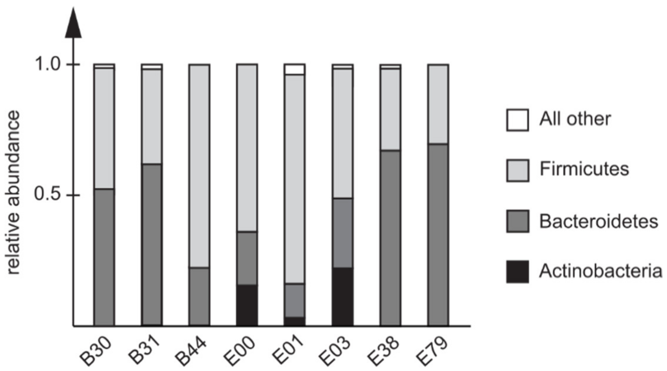
3.4. Pre-Travel Composition of the Fecal Microbiota in Travelers Infected with C. jejuni ST353CC Isolates

To further study the characteristics of travelers later infected with related C. jejuni ST353CC isolates, the pre-travel fecal microbiota results [24] of these individuals were compared. The bacterial alpha-diversity, expressed using the Shannon H index, did not differ between the individuals infected with ST-9438 isolates, and those that were not, in fecal samples collected before traveling. Moreover, prior to travel there was no difference in the alpha-diversity of the fecal microbiota for individuals who later reported no, or only minor symptoms (1–2 on the symptom scale) when compared to those who developed severe symptoms (3–5 on the symptom scale).
Three major bacterial phyla were detected in the fecal samples collected prior to traveling: Actinobacteria, Bacteroidetes, and Firmicutes. There was a clear individual variation in the relative abundances but, in line with the alpha-diversity data, there were no apparent correlations between microbial community compositions prior to the travel, and the development of severe symptoms (Figure 3).

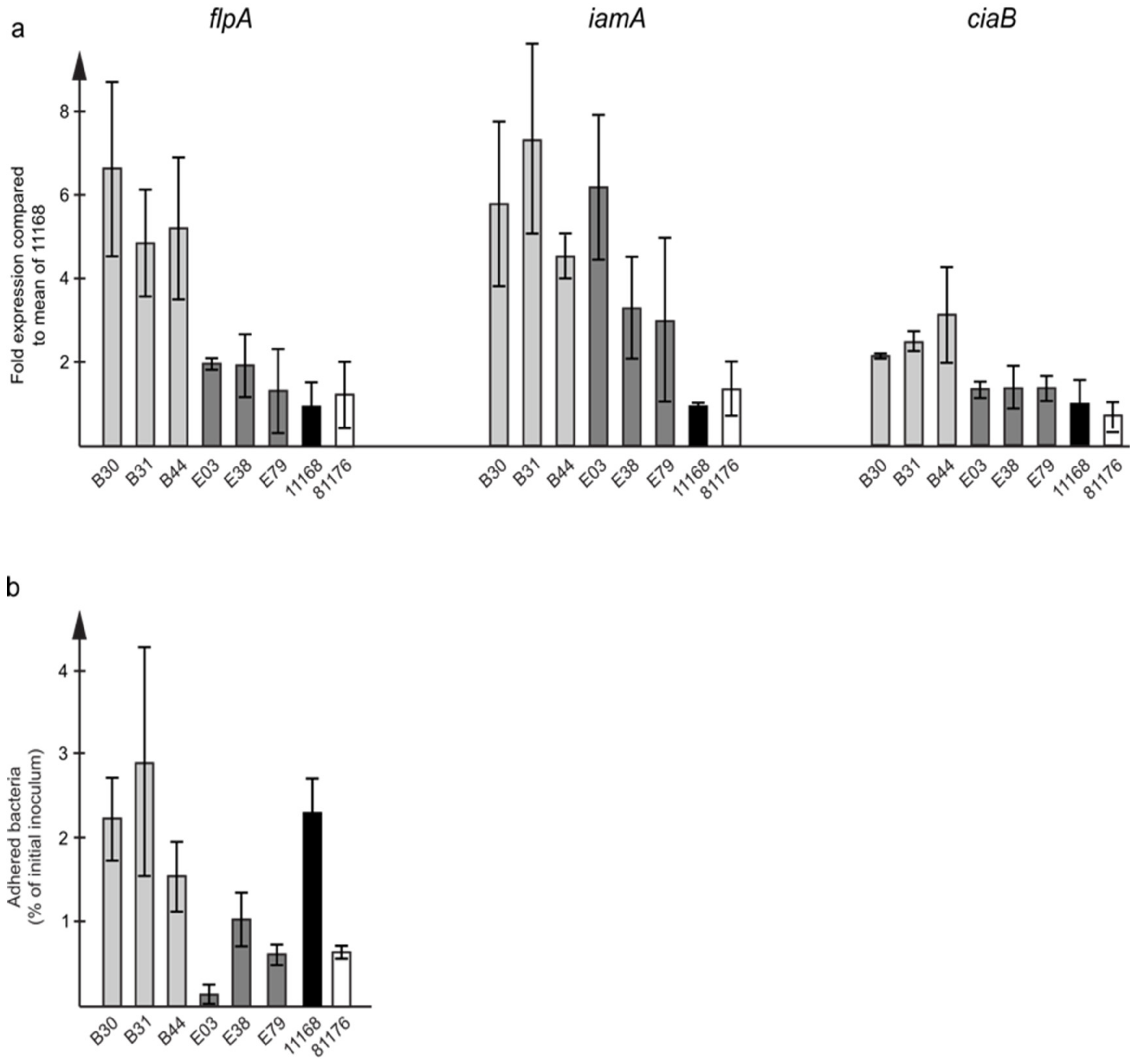
3.5. In Vitro Characteristics of C. jejuni ST353CC Isolates

Six viable ST353CC isolates, three from individuals infected with ST-9438 isolates who exhibited only minor symptoms (B30, B31, and B44), and three from individuals infected with isolates belonging to various STs who exhibited severe symptoms (E03, E38 and E79), were available for phenotypic analyses (Table 1). These isolates were analyzed for in vitro characteristics together with the C. jejuni 11168 and 81-176 reference strains.
When the expression of virulence genes involved in adhesion and invasion was measured, ST-9438 isolates had higher expression levels of the flpA, ciaB and iamA genes (Figure 4a), while the expression of cadF was similar in both groups.
The adhesion of the isolates to human HT-29 colon cancer cells was assessed using our previously established in vitro infection model [20]. The ST-9438 isolates clearly had higher levels of adhesion compared to the others (Figure 4b). However, the levels of induced IL-8 in the cells were similar for both groups.

4. Discussion

C. jejuni isolates of eleven healthy young individuals who had travelled to Bangladesh or Ecuador [24], were whole-genome sequenced and shown to belong to three globally distributed CCs: ST353CC, ST354CC, and ST607CC [33,34,35]. Despite the remote travel destinations and the high molecular diversity of the C. jejuni population, eight isolates, three from Bangladesh and five from Ecuador, were found to be genetically related, as they belonged to the same clonal complex (ST353CC). Here, we analyzed these ST353CC isolates as well as some characteristics of the infected hosts.
The clonal complex ST353CC accounts for 4.4% of all C. jejuni isolates in PubMLST and is globally distributed [7]. In our comparative genomic analysis, we identified differences between the ST353CC isolates. Three isolates, which all belonged to the same sequence type (ST-9438), were associated with less severe symptoms but had higher quantities of unique genetic content compared to the isolates relating to more severe infections. This is in line with our earlier findings on clinical ST21CC isolates, which harbored less accessory genetic content when isolated from blood in severe infections, when compared to isolates cultivated from fecal samples only [21].
As far as putative virulence genes were concerned, most that are involved in capsule biosynthesis, motility, chemotaxis, adhesion, invasion, stress response, and iron acquisition were equally present in all samples. To better understand the role of these genes in virulence, we looked at the expression levels of selected genes involved in adhesion and invasion. Higher expression levels for these genes were detected in the ST-9438 isolates despite the lower symptom scores in travelers who were infected with these isolates. Furthermore, in our in vitro infection model, higher levels of attached bacteria were demonstrated for the ST-9438 isolates. These findings, together with the results from our previous study on C. coli [19], show a direct correlation between the expression levels of genes relating to adhesion, and actual adherence capacity to human cells in an in vitro assay.
For cadF, which codes for the fibronectin-binding protein involved in adhesion, the expression levels were similar for all ST353CC isolates, irrespective of sequence type. However, we detected an interesting difference in the amino acid sequence of the CadF protein. The ST-9438 isolates had an R135C substitution in the fibronectin-binding site FRLS [36], possibly altering the fibronectin-binding capacity and contributing to their higher adhesion levels.
Altogether, our results emphasize the importance of looking at gene expression levels and gene product functions, rather than merely demonstrating the presence of a gene to understand the impact of experimental results regarding virulence. However, the picture as a whole seems to be even more complicated, and far from clear, as an inverse correlation was demonstrated between the levels of bacterial adhesion to human cells in vitro, and the severity of symptoms in individuals infected with the corresponding isolate.
Bacterial pathogenesis in general, and Campylobacter infection in particular, is difficult to study as the development of infection is not only influenced by bacterial characteristics, but is very likely to be influenced by characteristics of the host as well, including factors such as the composition of intestinal microbiota. Studies using murine infection models have indicated a correlation between the composition of intestinal microbiota and Campylobacter infection [37]. We have previously shown that the composition of intestinal microbiota had a significant impact on the susceptibility of humans to Campylobacter infection [23,24]. For instance, the relative abundance of Bacteroides was significantly higher among poultry abattoir workers who later became Campylobacter–positive as compared to subjects who remained uninfected [23].
In the present study, there were no apparent differences in either microbial diversity or community composition for study participants who later developed severe symptoms, compared to those with minor symptoms. Although no consistent difference has been found in the overall bacterial composition of the microbiota of vegetarians and vegans compared to omnivores, an increased abundance of Bacteroidetes, and of OTUs belonging to this phylum, has been demonstrated in vegetarians and vegans elsewhere [38,39,40,41].
It remains to be studied in a much larger number of individuals whether diet, or if specific microbial taxa, could be related to the development of more severe symptoms in Campylobacter–infected subjects, or whether C. jejuni isolates of ST-9348 cause less severe symptoms. After all, the comparative genomic analyses did not identify any specific or unique genes in the isolates that could have explained the more severe symptoms in infected individuals.

5. Conclusions

Here, we analyzed the bacterial and host characteristics for a very limited number of international travelers infected with genetically related (ST353CC) C. jejuni isolates. Travelers infected with ST-9438 isolates developed no, or only mild, symptoms. This is despite their Campylobacter isolates seeming to have more pathogenic potential in vitro compared to isolates of other sequence types within ST353CC, which had infected travelers who developed severe symptoms. Whether an individual’s characteristics (such as gut microbiota) might be related to the severity of symptoms in Campylobacter infection needs to be confirmed by much larger studies.

Supplementary Materials

The following are available online at https://www.mdpi.com/article/10.3390/microorganisms9122540/s1, Figure S1: BRIG analysis of plasmids identified in C. jejuni ST353CC isolates B30, B31 and B44 compared to the pTet plasmid from C. jejuni 81-176, Figure S2: Chromosomal location of a 2862 nucleotide long fragment containing the tetO gene in non-ST-9438 isolates, Table S1: Screening of putative virulence genes in C. jejuni ST353CC isolates sequenced in this study, Table S2: Results from comparative genomic analyses identifying genes unique for the C. jejuni ST353CC isolates sequenced in this study.

Author Contributions

Conceptualization, C.J. and H.R.; validation, C.J.; formal analysis, C.J., C.K. and J.D.; investigation, C.J., C.K. and A.N.; resources, L.E. and H.R; writing—original draft preparation, C.J.; writing—review and editing, C.J., C.K., J.D. and H.R.; visualization, C.J.; project administration, H.R.; funding acquisition, L.E. and H.R. All authors have read and agreed to the published version of the manuscript.

Funding

This research was funded by ALF grants from the Uppsala University Hospital (H.R.) and the Söderbergs Foundation (L.E.).

Data Availability Statement

All whole-genome sequences are available in GenBank [28] under the accession numbers shown in Table 1. Primers are available from the authors upon request. Additional data supporting the conclusions of this article are included within the article (and its Supplementary Materials).

Acknowledgments

We thank Emma Davies for the plasmid isolation and agarose gel electrophoresis. The results of the study have been presented as part of the doctoral thesis of Christian Kampmann.

Conflicts of Interest

The authors declare no conflict of interest.

References

  1. EFSA; ECDC. The European Union One Health 2018 Zoonoses Report. EFSA J. 2019, 17, 5926. [Google Scholar]
  2. Humphrey, T.; O’Brien, S.; Madsen, M. Campylobacters as zoonotic pathogens: A food production perspective. Int. J. Food Microbiol. 2007, 117, 237–257. [Google Scholar] [CrossRef] [PubMed]
  3. Blaser, M.J. Epidemiologic and clinical features of Campylobacter jejuni infections. J. Infect. Dis. 1997, 176 (Suppl. 2), S103–S105. [Google Scholar] [CrossRef] [Green Version]
  4. Hannu, T.; Mattila, L.; Rautelin, H.; Pelkonen, P.; Lahdenne, P.; Siitonen, A.; Leirisalo-Repo, M. Campylobacter-triggered reactive arthritis: A population-based study. Rheumatology 2002, 41, 312–318. [Google Scholar] [CrossRef] [PubMed] [Green Version]
  5. Rodriguez, L.A.; Ruigomez, A. Increased risk of irritable bowel syndrome after bacterial gastroenteritis: Cohort study. BMJ 1999, 318, 565–566. [Google Scholar] [CrossRef] [PubMed] [Green Version]
  6. McCarthy, N.; Giesecke, J. Incidence of Guillain-Barre syndrome following infection with Campylobacter jejuni. Am. J. Epidemiol. 2001, 153, 610–614. [Google Scholar] [CrossRef]
  7. PubMLST. Sequence Query—Campylobacter Locus/Sequence Definitions. 2021. Available online: https://pubmlst.org/bigsdb?db=pubmlst_campylobacter_seqdef&page=sequenceQuery (accessed on 5 September 2021).
  8. Gripp, E.; Hlahla, D.; Didelot, X.; Kops, F.; Maurischat, S.; Tedin, K.; Alter, T.; Ellerbroek, L.; Schreiber, K.; Schomburg, D.; et al. Closely related Campylobacter jejuni strains from different sources reveal a generalist rather than a specialist lifestyle. BMC Genom. 2011, 12, 584. [Google Scholar] [CrossRef] [Green Version]
  9. Sheppard, S.K.; Dallas, J.F.; MacRae, M.; McCarthy, N.D.; Sproston, E.L.; Gormley, F.J.; Strachan, N.J.; Ogden, I.D.; Maiden, M.C.; Forbes, K.J. Campylobacter genotypes from food animals, environmental sources and clinical disease in Scotland 2005/6. Int. J. Food Microbiol. 2009, 134, 96–103. [Google Scholar] [CrossRef] [Green Version]
  10. Epping, L.; Antão, E.M.; Semmler, T. Population biology and comparative genomics of Campylobacter species. In Fighting Campylobacter Infections. Current Topics in Microbiology and Immunology; Backert, S., Ed.; Springer: Cham, Switzerland, 2021; p. 431. [Google Scholar]
  11. Flanagan, R.C.; Neal-McKinney, J.M.; Dhillon, A.S.; Miller, W.G.; Konkel, M.E. Examination of Campylobacter jejuni putative adhesins leads to the identification of a new protein, designated FlpA, required for chicken colonization. Infect. Immun. 2009, 77, 2399–2407. [Google Scholar] [CrossRef] [Green Version]
  12. Konkel, M.E.; Garvis, S.G.; Tipton, S.L.; Anderson, D.E., Jr.; Cieplak, W., Jr. Identification and molecular cloning of a gene encoding a fibronectin-binding protein (CadF) from Campylobacter jejuni. Mol. Microbiol. 1997, 24, 953–963. [Google Scholar] [CrossRef]
  13. Konkel, M.E.; Larson, C.L.; Flanagan, R.C. Campylobacter jejuni FlpA binds fibronectin and is required for maximal host cell adherence. J. Bacteriol. 2010, 192, 68–76. [Google Scholar] [CrossRef] [PubMed] [Green Version]
  14. Ziprin, R.L.; Young, C.R.; Stanker, L.H.; Hume, M.E.; Konkel, M.E. The absence of cecal colonization of chicks by a mutant of Campylobacter jejuni not expressing bacterial fibronectin-binding protein. Avian Dis. 1999, 43, 586–589. [Google Scholar] [CrossRef]
  15. Carvalho, A.C.; Ruiz-Palacios, G.M.; Ramos-Cervantes, P.; Cervantes, L.E.; Jiang, X.; Pickering, L.K. Molecular characterization of invasive and noninvasive Campylobacter jejuni and Campylobacter coli isolates. J. Clin. Microbiol. 2001, 39, 1353–1359. [Google Scholar] [CrossRef] [PubMed] [Green Version]
  16. Konkel, M.E.; Kim, B.J.; Rivera-Amill, V.; Garvis, S.G. Identification of proteins required for the internalization of Campylobacter jejuni into cultured mammalian cells. Adv. Exp. Med. Biol. 1999, 473, 215–224. [Google Scholar]
  17. Ziprin, R.L.; Young, C.R.; Byrd, J.A.; Stanker, L.H.; Hume, M.E.; Gray, S.A.; Kim, B.J.; Konkel, M.E. Role of Campylobacter jejuni potential virulence genes in cecal colonization. Avian Dis. 2001, 45, 549–557. [Google Scholar] [CrossRef]
  18. Johansson, C.; Nilsson, A.; Kaden, R.; Rautelin, H. Campylobacter coli clade 3 isolates induce rapid cell death in vitro. Appl. Environ. Microbiol. 2019, 85, e02993-18. [Google Scholar] [CrossRef] [Green Version]
  19. Johansson, C.; Nilsson, A.; Kaden, R.; Rautelin, H. Differences in virulence gene expression between human blood and stool Campylobacter coli clade 1 ST828CC isolates. Gut Pathog. 2019, 11, 42. [Google Scholar] [CrossRef]
  20. Nilsson, A.; Johansson, C.; Skarp, A.; Kaden, R.; Engstrand, L.; Rautelin, H. Genomic and phenotypic characteristics of Swedish C. jejuni water isolates. PLoS ONE 2017, 12, e0189222. [Google Scholar] [CrossRef] [Green Version]
  21. Skarp, C.P.A.; Akinrinade, O.; Kaden, R.; Johansson, C.; Rautelin, H. Accessory genetic content in Campylobacter jejuni ST21CC isolates from feces and blood. Int. J. Med. Microbiol. 2017, 307, 233–240. [Google Scholar] [CrossRef]
  22. Nilsson, A.; Skarp, A.; Johansson, C.; Kaden, R.; Engstrand, L.; Rautelin, H. Characterization of Swedish Campylobacter coli clade 2 and clade 3 water isolates. Microbiologyopen 2018, 7, e00583. [Google Scholar] [CrossRef] [PubMed] [Green Version]
  23. Dicksved, J.; Ellström, P.; Engstrand, L.; Rautelin, H. Susceptibility to Campylobacter infection is associated with the species composition of the human fecal microbiota. mBio 2014, 5, e01212-14. [Google Scholar] [CrossRef] [Green Version]
  24. Kampmann, C.; Dicksved, J.; Engstrand, L.; Rautelin, H. Composition of human faecal microbiota in resistance to Campylobacter infection. Clin. Microbiol. Infect. 2016, 22, 61.e1–61.e8. [Google Scholar] [CrossRef] [Green Version]
  25. Riddle, M.S.; DuPont, H.L.; Connor, B.A. ACG Clinical Guideline: Diagnosis, treatment, and prevention of acute diarrheal infections in adults. Am. J. Gastroenterol. 2016, 111, 602–622. [Google Scholar] [CrossRef]
  26. Riddle, M.S.; Connor, B.A.; Beeching, N.J.; DuPont, H.L.; Hamer, D.H.; Kozarsky, P.; Libman, M.; Steffen, R.; Taylor, D.; Tribble, D.R.; et al. Guidelines for the prevention and treatment of travelers’ diarrhea: A graded expert panel report. J. Travel Med. 2017, 24 (Suppl. 1), S63–S80. [Google Scholar] [CrossRef] [PubMed] [Green Version]
  27. Zerbino, D.R.; Birney, E. Velvet: Algorithms for de novo short read assembly using de Bruijn graphs. Genome Res. 2008, 18, 821–829. [Google Scholar] [CrossRef] [PubMed] [Green Version]
  28. National Center for Biotechnology Information. Genbank. 2019. Available online: https://www.ncbi.nlm.nih.gov/genbank/ (accessed on 19 June 2019).
  29. Huson, D.H.; Bryant, D. Application of phylogenetic networks in evolutionary studies. Mol. Biol. Evol. 2006, 23, 254–267. [Google Scholar] [CrossRef]
  30. Ågren, J.; Sundstrom, A.; Hafstrom, T.; Segerman, B. Gegenees: Fragmented alignment of multiple genomes for determining phylogenomic distances and genetic signatures unique for specified target groups. PLoS ONE 2012, 7, e39107. [Google Scholar] [CrossRef]
  31. Alikhan, N.F.; Petty, N.K.; Ben Zakour, N.L.; Beatson, S.A. BLAST Ring Image Generator (BRIG): Simple prokaryote genome comparisons. BMC Genom. 2011, 12, 402. [Google Scholar] [CrossRef] [Green Version]
  32. National Center for Biotechnology Information. COG List—Camjej 2019. Available online: https://ftp.ncbi.nih.gov/pub/COG/COG2014/static/lists/listCamjej.html (accessed on 23 August 2019).
  33. Islam, Z.; van Belkum, A.; Wagenaar, J.A.; Cody, A.J.; de Boer, A.G.; Sarker, S.K.; Jacobs, B.C.; Talukder, K.A.; Endtz, H.P. Comparative population structure analysis of Campylobacter jejuni from human and poultry origin in Bangladesh. Eur. J. Clin. Microbiol. Infect. Dis. 2014, 33, 2173–2181. [Google Scholar] [CrossRef] [Green Version]
  34. Vasco, K.; Graham, J.P.; Trueba, G. Detection of zoonotic enteropathogens in children and domestic animals in a semirural community in Ecuador. Appl. Environ. Microbiol. 2016, 82, 4218–4224. [Google Scholar] [CrossRef] [PubMed] [Green Version]
  35. Vinueza-Burgos, C.; Wautier, M.; Martiny, D.; Cisneros, M.; Van Damme, I.; De Zutter, L. Prevalence, antimicrobial resistance and genetic diversity of Campylobacter coli and Campylobacter jejuni in Ecuadorian broilers at slaughter age. Poult. Sci. 2017, 96, 2366–2374. [Google Scholar] [CrossRef]
  36. Konkel, M.E.; Christensen, J.E.; Keech, A.M.; Monteville, M.R.; Klena, J.D.; Garvis, S.G. Identification of a fibronectin-binding domain within the Campylobacter jejuni CadF protein. Mol. Microbiol. 2005, 57, 1022–1035. [Google Scholar] [CrossRef] [PubMed]
  37. Bereswill, S.; Fischer, A.; Plickert, R.; Haag, L.M.; Otto, B.; Kühl, A.A.; Dashti, J.I.; Zautner, A.E.; Muñoz, M.; Loddenkemper, C.; et al. Novel murine infection models provide deep insights into the “menage a trois” of Campylobacter jejuni, microbiota and host innate immunity. PLoS ONE 2011, 6, e20953. [Google Scholar] [CrossRef]
  38. De Filippis, F.; Pellegrini, N.; Vannini, L.; Jeffery, I.B.; La Storia, A.; Laghi, L.; Serrazanetti, D.I.; Di Cagno, R.; Ferrocino, I.; Lazzi, C.; et al. High-level adherence to a Mediterranean diet beneficially impacts the gut microbiota and associated metabolome. Gut 2016, 65, 1812–1821. [Google Scholar] [CrossRef]
  39. Gentile, C.R.; Weir, T.L. The gut microbiota at the intersection of diet and human health. Science 2018, 362, 776–780. [Google Scholar] [CrossRef] [Green Version]
  40. Losasso, C.; Eckert, E.M.; Mastrorilli, E.; Villiger, J.; Mancin, M.; Patuzzi, I.; Di Cesare, A.; Cibin, V.; Barrucci, F.; Pernthaler, J.; et al. Assessing the influence of vegan, vegetarian and omnivore oriented westernized dietary styles on human gut microbiota: A cross sectional study. Front. Microbiol. 2018, 9, 317. [Google Scholar] [CrossRef] [PubMed]
  41. Franco-de-Moraes, A.C.; de Almeida-Pititto, B.; da Rocha Fernandes, G.; Gomes, E.P.; da Costa Pereira, A.; Ferreira, S.R.G. Worse inflammatory profile in omnivores than in vegetarians associates with the gut microbiota composition. Diabetol. Metab. Syndr. 2017, 9, 62. [Google Scholar] [CrossRef] [Green Version]
Figure 1. ANI-based whole-genome sequence phylogenetic tree of the eleven isolates sequenced in this study, together with the C. jejuni 11168 and 81-176 reference strains, showing the separation of ST353CC, ST354CC, and ST607CC.
Microorganisms 09 02540 g001
Figure 2. Symptom scores in individuals infected with C. jejuni ST353CC isolates. Three individuals (B30, B31, and B44) were infected with ST-9438 isolates and had traveled to Bangladesh. The rest of the individuals had traveled to Ecuador. Symptoms score 1–5: 1 indicates no symptoms, and 5 indicated the most severe symptoms.
Microorganisms 09 02540 g002
Figure 3. Composition of fecal microbiota prior to travel in individuals later infected with C. jejuni ST353CC isolates. The relative abundance of detectable phyla is shown for each individual. Three individuals (B30, B31, and B44) included meat in their diet whereas the rest of the subjects adhered to a non-meat diet.
Microorganisms 09 02540 g003
Figure 4. In vitro bacterial characteristics of C. jejuni ST353CC isolates. (a) RNA expression of putative virulence genes flpA, iamA, and ciaB involved in adhesion and invasion, analyzed by RT-qPCR. Data are expressed relative to the C. jejuni 11168 reference strain, and shown as mean of six PCR-experiments for each isolate together with data for the 11168 and 81-176 reference strains. (b) HT-29 cells were infected with C. jejuni ST353CC isolates at an MOI (multiplicity of infection) of 100. The C. jejuni 11168 and 81-176 reference strains were included for comparison. Adhered bacteria at 3 h post-infection were quantified using qPCR and expressed as a percentage of the starting inoculum. The graph shows the mean of three independent infection experiments for each isolate.
Microorganisms 09 02540 g004
Table 1. Campylobacter–positive individuals and their C. jejuni isolates (n = 11).
Travel DestinationGenderAge
(Years)
IsolateCC/ST #Accession
BangladeshF24B29354/354JAGFPS000000000
F24B30353/9438 ^JAGFPT000000000
F28B31353/9438 ^JAGFPR000000000
F26B44353/9438 ^JAGFPU000000000
EcuadorF29E00 *353/462JAGFPV000000000
M24E01 *353/9336JAGFPN000000000
F20E03353/9336JAGFPO000000000
F20E38353/9437 ^JAGFPP000000000
F23E61607/607JAGFPQ000000000
F19E63607/607JAGFPL000000000
F22E79353/3515JAGFPM000000000
(# according to MLST assignment; * only DNA available; ^ novel STs).
Table 2. Scoring of intestinal symptoms based on questionnaires filled out by travelers.
ScoreSymptomDescription
1No symptoms
2Mild symptomsSlight abdominal discomfort, ≤3 loose stools in 24 h, symptoms did not force any change in planned daily activities
3Severe diarrhea>3 loose stools in 24 h + enteric symptoms (e.g., fecal urgency, nausea, abdominal pain/cramps, emesis), symptoms did not force a significant change in planned daily activities, duration ≤3 consecutive days
4Severe diarrhea>3 loose stools in 24 h + enteric symptoms for >3 consecutive days, symptoms forced a significant change in planned daily activities and/or affected general health
5Severe diarrheaBloody diarrhea, and/or fever, and/or severely affected general health
If no exact fecal frequency was provided, score was based on the total information and graded down.
Publisher’s Note: MDPI stays neutral with regard to jurisdictional claims in published maps and institutional affiliations.

