Article

Diversity of Pectobacteriaceae Species in Potato Growing Regions in Northern Morocco

1
Department of Biology, Faculty of Sciences, Moulay Ismaïl University, 50000 Meknes, Morocco
2
Institute for Integrative Biology of the Cell (I2BC), Université Paris-Saclay, CEA, CNRS, 91198 Gif-sur-Yvette, France
3
Biotechnology Research Unit, CRRA-Rabat, National Institut for Agricultural Research (INRA), 10101 Rabat, Morocco
4
National Federation of Seed Potato Growers (FN3PT-RD3PT), 75008 Paris, France
5
Department of Plant Pathology, University of Agriculture Faisalabad Sub-Campus Depalpur, 38000 Okara, Pakistan
6
INRA, US 1426, GeT-PlaGe, Genotoul, 31320 Castanet-Tolosan, France
*
Author to whom correspondence should be addressed.
Microorganisms 2020, 8(6), 895; https://doi.org/10.3390/microorganisms8060895
Submission received: 28 April 2020 / Revised: 30 May 2020 / Accepted: 9 June 2020 / Published: 13 June 2020
(This article belongs to the Special Issue Dickeya and Pectobacterium: Ecology, Pathology and Plant Protection)

Abstract

:
Dickeya and Pectobacterium pathogens are causative agents of several diseases that affect many crops worldwide. This work investigated the species diversity of these pathogens in Morocco, where Dickeya pathogens have only been isolated from potato fields recently. To this end, samplings were conducted in three major potato growing areas over a three-year period (2015–2017). Pathogens were characterized by sequence determination of both the gapA gene marker and genomes using Illumina and Oxford Nanopore technologies. We isolated 119 pathogens belonging to P. versatile (19%), P. carotovorum (3%), P. polaris (5%), P. brasiliense (56%) and D. dianthicola (17%). Their taxonomic assignation was confirmed by draft genome analyses of 10 representative strains of the collected species. D. dianthicola were isolated from a unique area where a wide species diversity of pectinolytic pathogens was observed. In tuber rotting assays, D. dianthicola isolates were more aggressive than Pectobacterium isolates. The complete genome sequence of D. dianthicola LAR.16.03.LID was obtained and compared with other D. dianthicola genomes from public databases. Overall, this study highlighted the ecological context from which some Dickeya and Pectobacterium species emerged in Morocco, and reported the first complete genome of a D. dianthicola strain isolated in Morocco that will be suitable for further epidemiological studies.

1. Introduction

Pectinolytic Pectobacterium and Dickeya spp. are causative agents of severe diseases in a wide range of plants of high economic value [1,2]. On potato tubers and stems, the diseases caused by pectinolytic pathogens are soft rot and blackleg, respectively. These pathogens produce a large set of extracellular enzymes that degrade the plant cell wall, resulting in plant tissue decay and maceration. This rotting process causes losses in the production of potato tubers sold both on the food market and as certified seed tubers [3]. The pathogens may be acquired by the host plants from soil and/or from contaminated seed tubers [4]. On plants, pathogen populations remain at a low level in asymptomatic plant tissues, and may become particularly destructive when environmental conditions favor their proliferation and the expression of virulence factors.
P. atrosepticum was considered as the primary pathogen responsible for the rotting of stored potato tubers and wilting of potato plants under temperate climates [4]. Other Pectobacterium species frequently associated with damage of potato crops are P. carotovorum, P. brasiliense, P. parmentieri and P. polaris [5,6,7,8,9]. Some Pectobacterium species have also been characterized in some specific areas, such as P. peruviense strains isolated from tubers in Peru, and P. punjabense species from symptomatic potato plants in Pakistan [10,11]. P. odoriferum exhibits a very wide host range, including potato plants [12], while some other species were characterized by a more restricted host range, at least in the fields. Thus, P. wasabiae was isolated from symptomatic Japanese horseradish [13]; P. betavasculorum was reported almost exclusively on sugar beet [14]; P. aroidearum exhibits a preference for some monocotyledonous plants [15]; P. zantedeschiae strains were isolated from Zantedeschia spp. (Calla lily) [16]; and P. actinidiae from symptomatic Actinidia chinensis (kiwi fruit) [17]. Recently, some other species, isolated from surface waters, have also been described: P. fontis, P. aquaticum and P. versatile [7,18,19]. Altogether, 16 Pectobacterium species have been described so far [7].
A limited number of Dickeya species, i.e., D. dianthicola, D. dadantii and D. solani, have been associated with symptoms on potatoes. D. dianthicola was first reported on potatoes in the Netherlands in the 1970s, and has been detected since then in many other European countries [20]. D. dadantii causes soft rot disease in several members of the Solanaceae family, including the potato [21]. Another virulent species, namely D. solani, spread rapidly throughout Western Europe [22] and in Russia [23], and into other countries such as Turkey [24], Georgia [25] and Brazil [26]. During the past decade, the taxonomy of Dickeya and Pectobacterium species was revisited following genomic studies bearing on international culture collections and diverse ecosystems around the world [27,28]. By now, the genus Dickeya encompasses 10 species: D. aquatica, D. chrysanthemi, D. dadantii, D. dianthicola, D. fangzhongdai, D. lacustris, D. paradisiaca, D. solani, D. undicola, and D. zeae [22,29,30,31,32,33,34]. Bacteria belonging to this genus cause plant diseases in temperate, tropical and subtropical climates [35].
The unambiguous identification of Dickeya and Pectobacterium species is crucial for epidemiological purposes, to develop appropriate prophylactic approaches and quality controls in national and international trade exchanges. Multi-Locus Sequence Analysis (MLSA) provides relevant information for a better understanding of speciation, and hence for proposing pertinent taxa delineations [15,36]. MLSA may exploit gene sequences, obtained by PCR-sequencing of several loci or by whole genome sequencing. Among the loci commonly included in MLSA, the rrs sequence is poorly informative at a species level, while the gapA gene appeared as an appropriate marker to discriminate the different Dickeya and Pectobacterium species [10,18,19,31,32,37]. Taxonomy of Dickeya and Pectobacterium gained precision and robustness with additional genome analyses, such as average nucleotide identity (ANI) and in silico DNA–DNA hybridization (isDDH) [38]. Comparative genomics is also used to identify species-specific DNA regions. Analysis of the functions encoded by these DNA regions allows the prediction of species-specific metabolic traits. This knowledge contributes to the understanding of both the taxonomy and ecology of the Dickeya and Pectobacterium pathogens [22,30,31,32,39].
P. atrosepticum, P. carotovorum and P. brasiliense were described in Morocco as the main causative agents of blackleg and soft rot diseases in potato crop [40,41,42,43]. In 2016, D. dianthicola was described for the first time in the North of Morocco [44]. In this respect, the main objectives of this study were: (i) to investigate the species composition of the Moroccan Dickeya and Pectobacterium populations, collected between 2015 and 2017 from diseased potato tubers and stems, (ii) to compare the aggressiveness of some identified pathogens belonging to different species, and (iii) to propose a complete genome of the emerging pathogen D. dianthicola in Morocco, that could be used for further studies as a reference genome. This work represents the most important sampling effort of the Pectobacterium and Dickeya potato pathogens in Morocco over the past decade.

2. Materials and Methods

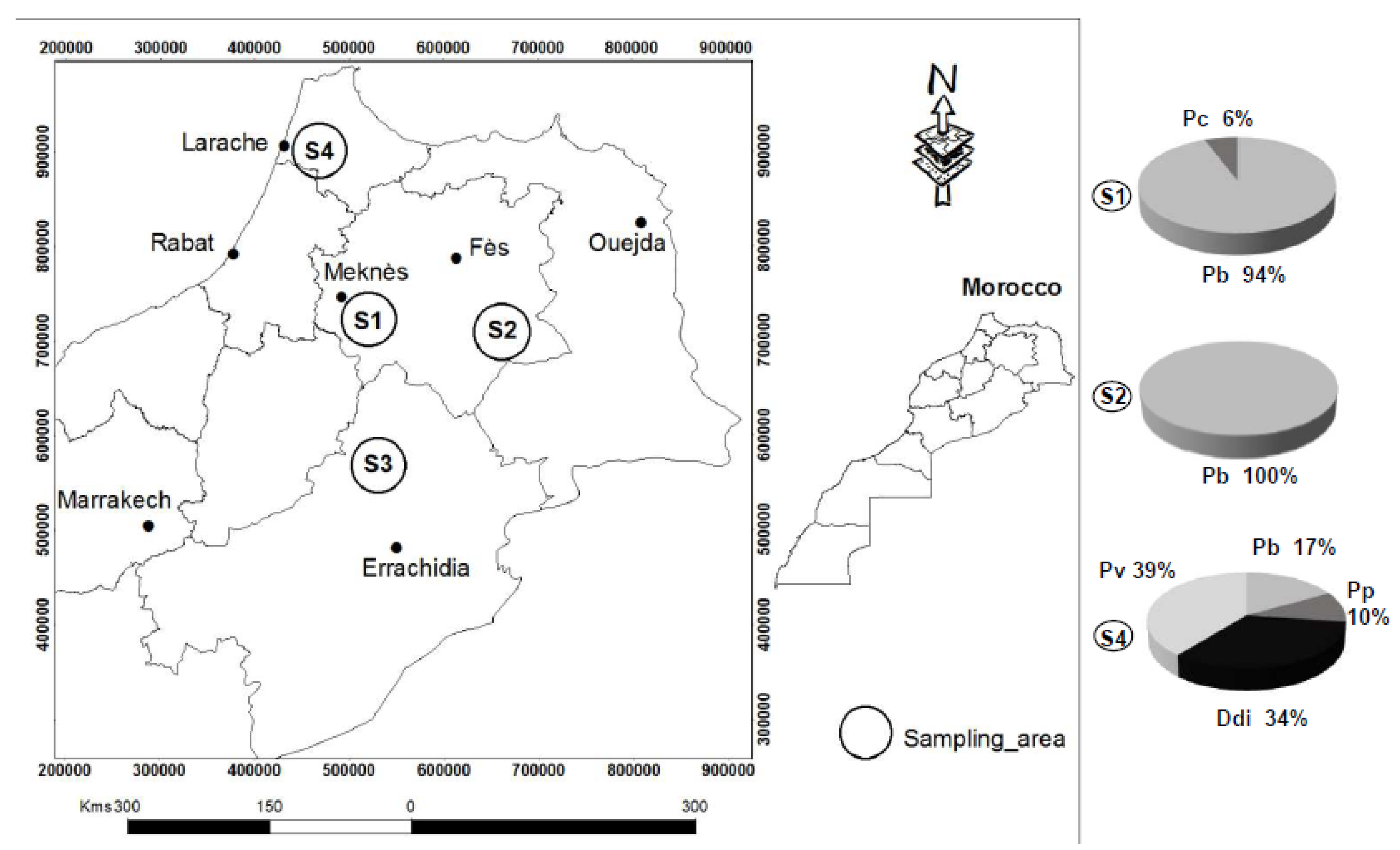
2.1. Sampling and Isolation of Pectinolytic Bacteria

In 2015, 2016 and 2017, blackleg symptoms were searched for in potato fields in four regions (Meknes, Guigo, Boumia and Larache) in Northern Morocco. Pectinolytic bacteria were isolated from symptomatic plant tissues using crystal violet pectate (CVP) medium as described previously [45]. The CVP plates were incubated at 28 °C for 3 days and colonies that had formed pits were re-streaked onto Tryptone (5 g/L) yeast extract (3 g/L) agar medium (TY). The purified isolates were spotted again on CVP to confirm the pectinolytic activity. The obtained cultures from single colonies were stored in 25% glycerol at −80 °C.

2.2. Molecular Characterization of Pectobacterium and Dickeya Isolates

The primer couples Y1/Y2 and ADE1/ADE2 (Table S1) were used for the identification of isolates belonging to Pectobacterium and Dickeya genera [46,47]. The reaction was carried out in a final volume of 25 μL, containing 1 μL of bacterial DNA (50 ng/µL), 2.5 μL of PCR buffer (10×), 2 μL of MgCl2 (25 mM), 2.5 µL of dNTPs (1 mM), 1U Taq polymerase and 1 µL of each primer (1 µM) and water. The temperature settings for PCR were the same as described before [46,47]. The analysis of PCR products was done by electrophoresis on 2% agarose gels, using PCR products of P. atrosepticum CFBP1526T and D. solani IPO2222T as control along with the 1 Kb DNA ladder.
Positive strains for either Y1/Y2 or ADE1/ADE2 PCR were further characterized using the gapA barcode procedure [37]. All the gapA PCR products obtained with gapA-F/gapA-R primers (Table S1) were sequenced using Sanger technology (GATC Biotech, Konstanz, Germany). The sequences were trimmed using the CLC genomic workbench (V10.1.1, Aarhus, Denmark) and aligned using ClustalW. The phylogenetic analysis of the gapA gene was performed as follows: the evolutionary distances were computed using the maximum composite likelihood method (Mega7 software, Pennsylvania State University Park, PA, USA) with 1000 bootstrap. The obtained sequences were deposited in GenBank (Table S2).

2.3. Genome Sequencing

A total of 10 strains representing 5 species identified with gapA sequencing were selected for genome sequencing (Table 1). DNA of the 10 isolates (listed in Table 1) was extracted from overnight cultures in TY medium using the MasterPure™ Complete DNA and RNA Purification Kit (Epicenter, Madison, WI, USA) followed by an ethanol precipitation. The quantity and quality control of the DNA was completed using a NanoDrop (Wilmington, DE, USA) device and 1.0% agarose gel electrophoresis.
Paired-end libraries (500 bp in insert size) were constructed for each strain, and DNA sequencing was performed by Illumina NextSeq technology. Sequencing of the library was carried out using the 2 × 75 bp paired-end read module. Illumina sequencing was performed at the I2BC sequencing platform (Gif-sur-Yvette, France).
In the case of the D. dianthicola strain LAR.16.03.LID, Nanopore sequencing was also performed. Library preparation and sequencing were performed at the GeT-PlaGe core facility, INRA Toulouse, using the “1D Native barcoding genomic DNA kit (EXP-NBD103 and SQK-LSK108)”, according to the manufacturer’s instructions. At each step, DNA was quantified using the Qubit dsDNA HS Assay Kit (Life Technologies). DNA purity was tested using a NanoDrop device (Thermofisher, Waltham, MA, USA) and size distribution and degradation was assessed using the fragment analyzer (AATI) High Sensitivity DNA Fragment Analysis Kit. Purification steps were performed using AMPure XP beads (Beckman Coulter). Quantities of 5 µg of each DNA (five samples) were purified then sheared at 20 kb using the Megaruptor1 system (Diagenode, Seraing, Belgium). A DNA damage repair step was performed on 3 µg of sample. Then END-repair and dA-tailing of double stranded DNA fragments were performed on 1 µg of each sample. Then, a specific index was ligated to each sample. The library was generated by an equimolar pooling of these barcoded samples. Then adapters were ligated to the library. The library was loaded on a R9.4.1 flowcell and sequenced on MinION instrument at 0.15 pmol within 48 h.

2.4. Genome Assembly

Assembly of the Illumina reads was performed using the CLC Genomics Workbench v10.1.1 software (CLCInc, Aarhus, Denmark). After quality (quality score threshold 0.05) and length (above 40 nucleotides) trimming of the reads, contigs were generated by de novo assembly (CLC parameters: automatic determination of the word and bubble sizes with no scaffolding). The draft genome sequences of each strain were deposited at NCBI and annotated using the NCBI Prokaryotic Genome Annotation Pipeline. Statistics of all the ten draft genomes are presented in Table 1.
Assembly of the Nanopore reads was performed as follows. Fast5s from Nanopore sequencing were obtained with MinKNOW version 1.10.23 and were basecalled with ONT Albacore Sequencing Pipeline Software version 2.1.10 and reads passing the internal test were used for subsequent analysis. Porechop 0.2.1 (https://github.com/rrwick/Porechop) was used for adaptor trimming. Illumina paired-end reads were processed with trim_galore 0.4.0 (https://github.com/FelixKrueger/TrimGalore), to trim adaptor sequences. Nanopore reads were assembled using Canu 1.7 [48] with the “genomeSize = 5 m” and “minReadLength = 3000” options. For Nanopore-only assembly, one output contig was obtained, then polished three times using Pilon 1.22 (https://github.com/broadinstitute/pilon), with the “--mindepth 25” option. The contig was finally circularized using Circlator 1.5.1 (https://github.com/sanger-pathogens/circlator).

2.5. Genome Analysis

Phylogenetic and molecular evolutionary analyses were conducted using MEGA, version 7. An MLSA was performed using 13 concatenated housekeeping genes (fusA, rpoD, acnA, purA, gyrB, recA, mdh, mtlD, groEL, secY, glyA, gapA, rplB) retrieved from all the Pectobacterium spp. and Dickeya spp. strains to confirm their phylogenetic position within the reference strains P. atrosepticum ICMP1526T, P. betavasculorum NCPPB2795T, P. parmentieri RNS 08-42.1AT, P. wasabiae CFBP 3304T, P. actinidiae KKH3, P. brasiliense LMG21371T, P. odoriferum BCS7, P. aroidearum PC1, D. dianthicola NCPPB 453T, D. dadantii NCPPB 898T, and D. solani IPO2222T. The average nucleotide identity (ANI) value was calculated as previously proposed using the ANI calculator (http://enveomics.ce.gatech.edu/ani/, Atlanta, GA, USA). The in-silico DNA–DNA hybridization (isDDH) was evaluated using genome sequence-based species delineation (http://ggdc.dsmz.de/, Braunschweig, Germany) (Table 2).
The genome map of the D. dianthicola strain LAR.16.03.LID was generated using CGView Server [48]. Synteny analysis of the complete genomes of D. dianthicola LAR.16.03.LID, D. dianthicola ME23 and D. dianthicola RNS049 was performed using the MAUVE software [49]. Paired end reads for the strain LAR.16.03.LID were mapped against the two complete genome sequences of D. dianthicola strains ME23 and RNS049 with threshold set as 0.8 of identity on 0.5 of read length using CLC Genomics Workbench version 10.1.1 software. The mappings were used for detection of variations (SNPs and InDels) using the basic variant calling tool from the CLC genomic workbench version 10.1.1.
The presence of clustered regularly interspaced short palindromic repeats (CRISPRs) was determined using CRISPRfinder (http://crispr.i2bc.paris-saclay.fr/Server/, Orsay, France) [50]. The prophage identification tool PHAge Search Tool—Enhanced Release (PHASTER) was used to check for the regions containing prophage-like elements in bacterial genomes (http://phaster.ca/, Edmonton, AB, Canada) [51]. The Predicted resistome was checked using Resistance Gene Identifier tool (https://card.mcmaster.ca/analyze/rgi, Hamilton, ON, Canada). Finally, genomic regions containing secondary metabolite biosynthesis gene clusters were identified using the AntiSMASH server (version 4.1.0, https://doi.org/10.1093/nar/gkv437, Hørsholm, Denmark).
To investigate the phylogenetic position of the Moroccan D. dianthicola strain against the available genomes of this species in NCBI, an MLSA was generated using 15 housekeeping genes (fusA, rpoD, leuS, rpoS, purA, infB, gyrB, recA, groEL, secY, glyA, gapA, rplB, dnaX, gyrA) with the MEGA7 software.

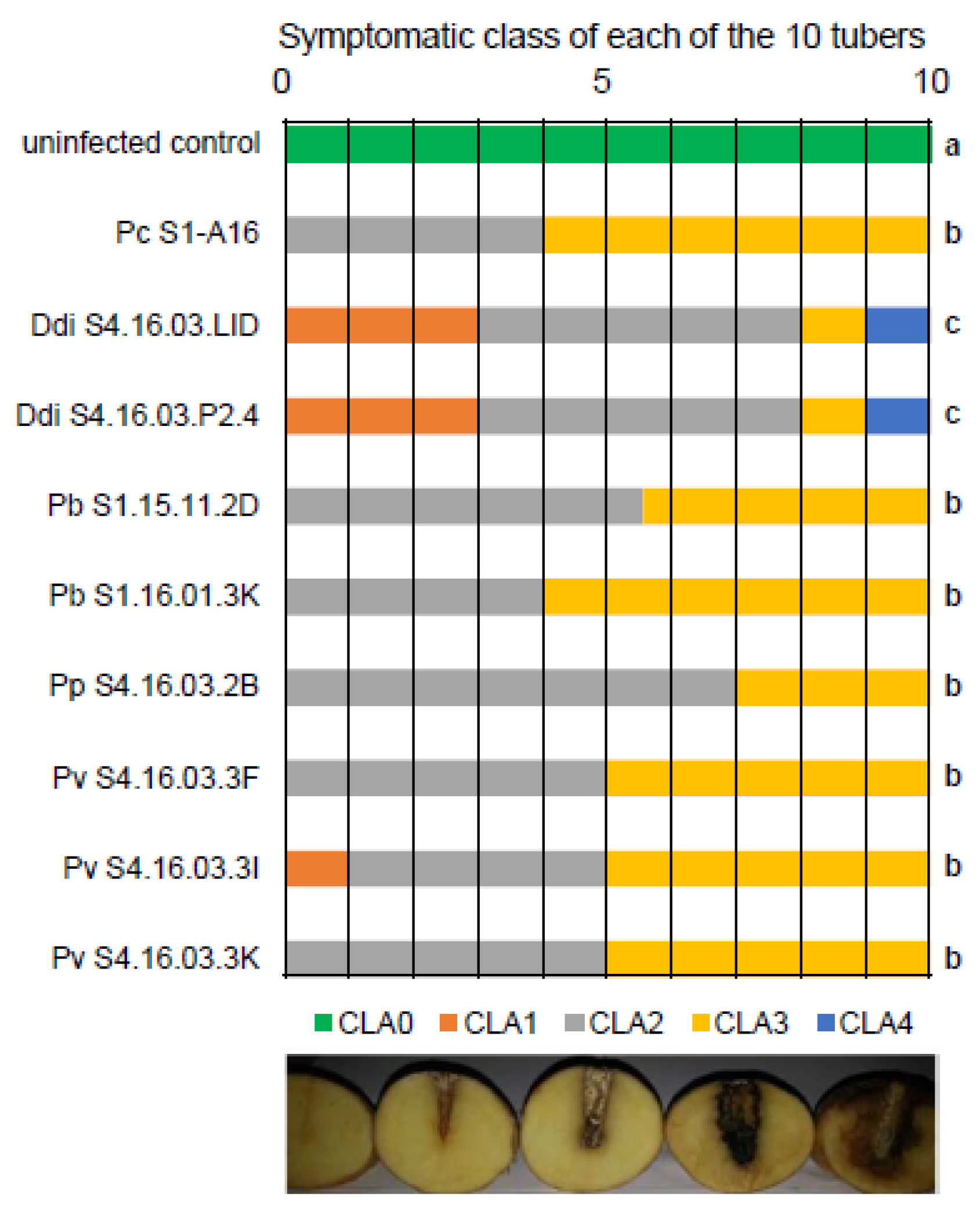
2.6. Potato Tuber Rotting Assays

Bacterial strains from Morocco were cultivated in TY broth for 24 h at 28 °C in a rotary shaker set at 125 rpm. Bacterial cultures were washed twice, resuspended in 0.8% NaCl, and the optical density was adjusted to OD600 = 1.0. Potato tubers (cv. Bintje) were surface-disinfected by submerging them into a 5% sodium hypochlorite solution for 5 min. They were subsequently rinsed twice in distilled water and air dried at room temperature one day before inoculation. A total of 10 potato tubers were infected with 10 µL of cell suspension of each strain, along with 10 tubers with NaCl 0.8% alone as a negative control. After 5 days of incubation at 24 °C, the tubers were cut vertically through the inoculation points. Disease symptoms were evaluated to define five aggressiveness classes [52]. Significance of the observed differences was assessed using a Kruskal–Wallis test (p < 0.05).

3. Results

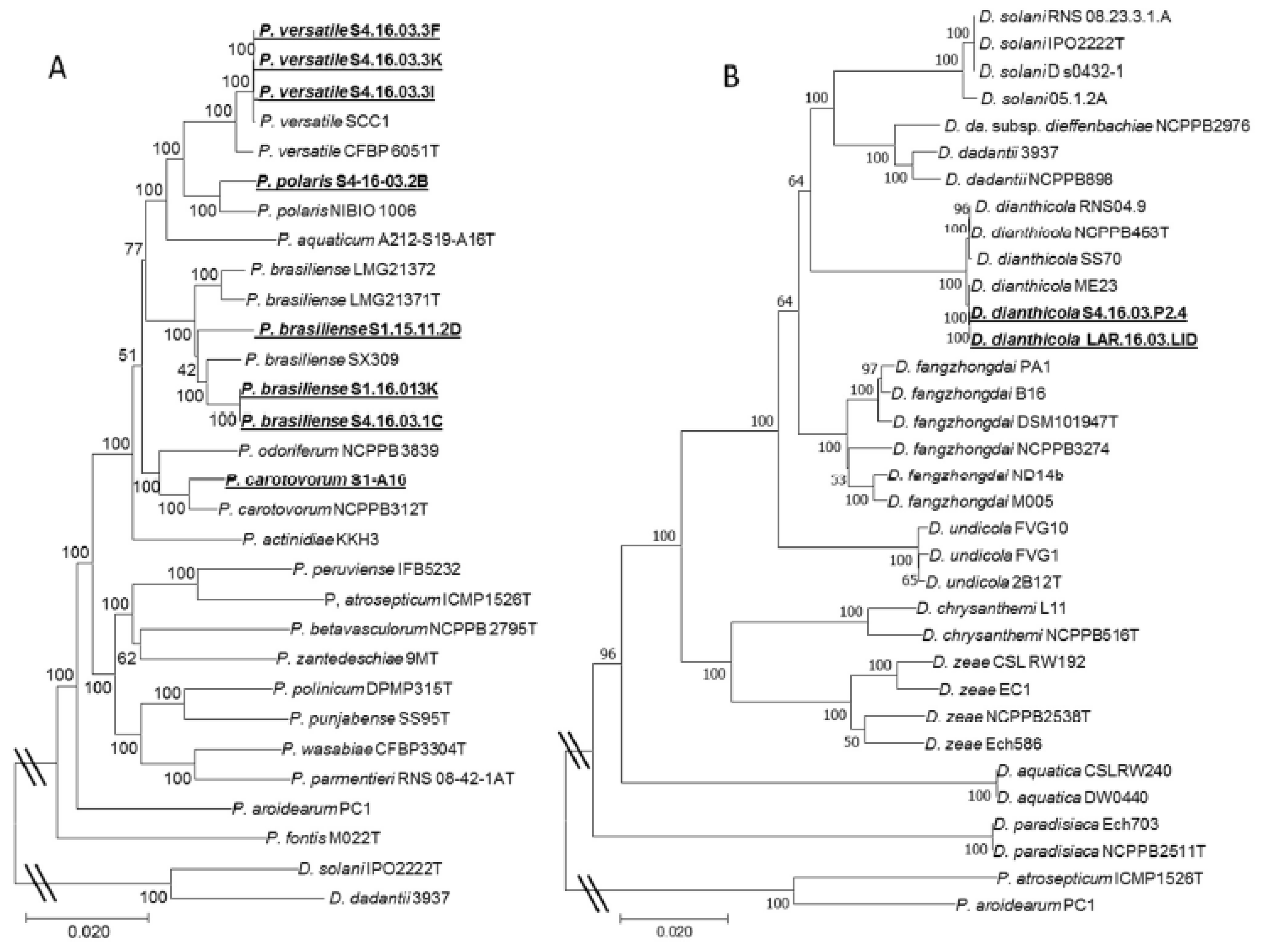
3.1. Diversity of the Pectinolytic Dickeya and Pectobacterium in Northern Morocco

From 2015 to 2017, our field inspections revealed the occurrence of blackleg symptoms in several potato growing areas, located in many townships distributed in three regions (Meknes, S1; Guigo, S2; Larache, S4) in northern Morocco (Figure 1). No symptoms were found in fields in the Boumia region (S3). Out of 200 strains isolated from plant symptoms, 140 provoked cavities on the pectate-containing medium. These were tested by PCR to evaluate whether they belonged to the Pectobacterium and Dickeya genera: 119 isolates generated amplification signals for either the Y1/Y2 or ADE1/ADE2 primer couples. Most of the isolates (83%) generated a signal with the Pectobacterium primers Y1/Y2, while the others (17%) did so with the Dickeya-specific ADE1/ADE2 primers.
All these PCR-positive Pectobacterium/Dickeya isolates were further characterized at species level based on their gapA gene sequence. Phylogenetic analyses using the Neighbor-Joining method (Mega7) of the gapA sequences are presented in the Figure S1. The regional diversity of the Dickeya and Pectobacterium isolates is summarized in Figure 1 (a detailed list is given in Table S2). The samples of the Larache region (S4) showed the highest diversity of taxons with the presence of D. dianthicola (20 isolates), P. polaris (6 isolates), P. brasiliense (5 isolates) and P. carotovorum (23 isolates). On the other hand, our investigations revealed the presence of only two species, P. brasiliense (53 isolates) and P. carotovorum (3 isolates), in the Meknes region (S1), and only a single one, P. brasiliense (9 isolates), in the Guigo region (S2).
Important information was also collected from the farmers regarding the potato variety, origin of the seed tubers (local production or importation), irrigation mode (surface water from a dam or underground water from wells) and geography (the sampled regions). We tested whether a correlation between these different parameters and the diversity of pathogens (the combination of Dickeya and Pectobacterium species) existed. Statistical analysis with the SAS (Statistical Analysis System, version 9.00, SAS Institute, 2002, Cary, NC, USA) software (Qui2 test with p < 0.05) revealed that the higher diversity of pathogens was associated with three confounding factors: geography (the unique Larache region), surface water irrigation and imported seed tubers (Table S1).

3.2. Draft Genomes of 10 Pectinolytic Bacteria from Northern Morocco

A draft genome (Illumina technology) was used to consolidate the taxonomic position of 10 isolates belonging to the collected taxons. Between 705,755 and 17,016,482 trimmed reads were used for the contigs assembly of each of the 10 genomes. Characteristics of the draft genomes are presented in Table 1. Genome data were exploited to retrieve 13 housekeeping genes from each genome using BLAST. The concatenated genes were used for MLSA. The MLSA tree (Figure 2) showed a similar topology to the one generated by the gapA analysis (Figure S1).
Genomic data were also used to calculate ANI and isDDH values. Most of the Moroccan strains exhibited an ANI value higher than 95%, and an isDDH value higher than 70% with the closest type strains, confirming their taxonomic assignation. Strains belonging to the P. versatile clade showed an isDDH lower than 70%, but an ANI value higher than 95% with the strain SCC1. Recently, another study confirmed the classification of the strains S4.16.03.3I (= CFBP8660), S4.16.03.3F (= CFBP8659) and SCC1 into the P. versatile species [7].

3.3. Aggressiveness of the Pectinolytic Bacteria from Northern Morocco

Of the 10 Moroccan strains whose genome sequence is available (Table 1), all but 1 (P. brasiliense S4.16.03.1C) were tested for aggressiveness on potato tubers. P. brasiliense strain S4.16.03.1C was isolated from the same field as P. brasiliense strain S1.16.01.3K, and they showed 100% identify by ANI (Table 2). Hence, we retained one of the two for tuber maceration assays. For each of the nine strains, 10 tubers were inoculated, and the resulting maceration symptoms were classified into five symptomatic classes (Figure 3). In addition, 10 tubers were used as uninfected control. The aggressiveness was compared between all the strains using a Kruskal–Wallis test. All the pathogens provoked symptoms that are different to the control condition. No significant difference was observed between strains belonging to the same species. In contrast, D. dianthicola strains showed a higher aggressiveness when compared with Pectobacterium strains (p value < 0.05).

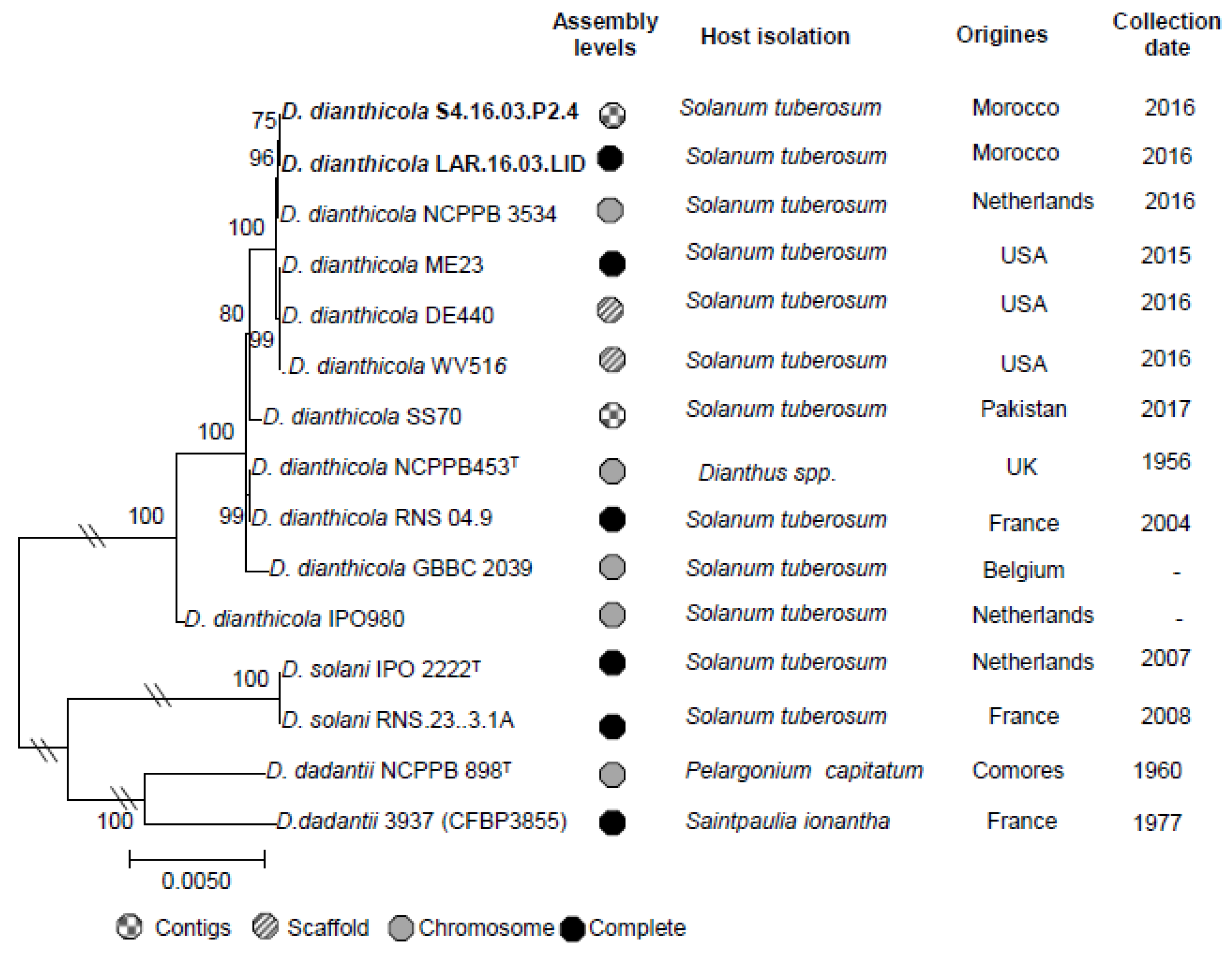
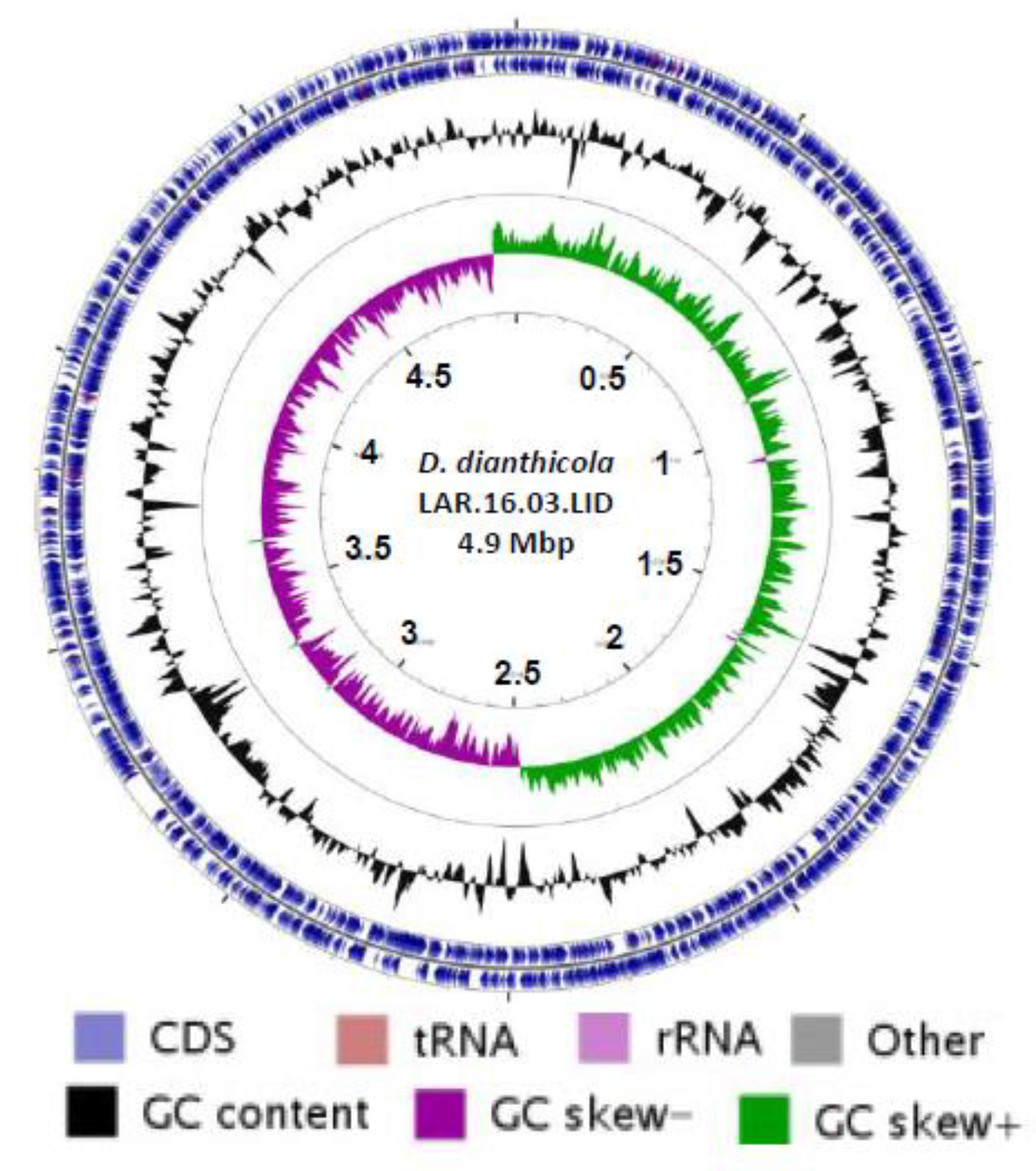
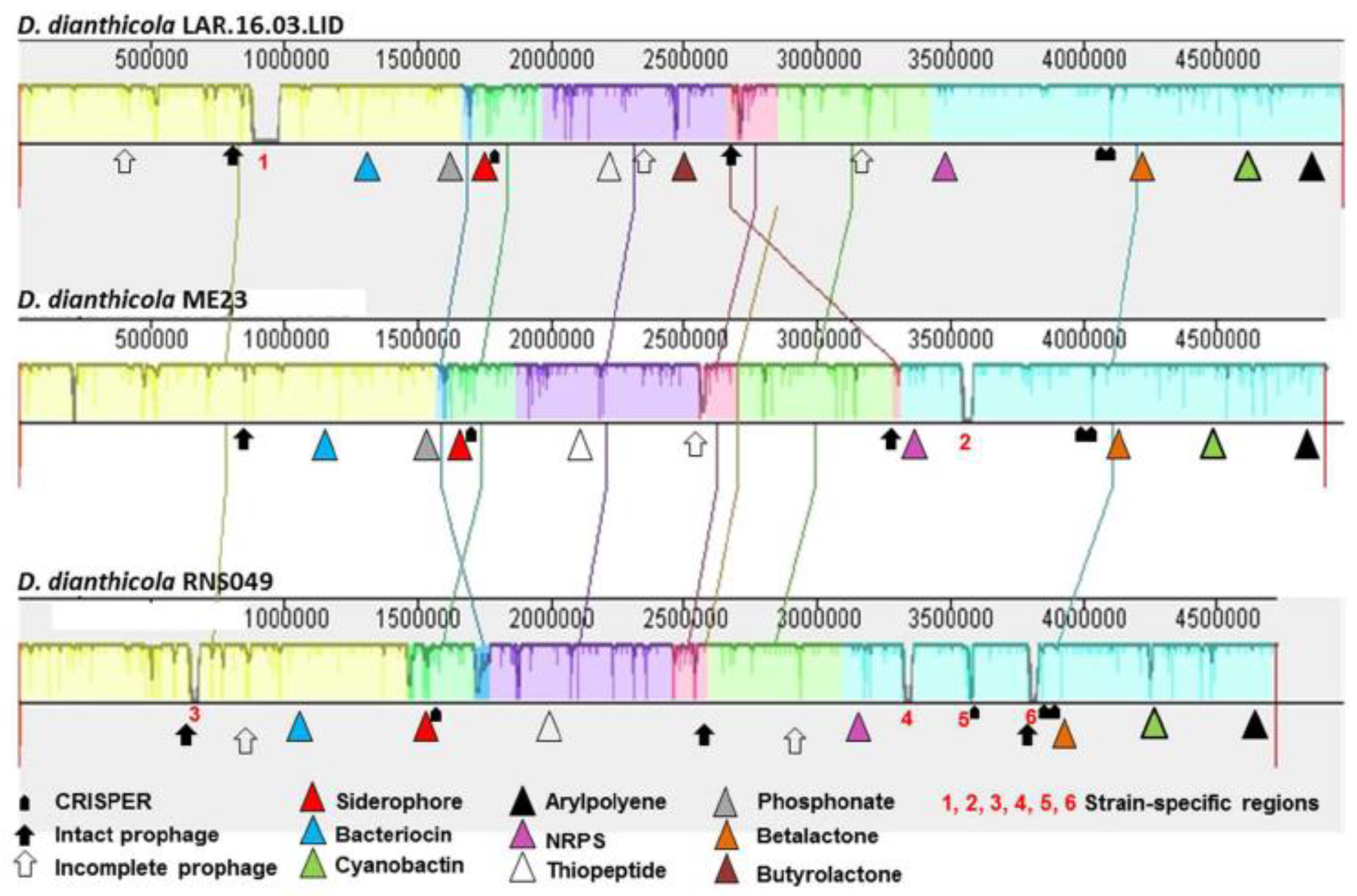
3.4. Complete Genome of D. dianthicola LAR.16.03.LID

The genome of D. dianthicola LAR.16.03.LID was the first complete genome of a D. dianthicola strain collected in Morocco, and the third D. dianthicola genome in the NCBI database that already hosted those of strains ME23 and RNS04.9 (Figure 4). Phylogenetic relationships between all D. dianthicola genomes available in NCBI were determined using MLSA. In the phylogenetic tree, the two Moroccan strains LAR.16.03.LID and S4.16.03.P2.4 appeared to be highly related. This could be explained by the close isolate locations, that were two potato fields separated only by a road. The two Moroccan D. dianthicola strains clustered with D. dianthicola strain ME23, which has been recently collected in a potato field in USA (Figure 4). The genomic relationship between D. dianthicola ME23 and D. dianthicola LAR.16.03.LID was confirmed using SNP/InDels calling. The SNP and InDel number in the LAR.16.03.LID genome reached 12,259 and 16,335, using the ME23 and D. dianthicola RNS04.9 genomes as a reference, respectively. The LAR.16.03.LID genome was annotated using the NCBI Prokaryotic Genome Annotation Pipeline. A graphical genome map is provided in Figure 5. The D. dianthicola LAR.16.03.LID genome exhibited a high synteny with that of D. dianthicola strains ME23 and RNS049, with the exception of some large insertion/deletions scattered in the genomes (Figure 6). These regions contained strain-specific genes with no counterpart in the other D. dianthicola genomes. In strains LAR.16.03LID and ME23, the analyses evidenced one strain-specific region that contained some mobile elements, such as genes from transposons and prophages; strain-specific regions 1 and 2 are presented in Figure 6. Additional information about these strain-specific regions are available in Table S3-1 and Table S3-2. Strain RNS049 exhibited four strain-specific regions which also contain mobile elements (Figure 6 and Table S3-1–6). Phaster analysis suggested the presence of intact prophages (Figure 6 and Table S4). The CRISPR elements are very important features of bacterial genomes as they provide acquired immunity against viruses and plasmids [53]. The three D. dianthicola genomes hosted three or four CRISPR loci (Figure 6 and Table S5).
The D. dianthicola LAR.16.03.LID genome exhibited an arsenal of virulence genes similar to that described for D. dianthicola RNS049 [39]. All the pectinase-encoding genes described in the model strain D. dadantii 3937 [54] were conserved in the three D. dianthicola genomes, with the noticeable exceptions of the lacking pehK gene (which encodes a predicted polygalacturonase) and the presence of a truncated form of the pelA gene (which encodes Pectate lyase A). Aside macerating enzymes determinants, other genes implicated in different stages of the host infection were conserved in the three D. dianthicola strains, including those involved in the resistance to oxidative stress, acidic pH (cfa, asr) and antimicrobial peptides (arnB-T, sapABCDF), synthesis of cell envelope components (such as bscABCD and wza-wzb-wzc), and siderophore synthesis and uptake (acsF-A and cbrABCDE for achromobactin and fct-cbsCEBA for chrysobactin) [54].
We strengthened this analysis using the resistance gene identifier (RGI) and AntiSMASH softwares. We searched for genes involved in the resistance to different families of antimicrobial compounds. No differences were observed between the three D. dianthicola genomes (Table S6). The AntiSMASH analysis identified many secondary metabolite biogenesis clusters in D. dianthicola genomes that were already described in several Dickeya species, like those responsible for the synthesis of siderophores, cyanobactin with cytotoxic activity, bacteriocin, nonribosomal peptide-synthetase (NRPS) and arylpolyene (Figure 6 and Table S7).

4. Discussion

The main objective of this study was to characterize the pectinolytic populations isolated from symptomatic potato plants in Morocco between 2015 and 2017. A set of 119 pectinolytic bacteria, belonging to the genus Pectobacterium or Dickeya, were isolated and characterized using the gapA gene marker in combination with MLSA and ANI. Most of the isolates (83%) belonged to the Pectobacterium genus: the P. brasiliense species dominated in the Meknes and Guigo regions, while D. dianthicola was identified in the Larache region only.
P. brasiliense caused major economic losses to several crops (potato, cucumber, paprika, etc.) in many countries, including Canada, USA, South Africa, China, Korea and New Zealand [55,56,57,58,59,60]. The wide host range of this pathogen could facilitate its survival even in harsh environments, by parasitizing many alternative host plants. In several studies, P. brasiliense isolates have been shown to be more aggressive than other Pectobacterium spp., except in the case of three Canadian strains exhibiting low aggressiveness [61]. In our study, the P. brasiliense isolates were as virulent as the other Pectobacterium strains. P. brasiliense was described in Morocco in 2012, and by now is the dominant species in two regions (S1 and S2). In the S2 region, the farmers use seed tubers produced in the S1 region, confirming the effective adaptation of this pathogen to the northern parts of Morocco.
The northern region (S4) exhibited the highest diversity of the pathogens. P. versatile and P. Polaris, described for the first time in Morocco, along with P. brasiliense and D. dianthicola, were isolated in the region S4. In this region, the majority of seed tubers were imported, and the irrigation water was derived from a dam. Either one or both agronomic practices could contribute to the wider diversity of pathogens in the Larache (S4) region than in the other investigated regions. While previous studies identified the P. carotovorum species as the most prevalent soft rot pathogens in Morocco [41,42], our study extended the diversity to other Pectobacterium and Dickyea species, including the recently described species P. versatile [7]. This species encompasses isolates, including the P. versatile strain SCC1 isolated in 1980 in Finland [62], which had been collected from a wide diversity of environments (host plants, surface waters) and geographic areas [7]. The presence of members of this clade in the potato field could be linked to irrigation, as P. versatile is also able to survive in this environment. This hypothesis remains to be investigated by sampling water from the dam. In addition, our study extended the worldwide distribution of this species to Morocco.
The international distribution of the genus Pectobacterium increases concerns about the economic losses caused by this bacterium to the potato growers. A study in the neighboring country Algeria, carried out between 2014 and 2015, revealed the presence of pectinolytic bacteria causing soft rot in potatoes that belonged to P. brasiliense and P. carotovorum, as judged by MLSA [63]. Ozturk et al. reported the presence of P. atrosepticum, P. brasiliense, P. carotovorum and P. parmentieri species in Turkey [64]. In Europe, the prevalence of different species belonging to the genera Pectobacterium and Dickeya detected in diseased potato plants differs from year to year and between countries, five bacterial species being the main causative agents of blackleg, namely P. atrosepticum, P. parmentieri, P. brasilense, D. solani and D. dianthicola [65].
The D. dianthicola species has been detected in Morocco in 2017 [44], and described as the main species causing losses in potato in North America [66]. More studies are needed for monitoring the spread of this highly aggressive pathogen. To reach this objective, we used Illumina and ONT sequencing technologies to assemble a complete genome of one D. dianthicola isolate that could be used, in the future, as a reference for studying the clonal variability of D. dianthicola populations in Morocco and elsewhere. Comparison of the three complete genomes available indicated the presence of several clusters that encode the biosynthesis of a number of secondary metabolites implicated in stress defense, possibly playing an important role during plant–bacteria interactions. For instance, bacteriocins are small molecules with bactericidal activity usually restricted to closely related species, increasing the competition during infection, while the production of arylpolyene, implicated in the protection against reactive oxygen species [67], has been recently described in D. fangzhongdai genomes [68].

5. Conclusions

This study revealed a wide diversity of Pectobacterium and Dickeya pathogens in northern Morocco, including P. polaris and P. versatile, that are reported for the first time in this country. In tuber maceration assays, the tested isolates of the emerging pathogen D. dianthicola were more aggressive than the Pectobacterium isolates. This feature should alert stakeholders to the threat that this pathogen poses to potato tuber production in northern Morocco. The nucletotide sequence data of the Dickeya and Pectobacterium Moroccan isolates, including a complete genome of D. dianthicola, 10 draft genomes and 119 partial sequences of the gapA gene, were deposited in a public database (NCBI GenBank) to be used as genetic resources for monitoring the spread of these pathogens in Northern Africa and elsewhere.

Supplementary Materials

The following are available online at https://www.mdpi.com/2076-2607/8/6/895/s1. Figure S1: Phylogenetic analysis of the gapA barcode, Table S1: Primers used in this study, Table S2: Strains of Pectobacterium and Dickeya isolated from different regions in Morocco, Table S3-1: Strain-specific region number 1 identified by Mauve, Table S3-2: Strain-specific region number 2 identified by Mauve, Table S3-3: Strain-specific region number 3 identified by Mauve, Table S3-4: Strain-specific region number 4 identified by Mauve, Table S3-5: Strain-specific region number 5 identified by Mauve, Table S3-6: Strain-specific region number 6 identified by Mauve, Table S4: Secondary metabolite gene clusters identified with antismath, Table S5: Prophage in Dickeya dianthicola identified using Phaster, Table S6: CRISPR identification in Dickeya dianthicola using CRISPER Finder, Table S7: Resistome genes identified in Dickeya dianthicola genomes.

Author Contributions

D.F. and M.M. supervised the data production and analysis, S.O. carried out isolation, taxonomical characterization and genome assembly and analysis, S.K. participated in Sanger sequence analysis, S.O. and S.S. conducted potato maceration assays, K.R. and S.K. conducted statistical analysis, C.L.-R. and C.V. carried out the ONT sequencing and assembly of the complete genome, S.O. and D.F. wrote the manuscript. All authors have read and agreed to the published version of the manuscript.

Funding

This work was supported by The French National Agency for Research (ANR-15-CE21-0003) and a cooperative project between France and Morocco (PRAD 14-02, Campus France n°30229ZK). Research in the I2BC was supported by the LabEx Saclay Plant Sciences-SPS (ANR-10-LABX-0040-SPS).

Acknowledgments

Yves Dessaux and Peter Mergaert (I2BC) are thanked for their advice during the preparation of the manuscript of which an uncorrected preprint was deposited (04 Feb, 2020) in Research Square database (https://www.researchsquare.com/article/rs-13222/v1; DOI 10.21203/rs.2.22648/v1) before a submission to Microorganisms Journal.

Conflicts of Interest

The authors declare no conflict of interest.

References

  1. Czajkowski, R.; Pérombelon, M.C.M.; Van Veen, J.A.; Van der Wolf, J.M. Control of blackleg and tuber soft rot of potato caused by Pectobacterium and Dickeya species: A review. Plant Pathol. 2011, 60, 999–1013. [Google Scholar] [CrossRef]
  2. Mansfield, J.; Genin, S.; Magori, S.; Citovsky, V.; Sriariyanum, M.; Ronald, P.; Dow, M.; Verdier, V.; Beer, S.V.; Machado, M.A.; et al. Top 10 plant pathogenic bacteria in molecular plant pathology. Mol. Plant Pathol. 2012, 13, 614–629. [Google Scholar] [CrossRef] [PubMed] [Green Version]
  3. Leonard, S.; Hommais, F.; Nasser, W.; Reverchon, S. Plant–phytopathogen interactions: Bacterial responses to environmental and plant stimuli. Environ. Microbiol. 2017, 19, 1689–1716. [Google Scholar] [CrossRef] [PubMed] [Green Version]
  4. Pérombelon, M.C.M. Potato diseases caused by soft rot erwinias: An overview of pathogenesis. Plant Pathol. 2002, 51, 1–12. [Google Scholar] [CrossRef]
  5. De Boer, S.H.; Verdonck, L.; Vruggink, H.; Harju, P.; Bång, H.O.; Ley, J. De Serological and biochemical variation among potato strains of Erwinia carotovora subsp. atroseptica and their taxonomic relationship to other E. carotovora strains. J. Appl. Bacteriol. 1987, 63, 487–495. [Google Scholar] [CrossRef]
  6. Khayi, S.; Cigna, J.; Chong, T.M.; Qu�tu-Laurent, A.; Chan, K.G.; Helias, V.; Faure, D. Transfer of the potato plant isolates of Pectobacterium wasabiae to Pectobacterium parmentieri sp. nov. Int. J. Syst. Evol. Microbiol. 2016, 66, 5379–5383. [Google Scholar] [CrossRef]
  7. Portier, P.; Pédron, J.; Taghouti, G.; Fischer-Le Saux, M.; Caullireau, E.; Bertrand, C.; Laurent, A.; Chawki, K.; Oulgazi, S.; Moumni, M.; et al. Elevation of Pectobacterium carotovorum subsp. odoriferum to species level as Pectobacterium odoriferum sp. nov., proposal of Pectobacterium brasiliense sp. nov. and Pectobacterium actinidiae sp. nov., emended description of Pectobacterium carotovorum. Int. J. Syst. Evol. Microbiol. 2019, 69, 3207–3216. [Google Scholar] [CrossRef]
  8. Smith, C. Variation in the Pathogenicity and Aggressiveness of Strains of Erwinia carotovora subsp. carotovora Isolated from Different Hosts. Plant Dis. 2007, 74, 505. [Google Scholar] [CrossRef]
  9. Dees, M.W.; Lysøe, E.; Rossmann, S.; Perminow, J.; Brurberg, M.B. Pectobacterium polaris sp. nov., isolated from potato (Solanum tuberosum). Int. J. Syst. Evol. Microbiol. 2017, 67, 5222–5229. [Google Scholar] [CrossRef]
  10. Sarfraz, S.; Riaz, K.; Oulghazi, S.; Cigna, J.; Sahi, S.T.; Khan, S.H.; Faure, D. Pectobacterium punjabense sp. nov., isolated from blackleg symptoms of potato plants in Pakistan. Int. J. Syst. Evol. Microbiol. 2018, 68, 3551–3556. [Google Scholar] [CrossRef]
  11. Waleron, M.; Misztak, A.; Waleron, M.; Franczuk, M.; Wielgomas, B.; Waleron, K. Transfer of Pectobacterium carotovorum subsp. carotovorum strains isolated from potatoes grown at high altitudes to Pectobacterium peruviense sp. nov. Syst. Appl. Microbiol. 2018, 41, 85–93. [Google Scholar] [CrossRef] [PubMed]
  12. Waleron, M.; Waleron, K.; Lojkowska, E. Characterization of Pectobacterium carotovorum subsp. odoriferum causing soft rot of stored vegetables. Eur. J. Plant Pathol. 2014, 139, 457–469. [Google Scholar] [CrossRef] [Green Version]
  13. Goto, M.; Matsumoto, K. Erwinia carotovora subsp. wasabiae subsp. nov. Isolated from Diseased Rhizomes and Fibrous Roots of Japanese Horseradish (Eutrema wasabi Maxim.). Int. J. Syst. Bacteriol. 1987, 37, 130–135. [Google Scholar] [CrossRef] [Green Version]
  14. Gardan, L.; Gouy, C.; Christen, R.; Samson, R. Elevation of three subspecies of Pectobacterium carotovorum to species level: Pectobacterium atrosepticum sp. nov., Pectobacterium betavasculorum sp. nov. and Pectobacterium wasabiae sp. nov. Int. J. Syst. Evol. Microbiol. 2003, 53, 381–391. [Google Scholar] [CrossRef] [PubMed]
  15. Nabhan, S.; de Boer, S.H.; Maiss, E.; Wydra, K. Pectobacterium aroidearum sp. nov., a soft rot pathogen with preference for monocotyledonous plants. Int. J. Syst. Evol. Microbiol. 2013, 63, 2520–2525. [Google Scholar] [CrossRef] [Green Version]
  16. Waleron, M.; Misztak, A.; Waleron, M.; Franczuk, M.; Jońca, J.; Wielgomas, B.; Mikiciński, A.; Popović, T.; Waleron, K. Pectobacterium zantedeschiae sp. nov. a new species of a soft rot pathogen isolated from Calla lily (Zantedeschia spp.). Syst. Appl. Microbiol. 2019, 42, 275–283. [Google Scholar] [CrossRef]
  17. Koh, Y.J.; Kim, G.H.; Lee, Y.S.; Sohn, S.H.; Koh, H.S.; Kwon, S.; Heu, S.; Jung, J.S. Pectobacterium carotovorum subsp. actinidiae subsp. nov., a new bacterial pathogen causing canker-like symptoms in yellow kiwifruit, Actinidia chinensis. N. Z. J. Crop Hortic. Sci. 2012, 40, 269–279. [Google Scholar] [CrossRef] [Green Version]
  18. Oulghazi, S.; Cigna, J.; Lau, Y.Y.; Moumni, M.; Chan, K.G.; Faure, D. Transfer of the waterfall source isolate Pectobacterium carotovorum M022 to Pectobacterium fontis sp. nov., a deep-branching species within the genus Pectobacterium. Int. J. Syst. Evol. Microbiol. 2019, 69, 470–475. [Google Scholar] [CrossRef]
  19. Pédron, J.; Bertrand, C.; Taghouti, G.; Portier, P.; Barny, M.-A. Pectobacterium aquaticum sp. nov., isolated from waterways. Int. J. Syst. Evol. Microbiol. 2019, 69, 745–751. [Google Scholar] [CrossRef]
  20. Toth, I.K.; van der Wolf, J.M.; Saddler, G.; Lojkowska, E.; Hélias, V.; Pirhonen, M.; Tsror (Lahkim), L.; Elphinstone, J.G. Dickeya species: An emerging problem for potato production in Europe. Plant Pathol. 2011, 60, 385–399. [Google Scholar] [CrossRef]
  21. Tsror, L.; Erlich, O.; Lebiush, S.; Hazanovsky, M.; Zig, U.; Slawiak, M.; Grabe, G.; Van Der Wolf, J.M.; Van De Haar, J.J. Assessment of recent outbreaks of Dickeya sp. (syn. Erwinia chrysanthemi) slow wilt in potato crops in Israel. Eur. J. Plant Pathol. 2009, 123, 311–320. [Google Scholar]
  22. Van der Wolf, J.M.; Nijhuis, E.H.; Kowalewska, M.J.; Saddler, G.S.; Parkinson, N.; Elphinstone, J.G.; Pritchard, L.; Toth, I.K.; Lojkowska, E.; Potrykus, M.; et al. Dickeya solani sp. nov., a pectinolytic plant-pathogenic bacterium isolated from potato (Solanum tuberosum). Int. J. Syst. Evol. Microbiol. 2014, 64, 768–774. [Google Scholar] [CrossRef] [PubMed] [Green Version]
  23. Ignatov, A.; Karlov, A.; Dzhalilov, F. Spreading of the blackleg of potatoes in Russia caused by bacteria of Dickeya genus. Zaschita i Karantin Rastenij 2014, 11, 41–43. [Google Scholar]
  24. Ozturk, M.; Aksoy, H.M. First report of Dickeya solani associated with potato blackleg and soft rot in Turkey. J. Plant Pathol. 2017, 99, 298. [Google Scholar]
  25. Tsror [Lahkim], L.; Erlich, O.; Lebiush, S.; van der Wolf, J.; Czajkowski, R.; Mozes, G.; Sikharulidze, Z.; Ben Daniel, B. First report of potato blackleg caused by a biovar 3 Dickeya sp. in Georgia. New Dis. Rep. 2011, 23, 1. [Google Scholar] [CrossRef] [Green Version]
  26. Cardoza, Y.F.; Duarte, V.; Lopes, C.A. First report of blackleg of potato caused by Dickeya solani in Brazil. Plant Dis. 2017, 101, 243. [Google Scholar] [CrossRef]
  27. Nykyri, J.; Niemi, O.; Koskinen, P.; Nokso-Koivisto, J.; Pasanen, M.; Broberg, M.; Plyusnin, I.; Törönen, P.; Holm, L.; Pirhonen, M.; et al. Revised Phylogeny and Novel Horizontally Acquired Virulence Determinants of the Model Soft Rot Phytopathogen Pectobacterium wasabiae SCC3193. PLoS Pathog. 2012, 8, e1003013. [Google Scholar] [CrossRef] [Green Version]
  28. Zhang, Y.; Fan, Q.; Loria, R. A re-evaluation of the taxonomy of phytopathogenic genera Dickeya and Pectobacterium using whole-genome sequencing data. Syst. Appl. Microbiol. 2016, 39, 252–259. [Google Scholar] [CrossRef]
  29. Brady, C.L.; Cleenwerck, I.; Denman, S.; Venter, S.N.; Rodriguez-Palenzuela, P.; Coutinho, T.A.; De Vos, P. Proposal to reclassify Brenneria quercina (Hildebrand and Schroth 1967) Hauben et al. 1999 into a new genus, Lonsdalea gen. nov., as Lonsdalea quercina comb. nov., descriptions of Lonsdalea quercina subsp. quercina comb. nov., Lonsdalea quercina subsp. ib. Int. J. Syst. Evol. Microbiol. 2012, 62, 1592–1602. [Google Scholar] [CrossRef]
  30. Tian, Y.; Zhao, Y.; Yuan, X.; Yi, J.; Fan, J.; Xu, Z.; Hu, B.; De Boer, S.H.; Li, X. Dickeya fangzhongdai sp. nov., a plantpathogenic bacterium isolated from pear trees (Pyrus pyrifolia). Int. J. Syst. Evol. Microbiol. 2016, 66, 2831–2835. [Google Scholar] [CrossRef] [PubMed]
  31. Hugouvieux-Cotte-Pattat, N.; Jacot-Des-Combes, C.; Briolay, J. Dickeya lacustris sp. nov., a water-living pectinolytic bacterium isolated from lakes in France. Int. J. Syst. Evol. Microbiol. 2019, 69, 721–726. [Google Scholar] [CrossRef] [PubMed]
  32. Oulghazi, S.; Pédron, J.; Cigna, J.; Lau, Y.Y.; Moumni, M.; Van Gijsegem, F.; Chan, K.-G.; Faure, D. Dickeya undicola sp. nov., a novel species for pectinolytic isolates from surface waters in Europe and Asia. Int. J. Syst. Evol. Microbiol. 2019, 69, 2440–2444. [Google Scholar] [CrossRef] [PubMed]
  33. Parkinson, N.; DeVos, P.; Pirhonen, M.; Elphinstone, J. Dickeya aquatica sp. nov., isolated from waterways. Int. J. Syst. Evol. Microbiol. 2014, 64, 2264–2266. [Google Scholar] [CrossRef] [PubMed]
  34. Samson, R.; Legendre, J.; Richard, C.; Fischer-Le Saux, M.; Achouak, W.; Gardan, L. Transfer of Pectobacterium chrysanthemi (Burkholder, 1953) Brenner et al. 1973 and Brenneria paradisiaca to the genus Dickeya gen. nov. as Dickeya chrysanthemi comb. nov. and Dickeya paradisiaca comb. nov. and delineation of four novel species, Dickeya da. Int. J. Syst. Evol. Microbiol. 2005, 55, 1415–1427. [Google Scholar] [CrossRef]
  35. Golanowska, M.; Galardini, M.; Bazzicalupo, M.; Hugouvieux-Cotte-Pattat, N.; Mengoni, A.; Potrykus, M.; Slawiak, M.; Lojkowska, E. Draft Genome Sequence of a Highly Virulent Strain of the Plant Pathogen Dickeya solani, IFB0099. Genome Announc. 2015, 3, e00109-15. [Google Scholar] [CrossRef] [Green Version]
  36. Hanage, W.P.; Spratt, B.G.; Turner, K.M.E.; Fraser, C. Modelling bacterial speciation. Philos. Trans. R. Soc. B Biol. Sci. 2006, 361, 2039–2044. [Google Scholar] [CrossRef] [Green Version]
  37. Cigna, J.; Dewaegeneire, P.; Beury, A.; Gobert, V.; Faure, D. A gapA PCR-sequencing Assay for Identifying the Dickeya and Pectobacterium Potato Pathogens. Plant Dis. 2017, 101, 1278–1282. [Google Scholar] [CrossRef] [Green Version]
  38. Goris, J.; Konstantinidis, K.T.; Klappenbach, J.A.; Coenye, T.; Vandamme, P.; Tiedje, J.M. DNA–DNA hybridization values and their relationship to whole-genome sequence similarities. Int. J. Syst. Evol. Microbiol. 2007, 57, 81–91. [Google Scholar] [CrossRef] [Green Version]
  39. Raoul des Essarts, Y.; Pédron, J.; Blin, P.; Van Dijk, E.; Faure, D.; Van Gijsegem, F. Common and distinctive adaptive traits expressed in Dickeya dianthicola and Dickeya solani pathogens when exploiting potato plant host. Environ. Microbiol. 2019, 21, 1004–1018. [Google Scholar] [CrossRef]
  40. Anajjar, B.; Aitmhand, R.; Timinouni, M.; Ennaji, M.M. Caractérisation par PCR de deux souches d’Erwinia carotovora isolées de la rhizosphère de la pomme de terre dans la région du grand Casablanca au Maroc. EPPO Bull. 2007, 37, 175–180. [Google Scholar] [CrossRef]
  41. Faquihi, H.; Terta, M.; Amdan, M.; Achbani, E.H.; Ennaji, M.M.; Ait Mhand, R. Phenotypic and genotypic diversity of Pectobacterium carotovorum subsp carotovorum causing soft rot disease of potatoes in Morocco. Eur. J. Plant Pathol. 2015, 143, 801–811. [Google Scholar] [CrossRef]
  42. Terta, M.; El Karkouri, A.; Ait M’Hand, R.; Achbani, E.; Barakate, M.; Amdan, M.; Annajar, B.; El Hassouni, M.; Val, F.; Bouteau, F.; et al. Occurrence of Pectobacterium carotovorum strains isolated from potato soft rot in Morocco. Cell. Mol. Biol. 2010, 56, OL1324–OL1333. [Google Scholar] [PubMed]
  43. Terta, M.; Azelmat, S.; M’Hand, R.A.; Achbani, E.H.; Barakate, M.; Bouteau, F.; Ennaji, M.M. Molecular typing of Pectobacterium carotovorum isolated from potato tuber soft rot in Morocco. Ann. Microbiol. 2012, 62, 1411–1417. [Google Scholar] [CrossRef]
  44. Oulghazi, S.; Khayi, S.; Lafkih, N.; Massaoudi, Y.; El Karkouri, A.; El Hassouni, M.; Faure, D.; Moumni, M. First report of Dickeya dianthicola causing blackleg on potato in Morocco. Plant Dis. 2017, 101, 1671–1672. [Google Scholar] [CrossRef]
  45. Hélias, V.; Hamon, P.; Huchet, E.; Wolf, J.V.D.; Andrivon, D. Two new effective semiselective crystal violet pectate media for isolation of Pectobacterium and Dickeya. Plant Pathol. 2012, 61, 339–345. [Google Scholar] [CrossRef]
  46. Darrasse, A.; Priou, S.; Kotoujansky, A.; Bertheau, Y. PCR and restriction fragment length polymorphism of a pel gene as a tool to identify Erwinia carotovora in relation to potato diseases. Appl. Environ. Microbiol. 1994, 60, 1437–1443. [Google Scholar] [CrossRef] [Green Version]
  47. Nassar, A.; Darrasse, A.; Lemattre, M.; Kotoujansky, A.; Dervin, C.; Vedel, R.; Bertheau, Y. Characterization of Erwinia chrysanthemi by pectinolytic isozyme polymorphism and restriction fragment length polymorphism analysis of PCR- amplified fragments of pel genes. Appl. Environ. Microbiol. 1996, 62, 2228–2235. [Google Scholar] [CrossRef] [Green Version]
  48. Grant, J.R.; Stothard, P. The CGView Server: A comparative genomics tool for circular genomes. Nucleic Acids Res. 2008, 36, W181–W184. [Google Scholar] [CrossRef]
  49. Darling, A.C.E.; Mau, B.; Blattner, F.R.; Perna, N.T. Mauve: Multiple alignment of conserved genomic sequence with rearrangements. Genome Res. 2004, 14, 1394–1403. [Google Scholar] [CrossRef] [Green Version]
  50. Grissa, I.; Vergnaud, G.; Pourcel, C. CRISPRFinder: A web tool to identify clustered regularly interspaced short palindromic repeats. Nucleic Acids Res. 2007, 35, W52–W57. [Google Scholar] [CrossRef] [Green Version]
  51. Arndt, D.; Grant, J.R.; Marcu, A.; Sajed, T.; Pon, A.; Liang, Y.; Wishart, D.S. PHASTER: A better, faster version of the PHAST phage search tool. Nucleic Acids Res. 2016, 44, W16–W21. [Google Scholar] [CrossRef] [PubMed] [Green Version]
  52. Khayi, S.; Blin, P.; Pédron, J.; Chong, T.M.; Chan, K.G.; Moumni, M.; Hélias, V.; Van Gijsegem, F.; Faure, D. Population genomics reveals additive and replacing horizontal gene transfers in the emerging pathogen Dickeya solani. BMC Genom. 2015, 16, 788. [Google Scholar] [CrossRef] [PubMed] [Green Version]
  53. Horvath, P.; Barrangou, R. CRISPR/Cas, the immune system of bacteria and archaea. Science 2010, 327, 167–170. [Google Scholar] [CrossRef] [PubMed] [Green Version]
  54. Reverchon, S.; Nasser, W. Dickeya ecology, environment sensing and regulation of virulence programme. Environ. Microbiol. Rep. 2013, 5, 622–636. [Google Scholar]
  55. Choi, O.; Kim, J. Pectobacterium carotovorum subsp. brasiliense Causing Soft Rot on Paprika in Korea. J. Phytopathol. 2013, 161, 125–127. [Google Scholar] [CrossRef]
  56. De Boer, S.H.; Li, X.; Ward, L.J. Pectobacterium spp. Associated with Bacterial Stem Rot Syndrome of Potato in Canada. Phytopathology 2012, 102, 937–947. [Google Scholar] [CrossRef] [Green Version]
  57. Meng, X.; Chai, A.; Shi, Y.; Xie, X.; Ma, Z.; Li, B. Emergence of Bacterial Soft Rot in Cucumber Caused by Pectobacterium carotovorum subsp. brasiliense in China. Plant Dis. 2017, 101, 279–287. [Google Scholar] [CrossRef] [Green Version]
  58. Nunes Leite, L.; de Haan, E.G.; Krijger, M.; Kastelein, P.; van der Zouwen, P.S.; van den Bovenkamp, G.W.; Tebaldi, N.D.; van der Wolf, J.M. First report of potato blackleg caused by Pectobacterium carotovorum subsp. brasiliensis in the Netherlands. New Dis. Rep. 2014, 29, 24. [Google Scholar] [CrossRef] [Green Version]
  59. Panda, P.; Fiers, M.A.W.J.; Armstrong, K.; Pitman, A.R. First report of blackleg and soft rot of potato caused by Pectobacterium carotovorum subsp. brasiliensis in New Zealand. New Dis. Rep. 2012, 26, 15. [Google Scholar] [CrossRef] [Green Version]
  60. Van der Merwe, J.J.; Coutinho, T.A.; Korsten, L.; van der Waals, J.E. Pectobacterium carotovorum subsp. brasiliensis causing blackleg on potatoes in South Africa. Eur. J. Plant Pathol. 2010, 126, 175–185. [Google Scholar] [CrossRef] [Green Version]
  61. Li, X.S.; Yuan, K.X.; Cullis, J.; Lévesque, C.A.; Chen, W.; Lewis, C.T.; De Boer, S.H. Draft Genome Sequences for Canadian Isolates of Pectobacterium carotovorum subsp. brasiliense with Weak Virulence on Potato. Genome Announc. 2015, 3, e00240-15. [Google Scholar] [CrossRef] [PubMed] [Green Version]
  62. Niemi, O.; Laine, P.; Koskinen, P.; Pasanen, M.; Pennanen, V.; Harjunpää, H.; Nykyri, J.; Holm, L.; Paulin, L.; Auvinen, P.; et al. Genome sequence of the model plant pathogen Pectobacterium carotovorum SCC1. Stand. Genom. Sci. 2017, 12, 87. [Google Scholar] [CrossRef] [PubMed] [Green Version]
  63. Naas, H.; Sebaihia, M.; Orfei, B.; Rezzonico, F.; Buonaurio, R.; Moretti, C. Pectobacterium carotovorum subsp. brasiliense and Pectobacterium carotovorum subsp. carotovorum as causal agents of potato soft rot in Algeria. Eur. J. Plant Pathol. 2018, 151, 1027–1034. [Google Scholar] [CrossRef]
  64. Ozturk, M.; Aksoy, H.M.; Potrykus, M.; Lojkowska, E. Correction to: Genotypic and phenotypic variability of Pectobacterium strains causing blackleg and soft rot on potato in Turkey. Eur. J. Plant Pathol. 2018, 152, 1. [Google Scholar] [CrossRef]
  65. Van der Wolf, J.; de Haan, E.G.; Kastelein, P.; Krijger, M.; de Haas, B.H.; Velvis, H.; Mendes, O.; Kooman-Gersmann, M.; van der Zouwen, P.S. Virulence of Pectobacterium carotovorum subsp. brasiliense on potato compared with that of other Pectobacterium and Dickeya species under climatic conditions prevailing in the Netherlands. Plant Pathol. 2017, 66, 571–583. [Google Scholar] [CrossRef]
  66. Charkowski, A.O. The Changing Face of Bacterial Soft-Rot Diseases. Annu. Rev. Phytopathol. 2018, 56, 16éà. [Google Scholar] [CrossRef]
  67. Schöner, T.A.; Gassel, S.; Osawa, A.; Tobias, N.J.; Okuno, Y.; Sakakibara, Y.; Shindo, K.; Sandmann, G.; Bode, H.B. Aryl Polyenes, a Highly Abundant Class of Bacterial Natural Products, Are Functionally Related to Antioxidative Carotenoids. ChemBioChem 2016, 17, 247–253. [Google Scholar] [CrossRef]
  68. Alič, Š.; Pédron, J.; Dreo, T.; Van Gijsegem, F. Genomic characterisation of the new Dickeya fangzhongdai species regrouping plant pathogens and environmental isolates. BMC Genom. 2019, 20, 34. [Google Scholar] [CrossRef]
Figure 1. Species diversity of the Pectobacterium and Dickeya pathogens in the sampling areas of northern Morocco. Sampling of pathogens was performed in four potato growing areas in the northern Morocco: Meknes (S1), Guigo (S2), Boumia (S3) and Laraache (S4). The map of Morocco was generated by a free and open source geographic information system (https://www.qgis.org/fr/site/index.html). The pie charts represent diversity of the isolated pathogens. None of them were collected from the Boumia (S3) region. Legend: Ddi, Dickeya dianthicola; Pb, Pectobacterium brasiliense; Pc, Pectobacterium carotovorum; Pp, Pectobacterium polaris; Pv, Pectobacterium versatile.
Microorganisms 08 00895 g001
Figure 2. Phylogeny of the Moroccan strains based on MLSA. The phylogenetic trees were generated separately (A) for Pectobacterium and (B) for Dickeya strains. The alignment of the concatenated genes fusA, rpoD, rpoS, acnA, purA, recA, mdh, mtlD, groEL, secY, glyA, gapA and rplB was generated using ClustalW; the evolutionary history was inferred using the Neighbor-Joining method and the evolutionary distances were computed using the Maximum Composite Likelihood method. Phylogenetic analyses were conducted using MEGA7 software. The name of the Moroccan isolates is underlined.
Microorganisms 08 00895 g002
Figure 3. Virulence test of Pectobacterium and Dickeya on potato tubers. The symptoms provoked by each strain were compared by infecting 10 tubers per strain. The 10 non-inoculated tubers were used as control. Symptoms were classified into five classes (CLA0, CLA1, CLA2, CLA3, CLA4, according to increasing severity). The typology of these classes was illustrated by a picture of an example. Data were statistically analyzed by a Kruskal–Wallis test (α = 5%). Lower case letters on the right of the graph indicate statistical differences between the different inoculated pathogens.
Microorganisms 08 00895 g003
Figure 4. Phylogenetic analysis and characteristics of the Moroccan and NCBI Dickeya genomes. The genes fusA, rpoD, leuS, rpoS, purA, infB, gyrB, recA, groEL, secY, glyA, gapA, rplB, dnaX and gyrA were concatenated. The alignment was generated using ClustalW; the evolutionary history was inferred using the Neighbor-Joining method and the evolutionary distances were computed using the Maximum Composite Likelihood method. Phylogenetic analyses were conducted using MEGA7 software. The Moroccan D. dianthicola isolates are indicated in bold face.
Microorganisms 08 00895 g004
Figure 5. Circular map of the genome of Dickeya dianthicola LAR.16.03.LID. The genome size is 4,976,211 bp with 4223 predicted protein-coding genes. The GC content and GC skew are represented on the distance scale (in kbp) on the inner map. The arrows around the map indicate the deduced Coding DNA Sequences (CDS) and their orientation.
Microorganisms 08 00895 g005
Figure 6. Synteny between the complete genomes of D. dianthicola LAR.16.03.LID, RNS049 and ME23 strains. Synteny analysis was performed using MAUVE software. The numbers indicate the position of strain specific genomic regions. The secondary pathway gene clusters were searched using AntiSMASH, the prophages were identified using PHASTER, and CRISPER loci were localized using CRISPER finder.
Microorganisms 08 00895 g006
Table 1. Draft genome sequences of Pectobacterium and Dickeya strains isolated from Northern Morocco.
OrganismAccession NumberGenome
Size
N50 (pb)Number of
Contigs
CoverageNumber of
CDS
Number of
tRNAs
Pectobacterium polaris S4.16.03.2BQZDF000000004,862,009155,8656541435554
Pectobacterium brasiliense S1.16.01.3kQZDG000000004,946,598146,84474410433736
Pectobacterium brasiliense S1.15.11.2DQZDH000000004,818,836 99,39291420420635
Pectobacterium brasiliense S4.16.03.1CQZDI000000004,944,722139,66574467433637
Pectobacterium carotovorum S1-A16QZDJ000000004,835,633255,2063755426163
Pectobacterium versatile S4.16.03.3IQZDK000000004,854,0848262108246424734
Pectobacterium versatile S4.16.03.3KQZDL000000004,870,94090,195106237426234
Pectobacterium versatile S4.16.03.3FQZDM000000004,852,59589,731114143424440
Dickeya dianthicola S4.16.03.P2.4QZDN000000004,865,14792,028101415423837
Dickeya dianthicola LAR.16.03.LIDQZDO000000004,863,93971,707108344423837
Table 2. Pairwise Average Nucleotide Identity (ANI) and in-silico DNA-DNA Hybridization (is-DDH) values of Pectobacterium and Dickeya strains isolated from Northern Morocco.
ANI Values
Strains12345678910111213141516
1-Pp NIBIO 1006T 96.892.992.993.593.793.593.793.493.493.593.592.279.37979.1
2-Pp S4.16.03.2B73.30 9392.993.593.793.593.793.493.593.593.492.278.97979
3-Pc ICMP5702T52.352.10 97.292.692.992.892.895.195.295.295.294.878.978.978.9
4-Pc S1-A1652.251.9076.30 92.692.992.792.995.395.395.395.394.878.879.679.6
5-Pb LMG 21371T54.454.550.950.9 96.195.996.192.292.292.292.393.0078.679.579.5
6-Pb S4.16.03.1C5655.75251.768.5 96.310092.391.291.191.291.978.779.679.6
7-Pb S1.15.11.2D54.754.251.35167.169.4 96.392.392.292.392.391.879.179.179.1
8-Pb S1.16.01.3K5655.77651.768.510071.4 92.492.492.492.391.979.679.679.7
9-Pv SCC154.354.363.563.948.949.849.549.3 99.599.599.594.779.279.179.3
10-Pv S4.16.03.3F54.454.363.964.349.149.949.549.996.6 10010094.779.279.279.2
11-Pv S4.16.03.3k54.454.363.864.249.249.949.549.996.699.3 10094.779.379.379.4
12-Pv S4.16.03.3I54.454.363.864.249.149.949.549.896.510099.3 94.779.379.379.3
13-Po BCS749.24961.360.647.447.847.647.860.460.760.660.6 79.179.179.1
14-Ddi NCPPB 453T21.120.720.520.720.92120.62121.221212121.1 99.599.5
15-Ddi S4.16.03.P2.420.820.620.420.921.120.920.720.92121212120.895.6 100
16-Ddi LAR.16.03.LID20.820.620.42121.120.920.620.92121212120.895.6100
is-DDH
Strains: 1, P. polaris NIBIO1006T; 2, P. polaris S4.16.03.2B 3, P. carotovorum ICMP5702T; 4, P. carotovorum S1-A16; 5, P. brasiliense LMG21371T; 6, P. brasiliense S4.16.03.1C; 7, P. brasiliense S1.15.11.2D 8, P. brasiliense S1.16.01 3K; 9, P. versatile SCC1; 10, P. versatile S4.16.03.3F; 11, P. versatile S4.16.03.3k 12, P. versatile S4.16.03.3I; 13, P. odoriferum BCS7; 14, D. dianthicola NCPPB 453T, 15, D. dianthicola S4.16.03.P2.4; 16, D. dianthicola LAR.16.03.LID.

