Article

Selenite Cystine Agar for Enumeration of Inoculated Salmonella Serovars Recovered from Stressful Conditions during Antimicrobial Validation Studies

1
Robert M. Kerr Food & Agricultural Products Center, Oklahoma State University, Stillwater, OK 74078, USA
2
Department of Animal and Food Sciences, Oklahoma State University, Stillwater, OK 74078, USA
3
Los Angeles County, Department of Agricultural Commissioner/Weights & Measures, South Gate, CA 90280, USA
*
Author to whom correspondence should be addressed.
Microorganisms 2020, 8(3), 338; https://doi.org/10.3390/microorganisms8030338
Submission received: 21 January 2020 / Revised: 21 February 2020 / Accepted: 25 February 2020 / Published: 28 February 2020
(This article belongs to the Special Issue Antimicrobial Interventions for Raw and Processed Foods)

Abstract

:
Process validation studies often require the inoculation of select foodborne pathogens into targeted foods to determine the lethality of the process or antimicrobial ingredients, and quantitative recovery of surviving inoculum bacteria helps to make those assessments. Such processes introduce various stressors on the inoculated challenge microorganisms whereby traditional selective media are too harsh to enumerate the remaining viable and injured population quantitatively. Innate antibiotic resistance of challenge organisms has often been used to establish simple selective media (i.e., Tryptic Soy Agar/TSA + antibiotics) for recovering inoculated strains, but sometimes antibiotic resistant background microorganisms are higher than desired. Salmonella Thompson 120, Salmonella Heidelberg F5038BG1, Salmonella Hadar MF60404, Salmonella Enteritidis H3527, and Salmonella Typhimurium H3380 were characterized for antibiotic resistance and acid adaptation in Tryptic Soy Broth containing 0%, 0.25%, or 1.0% glucose. Sodium pyruvate was evaluated for recovery after stress but no enhancing effect was observed, possibly because the strains were acid-adapted. Selenite Cystine Broth, traditionally used as a selective enrichment broth, was used as the basis for Selenite Cystine Agar (SCA) in combination with three antibiotics to which our Salmonella are resistant. Serovars of Salmonella, both individually and in mixtures, were enumerated on TSA, SCA, Xylose Lysine Desoxycholate (XLD), and Hektoen Enteric (HE) selective agars (all containing the same antibiotics) after conditions of nutrient starvation, desiccation, acid stress, and thermal stress. The data show that quantitative enumeration of our Salmonella serovars on SCA was not significantly different (p > 0.05) than those achieved on TSA for all tested stress categories. Levels of Salmonella enumerated on XLD and/or HE were significantly different (p < 0.05) than on TSA and SCA and often more than 1–2-log lower, consistent with the inhibition of injured cells. These data confirm that SCA (+ antibiotics) is a suitable selective medium for enumeration of these acid-adapted Salmonella serovars as challenge organisms recovered from various conditions of stress.

1. Introduction

Microbial challenge studies of foods are often conducted using pathogens or spoilage microorganisms that are inoculated into targeted food products. After processing or some period of shelf life, the inoculated microorganisms are recovered and enumerated to determine whether the food formulation or food processing conditions inhibits (bactericidal), prevents growth (bacteriostatic), or allows survival and growth (no control) under the conditions of treatment. Such inoculates are often added to products that are not sterile and may include a background of other microorganisms from which the inoculum must be differentially enumerated. Sometimes, no selective media is required for the inoculated microorganisms if the level of inoculum is significantly higher (several orders of magnitude) than the underlying background. Enumeration of un-inoculated control samples provides proof that the background microbiota are well below the inoculum level in test samples.
The use of simple media containing one or more antibiotics for which the added strains are resistant can readily be employed to provide sufficient ‘knock down’ of background organisms. Antibiotic resistance can be generated by the selective recovery of low frequency (10-6) spontaneous mutations incurred during selective pressure of high cell levels plated on antibiotic containing media [1]. These mutational changes in DNA can eliminate target binding sites of antibiotics that normally bind to RNA polymerase (rifampin, rifamycin) or ribosomes (streptomycin, spectinomycin, gentamycin) affecting transcription or translation, respectively, and provide stable resistance to those antibiotics [2,3]. Numerous examples using this basic approach are found in the published literature [4,5,6,7,8], and is supported by the National Advisory Committee for the Microbial Criteria of Food (NACMCF) [9] as a method of selective recovery of inoculated strains from foods.
More elaborate approaches have examined whether selective/differential media used for ‘detection’ of foodborne pathogens may also be adapted for purposes of microbial ‘enumeration’. The use of such agars for differential enumeration has often lead to inaccurate underreporting of pathogenic populations because such media are harsh on injured cells and may result in significantly lower microbial counts [10,11]. Accurate enumeration can be enhanced by a variety of substances that may improve recovery of injured bacteria from stressed conditions. Sodium pyruvate, yeast extract, free radical scavengers (superoxide dismutase, catalase), cations (zinc), or diluent/media buffers may improve recovery of injured bacteria after stressed conditions [12,13,14,15,16,17]. In addition to media additives, various layered agar methods (i.e., direct overlay, thin agar overlay, and agar underlay method) have also been used to improve recovery of injured cells that might otherwise be suppressed by selective media [18,19,20,21].
The current work describes our efforts to characterize several selective media for enumeration of widely used Salmonella serovars that are often added as challenge inocula in process validation studies and recovered after exposure to acidic antimicrobial treatments, nutrient depletion, thermal treatment, or desiccation.

2. Materials and Methods

2.1. Bacterial Strains and Growth Conditions

Active cultures were grown in Tryptic Soy Broth (TSB, BD Bacto, Franklin Lakes, NJ, USA) in 9-mL tubes at 37°C. Cultures were maintained for storage by centrifugation (6,000xg, 5°C) of 9 mL of fresh, overnight cultures and cell pellets were resuspended in 2–3 mL of fresh sterile TSB containing 10% glycerol. Cell suspensions were placed into glass vials and stored in an ultra-low freezer (−80°C). Frozen stocks were revived by transferring 100 µL of the thawed cell suspension into 9 mL of TSB, incubating overnight at 37°C, and sub-cultured twice before use. Microbial enumeration for all assays was carried out on Tryptic Soy Agar (TSA, BD Bacto; 1.5% agar) and plated in duplicate.
Salmonella serovars used in this study included: Salmonella enterica subsp. enterica serotype Thompson 120 (chicken isolate), Salmonella enterica subsp. enterica serotype Heidelberg F5038BG1 (ham isolate), Salmonella enterica subsp. enterica serotype Hadar MF60404 (turkey isolate), Salmonella enterica subsp. enterica serotype Enteritidis H3527 (phage type 13a, clinical isolate), Salmonella enterica subsp. enterica serotype Typhimurium H3380 (DT 104 clinical isolate), and Salmonella enterica subsp. enterica serotype Montevideo FSIS 051 (beef isolate). These are well-characterized strains that have been used in numerous research publications involving antimicrobial interventions against Salmonella spp [22,23,24,25].
Acid adaptation of our Salmonella serovars was carried out according to Wilde et al. [26] in which cultures were inoculated in TSB augmented with 1% glucose prior to use [27]. Individual cultures were harvested by centrifugation, and resuspended with 0.1% buffered peptone water (BPW, BD Difco) and held refrigerated until use (5 °C). In situations where a mixed-inoculum was used, the centrifuged and resuspended individual cultures were mixed in equal proportions. All stress tests in this study were performed using acid-adapted Salmonella cultures in TSA containing 1% glucose as described above. The US Department of Agriculture, Food Safety Inspection Service (USDA-FSIS) ‘highly recommends’ the use of acid-adapted cultures when such inoculum strains would be used for stressed conditions to ensure that they are not easily overcome by acidic processing conditions.
Confirmation of pH effects of Salmonella grown in media containing glucose was examined in three different TSB media: TSB containing 0% glucose (BD Bacto, BD286220), 0.25% glucose (BD Bacto, BD211825), and 1% glucose (BD286220 + 1% glucose). All cultures were separately-inoculated into TSB media containing 0% glucose (in triplicate replication) and incubated overnight at 37 °C; these cultures in turn, were used to inoculate different replicative sets of TSB at 0%, 0.25%, and 1.0% glucose and pH levels of the various cultures were then recorded after 18 hrs at 37 °C.

2.2. Antibiotics, Disc Assay, and Media Validation of Antibiotic Resistance

Five Salmonella serovars were tested for innate antibiotic resistance using BD BBL Sensi-Discs (Becton-Dickenson Laboratories, Franklin Lakes, NJ, USA) consisting of sterile paper discs impregnated with specific levels of antibiotic [28]. Bacterial lawns were obtained for individual Salmonella serovars by seeding 0.1 mL of overnight culture into 10 mL molten/tempered TSA (0.75% agar), mixed, and overlaid onto pre-poured TSA (1.5% agar) in 150-mm petri plates. When the overlay was solidified, antibiotic discs were aseptically dispensed onto the bacterial lawns and plates were incubated overnight at 37 °C. Following incubation, cultures were evaluated for resistance (no zone) or degree of susceptibility based on subjective size of the inhibitory zone (slightly sensitive, sensitive, very sensitive).
Antibiotics examined included Amikacin (30 ug), Ampicillin (10 ug), Cefazolin (30 ug), Cefotaxime (30 ug), Cefoxitin (30 ug), Cephalothin (30 ug), Chloramphenicol (30 ug), Chloramphenicol (5 ug), Ciprofloxacin (5 ug), Clindamycin (2 ug), Colistin (10 ug), Erythromycin (15 ug), Ethionamide (25 ug), Furazolidone (100 ug), Gentamicin (10 ug), Isoniazid (5 ug), Nalidixic acid (30 ug), Nitrofurantoin (300 ug), Novobiocin (5 ug), Oxacillin (1 ug), Penicillin (10 units), Piperacillin (100 ug), Rifampin (5 ug), Streptomycin (10 ug), Streptomycin (50 ug), Tetracycline (30 ug), Tobramycin (10 ug), Vancomycin (30 ug) (BD Labs).
Antibiotic resistance was confirmed on agar by plating individual cultures grown in TSB (without antibiotics) for comparative enumeration onto TSA plates, with and without individual antibiotics. This was especially important for combinations of antibiotics to insure the absence of synergistic inhibitory activity when multiple antibiotics are combined. All assays were performed in triplicate replication.

2.3. Salmonella-Selective Agar Media Used for Enumeration of Salmonella spp. after Stressed Conditions

Four selective agar media were compared for enumeration of acid-adapted Salmonella serovars after various stress situations including nutrient depletion, acid stress, desiccation, and thermal stress to mimic conditions from which they may be recovered when examining various antimicrobial/processing conditions. The selective media included TSA (non-selective), Selenite Cystine Agar (SCA), Hektoen Enteric (HE), and Xylose Lysine Desoxycholate (XLD) agars. All four of these agar media contained three antibiotics: Spectinomycin (5 ug/mL), clindamycin (5 ug/mL), and novobiocin (50 ug/mL). Each of the individual Salmonella serovars were retrieved after consecutive passage (streak isolation) from one agar media to the other to insure clonal isolates would be tolerant of each media before growth and storage of cultures as frozen stocks.

2.4. Preliminary Studies: Evaluation of Sodium Pyruvate for Recovery of Injured Bacterial Cells

In addition to various selective media and antibiotics to which the Salmonella strains were resistant, sodium pyruvate (0.1%) was evaluated to determine if it enhanced the recovery of injured Salmonella. Optimal levels of sodium pyruvate for recovery of stressed/injured cells have been reported as low as 0.05% to as high as 1.0% [29,30,31].

2.4.1. Sodium Pyruvate Following Acid Adaptation and Nutrient Starvation

Acid-adapted cultures grown in TSB (1% glucose) were centrifuged and resuspended in a reduced volume of 0.1% BPW to concentrate cells approximately 10-fold and combined in equal amounts. The mixed culture suspension was further diluted in a 10-fold dilution series with 0.1% BPW and maintained at 4 °C for 10 days to induce starvation, as per Wesche et al. [32] and Dickson and Frank [33]. The stored dilutions were then plated on TSA, SCA, XLD, and HE and on the same agars containing 0.1% sodium pyruvate (i.e., TSA-SP, SCA-SP, XLD-SP, HE-SP), and incubated at 37 °C for 48 hrs before enumeration. Each of the agar media contained spectinomycin (5 ug/mL), novobiocin (50 ug/mL), and clindamycin (5 ug/mL).

2.4.2. Sodium Pyruvate Following Acid Adaptation, Salt Desiccation, and Acid Exposure

Mixed acid-adapted Salmonella serovars, as described above, were surface-inoculated (150 uL/side) onto ~100 gm beef pieces, vacuum-marinated (15 inches Hg) for 30 min in a vacuum tumbler (Biro VTS-43, Marblehead, OH, USA) in a biltong spice blend containing 2% salt and 3% vinegar (10% acetic acid) (% of ingredient is listed as a % of total formulation). Following marination, beef samples were stomached with 100 mL of neutralizing buffered peptone water (nBPW; Hardy Diagnostics, Santa Maria, CA, USA) from which additional 10-fold dilutions were made in 0.1% BPW. These dilutions were then plated on the same selective plates described above (with and without sodium pyruvate; all containing three antibiotics) and incubated at 37 °C for 48 hrs before enumeration.

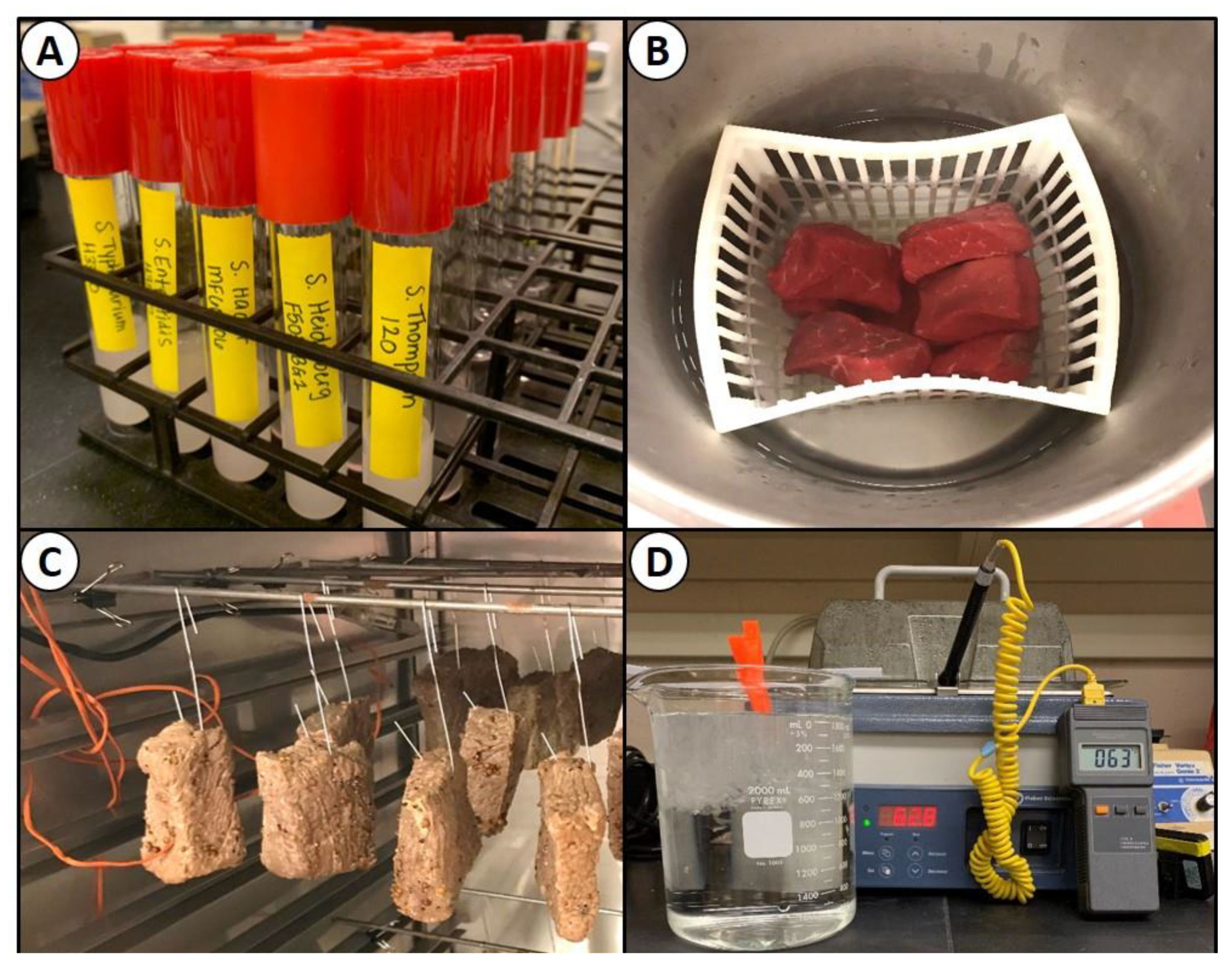
2.5. Stress Conditions for Enumeration

Multiple selective agar media (TSA, SCA, XLD, and HE) containing spectinomycin (5 ug/mL), clindamycin (5 ug/mL), and novobiocin (50 ug/mL) were compared for enumeration of Salmonella, either individually or in mixture, under different stress conditions including: nutrient depletion, acid stress, desiccation, and thermal treatment.

2.5.1. Nutrient Depletion and Cell Starvation

Each of five individual Salmonella serovars, in triplicate replication, were grown in TSB containing 1% glucose, serially diluted in 0.1% BPW, and surface plated immediately after harvesting; at this point, these were referred to as ‘fresh cells’. The Salmonella were plated onto four selective media (TSA, SCA, HE, and XLD), each containing three antibiotics: Spectinomycin (5 ug/mL), clindamycin (5 ug/mL), novobiocin (50 ug/mL). Plates were then incubated at 37 °C for 48 h. Nutrient starvation was assessed by maintaining the dilution tubes used for the fresh cell experiment at 4 °C and determining the bacterial populations after extended three- and six-week intervals (Figure 1A). The trials were performed in triplicate, with each dilution plated in duplicate, on TSA, SCA, HE, and XLD (all containing three antibiotics), and incubated for 48 h at 37 °C.

2.5.2. Antimicrobial (Acid) Stress

Intact, select grade, beef bottom-round sub-primal cuts (Ralph’s Packing Co., Perkins, OK, USA) were trimmed to approximately 0.75-in thick × 2-inch wide × 3-inch long ‘steaks’ at the R.M. Kerr Food & Agricultural Product Center (FAPC). Beef pieces used for this experiment were vacuum-packaged fresh, stored frozen (−20 °C), and thawed immediately before use. The beef pieces were inoculated by pipette with 150 uL of the 5-serovar Salmonella mixture on each side, and immediately spread with a ‘gloved finger’. Inoculated beef pieces were then incubated for 30 min at 4–5 °C to allow for bacterial attachment prior to use. Following incubation, the inoculated beef was dipped in white vinegar (5% acetic acid), lactic acid (5%), or sodium acid sulfate (SAS, 3%) for 30 s and excess liquid was allowed to drain before proceeding (Figure 1B). Meat samples were transferred to filter-stomaching bags, followed by the addition of 100 mL of 1% nBPW (Hardy Diagnostics) and then stomached for 90 sec in a Masticator paddle-blender (IUL Instruments, Barcelona, Spain). Samples were withdrawn, serially-diluted with 0.1% BPW, and dilutions surface plated (0.1 mL) in duplicate, on TSA, SCA, HE, and XLD plates (each containing spectinomycin, clindamycin, and novobiocin) and incubated at 37 °C for 48 h before enumeration. All trials were performed as separate experiments in triplicate replication.

2.5.3. Desiccation and Drying

Beef pieces, as described previously, were inoculated with 150 uL of a five-serovar Salmonella mixture on each side that was spread with a gloved finger and held at 4–5 °C for 30 min to allow for bacterial attachment. The beef pieces were then tumbled (without vacuum) in a biltong spice mixture (2% salt) for 5 min until pieces were evenly coated. Beef pieces were then hung in a temperature-controlled humidity oven (Hotpack, Warminster, PA, USA) at 23.9 °C (75 °F) and 55% relative humidity (RH) and allowed to dry for up to 4 days (Figure 1C). Beef was sampled after inoculation (0 days), and after 2 and 4 days of drying. Beef samples were stomached with 1% nBPW and then serially diluted in 0.1% BPW. Serial dilutions were plated in duplicate (0.1 mL) on the surface of TSA, SCA, HE, and XLD (each containing spectinomycin, clindamycin, and novobiocin) and incubated for 48 h at 37 °C. Treatments were performed in triplicate replication.

2.5.4. Thermal Stress

Salmonella serovar cultures were grown in TSB (+ 1% glucose) as described earlier, harvested by centrifugation, resuspended in fresh/sterile TSB (+ 1% glucose), mixed in equal volumes, and held on ice until used. Then, 1.0 mL of the mixed culture was heat-sealed as a thin layer in vacuum-package bags and heated at 145 °F (62.8 °C) for 75 sec (Figure 1D). The cultures were then removed to ice water to chill for 15 min and held at room temperature for 5 min. Dilutions were then made in 0.1% BPW and surface plated (0.1 mL) in duplicate onto TSA, SCA, HE, and XLD agars (each containing spectinomycin, clindamycin, and novobiocin) and incubated for 48 h at 37 °C. All treatments, including inoculations, were performed in triplicate replication.

2.6. Bacterial Injury

The degree of bacterial sublethal injury was determined by comparing microbial counts on nonselective media (TSA) to those on selective media (XLD) for various treatments in the prior trials according to the equation described by Wesche et al. [32]:
%   I n j u r y = [ ( c o u n t   o n   n o n s e l e c t i v e   a g a r ) ( c o u n t   o n   s e l e c t i v e   a g a r ) ] ( c o u n t   o n   n o n s e l e c t i v e   a g a r )   x   100

2.7. Statistical Analysis

Each trial was performed in triplicate replication and all replications were performed as autonomous and separate experiments using separately inoculated cultures and prepared plating media. All data were presented as the mean of triplicate replications with standard deviation of the mean represented by error bars. Statistical analysis was done using one-way analysis of variance (ANOVA) and the Holm–Sidak test for pairwise multiple comparisons to determine significant differences (p < 0.05). Data treatments with different letters are significantly different (p < 0.05); treatments with the same letter are not significantly different (p > 0.05).

3. Results

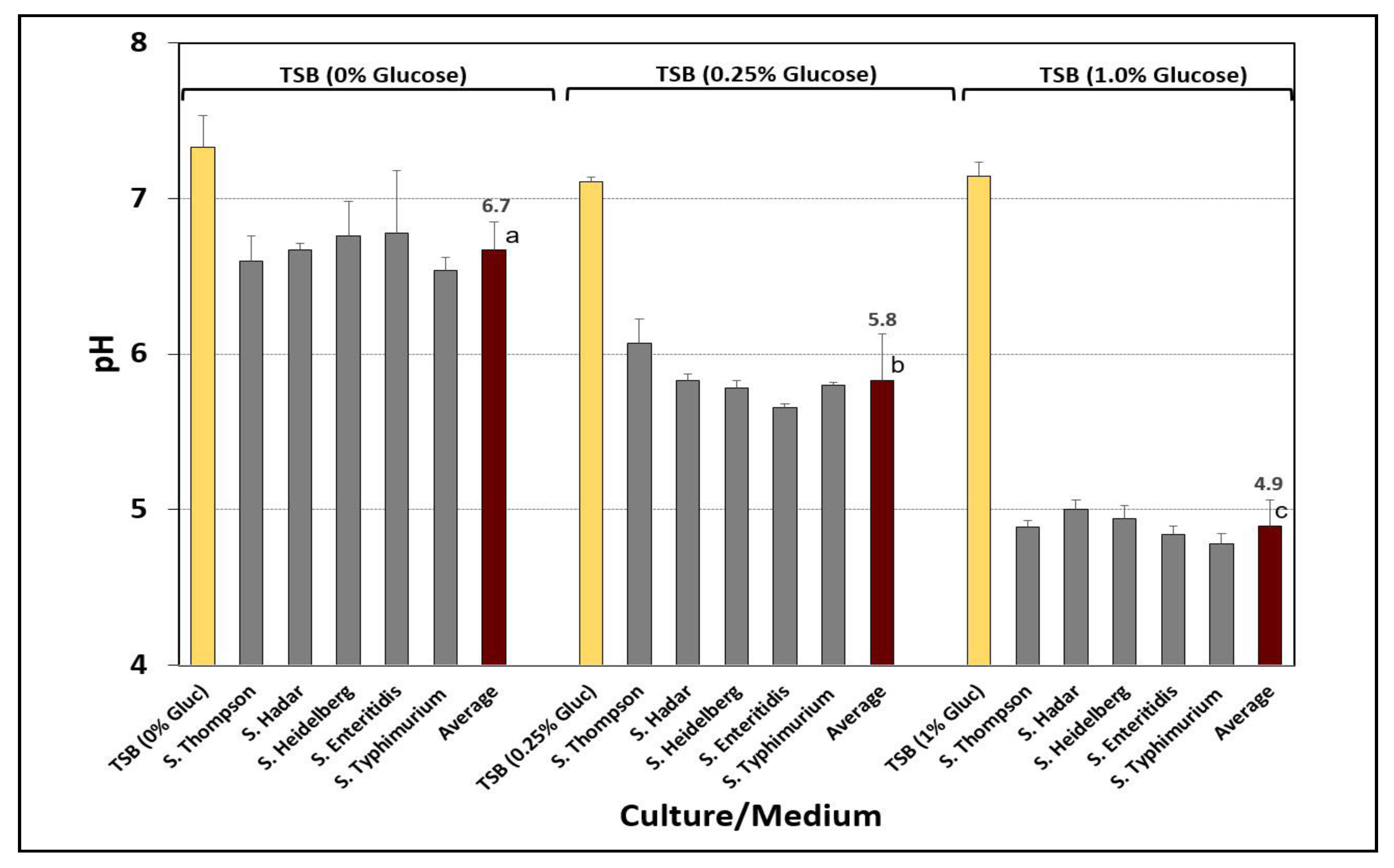
3.1. Acid Adaptation of Salmonella Cultures

Cultures were grown in TSB containing 1% glucose in order to ‘acid adapt’ them to low pH for all stress conditions used in this study. The pH of each of five different Salmonella serovar cultures was examined after growth in TSB, with and without glucose (0%, 0.25%, and 1.0% glucose). Growth in TSB without glucose resulted in spent culture broth pH near neutrality (i.e., average, pH 6.7) whereas the culture broth pH of those grown in TSB with glucose were significantly lower (i.e., 0.25% glucose, average, pH 5.8; 1.0% glucose, average, pH 4.9) (Figure 2).

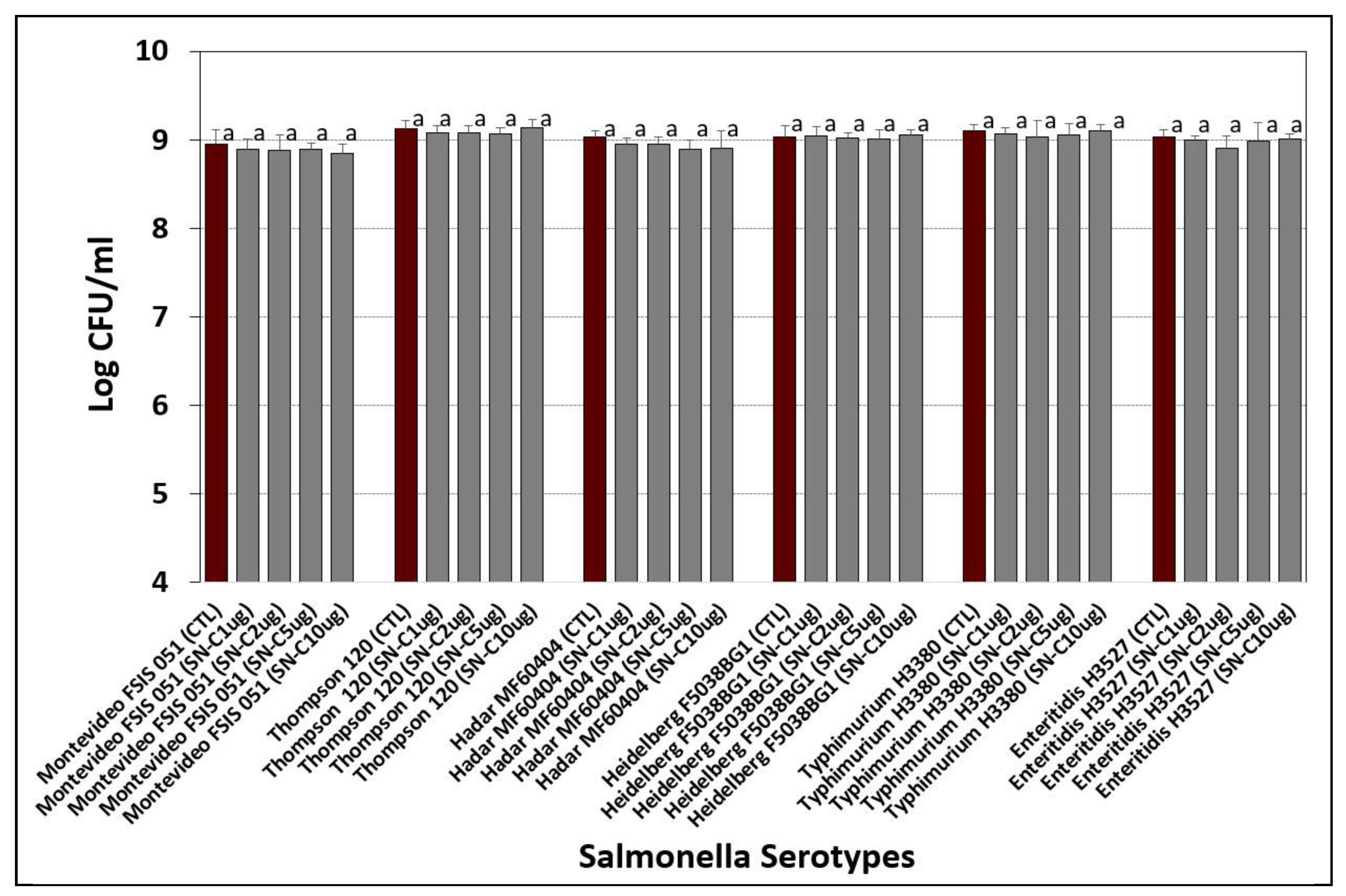
3.2. Determination of Salmonella Antibiotic Resistance (Disc Assay)

Antibiotic resistance for six Salmonella serovars was examined using BD BBL Sensi-Discs on lawns of individual Salmonella serovars and scored qualitatively for sensitivity or resistance to the antibiotic discs (Table 1; only those antibiotics for which four or more strains were resistant are shown). ‘Resistance’ was characterized as an antibiotic disc that showed no inhibition zone around the periphery of the disc while various degrees of sensitivity were subjectively attributed according to size of a visible inhibition zone.

3.3. Confirmation of Salmonella Antibiotic Resistance (Plating on Agar Containing Antibiotics)

Based on the antibiotic disc assay (Table 1), ‘on agar’ antibiotic resistance was confirmed by growing cultures in TSB (without antibiotics) and plating the cultures on TSA alone, and on TSA containing the chosen antibiotics (Figure 3). This was done to determine if there was any antagonism that might be at play when multiple antibiotics are added in combination [34,35,36,37].

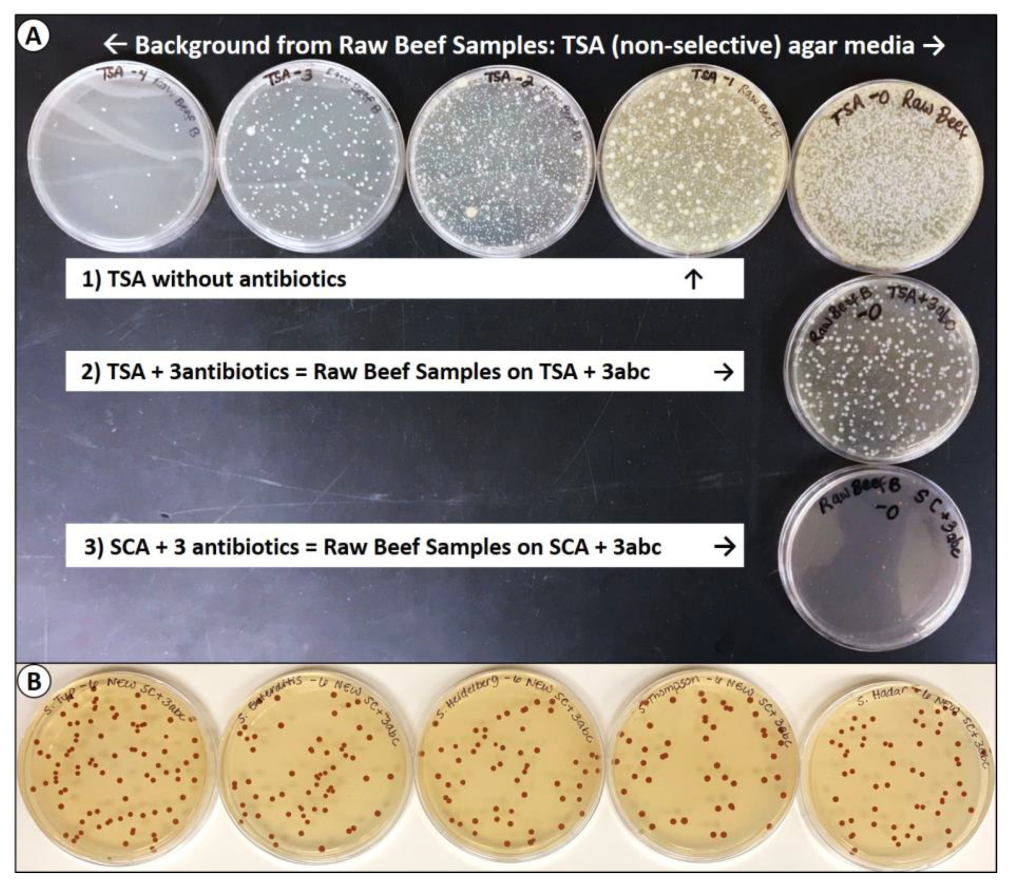
3.4. Selective Agar Media Containing Antibiotics

Based on the microbial platings of the individual Salmonella serovars on TSA containing spectinomycin, novobiocin, and clindamycin at 10-, 100-, and 10-ug/mL, respectively, we felt these levels, or even slightly lower in TSA or other media, would provide a good ‘selective medium’ for our specific Salmonella serovar inoculum in various applications with raw meat if we could demonstrate enumeration equivalent to that on TSA. Additional platings with HE and XLD agars, both with and without antibiotics, showed no significant differences (p > 0.05) (data not shown).

Tryptic Soy Agar and Selenite Cystine Agar Containing Antibiotics

TSA containing spectinomycin (5 ug/mL), clindamycin (5 ug/mL), and novobiocin (50 ug/mL) was examined in a variety of situations where multi-strain (serovar) combinations of Salmonella were used. However, we obtained very high background counts on some samples of raw meat on TSA without antibiotics (Figure 4A, top row). When plated on TSA containing the three antibiotics (i.e., 3abc), the background levels were reduced by approximately 3 logs, but still had significant levels of colonies on the lowest dilution (Figure 4A, middle row). This background was further minimized when we used SCA in combination with our antibiotics for which our Salmonella serovars were resistant (Figure 4A, bottom row). Each of the Salmonella serovars gave slightly different colony sizes on TSA + 3abc for the same incubation time (data not shown) whereas they grew luxuriously on the SC agar (SCA) giving equivalent-sized and perfectly round colony morphologies (Figure 4B).

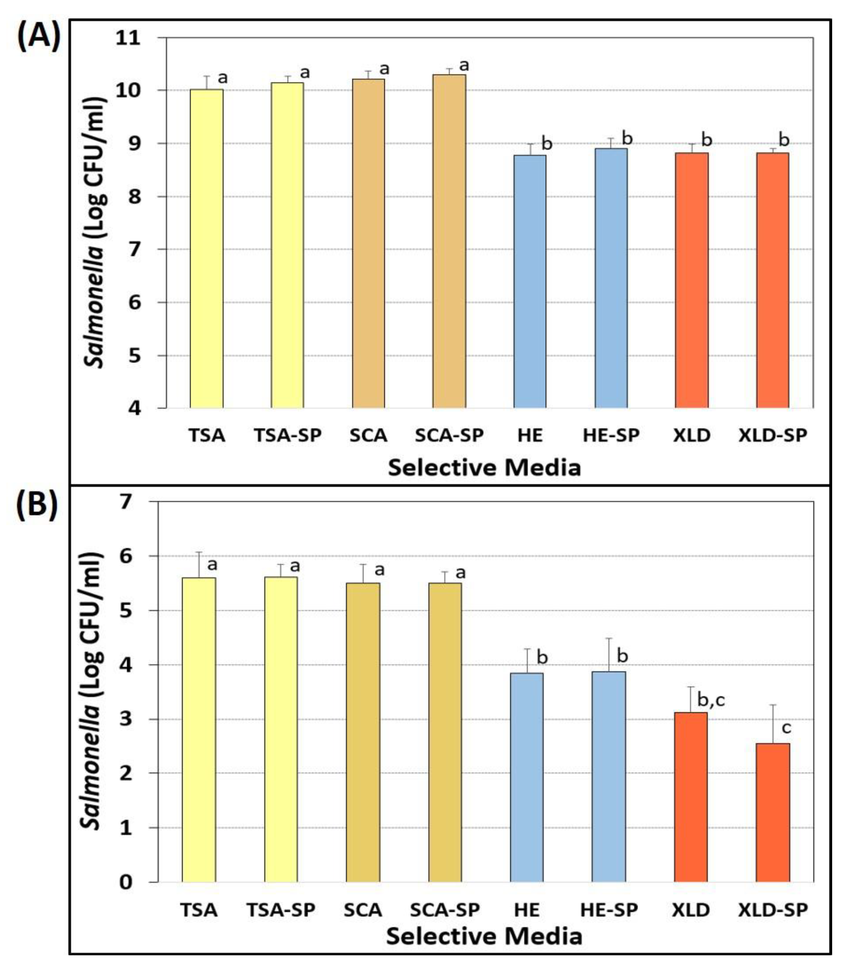
3.5. Comparitive Enumeration on Selective Agars with and without Sodium Pyruvate

Selective agars were formulated that consisted of TSA, TSA-SP, SCA, SCA-SP, XLD, XLD-SP, HE, and HE-SP, all containing three antibiotics (spectinomycin, 5 ug/mL; novobiocin, 50 ug/mL; clindamycin, 5 ug/mL). Individual Salmonella serovars, or equal mixtures of them, were enumerated on these four media after various stress conditions.

Evaluation of Sodium Pyruvate for Recovery of Injured Cells

Sodium pyruvate was examined as a possible supplemental ingredient for the recovery of injured bacterial cells from acid-adapted and nutrient-starved Salmonella (Figure 5A) as well as acid-adapted and 2% salt/3% vinegar-marinaded Salmonella (Figure 5B). When nutrient-starved Salmonella were plated on the various selective media, no statistical difference (p > 0.05) was observed between TSA, TSA-SP, SCA, and SCA-SP, even though the data for SCA was slightly higher than TSA (Figure 5A). However, microbial counts on both HE and XLD (with and without sodium pyruvate), were approximately 1.5-log lower and significantly different (p < 0.05) than TSA and SCA. No significant differences (p > 0.05) were observed between HE and HE-SP, nor between XLD and XLD-SP (Figure 5A).
When inoculated beef pieces were subjected to a salt/vinegar spice marinade and vacuum-tumbled for 30 min, we observed lower counts as would be expected from salt/vinegar marination (inhibition) and subsequent sample stomaching of beef in 100 mL buffer (dilution). Results show that no significant difference (p < 0.05) was observed among TSA- or SCA-based media (with or without sodium pyruvate), nor between HE and HE-SP or XLD and XLD-SP (Figure 5B). Again, enumerations on HE, HE-SP, XLD, and XLD-SP were significantly different (p < 0.05) and lower than on TSA and SCA (Figure 5B).

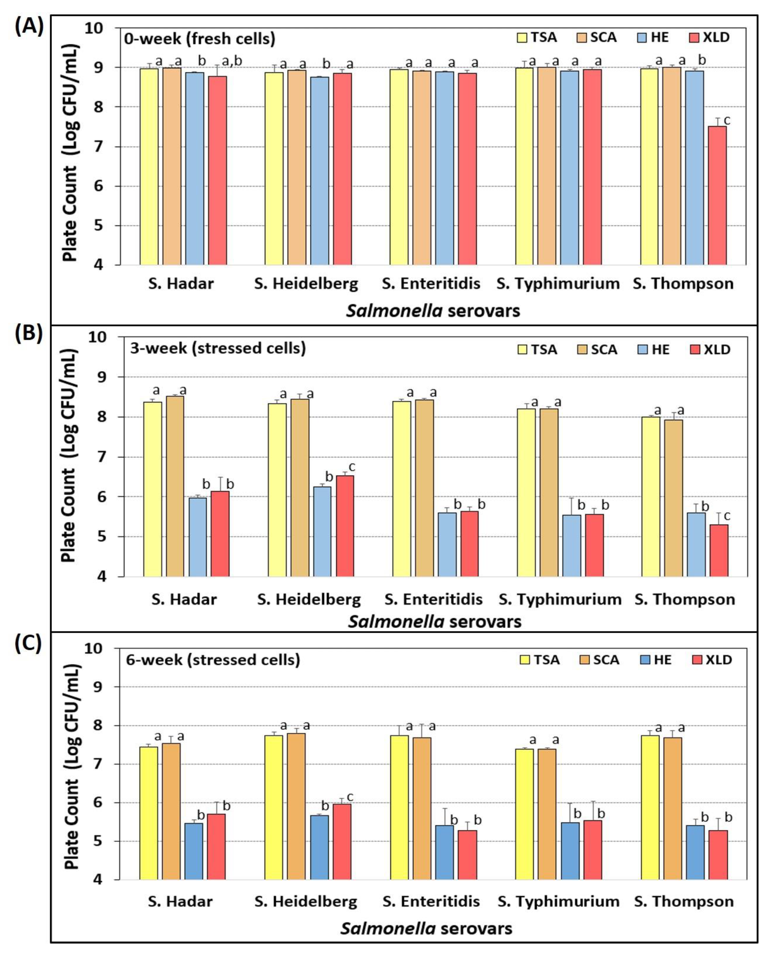
3.6. Salmonella Stress Conditions: Nutrient Depletion and Starvation

Individual Salmonella serovars (in triplicate) were plated on four selective media containing three antibiotics after fresh growth (18 hrs, 37 °C) in TSB (1% glucose) (Figure 6). No significant differences were observed between counts on TSA or SCA, whereas three of five serovars showed slightly lower, but no significant differences when enumerated on HE, and one of five serovars showed significantly lower counts on XLD of ~1.4-log lower levels (Figure 6A). When the same triplicate dilution tube series (in 0.1% BPW) was held at refrigeration temperature (4 °C) and plated again after 3- and 6-weeks, significant differences were observed for platings on HE and XLD which showed 2–3 log lower counts relative to platings on TSA and SCA (Figure 6B,C).

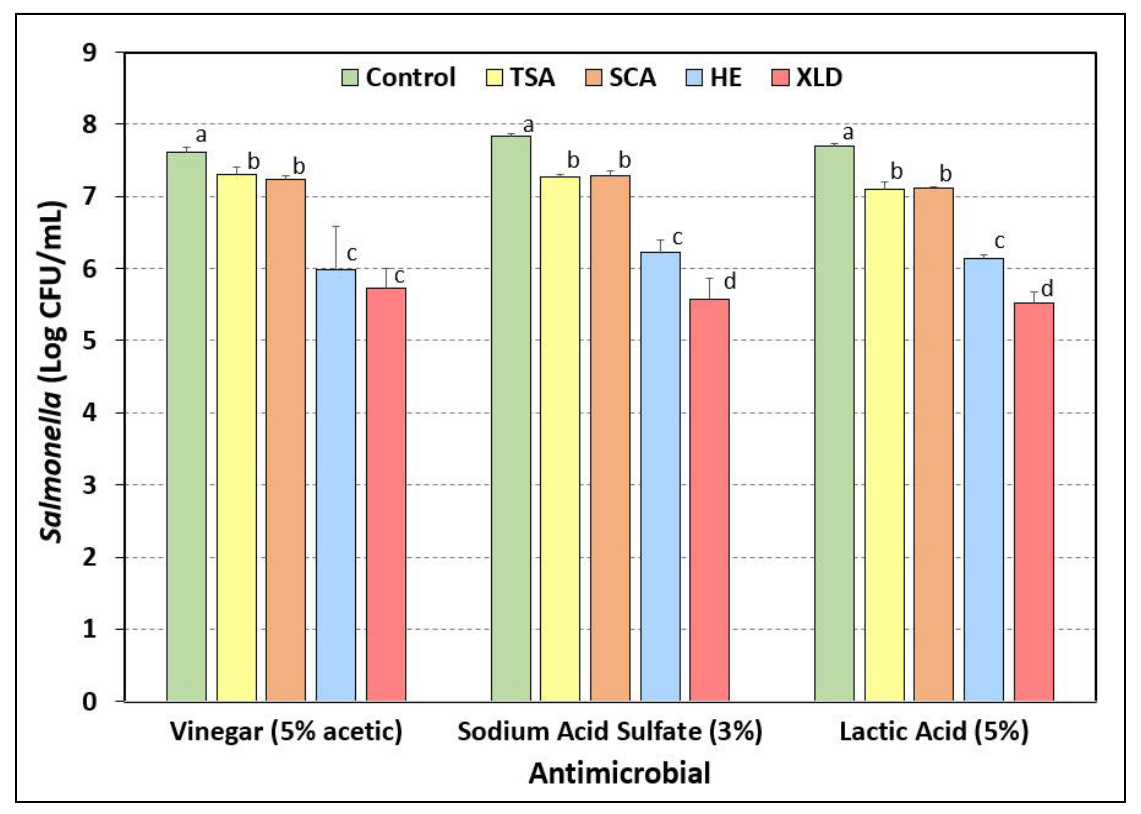
3.7. Salmonella Stress Conditions: Exposure to Acidic Antimicrobials

Beef pieces inoculated with a five-serovar mixture of Salmonella were subjected to 30-sec dip treatment in vinegar (5% acetic acid), sodium acid sulfate (3%), or lactic acid (5%). The beef pieces were stomached in neutralizing BPW, further diluted in 0.1% BPW, and plated onto each of four different selective media described above: TSA, SCA, HE, and XLD, all containing three antibiotics (spectinomycin, 5 ug/mL; novobiocin, 50 ug/mL; clindamycin, 5 ug/mL). Nearly the same results were obtained within each antimicrobial dip treatment: No significant differences (p > 0.05) were observed between TSA and SCA, which showed a modest 0.3–0.6-log reduction from the controls (Figure 7). However, enumeration on HE and XLD agars showed approximately 1.5–2.0 log lower counts than that obtained using the same dilutions for samples plated on TSA and SCA.

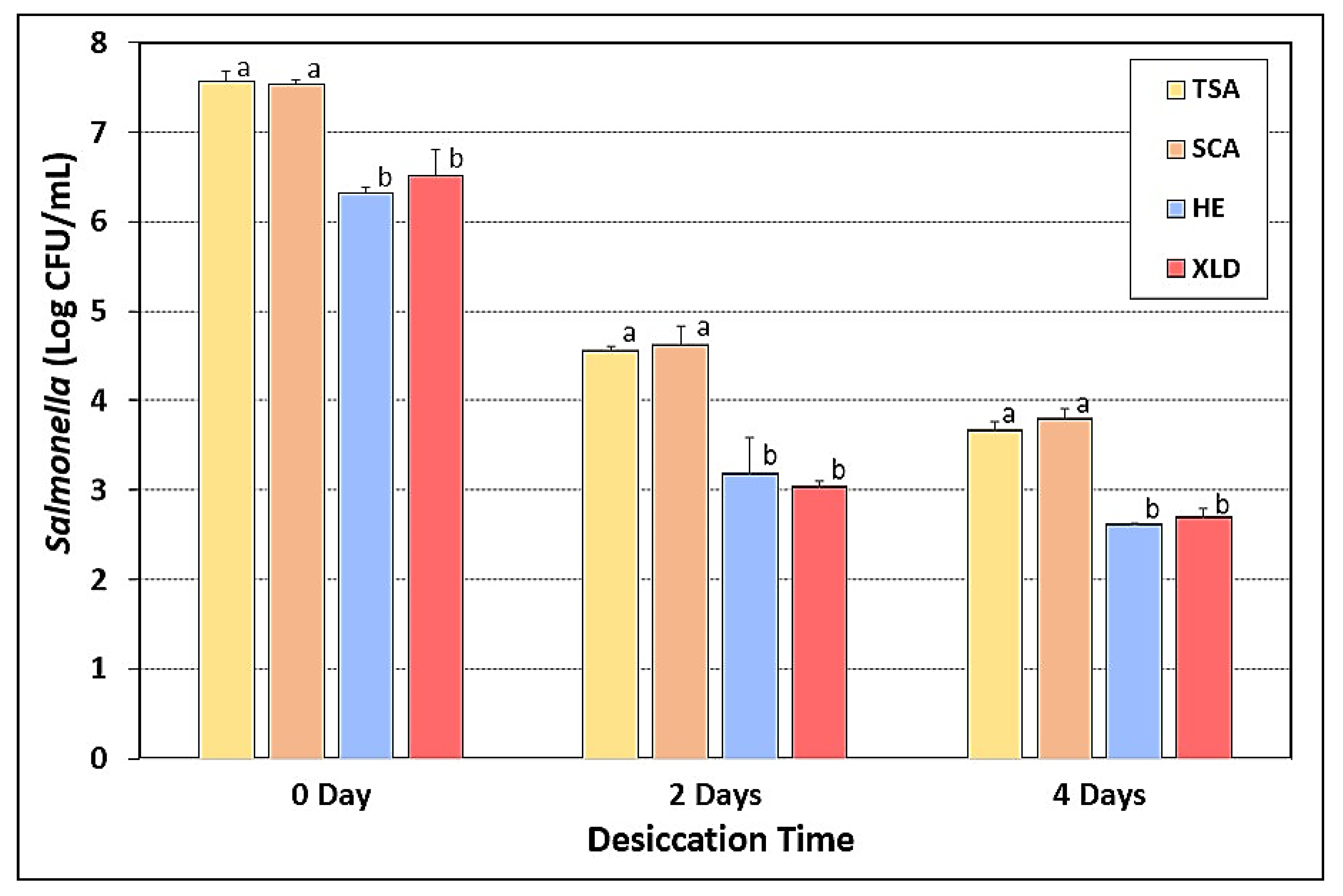
3.8. Salmonella Stress Conditions: Desiccation and Drying

Beef pieces inoculated with a five-serovar mixture of Salmonella were dry-marinaded with biltong spices and salt (no vinegar) before hanging in a humidity oven to be subjected to drying for up to 4 days at 75 °F and 55% RH. Samples retrieved at 0-, 2-, and 4-days of drying were plated on the four selective media described earlier (TSA, SCA, HE, and XLD, all containing three antibiotics). Again, the TSA and SCA based media had nearly identical counts, while HE and XLD based media both showed 1–1.5-log lower counts than the other selective media at each assay time (Figure 8).

3.9. Salmonella Stress Conditions: Thermal Stress

When 1-mL samples of the combined Salmonella serovars were subjected to heating at 145 °F (62.8 °C) for 75 sec in thin layer bags, equivalent counts were obtained on both TSA and SCA agar (~3.4–3.5-log reduction). Counts retrieved on HE and XLD agars showed microbial reductions of 4.75-log and 5.25-log, respectively (Figure 9).

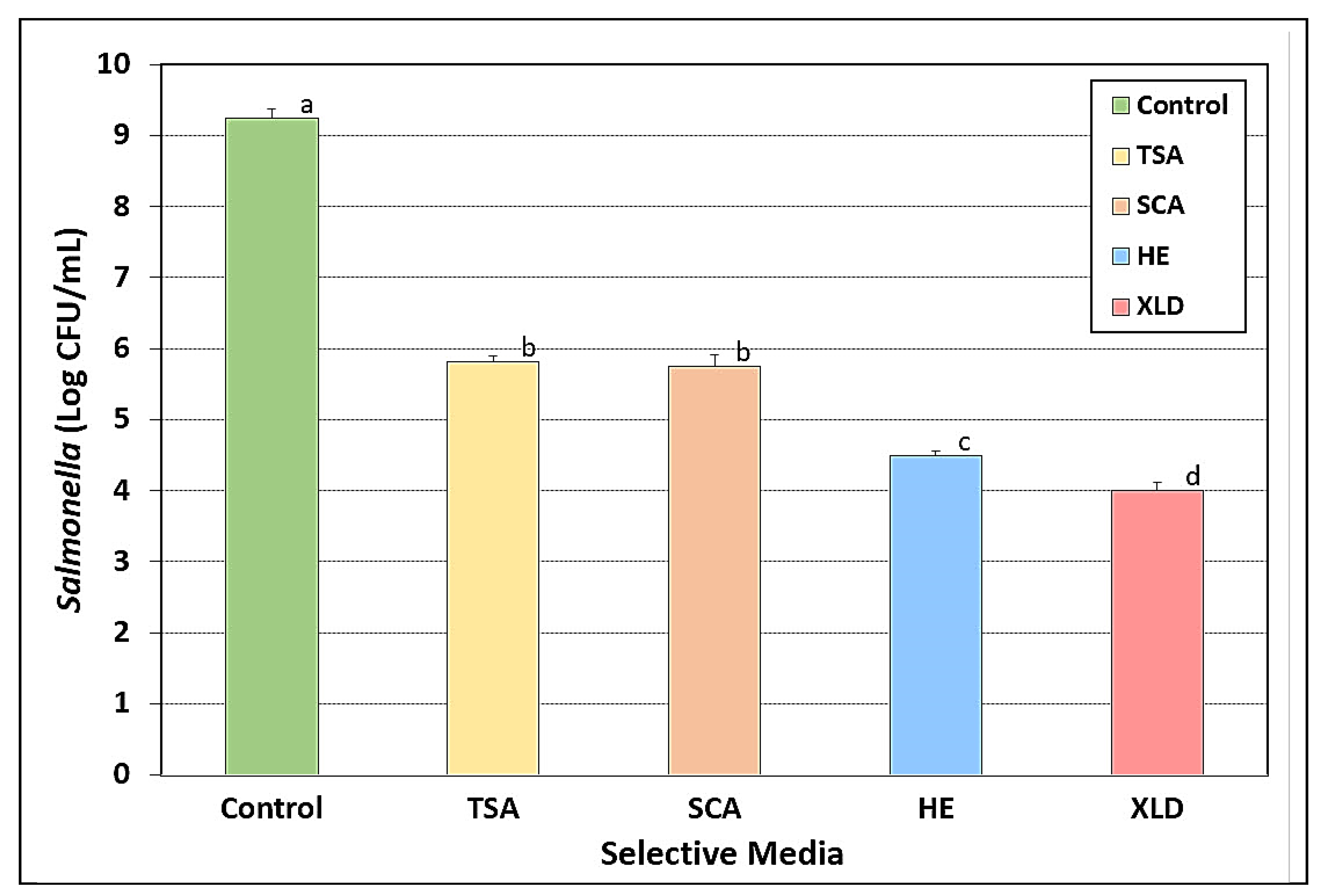
3.10. Bacterial Injury as Determined by Plating on Selective vs. Nonselective Media

When microbial counts of acid-adapted Salmonella were plated on TSA, and compared to platings on SCA, XLD, and HE, reduced levels of recovered counts were consistently obtained on XLD and HE (Table 2). This is consistent with the inhibition of sublethally injured cells by selective media components, as observed by Wesche et al. [32]. As determined with XLD and HE agars, injured cells were determined to comprise as much as 14%–19% of fresh acid-adapted cells and 73.0%–99.7% of the microbial populations of the remaining 11 trials after stressed conditions (Table 2). A similar comparison of acid-adapted Salmonella plated on SCA with those plated on TSA demonstrated a modest 4.4%–13.7% injury level in four of twelve trials, while showing 1.9%–46.7% enhancement of microbial recovery in eight of twelve trials (Table 2).

4. Discussion

In this study, various Salmonella serovars (S. Thompson 120, S. Heidelberg F5038BG1, S. Hadar MF60404, S. Enteritidis H3527, S. Typhimurium H3380) were characterized prior to their use as inocula in subsequent studies to evaluate antimicrobial interventions applied during the processing of dried beef (i.e., biltong, beef jerky). The Salmonella strains used in this study are widely distributed in academia and government research labs and have a long history of testing on the effects of antimicrobial interventions against Salmonella applied to meat and poultry products [22,23,24,25,32,38,39,40,41,42].
Characterization of antibiotic resistance demonstrated that they are not only multi-drug resistant but are also resistant to the same antibiotics. This enables the use of the common antibiotics for the preparation of selective media to recover them from inoculated food studies. TSA containing novobiocin, spectinomycin, and clindamycin was considered a useful generic selective agar media for these Salmonella serovars. The approach to use antibiotics to which the inoculum bacteria are resistant has long been an effective method of selective enumeration of inoculum strains recovered from non-sterile foods that also contain other microorganisms [4,5,6,7,8,9]. However, the appearance of high levels of multi-drug resistant background bacteria from un-inoculated raw beef on TSA containing the three antibiotics was cause for concern and precipitated a search for a more selective medium.
The intended use of the Salmonella serovars was as inocula prior to antimicrobial (acid) interventions on raw beef, and therefore, the strains were acid-adapted prior to use throughout this study. Acid adaptation of Salmonella serovars was reported in the early 1990s [43,44,45] whereby Salmonella pre-exposed to low pH were more resistant to acidic conditions than non-adapted cells. Foster and others [43,45] adjusted media with HCl to achieve low pH conditions. Subsequently, during investigations with enterohemorrhagic Escherichia coli and Listeria monocytogenes, Buchanan et al. [46,47] augmented media with glucose to allow the organisms to lower pH during growth to induce the acid adaptation response. The ability of Salmonella and Shigatoxigenic E. coli to adapt to stressful environments has significant implications in the safety of processed foods [43,48,49]. The Salmonella cultures used throughout this study were predisposed to acidic stress conditions by growth in media containing 1% glucose. This has been recommended by the NACMCF [9] for inoculated challenge studies and by USDA-FSIS when evaluating antimicrobial food processes involving acidic treatments.
Sodium pyruvate was initially considered as a possible media additive to allow recovery of injured cells since many intended applications would be stress-related. However, when examined in the context of nutrient depleted/starved cells, or when Salmonella-inoculated beef was exposed to the stress of salt (dehydration) and vinegar (acid) marination, no benefit was observed with sodium pyruvate. Similarly, neither Knabel and Thielen [50] nor Kirby and Davies [51] observed improved recovery of heat-injured Listeria or Salmonella, respectively, when using sodium pyruvate in their recovery medium. Various investigators have shown that acid adaptation plays a role in enhanced survival of Salmonella and other bacteria, offering cross-protection not only against acid stress, but desiccation, salt, and thermal stress as well [44,52,53,54]. Although other injury recovery additives may have been more effective than sodium pyruvate (i.e., catalase, yeast extract), we continued without additional additives, since the SCA medium was demonstrating comparable results to TSA. In our study, enumeration of Salmonella was consistently and significantly lower on XLD and HE agars, as they likely inhibited the recovery of injured cells [10,11,55]. XLD or HE would have solved our background microorganism problem as the Salmonella would have appeared as black colonies. However, our data showed lower counts (corresponding to injury levels of 89%–99%) when Salmonella were plated on XLD or HE and compared to levels recovered on TSA. Strantz and Zottola [55] were only able to improve recovery with XLD and HE media by plating cells on TSA, allowing them to sit for 4 hrs of recuperation, and then overlaying with XLD or HE agar. Subsequent work by various investigators led to several modifications of the selective overlay technique, including the agar underlay [21] and thin agar layer [19,20,56,57] methods.

5. Conclusions

The Salmonella serovars examined in this study were intended for use in USDA-FSIS validation studies on antimicrobial interventions for dried beef processing to achieve 5-log reduction of Salmonella. Hence, it was important to accurately enumerate Salmonella survivors during processing in lieu of potential background bacteria from raw meat. Selenite cystine broth, a medium routinely used as a selective enrichment broth for luxuriant growth of Salmonella spp., provided more suppression of background microorganisms from raw beef when used as an agar medium than did TSA (with antibiotics). The ability to quantitatively enumerate these Salmonella serovars on SCA containing antibiotics facilitated experimental procedures and eliminated the need for cumbersome multiple layered plating schemes. Enumeration with SCA was not significantly different than that obtained with TSA, which demonstrates the adequacy of SCA as a selective media for these Salmonella serovars, and more so when supplemented with antibiotics to which the strains are resistant. The use of SCA should allow quantitative enumeration of these acid adapted Salmonella serovars when recovered from inoculated food studies employing antimicrobial interventions in spite of the background organisms that might be present.

Author Contributions

The authors have participated in the following: conceptualization, P.M.M.; methodology, P.M.M., D.G., and C.E.K.; validation, C.E.K., D.G., and A.B.; formal analysis, P.M.M. and C.E.K.; investigation, C.E.K., D.G., and A.B.; resources, P.M.M.; data curation, P.M.M.; writing—original draft preparation, P.M.M. and C.E.K.; writing—review and editing, P.M.M. and C.E.K.; visualization, P.M.M.; supervision, P.M.M. and C.E.K.; project administration, P.M.M.; funding acquisition, P.M.M. All authors have read and agreed to the published version of the manuscript.

Funding

Funding for the study was contributed in part by the R.M. Kerr Food & Ag Products Center, the USDA National Institute of Food and Agriculture [Hatch Project #OKL03090], the Advance Foods-SE Gilliland Professorship in Microbial Food Safety (#21-57200), the Division of Agricultural Sciences and Natural Resources at Oklahoma State University, and the USDA Oklahoma Agricultural Experiment Station.

Conflicts of Interest

The authors declare no conflict of interest.

References

  1. Knopp, M.; Andersson, D.I. Predictable phenotypes of antibiotic resistance mutations. mBio 2018, 9, e00770-18. [Google Scholar] [CrossRef] [PubMed] [Green Version]
  2. Sun, J.; Zhu, D.; Xu, J.; Jia, R.; Chen, S.; Liu, M.; Zhao, X.; Yang, Q.; Wu, Y.; Zhang, S.; et al. Rifampin resistance and its fitness cost in Riemerella anatipestifer. BMC Microbiol. 2019, 19, 107. [Google Scholar] [CrossRef] [PubMed]
  3. Woodford, N.; Ellington, M.J. The emergence of antibiotic resistance by mutation. Clin. Microbiol. Infect. 2007, 13, 5–18. [Google Scholar] [CrossRef] [PubMed] [Green Version]
  4. Muriana, P.M.; Klaenhammer, T.R. Conjugal transfer of plasmid-encoded determinants for bacteriocin production and immunity in Lactobacillus acidophilus 88. Appl. Environ. Microbiol. 1987, 53, 553–560. [Google Scholar] [CrossRef] [PubMed] [Green Version]
  5. Mann, J.E.; Brashears, M.M. Validation of time and temperature values as critical limits for the control of Escherichia coli O157:H7 during the production of fresh ground beef. J. Food Prot. 2006, 69, 1978–1982. [Google Scholar] [CrossRef]
  6. Price, S.B.; Cheng, C.M.; Kaspar, C.W.; Wright, J.C.; Degraves, F.J.; Penfound, T.A.; Castanie-Cornet, M.P.; Foster, J.W. Role of rpoS in acid resistance and fecal shedding of Escherichia coli O157:H7. Appl. Environ. Microbiol. 2000, 66, 632–637. [Google Scholar] [CrossRef] [Green Version]
  7. Luchansky, J.B.; Porto-Fett, A.C.S.; Shoyer, B.; Phebus, R.K.; Thippareddi, H.; Call, J.E. Thermal inactivation of Escherichia coli O157:H7 in blade-tenderized beef steaks cooked on a commercial open-flame gas grill. J. Food Prot. 2009, 72, 1404–1411. [Google Scholar] [CrossRef]
  8. Flores, R.A. Distribution of Escherichia coli O157:H7 in beef processed in a table-top bowl cutter. J. Food Prot. 2004, 67, 246–251. [Google Scholar] [CrossRef]
  9. National Advisory Committee on the Microbiological Criteria for Foods. Parameters for determining inoculated pack/challenge study protocols. J. Food Prot. 2010, 73, 140–202. [Google Scholar] [CrossRef]
  10. Pao, S.; Kalantari, A.; Huang, G. Utilizing acidic sprays for eliminating Salmonella enterica on raw almonds. J. Food Sci. 2006, 71, M14–M19. [Google Scholar] [CrossRef]
  11. Gorski, L. Selective enrichment media bias the types of Salmonella enterica strains isolated from mixed strain cultures and complex enrichment broths. PLoS ONE 2012, 7, e34722. [Google Scholar] [CrossRef]
  12. Martin, S.E.; Flowers, R.S.; Ordal, Z.J. Catalase: Its effect on microbial enumeration. Appl. Environ. Microbiol. 1976, 32, 731–734. [Google Scholar] [CrossRef] [PubMed] [Green Version]
  13. Mcdonald, L.C.; Hackney, C.R.; Ray, B. Enhanced recovery of injured Escherichia coli by compounds that degrade hydrogen peroxide or block its formation. Appl. Environ. Microbiol. 1983, 45, 360–365. [Google Scholar] [CrossRef] [PubMed] [Green Version]
  14. Shi, H.; Zhang, R.; Lan, L.; Chen, Z.; Kan, J. Zinc mediates resuscitation of lactic acid-injured Escherichia coli by relieving oxidative stress. J. Appl. Microbiol. 2019, 127, 1741–1750. [Google Scholar] [CrossRef] [PubMed]
  15. Jacobson, A.P.; Wang, H.; Gill, V.S.; Duvall, R.; Arce, G.; Chirtel, S.; Hammack, T.S. Relative effectiveness of selected preenrichment media for the detection of Salmonella from leafy green produce and herbs. Food Microbiol. 2017, 63, 123–128. [Google Scholar] [CrossRef] [PubMed]
  16. Williams, M.S.; Ebel, E.D.; Hretz, S.A.; Golden, N.J. Adoption of neutralizing buffered peptone water coincides with changes in apparent prevalence of Salmonella and Campylobacter of broiler rinse samples. J. Food Prot. 2018, 81, 1851–1863. [Google Scholar] [CrossRef]
  17. Yan, Z.; Gurtler, J.B.; Kornacki, J.L. A solid agar overlay method for recovery of heat-injured Listeria monocytogenes. J. Food Prot. 2006, 69, 428–431. [Google Scholar] [CrossRef]
  18. Kang, D.H.; Fung, D.Y.C. Thin agar layer method for recovery of heat-injured Listeria monocytogenes. J. Food Prot. 1999, 62, 1346–1349. [Google Scholar] [CrossRef]
  19. Kang, D.H.; Fung, D.Y.C. Application of thin agar layer method for recovery of injured Salmonella Typhimurium. Int. J. Food Microbiol. 2000, 54, 127–132. [Google Scholar] [CrossRef]
  20. Wu, V.C.H.; Fung, D.Y.C.; Kang, D.H.; Thompson, L.K. Evaluation of thin agar layer method for recovery of acid-injured foodborne pathogens. J. Food Prot. 2001, 64, 1067–1071. [Google Scholar] [CrossRef] [Green Version]
  21. Kang, D.H.; Siragusa, G.R. Agar underlay method for recovery of sublethally hheat-injured bacteria. Appl. Environ. Microbiol. 1999, 65, 5334–5337. [Google Scholar] [CrossRef] [Green Version]
  22. Juneja, V.K.; Hwang, C.A.; Friedman, M. Thermal inactivation and postthermal treatment growth during storage of multiple Salmonella serotypes in ground beef as affected by sodium lactate and oregano oil. J. Food Sci. 2010, 75, M1–M6. [Google Scholar] [CrossRef] [PubMed]
  23. Carpenter, C.E.; Smith, J.V.; Broadbent, J.R. Efficacy of washing meat surfaces with 2% levulinic, acetic, or lactic acid for pathogen decontamination and residual growth inhibition. Meat Sci. 2011, 88, 256–260. [Google Scholar] [CrossRef]
  24. Juneja, V.K.; Eblen, B.S.; Marks, H.M. Modeling non-linear survival curves to calculate thermal inactivation of Salmonella in poultry of different fat levels. Int. J. Food Microbiol. 2001, 70, 37–51. [Google Scholar] [CrossRef]
  25. Juneja, V.K.; Yadav, A.S.; Hwang, C.A.; Sheen, S.; Mukhopadhyay, S.; Friedman, M. Kinetics of thermal destruction of Salmonella in ground chicken containing trans-cinnamaldehyde and carvacrol. J. Food Prot. 2012, 75, 289–296. [Google Scholar] [CrossRef] [PubMed]
  26. Wilde, S.; Jørgensen, F.; Campbell, A.; Rowbury, R.; Humphrey, T. Growth of Salmonella enterica Serovar Enteritidis PT4 in media containing glucose results in enhanced RpoS-independent heat and acid tolerance but does not affect the ability to survive air-drying on surfaces. Food Microbiol. 2000, 17, 679–686. [Google Scholar] [CrossRef]
  27. Calicioglu, M.; Sofos, J.N.; Samelis, J.; Kendall, P.A.; Smith, G.C. Effect of acid adaptation on inactivation of Salmonella during drying and storage of beef jerky treated with marinades. Int. J. Food Microbiol. 2003, 89, 51–65. [Google Scholar] [CrossRef]
  28. Sandle, T. 14—Antibiotics and preservatives. In Pharmaceutical Microbiology; Sandle, T., Ed.; Woodhead Publishing: Oxford, UK, 2016; pp. 171–183. [Google Scholar]
  29. Lee, R.M.; Hartman, P.A. Optimal pyruvate concentration for the recovery of coliforms from food and water. J. Food Prot. 1989, 52, 119–121. [Google Scholar] [CrossRef]
  30. Morishige, Y.; Fujimori, K.; Amano, F. Differential resuscitative effect of pyruvate and its analogues on VBNC (viable but non-culturable) Salmonella. Microbes Environ. 2013, 28, 180–186. [Google Scholar] [CrossRef] [Green Version]
  31. Gurtler, J.B.; Beuchat, L.R. Performance of media for recovering stressed cells of Enterobacter sakazakii as determined using spiral plating and ecometric techniques. Appl. Environ. Microbiol. 2005, 71, 7661–7669. [Google Scholar] [CrossRef] [Green Version]
  32. Wesche, A.M.; Marks, B.P.; Ryser, E.T. Thermal resistance of heat-, cold-, and starvation-injured Salmonella in irradiated comminuted Turkey. J. Food Prot. 2005, 68, 942–948. [Google Scholar] [CrossRef]
  33. Dickson, J.S.; Frank, J.F. Bacterial starvation stress and contamination of beef. Food Microbiol. 1993, 10, 215–222. [Google Scholar] [CrossRef]
  34. Bollenbach, T. Antimicrobial interactions: Mechanisms and implications for drug discovery and resistance evolution. Curr. Opin. Microbiol. 2015, 27, 1–9. [Google Scholar] [CrossRef] [PubMed] [Green Version]
  35. Ocampo, P.S.; Lázár, V.; Papp, B.; Arnoldini, M.; Abel Zur Wiesch, P.; Busa-Fekete, R.; Fekete, G.; Pál, C.; Ackermann, M.; Bonhoeffer, S. Antagonism between bacteriostatic and bactericidal antibiotics is prevalent. Antimicrob. Agents Chemother. 2014, 58, 4573–4582. [Google Scholar] [CrossRef] [Green Version]
  36. Singh, N.; Yeh, P.J. Suppressive drug combinations and their potential to combat antibiotic resistance. J. Antibiot. 2017, 70, 1033–1042. [Google Scholar] [CrossRef] [PubMed]
  37. Yilancioglu, K. Antimicrobial drug interactions: Systematic evaluation of protein and nucleic acid synthesis inhibitors. Antibiotics 2019, 8, 114. [Google Scholar] [CrossRef] [PubMed] [Green Version]
  38. Tuntivanich, V.; Orta-Ramirez, A.; Marks, B.P.; Ryser, E.T.; Booren, A.M. Thermal inactivation of Salmonella in whole muscle and ground turkey breast. J. Food Prot. 2008, 71, 2548–2551. [Google Scholar] [CrossRef] [PubMed]
  39. Velasquez, A.; Breslin, T.J.; Marks, B.P.; Orta-Ramirez, A.; Hall, N.O.; Booren, A.M.; Ryser, E.T. Enhanced thermal resistance of Salmonella in marinated whole muscle compared with ground pork. J. Food Prot. 2010, 73, 372–375. [Google Scholar] [CrossRef] [PubMed]
  40. Orta-Ramirez, A.; Marks, B.P.; Warsow, C.R.; Booren, A.M.; Ryser, E.T. Enhanced thermal resistance of Salmonella in whole muscle compared to ground beef. J. Food Sci. 2005, 70, m359–m362. [Google Scholar] [CrossRef]
  41. Warsow, C.R.; Orta-Ramirez, A.; Marks, B.P.; Ryser, E.T.; Booren, A.M. Single directional migration of Salmonella into marinated whole muscle turkey breast. J. Food Prot. 2008, 71, 153–156. [Google Scholar] [CrossRef]
  42. Mann, J.E.; Brashears, M.M. Contribution of humidity to the lethality of surface-attached heat-resistant Salmonella during the thermal processing of cooked ready-to-eat roast beef. J. Food Prot. 2007, 70, 762–765. [Google Scholar] [CrossRef] [PubMed]
  43. Foster, J.W. Salmonella acid shock proteins are required for the adaptive acid tolerance response. J. Bacteriol. 1991, 173, 6896–6902. [Google Scholar] [CrossRef] [PubMed] [Green Version]
  44. Leyer, G.J.; Johnson, E.A. Acid adaptation promotes survival of Salmonella spp. in cheese. Appl. Environ. Microbiol. 1992, 58, 2075–2080. [Google Scholar] [CrossRef] [PubMed] [Green Version]
  45. Foster, J.W.; Hall, H.K. Adaptive acidification tolerance response of Salmonella typhimurium. J. Bacteriol. 1990, 172, 771–778. [Google Scholar] [CrossRef] [Green Version]
  46. Buchanan, R.L.; Edelson, S.G. Culturing enterohemorrhagic Escherichia coli in the presence and absence of glucose as a simple means of evaluating the acid tolerance of stationary-phase cells. Appl. Environ. Microbiol. 1996, 62, 4009–4013. [Google Scholar] [CrossRef] [Green Version]
  47. Buchanan, R.L.; Golden, M.H.; Whiting, R.C.; Phillips, J.G.; Smith, J.L. Non-thermal inactivation models for Listeria monocytogenes. J. Food Sci. 1994, 59, 179–188. [Google Scholar] [CrossRef]
  48. Álvarez-Ordóñez, A.; Prieto, M.; Bernardo, A.; Hill, C.; López, M. The acid tolerance response of Salmonella spp.: An adaptive strategy to survive in stressful environments prevailing in foods and the host. Food Res. Intl. 2012, 45, 482–492. [Google Scholar] [CrossRef]
  49. Suehr, Q.J.; Chen, F.; Anderson, N.M.; Keller, S.E. Effect of pH on survival of Escherichia coli O157, Escherichia coli O121, and Salmonella enterica during desiccation and short-term storage. J. Food Prot. 2020, 83, 211–220. [Google Scholar] [CrossRef]
  50. Knabel, S.J.; Thielen, S.A. Enhanced recovery of severely heat-injured, thermotolerant Listeria monocytogenes from USDA and FDA primary enrichment media using a novel, simple, strictly anaerobic method. J. Food Prot. 1995, 58, 29–34. [Google Scholar] [CrossRef]
  51. Kirby, R.M.; Davies, R. Survival of dehydrated cells of Salmonella typhimurium LT2 at high temperatures. J. Appl. Bacteriol. 1990, 68, 241–246. [Google Scholar] [CrossRef] [PubMed]
  52. Leyer, G.J.; Johnson, E.A. Acid adaptation induces cross-protection against environmental stresses in Salmonella typhimurium. Appl. Environ. Microbiol. 1993, 59, 1842–1847. [Google Scholar] [CrossRef] [PubMed] [Green Version]
  53. Greenacre, E.J.; Brocklehurst, T.F. The acetic acid tolerance response induces cross-protection to salt stress in Salmonella typhimurium. Intl. J. Food Microbiol. 2006, 112, 62–65. [Google Scholar] [CrossRef] [PubMed]
  54. Bacon, R.T.; Ransom, J.R.; Sofos, J.N.; Kendall, P.A.; Belk, K.E.; Smith, G.C. Thermal iinactivation of susceptible and multiantimicrobial-resistant Salmonella strains grown in the absence or presence of glucose. Appl. Environ. Microbiol. 2003, 69, 4123–4128. [Google Scholar] [CrossRef] [PubMed] [Green Version]
  55. Strantz, A.A.; Zottola, E.A. A modified plating technique for the recovery and enumeration of stressed Salmonella typhimurium Hf. J. Food Prot. 1989, 52, 712–714. [Google Scholar] [CrossRef] [PubMed]
  56. Ferreira, F.S.; Horvath, M.B.; Tondo, E.C. Assessing the growth and recovery of Salmonella Enteritidis SE86 after sodium dichloroisocyanurate exposure. Braz. J. Microbiol. 2013, 44, 785–790. [Google Scholar] [CrossRef] [Green Version]
  57. Chen, Z.; Diao, J.; Dharmasena, M.; Ionita, C.; Jiang, X.; Rieck, J. Thermal inactivation of desiccation-adapted Salmonella spp. in aged chicken litter. Appl. Environ. Microbiol. 2013, 79, 7013–7020. [Google Scholar] [CrossRef] [Green Version]
Figure 1. Stress conditions for Salmonella serovars: (A) nutrient depletion/starvation by extended refrigeration of individual Salmonella serovars in 0.1% BPW; (B) acid stress by dip treatment of Salmonella-inoculated beef in acidic solutions (vinegar, 5%; 3% sodium acid sulfate; 5% lactic acid); (C) desiccation of Salmonella-inoculated and spice-coated beef at 23.9 °C (75 °F) and 55% RH in a temperature-controlled humidity oven; (D) thermal heating of vacuum-packaged bags containing Salmonella inocula at 62.8 °C (145 °F) for 75 sec.
Microorganisms 08 00338 g001
Figure 2. Analysis of spent broth pH for Salmonella cultures grown in Tryptic Soy Broth (TSB) containing 0%, 0.25%, or 1% glucose incubated at 37 °C for 18 hrs. The data bars in each set are pH values for the medium before inoculation, the five individual cultures after growth, and the average pH of the five cultures. Cultures include S. Thompson 120, S. Heidelberg F5038BG1, S. Hadar MF60404, S. Enteritidis H3527, and S. Typhimurium H3380. Data are presented as the mean of triplicate replications and error bars represent the standard deviation from the mean. Means (for average pH) with different letters are significantly different, as determined by one-way ANOVA using the Holm–Sidak test for pairwise multiple comparisons to determine significant differences (p < 0.05); means with different letters are significantly different (p < 0.05).
Microorganisms 08 00338 g002
Figure 3. Evaluation of Salmonella serovars on TSA alone as Control (CTL) vs. TSA containing spectinomycin (S, 10 ug/mL), novobiocin (N, 100 ug/mL), and increasing amounts of clindamycin (C, 1-, 2-, 5-, and 10 ug/mL). Cultures include S. Montevideo FSIS 051, S. Thompson 120, S. Heidelberg F5038BG1, S. Hadar MF60404, S. Enteritidis H3527, and S. Typhimurium H3380. Cultures were grown 18–20 hrs at 37 °C in TSB (without antibiotics), 10-fold dilutions in 0.1% BPW, and plated on the various media as indicated. Data are presented as the mean of triplicate replications and error bars represent the standard deviation from the mean. Within the same serotype, comparisons of means with the same letters are not significantly different (p > 0.05) as determined by one-way ANOVA using the Holm–Sidak test for pairwise multiple comparisons to determine significant differences.
Microorganisms 08 00338 g003
Figure 4. Comparison of TSA (without antibiotics) vs. TSA and selinite cystine agar (SCA) (with antibiotics). (A) Plating of samples taken from fresh raw meat onto TSA without antibiotics (top row), vs. TSA containing antibiotics (2nd row), vs. SCA containing antibiotics (3rd row). The “3abc” represents the ‘three antibiotics’ described previously: Spectinomycin (5 ug/mL), novobiocin (50 ug/mL), and clindamycin (5 ug/mL). (B) Individual Salmonella serovars surface plated (10−6 dilution) on SCA containing three antibiotics.
Microorganisms 08 00338 g004
Figure 5. Evaluation of sodium pyruvate for recovery of injured Salmonella (A) after 10-day nutrient starvation or (B) after 30-min salt and vinegar (acid) marinade treatment on inoculated beef pieces. Enumeration of mixed-Salmonella serovars on TSA, SCA, Hektoen Enteric (HE), Xylose Lysine Desoxycholate (XLD), and on the same media containing 0.1% sodium pyruvate (TSA-SP, SCA-SP, HE-SP, XLD-SP). All media contained spectinomycin (5 ug/mL), novobiocin (50 ug/mL), and clindamycin (5 ug/mL). Cultures included S. Thompson 120, S. Heidelberg F5038BG1, S. Hadar MF60404, S. Enteritidis H3527, and S. Typhimurium H3380. Data are presented as the mean of triplicate replications, and error bars represent the standard deviation from the mean. Comparisons of means with different letters are significantly different (p < 0.05) as determined by one-way ANOVA using the Holm–Sidak test for pairwise multiple comparisons; means with the same letter are not significantly different (p > 0.05).
Microorganisms 08 00338 g005
Figure 6. Comparison of four selective media for enumeration of individual Salmonella serovars after extended nutrient starvation for 0-weeks (A), 3-weeks (B), and 6-weeks (C) at 4 °C. Each serovar was plated on TSA, SCA, HE, and XLD containing spectinomycin (5 ug/mL), novobiocin (50 ug/mL), and clindamycin (5 ug/mL). Cultures included S. Thompson 120, S. Heidelberg F5038BG1, S. Hadar MF60404, S. Enteritidis H3527, and S. Typhimurium H3380. Data are presented as the mean of triplicate replications of cultures and their dilutions, and error bars represent the standard deviation from the mean. Within the same serotype, comparisons of means with different letters are significantly different (p < 0.05) as determined by one-way ANOVA using the Holm–Sidak test for pairwise multiple comparisons; means with the same letter are not significantly different (p > 0.05).
Microorganisms 08 00338 g006
Figure 7. Acid-stress response of five-serovar mixtures of Salmonella inoculated on the surface of raw beef and subject to 30-sec dip treatment in vinegar (5% acetic acid), sodium acid sulfate (3%), and lactic acid (5%). Treated samples were then plated on TSA, SCA, HE, and XLD containing spectinomycin (5 ug/mL), novobiocin (50 ug/mL), and clindamycin (5 ug/mL). Cultures in the mixture included S. Thompson 120, S. Heidelberg F5038BG1, S. Hadar MF60404, S. Enteritidis H3527, and S. Typhimurium H3380. Data are presented as the mean of triplicate replications and error bars represent the standard deviation from the mean. Control represents inoculum level on beef pieces before antimicrobial treatment. Means with different letters are significantly different (p < 0.05) as determined by one-way ANOVA using the Holm–Sidak test for pairwise multiple comparisons to determine significant differences; means with the same letter are not significantly different (p > 0.05).
Microorganisms 08 00338 g007
Figure 8. Desiccation-stress response of five-serovar mixtures of Salmonella inoculated on the surface of raw beef pieces and subjected up to 4-days drying in a humidity chamber at 23.9 °C (75 °F) and 55% RH. Treated samples were then plated on TSA, SCA, HE, and XLD containing spectinomycin (5 ug/mL), novobiocin (50 ug/mL), and clindamycin (5 ug/mL). Cultures in the mixture included S. Thompson 120, S. Heidelberg F5038BG1, S. Hadar MF60404, S. Enteritidis H3527, and S. Typhimurium H3380. Data are presented as the mean of triplicate replications and error bars represent the standard deviation from the mean. Means with different letters are significantly different (p < 0.05) as determined by one-way ANOVA using the Holm–Sidak test for pairwise multiple comparisons to determine significant differences; means with the same letter are not significantly different (p > 0.05).
Microorganisms 08 00338 g008
Figure 9. Thermal-stress response of five-serovar mixture of Salmonella heated at 62.8 °C (145 °F) for 75 sec. Treated samples were then plated on TSA, SCA, HE, and XLD; all contained spectinomycin (5 ug/mL), novobiocin (50 ug/mL), and clindamycin (5 ug/mL). Cultures in the mixture included S. Thompson 120, S. Heidelberg F5038BG1, S. Hadar MF60404, S. Enteritidis H3527, and S. Typhimurium H3380. The control is the level of the mixed culture before heating. Data are presented as triplicate replications and error bars represent standard deviation from the mean. Means with different letters are significantly different (p < 0.05) as determined by one-way ANOVA using the Holm–Sidak test for pairwise multiple comparisons to determine significant differences; means with the same letter are not significantly different (p > 0.05).
Microorganisms 08 00338 g009
Table 1. Antibiotic disc assay of select Salmonella serovars showing multidrug resistance (Res, resistant; Sens, sensitive; S-sens, slightly sensitive; V-sens, very sensitive).
Salmonella SerovarsClinNovoOxaPenVanSpecAmoAmpPipStr
CC 2*NB 5OX 1P 10VA 3010 ugAMC 30AM 10PIP 100S 10
S. Enteritidis H3527ResResResResResResSensSensSensSens
S. Hadar MF60404ResResResResResResResResResRes
S. Heidelberg F5038BG1ResResResResResResResResResRes
S. Montevideo FSIS 051ResResResResResResSensS-SensV-SensS-Sens
S. Thompson 120ResResResResResResResResResRes
S. Typhimurium H3380ResResResResResResResResResRes
Antibiotics: Clin (Clindamycin), Novo (Novobiocin), Oxa (Oxacillin), Pen(Penicillin), Van (Vancomycin), Spec (Spectinomycin), Amo (Amoxicillin), Amp (Ampicillin), Pip (Piperacillin), Str (Streptomycin). * BD BBL Sensi-Disc product designations.
Table 2. Determination of % injury (-%) or enhancement (+%) of Salmonella by comparison of microbial counts on selective media (SCA, XLD, HE) to non-selective meda (TSA).
Process% Injury (-) or %Enhancement (+)
SCAXLDHE
Sodium pyruvate trials
  • Nutrient depletion/starvation
  • Acid/vinegar marinade

+46.5
−13.7

−94.0
−99.7

−94.9
−98.5
Extended nutrient depletion/starvation
  • Fresh cells
  • 3-weeks
  • 6-weeks

+1.93
+16.1
+6.6

−14.4
−99.4
−98.6

−19.0
−99.7
−99.2
Acidic stress (inoculated beef, 30-sec dip)
  • Vinegar (5% acetic acid)
  • Lactic acid (5%)
  • Sodium acid sulfate (3%)

−4.4
+4.8
+2.7

−95.1
−97.3
−97.9

−96.5
−89.1
−73.0
Desiccation stress (salt/spiced beef, dried)
  • 0-days
  • 2-days
  • 4-days

−6.5
+17.6
+31.8

−91.1
−96.9
−89.3

−94.4
−95.8
−91.1
Thermal stress (62.8 °C/145 °F for 75 sec)−10.6−99.2−99.6

