Article

Branched Chondroitin Sulfate Oligosaccharides Derived from the Sea Cucumber Acaudina molpadioides Stimulate Neurite Outgrowth

1
State Key Laboratory of Phytochemistry and Plant Resources in West China, Kunming Institute of Botany, Chinese Academy of Sciences, Kunming 650201, China
2
University of Chinese Academy of Sciences, Beijing 100049, China
3
State Key Laboratory for Conservation and Utilization of Bio-Resources, Key Laboratory for Microbial Resources of the Ministry of Education, School of Life Sciences, Yunnan University, Kunming 650091, China
4
School of Pharmaceutical Sciences, South-Central Minzu University, Wuhan 430074, China
*
Authors to whom correspondence should be addressed.
These authors contributed equally to this work.
Mar. Drugs 2022, 20(10), 653; https://doi.org/10.3390/md20100653
Submission received: 1 October 2022 / Revised: 14 October 2022 / Accepted: 17 October 2022 / Published: 21 October 2022
(This article belongs to the Special Issue Bioactive Compounds from Sea Cucumbers)

Abstract

:
Fucosylated chondroitin sulfate (FCS) from the sea cucumber Acaudina molpadioides (FCSAm) is the first one that was reported to be branched by disaccharide GalNAc-(α1,2)-Fuc3S4S (15%) and sulfated Fuc (85%). Here, four size-homogenous fractions, and seven oligosaccharides, were separated from its β-eliminative depolymerized products. Detailed NMR spectroscopic and MS analyses revealed the oligomers as hexa-, hepta-, octa-, and nonasaccharide, which further confirmed the precise structure of native FCSAm: it was composed of the CS-E-like backbone with a full content of sulfation at O-4 and O-6 of GalNAc in the disaccharide repeating unit, and the branches consisting of sulfated fucose (Fuc4S and Fuc2S4S) and heterodisaccharide [GalNAc-(α1,2)-Fuc3S4S]. Pharmacologically, FCSAm and its depolymerized derivatives, including fractions and oligosaccharides, showed potent neurite outgrowth-promoting activity in a chain length-dependent manner. A comparison of analyses among oligosaccharides revealed that the sulfate pattern of the Fuc branches, instead of the heterodisaccharide, could affect the promotion intensity. Fuc2S4S and the saccharide length endowed the neurite outgrowth stimulation activity most.

1. Introduction

Neurons are highly differentiated terminal cells that cannot regenerate. If compounds can promote neuronal growth, it will be meaningful for treatment of neuronal damage and degeneration. Chondroitin sulfate (CS) is a class of glycosaminoglycans and is involved in cell division and neuronal development. CS proteoglycans (CS-PGs) were defined as inhibitory molecules of neuron growth in early experiments, and the use of chondroitinase ABC could attenuate the inhibitory effect on neuron growth [1,2,3]. However, some researches have reported that CS could promote neuronal growth [4]. According to the sulfation sites on the saccharide chain, CS could be classified as type A, B, C, D, E, et al. (Figure 1A). CS-A, CS-B, and CS-C were considered to inhibit neuron growth, while CS-D and CS-E exhibited neurite outgrowth-promoting activity. Some reports also proved that CS-E strongly inhibited nerve regeneration [5,6,7,8,9,10]. It has been known that commercially available CS retains heterogeneity, including CS-A (50–80% of A-type unit), CS-C (50–70% of C-type unit), CS-D (20–40% of D-type unit) and CS-E (63.6% of E-type unit). The low purity of saccharides may contribute to the contradictory results [11].
In regard to the CS analogs from invertebrates, the keratan sulfate disaccharide-branched CS-E from a clam, Mactra chinensis, stimulated the neurite outgrowth of hippocampal neurons [11]. A fucosylated CS from the sea cucumber Apostichopus japonicus exhibited neurite outgrowth-promoting activity [12].
Fucosylated CS (FCS) is a characterized CS-E derivative that has been found ubiquitously and exclusively in sea cucumbers thus far. It possesses the CS-E-like backbone with repeating disaccharide units [D-GalNAc4S6S-β(1,4)-D-GlcA-β(1,3)], where D-GalNAc are sulfated in O-4 and O-6 sites [13]. The O-3 of D-GlcA in each backbone are branched by sulfated fucose or disaccharide [14,15,16,17,18,19]. Structural differences of FCS from different sea cucumber species are embodied in the size, the composition, the sulfate substitution of branches (Figure 1B). Besides structure, another aspect of FCS that appeals to researchers is that FCS has wide-ranging biological activities, such as anti-coagulant, anti-thrombotic, anti-virus, and anti-cancer effects [13,20,21,22]. Previous research proposed that the FCS from the sea cucumber A. japonicus exhibited neurite outgrowth-promoting activity, and recognized the potential roles of the fucose branch in neurite outgrowth. A chemically synthesized FCS trisaccharide that is composed of a CS-E disaccharide unit with a sulfated fucose branch also exhibited comparable stimulation activity [12]. The effects of the FCS originated from other sea cucumbers on neurite’s growth, and the structure-function relationships remain unresolved and deserve detailed investigations.
FCSAm was obtained from the sea cucumber Acaudina molpadioides in our previous work [23]. Oligosaccharides from the β-eliminative depolymerized FCSAm revealed that its backbone has a full content of type E, which was branched by monosaccharide [Fuc2S4S and Fuc4S], and the disaccharide [GalNAc-(α1,2)-Fuc3S4S] on O-3 of each GlcA. This is the first report of such a disaccharide branch in natural FCS. FCSAm showed the variety of branches with sulfated substitutions, as well as the novel disaccharide.
In this work, further investigation on this unique CS analog (FCSAm) was conducted. From its β-eliminative depolymerized products, the size-homogenous fractions with a higher degree of polymerization were obtained. Subsequently, charge-separation by SAX-HPLC was employed to yield hexa-, hepta-, octa-, and nonasaccharide. These oligosaccharides clearly revealed the structural features of natural FCSAm, and further verified the characteristics of its branches which is in line with our previous report. In view of the novelty of FCSAm, and the structural diversity of its derivatives, including the size-homogenous fractions and oligosaccharides, the effects on neurite outgrowth were evaluated and the structure-activity relationships were also discussed.

2. Results and Discussion

2.1. Preparation of Homogenous Fractions and Oligosaccharides from the Depolymerized FCSAm

β-Eliminative depolymerization has been established as an effective method for deciphering the structures of FCS from sea cucumbers. Here the FCSAm was employed as the depolymerization process described in Section 3.2 to prepare its low-molecular-weight product, dFCSAm. As shown in Figure 2A, dFCSAm was composed of a series of fractions with different sizes, reflected by its HPGPC profile. For the sake of their structural identification, dFCSAm was fractionated by GPC using Bio-Gel P-10 & P-6 columns, until each fraction exhibited a relatively single and symmetric peak on the HPGPC analysis chromatogram. Finally, five fractions (F1–F5) were obtained (Figure 2A).
F1 and F2 have been further analyzed in our previous work. Both fractions consisted of the oligomers with the same or approximate size. From F1, three trisaccharides were obtained. And from F2, two tetra-, one penta-, and two hexa-saccharides were purified by the aid of SAX separation (oligosaccharides 17). For the fractions with larger size, theoretically, its oligosaccharide composition should be more complex and variable. Though F3–F5 presented the homogeneities in size, further analysis based on the charge difference showed the characteristic of a multi-component. In this work, F3 was further charge-separated on the Dionex Ionpac™ AS11-HC Semi-prep column repeatedly to yield oligosaccharides 813. Chromatographic analysis indicated that 813 running over the SAX column presented a single chromatographic peak, respectively (Figure 2B). The same separation strategy has been adopted for fraction F4, while, unfortunately, the separated components are mixtures as well.

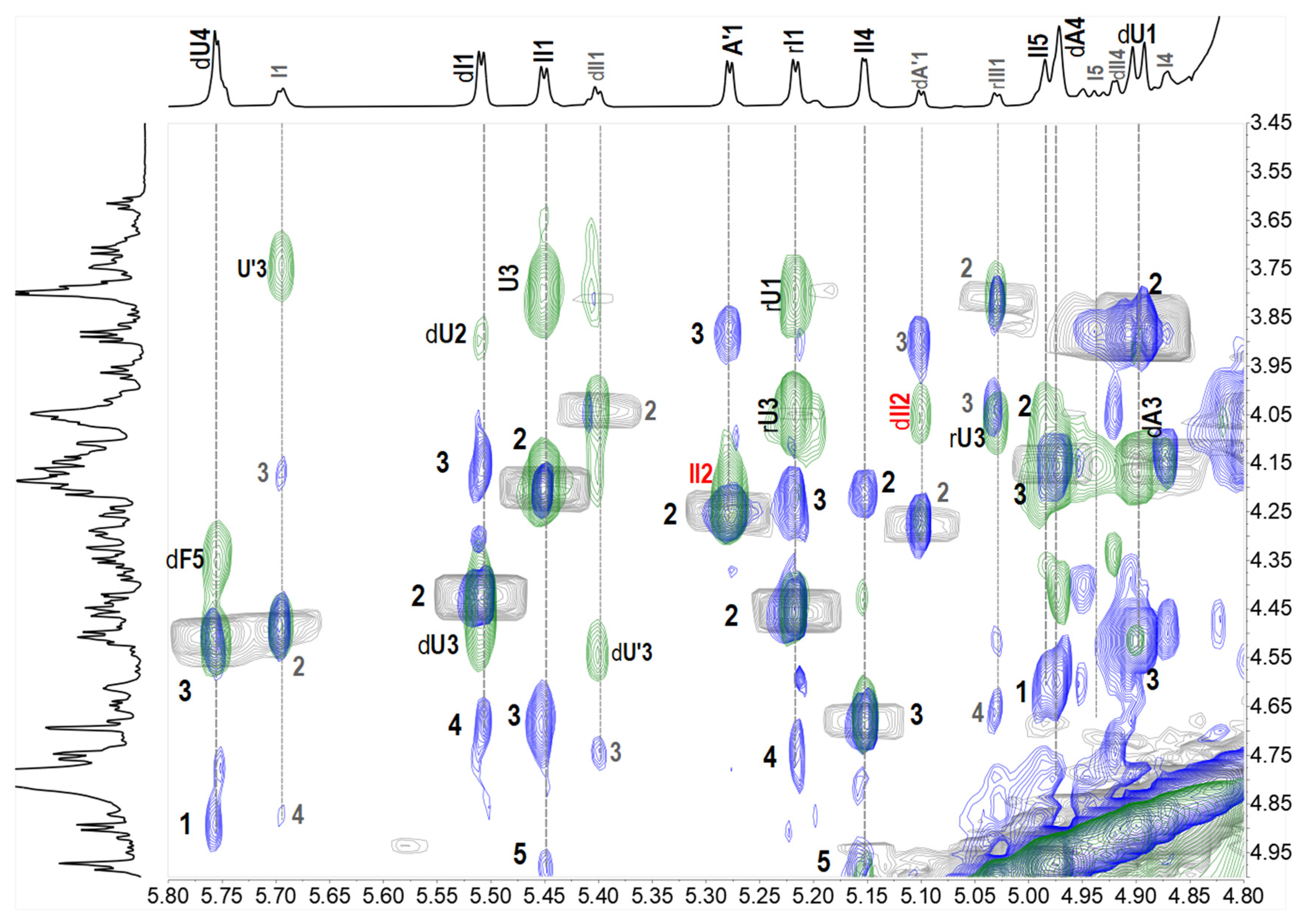
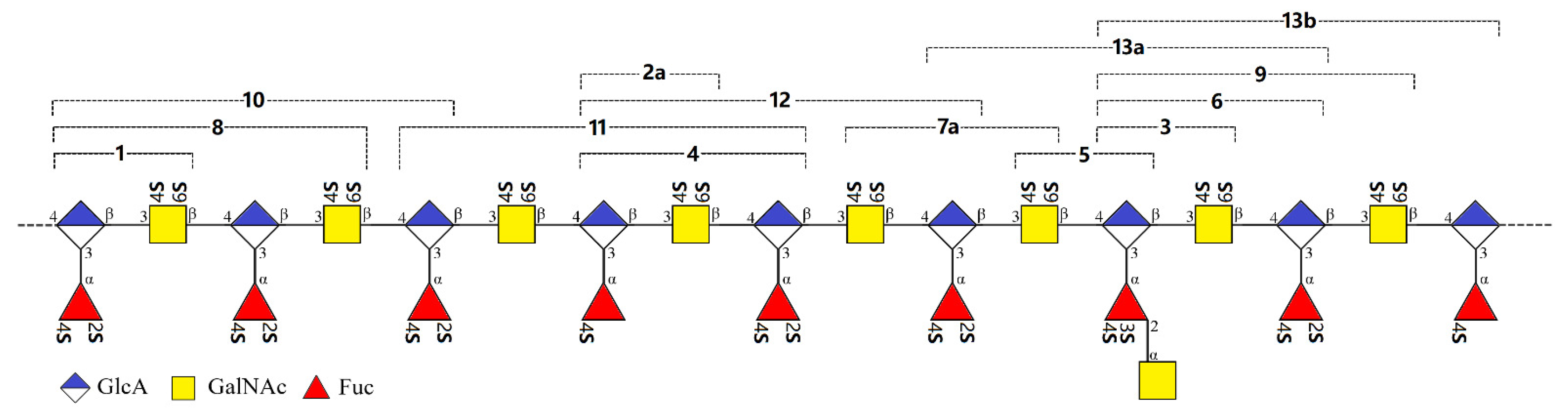
2.2. Structural Elucidation of the Oligosaccharides

The structures of oligosaccharides 813 were elucidated by the aid of their 1D & 2D NMR spectra (Figure 3, Figure 4, Figure 5 and Figure 6, and Figures S2 and S3) and ESI-Q-TOF MS spectra (Figure S5).
The spectra of compound 8 indicated it was a hexasaccharide with the structure of L-Fuc2S4S-(α1,3)-L-Δ4,5GlcA-(α1,3)-D-GalNAc4S6S-(β1,4)-[L-Fuc2S4S-(α1,3)-]-D-GlcA-(β1,3)-D-GalNAc4S6S-ol, the same as the one we have reported before [18]. The distinctive methyl groups signaling at ~2.0 ppm (-COCH3) and ~1.3 ppm (-CH3) in the 1H NMR spectrum revealed that 8 contained two GalNAc residues and two Fuc residues. The anomeric protons at δH 5.68 and 5.50 ppm, derived from the two Fuc residues, indicated the sulfation at O-2 and O-4 positions. The downfield shift signals of the respective protons (I2, 4.49 ppm; I4, 4.85 ppm; dI2, 4.42 ppm; dI4, 4.69 ppm) reconfirmed the substitution of sulfate esters. For oligosaccharide 9, as shown in Figure 3, the appearance of one more set of anomeric signals (at the region of δH 5.0–5.7 ppm, δC 100–106 ppm), accompanying with one more methyl signal at δH 2.09 ppm, indicated that 9 possessed one more GalNAc residue (marked as A’) than that in 8. Another signal at δH 5.40 ppm was from the anomeric proton of Fuc residue (designated as II) with the sulfation at O-3 and O-4 positions, which were confirmed by the resonance signals (II3: 4.74, 76.7 ppm; II4: 4.91, 81.9 ppm). Every resonance of each residue was determined by the HSQC spectrum (Figure 4). The correlation of A’1 (δH 5.09 ppm) and II2 (δC 75.1 ppm) in the HMBC spectrum indicated that residue A’1 connected to II by an α1,2 linkage, and this linkage could be reconfirmed by the correlation of A’1 (δH 5.09 ppm) and II2 (δH 4.04 ppm) in the ROESY spectrum (Figure 4). Thus, 9 was proved to be a heptasaccharide with the structure of D-GalNAc-(α1,2)-L-Fuc3S4S-(α1,3)-L-Δ4,5GlcA-(α1,3)-D-GalNAc4S6S-(β1,4)-[L-Fuc2S4S-(α1,3)-]-D-GlcA-(β1,3)-D-GalNAc4S6S-ol.
The 1D spectra of 1012 occurred similarly in the number of characteristic signals, including the methyl groups (two -COCH3 of GalNAc, δH~2.0 ppm, and three -CH3 of Fuc, δH~1.3 ppm), the anomeric resonances (three signals, at δH 5.0–5.7 ppm; seven signals at δC 99–107 ppm), and the two carbon signals of C-2 of GalNAc residues (δC~54 ppm) (Figure 5). This suggested that 1012 shared the common skeleton of an octasaccharide. Detailed analyses based on their 2D NMR spectra lead to the full assignments of all the signals, which are shown in Figure 5 and Table 1. The structural differences between 1012 existed in the sulfation form of the Fuc branches. According to the downfield shift signals of H2/C2 and H4/C4 (marked in Italic, Table 1), three Fuc2S4S (dI, I, and rI represented the Fuc2S4S locating at the non-reducing end, the middle, and the reducing end, respectively) were determined in oligosaccharide 10, while in 11 and 12, an Fuc4S at the middle and the non-reducing end were elucidated and designated as III and dIII, respectively. The structures of 1012 were determined as L-Fuc2S4S-(α1,3)-L-Δ4,5GlcA-(α1,3)-D-GalNAc4S6S-(β1,4)-[L-Fuc2S4S-(α1,3)-]-D-GlcA-(β1,3)-D-GalNAc4S6S-(β1,4)-[L-Fuc2S4S-(α1,3)-]-D-GlcA-ol, L-Fuc2S4S-(α1,3)-L-Δ4,5GlcA-(α1,3)-D-GalNAc4S6S-(β1,4)-[L-Fuc4S-(α1,3)-]-D-GlcA- (β1,3)-D-GalNAc4S6S-(β1,4)-[L-Fuc2S4S-(α1,3)-]-D-GlcA-ol, and L-Fuc4S-(α1,3)-L-Δ4,5GlcA-(α1,3)-D-GalNAc4S6S-(β1,4)-[L-Fuc2S4S-(α1,3)-]-D-GlcA-(β1,3)-D-GalNAc4S6S-(β1,4)-[L-Fuc2S4S-(α1,3)-]-D-GlcA-ol. The full assignments are displayed in Table 1. The structures of 812 were also confirmed according to the accurate molecular mass information of the excimer ion peaks, as shown in Table 2.
For 13, two sets of signals different in the integration area reflected in its 1H NMR spectrum, suggesting that 13 was a mixture. In terms of the prevailing component, compared with 10, three more signals were observed at the region of 5.0–5.7 ppm besides the H-1 of Fuc2S4S at the non-reducing and reducing ends (5.51 and 5.22 ppm). According to the superimposed spectra of 1H-1H COSY/TOCSY/ROESY of 13 (Figure 6), the protons at 5.15 and 5.45 ppm came from the same spin coupling system. Detailed analysis on the COSY and TOCSY spectra showed that it was an Fuc residue with sulfation at O-3 and O-4 (Fuc3S4S, designated as II). The H-5 of II (4.98 ppm) indicated that it is in the middle of the saccharide chain. The proton at 5.28 ppm, combining with the extra carbon signal at 100.5 ppm, and the extra methyl signal at ~2.0 ppm, outlined the presence of a GalNAc residue (A’). A’ connected to II by an α1,2 linkage, revealing from the correlation peak of A’1 (5.28 ppm) and II2 (4.15 ppm) in the ROESY spectrum. Thus, the main component in 13 (designated as 13a) was deduced as a nonasaccharide with the structure of L-Fuc2S4S-(α1,3)-L-Δ4,5GlcA-(α1,3)-D-GalNAc4S6S-(β1,4)-[D-GalNAc-(α1,2)-L-Fuc3S4S-(α1,3)-]-D-GlcA-(β1,3)-D-GalNAc4S6S-(β1,4)-[L-Fuc2S4S-(α1,3)-]-D-GlcA-ol. For the minor component, it was also elucidated as a nonasaccharide with the structure of D-GalNAc-(α1,2)-L-Fuc3S4S-(α1,3)-L-Δ4,5GlcA-(α1,3)-D-GalNAc4S6S-(β1,4)-[L-Fuc2S4S-(α1,3)-]-D-GlcA-(β1,3)-D-GalNAc4S6S-(β1,4)-[L-Fuc4S-(α1,3)-]-D-GlcA-ol (13b). The structures of oligosaccharides 1013 were presented in Figure 7.

2.3. Structural Confirmation of the Native FCSAm

In our last work, from the size-homogeneous fractions with low degree of polymerization that separated from the β-eliminative depolymerized product dFCSAm, we have elucidated a series of purified oligosaccharides, including tri-, tetra-, penta-, and hexasaccharides [23]. The branches contained the mono- and disaccharide [Fuc4S, Fuc2S4S, and GalNAc-(α1,2)-Fuc3S4S]. Here, we have purified and elucidated the oligomers 813 consisting of hexa-, hepta-, octa-, and nonasaccharide from the fraction F3 with a higher degree of polymerization. 813 presented a continuation with the structural features summarized before which are composed of the central core of 3)-D-GalNAc4S6S-(β1,4)-D-GlcA-(β1, and the sulfated branches [Fuc4S, Fuc2S4S, and GalNAc-(α1,2)-Fuc3S4S]. Oligosaccharides that contain the heterodisaccharide branch were confirmed again. These results further supported our structural deduction of the natural FCSAm: its backbone consists of a repeating disaccharide unit of GlcA and GalNAc linked alternatively by β1,4 and β1,3, wherein all the GalNAc residues were sulfated at O-4 and O-6, the same as CS-E; all the GlcA residues were branched and all the branches glycosylated at O-3 of GlcA, instead of any other sites; the branch types included sulfated fucose (Fuc4S and Fuc2S4S) and heterodisaccharide [GalNAc-(α1,2)-Fuc3S4S]; the disaccharide branches distributed randomly, but there was no case that such disaccharide branches adjacently distributed in the saccharide chain. All of the reducing end residues (Fuc) of the heterodisaccharide branch were sulfated both at O-3 and O-4, while no sulfation occurred on any sites of the non-reducing end residues (GalNAc) (Figure 8). The sulfation pattern of the heterodisaccharide branch seemed conserved, markedly differing from that reported in the FCS from the sea cucumbers Holothuria nobilis and Ludwigothurea grisea [17,19].

2.4. Neurite Outgrowth-Promoting Activity of FCSAm and Its Derivatives

FCSAm and a variety of its derivatives, including the size-homogenous fractions with different molecular weights and the purified oligosaccharides with different sulfate substitutions, laid the substance that is fundamental for the further analysis of the influence of structural characteristics on the neurite outgrowth-promoting activity.
As shown in Figure 9 and Table 3, natural FCSAm and its depolymerized product dFCSAm showed significant activity in promoting neurite outgrowth. Compared with blank control and CS-E groups, the length increased by 50% at the concentration of 50 μg/mL. Meanwhile, FCSAm, dFCSAm and CS-E showed an aggregation phenomenon under the condition (Figure 9C). While for the size-homogenous fractions F2–F5 obtained after GPC treatment, they all showed the promotion activities, and the promoting effects increased with the increase of molecular weight from 1.5 to 4.4 kDa. Among them, F5 exhibited more than two-times the outgrowth promoting activity than CS-E, and no obvious aggregation phenomenon occurred. In contrast, FCSAm did not show a significantly stronger growth promoting effect than fractions, as expected, indicating that an optimum chain length is required for promotion. For dFCSAm, the presence of the saccharides with large Mw (Figure 2A) should be responsible for the weaker promotion effect than F5.
For the purified oligosaccharides from tri- to nonasaccharides, different concentrations (4.4, 13.3, 40 μM) of oligosaccharides were coated on PDL to evaluate the effects. As shown in Figure 10 and Table 4, compared with the control, all of the oligosaccharides showed neurite outgrowth-promoting activity to different degrees, which basically acted in a concentration-dependent manner. In response to the trisaccharide 1, the length of neurons increased by approximately 50%, which was consistent with the previous report [12]. In addition, at the same concentration, the activity of oligosaccharides on neuron growth increased with the increase of the chain length. By comparing the structural characteristics and activity effects of 10, 11 and 12, the sulfate ester enhanced the activity at low (4.4 μM) and medium (13.3 μM) concentrations. Moreover, the residue Fuc2S4S, instead of Fuc4S, located at the middle of octasaccharide (1012) showed greater promotion. By contrast, whether the branch located at non-reducing end is Fuc2S4S or Fuc4S, the octasaccharides (10 and 12) showed a similar enhancement effect on neurite outgrowth. For the novel disaccharide branch [GalNAc-(α1,2)-Fuc3S4S], it did not show an obvious difference in the promotion or inhibition effects, which could be concluded from the comparison of analyses of 1 & 3, 8 & 9 and 1013.

3. Materials and Methods

3.1. Materials

Fucosylated chondroitin sulfate from the body walls of A. molpadioides were obtained in our previous work. Bio-Gel P-10/P-6/P-2 and Sephadex G-25 were purchased from Bio-Rad Laboratories and GE Healthcare Life Sciences, respectively. CS-E [60% of E-type unit, Code# CSR-NaCS-E2(SqC)10] was bought from Cosmo Bio Co., Ltd. Neurobasal (21103049), B27 (17504044), glutamax (35050061), penicillin and streptomycin (15140122) were obtained from Gibco. Poly-D-lysine (PDL, P6407), DNase I (D4513) Anti-tublin Ш (T8578) and Bovine serum albumin (BSA, B2064) were purchased from Sigma. PBS (092810305) and Triton X-100 (QR12914) are from MP. Papain (LS003119) was obtained from Worthington. Alexa flour 488 anti-mouse IgG (ab15105) came from Abcam. Pregnant Sprague-Dawley rats were obtained from SPF (Beijing) Biotechnology Co. Ltd. (Beijing, China). All other reagents were of analytical grade and obtained commercially.

3.2. Depolymerization of FCSAm

The β-eliminative depolymerization of FCSAm was employed according to an established method. Specifically, 6.5 g FCSAm was dissolved in 97.5 mL of water and then reacted with 245 mL of benzethonium chloride solution (62.5 mg/mL) under stirring at room temperature. After centrifugation (4000 rpm × 15 min) and dryness under reduced pressure at 40 °C to constant weight, 17.5 g benzethonium salts of FCSAm were obtained. They were then dissolved in 88 mL dimethyl formamide, and 6.23 mL benzyl chloride was added at 35 °C for 24 h. After that, 53.7 mL of freshly prepared EtONa/EtOH (0.16 M) was added into the reaction system for another 30 min at room temperature at 300 rpm. Subsequently, 120 mL of saturated sodium chloride solution and 960 mL EtOH were added and the precipitate was collected by centrifugation. In order to remove the benzyl groups, the resulting precipitate was dissolved in 360 mL H2O, and 16 mL freshly prepared 6M NaOH was added into the solution at 25 °C for 30 min. Meanwhile, 1385 mg of sodium borohydride was added to the solution to reduce the hemiacetals of the resulting saccharides. After that, the solution was neutralized by HCl. Finally, the β-eliminative depolymerized product was obtained after desalting by Sephadex G-25.

3.3. Preparation of Size-Homogenous Fractions and Oligosaccharides from Depolymerizated FCSAm

dFCSAm was subjected to a Bio-Gel P-10 column and eluted by 0.2 M sodium chloride solution in batches. The sample in tubes was detected at the UV wavelength of 232 nm, and according to the absorption values, the elution curve was plotted. After repeating separation using Bio-Gel P-10 or P-6 column, the fractions with a similar size were pooled and lyophilized separately. The obtained five size-homogenous fractions were designated as F1~F5 in order of size from lowest to highest.
F3 was further purified by strong anion exchange chromatography using a Dionex Ionpac AS11-HC Semi-prep column. The gradient elution program was as follows: 0−60 min, 0−60% elution B (2 M NaCl, pH 3.5); elution A was H2O (pH 3.5). The flow rate was 3.5 mL/min. The preparation was employed under the online UV detection at λ232 nm. Next, the oligosaccharides were desalted by GPC on a Bio-Gel P-2 or Sephadex G-25 column and lyophilized.

3.4. Structural Elucidation of the Oligosaccharides

The structures of the oligosaccharides were determined by 1D & 2D NMR spectra and ESI-Q-TOF-MS data, which were recorded at 298 K with a Bruker Advance 600/800 MHz spectrometer equipped with a 13C/1H dual probe in FT mode, and with a 6540 UHD Accurate-Mass Q-TOF LC/MS spectrometer. For the NMR data acquisition, each sample was dissolved in deuterium oxide (D, 99.9%).

3.5. Neuronal Cultures and Coating Plates

Neurobasal medium contained B27 supplement (1×), 2 mM glutamax, penicillin and streptomycin (1×). The plates were pre-coated with 50 μg/mL PDL at 37 °C for 6 h, washed three times with double distilled water and then coated with the sample overnight at 37 °C. The cortical tissue of rats at embryonic day (E) 18 were isolated under a dissecting microscope, removed quickly, and placed in cold PBS and gently chopped, then incubated in a 1 mg/mL papain and DNase I (20 IU/mL) solution at 37 °C for 30 min. Supplementing with neurobasal medium, the cells were eventually plated at 10,000/cm2 into plates. The cultures were maintained at 37 °C in a humidified 5% CO2 atmosphere [24,25,26].

3.6. Immunofluorescence

After 2 days, neurons were fixed with 4% paraformaldehyde for 30 min, washed with PBS three times, incubated in 0.2% Triton X-100 for 15 min, blocked with 1% BSA and incubated overnight with anti-tublin Ш antibody at 4 °C. The second day, the cells were washed in PBS three times. The second antibody was then incubated at room temperature for 1 h and then rinsed in PBS three times. The nuclei were stained with DAPI for 10 min. The neurons were washed three times with PBS, and then, the cell morphology was detected on an IncuCyte S3 live-cell analysis system [12,27].

3.7. Statistical Analysis

Only cells with neurites longer than one cell body diameter were counted. The length of the longest neurite (50–100 cells) were measured by Image J software and expressed as percentage of growth or length, relative to control (* p < 0.05, ** p < 0.01, or *** p < 0.001). The percentage of growth rate was calculated as follows: growth rate (%) = (length of tested group–length of control group)/control group × 100%. All data were expressed as mean ± SD. A statistical analysis was performed using IBM SPSS Statistics 20. Statistical significances were evaluated with a two-sided unpaired Student’s t-test for two-group comparisons, and a one-way ANOVA followed by the Dunnett post hoc test for multigroup comparisons. p < 0.05 was considered statistically significant [12,28].

4. Conclusions

Fucosylated chondroitin sulfate (FCS) from the sea cucumber Acaudina molpadioides has been obtained (FCSAm), and its precise structure was preliminary investigated in our previous work. An unusual disaccharide branch [GalNAc-(α1,2)-Fuc3S4S] was first found in natural FCS, and sulfated fucose Fuc2S4S and Fuc4S were also elucidated as the branches linked to O-3 of GlcA in the central core. In view of the novelty and variety of the branch type of FCSAm, further investigation of structure and activity were conducted in this study. From the glycosidic bond-selectively depolymerized product (dFCSAm), several size-homogenous fractions (F2–F5) with the molecular weight increasing from 1.5 kDa to 4.4 kDa were obtained by GPC separation. Furthermore, seven oligosaccharides were purified after repeat charge-separation from fraction F3. A detailed analyses of the NMR and MS spectroscopic data clearly presented their structures as hexa-, hepta-, octa-, and nonasaccharide. Combined with the oligomers including tri-, tetra-, penta-, and hexasaccharide obtained from fractions F1 and F2, the precise structure of native FCSAm was confirmed. Its CS-E-like backbone was composed of the repeating disaccharide unit [3)-GalNAc4S6S-(β1,4)-GlcA-(β1,], where all the GlcA residues were branched by sulfated fucose [Fuc2S4S (60%) and Fuc2S4S (25%)] and heterodisaccharide [GalNAc-(α1,2)-Fuc3S4S (15%)]. The disaccharide branches dispersively distributed in the saccharide chain rather than clustered.
Based on the fractions and oligosaccharides with rich structural features, the effects on the cortical neuron outgrowth were evaluated. The natural FCSAm and its depolymerized products dFCSAm showed neurite outgrowth promotion and cell aggregation simultaneously. For the size-homogonous fraction (F2–F5), F2 could stimulate the outgrowth, and the promoting effects increased with the increase of molecular weight. A comparison of analyses among the purified oligosaccharides also showed that the promoting activity was positively correlated with chain length, while an optimum chain length is required for maximum growth without aggregation. In addition, the branch Fuc2S4S significantly contributed to the promotion, whereas the novel disaccharide branch [GalNAc-(α1,2)-Fuc3S4S] did not show obvious difference in promotion or inhibition effects. The results expanded the range of FCS activity analysis and provided some guidance for drug research in neuro-systemic diseases.

Supplementary Materials

The following are available online at https://www.mdpi.com/article/10.3390/md20100653/s1, Figure S1. 1H/13C NMR spectra of oligosaccharide 9; Figures S2–S4. HSQC spectrum and signal assign-ments of oligosaccharide 1012; Figure S5. MS spectra and signal assignments of oligosaccharides 813.

Author Contributions

Conceptualization, J.Z.; methodology, W.W. and H.M.; software, L.Z. and L.Y.; formal analysis, W.W., H.M., S.L., R.Y. and J.Z.; investigation, W.W. and H.M.; data curation, W.W. and H.M.; writing—original draft preparation, W.W. and R.Y.; writing—review and editing, R.Y. and J.Z.; supervision, R.Y. and J.Z.; funding acquisition, R.Y. and J.Z. All authors have read and agreed to the published version of the manuscript.

Funding

This research was funded by the National Natural Science Foundation of China (82073725 and 31900926).

Institutional Review Board Statement

Animal experiments were approved by the Animal Ethics Committee of the Kunming Institute of Botany, Chinese Academy of Sciences (SYXK (Dian), K2018-0005).

Data Availability Statement

All data presented in this study are available from the corresponding author on reasonable request.

Conflicts of Interest

The authors declare that they have no conflict of interest.

References

  1. Dow, K.E.; Mirski, S.; Roder, J.; Riopelle, R. Neuronal proteoglycans: Biosynthesis and functional interaction with neurons in vitro. J. Neurosci. 1988, 8, 3278–3289. [Google Scholar] [CrossRef] [PubMed] [Green Version]
  2. Snow, D.M.; Letourneau, P.C. Neurite outgrowth on a step gradient of chondroitin sulfate proteoglycan (CS-PG). J. Neurosci. 1992, 23, 322–336. [Google Scholar] [CrossRef] [PubMed]
  3. Zuo, J.; Neubauer, D.; Dyess, K.; Ferguson, T.A.; Muir, D. Degradation of chondroitin sulfate proteoglycan enhances the neurite-promoting potential of spinal cord tissue. Exp. Neurol. 1998, 154, 654–662. [Google Scholar] [CrossRef] [PubMed]
  4. Maeda, N.; Noda, M. 6B4 proteoglycan/phosphacan is a repulsive substratum but promotes morphological differentiation of cortical neurons. Development 1996, 122, 647–658. [Google Scholar] [CrossRef]
  5. Hikino, M.; Mikami, T.; Faissner, A.; Vilela-Silva, A.-C.E.S.; Pavão, M.S.G.; Sugahara, K. Oversulfated dermatan sulfate exhibits neurite outgrowth-promoting activity toward embryonic mouse hippocampal neurons. J. Biol. Chem. 2003, 278, 43744–43754. [Google Scholar] [CrossRef] [Green Version]
  6. Sugahara, K.; Mikami, T.; Uyama, T.; Mizuguchi, S.; Nomura, K.; Kitagawa, H. Recent advances in the structural biology of chondroitin sulfate and dermatan sulfate. Curr. Opin. Struct. Biol. 2003, 13, 612–620. [Google Scholar] [CrossRef]
  7. Tully, S.E.; Mabon, R.; Gama, C.I.; Tsai, S.M.; Liu, X.; Hsieh-Wilson, L.C. A chondroitin sulfate small molecule that stimulates neuronal growth. J. Am. Chem. Soc. 2004, 126, 7736–7737. [Google Scholar] [CrossRef] [Green Version]
  8. Gilbert, R.J.; McKeon, R.J.; Darr, A.; Calabro, A.; Hascall, V.C.; Bellamkonda, R.V. CS-4,6 is differentially upregulated in glial scar and is a potent inhibitor of neurite extension. Mol. Cell. Neurosci. 2005, 29, 545–558. [Google Scholar] [CrossRef]
  9. Kim, M.H.; Park, S.R.; Choi, B.H. Comparative analysis of the expression of chondroitin sulfate subtypes and their inhibitory effect on axonal growth in the embryonic, adult, and injured rat brains. Tissue Eng. Regener. Med. 2020, 18, 165–178. [Google Scholar] [CrossRef]
  10. Clement, A.M.; Sugahara, K.; Faissner, A. Chondroitin sulfate E promotes neurite outgrowth of rat embryonic day 18 hippocampal neurons. Neurosci. Lett. 1999, 269, 125–128. [Google Scholar] [CrossRef]
  11. Higashi, K.; Takeda, K.; Mukuno, A.; Okamoto, Y.; Masuko, S.; Linhardt, R.J.; Toida, T. Identification of keratan sulfate disaccharide at C-3 position of glucuronate of chondroitin sulfate from Mactra chinensis. Biochem. J. 2016, 473, 4145–4158. [Google Scholar] [CrossRef] [PubMed] [Green Version]
  12. Shida, M.; Mikami, T.; Tamura, J.-I.; Kitagawa, H. A characteristic chondroitin sulfate trisaccharide unit with a sulfated fucose branch exhibits neurite outgrowth-promoting activity: Novel biological roles of fucosylated chondroitin sulfates isolated from the sea cucumber Apostichopus japonicus. Biochem. Biophys. Res. Commun. 2017, 487, 678–683. [Google Scholar] [CrossRef] [PubMed]
  13. Xu, H.; Zhou, Q.; Liu, B.; Chen, F.; Wang, M. Holothurian fucosylated chondroitin sulfates and their potential benefits for human health: Structures and biological activities. Carbohydr. Polym. 2022, 275, 118691. [Google Scholar] [CrossRef] [PubMed]
  14. Li, H.; Yuan, Q.; Lv, K.; Ma, H.; Gao, C.; Liu, Y.; Zhang, S.; Zhao, L. Low-molecular-weight fucosylated glycosaminoglycan and its oligosaccharides from sea cucumber as novel anticoagulants: A review. Carbohydr. Polym. 2021, 251, 117034. [Google Scholar] [CrossRef] [PubMed]
  15. Zhao, L.; Wu, M.; Xiao, C.; Yang, L.; Zhou, L.; Gao, N.; Li, Z.; Chen, J.; Chen, J.; Liu, J.; et al. Discovery of an intrinsic tenase complex inhibitor: Pure nonasaccharide from fucosylated glycosaminoglycan. Proc. Natl. Acad. Sci. USA 2015, 112, 8284–8289. [Google Scholar] [CrossRef] [PubMed] [Green Version]
  16. Yin, R.; Zhou, L.; Gao, N.; Li, Z.; Zhao, L.; Shang, F.; Wu, M.; Zhao, J. Oligosaccharides from depolymerized fucosylated glycosaminoglycan: Structures and minimum size for intrinsic factor Xase complex inhibition. J. Biol. Chem. 2018, 293, 14089–14099. [Google Scholar] [CrossRef] [PubMed] [Green Version]
  17. Li, S.; Zhong, W.; Pan, Y.; Lin, L.; Cai, Y.; Mao, H.; Zhang, T.; Li, S.; Chen, R.; Zhou, L.; et al. Structural characterization and anticoagulant analysis of the novel branched fucosylated glycosaminoglycan from sea cucumber Holothuria nobilis. Carbohydr. Polym. 2021, 269, 118290. [Google Scholar] [CrossRef]
  18. Yin, R.; Zhou, L.; Gao, N.; Lin, L.; Sun, H.; Chen, D.; Cai, Y.; Zuo, Z.; Hu, K.; Huang, S.; et al. Unveiling the disaccharide-branched glycosaminoglycan and anticoagulant potential of its derivatives. Biomacromolecules 2021, 22, 1244–1255. [Google Scholar] [CrossRef]
  19. Yin, R.; Pan, Y.; Cai, Y.; Yang, F.; Gao, N.; Ruzemaimaiti, D.; Zhao, J. Re-understanding of structure and anticoagulation: Fucosylated chondroitin sulfate from sea cucumber Ludwigothurea grisea. Carbohydr. Polym. 2022, 294, 119826. [Google Scholar] [CrossRef]
  20. Huang, N.; Wu, M.; Zheng, C.; Zhu, L.; Zhao, J.; Zheng, Y. The depolymerized fucosylated chondroitin sulfate from sea cucumber potently inhibits HIV replication via interfering with virus entry. Carbohydr. Res. 2013, 380, 64–69. [Google Scholar] [CrossRef]
  21. Li, X.; Luo, L.; Cai, Y.; Yang, W.; Lin, L.; Li, Z.; Gao, N.; Purcell, S.W.; Wu, M.; Zhao, J. Structural elucidation and biological activity of a highly regular fucosylated glycosaminoglycan from the edible sea cucumber Stichopus herrmanni. J. Agric. Food Chem. 2017, 65, 9315–9323. [Google Scholar] [CrossRef] [PubMed]
  22. Pomin, V.H.; Vignovich, W.P.; Gonzales, A.V.; Vasconcelos, A.A.; Mulloy, B. Galactosaminoglycans: Medical applications and drawbacks. Molecules 2019, 24, 2803. [Google Scholar] [CrossRef] [PubMed] [Green Version]
  23. Mao, H.; Cai, Y.; Li, S.; Sun, H.; Lin, L.; Pan, Y.; Yang, W.; He, Z.; Chen, R.; Zhou, L.; et al. A new fucosylated glycosaminoglycan containing disaccharide branches from Acaudina molpadioides: Unusual structure and anti-intrinsic tenase activity. Carbohydr. Polym. 2020, 245, 116503. [Google Scholar] [CrossRef]
  24. Brewer, G.J.; Torricelli, J.; Evege, E.; Price, P. Optimized survival of hippocampal neurons in B27-supplemented neurobasal™, a new serum-free medium combination. J. Neurosci. Res. 1993, 35, 567–576. [Google Scholar] [CrossRef] [PubMed]
  25. Brewer, G.J.; Torricelli, J.R. Isolation and culture of adult neurons and neurospheres. Nat. Protoc. 2007, 2, 1490–1498. [Google Scholar] [CrossRef] [PubMed]
  26. Yang, H.; Cong, R.; Na, L.; Ju, G.; You, S.-W. Long-term primary culture of highly-pure rat embryonic hippocampal neurons of low-density. Neurochem. Res. 2010, 35, 1333–1342. [Google Scholar] [CrossRef]
  27. Gama, C.I.; Tully, S.E.; Sotogaku, N.; Clark, P.M.; Rawat, M.; Vaidehi, N.; Goddard, W.A.; Nishi, A.; Hsieh-Wilson, L.C. Sulfation patterns of glycosaminoglycans encode molecular recognition and activity. Nat. Chem. Biol. 2006, 2, 467–473. [Google Scholar] [CrossRef] [Green Version]
  28. Sotogaku, N.; Tully, S.E.; Gama, C.I.; Higashi, H.; Tanaka, M.; Hsieh-Wilson, L.C.; Nishi, A. Activation of phospholipase C pathways by a synthetic chondroitin sulfate-E tetrasaccharide promotes neurite outgrowth of dopaminergic neurons. J. Neurochem. 2007, 103, 749–760. [Google Scholar] [CrossRef]
Figure 1. Structures of typical CS (A), and FCS from sea cucumbers (B).
Marinedrugs 20 00653 g001
Figure 2. HPGPC profiles of dFCSAm, F2–F5 (Superdex peptide 10/300 GL column) (A); HPLC profiles of oligosaccharides 813 (Dionex IonPac™ AS11-HC column) (B).
Marinedrugs 20 00653 g002
Figure 3. 1D NMR spectra and chemical shifts assignments of 8 and 9.
Marinedrugs 20 00653 g003
Figure 4. 1H-13C HSQC, partial 1H-13C HMBC, 1H-1H ROESY spectra and signal assignments of 9.
Marinedrugs 20 00653 g004
Figure 5. 1D (1H, (A); 13C, (B)) NMR spectra and chemical shifts assignments of 10–13.
Marinedrugs 20 00653 g005
Figure 6. Partial superimposed spectra of 1H-1H COSY/TOCSY/ROESY of 13 (COSY-gray, TOCSY-blue, ROESY-green).
Marinedrugs 20 00653 g006
Figure 7. Structures of oligosaccharides 813.
Marinedrugs 20 00653 g007
Figure 8. Diagram of the structure of FCSAm and the oligosaccharides obtained through β-elimination depolymerization. The structures of 17 were presented in the literature [23].
Marinedrugs 20 00653 g008
Figure 9. Effects of FCSAm, dFCSAm, and the size-homogeneous fractions F2–F5 on neurite outgrowth (n = 4–6) (A,B). Cells were cultured for 2 days in plates coated with PDL (50 μg/mL) alone (control) or size-homogeneous fractions F2–F5 (50 μg/mL). The length (n = 50–100 cells) of the longest neurite (mean ± SD) was measured by Image J software and expressed as percentage of growth relative to control (* p < 0.05, ** p < 0.01); the aggregation phenomenon of FCSAm, and dFCSAm, and neurite and cell body aggregation are indicated by yellow and green arrows, respectively (C). Scale bar, 200 μm.
Marinedrugs 20 00653 g009
Figure 10. Effects of oligosaccharides 1, 36, and 813 from dFCSAm on neurite outgrowth (n = 3). Neurons were cultured in 96 plates for 2 days in plates coated with PDL (50 μg/mL) alone (control) or with oligosaccharides. The neurons were fixed and visualized with 4% paraformaldehyde and TritonX-100, and incubated with tublin Ш (green) and DAPI (blue) (A). The length (n = 50–100 cells) of the longest neurite (mean ± SD) was measured by Image J software and expressed as length, relative to control (*, # p < 0.05, **, ## p < 0.01, or *** p < 0.001) (B). Scale bar, 200 μm.
Marinedrugs 20 00653 g010
Table 1. 1H (800 MHz) and 13C (200 MHz) NMR data for 9, 10, 11, 12 and 13a in D2O.
Comp.ResidueδH, ppmδC, ppm
H1H2H3H4H5H6H8C1C2C3C4C5C6C7C8
9A’5.094.273.894.004.173.78/3.742.09102.452.171.071.574.664.3178.025.1
II5.404.044.744.914.341.31 101.175.176.781.969.218.6
dU4.903.844.535.74 105.873.378.9109.4149.6171.6
A4.624.124.184.984.064.30/4.252.06102.454.278.779.274.570.2177.825.3
I5.684.504.154.864.961.38 99.677.969.383.969.118.5
U4.603.733.793.943.79 105.877.180.378.179.0178.0
rA3.704.274.284.504.414.152.0162.554.477.880.670.372.4177.024.8
10dI5.514.444.154.704.361.31 99.077.769.283.569.118.4
dU4.913.904.515.76 105.973.179.1109.1149.8171.6
dA4.614.124.154.984.044.31/4.202.06102.654.178.779.175.070.1177.725.3
I5.704.494.164.884.941.39 99.377.869.383.969.118.6
U4.483.673.763.963.70 106.676.679.978.179.7177.7
rA4.784.074.014.834.064.30/4.202.06103.554.378.979.174.670.5177.725.3
rI5.224.464.214.744.591.33 101.378.069.383.869.518.7
rU3.834.054.054.174.32 65.173.380.683.075.0180.0
11dI5.504.424.144.694.351.30 99.077.769.183.469.018.4
dU4.903.884.505.75 105.973.179.1109.1149.8171.6
dA4.604.114.164.964.044.31/4.182.05102.554.178.779.174.670.5177.725.3
III5.403.804.044.774.881.37 101.371.171.583.969.018.7
U4.483.623.694.003.69 106.376.680.278.379.8177.7
rA4.794.044.044.814.044.28/4.182.05103.454.378.279.174.770.5177.725.3
rI5.224.444.204.734.581.32 101.278.069.283.869.518.7
rU3.804.034.034.154.32 65.173.480.782.874.9180.1
12dIII5.203.804.014.624.331.29 101.471.171.283.469.118.6
dU4.943.884.415.75 105.572.978.8109.5149.3171.7
dA4.614.114.164.954.064.30/4.202.06102.554.278.779.075.070.0177.425.3
I5.694.494.164.864.931.37 99.377.869.183.969.018.6
U4.473.663.753.953.69 106.676.580.078.279.7177.7
rA4.774.024.024.824.034.29/4.182.06103.554.378.979.074.670.5177.725.3
rI5.224.454.214.744.581.32 101.378.069.383.869.518.7
rU3.794.034.044.174.31 65.173.380.783.074.9180.0
13adI5.514.434.104.694.351.30 98.977.769.183.569.118.4
dU4.903.894.525.76 106.073.179.0109.0149.9171.6
dA4.614.174.164.974.064.38/4.302.06102.654.278.779.074.570.5177.825.4
A’5.284.253.884.114.183.78/3.742.09100.552.170.971.474.564.3177.825.4
II5.454.154.695.154.981.43 101.274.778.182.269.218.7
U4.503.623.803.963.71 105.676.380.478.080.1177.8
rA4.804.054.134.764.064.29/4.202.05103.354.376.279.174.870.5178.125.3
rI5.224.464.214.754.641.33 101.078.069.283.869.518.7
rU3.804.024.054.194.33 65.173.580.582.875.1180.1
Values in italic and bold type indicate positions of sulfation and glycosylation, respectively.
Table 2. Negative-ion ESI-MS of oligosaccharides 813.
Comp.Molecular Ionsm/zMolecular FormulaMw
ObservedCalculated
8[M−3Na]3−
[M−4Na + H]3−
614.2876
606.9607
614.2869
606.9596
C40H54O54N2S8Na101913.2097
9[M−3Na + H]2−
[M−4Na + H]3−[M−3Na]3−
1023.4730
674.6529
681.9800
1023.4737
674.6527
681.9800
C48H67O59N3S8Na102116.4037
10[M−4Na + H]3−
[M−5Na + H]4−
[M−6Na + H]5−
789.6083
586.4597
464.5697
789.6094
586.4597
464.5700
C52H69O70N2S10Na132461.5350
11[M−3Na]3−
[M−4Na]4−
762.9568
566.4712
762.9571
566.4705
C52H70O67N2S9Na122359.4962
12[M−4Na + H]3−
[M−5Na + H]4−
[M−6Na + H]5−
755.6310
560.9767
444.1845
755.6298
560.9751
444.1822
C52H70O67N2S9Na122359.4962
13a[M−4Na + H]3−857.3014857.3025C60H82O75N3S10Na132664.7290
[M−5Na + H]4−
[M−4Na]4−
637.2300
642.7250
637.2296
642.7251
13b[M−3Na]3−830.6498830.6503C60H83O72N3S9Na122562.6902
[M−4Na + H]3−
[M−4Na]4−
823.3222
617.2408
823.3229
617.2404
Table 3. Statistical analysis of neurite outgrowth assays performed on FCSAm, dFCSAm, and size-homogeneous fractions (%, mean ± SD).
TreatmentMw (kDa)Conc. (μg/mL)Neurite Length (% of Control)
F2~1.5 a50152.10 ± 21.18
F3~2.5a50179.70 ± 6.83 **
F4~3.5 a50188.60 ± 9.83 **
F5~4.4a50218.50 ± 13.24 **
dFCSAm4.13 b50150.40 ± 13.07
FCSAm51.7b50148.90 ± 9.13 *
CS-E72c50112.00 ± 27.06
Control/-100 ± 3.10
* p < 0.05, ** p < 0.01 vs. control. a Data was calculated according to the number of structural unit. b Data was from our previous work [23]. c Data was from the Certification of Analysis provided by the manufacturer.
Table 4. Statistical analysis of neurite outgrowth assays performed on the oligosaccharides (μm, mean ± SD).
Conc. (μM)134568
4.4116.30 ± 18.76130.80 ± 12.35138.80 ± 21.85129.40 ± 14.98144.70 ± 13.40146.10 ± 5.16
13.3140.20 ± 13.45146.00 ± 16.81141.20 ± 16.85147.10 ± 15.19175.60 ± 9.89 ***177.90 ± 16.96 ***
40149.10 ± 16.68159.80 ± 4.43 **164.30 ± 10.43 **171.80 ± 23.78 ***186.40 ± 4.10 ***189.70 ± 18.06 ***
Conc. (μM)910111213
4.4146.00 ± 12.51167.80 ± 25.99133.60 ± 14.12160.30 ± 20.22 **160.80 ± 7.96 **
13.3206.00 ± 6.56 ***202.90 ± 10.72 ***157.10 ± 11.42 *187.60 ± 12.16 ***196.20 ± 15.59 ***
40193.40 ± 18.15 ***206.60 ± 25.99 ***203.20 ± 11.42 ***217.40 ± 22.29 ***216.60 ± 31.12 ***
* p < 0.05, ** p < 0.01, or *** p < 0.001 vs. control.
Publisher’s Note: MDPI stays neutral with regard to jurisdictional claims in published maps and institutional affiliations.

