Comparison between Plasma Electrolytic Oxidation Coating and Sandblasted Acid-Etched Surface Treatment: Histometric, Tomographic, and Expression Levels of Osteoclastogenic Factors in Osteoporotic Rats


 , , ,
, , , 
Abstract
:1. Introduction
2. Materials and Methods
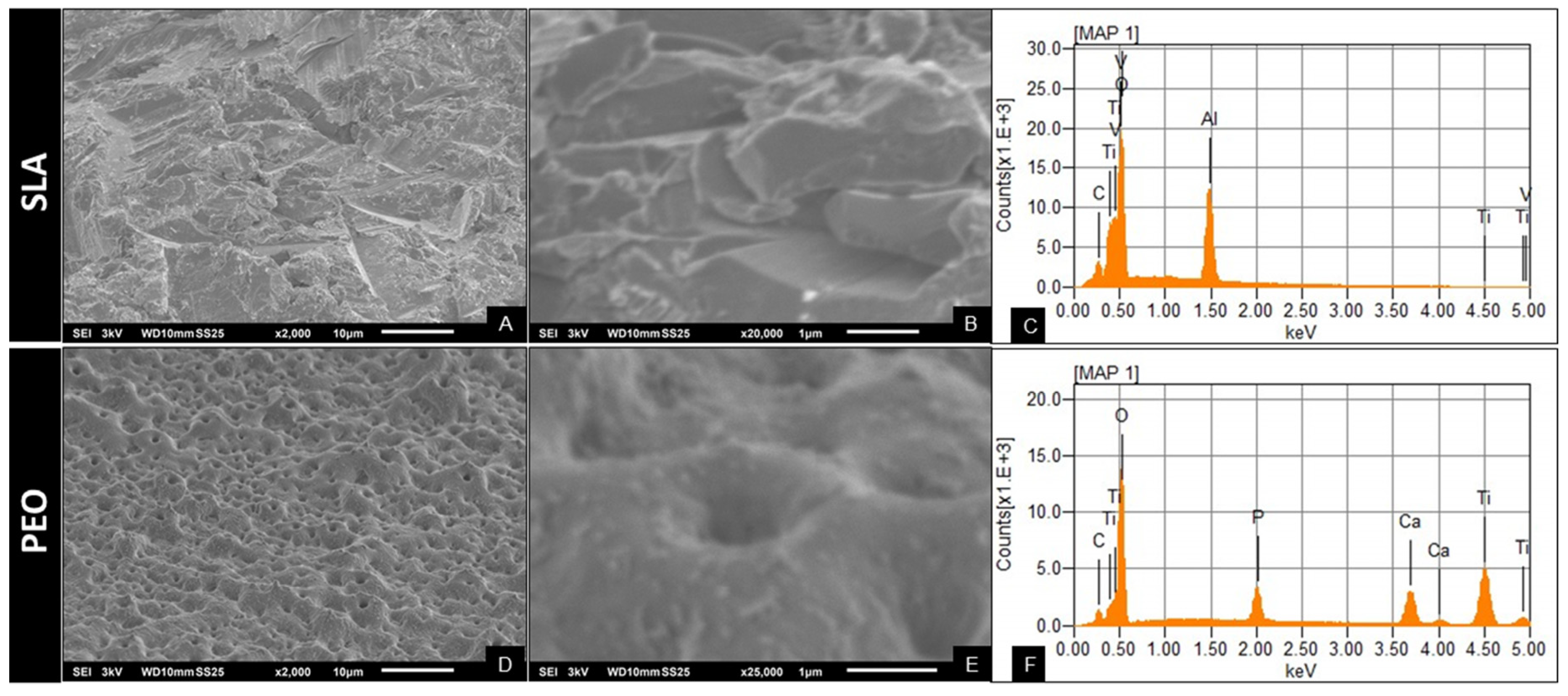
2.1. Surface Preparation and Characterization
2.2. Implant Surface Characterization
2.3. Animal Study
2.4. Osteoporosis Induction
2.5. Implant Placement
2.6. Application of Fluorochromes for Analysis Bone Turnover
2.7. Analysis of Peri-Implant Bone Repair
2.8. Molecular Analysis of Peri-Implant Repair (Real-Time PCR)
2.9. Computed Microtomography
2.10. Laser Confocal Microscopy (Peri-Implant Bone Turnover)
2.11. Histometry
2.12. Statistical Analysis
3. Results
3.1. ELISA Test
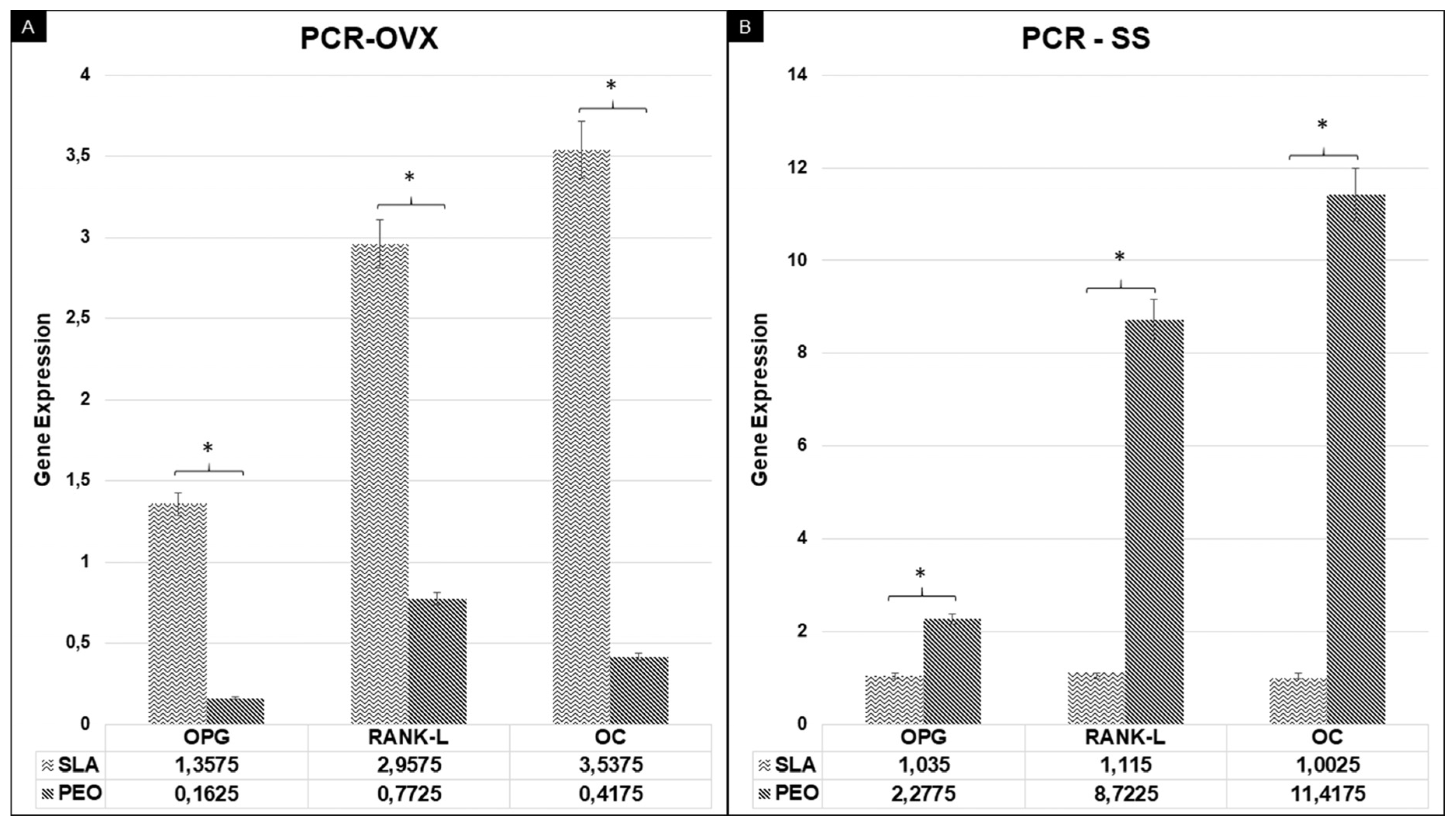
3.2. Real-Time PCR
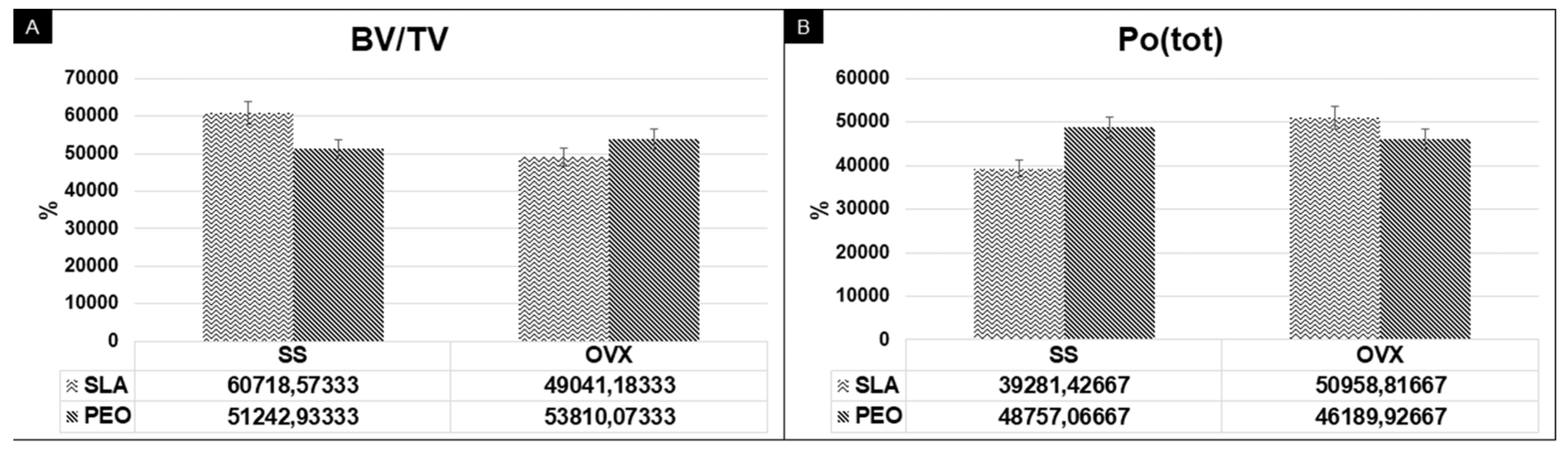
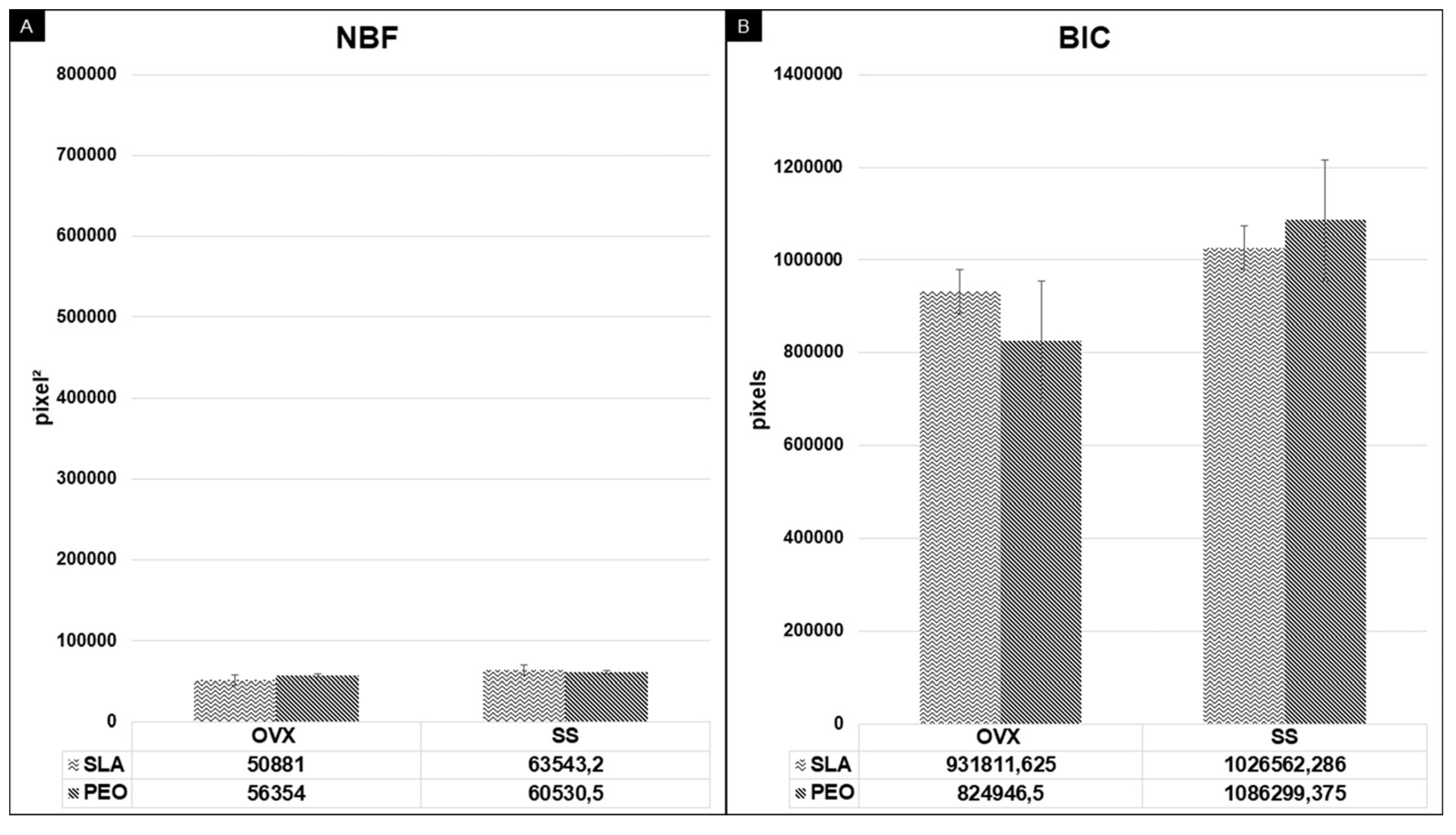
3.3. Computed Microtomography
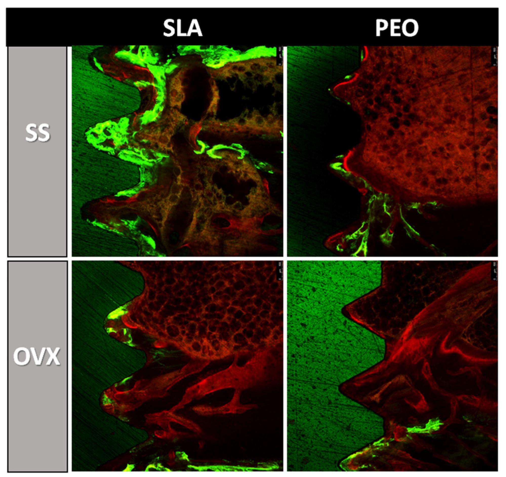
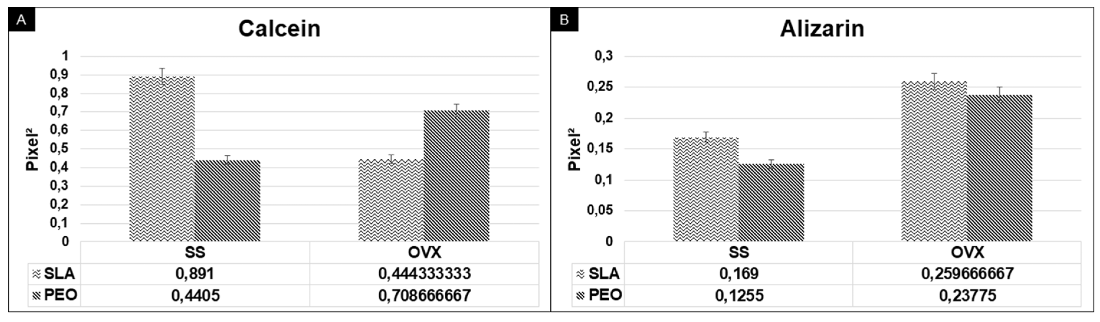
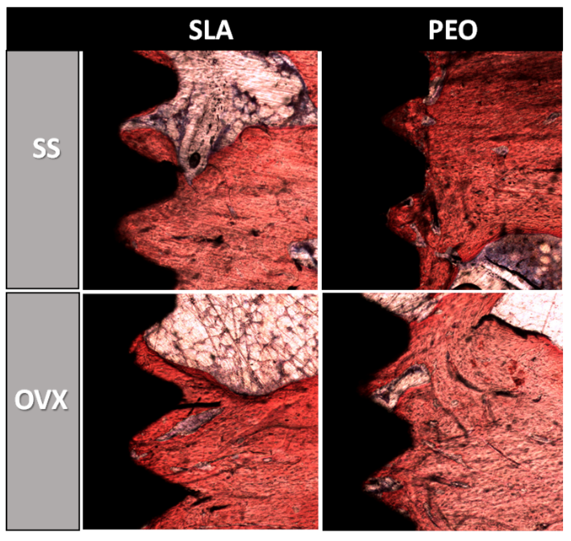
3.4. Peri-Implant Bone Turnover
3.5. Histometry
4. Discussion
5. Conclusions
Novelty Statement
Author Contributions
Funding
Conflicts of Interest
References
- Krzakala, A.; Kazek-Kęsik, A.; Simka, W. Application of plasma electrolytic oxidation to bioactive surface formation on titanium and its alloys. RSC Adv. 2013, 3, 19725–19743. [Google Scholar] [CrossRef]
- Laurindo, C.A.; Torres, R.D.; Mali, S.A.; Gilbert, J.L.; Soares, P. Incorporation of Ca and P on anodized titanium surface: Effect of high current density. Mater. Sci. Eng. C Mater. Biol. Appl. 2014, 37, 223–231. [Google Scholar] [CrossRef]
- Hwang, I.-J.; Choe, H.-C.; Brantley, W.A. Electrochemical characteristics of Ti-6Al-4V after plasma electrolytic oxidation in solutions containing Ca, P, and Zn ions. Surf. Coat. Technol. 2017, 320, 458–466. [Google Scholar] [CrossRef]
- Krząkała, A.; Służalska, K.; Widziołek, M.; Szade, J.; Winiarski, A.; Dercz, G.; Kazek, A.; Tylko, G.; Michalska, J.; Iwaniak, A.; et al. Formation of bioactive coatings on a Ti–6Al–7Nb alloy by plasma electrolytic oxidation. Electrochim. Acta 2013, 104, 407–424. [Google Scholar] [CrossRef]
- Lederer, S.; Lutz, P.; Fürbeth, W. Surface modification of Ti 13Nb 13Zr by plasma electrolytic oxidation. Surf. Coat. Technol. 2018, 335, 62–71. [Google Scholar] [CrossRef]
- Albrektsson, T.; Wennerberg, A. Oral implant surfaces: Part 1—Review focusing on topographic and chemical properties of different surfaces and In Vivo responses to them. Int. J. Prosthodont. 2004, 17, 536–543. [Google Scholar]
- Faverani, L.P.; Barao, V.A.; Pires, M.F.; Yuan, J.C.; Sukotjo, C.; Mathew, M.T.; Assunção, W.G. Corrosion kinetics and topography analysis of Ti-6Al-4V alloy subjected to different mouthwash solutions. Mater. Sci. Eng. C Mater. Biol. Appl. 2014, 43, 1–10. [Google Scholar] [CrossRef]
- Tavares, M.G.; de Oliveira, P.T.; Nanci, A.; Hawthorne, A.C.; Rosa, A.L.; Xavier, S.P. Treatment of a commercial, machined surface titanium implant with H2SO4/H2O2 enhances contact osteogenesis. Clin. Oral Implant. Res. 2007, 18, 452–458. [Google Scholar] [CrossRef]
- Messer, R.L.W.; Tackas, G.; Mickalonis, J.; Brown, Y.; Lewis, J.B.; Wataha, J.C. Corrosion of Machined Titanium Dental Implants under Inflammatory Conditions. J. Biomed. Mater. Res. Part B 2009, 88B, 474–481. [Google Scholar] [CrossRef]
- Faverani, L.P.; Assuncao, W.G.; de Carvalho, P.S.; Yuan, J.C.; Sukotjo, C.; Mathew, M.T.; Barao, V.A. Effects of dextrose and lipopolysaccharide on the corrosion behavior of a Ti-6Al-4V alloy with a smooth surface or treated with double-acid-etching. PLoS ONE 2014, 9, e93377. [Google Scholar] [CrossRef] [Green Version]
- Barao, V.A.; Ricomini-Filho, A.P.; Faverani, L.P.; Del Bel Cury, A.A.; Sukotjo, C.; Monteiro, D.R.; Yuan, J.C.; Mathew, M.T.; do Amaral, R.C.; Mesquita, M.F.; et al. The role of nicotine, cotinine and caffeine on the electrochemical behavior and bacterial colonization to cp-Ti. Mater. Sci. Eng. C Mater. Biol. Appl. 2015, 56, 114–124. [Google Scholar] [CrossRef] [PubMed]
- Ferreira Ribeiro, C.; Cogo-Muller, K.; Franco, G.C.; Silva-Concilio, L.R.; Sampaio Campos, M.; de Mello Rode, S.; Claro Neves, A.C. Initial oral biofilm formation on titanium implants with different surface treatments: An In Vivo study. Arch. Oral Biol. 2016, 69, 33–39. [Google Scholar] [CrossRef] [Green Version]
- Mendes, M.W.D.; Agreda, C.G.; Bressiani, A.H.A.; Bressiani, J.C. A new titanium based alloy Ti-27Nb-13Zr produced by powder metallurgy with biomimetic coating for use as a biomaterial. Mater. Sci. Eng. C Mater. 2016, 63, 671–677. [Google Scholar] [CrossRef] [PubMed]
- Queiroz, T.P.; Souza, F.A.; Guastaldi, A.C.; Margonar, R.; Garcia, I.R.; Hochuli-Vieira, E. Commercially pure titanium implants with surfaces modified by laser beam with and without chemical deposition of apatite. Biomechanical and topographical analysis in rabbits. Clin. Oral Implant. Res. 2013, 24, 896–903. [Google Scholar] [CrossRef]
- Souza, F.A.; Queiroz, T.P.; Guastaldi, A.C.; Garcia, R.; Magro, O.; Nishioka, R.S.; Sisti, K.E.; Sonoda, C.K. Comparative In Vivo study of commercially pure Ti implants with surfaces modified by laser with and without silicate deposition: Biomechanical and scanning electron microscopy analysis. J. Biomed. Mater. Res. Part B 2013, 101B, 76–84. [Google Scholar] [CrossRef] [PubMed]
- Luo, W.; Zhao, J.; Meng, X.; Ma, S.; Sun, Q.; Guo, T.; Wang, Y.; Zhou, Y. Effect of Zinc Doped Calcium Phosphate Coating on Bone Formation and the Underlying Biological Mechanism. Sheng Wu Yi Xue Gong Cheng Xue Za Zhi = J. Biomed. Eng. = Shengwu Yixue Gongchengxue Zazhi 2015, 32, 1359–1363. [Google Scholar]
- Yerokhin, A.L.; Nie, X.; Leyland, A.; Matthews, A.; Dowey, S.J. Plasma electrolysis for surface engineering. Surf. Coat. Technol. 1999, 122, 73–93. [Google Scholar] [CrossRef]
- Walsh, F.C.; Low, C.T.J.; Wood, R.J.K.; Stevens, K.T.; Archer, J.; Poeton, A.R.; Ryder, A. Plasma electrolytic oxidation (PEO) for production of anodised coatings on lightweight metal (Al, Mg, Ti) alloys. Trans. IMF Int. J. Surf. Eng. Coat. 2009, 87, 122–135. [Google Scholar] [CrossRef]
- Faverani, L.P.; Barao, V.A.; Ramalho-Ferreira, G.; Ferreira, M.B.; Garcia-Junior, I.R.; Assuncao, W.G. Effect of bleaching agents and soft drink on titanium surface topography. J. Biomed. Mater. Res. B Appl. Biomater. 2014, 102, 22–30. [Google Scholar] [CrossRef]
- Akatsu, T.; Yamada, Y.; Hoshikawa, Y.; Onoki, T.; Shinoda, Y.; Wakai, F. Multifunctional porous titanium oxide coating with apatite forming ability and photocatalytic activity on a titanium substrate formed by plasma electrolytic oxidation. Mater. Sci. Eng. C Mater. Biol. Appl. 2013, 33, 4871–4875. [Google Scholar] [CrossRef]
- Marques, I.D.; da Cruz, N.C.; Landers, R.; Yuan, J.C.; Mesquita, M.F.; Sukotjo, C.; Mathew, M.T.; Barão, V.A. Incorporation of Ca, P, and Si on bioactive coatings produced by plasma electrolytic oxidation: The role of electrolyte concentration and treatment duration. Biointerphases 2015, 10, 041002. [Google Scholar] [CrossRef] [PubMed]
- Marques, I.D.; Barao, V.A.; da Cruz, N.C.; Yuan, J.C.; Mesquita, M.F.; Ricomini-Filho, A.P.; Sukotjo, C.; Mathew, M.T. Electrochemical behavior of bioactive coatings on cp-Ti surface for dental application. Corros. Sci. 2015, 100, 133–146. [Google Scholar] [CrossRef] [PubMed] [Green Version]
- He, J.; Zhao, B.; Deng, C.; Shang, D.; Zhang, C. Assessment of implant cumulative survival rates in sites with different bone density and related prognostic factors: An 8-year retrospective study of 2684 implants. Int. J. Oral Maxillofac. Implant. 2015, 30, 360–371. [Google Scholar] [CrossRef] [PubMed] [Green Version]
- Hurst, D. Evidence unclear on whether Type I or II diabetes increases the risk of implant failure. Evid. Based Dent. 2014, 15, 102–103. [Google Scholar] [CrossRef] [PubMed]
- Chrcanovic, B.R.; Albrektsson, T.; Wennerberg, A. Diabetes and oral implant failure: A systematic review. J. Dent. Res. 2014, 93, 859–867. [Google Scholar] [CrossRef] [PubMed]
- Aguilar, E.A.; Barry, S.D.; Cefalu, C.A.; Abdo, A.; Hudson, W.P.; Campbell, J.S.; Reske, T.M.; Bonafede, M.; Wilson, K.; Stolshek, B.S.; et al. Osteoporosis Diagnosis and Management in Long-Term Care Facility. Am. J. Med. Sci. 2015, 350, 357–363. [Google Scholar] [CrossRef]
- Lehnert, H. Underestimated osteoporosis: New directions in diagnosis and therapy. Dtsch. Med. Wochenschr. 2015, 140, 1660. [Google Scholar]
- Zhou, B.; Lu, Y.; Hajifathalian, K.; Bentham, J.; Di Cesare, M.; Danaei, G.; Bixby, H.; Cowan, M.J.; Ali, M.K.; Taddei, C.; et al. Worldwide trends in diabetes since 1980: A pooled analysis of 751 population-based studies with 4.4 million participants. Lancet 2016, 387, 1513–1530. [Google Scholar] [CrossRef] [Green Version]
- Kilkenny, C.; Browne, W.J.; Cuthill, I.C.; Emerson, M.; Altman, D.G. Improving bioscience research reporting: The ARRIVE guidelines for reporting animal research. Osteoarthr. Cartil. 2012, 20, 256–260. [Google Scholar] [CrossRef] [Green Version]
- Luvizuto, E.R.; Dias, S.M.; Queiroz, T.P.; Okamoto, T.; Garcia, I.R.; Okamoto, R.; Dornelles, R.C. Osteocalcin immunolabeling during the alveolar healing process in ovariectomized rats treated with estrogen or raloxifene. Bone 2010, 46, 1021–1029. [Google Scholar] [CrossRef]
- Luvizuto, E.R.; Queiroz, T.P.; Dias, S.M.; Okamoto, T.; Dornelles, R.C.; Garcia, I.R.; Okamoto, R. Histomorphometric analysis and immunolocalization of RANKL and OPG during the alveolar healing process in female ovariectomized rats treated with oestrogen or raloxifene. Arch. Oral Biol. 2010, 55, 52–59. [Google Scholar] [CrossRef] [PubMed]
- Kalu, D.N. The ovariectomized rat model of postmenopausal bone loss. Bone Miner. 1991, 15, 175–191. [Google Scholar] [CrossRef]
- French, D.L.; Muir, J.M.; Webber, C.E. The ovariectomized, mature rat model of postmenopausal osteoporosis: An assessment of the bone sparing effects of curcumin. Phytomedicine 2008, 15, 1069–1078. [Google Scholar] [CrossRef] [PubMed]
- Glosel, B.; Kuchler, U.; Watzek, G.; Gruber, R. Review of dental implant rat research models simulating osteoporosis or diabetes. Int. J. Oral Maxillofac. Implant. 2010, 25, 516–524. [Google Scholar]
- Thompson, D.D.; Simmons, H.A.; Pirie, C.M.; Ke, H.Z. FDA Guidelines and animal models for osteoporosis. Bone 1995, 17, 125s–133s. [Google Scholar] [CrossRef]
- Faverani, L.P.; Polo, T.O.B.; Ramalho-Ferreira, G.; Momesso, G.A.C.; Hassumi, J.S.; Rossi, A.C.; Freire, A.R.; Prado, F.B.; Luvizuto, E.R.; Gruber, R.; et al. Raloxifene but not alendronate can compensate the impaired osseointegration in osteoporotic rats. Clin. Oral Investig. 2017. [Google Scholar] [CrossRef]
- Ramalho-Ferreira, G.; Faverani, L.P.; Grossi-Oliveira, G.A.; Okamoto, T.; Okamoto, R. Alveolar bone dynamics in osteoporotic rats treated with raloxifene or alendronate: Confocal microscopy analysis. J. Biomed. Opt. 2015, 20, 038003. [Google Scholar] [CrossRef]
- Palin, L.P.; Polo, T.O.B.; Batista, F.R.S.; Gomes-Ferreira, P.H.S.; Garcia, I.R., Jr.; Rossi, A.C.; Freire, A.; Faverani, L.P.; Sumida, D.H.; Okamoto, R. Daily melatonin administration improves osseointegration in pinealectomized rats. J. Appl. Oral Sci. 2018, 26, e20170470. [Google Scholar] [CrossRef] [Green Version]
- He, J.; Feng, W.; Zhao, B.H.; Zhang, W.; Lin, Z. In Vivo Effect of Titanium Implants with Porous Zinc-Containing Coatings Prepared by Plasma Electrolytic Oxidation Method on Osseointegration in Rabbits. Int. J. Oral Maxillofac. Implant. 2018, 33, 298–310. [Google Scholar] [CrossRef] [Green Version]
- Ogawa, E.S.; Matos, A.O.; Beline, T.; Marques, I.S.; Sukotjo, C.; Mathew, M.T.; Rangel, E.C.; Cruz, N.C.; Mesquita, M.F.; Consani, R.X.; et al. Surface-treated commercially pure titanium for biomedical applications: Electrochemical, structural, mechanical and chemical characterizations. Mater. Sci. Eng. C Mater. Biol. Appl. 2016, 65, 251–261. [Google Scholar] [CrossRef] [Green Version]
- He, T.; Cao, C.; Xu, Z.; Li, G.; Cao, H.; Liu, X.; Zhang, C.; Dong, Y. A comparison of micro-CT and histomorphometry for evaluation of osseointegration of PEO-coated titanium implants in a rat model. Sci. Rep. 2017, 7, 16270. [Google Scholar] [CrossRef] [PubMed] [Green Version]
- Lu, X.; Mohedano, M.; Blawert, C.; Matykina, E.; Arrabal, R.; Kainer, K.U.; Zheludkevich, M.L. Plasma electrolytic oxidation coatings with particle additions–A review. Surf. Coat. Technol. 2016, 307, 1165–1182. [Google Scholar] [CrossRef]
- Zaporozhets, T.S.; Puz’, A.V.; Sinebryukhov, S.L.; Gnedenkov, S.V.; Smolina, T.P.; Besednova, N.N. Biocompatibility of Modified Osteoinductive Calcium-Phosphate Coatings of Metal Implants. Bull. Exp. Biol. Med. 2017, 162, 366–369. [Google Scholar] [CrossRef] [PubMed]
- Ramalho-Ferreira, G.; Faverani, L.P.; Prado, F.B.; Garcia, I.R., Jr.; Okamoto, R. Raloxifene enhances peri-implant bone healing in osteoporotic rats. Int. J. Oral Maxillofac. Surg. 2015, 44, 798–805. [Google Scholar] [CrossRef]
- Nagay, B.E.; Dini, C.; Cordeiro, J.M.; Ricomini-Filho, A.P.; de Avila, E.D.; Rangel, E.C.; Da Cruz, N.C.; Barão, V.A. Visible-Light-Induced Photocatalytic and Antibacterial Activity of TiO2 Codoped with Nitrogen and Bismuth: New Perspectives to Control Implant-Biofilm-Related Diseases. ACS Appl. Mater. Interfaces 2019, 11, 18186–18202. [Google Scholar] [CrossRef]


© 2020 by the authors. Licensee MDPI, Basel, Switzerland. This article is an open access article distributed under the terms and conditions of the Creative Commons Attribution (CC BY) license (http://creativecommons.org/licenses/by/4.0/).
Share and Cite
Momesso, G.A.C.; de Souza Santos, A.M.; Fonseca e Santos, J.M.; da Cruz, N.C.; Okamoto, R.; Barão, V.A.R.; Siroma, R.S.; Shibli, J.A.; Perez Faverani, L. Comparison between Plasma Electrolytic Oxidation Coating and Sandblasted Acid-Etched Surface Treatment: Histometric, Tomographic, and Expression Levels of Osteoclastogenic Factors in Osteoporotic Rats. Materials 2020, 13, 1604. https://doi.org/10.3390/ma13071604
Momesso GAC, de Souza Santos AM, Fonseca e Santos JM, da Cruz NC, Okamoto R, Barão VAR, Siroma RS, Shibli JA, Perez Faverani L. Comparison between Plasma Electrolytic Oxidation Coating and Sandblasted Acid-Etched Surface Treatment: Histometric, Tomographic, and Expression Levels of Osteoclastogenic Factors in Osteoporotic Rats. Materials. 2020; 13(7):1604. https://doi.org/10.3390/ma13071604
Chicago/Turabian StyleMomesso, Gustavo Antonio Correia, Anderson Maikon de Souza Santos, João Matheus Fonseca e Santos, Nilson Cristino da Cruz, Roberta Okamoto, Valentim Adelino Ricardo Barão, Rafael Shinoske Siroma, Jamil Awad Shibli, and Leonardo Perez Faverani. 2020. "Comparison between Plasma Electrolytic Oxidation Coating and Sandblasted Acid-Etched Surface Treatment: Histometric, Tomographic, and Expression Levels of Osteoclastogenic Factors in Osteoporotic Rats" Materials 13, no. 7: 1604. https://doi.org/10.3390/ma13071604