
Toughening of Bioceramic Composites for Bone Regeneration


Institute of Science and Technology for Ceramics-National Research Council of Italy (ISTEC-CNR), 48018 Faenza, Italy
*
Authors to whom correspondence should be addressed.
†
Co-first author, these authors contributed equally to this work.
J. Compos. Sci. 2021, 5(10), 259; https://doi.org/10.3390/jcs5100259
Submission received: 29 July 2021
/
Revised: 21 September 2021
/
Accepted: 24 September 2021
/
Published: 29 September 2021
(This article belongs to the Special Issue Bioceramic Composites)
Abstract
:Bioceramics are widely considered as elective materials for the regeneration of bone tissue, due to their compositional mimicry with bone inorganic components. However, they are intrinsically brittle, which limits their capability to sustain multiple biomechanical loads, especially in the case of load-bearing bone districts. In the last decades, intense research has been dedicated to combining processes to enhance both the strength and toughness of bioceramics, leading to bioceramic composite scaffolds. This review summarizes the recent approaches to this purpose, particularly those addressed to limiting the propagation of cracks to prevent the sudden mechanical failure of bioceramic composites.
1. Introduction
Bone tissue is classified as a calcified connective tissue with several important roles in the human body, including storing minerals, protecting vital organs, enabling movement, providing internal support, and providing the sites of attachment for muscles and tendons [1,2]. Bone can be considered as a natural composite made of inorganic components (naturally doped calcium phosphates, ~70 wt %), organics (Collagen Type I, non-collagenous proteins, proteoglycans, cells, ~22 wt %), and water (~8 wt %) [2,3,4].
The complex metabolism and 3D hierarchic structure of bone tissue give it an innate ability to heal from minor defects. However, the natural healing process of bone is limited when major injuries due to traumas or metabolic or neoplastic bone pathologies occur [1,2]. In such instances, the orthopedic surgeon is challenged to find out adequate regenerative approaches [3]. The use of natural bone grafting (i.e., autologous or heterologous bone) can be pursued to replace the bone defect. Autografts are considered as the gold standard for bone grafting, as they closely resemble the natural bone structure, without immunogenic response. Despite these benefits, some limitations are evident, including the morbidity of the donor site, increased operation time, increased blood loss, and risk of immunogenicity and pathogenicity [4,5,6,7]. In addition, the sterilization and irradiation processes of natural bone grafts have been reported as critical steps that limit their bioactivity [8,9,10,11].
In this context, a great deal of research effort has been devoted in the last decades towards the synthesis of synthetic scaffolds [12,13,14,15]. The naturally occurring mineral phase in bone tissue is represented by poorly crystalline calcium phosphates with the crystal structure of hydroxyapatite (HA). HA can be synthesized in laboratory, and it is currently under study for the development of bone grafts, due to its excellent biocompatibility, osteoconductivity, and osteoinductivity [16,17,18,19,20,21,22].
In the last decades, bioceramics have been considered as ideal candidates for bone grafting due to their ability to locally deliver biomolecules in vivo. Calcium phosphates are a major member of bioceramics, covering a wide range of biomedical applications in tissue engineering, including orthopedic and dental surgeries [23,24,25,26].
Bioceramics must meet strict criteria to be approved for biomedical applications, such as biocompatibility, bioactivity, and an absence of proinflammatory features [27]. The classification of bioceramics is generally based on their chemical composition, as well as on the basis of their interaction with natural tissues; thus, bioceramics can be considered as bioinert or bioactive, considering biodegradability as an added value that enables the replacement of damaged bone parts with new ones during the scaffold bioresorption [28,29,30,31,32]. In this respect, recent studies demonstrated that the modulation of composition and textural properties can be considered as a valuable strategy to control material resorption and bone formation [33,34].
Bioinert ceramics, including alumina, zirconia, and silicon nitride, are not able to undergo any modification upon implantation, and thus maintain their chemical structure and represent a foreign body within the biological environment [35,36]. In contrast, bioactive ceramics have the capability to form chemical bonds with the surrounding tissues, and actively interact with the surrounding environment [37,38]. Among them, calcium phosphates (CaPs), bioglasses, and calcium silicate (Ca-Si) bioceramics are intensively studied for skeletal bone regeneration applications [22,39,40,41,42,43].
The osteogenic capability of bioceramic scaffolds is significantly correlated to their intrinsic pore size distribution and interconnection, enabling cell infiltration, migration, and neo-vascularization. The pore distribution and geometry of the scaffold strongly influence the ability of cells to penetrate, proliferate, and differentiate as well as the rate of scaffold degradation [44,45,46,47,48]. In spite of their great potential, a main drawback associated with bioceramics is their intrinsic brittleness, i.e., incapacity to withstand deformation without rupture, which is a major problem that can potentially cause a sudden failure of the scaffold structure under physiological mechanical loading. This is particularly relevant for porous calcium phosphates that associate brittleness to limited fracture strength, in comparison with inert ceramics such as zirconia or alumina [38,49,50]. In the last century, an intense research effort has been devoted to the reinforcement of bioceramics for different applications. In this respect, various approaches have been proposed, including modified sintering treatments [51,52,53], combination with polymeric phases to produce composites [49,54,55], the addition of fibers or the development of additive manufacturing as a 3D technique to prepare complex-shaped bioceramic structures [34,56,57,58,59,60]. A major approach to this purpose is the addition of ceramic particles, whiskers, and fibers to the ceramic matrix to improve the fracture toughness [61,62,63,64,65]. Ceramic fibers selected for their lightweight, adequate strength and modulus, and biocompatibility have been tested in the last decade for improving the mechanical properties of bioceramics [66,67,68,69]. The key factor influencing the performance of the final material is represented by the interfacial adhesion between fibers and the surrounding matrix [70]. The main factors affecting the fabrication of fiber-reinforced scaffolds include the chemical composition of fibers and matrix, the physical interaction between them, and the amount and alignment of fibers [59,71,72]. These factors affect the mechanical strength and degradation properties of the scaffold, leading to changes in the cell response. In this respect, many studies have reported the biocompatibility of fiber-reinforced ceramics both in vitro and in vivo [73,74,75,76,77].
It was observed that smooth fibers with a chemically inert surface are provided with less reactive functional groups, resulting in poor adhesion with the matrix [78]. Some studies reported chemical approaches to activate the fibers’ surfaces, in order to strengthen this interaction [59,79,80,81,82,83,84,85].
The present review summarizes the relevant progress made on the mechanical reinforcement of bioceramic composites. The fabrication techniques for these scaffolds, along with the current strategies for toughening mechanisms, are described. Furthermore, the concerns related to porosity along with the mechanical and biological properties of fibrous ceramics are reported. As the advances in bone tissue engineering move toward application in the clinical setting, achieving adequate bioceramic toughness for clinical applications is particularly critical. In this context, recent computational approaches have been proposed in order to predict the crack propagation pathways, while increasing the toughness of ceramic-based bioinspired materials [86].
2. Bioceramic Composites in Bone Regeneration
2.1. Bone Tissue Formation and Remodeling
The adult human skeleton is made up of 206 bones. Each bone is a very complex hierarchical structure consisting of osteon, lamellar, fibrils, and mineral and collagen fibers (Figure 1). The bone is a dynamic tissue that continuously remodels during the life span of an individual: the term “bone remodeling” refers to a complex biochemical process involving the degradation of the mineralized bone via osteoclasts followed by the deposition of newly formed bone matrix by osteoblasts [87]. Due to this remodeling, the timing for complete bone tissue renewal is about 5 to 10 years [88,89,90]. This helps it in adapting to ever changing biomechanical forces by replacing the old or micro-damaged bone with a new and mechanically stronger bone, thereby preserving bone strength [91]. The bone has a unique ability to shift the intricate balance between osteoclastic and osteoblastic activities depending upon the external stimuli [92,93,94]. Such mechano-transduction signals can amplify the osteoblastic activity, resulting in an enhanced deposition of bone matrix [44]. In other circumstances, this equilibrium can be triggered by a chemical rather than a mechanical signal [44,95,96]. After receiving the mechanical signal, osteoclasts are deployed to the bone to initiate resorption. This process results in the release of calcium or phosphate to the body fluid as it is crucial for specific metabolic reactions. It is also assumed that chemokines are responsible for the differentiation-fusion of monocytes into osteoclasts and for carrying out the subsequent osteoclastic activity [97,98,99].
In this context, the regulation of osteoblasts-osteoclasts mediated processes plays a key role in achieving effective bone tissue regeneration [100]. Bioceramic composites can be engineered for better resorption and bone remodeling by mixing different ceramic materials [101]. The incorporation of strontium (Sr) into bioceramic composites can improve the bone tissue density via increasing osteoblast function and inhibiting osteoclast activity [102,103,104,105]. In addition, the surface topography also affects the resorption capacity of osteoclasts [104,105]. It was observed that human peripheral blood monocyte derived osteoclasts were more actively resorbed onto sub-micro structured β-TCP compared to microscale topography [106].
2.2. Classification of Bioceramic Composites
One major classification of bioceramics relies on the biochemical reactions occurring between the implanted scaffold and the surrounding tissue, particularly on the increasing capacity to be resorbed upon implantation in vivo. In this way, bioceramics can be considered as bioinert, bioactive, or bioresorbable materials [107]. Zirconia (ZrO2) and alumina (Al2O3) are examples of inert bioceramics with minimal adverse reactions on tissue and body organs after coming into contact with the human physiology [108]. They have inherently low levels of reactivity compared with other materials, such as polymers and metals, as well as surface reactive or resorbable ceramics. Strategies for the improvement of the biocompatibility of inert ceramics were proposed, such as surface modifications, coatings, or ion doping [109,110,111,112].
In contrast, bioactive ceramics, such as bioglasses, show an ability to superficially bond with the surrounding bone, thus improving their interfacial strength [103,113]. The ability to bond to bone tissue is a unique property of bioactive ceramics. Analyses of the bone implant interface revealed that the presence of hydroxyapatite is one of the key features in the bonding zone [103].
Bioresorbable bioceramics represent a further improvement in their long-term interaction with surrounding tissues, because in addition to their chemical similarity to the mineral component of bone, they are able to be gradually resorbed and replaced by new bone tissue over time. The in vivo behavior of ceramic bone substitutes includes three main steps: (i) solubility: if the compound is soluble in physiological conditions, dissolution and removal can occur; (ii) the dissolution kinetics, related to the speed at which the particular ceramic is removed from the body; and (iii) conversion into another compound via a dissolution–precipitation mechanism [114]. Bioresorbable bioceramics are represented by calcium sulphates (Plaster of Paris) and calcium phosphates, especially with ion doping with Sr, Mg, Si, and Zn, which can improve their biological activity [19,115,116,117,118,119,120]. However, there are some drawbacks of calcium phosphates, such as their poor mechanical strength, differences between the bone regeneration and degradation rate, inflammatory reaction of synthetic bioceramics, and limited ingrowth due to pore size [121,122,123].
In this context, the possibility to introduce additional inorganic phases in bioceramics opened a wider choice of materials for their use as implants. Some of these materials include ceramic/ceramic, ceramic/metal, and ceramic/polymer composites. However, ceramic/polymer composites have been observed to release toxic components in the surrounding tissues, while metals undergo corrosion-related issues as the ceramic coating on the metallic implant degrades over time [124,125,126]. Ceramic/ceramic composites are thought to have a better performance because they resemble bone minerals and exhibit high biocompatibility [127,128,129]. Nevertheless, the biological activity of bioceramic composites has to be defined, especially considering the specific implantation site [130]. Bioceramic composites have exceptional biocompatibility and are non-toxic [131,132,133]. Some additional features of bioceramics composites include their hydrophilicity and antibacterial properties [134,135,136].
2.3. Surface Chemistry
The effective chemical interaction between the surfaces of the implanted scaffolds and the surrounding tissues plays a crucial role in the regeneration of bone tissue [137,138]. The three different types of bioceramics (bioinert, bioresorbable, and bioactive) have significantly different superficial interactions [139]. It is worth mentioning here that these fundamental differences in surface chemistry result in different interacting conditions at the biomolecular interface with cells and proteins [23,140,141].
The implantation of a scaffold into a biological environment is followed by the hydration and hydrolysis of the surfaces, typically leading to the formation of chemical bonds, containing either hydrogen or hydroxyl groups, with a rate dependent on the pH environment [142].
The superficial properties essentially modulate the interaction with water molecules and the mechanisms of adsorption of biological macromolecules (e.g., proteins). This interaction ultimately determines the interplay between the implanted bioceramics and bone cells.
It was reported that an electrostatic attraction primarily affects the protein adsorption on bioceramic surfaces, and effective surface charge modulation can be achieved by the immobilization of biomolecules such as bisphosphonates (BPs), amino acids, or carboxylic acid on the bioceramic surface [140]. Additional factors affecting the protein adsorption and cell adhesion include surface wettability and surface energy. The tuning of the chemical and morphological features of bioceramics can be performed by chemical or physical surface modifications, including atomic layer deposition, chemical vapor deposition, plasma vapor deposition, and electrochemical deposition [141,143,144].
Chemical treatments generally result in the formation of coating layers or the induction of specific chemical functional groups (e.g., carbidization, nitration, oxidation), while physical modifications result in micro- to nanoscale morphological or topographical alterations via a multitude of processes (e.g., machining, grit-blasting, and etching) [145,146,147].
2.4. Mechanical Properties
The mechanical properties of bioceramics, including compressive strength, stiffness, fracture toughness, and fatigue resistance, represent the key factors for effective bone regeneration [148]. These criteria include “static” mechanical properties (e.g., stiffness, hardness, strength), as well as “dynamic” mechanical properties (e.g., fatigue cycle resistance, crack propagation stability, and fracture toughness) [149].
A major concept in defining mechanical properties of ceramics is the difference between strength and toughness. They are frequently considered to be overlapped, despite the fact that they are mutually exclusive—strength is a stress representing the intrinsic capability of a material to resist to irreversible deformations, while toughness refers to the energy required to induce a fracture [150].
Toughness can also be determined using fracture mechanics methods, which determine the critical value of a crack-driving force, such as the stress intensity K, strain-energy release rate G, or nonlinear elastic J-integral, required to initiate and/or propagate a previously formed crack.
However, the intrinsic brittleness of ceramics basically limit the capability to improve the toughness, primarily because they cannot be toughened by promoting plasticity [151].
The compressive strength of the human cortical bone is reportedly in the range 90–209 MPa, while the reported flexural strength is 135–193 MPa [152,153]. The mechanical strength of bioceramics is reported to be in the range of cortical and cancellous bones [154]. The ideal scaffolds for bone regeneration should be designed considering this feature, but also considering that extensive bone penetration in a porous scaffold will increase the mechanical properties of the bone-scaffold construct until reaching physiological levels [44]. In particular, fracture toughness is important because it refers to the ability of the scaffold to contrast the propagation of a crack defect [155]. Hence, compressive strength and fracture toughness are relevant properties to be considered for effective bone regeneration [156,157]. The particle size, composition, porosity, and crystallinity of bioceramics significantly affect their mechanical performance—an increase of porosity and particle size leads to the decrease of mechanical properties [158,159,160].
The fracture toughness of cortical bone (KIc = 2–12 MPa∙m0.5) is higher than that of ceramics or inorganic glass [160,161,162]. Numerous methods have been developed over time to measure the fracture toughness and hardness [163,164,165]. The low fracture toughness and poor mechanical strength of bioceramics limits their usage in load-bearing applications [166,167]. It was reported that the fracture toughness and flexural strength of bioceramics increase in wet environments [168].
The toughness and flexibility of bone tissue can be ascribed to the complex biomineralization of collagen fibers with apatitic crystals, associated to the multi-scale hierarchical architecture [168]. The toughness of bioceramics can be improved by including additional biocompatible phases [169,170], crack bridging, or phase transformation, in order to control the crack growth [171,172,173]. The dispersion of second phase such as fibers, whiskers, and particles for creating toughness in bioceramics was also reported [174,175,176,177].
The mechanisms for increasing the toughness of ceramics can be classified as either intrinsic or extrinsic. Intrinsic toughening is primarily related to plasticity, that is, enlarging the plastic zone, mainly against the initiation of a crack. Conversely, extrinsic toughening acts to limit an initiated crack, reduce the stress and strain fields at the crack tip, preventing further opening, including crack bridging by fibers or ductile phases in composites.
A significant increase of flexural strength, flexural modulus, and fracture toughness of ceramic dental composites was also reported through the addition of zirconia-silica (ZS) or zirconia-yttria-silica (ZYS) nanofibers (2.5 wt % or 5.0 wt %) [178,179].
Bioceramic composites made from HA and TZP (tetragonal zirconia polycrystal) powders coated with Al2O3 also exhibited significantly higher strength and fracture toughness, due to the integration of ZrO2 (15 vol %) and Al2O3 (30 vol %) [180].
The microstructural and mechanical changes of Al2O3 matrixes, after the incorporation of Cr2O3, was also studied, resulting in improved hardness and elastic modulus, while fracture toughness deteriorated with the addition of 2 mol % Cr2O3 particles [181].
It was also reported that Zr–Ti–Nb–Cu–Be glasses containing 42–67 vol % dendrites exhibited 100–160 MPa√m toughness at tensile yield strengths of 1.1–1.5 GPa [182]. A monolithic and amorphous Pd–Ag–P–Si–Ge glass alloy with 1.5 GPa tensile strength and 200 MPa∙m0.5 toughness properties was also recently reported; its properties were a result of the generation of shear band after loading, which resembles large-scale plasticity [183]. Nevertheless, it has drawbacks related to critical processing and production costs [150].
Moreover, researchers produced novel dental restorative composites by using hydroxyapatite whiskers. They reported that the efficiency of reinforcement depends on the filler morphology. Hydroxyapatite has good wettability with polymer which leads to increased toughness in comparison to nano-size HA powder [184,185]. Two composite materials have been produced by using ZrO2-Al2O3 system: zirconia toughened alumina (ZTA) and alumina toughened zirconia (ATZ) [186,187,188,189,190,191]. The ZTA ceramic composites with 0.5 wt % MgO content exhibited the best attributes, such as a fracture toughness value of 9.14 MPa∙m0.5 and a hardness value of 1591 HV. Similarly, the effect of TiO2 phase composition and mechanical properties of Ca-TZP (calcium stabilized tetragonal zirconia) ceramic have been observed, with fracture toughness values up to 9.1 MPa√m after reinforcement with TiO2 in the range of 0.5–0.65 mol % [192].
A great research effort for the reinforcement of bioceramics with carbon fibers (CF) has been established, due to their excellent biocompatibility and mechanical properties [193,194,195]. The addition of CF to HA matrix effectively improves the bending strength and fracture toughness of HA [177,196]. ZrO2-HAp composites (40 and 60 vol % of ZrO2) were fabricated and evaluated, demonstrating the reinforcing effect of ZrO2 [174].
3. Toughening Strategies for Bioceramic Composites
The brittleness of bioceramics significantly limits their applications because in addition to strength, adequate toughness is required to sustain the biomechanical loads [86].
Any crystallographic defect or irregularity within the crystal structure represents the main cause for dislocations, the mechanisms of which are related to the Peierls–Nabarro (PN) barrier energy that defines the fracture toughness of a material [197].
Metals contain mobile dislocations, leading to local plasticity and desirable toughness [197,198], while ceramics are characterize by the immobility of dislocations and low fracture toughness, especially at room temperature [199]. In this respect, the high-strength ionic bond typical for ceramic structures plays a crucial role, limiting atomic slip systems.
The mechanical performance of bioceramics is closely related to several factors, including microstructure, chemical composition, ionic impurities, and structural defects. The strategies to improve the toughness or fracture strength of ceramics refer to the capacity to control or limit the propagation of cracks along the powder particles and grains.
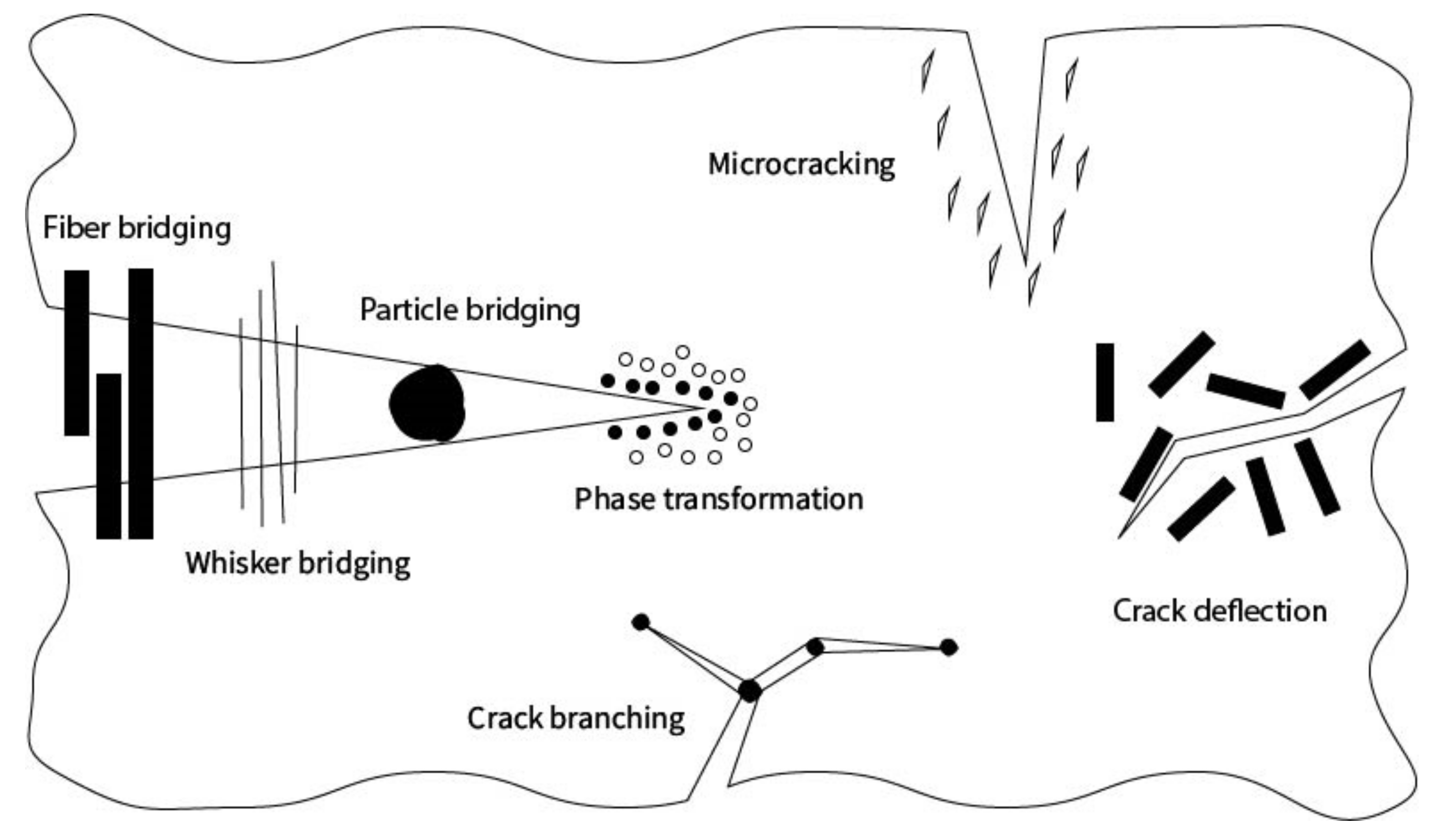
Several methods have been reported to improve the toughness of ceramics, including crack-bridging, crack-deflection, microcrack-induced toughening, generation of phase transformations, and reduction of the defect size (Figure 2).
Moreover, the basic scaffold structure can be combined with polymer coatings, or interpenetrating polymer-bioactive ceramic microstructures can be formed to improve the toughness of the ceramic as a simple and effective approach [200].
3.1. Phase Transformation
Toughening phenomena related to phase transformation are well known for zirconia-containing composites, where the phase transition from the tetragonal to monoclinic phase is the essential mechanism behind the enhanced toughness of zirconia used for the development of dental and orthopedic implants. This approach is based on stress induced phase transformations, which are mainly responsible for microstructural additional compressive stresses during the propagation of cracks, which increase the crack growth resistance KIc.
Similar to precipitation hardening, the stabilization of particles in a metastable and thermodynamically unfavorable state requires overcoming an energy nucleation barrier. In this case, the modulation of particle volume can be achieved by the application of adequate tensile stress at the crack tip. Phase transformation is initiated by the presence of sufficiently large elastic energy. As the particle was metastable prior to the transformation, the decrease in stress due to an increase in volume does not hamper the process of transformation [201]. Moreover, compressive stress in radial direction and tensile stress in circumferential direction around a particle are superimposed to the external load during the transformation. These compressive residual stresses may result in the reduction of stress on the crack and hence may partially or completely close the crack. In addition, as the tensile stress is applied in circumferential direction around a particle it can generate microcracks, further leading to the dissipation of energy [202,203].
The stress induced transformation is also related to the free enthalpy reduction [204]. The addition of hydrostatic tensile stress strongly decreases the enthalpy of the phase with a larger volume. This, in turn, increases the driving force for the transformation, enabling the particle to overcome the nucleation barrier [205,206].
3.2. Defect Size Reduction
The main limitation of bioceramics’ mechanical properties is related to their brittleness [207,208,209]. Ceramics generally exhibit higher compressive strength than tensile strength, essentially due to limitations in stress concentrations and crack propagation when micropores are flattened instead of dilated [210]. Bioceramics are characterized by very limited strain to failure and toughness, compared with ductile material counterparts (e.g., metals, polymers). Tensile stress could cause a fracture to propagate through the material, often causing failure in ceramic material.
Many defects can occur during the production, finishing, and application of ceramic because of the foreign particles, porous regions, or large grain sizes (Figure 3) [211].
A great research effort has been dedicated to the design of ceramic microstructures with increased toughness and damage tolerance. In this context, the reduction of defect sizes can also be obtained by the incorporation of various ions such as nickel, silver, tantalum [212], and strontium [212,213,214].
Some requirements for reducing the size of defects include an efficient, fast, and reliable fine grinding, a compact design, and the versatility of the process [215].
The fracture mechanics of bioceramics is mainly affected by the powder particle size distribution. Grain size is usually tuned towards monomodal or multimodal distributions, in order to increase the packing density of particles [216]. It was reported that the largest grain may control the size of largest flaw [217]. Alternatively, grain size can be measured at the fracture origin [218]. The microstructure is affected by multiple factors (e.g., powder impurities, thermal treatments of powders, sieving size), complicating the possibility to understand the role of each factor [24]. This methodology has been in use for the production of ceramics to obtain a more homogenous microstructure [219,220,221].
3.3. Crack Deflection
The propagation of cracks in bioceramics is a critical issue that can cause sudden failure of large structures. Crack deflection can be used as a strategy to increase the toughness of bioceramics. Some local areas in the bioceramics exhibit low resistance to crack propagation, resulting in crack deflection [222]. In particular, when a crack is deflected, the surface of the crack increases, leading to more energy required for crack propagation and an increase of fracture toughness [221,223]. The prediction of a crack path as the crack approaches a fiber can be based on an energy criterion or a stress criterion [217,219,220,224,225,226,227]. Young’s modulus mismatches are also reported as mechanisms of crack deflection [211]. The microstructural paths for crack propagation in bioceramics generally reflect the grain boundaries [211,217,228,229,230].
The evaluation of crack deflection by disks, rods, and spheres [217] showed that (1) increased toughness as a result of crack deflection is dependent on particle shape and volume fraction, and is independent of particle size [231]; (2) a rod-shaped morphology is the most effective, followed by disk and sphere, for increasing the toughness [232]. An increase in volume fraction of up to 20% increases the toughness, however, very little increase was observed with a higher volume fraction.
The toughness significantly increases when using rods compared to toughness without deflecting particles [226,233]. Another cause of crack deflection is partial bridging by grains, occurring when a grain/whisker causes the deflection of a crack around it, hence leaving the grain/whisker to bridge the crack [234].
Interactions between crack bridging and crack deflection in silicon nitride containing rod-shaped grains and whiskers toughened alumina were observed, demonstrating that crack deflection is crucial for the development of crack bridging [235].
3.4. Microcrack Formation and Crack Branching
Stress-induced microcrack formation and crack branching represent irretrievable deformation phenomena associated with energy dissipation [151,236]. Microcracks appear to debond at a poorly bonded matrix particle interface [237,238]. The stress energy near the tip of the fracture can result in the formation of micro cracks at weak areas in the bioceramic, for example, due to the undesired orientation of grain boundaries.
It was observed that a microcrack can decreased crack resistance at the macrocrack tip, which encourages crack progression [239]. The microcrack’s effect on fracture propagation can be examined in two ways: energy dissipation owing to microcrack generation [170] and change in local stress intensity factor by simulating the interaction of microcracks with the main cracks [240,241]. The crack shielding phenomena has a role in microcrack toughening because of two aspects: the material’s lower elastic modulus as a result of microcracking and the microcracking-induced dilatation [242].
3.5. Crack Bridges Formation
A variety of reinforcing phases can improve the fracture toughness of bioceramic composites. The reinforcement in this case bridges the crack surfaces that effectively pins the crack and increases resistance for any further extension of the crack [243]. It was observed that these reinforcing phases bridge the crack in the region behind the crack tip [243]. When the two opposing crack surfaces interact during crack propagation, an increase in energy dissipation occurs during the propagation of a crack.
This behavior was observed in coarse-grained microstructures with intercrystallite crack propagation [243,244]. Different varieties of ceramic whiskers (high-strength crystals with length/diameter ratios of 10 or more), particulates, or fibers can be added to the matrix material of the host in order to generate a composite that can improve fracture toughness. This reinforcement strategy relies on two different mechanisms: (i) the presence of additional particles or fibers represents a deflection stimulus for opening cracks, against its propagation [238,245,246]; (ii) in case of weak bonds between the matrix and reinforcement phase, crack propagation energy can be absorbed by pulling out the fiber from its original location, thus preventing crack propagation by forming a bridge in a crack and holding the two face together [247].
3.5.1. Particles
Crack bridging is generally induced by the addition of particles that can confer ductile behavior (e.g., particles with lower Young’s Modulus), as in this condition, additional work is required to achieve deformations and crack propagation [248]. Moreover, the addition of ductile particles in bioceramics can also significantly increase their fracture toughness by forming crack bridging behind the crack tip via a discontinuous but strong reinforcing second phase that imposes a closure force on the crack [249]. The mathematical description of non-linear fracture processes and stress transfer across cracks was proposed in the Dugdale–Barenblatt model, useful for estimating the effect of particles addition in increasing toughness [250]. This model encounters the behavior of crack extension when intersecting the particles: the primary crack propagation is impeded by particles, thus retarding its interaction with the surrounding cracks [246].
The doping of HA with strontium-doped particles can improve the mechanical properties [213,214]. The compressive strength was improved from 50 MPa to 66.57 MPa up to 5 mol % Sr/(Sr + Ca) doping [251]. The incorporation of Sr2+ in HA lattice replaces Ca2+ with Sr2+ and form a Ca10−nSrn(PO4)6(OH)2 (Sr-HA). This decreases the crystallization size and crystallization rate of HA and changes the lattice constant.
The addition of titanium particles was also proposed as a promising approach to improve the mechanical properties [252]. Titanium and its alloys are considered as some of the most attractive and important materials due to their unique properties, such as high tensile strength, resistance to body fluid effects, flexibility, and high corrosion resistance. They exhibit a unique combination of strength and biocompatibility, which makes them suitable for biomedical applications. Commercially pure titanium (c.p. Ti) is prominent in dental implants and Ti-6Al-4V is dominant in orthopedics applications [253].
3.5.2. Whiskers
A whisker is a single crystal in the form of a fiber. Whiskers can be considered as a sub-group of random fibers, possessing shorter lengths compared to conventional fibers. They are defect free and thus stronger and stiffer than fibers. Due to these properties, there is a more pronounced difference in the mechanical properties of a whisker when compared to bulk materials [254,255]. Materials are crystallized on a very small scale for the production of whiskers. Internal alignment within each whisker is observed to be extremely high. The processes that cause toughening in whisker-reinforced ceramics are considered to be fundamentally similar to those in ceramic matrix reinforced with aligned continuous fibers [256,257,258].
3.5.3. Nanosheets
Recently, regenerative medicine focused on the nanosheets applications owing to their excellent biocompatibility and unique mechanical and physicochemical properties. Two-dimensional (2D) structures of nanosheets (e.g., 1–100 nm thickness) are characterized by a large surface-to-volume ratio, ultrathin structure, and enhanced mechanical strength, which can be substituted with a large number of functional biomolecules [259,260,261]. They express a greater ability to interact with polymers through hydrophobic interaction, Van der Waals force, physical adsorption, and electrostatic attraction. The mechanical strength and biocompatibility of scaffolds can be improved by combinations of nanosheets with ceramics polymers [262].
Nanosheets are categorized into monolayered hydroxide nanosheets (MLDHs), polymeric nanosheets, metallic nanosheets, and nonmetallic nanosheets. Metallic and non-metallic nanosheets are used for tissue engineering. They have desirable features for tissue engineering, such as biocompatibility, mechanical strength, and photothermal and colloidal stability [78,145,263,264]. Molybdenum disulfide (MoS2), manganese dioxide (MnO2), and magnesium phosphate (MgPO4) are frequently used metallic compounds, while the commonly used non-metallic components include graphene (GN), graphene oxide (GO), and black phosphorus (BP) [262,265].
Graphene oxide nanosheets (GOns) have improved mechanical ability, a large surface-to-volume ratio, protein adsorption, and biocompatibility, all of which are important properties required in tissue engineering [266]. Surface roughness, protein absorption, hydrophilicity, and cell adhesion can be improved by adding extracellular matrix (ECM) components such as Col and HAp to the above nanostructured composites [267].
Molybdenum disulfide (MoS2) nanosheets exhibit excellent mechanical properties, including 300 GPa Young’s modulus, a tensile strength of over 23 GPa, and excellent elasticity [266,268]. Graphene nanosheets are mostly used to strengthen HAp scaffolds. After nanosheets were incorporated into the scaffolds, the elastic modulus of the composite was increased by 40% to 141 ± 8.50 GPa and the fracture toughness was increased by 80% to 1.06 ± 0.03 MPa [269]. Plasma spray was used to create a graphene nanosheet (GNS) reinforced HA on Ti6Al4V substrate. The resulting GNS/HA composite coating has increased strength and toughness, with ~32.3% and ~54.7%, increases in fracture toughness and indentation yield strength, respectively [68]. The composite coating’s improved strength and toughness were attributed to synergetic toughening and strengthening mechanisms such as load transfer, graphene nanosheet (GNS) pull-out, GNS inter-layer sliding, crack branching, and GNS bridging. Moreover, the frequent crack deflection when a crack comes into contact with GNS could tailor the trade-off between strength and toughness through crack branching and GNS bridging [68].
3.5.4. Fibers
Fibers in ceramic matrix composites (CMC) help increase the fracture toughness, due to their excellent mechanical properties [270]. Different types of fibers on the basis of length, i.e., particulates and fiber network, continuous fibers, and short fibers, can be used for the processing of ceramic matrix composites. For bridging by brittle short fibers, an increase in interfacial shear forces is observed until it either causes the particle to break or debond from the matrix. This interfacial debonding, when followed by the subsequent frictional pulling out process, has a great impact on the toughness of the material. Hull and Clyne (1996) expressed the fracture energy related to fibers pull-out with the following formula:
where G represents the interfacial shear strength, r is fiber radius, l is pull-out length, and N is the number of fibers per unit area.
In bioceramics, the mechanism for fiber reinforcement involves fiber bridging the crack after its appearance due to stress, impeding its further propagation. Furthermore, the frictional sliding of fibers against the matrix during pullout further consumes the applied force that results in increased fracture toughness. The addition of different types of fibers (e.g., carbon, e-glass, aramid, and polyglactin) increased the strength of bioceramics and resulted in an increase of approximately two orders of magnitude in the fracture work [271].
Carbon fibers are the preferred choice among researchers compared to all other types of fibers due to their high strength-to-weight ratio, thermophysical properties, sorption, and high elastic modulus [272]. Carbon fibers are crystalline filaments of carbon that have a regular hexagonal pattern of carbon sheets. Moreover, due to their inherent biocompatibility (in vivo and in vitro), they are extensively used in the production of artificial heart valves, purulent wounds, in the treatment of bone fractures, and for making bio composites. Carbon fibers are produced by high temperature conversion during the pyrolysis of carbon-rich precursors.
The fracture toughness of bioceramic composites can be increased by adding carbon fibers. A 300% increase in fracture toughness of alumina-single-walled carbon nanotubes (SWCNTs) composites was reported [221]. In another report, a 69% improvement in fracture toughness for silica-CNT composites by loading only 0.05 wt % CNTs was obtained [273]. A significant increase in the fracture toughness of BaTiO3-CNTs composites was described when loading 0.5, 1, and 3 wt %, respectively [274]. Wang also reported a moderate improvement of 15% for ZrB2-SiC-multi-walled carbon nanotube (MWCNT) nanocomposite (2 wt %) [275]. He successfully manufactured composites comprising of micrometer-sized carbon fibers (CFs) and also made biocompatible nanocrystalline calcium hydroxyapatite that contained carbon fibers by 1.0, 2.0, and 5.0 wt %. Moreover, he reported the manufacturing of a HAp-carbon fiber composite via hot pressing by using high temperature, pressure, and argon atmosphere. The resulting bioceramic composite had improved fracture toughness and strength [276]. In another instance, the microabrasion resistance of carbon fiber based reinforced and non-reinforced hydroxyapatite was worked on. Commercial grade Hap and carbon fibers were used by hot pressing. The researchers used a temperature of 1000–1150 °C and 25 MPa pressure with 15 min pressing time in an argon atmosphere. Most researchers have used the microhardness indentation method to the measure fracture toughness (KIc) of carbon-based bioceramic composites due to the small sample sizes [277].
A chemical treatment performed to activate the fiber surface to improve the adhesion adhesion with surrounding matrix concerned the conditioning of the fibers surface, using molecules such as carboxylic acid, sulfuric acid, nitric acid, alkali, formaldehyde, and isocyanate [276,278,279,280,281].
The main drawbacks associated with the use of fiber-reinforced bioceramics include the tendency of fibers to agglomerate due to their high Van-der-Walls forces of interaction among carbon particles and light weight, and the low interfacial adhesion between the fibers and the matrix. This tendency to agglomerate has obstructed their application in various fields. In this context, surface functionalization/modification processes that can reduce this agglomeration tendency and increase the fiber–bioceramics interfacial adhesion through covalent or ionic bonding were proposed [276]. Several functionalization strategies were reported for fibers, including wet oxidation (oxidation using potassium permanganate, hydrogen peroxide, sulfuric acid, nitric acid, etc.), dry oxidation (oxidation by using plasma, air, ozone, etc.), surface adsorption, and encapsulation [282].
The oxidation of carbon fibers can be carried out in both wet and dry conditions [282]. Strong acids, such as H2SO4, HNO3 or a mixture of the two with a strong oxidant, i.e., KMnO4, is used for the wet oxidation of CFs and ozone or reactive plasma is used for the oxidation of CFs in dry conditions. Wet oxidation is the most cost-effective process for the surface modification of CFs. A few studies have indicated that the addition/activation of some functional groups on CF surface favors the bonding between bioceramics and carbon fibers; in particular, defects caused by oxidants on the surface of carbon fibers are stabilized by bonding with hydroxyl (-OH) or carboxylic acid (COOH) [282,283].
4. Processing Approaches towards Ceramic Toughening
Several approaches have been developed to improve the mechanical properties of bioceramics [26]. The accurate processing of toughened bioceramic composites involves many steps, from raw materials to the semi-finished processing, including the synthesis of powders, controlled drying, calcination, the debonding of organic components, the addition of second phases, and thermal sintering [284]. The intrinsic features of the ceramic powders significantly influence each physical (e.g., density, porosity), microstructural (e.g., shape of grains, grain size, grain boundaries), mechanical (e.g., strength, hardness, toughness, resistance to fatigue failure,), and chemical (e.g., dissolution, hydrolysis) property of the final bioceramic composite scaffold.
Essential criteria for the effective preparation and reinforcement of bioceramic composites are the homogeneous mixing of the matrix and reinforcement phase and a controlled particle size distribution to optimize the packing density of particles while avoiding agglomeration.
The preparation of powders involves several approaches, classified into dry and wet chemical methods. The formulation technique has a significant impact on surface characteristics, powder morphology, stoichiometry, and crystallinity. Dry methods involve three main types of chemical reactions: thermal decomposition, oxidation/reduction, and solid-state reactions. In contrast, various methods can be used for the liquid or wet reaction of bioceramic powders such as hydrothermal synthesis, precipitation, liquid drying, and sol–gel synthesis [24,285]. The preparation of bioceramic powders, in particular hydroxyapatite, mainly involves wet chemical methods, especially hydrothermal synthesis and solid-state reactions [286].
A promising approach for the toughening of bioceramics is the addition of carbon fibers into the matrix. The manipulation of carbon fibers can be performed by the solution powder mixing technique to prepare polymer–carbon composite materials [287] or biomimetic mineralization to improve the biocompatibility and bone inductivity [83,288].
The consolidation of bioceramic scaffolds is modulated by thermal treatments capable of improving the interactions among the particles. Sintering is a high-temperature treatment that can compact the ceramic particles of a pre-shaped green body or powder to consolidate a solid structure [289]. The major goal of sintering is the densification; fine and uniform microstructure and bioceramics are typically sintered at temperatures ranging from 500 to 1200 °C. The high temperature of sintering provides adequate energy to force material transport processes such as the migration of grain boundaries via the diffusion of atoms or evaporation–condensation phenomena, with the aim of reducing the superficial energy of ceramic particles and eliminating the pores [290]. The sintering can be performed in different atmospheres, including inert gas or air [291].
Semi-finished processing techniques for bioceramic composites involve a myriad of techniques, including hand layup, spray up, injection molding, resign transfer molding, compression molding, filament winding, and pultrusion, according to the type of filler (particles, whiskers, and fibers) [175,176].
A recently reported approach also explored the possibility to increase the toughness of bioceramics by introducing a large and controlled density of dislocations, thus leading to local plasticity [292]. It was observed that conventional sintering, the standard densification method for ceramics, actually yields ceramics virtually free of dislocations and dislocation sources. In other words, the brittleness of ceramics appears as merely a consequence of the established conventional production method.
5. Conclusions
The limited toughness of bioceramics highlights a relevant clinical need, especially when the regeneration of load-bearing bone portions is required. Despite the multitude of approaches that have been explored in the past decades, further research is still needed to improve the performance of sintered bioceramics for clinical use. In particular, fiber reinforcement is a promising approach, even though some critical issues still remain, mainly related to the achievement of a strong interface between fibers and the surrounding matrix and to the thermal fiber decomposition. In this respect, processes based on the activation of the fibers’ surface or dislocation-toughening have been proposed and are promising for improving the reinforcement–matrix interface. Relevant research targets for material scientists in the future will be to focus on new forming processes that can generate reinforced ceramics with tailored porous architecture, thus enabling advanced applications in bone surgery.
Author Contributions
Conceptualization, M.D., S.S. and A.T.; writing—review and editing, Z.A., M.D.; supervision, S.S. and M.D. All authors have read and agreed to the published version of the manuscript.
Funding
This research received no external funding.
Conflicts of Interest
The authors declare no conflict of interest.
References
- Annamalai, R.T.; Hong, X.; Schott, N.G.; Tiruchinapally, G.; Levi, B.; Stegemann, J.P. Injectable osteogenic microtissues containing mesenchymal stromal cells conformally fill and repair critical–size defects. Biomaterials 2019, 208, 32–44. [Google Scholar] [CrossRef]
- Oryan, A.; Monazzah, S.; Bigham-Sadegh, A. Bone injury and fracture healing biology. Biomed. Environ. Sci. 2015, 28, 57–71. [Google Scholar]
- Bose, S.; Sarkar, N. Natural medicinal compounds in bone tissue engineering. Trends Biotechnol. 2020, 38, 404–417. [Google Scholar] [CrossRef]
- Shafiei, Z.; Bigham, A.; Dehghani, S.; Nezhad, S.T. Fresh cortical autograft versus fresh cortical allograft effects on experimental bone healing in rabbits: Radiological, histopathological and biomechanical evaluation. Cell Tissue Bank. 2009, 10, 19–26. [Google Scholar] [CrossRef] [PubMed]
- Vaccaro, A.R. The role of the osteoconductive scaffold in synthetic bone graft. Orthopedics 2002, 25, s571–s578. [Google Scholar] [CrossRef] [PubMed]
- Chiarello, E.; Cadossi, M.; Tedesco, G.; Capra, P.; Calamelli, C.; Shehu, A.; Giannini, S. Autograft, allograft and bone substitutes in reconstructive orthopedic surgery. Aging Clin. Exp. Res. 2013, 25, 101–103. [Google Scholar] [CrossRef] [PubMed]
- Bal, Z.; Kaito, T.; Korkusuz, F.; Yoshikawa, H. Bone regeneration with hydroxyapatite-based biomaterials. Emergent Mater. 2020, 3, 521–544. [Google Scholar] [CrossRef]
- Blokhuis, T.; Arts, J.C. Bioactive and osteoinductive bone graft substitutes: Definitions, facts and myths. Injury 2011, 42, S26–S29. [Google Scholar] [CrossRef]
- Campana, V.; Milano, G.; Pagano, E.; Barba, M.; Cicione, C.; Salonna, G.; Lattanzi, W.; Logroscino, G. Bone substitutes in orthopaedic surgery: From basic science to clinical practice. J. Mater. Sci. Mater. Med. 2014, 25, 2445–2461. [Google Scholar] [CrossRef]
- Haugen, H.J.; Lyngstadaas, S.P.; Rossi, F.; Perale, G. Bone grafts: Which is the ideal biomaterial? J. Clin. Periodontol. 2019, 46, 92–102. [Google Scholar] [CrossRef]
- Jahangir, A.A.; Nunley, R.M.; Mehta, S.; Sharan, A. Bone-graft substitutes in orthopaedic surgery. AAos Now 2008, 2, 35–37. [Google Scholar]
- Oryan, A.; Alidadi, S.; Moshiri, A.; Maffulli, N. Bone regenerative medicine: Classic options, novel strategies, and future directions. J. Orthop. Surg. Res. 2014, 9, 1–27. [Google Scholar] [CrossRef] [Green Version]
- Matassi, F.; Nistri, L.; Paez, D.C.; Innocenti, M. New biomaterials for bone regeneration. Clin. Cases Miner. Bone Metab. 2011, 8, 21. [Google Scholar]
- Basha, R.Y.; TS, S.K.; Doble, M. Design of biocomposite materials for bone tissue regeneration. Mater. Sci. Eng. C 2015, 57, 452–463. [Google Scholar] [CrossRef]
- Ghanbari, H.; Vakili-Ghartavol, R. Bone regeneration: Current status and future prospects. Adv. Tech. Bone Regen. 2016, 3–26. [Google Scholar] [CrossRef] [Green Version]
- Eslami, H.; Tahriri, M.; Moztarzadeh, F.; Bader, R.; Tayebi, L. Nanostructured hydroxyapatite for biomedical applications: From powder to bioceramic. J. Korean Ceram. Soc. 2018, 55, 597–607. [Google Scholar] [CrossRef] [Green Version]
- Boskey, A.; Camacho, N.P. FT-IR imaging of native and tissue-engineered bone and cartilage. Biomaterials 2007, 28, 2465–2478. [Google Scholar] [CrossRef] [Green Version]
- Dorozhkin, S.V. Calcium Orthophosphates: Applications in Nature, Biology, and Medicine; CRC Press: Boca Raton, FL, USA, 2012. [Google Scholar]
- Zhang, X.; Gubbels, G.M.; Terpstra, R.; Metselaar, R. Toughening of calcium hydroxyapatite with silver particles. J. Mater. Sci. 1997, 32, 235–243. [Google Scholar] [CrossRef]
- Tsapikouni, T.S.; Missirlis, Y.F. Protein-material interactions: From micro-to-nano scale. Mater. Sci. Eng. B 2008, 152, 2–7. [Google Scholar] [CrossRef]
- Dorozhkin, S.V. Amorphous calcium orthophosphates: Nature, chemistry and biomedical applications. Int. J. Mater. Chem. 2012, 2, 19–46. [Google Scholar] [CrossRef]
- Jeong, J.; Kim, J.H.; Shim, J.H.; Hwang, N.S.; Heo, C.Y. Bioactive calcium phosphate materials and applications in bone regeneration. Biomater. Res. 2019, 23, 1–11. [Google Scholar] [CrossRef] [Green Version]
- Brunello, G.; Panda, S.; Schiavon, L.; Sivolella, S.; Biasetto, L.; Del Fabbro, M. The impact of bioceramic scaffolds on bone regeneration in preclinical in vivo studies: A systematic review. Materials 2020, 13, 1500. [Google Scholar] [CrossRef] [Green Version]
- Kokubo, T. Bioceramics and Their Clinical Applications; Elsevier: Amsterdam, The Netherlands, 2008. [Google Scholar]
- Heydary, H.A.; Karamian, E.; Poorazizi, E.; Heydaripour, J.; Khandan, A. Electrospun of polymer/bioceramic nanocomposite as a new soft tissue for biomedical applications. J. Asian Ceram. Soc. 2015, 3, 417–425. [Google Scholar] [CrossRef] [Green Version]
- Tavoni, M.; Dapporto, M.; Tampieri, A.; Sprio, S. Bioactive Calcium Phosphate-Based Composites for Bone Regeneration. J. Compos. Sci. 2021, 5, 227. [Google Scholar] [CrossRef]
- Le Guéhennec, L.; Layrolle, P.; Daculsi, G. A review of bioceramics and fibrin sealant. Eur. Cells Mater. 2004, 8, 1e11. [Google Scholar] [CrossRef] [PubMed]
- De Aza, P.; De Aza, A.; De Aza, S. Crystalline bioceramic materials. Bol. Soc. Esp. Ceram. 2005, 44, 135–145. [Google Scholar] [CrossRef]
- Utneja, S.; Nawal, R.R.; Talwar, S.; Verma, M. Current perspectives of bio-ceramic technology in endodontics: Calcium enriched mixture cement—Review of its composition, properties and applications. Restor. Dent. Endod. 2015, 40, 1–13. [Google Scholar] [CrossRef] [Green Version]
- Park, J.B.; Bronzino, J.D. (Eds.) Biomaterials—Principles and Applications; CRC Press: Boca Raton, FL, USA, 2003. [Google Scholar]
- Gul, H.; Khan, M.; Khan, A.S. Bioceramics: Types and clinical applications. In Handbook of Ionic Substituted Hydroxyapatites; Elsevier: Amsterdam, The Netherlands, 2020; pp. 53–83. [Google Scholar]
- Huang, J.; Best, S. Ceramic biomaterials for tissue engineering. In Tissue Engineering Using Ceramic Polymymers, 2nd ed.; Woodhead Publishing: Sawston, UK, 2014; pp. 3–34. [Google Scholar] [CrossRef]
- Jiang, X.; Yao, Y.; Tang, W.; Han, D.; Zhang, L.; Zhao, K.; Wang, S.; Meng, Y. Design of dental implants at materials level: An overview. J. Biomed. Mater. Res. Part A 2020, 108, 1634–1661. [Google Scholar] [CrossRef]
- Shao, H.; Ke, X.; Liu, A.; Sun, M.; He, Y.; Yang, X.; Fu, J.; Liu, Y.; Zhang, L.; Yang, G. Bone regeneration in 3D printing bioactive ceramic scaffolds with improved tissue/material interface pore architecture in thin-wall bone defect. Biofabrication 2017, 9, 025003. [Google Scholar] [CrossRef]
- Udduttula, A.; Zhang, J.V.; Ren, P.-G. Bioinert Ceramics for Biomedical Applications; Biomedical Sci. and Tech. Series; Wiley–Scrivener: Salem, MA, USA, 2019. [Google Scholar]
- Doremus, R. Bioceramics. J. Mater. Sci. 1992, 27, 285–297. [Google Scholar] [CrossRef]
- Sainz, M.; Pena, P.; Serena, S.; Caballero, A. Influence of design on bioactivity of novel CaSiO3–CaMg (SiO3) 2 bioceramics: In vitro simulated body fluid test and thermodynamic simulation. Acta Biomater. 2010, 6, 2797–2807. [Google Scholar] [CrossRef] [PubMed]
- Gao, C.; Yao, M.; Shuai, C.; Feng, P.; Peng, S. Advances in biocermets for bone implant applications. Bio-Des. Manuf. 2020, 3, 1–24. [Google Scholar] [CrossRef]
- Wang, G.; Lu, Z.; Zreiqat, H. Bioceramics for skeletal bone regeneration. In Bone Substitute Biomaterials; Elsevier: Amsterdam, The Netherlands, 2014; pp. 180–216. [Google Scholar]
- Zadehnajar, P.; Mirmusavi, M.; Soleymani Eil Bakhtiari, S.; Bakhsheshi-Rad, H.; Karbas, S.; RamaKrishna, S.; Berto, F. Recent advances on akermanite calcium-silicate ceramic for biomedical applications. Int. J. Appl. Ceram. Technol. 2021, 1–20. [Google Scholar] [CrossRef]
- Al-Harbi, N.; Mohammed, H.; Al-Hadeethi, Y.; Bakry, A.S.; Umar, A.; Hussein, M.A.; Abbassy, M.A.; Vaidya, K.G.; Berakdar, G.A.; Mkawi, E.M. Silica–based bioactive glasses and their applications in hard tissue regeneration: A review. Pharmaceuticals 2021, 14, 75. [Google Scholar] [CrossRef] [PubMed]
- Gerhardt, L.-C.; Boccaccini, A.R. Bioactive glass and glass-ceramic scaffolds for bone tissue engineering. Materials 2010, 3, 3867–3910. [Google Scholar] [CrossRef] [Green Version]
- Punj, S.; Singh, J.; Singh, K. Ceramic biomaterials: Properties, state of the art and future prospectives. Ceram. Int. 2021, 47, 28059–28074. [Google Scholar] [CrossRef]
- Ginebra, M.-P.; Espanol, M.; Maazouz, Y.; Bergez, V.; Pastorino, D. Bioceramics and bone healing. EFORT Open Rev. 2018, 3, 173–183. [Google Scholar] [CrossRef]
- Loh, Q.L.; Choong, C. Three-dimensional scaffolds for tissue engineering applications: Role of porosity and pore size. Tissue Eng. Part B Rev. 2013, 19, 485–502. [Google Scholar] [CrossRef] [Green Version]
- Diez-Escudero, A.; Espanol, M.; Beats, S.; Ginebra, M.-P. In vitro degradation of calcium phosphates: Effect of multiscale porosity, textural properties and composition. Acta Biomater. 2017, 60, 81–92. [Google Scholar] [CrossRef] [Green Version]
- Mastrogiacomo, M.; Scaglione, S.; Martinetti, R.; Dolcini, L.; Beltrame, F.; Cancedda, R.; Quarto, R. Role of scaffold internal structure on in vivo bone formation in macroporous calcium phosphate bioceramics. Biomaterials 2006, 27, 3230–3237. [Google Scholar] [CrossRef]
- Vitale-Brovarone, C.; Baino, F.; Verné, E. High strength bioactive glass-ceramic scaffolds for bone regeneration. J. Mater. Sci. Mater. Med. 2009, 20, 643–653. [Google Scholar] [CrossRef] [Green Version]
- Mouriño, V.; Cattalini, J.P.; Roether, J.A.; Dubey, P.; Roy, I.; Boccaccini, A.R. Composite polymer-bioceramic scaffolds with drug delivery capability for bone tissue engineering. Expert Opin. Drug Deliv. 2013, 10, 1353–1365. [Google Scholar] [CrossRef] [PubMed]
- Sadeghzade, S.; Emadi, R.; Ahmadi, T.; Tavangarian, F. Synthesis, characterization and strengthening mechanism of modified and unmodified porous diopside/baghdadite scaffolds. Mater. Chem. Phys. 2019, 228, 89–97. [Google Scholar] [CrossRef]
- Descamps, M.; Boilet, L.; Moreau, G.; Tricoteaux, A.; Lu, J.; Leriche, A.; Lardot, V.; Cambier, F. Processing and properties of biphasic calcium phosphates bioceramics obtained by pressureless sintering and hot isostatic pressing. J. Eur. Ceram. Soc. 2013, 33, 1263–1270. [Google Scholar] [CrossRef]
- Bellucci, D.; Cannillo, V.; Sola, A. A new potassium-based bioactive glass: Sintering behaviour and possible applications for bioceramic scaffolds. Ceram. Int. 2011, 37, 145–157. [Google Scholar] [CrossRef]
- Perera, F.H.; Martinez-Vazquez, F.J.; Miranda, P.; Ortiz, A.L.; Pajares, A. Clarifying the effect of sintering conditions on the microstructure and mechanical properties of β–tricalcium phosphate. Ceram. Int. 2010, 36, 1929–1935. [Google Scholar] [CrossRef]
- Misra, S.K.; Valappil, S.P.; Roy, I.; Boccaccini, A.R. Polyhydroxyalkanoate (PHA)/inorganic phase composites for tissue engineering applications. Biomacromolecules 2006, 7, 2249–2258. [Google Scholar] [CrossRef]
- Yunos, D.M.; Bretcanu, O.; Boccaccini, A.R. Polymer-bioceramic composites for tissue engineering scaffolds. J. Mater. Sci. 2008, 43, 4433–4442. [Google Scholar] [CrossRef]
- Wöltje, M.; Brünler, R.; Böbel, M.; Ernst, S.; Neuss, S.; Aibibu, D.; Cherif, C. Functionalization of silk fibers by PDGF and bioceramics for bone tissue regeneration. Coatings 2020, 10, 8. [Google Scholar] [CrossRef] [Green Version]
- Gómez-Cerezo, M.N.; Peña, J.; Ivanovski, S.; Arcos, D.; Vallet-Regí, M.; Vaquette, C. Multiscale porosity in mesoporous bioglass 3D-printed scaffolds for bone regeneration. Mater. Sci. Eng. C 2021, 120, 111706. [Google Scholar] [CrossRef]
- De Lacerda Schickert, S.; Jansen, J.A.; Bronkhorst, E.M.; van den Beucken, J.J.; Leeuwenburgh, S.C. Stabilizing dental implants with a fiber–reinforced calcium phosphate cement: An in vitro and in vivo study. Acta Biomater. 2020, 110, 280–288. [Google Scholar] [CrossRef]
- Zhao, X.; Zheng, J.; Zhang, W.; Chen, X.; Gui, Z. Preparation of silicon coated-carbon fiber reinforced HA bio-ceramics for application of load-bearing bone. Ceram. Int. 2020, 46, 7903–7911. [Google Scholar] [CrossRef]
- Zhao, X.; Chen, X.; Gui, Z.; Zheng, J.; Yang, P.; Liu, A.; Wei, S.; Yang, Z. Carbon fiber reinforced hydroxyapatite composites with excellent mechanical properties and biological activities prepared by spark plasma sintering. Ceram. Int. 2020, 46, 27446–27456. [Google Scholar] [CrossRef]
- O’Masta, M.R.; Stonkevitch, E.; Porter, K.A.; Bui, P.P.; Eckel, Z.C.; Schaedler, T.A. Additive manufacturing of polymer-derived ceramic matrix composites. J. Am. Ceram. Soc. 2020, 103, 6712–6723. [Google Scholar] [CrossRef]
- Cui, K.; Zhang, Y.; Fu, T.; Wang, J.; Zhang, X. Toughening mechanism of mullite matrix composites: A Review. Coatings 2020, 10, 672. [Google Scholar] [CrossRef]
- Xing, H.; Zou, B.; Wang, X.; Hu, Y.; Huang, C.; Xue, K. Fabrication and characterization of SiC whiskers toughened Al2O3 paste for stereolithography 3D printing applications. J. Alloy Compd. 2020, 828, 154347. [Google Scholar] [CrossRef]
- Lamon, J. Reinforcement of ceramic matrix composites by ceramic continuous fibers. In Composite Reinforcements for Optimum Performance; Elsevier: Amsterdam, The Netherlands, 2021; pp. 55–93. [Google Scholar]
- Bahl, S. Fiber reinforced metal matrix composites—A review. Mater. Today Proc. 2021, 39, 317–323. [Google Scholar] [CrossRef]
- Petre, D.G.; Leeuwenburgh, S.C. The use of fibers in bone tissue engineering. Tissue Eng. Part B Rev. 2021. [Google Scholar] [CrossRef] [PubMed]
- Nurulhuda, A.; Sudin, I.; Ngadiman, N.H.A. Fabrication a novel 3D tissue engineering scaffold of Poly (ethylene glycol) diacrylate filled with Aramid Nanofibers via Digital Light Processing (DLP) technique. J. Mech. Eng. 2020, 9, 1–12. [Google Scholar]
- Chen, B.; Wu, S.; Ye, Q. Fabrication and characterization of biodegradable KH560 crosslinked chitin hydrogels with high toughness and good biocompatibility. Carbohydr. Polym. 2021, 259, 117707. [Google Scholar] [CrossRef]
- Biswal, T.; BadJena, S.K.; Pradhan, D. Synthesis of polymer composite materials and their biomedical applications. Mater. Today Proc. 2020, 30, 305–315. [Google Scholar] [CrossRef]
- Shesan, O.J.; Stephen, A.C.; Chioma, A.G.; Neerish, R.; Rotimi, S.E. Fiber-matrix relationship for composites preparation. In Renewable and Sustainable Composites; IntechOpen: London, UK, 2019; pp. 1–30. [Google Scholar]
- Rahman, R.; Syed Putra, S.Z.F.; Abd Rahim, S.Z.; Nainggolan, I.; Jeż, B.; Nabiałek, M.; Musa, L.; Sandu, A.V.; Vizureanu, P.; Al Bakri Abdullah, M.M. The Influence of MMA Esterification on Interfacial Adhesion and Mechanical Properties of Hybrid Kenaf Bast/Glass Fiber Reinforced Unsaturated Polyester Composites. Materials 2021, 14, 2276. [Google Scholar] [CrossRef]
- Shuai, C.; Yu, L.; Feng, P.; Gao, C.; Peng, S. Interfacial reinforcement in bioceramic/biopolymer composite bone scaffold: The role of coupling agent. Colloids Surf. B Biointerfaces 2020, 193, 111083. [Google Scholar] [CrossRef] [PubMed]
- Moussa, H.; El Hadad, A.; Sarrigiannidis, S.; Saad, A.; Wang, M.; Taqi, D.; Al-Hamed, F.S.; Salmerón-Sánchez, M.; Cerruti, M.; Tamimi, F. High toughness resorbable brushite-gypsum fiber-reinforced cements. Mater. Sci. Eng. C 2021, 127, 112205. [Google Scholar] [CrossRef] [PubMed]
- Raut, H.K.; Das, R.; Liu, Z.; Liu, X.; Ramakrishna, S. Biocompatibility of Biomaterials for Tissue Regeneration or Replacement. Biotechnol. J. 2020, 15, 2000160. [Google Scholar] [CrossRef] [PubMed]
- Kumar, P.; Saini, M.; Dehiya, B.S.; Umar, A.; Sindhu, A.; Mohammed, H.; Al-Hadeethi, Y.; Guo, Z. Fabrication and in-vitro biocompatibility of freeze-dried CTS–nHA and CTS–nBG scaffolds for bone regeneration applications. Int. J. Biol. Macromol. 2020, 149, 1–10. [Google Scholar] [CrossRef]
- Foroutan, F.; Nikolaou, A.; Kyffin, B.A.; Elliott, R.M.; Felipe-Sotelo, M.; Gutierrez-Merino, J.; Carta, D. Multifunctional phosphate–based glass fibres prepared via electrospinning of coacervate precursors: Controlled delivery, biocompatibility and antibacterial activity. Materialia 2020, 14, 100939. [Google Scholar] [CrossRef]
- Lopez de Armentia, S.; Del Real, J.C.; Paz, E.; Dunne, N. Advances in Biodegradable 3D Printed Scaffolds with Carbon–Based Nanomaterials for Bone Regeneration. Materials 2020, 13, 5083. [Google Scholar] [CrossRef] [PubMed]
- Li, C.; Wang, H.; Zhao, X.; Fu, Y.; He, X.; Song, Y. Investigation of Mechanical Properties for Basalt Fiber/Epoxy Resin Composites Modified with La. Coatings 2021, 11, 666. [Google Scholar] [CrossRef]
- Yusufu, M.; Ariahu, C.; Igbabul, B. Production and characterization of activated carbon from selected local raw materials. Afr. J. Pure Appl. Chem. 2012, 6, 123–131. [Google Scholar] [CrossRef]
- Bansal, S.A.; Karimi, J.; Singh, A.P.; Kumar, S. Carbon Fibers: Surface Modification Strategies and Biomedical Applications. In Advanced Manufacturing and Processing Technology; CRC Press: Boca Raton, FL, USA, 2020; pp. 207–224. [Google Scholar]
- Maurer, E.I. Surface Modification of Carbon Structures for Biological Applications. Master’s Thesis, Wright State University, Dayton, OH, USA, 2010. [Google Scholar]
- Prokopowicz, M.; Żeglinski, J.; Szewczyk, A.; Skwira, A.; Walker, G. Surface-activated fibre-like SBA–15 as drug carriers for bone diseases. AAPS PharmSciTech 2019, 20, 1–13. [Google Scholar] [CrossRef] [PubMed]
- El-Fiqi, A.; Seo, S.-J.; Kim, H.-W. Mineralization of fibers for bone regeneration. In Biomineralization and Biomaterials; Elsevier: Amsterdam, The Netherlands, 2016; pp. 443–476. [Google Scholar]
- Boda, S.K.; Almoshari, Y.; Wang, H.; Wang, X.; Reinhardt, R.A.; Duan, B.; Wang, D.; Xie, J. Mineralized nanofiber segments coupled with calcium–binding BMP–2 peptides for alveolar bone regeneration. Acta Biomater. 2019, 85, 282–293. [Google Scholar] [CrossRef] [PubMed]
- Metwally, S.; Ferraris, S.; Spriano, S.; Krysiak, Z.J.; Kaniuk, Ł.; Marzec, M.M.; Kim, S.K.; Szewczyk, P.K.; Gruszczyński, A.; Wytrwal–Sarna, M. Surface potential and roughness controlled cell adhesion and collagen formation in electrospun PCL fibers for bone regeneration. Mater. Des. 2020, 194, 108915. [Google Scholar] [CrossRef]
- Rahimizadeh, A.; Sarvestani, H.Y.; Li, L.; Robles, J.B.; Backman, D.; Lessard, L.; Ashrafi, B. Engineering toughening mechanisms in architectured ceramic–based bioinspired materials. Mater. Des. 2021, 198, 109375. [Google Scholar] [CrossRef]
- Caon, M. Skeletal System. In Examination Questions and Answers in Basic Anatomy and Physiology; Springer: New York, NY, USA, 2020; pp. 185–212. [Google Scholar]
- Office of the Surgeon General. The basics of bone in health and disease. In Bone Health and Osteoporosis: A Report of the Surgeon General; Office of the Surgeon General (US): Washington, DC, USA, 2004. [Google Scholar]
- Langdahl, B.; Ferrari, S.; Dempster, D.W. Bone modeling and remodeling: Potential as therapeutic targets for the treatment of osteoporosis. Ther. Adv. Musculoskelet. Dis. 2016, 8, 225–235. [Google Scholar] [CrossRef] [Green Version]
- Ronald Watson, V.P. (Ed.) Bioactive Food as Dietary Interventions for Arthritis and Related Inflammatory Diseases; Elsevier: Amsterdam, The Netherlands, 2019. [Google Scholar]
- Taichman, R.S. Blood and bone: Two tissues whose fates are intertwined to create the hematopoietic stem–cell niche. Blood 2005, 105, 2631–2639. [Google Scholar] [CrossRef] [PubMed]
- Pérez-Amodio, S.; Engel, E. Bone biology and regeneration. Bio-Ceram. Clin. Appl. 2014, 315–342. [Google Scholar] [CrossRef]
- Hapidin, H.; Romli, N.A.A.; Abdullah, H. Proliferation study and microscopy evaluation on the effects of tannic acid in human fetal osteoblast cell line (hFOB 1.19). Microsc. Res. Tech. 2019, 82, 1928–1940. [Google Scholar] [CrossRef]
- Maes, C.; Kronenberg, H.M. Postnatal bone growth: Growth plate biology, bone formation, and remodeling. In Pediatric Bone; Elsevier: Amsterdam, The Netherlands, 2012; pp. 55–82. [Google Scholar]
- Alisafaei, F.; Chen, X.; Leahy, T.; Janmey, P.A.; Shenoy, V.B. Long-range mechanical signaling in biological systems. Soft Matter 2021, 17, 241–253. [Google Scholar] [CrossRef] [PubMed]
- Ribeiro, F.O.; Gómez-Benito, M.J.; Folgado, J.; Fernandes, P.R.; García-Aznar, J.M. In silico mechano-chemical model of bone healing for the regeneration of critical defects: The effect of BMP–2. PLoS ONE 2015, 10, e0127722. [Google Scholar] [CrossRef] [Green Version]
- Wong, J.Y.; Leach, J.B.; Brown, X.Q. Balance of chemistry, topography, and mechanics at the cell-biomaterial interface: Issues and challenges for assessing the role of substrate mechanics on cell response. Surf. Sci. 2004, 570, 119–133. [Google Scholar] [CrossRef]
- Glimcher, M.J. Bone: Nature of the calcium phosphate crystals and cellular, structural, and physical chemical mechanisms in their formation. Rev. Mineral. Geochem. 2006, 64, 223–282. [Google Scholar] [CrossRef]
- Takayanagi, H. Osteoimmunology: Shared mechanisms and crosstalk between the immune and bone systems. Nat. Rev. Immunol. 2007, 7, 292–304. [Google Scholar] [CrossRef] [PubMed]
- Park, H.; Park, K. Biocompatibility issues of implantable drug delivery systems. Pharm. Res. 1996, 13, 1770–1776. [Google Scholar] [CrossRef] [PubMed]
- Civinini, R.; Capone, A.; Carulli, C.; Matassi, F.; Nistri, L.; Innocenti, M. The kinetics of remodeling of a calcium sulfate/calcium phosphate bioceramic. J. Mater. Sci. Mater. Med. 2017, 28, 1–5. [Google Scholar] [CrossRef] [PubMed]
- Hodges, R.M.; MacDonald, N.S.; Nusbaum, R.; Stearns, R.; Ezmirlian, F.; Spain, P.; McArthur, C. The strontium content of human bones. J. Biol. Chem. 1950, 185, 519–524. [Google Scholar] [CrossRef]
- Ducheyne, P.; Qiu, Q.J.B. Bioactive ceramics: The effect of surface reactivity on bone formation and bone cell function. Biomaterials 1999, 20, 2287–2303. [Google Scholar] [CrossRef]
- El-Hajj Fuleihan, G. Strontium ranelate—A novel therapy for osteoporosis or a permutation of the same? N. Engl. J. Med. 2004, 350, 504–506. [Google Scholar] [CrossRef]
- Sips, A.; van der Vijgh, W.; Barto, R.; Netelenbos, J.C. Intestinal strontium absorption: From bioavailability to validation of a simple test representative for intestinal calcium absorption. Clin. Chem. 1995, 41, 1446–1450. [Google Scholar] [CrossRef]
- Steffi, C.; Shi, Z.; Kong, C.H.; Wang, W. Modulation of Osteoclast Interactions with Orthopaedic Biomaterials. J. Funct. Biomater. 2018, 9, 18. [Google Scholar] [CrossRef] [PubMed] [Green Version]
- Osaka, A.; Narayan, R. Bioceramics: From Macro to Nanoscale; Elsevier: Amsterdam, The Netherlands, 2020. [Google Scholar]
- Heness, G.; Ben-Nissan, B. Innovative bioceramics. Mater. Forum 2004, 27, 107–114. [Google Scholar]
- Pobloth, A.-M.; Mersiowsky, M.J.; Kliemt, L.; Schell, H.; Dienelt, A.; Pfitzner, B.M.; Burgkart, R.; Detsch, R.; Wulsten, D.; Boccaccini, A.R. Bioactive coating of zirconia toughened alumina ceramic implants improves cancellous osseointegration. Sci. Rep. 2019, 9, 1–16. [Google Scholar] [CrossRef]
- Singh, V.K.; Reddy, B.R. Synthesis and characterization of bioactive zirconia toughened alumina doped with HAp and fluoride compounds. Ceram. Int. 2012, 38, 5333–5340. [Google Scholar] [CrossRef]
- Afzal, A. Implantable zirconia bioceramics for bone repair and replacement: A chronological review. Mater. Express 2014, 4, 1–12. [Google Scholar] [CrossRef]
- Moxon, K.A.; Kalkhoran, N.M.; Markert, M.; Sambito, M.A.; McKenzie, J.; Webster, J.T. Nanostructured surface modification of ceramic–based microelectrodes to enhance biocompatibility for a direct brain-machine interface. IEEE Trans. Biomed. Eng. 2004, 51, 881–889. [Google Scholar] [CrossRef]
- Baino, F.; Hamzehlou, S.; Kargozar, S. Bioactive glasses: Where are we and where are we going? J. Funct. Biomater. 2018, 9, 25. [Google Scholar] [CrossRef] [Green Version]
- Yamamuro, T. Bioceramics. In Biomechanics and Biomaterials in Orthopedics; Poitout, D.G., Ed.; Springer: London, UK, 2004; pp. 22–33. [Google Scholar]
- Hench, L.L. Bioceramics: From concept to clinic. J. Am. Ceram. Soc. 1991, 74, 1487–1510. [Google Scholar] [CrossRef]
- Turnbull, G.; Clarke, J.; Picard, F.; Riches, P.; Jia, L.; Han, F.; Li, B.; Shu, W. 3D bioactive composite scaffolds for bone tissue engineering. Bioact. Mater. 2018, 3, 278–314. [Google Scholar] [CrossRef] [PubMed] [Green Version]
- Gao, C.; Deng, Y.; Feng, P.; Mao, Z.; Li, P.; Yang, B.; Deng, J.; Cao, Y.; Shuai, C.; Peng, S. Current progress in bioactive ceramic scaffolds for bone repair and regeneration. Int. J. Mol. Sci. 2014, 15, 4714–4732. [Google Scholar] [CrossRef]
- Juhasz, J.A.; Best, S.M. Bioactive ceramics: Processing, structures and properties. J. Mater. Sci. 2012, 47, 610–624. [Google Scholar] [CrossRef]
- Kitsugi, T.; Yamamuro, T.; Nakamura, T.; Kokubo, T.; Takagi, M.; Shibuya, T.; Takeuchi, H.; Ono, M. Bonding behavior between two bioactive ceramics in vivo. J. Biomed. Mater. Res. 1987, 21, 1109–1123. [Google Scholar] [CrossRef] [PubMed]
- Baino, F.; Novajra, G.; Vitale-Brovarone, C. Bioceramics and scaffolds: A winning combination for tissue engineering. Front. Bioeng. Biotechnol. 2015, 3, 202. [Google Scholar] [CrossRef] [Green Version]
- Kaya, C.; Singh, I.; Boccaccini, A.R. Multi-walled carbon nanotube-reinforced hydroxyapatite layers on Ti6Al4V medical implants by Electrophoretic Deposition (EPD). Adv. Eng. Mater. 2008, 10, 131–138. [Google Scholar] [CrossRef]
- Furko, M.; Balázsi, C. Calcium phosphate based bioactive ceramic layers on implant materials preparation, properties, and biological performance. Coatings 2020, 10, 823. [Google Scholar] [CrossRef]
- Paital, S.R.; Dahotre, N.B. Calcium phosphate coatings for bio-implant applications: Materials, performance factors, and methodologies. Mater. Sci. Eng. R Rep. 2009, 66, 1–70. [Google Scholar] [CrossRef]
- Gupta, V.; Yuan, J.; Pronin, A. Recent developments in the laser spallation technique to measure the interface strength and its relationship to interface toughness with applications to metal/ceramic, ceramic/ceramic and ceramic/polymer interfaces. J. Adhes. Sci. Technol. 1994, 8, 713–747. [Google Scholar] [CrossRef]
- Adanur, S. Textile Structural Composites; Technomic Publishing Company, Inc.: Lancaster, PA, USA, 1995; pp. 231–268. [Google Scholar]
- Iannazzo, D.; Pistone, A.; Salamò, M.; Galvagno, S. Hybrid ceramic/polymer composites for bone tissue regeneration. In Hybrid Polymer Composite Materials; Elsevier: Amsterdam, The Netherlands, 2017; pp. 125–155. [Google Scholar]
- Naslain, R.; Langlais, F. CVD-processing of ceramic-ceramic composite materials. In Tailoring Multiphase and Composite Ceramics; Springer: New York, NY, USA, 1986; pp. 145–164. [Google Scholar]
- Steiner, P.J.; Kelly, J.R.; Giuseppetti, A.A. Compatibility of ceramic-ceramic systems for fixed prosthodontics. Int. J. Prosthodont. 1997, 10, 375–380. [Google Scholar]
- Deng, Y.; Miranda, P.; Pajares, A.; Guiberteau, F.; Lawn, B.R. Fracture of ceramic/ceramic/polymer trilayers for biomechanical applications. J. Biomed. Mater. Res. Part A 2003, 67, 828–833. [Google Scholar] [CrossRef]
- Abarrategi, A.; Moreno-Vicente, C.; Martínez-Vázquez, F.J.; Civantos, A.; Ramos, V.; Sanz-Casado, J.V.; Martínez-Corriá, R.; Perera, F.H.; Mulero, F.; Miranda, P. Biological properties of solid free form designed ceramic scaffolds with BMP–2: In vitro and in vivo evaluation. PLoS ONE 2012, 7, e34117. [Google Scholar] [CrossRef] [PubMed] [Green Version]
- Elshahawy, W. Biocompatibility. In Advances in Ceramics-Electric and Magnetic Ceramics, Bioceramics, Ceramics and Environment; IntechOpen: London, UK, 2011. [Google Scholar]
- Chellappa, M.; Anjaneyulu, U.; Manivasagam, G.; Vijayalakshmi, U. Preparation and evaluation of the cytotoxic nature of TiO2 nanoparticles by direct contact method. Int. J. Nanomed. 2015, 10, 31. [Google Scholar]
- Kalaskar, D.; Seifalinan, A.; Salmasi, S.; Prinsloo, N. Inorganic Biomaterials Characterization; Smithers Rapra: Shrewsbury, UK, 2014. [Google Scholar]
- Durdu, S. Characterization, bioactivity and antibacterial properties of copper-based TiO2 bioceramic coatings fabricated on titanium. Coatings 2019, 9, 1. [Google Scholar] [CrossRef] [Green Version]
- Yusoff, M.F.M.; Kasim, N.H.A.; Himratul-Aznita, W.H.; Saidin, S.; Genasan, K.; Kamarul, T.; Radzi, Z. Physicochemical, antibacterial and biocompatibility assessments of silver incorporated nano–hydroxyapatite synthesized using a novel microwave-assisted wet precipitation technique. Mater. Charact. 2021, 178, 111169. [Google Scholar] [CrossRef]
- Kumar, P.; Dehiya, B.S.; Sindhu, A. Bioceramics for hard tissue engineering applications: A review. Int. J. Appl. Eng. Res. 2018, 13, 2744–2752. [Google Scholar]
- El–Ghannam, A. Bone reconstruction: From bioceramics to tissue engineering. Expert Rev. Med. Devices 2005, 2, 87–101. [Google Scholar] [CrossRef]
- Sailaja, G.; Ramesh, P.; Vellappally, S.; Anil, S.; Varma, H. Biomimetic approaches with smart interfaces for bone regeneration. J. Biomed. Sci. 2016, 23, 1–13. [Google Scholar] [CrossRef] [Green Version]
- Lam, R.H.; Chen, W. Biomedical Devices: Materials, Design, and Manufacturing; Springer: New York, NY, USA, 2019. [Google Scholar]
- Wang, K.; Zhou, C.; Hong, Y.; Zhang, X. A review of protein adsorption on bioceramics. Interface Focus 2012, 2, 259–277. [Google Scholar] [CrossRef] [PubMed]
- Furkó, M.; Balázsi, K.; Balázsi, C. Comparative study on preparation and characterization of bioactive coatings for biomedical applications—A review on recent patents and literature. Rev. Adv. Mater. Sci. 2017, 48, 25–51. [Google Scholar]
- Pezzotti, G. Surface chemistry of bioceramics: The missing key. In Bioceramics; Elsevier: Amsterdam, The Netherlands, 2021; pp. 297–324. [Google Scholar]
- Schmitz, T. Functional Coatings by Physical Vapor Deposition (PVD) for Biomedical Applications. Ph.D. Thesis, Universität Würzburg, Würzburg, Germany, 2016. [Google Scholar]
- Awasthi, S.; Pandey, S.K.; Arunan, E.; Srivastava, C. A review on hydroxyapatite coatings for the biomedical applications: Experimental and theoretical perspectives. J. Mater. Chem. B 2021, 9, 228–249. [Google Scholar] [CrossRef] [PubMed]
- Bose, S.; Robertson, S.F.; Bandyopadhyay, A. Surface modification of biomaterials and biomedical devices using additive manufacturing. Acta Biomater. 2018, 66, 6–22. [Google Scholar] [CrossRef]
- Kargozar, S.; Kermani, F.; Mollazadeh Beidokhti, S.; Hamzehlou, S.; Verné, E.; Ferraris, S.; Baino, F. Functionalization and surface modifications of bioactive glasses (BGs): Tailoring of the biological response working on the outermost surface layer. Materials 2019, 12, 3696. [Google Scholar] [CrossRef] [PubMed] [Green Version]
- Fabris, D.; Lasagni, A.F.; Fredel, M.C.; Henriques, B. Direct laser interference patterning of bioceramics: A short review. Ceramics 2019, 2, 578–586. [Google Scholar] [CrossRef] [Green Version]
- Morgan, E.F.; Unnikrisnan, G.U.; Hussein, A.I. Bone mechanical properties in healthy and diseased states. Annu. Rev. Biomed. Eng. 2018, 20, 119–143. [Google Scholar] [CrossRef]
- Dee, P.; You, H.Y.; Teoh, S.-H.; Le Ferrand, H. Bioinspired approaches to toughen calcium phosphate-based ceramics for bone repair. J. Mech. Behav. Biomed. Mater. 2020, 112, 104078. [Google Scholar] [CrossRef]
- Ritchie, R.O. The conflicts between strength and toughness. Nat. Mater. 2011, 10, 817–822. [Google Scholar] [CrossRef]
- Evans, A.G. Perspective on the Development of High-Toughness Ceramics. J. Am. Ceram. Soc. 1990, 73, 187–206. [Google Scholar] [CrossRef]
- Elices, M. Structural Biological Materials: Design and Structure-Property Relationships; Elsevier: Amsterdam, The Netherlands, 2000. [Google Scholar]
- Ginebra, M.P.; Montufar, E.B. Cements as bone repair materials. In Bone Repair Biomaterials; Planell, J.A., Best, S.M., Lacroix, D., Merolli, A., Eds.; Woodhead Publishing: Cambridge, UK, 2009; pp. 271–308. [Google Scholar]
- Barralet, J.E.; Hofmann, M.; Grover, L.M.; Gbureck, U.J.A.M. High-strength apatitic cement by modification with α-hydroxy acid salts. Adv. Mater. 2003, 15, 2091–2094. [Google Scholar] [CrossRef]
- Vaidya, A.; Pathak, K. Mechanical stability of dental materials. In Applications of Nanocomposite Materials in Dentistry; Elsevier: Amsterdam, The Netherlands, 2019; pp. 285–305. [Google Scholar]
- Galeano, M.; Altavilla, D.; Bitto, A.; Minutoli, L.; Calò, M.; Cascio, P.L.; Polito, F.; Giugliano, G.; Squadrito, G.; Mioni, C. Recombinant human erythropoietin improves angiogenesis and wound healing in experimental burn wounds. Crit. Care Med. 2006, 34, 1139–1146. [Google Scholar] [CrossRef]
- Luo, J.-D.; Wang, Y.-Y.; Fu, W.-L.; Wu, J.; Chen, A.F. Gene therapy of endothelial nitric oxide synthase and manganese superoxide dismutase restores delayed wound healing in type 1 diabetic mice. Circulation 2004, 110, 2484–2493. [Google Scholar] [CrossRef] [Green Version]
- Raucci, M.; Giugliano, D.; Ambrosio, L. Fundamental properties of bioceramics and biocomposites. Handb. Bioceram. Biocomposites 2016, 35–58. [Google Scholar] [CrossRef]
- Prakasam, M.; Locs, J.; Salma-Ancane, K.; Loca, D.; Largeteau, A.; Berzina-Cimdina, L. Fabrication, properties and applications of dense hydroxyapatite: A review. J. Funct. Biomater. 2015, 6, 1099–1140. [Google Scholar] [CrossRef] [Green Version]
- Salvini, V.R.; Pandolfelli, V.C.; Spinelli, D. Mechanical properties of porous ceramics. Recent Adv. Porous Ceram. 2018, 34, 171–199. [Google Scholar] [CrossRef] [Green Version]
- Kang, T.Y.; Seo, J.Y.; Ryu, J.H.; Kim, K.M.; Kwon, J.S. Improvement of the mechanical and biological properties of bioactive glasses by the addition of zirconium oxide (ZrO2) as a synthetic bone graft substitute. J. Biomed. Mater. Res. Part A 2021, 109, 1196–1208. [Google Scholar] [CrossRef] [PubMed]
- Kaur, G. Biomedical, Therapeutic and Clinical Applications of Bioactive Glasses; Woodhead Publishing: Cambridge, UK, 2018. [Google Scholar]
- Shang-Xian, W. Compliance and stress-intensity factor of chevron-notched three-point bend specimen. In Chevron-Notched Specimens: Testing and Stress Analysis; ASTM International: West Conshohocken, PA, USA, 1984. [Google Scholar]
- Quinn, G.; Salem, J.; Baron, I.; Cho, K.; Foley, M.; Fang, H. Fracture toughness of advanced ceramics at room temperature. J. Res. Natl. Inst. Stand. Technol. 1992, 97, 579. [Google Scholar] [CrossRef]
- Quinn, G.D.; Salem, J.A. Effect of lateral cracks on fracture toughness determined by the surface-crack-in-flexure method. J. Am. Ceram. Soc. 2002, 85, 873–880. [Google Scholar] [CrossRef]
- Tancret, F.; Bouler, J.-M.; Chamousset, J.; Minois, L.-M. Modelling the mechanical properties of microporous and macroporous biphasic calcium phosphate bioceramics. J. Eur. Ceram. Soc. 2006, 26, 3647–3656. [Google Scholar] [CrossRef]
- Bouler, J.M.; Trécant, M.; Delécrin, J.; Royer, J.; Passuti, N.; Daculsi, G. Macroporous biphasic calcium phosphate ceramics: Influence of five synthesis parameters on compressive strength. J. Biomed. Mater. Res. 1996, 32, 603–609. [Google Scholar] [CrossRef]
- Groot, K.; Klein, C.; Wolke, J. Chemistry of Calcium Phosphate Bioceramics; CRC Press: Boca Raton, FL, USA, 1990. [Google Scholar]
- Dapporto, M.; Tampieri, A.; Sprio, S. Composite Calcium Phosphate/Titania Scaffolds in Bone Tissue Engineering. In Application of Titanium Dioxide; IntechOpen: London, UK, 2017; pp. 43–59. [Google Scholar]
- Sprio, S.; Guicciardi, S.; Dapporto, M.; Melandri, C.; Tampieri, A. Synthesis and mechanical behavior of β–tricalcium phosphate/titania composites addressed to regeneration of long bone segments. J. Mech. Behav. Biomed. Mater. 2013, 17, 1–10. [Google Scholar] [CrossRef]
- Danilenko, I.; Lasko, G.; Brykhanova, I.; Burkhovetski, V.; Ahkhozov, L. The Peculiarities of Structure Formation and Properties of Zirconia–Based Nanocomposites with Addition of Al2O3 and NiO. Nanoscale Res. Lett. 2017, 12, 1–10. [Google Scholar] [CrossRef] [PubMed] [Green Version]
- Benzaid, R.; Chevalier, J.; Saâdaoui, M.; Fantozzi, G.; Nawa, M.; Diaz, L.A.; Torrecillas, R. Fracture toughness, strength and slow crack growth in a ceria stabilized zirconia–alumina nanocomposite for medical applications. Biomaterials 2008, 29, 3636–3641. [Google Scholar] [CrossRef]
- Senatov, F.; Niaza, K.; Stepashkin, A.; Kaloshkin, S. Low-cycle fatigue behavior of 3d-printed PLA-based porous scaffolds. Compos. Part B Eng. 2016, 97, 193–200. [Google Scholar] [CrossRef]
- Zhang, J.; Iwasa, M.; Kotobuki, N.; Tanaka, T.; Hirose, M.; Ohgushi, H.; Jiang, D. Fabrication of hydroxyapatite–zirconia composites for orthopedic applications. J. Am. Ceram. Soc. 2006, 89, 3348–3355. [Google Scholar] [CrossRef]
- Zhao, X.; Yang, J.; Xin, H.; Wang, X.; Zhang, L.; He, F.; Liu, Q.; Zhang, W. Improved dispersion of SiC whisker in nano hydroxyapatite and effect of atmospheres on sintering of the SiC whisker reinforced nano hydroxyapatite composites. Mater. Sci. Eng. C 2018, 91, 135–145. [Google Scholar] [CrossRef] [PubMed]
- Wu, M.; Wang, Q.; Liu, X.; Liu, H. Biomimetic synthesis and characterization of carbon nanofiber/hydroxyapatite composite scaffolds. Carbon 2013, 51, 335–345. [Google Scholar] [CrossRef]
- Wang, X.; Zhao, X.; Zhang, L.; Wang, W.; Zhang, J.; He, F.; Yang, J. Design and fabrication of carbon fibers with needle-like nano–HA coating to reinforce granular nano–HA composites. Mater. Sci. Eng. C 2017, 77, 765–771. [Google Scholar] [CrossRef] [PubMed]
- Guo, G.; Fan, Y.; Zhang, J.-F.; Hagan, J.L.; Xu, X. Novel dental composites reinforced with zirconia–silica ceramic nanofibers. Dent. Mater. 2012, 28, 360–368. [Google Scholar] [CrossRef] [PubMed] [Green Version]
- Hodgson, S.; Cawley, J. The effect of titanium oxide additions on the properties and behaviour of Y–TZP. J. Mater. Process. Technol. 2001, 119, 112–116. [Google Scholar] [CrossRef]
- Kong, Y.-M.; Kim, S.; Kim, H.-E.; Lee, I.-S. Reinforcement of Hydroxyapatite Bioceramic by Addition of ZrO2 Coated with Al2O3. J. Am. Ceram. Soc. 1999, 82, 2963–2968. [Google Scholar] [CrossRef]
- Riu, D.-H.; Kong, Y.-M.; Kim, H.-E. Effect of Cr2O3 addition on microstructural evolution and mechanical properties of Al2O3. J. Eur. Ceram. Soc. 2000, 20, 1475–1481. [Google Scholar] [CrossRef]
- Launey, M.E.; Hofmann, D.C.; Suh, J.-Y.; Kozachkov, H.; Johnson, W.L.; Ritchie, R.O. Fracture toughness and crack–resistance curve behavior in metallic glass–matrix composites. Appl. Phys. Lett. 2009, 94, 241910. [Google Scholar] [CrossRef]
- Demetriou, M.D.; Launey, M.E.; Garrett, G.; Schramm, J.P.; Hofmann, D.C.; Johnson, W.L.; Ritchie, R.O. A damage–tolerant glass. Nat. Mater. 2011, 10, 123–128. [Google Scholar] [CrossRef]
- Juhasz, J.; Best, S.; Masakazu, K.; Miyata, N.; Kokubo, T.; Nakamura, T.; Bonfield, W. Mechanical properties of glass–ceramic AW–polyethylene composites: Effect of filler content. In Key Engineering Materials; Ben-Nissan, B., Sher, D., Walsh, W., Eds.; Trans Tech Publications Ltd.: Bäch, Switzerland, 2003; pp. 947–950. [Google Scholar]
- Xu, H.H.; Martin, T.; Antonucci, J.M.; Eichmiller, E. Ceramic whisker reinforcement of dental resin composites. J. Dent. Res. 1999, 78, 706–712. [Google Scholar] [CrossRef]
- De Aza, A.; Chevalier, J.; Fantozzi, G.; Schehl, M.; Torrecillas, R. Crack growth resistance of alumina, zirconia and zirconia toughened alumina ceramics for joint prostheses. Biomaterials 2002, 23, 937–945. [Google Scholar] [CrossRef]
- Ruhle, M.; Krause, B.; Strecker, A.; Waidelich, D. In situ observations of stress–induced phase transformations in ZrO2–containing ceramics. In Science and Technology of Zirconia II; Max Planck Institut fur Metallforschung, Institut fur Werkstoffwissenschaften: Stuttgart, Germany, 1983. [Google Scholar]
- Karihaloo, B.L. Contribution of t → m Phase Transformation to the Toughening of ZTA. J. Am. Ceram. Soc. 1991, 74, 1703–1706. [Google Scholar] [CrossRef]
- Becher, P.F.; Alexander, K.B.; Bleier, A.; Waters, S.B.; Warwick, W.H. Influence of ZrO2 grain size and content on the transformation response in the Al2O3—ZrO2 (12 mol % CeO2) System. J. Am. Ceram. Soc. 1993, 76, 657–663. [Google Scholar] [CrossRef]
- Gregori, G.; Burger, W.; Sergo, V. Piezo–spectroscopic analysis of the residual stresses in zirconia–toughened alumina ceramics: The influence of the tetragonal–to–monoclinic transformation. Mater. Sci. Eng. A 1999, 271, 401–406. [Google Scholar] [CrossRef]
- Kosmac, T.; Swain, M.V.; Claussen, N. Role of tetragonal and monoclinic ZrO/sub 2/particles in the fracture toughness of Al/sub 2/O/sub 3/–ZrO/sub 2/composites. Mater. Sci. Eng. 1985, 71, 57–64. [Google Scholar] [CrossRef]
- Zhigachev, A.O.; Rodaev, V.V.; Zhigacheva, D.V. The effect of titania doping on structure and mechanical properties of calcia–stabilized zirconia ceramic. J. Mater. Res. Technol. 2019, 8, 6086–6093. [Google Scholar] [CrossRef]
- Ślósarczyk, A.; Klisch, M.; Błażewicz, M.; Piekarczyk, J.; Stobierski, L.; Rapacz–Kmita, A. Hot pressed hydroxyapatite–carbon fibre composites. J. Eur. Ceram. Soc. 2000, 20, 1397–1402. [Google Scholar] [CrossRef]
- Dorner–Reisel, A.; Berroth, K.; Neubauer, R.; Nestler, K.; Marx, G.; Scislo, M.; Müller, E.; Slosarcyk, A. Unreinforced and carbon fibre reinforced hydroxyapatite: Resistance against microabrasion. J. Eur. Ceram. Soc. 2004, 24, 2131–2139. [Google Scholar] [CrossRef]
- Zhang, L.; Pei, L.; Li, H.; Zhu, F. Design and fabrication of pyrolytic carbon–SiC–fluoridated hydroxyapatite–hydroxyapatite multilayered coating on carbon fibers. Appl. Surf. Sci. 2019, 473, 571–577. [Google Scholar] [CrossRef]
- Zhang, Y.; Tan, S.; Yin, Y. C–fibre reinforced hydroxyapatite bioceramics. Ceram. Int. 2003, 29, 113–116. [Google Scholar] [CrossRef]
- Chan, K.S. Relationships of fracture toughness and dislocation mobility in intermetallics. Metall. Mater. Trans. A 2003, 34, 2315–2328. [Google Scholar] [CrossRef]
- Po, G.; Cui, Y.; Rivera, D.; Cereceda, D.; Swinburne, T.D.; Marian, J.; Ghoniem, N. A phenomenological dislocation mobility law for bcc metals. Acta Mater. 2016, 119, 123–135. [Google Scholar] [CrossRef] [Green Version]
- Danzer, R.; Lube, T.; Supancic, P.; Damani, R. Fracture of ceramics. Adv. Eng. Mater. 2008, 10, 275–298. [Google Scholar] [CrossRef]
- Philippart, A.; Boccaccini, A.R.; Fleck, C.; Schubert, D.W.; Roether, J.A. Toughening and functionalization of bioactive ceramic and glass bone scaffolds by biopolymer coatings and infiltration: A review of the last 5 years. Expert Rev. Med. Devices 2015, 12, 93–111. [Google Scholar] [CrossRef]
- Chevalier, J.; Gremillard, L.; Virkar, A.V.; Clarke, D.R. The tetragonal-monoclinic transformation in zirconia: Lessons learned and future trends. J. Am. Ceram. Soc. 2009, 92, 1901–1920. [Google Scholar] [CrossRef]
- Budiansky, B.; Truskinovsky, L. On the mechanics of stress–induced phase transformation in zirconia. J. Mech. Phys. Solids 1993, 41, 1445–1459. [Google Scholar] [CrossRef]
- Raut, V.; Glen, T.; Rauch, H.; Yu, H.; Boles, S. Stress–induced phase transformation in shape memory ceramic nanoparticles. J. Appl. Phys. 2019, 126, 215109. [Google Scholar] [CrossRef]
- Jin, X.-J. Martensitic transformation in zirconia containing ceramics and its applications. Curr. Opin. Solid State Mater. Sci. 2005, 9, 313–318. [Google Scholar] [CrossRef]
- Bhattacharyya, A.; Weng, G. An energy criterion for the stress–induced martensitic transformation in a ductile system. J. Mech. Phys. Solids 1994, 42, 1699–1724. [Google Scholar] [CrossRef]
- Marshall, D.; Evans, A.; Drory, M. Transformation toughening in ceramics. Fract. Mech. Ceram. 1983, 6, 289–307. [Google Scholar]
- Wang, C.; Zhou, X.; Wang, M. Influence of sintering temperatures on hardness and Young’s modulus of tricalcium phosphate bioceramic by nanoindentation technique. Mater. Charact. 2004, 52, 301–307. [Google Scholar] [CrossRef]
- Li, W.; Rungsiyakull, C.; Zhang, Z.P.; Zhou, S.W.; Swain, M.V.; Ichim, I.; Li, Q. Computational fracture modelling in bioceramic structures. In Advanced Materials Research; Trans Tech Publications Ltd.: Bäch, Switzerland, 2011; pp. 853–856. [Google Scholar]
- Lin, K.; Sheikh, R.; Romanazzo, S.; Roohani, I. 3D printing of bioceramic scaffolds—Barriers to the clinical translation: From promise to reality, and future perspectives. Materials 2019, 12, 2660. [Google Scholar] [CrossRef] [PubMed] [Green Version]
- Piggott, M. Load Bearing Fibre Composites; Springer Science & Business Media: New York, NY, USA, 2002. [Google Scholar]
- Rösler, J.; Harders, H.; Bäker, M. Mechanical Behaviour of Engineering Materials: Metals, Ceramics, Polymers, and Composites; Springer Science & Business Media: New York, NY, USA, 2007. [Google Scholar]
- Shirazi, S.F.S.; Gharehkhani, S.; Metselaar, H.S.C.; Nasiri–Tabrizi, B.; Yarmand, H.; Ahmadi, M.; Abu Osman, N.A. Ion size, loading, and charge determine the mechanical properties, surface apatite, and cell growth of silver and tantalum doped calcium silicate. RSC Adv. 2016, 6, 190–200. [Google Scholar] [CrossRef]
- Guo, D.; Mao, M.; Qi, W.; Li, H.; Ni, P.; Gao, G.; Xu, K. The influence of Sr and H 3 PO 4 concentration on the hydration of SrCaHA bone cement. J. Mater. Sci. Mater. Med. 2011, 22, 2631–2640. [Google Scholar] [CrossRef]
- Guo, D.; Xu, K.; Zhao, X.; Han, Y. Development of a strontium–containing hydroxyapatite bone cement. Biomaterials 2005, 26, 4073–4083. [Google Scholar] [CrossRef]
- Just, A.; Yang, M. Attrition dry milling in continuous and batch modes. In Proceedings of the Powder and Bulk Solids Conference/Exhibition, Chicago, IL, USA, 6–8 May 1997. [Google Scholar]
- Wang, K.; Wang, D.; Han, F. Effect of crystalline grain structures on the mechanical properties of twinning–induced plasticity steel. Acta Mech. Sin. 2016, 32, 181–187. [Google Scholar] [CrossRef]
- Wachtman, J.B.; Cannon, W.R.; Matthewson, M.J. Mechanical Properties of Ceramics; John Wiley & Sons: Hoboken, NJ, USA, 2009. [Google Scholar]
- Kambale, K.; Mahajan, A.; Butee, S. Effect of grain size on the properties of ceramics. Met. Powder Rep. 2019, 74, 130–136. [Google Scholar] [CrossRef]
- Casellas, D.; Nagl, M.; Llanes, L.; Anglada, M. Microstructural Coarsening of Zirconia-Toughened Alumina Composites. J. Am. Ceram. Soc. 2005, 88, 1958–1963. [Google Scholar] [CrossRef]
- Elias, C.N.; Duailibi Filho, J.; Oliveira, L.G.D. Mechanical properties of alumina–zirconia composites for ceramic abutments. Mater. Res. 2004, 7, 643–649. [Google Scholar]
- Shah, M.B. Mechanistic Aspects of Fracture and Fatigue in Resin Based Dental Restorative Composites. Ph.D. Thesis, Oregon State University, Corvallis, OR, USA, 2009. [Google Scholar]
- Wiederhorn, S. Brittle fracture and toughening mechanisms in ceramics. Annu. Rev. Mater. Sci. 1984, 14, 373–403. [Google Scholar] [CrossRef]
- Basu, B.; Balani, K. Advanced Structural Ceramics; John Wiley & Sons: Hoboken, NJ, USA, 2011. [Google Scholar]
- Bhat, M.; Kaur, B.; Kumar, R.; Bamzai, K.; Kotru, P.; Wanklyn, B. Effect of ion irradiation on dielectric and mechanical characteristics of ErFeO3 single crystals. Nucl. Instrum. Methods Phys. Res. Sect. B Beam Interact. Mater. At. 2005, 234, 494–508. [Google Scholar] [CrossRef]
- Parthasarathy, S.; Mertens, J.; Moore, C.; Weisner, C. Utilization and cost impact of integrating substance abuse treatment and primary care. Med. Care 2003, 41, 357–367. [Google Scholar] [CrossRef] [PubMed]
- Rice, R.W. Mechanical Properties of Ceramics and Composites: Grain and Particle Effects; CRC Press: Boca Raton, FL, USA, 2000. [Google Scholar]
- Souza, R.C.; Barboza, M.J.R.; Baptista, C.A.R.P.; Elias, C.N.; Dos Santos, C.; Strecker, K. Performance of 3Y–TZP bioceramics under cyclic fatigue loading. Mater. Res. 2008, 11, 89–92. [Google Scholar] [CrossRef] [Green Version]
- Haddad, Y.M. Mechanical Behaviour of Engineering Materials: Volume 2: Dynamic Loading and Intelligent Material Systems; Springer Science & Business Media: New York, NY, USA, 2013. [Google Scholar]
- Pezzotti, G.; Enomoto, Y.; Zhu, W.; Boffelli, M.; Marin, E.; McEntire, B.J. Surface toughness of silicon nitride bioceramics: I, Raman spectroscopy–assisted micromechanics. J. Mech. Behav. Biomed. Mater. 2016, 54, 328–345. [Google Scholar] [CrossRef]
- Krüger, R.; Groll, J. Fiber reinforced calcium phosphate cements—On the way to degradable load bearing bone substitutes? Biomaterials 2012, 33, 5887–5900. [Google Scholar] [CrossRef] [PubMed]
- Fu, S.-Y.; Feng, X.-Q.; Lauke, B.; Mai, Y.-W. Effects of particle size, particle/matrix interface adhesion and particle loading on mechanical properties of particulate–polymer composites. Compos. Part B Eng. 2008, 39, 933–961. [Google Scholar] [CrossRef]
- Elghazel, A.; Taktak, R.; Bouaziz, J. Investigation of Mechanical Behaviour of a Bioceramic. In Fracture Mechanics: Properties, Patterns and Behaviours; Alves, L., Ed.; Intech: London, UK, 2016; p. 277. [Google Scholar]
- Shokrieh, M.; Ghoreishi, S.; Esmkhani, M. Toughening mechanisms of nanoparticle–reinforced polymers. In Toughening Mechanisms in Composite Materials; Elsevier: Amsterdam, The Netherlands, 2015; pp. 295–320. [Google Scholar]
- Da Costa Neto, C.A. Creep and Mechanical Behavior of Silicon Nitride Whisker–Reinforced Silicon Nitride Composite Ceramics; Illinois Institute of Technology: Chicago, IL, USA, 1996. [Google Scholar]
- Rödel, J. Interaction between crack deflection and crack bridging. J. Eur. Ceram. Soc. 1992, 10, 143–150. [Google Scholar] [CrossRef]
- Gu, W.-H.; Faber, K.; Steinbrech, R. Microcracking and R–curve behavior in SiC–TiB2 composites. Acta Metall. Mater. 1992, 40, 3121–3128. [Google Scholar] [CrossRef]
- Evans, A.; Williams, S.; Beaumont, P. On the toughness of particulate filled polymers. J. Mater. Sci. 1985, 20, 3668–3674. [Google Scholar] [CrossRef]
- Evans, A.; Faber, K. Crack growth resistance curve of non–phase transforming ceramics. J. Am. Ceram. Soc. 1984, 67, 255–260. [Google Scholar] [CrossRef]
- Evans, A.G.; Faber, K.T. Toughening of ceramics by circumferential microcracking. J. Am. Ceram. Soc. 1981, 64, 394–398. [Google Scholar] [CrossRef]
- Dolgopolsky, A.; Karbhari, V.; Kwak, S. Microcrack induced toughening—An interaction model. Acta Metall. 1989, 37, 1349–1354. [Google Scholar] [CrossRef]
- Kachanov, M.; Montagut, E. Interaction of a crack with certain microcrack arrays. Eng. Fract. Mech. 1986, 25, 625–636. [Google Scholar] [CrossRef]
- Bengisu, M.; Inal, O.T. Whisker toughening of ceramics: Toughening mechanisms, fabrication, and composite properties. Annu. Rev. Mater. Sci. 1994, 24, 83–124. [Google Scholar] [CrossRef]
- Becher, P.F. Crack bridging processes in toughened ceramics. In Toughening Mechanisms in Quasi–Brittle Materials; Springer: New York, NY, USA, 1991; pp. 19–33. [Google Scholar]
- Becher, P.F. Reinforced Ceramics Employing Discontinuous Phases; Oak Ridge National Lab.: Oak Ridge, TN, USA, 1990. [Google Scholar]
- Logsdon, W.; Liaw, P.K. Tensile, fracture toughness and fatigue crack growth rate properties of silicon carbide whisker and particulate reinforced aluminum metal matrix composites. Eng. Fract. Mech. 1986, 24, 737–751. [Google Scholar] [CrossRef]
- Ohji, T.; Jeong, Y.K.; Choa, Y.H.; Niihara, K. Strengthening and toughening mechanisms of ceramic nanocomposites. J. Am. Ceram. Soc. 1998, 81, 1453–1460. [Google Scholar] [CrossRef]
- Phillips, D. Interfacial bonding and the toughness of carbon fibre reinforced glass and glass–ceramics. J. Mater. Sci. 1974, 9, 1847–1854. [Google Scholar] [CrossRef]
- Basista, M.; Węglewski, W. Modelling of damage and fracture in ceramic matrix composites—An overview. J. Theor. Appl. Mech. 2006, 44, 455–484. [Google Scholar]
- Clegg, R.E.; Paterson, G. Ductile particle toughening of hydroxyapatite ceramics using platinum particles. In Proceedings of the SIF2004 Structural Integrity and Fracture, Brisbane, Australia, 26–29 September 2004. [Google Scholar]
- Barenblatt, G.I. The mathematical theory of equilibrium cracks in brittle fracture. In Advances in Applied Mechanics; Elsevier: Amsterdam, The Netherlands, 1962; Volume 7, pp. 55–129. [Google Scholar]
- Schumacher, M.; Gelinsky, M. Strontium modified calcium phosphate cements–approaches towards targeted stimulation of bone turnover. J. Mater. Chem. B 2015, 3, 4626–4640. [Google Scholar] [CrossRef] [Green Version]
- Shi, H.; Zhou, Z.; Li, W.; Fan, Y.; Li, Z.; Wei, J. Hydroxyapatite based materials for bone tissue engineering: A brief and comprehensive introduction. Crystals 2021, 11, 149. [Google Scholar] [CrossRef]
- Kulkarni, M.; Mazare, A.; Gongadze, E.; Perutkova, Š.; Kralj–Iglič, V.; Milošev, I.; Schmuki, P.; Iglič, A.; Mozetič, M.J.N. Titanium nanostructures for biomedical applications. Nanotechnology 2015, 26, 062002. [Google Scholar] [CrossRef]
- Milewski, J.V. Whiskers and short fiber technology. Polym. Compos. 1992, 13, 223–236. [Google Scholar] [CrossRef]
- Jones, R.M. Mechanics of Composite Materials; CRC Press: Boca Raton, FL, USA, 2018. [Google Scholar]
- Belitskus, D. Fiber and Whisker Reinforced Ceramics for Structural Applications; CRC Press: Boca Raton, FL, USA, 1993. [Google Scholar]
- Hillig, W. Strength and toughness of ceramic matrix composites. Annu. Rev. Mater. Sci. 1987, 17, 341–383. [Google Scholar] [CrossRef]
- Wereszczak, A.A. Toughening Mechanisms in Whisker/Short Fiber–Reinforced Ceramic Matrix Composites at Elevated Temperatures. Ph.D. Thesis, University of Delaware, Newark, DE, USA, 1992. [Google Scholar]
- Miyamoto, A.; Lee, S.; Cooray, N.F.; Lee, S.; Mori, M.; Matsuhisa, N.; Jin, H.; Yoda, L.; Yokota, T.; Itoh, A.; et al. Inflammation–free, gas–permeable, lightweight, stretchable on–skin electronics with nanomeshes. Nat. Nanotechnol. 2017, 12, 907–913. [Google Scholar] [CrossRef]
- Phakatkar, A.H.; Shirdar, M.R.; Qi, M.-L.; Taheri, M.M.; Narayanan, S.; Foroozan, T.; Sharifi-Asl, S.; Huang, Z.; Agrawal, M.; Lu, Y.-P.; et al. Novel PMMA bone cement nanocomposites containing magnesium phosphate nanosheets and hydroxyapatite nanofibers. Mater. Sci. Eng. C 2020, 109, 110497. [Google Scholar] [CrossRef]
- Wu, J.; Cao, L.; Liu, Y.; Zheng, A.; Jiao, D.; Zeng, D.; Wang, X.; Kaplan, D.L.; Jiang, X. Functionalization of silk fibroin electrospun scaffolds via BMSC affinity peptide grafting through oxidative self–polymerization of dopamine for bone regeneration. ACS Appl. Mater. Interfaces 2019, 11, 8878–8895. [Google Scholar] [CrossRef] [PubMed]
- Li, X.; Zhao, Y.; Li, D.; Zhang, G.; Long, S.; Wang, H. Hybrid dual crosslinked polyacrylic acid hydrogels with ultrahigh mechanical strength, toughness and self–healing properties via soaking salt solution. Polymer 2017, 121, 55–63. [Google Scholar] [CrossRef]
- Fujie, K.; Yamada, T.; Ikeda, R.; Kitagawa, H. Introduction of an ionic liquid into the micropores of a metal–organic framework and its anomalous phase behavior. Angew. Chem. 2014, 126, 11484–11487. [Google Scholar] [CrossRef]
- Peng, S.; Cao, L.; He, S.; Zhong, Y.; Ma, H.; Zhang, Y.; Shuai, C. An overview of long noncoding RNAs involved in bone regeneration from mesenchymal stem cells. Stem Cells Int. 2018, 2018, 1–11. [Google Scholar] [CrossRef] [PubMed]
- Fujie, T.; Ricotti, L.; Desii, A.; Menciassi, A.; Dario, P.; Mattoli, V. Evaluation of substrata effect on cell adhesion properties using freestanding poly (l–lactic acid) nanosheets. Langmuir 2011, 27, 13173–13182. [Google Scholar] [CrossRef] [PubMed]
- Feng, L.-L.; Yu, G.; Wu, Y.; Li, G.-D.; Li, H.; Sun, Y.; Asefa, T.; Chen, W.; Zou, X. High–index faceted Ni3S2 nanosheet arrays as highly active and ultrastable electrocatalysts for water splitting. J. Am. Chem. Soc. 2015, 137, 14023–14026. [Google Scholar] [CrossRef] [PubMed]
- Saranya, N.; Moorthi, A.; Saravanan, S.; Devi, M.P.; Selvamurugan, N. Chitosan and its derivatives for gene delivery. J. Biol. Macromol. 2011, 48, 234–238. [Google Scholar] [CrossRef]
- Feng, P.; Kong, Y.; Yu, L.; Li, Y.; Gao, C.; Peng, S.; Pan, H.; Zhao, Z.; Shuai, C. Molybdenum disulfide nanosheets embedded with nanodiamond particles: Co–dispersion nanostructures as reinforcements for polymer scaffolds. Appl. Mater. Today 2019, 17, 216–226. [Google Scholar] [CrossRef]
- Zhang, X.; Xie, X.; Wang, H.; Zhang, J.; Pan, B.; Xie, Y. Enhanced photoresponsive ultrathin graphitic–phase C3N4 nanosheets for bioimaging. J. Am. Chem. Soc. 2013, 135, 18–21. [Google Scholar] [CrossRef]
- Zhang, C. Understanding the wear and tribological properties of ceramic matrix composites. In Advances in Ceramic Matrix Composites; Elsevier: Amsterdam, The Netherlands, 2014; pp. 312–339. [Google Scholar]
- Mittal, G.; Rhee, K.Y.; Mišković–Stanković, V.; Hui, D. Reinforcements in multi–scale polymer composites: Processing, properties, and applications. Compos. Part B Eng. 2018, 138, 122–139. [Google Scholar] [CrossRef]
- Prashanth, S.; Subbaya, K.; Nithin, K.; Sachhidananda, S. Fiber reinforced composites—A review. J. Mater. Sci. Eng. 2017, 6, 2–6. [Google Scholar]
- Berguiga, L.; Bellessa, J.; Vocanson, F.; Bernstein, E.; Plenet, J. Carbon nanotube silica glass composites in thin films by the sol–gel technique. Opt. Mater. 2006, 28, 167–171. [Google Scholar] [CrossRef]
- Huang, Q.; Gao, L.; Sun, J. Effect of adding carbon nanotubes on microstructure, phase transformation, and mechanical property of BaTiO3 ceramics. J. Am. Ceram. Soc. 2005, 88, 3515–3518. [Google Scholar] [CrossRef]
- Wang, X.; Padture, N.P.; Tanaka, H. Contact–damage–resistant ceramic/single–wall carbon nanotubes and ceramic/graphite composites. Nat. Mater. 2004, 3, 539–544. [Google Scholar] [CrossRef]
- Zakharov, N.; Safonova, A.; Orlov, M.; Demina, L.; Aliev, A.; Kiselev, M.; Matveev, V.; Shelekhov, E.; Zakharova, T.; Kuznetsov, N. Synthesis and properties of calcium hydroxyapatite/carbon fiber composites. Russ. J. Inorg. Chem. 2017, 62, 1162–1172. [Google Scholar] [CrossRef]
- Tian, W.-B.; Kan, Y.-M.; Zhang, G.-J.; Wang, P.-L. Effect of carbon nanotubes on the properties of ZrB2–SiC ceramics. Mater. Sci. Eng. A 2008, 487, 568–573. [Google Scholar] [CrossRef]
- Williams, P.T.; Reed, A.R. Development of activated carbon pore structure via physical and chemical activation of biomass fibre waste. Biomass Bioenergy 2006, 30, 144–152. [Google Scholar] [CrossRef]
- Sarkar, C.; Sahu, S.K.; Sinha, A.; Chakraborty, J.; Garai, S. Facile synthesis of carbon fiber reinforced polymer–hydroxyapatite ternary composite: A mechanically strong bioactive bone graft. Mater. Sci. Eng. C 2019, 97, 388–396. [Google Scholar] [CrossRef]
- Shen, L.; Yang, H.; Ying, J.; Qiao, F.; Peng, M. Preparation and mechanical properties of carbon fiber reinforced hydroxyapatite/polylactide biocomposites. J. Mater. Sci. Mater. Med. 2009, 20, 2259–2265. [Google Scholar] [CrossRef] [PubMed]
- Uddin, M.N.; Dhanasekaran, P.S.; Asmatulu, R. Mechanical properties of highly porous PEEK bionanocomposites incorporated with carbon and hydroxyapatite nanoparticles for scaffold applications. Prog. Biomater. 2019, 8, 211–221. [Google Scholar] [CrossRef] [PubMed] [Green Version]
- Nayak, L.; Rahaman, M.; Giri, R. Surface modification/functionalization of carbon materials by different techniques: An overview. In Carbon–Containing Polymer Composites; Springer: Singapore, 2019; pp. 65–98. [Google Scholar] [CrossRef]
- Rasheed, A.; Howe, J.Y.; Dadmun, M.D.; Britt, P.F. The efficiency of the oxidation of carbon nanofibers with various oxidizing agents. Carbon 2007, 45, 1072–1080. [Google Scholar] [CrossRef]
- Agrawal, D. Microwave sintering of ceramics, composites and metal powders. In Sintering of Advanced Materials; Elsevier: Amsterdam, The Netherlands, 2010; pp. 222–248. [Google Scholar]
- Ring, T.A. Fundamentals of Ceramic Powder Processing and Synthesis; Elsevier: Amsterdam, The Netherlands, 1996. [Google Scholar]
- Hench, L.L. An Introduction to Bioceramics; World Scientific: Singapore, 1993; Volume 1. [Google Scholar]
- Rahaman, M.; Aldalbahi, A.; Bhagabati, P. Preparation/Processing of Polymer–Carbon Composites by Different Techniques. In Carbon–Containing Polymer Composites; Springer: Singapore, 2019; pp. 99–124. [Google Scholar]
- Zhang, H.; Liu, J.; Yao, Z.; Yang, J.; Pan, L.; Chen, Z. Biomimetic mineralization of electrospun poly(lactic–co–glycolic acid)/multi–walled carbon nanotubes composite scaffolds in vitro. Mater. Lett. 2009, 63, 2313–2316. [Google Scholar] [CrossRef]
- Rahaman, M.N. Sintering of Ceramics; CRC Press: Boca Raton, FL, USA, 2007. [Google Scholar]
- Angelo, P.; Subramanian, R. Powder Metallurgy: Science, Technology and Applications; PHI Learning Pvt. Ltd.: New Delhi, India, 2008. [Google Scholar]
- Boda, S.K.; Basu, B.; Sahoo, B. Structural and magnetic phase transformations of hydroxyapatite–magnetite composites under inert and ambient sintering atmospheres. J. Phys. Chem. C 2015, 119, 6539–6555. [Google Scholar] [CrossRef]
- Porz, L.; Klomp, A.J.; Fang, X.; Li, N.; Yildirim, C.; Detlefs, C.; Bruder, E.; Höfling, M.; Rheinheimer, W.; Patterson, E.A.; et al. Dislocation–toughened ceramics. Mater. Horiz. 2021, 8, 1528–1537. [Google Scholar] [CrossRef]
Figure 1.
Hierarchical structure and mechanism of the formation and remodeling of long bone.

Figure 2.
Different strategies to improve the toughness of bioceramics.

Figure 3.
Different types of processing-derived defects in bioceramics.

Publisher’s Note: MDPI stays neutral with regard to jurisdictional claims in published maps and institutional affiliations. |
© 2021 by the authors. Licensee MDPI, Basel, Switzerland. This article is an open access article distributed under the terms and conditions of the Creative Commons Attribution (CC BY) license (https://creativecommons.org/licenses/by/4.0/).
Share and Cite
MDPI and ACS Style
Abbas, Z.; Dapporto, M.; Tampieri, A.; Sprio, S. Toughening of Bioceramic Composites for Bone Regeneration. J. Compos. Sci. 2021, 5, 259. https://doi.org/10.3390/jcs5100259
AMA Style
Abbas Z, Dapporto M, Tampieri A, Sprio S. Toughening of Bioceramic Composites for Bone Regeneration. Journal of Composites Science. 2021; 5(10):259. https://doi.org/10.3390/jcs5100259
Chicago/Turabian StyleAbbas, Zahid, Massimiliano Dapporto, Anna Tampieri, and Simone Sprio. 2021. "Toughening of Bioceramic Composites for Bone Regeneration" Journal of Composites Science 5, no. 10: 259. https://doi.org/10.3390/jcs5100259