Review

First Stabilize and then Gradually Recruit: A Paradigm Shift in Protective Mechanical Ventilation for Acute Lung Injury

1
Department of Surgery, Upstate Medical University, Syracuse, NY 13210, USA
2
Departments of Anesthesia, Radiology and Biomedical Engineering, University of Iowa, Iowa City, IA 52242, USA
3
Department of Medicine, R Adams Cowley Shock Trauma Center, University of Maryland Medical Center, Baltimore, MD 21201, USA
4
Department of Medicine, Upstate Medical University, Syracuse, NY 13210, USA
5
Brody School of Medicine, Department of Internal Medicine, East Carolina University, Greenville, NC 27834, USA
6
Department of Adult Critical Care, Guy’s and St Thomas’ NHS Foundation Trust, King’s Partners, St Thomas’ Hospital, London SE1 7EH, UK
7
Department of Biomedical Engineering, University of Iowa, Iowa City, IA 52242, USA
*
Author to whom correspondence should be addressed.
J. Clin. Med. 2023, 12(14), 4633; https://doi.org/10.3390/jcm12144633
Submission received: 25 May 2023 / Revised: 15 June 2023 / Accepted: 21 June 2023 / Published: 12 July 2023
(This article belongs to the Special Issue New Insights into Acute Respiratory Distress Syndrome)

Abstract

:
Acute respiratory distress syndrome (ARDS) is associated with a heterogeneous pattern of injury throughout the lung parenchyma that alters regional alveolar opening and collapse time constants. Such heterogeneity leads to atelectasis and repetitive alveolar collapse and expansion (RACE). The net effect is a progressive loss of lung volume with secondary ventilator-induced lung injury (VILI). Previous concepts of ARDS pathophysiology envisioned a two-compartment system: a small amount of normally aerated lung tissue in the non-dependent regions (termed “baby lung”); and a collapsed and edematous tissue in dependent regions. Based on such compartmentalization, two protective ventilation strategies have been developed: (1) a “protective lung approach” (PLA), designed to reduce overdistension in the remaining aerated compartment using a low tidal volume; and (2) an “open lung approach” (OLA), which first attempts to open the collapsed lung tissue over a short time frame (seconds or minutes) with an initial recruitment maneuver, and then stabilize newly recruited tissue using titrated positive end-expiratory pressure (PEEP). A more recent understanding of ARDS pathophysiology identifies regional alveolar instability and collapse (i.e., hidden micro-atelectasis) in both lung compartments as a primary VILI mechanism. Based on this understanding, we propose an alternative strategy to ventilating the injured lung, which we term a “stabilize lung approach” (SLA). The SLA is designed to immediately stabilize the lung and reduce RACE while gradually reopening collapsed tissue over hours or days. At the core of SLA is time-controlled adaptive ventilation (TCAV), a method to adjust the parameters of the airway pressure release ventilation (APRV) modality. Since the acutely injured lung at any given airway pressure requires more time for alveolar recruitment and less time for alveolar collapse, SLA adjusts inspiratory and expiratory durations and inflation pressure levels. The TCAV method SLA reverses the open first and stabilize second OLA method by: (i) immediately stabilizing lung tissue using a very brief exhalation time (≤0.5 s), so that alveoli simply do not have sufficient time to collapse. The exhalation duration is personalized and adaptive to individual respiratory mechanical properties (i.e., elastic recoil); and (ii) gradually recruiting collapsed lung tissue using an inflate and brake ratchet combined with an extended inspiratory duration (4–6 s) method. Translational animal studies, clinical statistical analysis, and case reports support the use of TCAV as an efficacious lung protective strategy.

Graphical Abstract

1. Introduction

Acute respiratory distress syndrome (ARDS) remains a significant clinical problem, with the primary treatment being supportive with positive pressure mechanical ventilation [1]. Unfortunately, mechanical ventilation is associated with unintended ventilator-induced lung injury (VILI), which can significantly increase mortality [2] despite current protective ventilation strategies [3,4,5,6]. Pulmonary edema and surfactant deactivation alter the inflation and deflation mechanics of alveoli and alveolar ducts (Figure 1) [7,8,9,10,11]. Early evidence based on computerized tomographic (CT) imaging characterized ARDS by two distinct anatomic and functional lung compartments: (1) a dependent compartment of nonaerated tissue arising from edema, surfactant dysfunction, and atelectasis; and (2) a non-dependent aerated compartment with smaller volume compared with a normal adult lung (i.e., the “baby lung”) [12]. Such understanding led to the ARDSnet “protective lung approach” (PLA) using low tidal volumes and plateau pressure (Pplat) to protect the baby lung from overdistension (i.e., volutrauma). Marini and Gattinoni recently described the role of mechanical ventilation in the pathogenesis of VILI as a positive feedback mechanism, whereby overdistension of the baby lung leads to localized injury and subsequent loss of aeration, resulting in a shrinking baby lung with higher susceptibility to further overdistension [13]. The progressive loss of aerated tissue due to lung collapse is associated with an increased risk for VILI, which pushes the lung into a “VILI Vortex”. Attempts have been made to prevent the lung from entering the VILI Vortex using an “open lung approach” (OLA) via the application of high positive end-expiratory pressure (PEEP), with or without recruitment maneuvers (RMs). Despite the PLA and OLA lung protective strategies, the mortality associated with ARDS has remained essentially unchanged for two decades [14,15,16,17].
The PLA used volume assist control mode in the original ARDS Network (ARDSnet) randomized controlled trial (RCT) (Figure 2A), which soon became a standard-of-care for mechanical ventilation patients with ARDS [27]. Collapsed tissue in the dependent compartment significantly reduces the volume of functional lung tissue available for gas exchange and increases parenchymal strain, which may initiate the VILI Vortex [13,28]. Strain is defined as the size change of a structure to an applied load. Thus, the change in the size of alveoli and alveolar ducts with the applied force of mechanically delivered tidal volume (VT) is the parenchymal strain. The conceptual goals of a PLA are to protect aerated tissue from overdistension (i.e., volutrauma), while applying sufficient PEEP to prevent progressive lung collapse [27]. The bulk of the atelectatic tissue is effectively allowed to “rest”, remaining collapsed throughout the ventilatory cycle [27]. Although the original ARDSnet trial demonstrated a significant reduction in mortality for patients ventilated with a tidal volume (VT) of 6 mL kg−1 compared with those ventilated with 12 mL kg−1 [27], subsequent analyses suggested that a fixed, limited VT was not always protective, but rather that outcomes were better predicted by the ratio of VT to lung compliance [29].
Deans et al. analyzed the outcomes in 2587 patients that were excluded from the original ARDSnet trial for procedural reasons but were treated with the standard-of-care at the time (i.e., VT ≅ 10 mL kg−1) [29]. The group receiving standard-of-care ventilation had similar mortality to patients receiving low VT. Moreover, high VT increased mortality in patients with low respiratory system compliance (CRS), yet reduced mortality in those with high CRS [29]. Thus, whether ventilation is protective or injurious depends on not only the size of VT, but also CRS and the end-expiratory lung volume (EELV) into which VT is being delivered.
Hence, Driving Pressure (∆P) has been proposed to be a more robust indicator of the impact of VT for a given CRS (i.e., ∆P = VT/CRS). Moreover, ∆P is better at stratifying the mortality risk of ARDS than VT alone [4,31,32,33,34,35]. The combination of VT and CRS on mortality was studied in a retrospective analysis on 1096 ARDS patients using the Bayesian multivariable logistic regression [36], which showed that the impact of VT on mortality was dependent on CRS, and suggested that ∆P should be used to guide protective lung ventilation [4,31,32,33,34,35,36]. Thus, EELV and lung pathophysiology (as reflected in CRS) are what predisposes the lung to mechanical damage during ventilation, rather than specific ventilator settings alone.

2. Conceptual Approaches to Protective Lung Ventilation

Although the PLA is designed to reduce overdistension of the baby lung and is the current standard-of-care in ARDS, the mortality associated with the syndrome remains high [3,4,5,6], as overdistension of the baby lung may not be the primary mechanism driving VILI. Previous studies have shown that normal aerated lung tissue is very resistant to high strain and overdistension-induced tissue damage [37,38,39,40,41,42,43,44,45]. Moreover, the presence of repetitive alveolar collapse and expansion (RACE) [46] and micro-atelectasis results in stress concentrators scattered throughout the lung (Videos S1 and S2) [8,9,10,47,48], which cannot be seen with conventional chest radiography or CT imaging [49]. These phenomena have been termed “hidden micro-atelectasis” [11], which, when present, may result in additional pathophysiologic complications. Indeed, it is well-known that persistent atelectasis is associated with multiple pathologies (Table 1).

2.1. Protective Lung Approach

The PLA is constrained to ventilating an injured lung with heterogeneously distributed regions of collapse since it does not attempt to reopen the lung. Such heterogeneity, combined with low VT, further contributes to the progressive loss of aerated volume and drives the VILI Vortex (Figure 3) [18]. Laffey et al. investigated potentially adjustable ventilator settings associated with ARDS mortality in 2377 patients from 50 countries enrolled in the LUNG SAFE study [4]. Ventilator settings associated with increased mortality were higher peak pressures, plateau pressures, PEEP, ∆P, and respiratory rate. By focusing on protocolized ventilator settings (i.e., VT = 6 mL kg−1, Pplat < 30 cmH2O), rather than the unique pathophysiology and disease processes of a given patient, the clinician may fail to individualize appropriate therapy. For example, low VT applied to a patient with high CRS has been shown to increase mortality [29]. Parameters that take into account changes in lung physiology, such as Driving Pressure, have been shown to be better predictors of mortality.

2.2. Open Lung Approach

By contrast, the OLA is designed to reopen collapsed lung tissue using higher PEEP, with or without recruitment maneuvers (RMs) to release the constraints of ventilating a heterogeneous lung [14,15,16,17]. However, OLA failed to reduce ARDS-related mortality below that of the original ARDSnet low VT strategy [27]. High-frequency oscillatory ventilation (HFOV) may also be viewed as an OLA strategy achieved by an entirely different ventilator method, but thus far has not shown superiority to the original ARDSnet protocol [65,66,67]. Several possible explanations have been proposed for these results, including (1) maintenance of an open lung with mechanical ventilation does not necessarily protect it from VILI. This is not likely since physiologic studies have shown that an open lung is highly resistant to injury during mechanical ventilation [68]; (2) the ventilation methods used to open the lung did so incompletely or transiently and, therefore, heterogeneity remained [69]; (3) high distending pressures during HFOV may result in hemodynamic compromise [70]; and (4) the maldistribution of oscillatory flow in a heterogeneously injured lung, resulting in regions of high parenchymal strain [71].
Thus, from multiple physiological perspectives, the efficacy of the LVT PLA method is problematic. The outcome data support the physiologic-based assumptions, with many statistical and meta-analyses showing that the LVT ventilation strategy has not further lowered mortality in patients with established ARDS [3,4,6,72,73,74,75] below that in the ARDSnet study published 23 years ago [27].
Data from previous OLA clinical trials support the hypothesis that the recruitment methods did not effectively open the lung, since there was no clear evidence of long-term lung recruitment based on lung imaging, arterial blood gases, or estimates of CRS. These negative trials have led some to suggest that the OLA should be abandoned, although the likely reason for failure was that the goals of the strategy were not achieved [76]. If regional RACE and micro-atelectasis result in stress concentrators that propagate VILI, then the most effective way to reduce VILI is to both reopen and stabilize the lung [11,37,40,41,47,48,77]. Danti et al. demonstrated that the combination of PEEP and RM duration plays a role in the mortality associated with ARDS. They found that: (1) higher PEEP without an RM was superior to lower PEEP; (2) higher PEEP with a prolonged RM was inferior to higher PEEP alone; and (3) higher PEEP with a brief RM was superior to higher PEEP alone. These data reveal the complex relationship between PEEP and RMs and are another explanation for why the OLA has not further reduced mortality in ARDS [78].

2.3. The Acutely Injured Lung Becomes Time- and Pressure-Dependent

ARDS causes the lung to become both time- and pressure-dependent. This means that it will take more time to recruit collapsed alveoli and less time for them to collapse at any given airway pressure. Changes in the time constants associated with alveolar opening and collapse result in greater susceptibility to progressive atelectasis and reductions in EELV, which decreases CRS and increases ∆P for any given VT [4,31,32,33,34,35]. The OLA attempts to eliminate atelectasis and normalize EELV; however, as discussed above, the OLA has not further reduced ARDS-related mortality [78]. Combined, these studies suggest that a better ventilation strategy must be developed to break the constraints of ventilating a heterogeneously injured lung (Figure 3) [64].

2.4. Stabilize Lung Approach

We postulate that the ventilation strategy necessary to break the constraints of ventilating a heterogeneously injured lung must (i) first stabilize the lung by halting RACE, since atelectrauma is a primary VILI mechanism, and then (ii) reopen the lung gradually so that it may heal in a more natural, inflated state (even though it is being supported by an unnatural mechanical ventilator) [18]. Unfortunately, identifying such a ventilation strategy has been elusive. If instability and collapse are the engines that drive the VILI Vortex, the goal of any protective ventilation strategy should be to eliminate instability as soon as possible. When atelectrauma is minimized, collapsed (but potentially recruitable) tissue may be reinflated slowly and safely over hours or even days. (Figure 3) [8,9,10,11].
Time-controlled adaptive ventilation (TCAV) is a strategy designed to stabilize lung tissue at risk for RACE, then gradually reopen collapsed tissue (Figure 3). TCAV utilizes a continuous positive airway pressure (CPAP) that is periodically interrupted by brief Release Phases (Figure 2B). The extended time at inspiration combined with an inflate and break ratchet method will gradually reopen collapsed tissue. The duration of this Release Phase is very brief and acts as a break to prevent newly recruited tissue from re-collapsing. The precise timing of the Release Phase necessary to prevent RACE is personalized and adaptive, based on evolving changes in CRS [79,80]. TCAV is the most investigated method for setting the parameters of airway pressure release ventilation (APRV), and has been comprehensively studied and reviewed by multiple investigators (e.g., TCAVnetwork.org) [81,82,83,84,85,86,87,88]. TCAV is a personalized approach to ventilation and differs from other methods to set APRV parameters, which may not stabilize and open the lung as effectively [40,89]. The positive and negative attributes of the APRV mode are beyond the scope of this paper but have been addressed in detail in a recent publication [90].

3. Stabilize the Lung Approach

3.1. Time-Dependent Alveolar Collapse

The tendency for ongoing lung collapse is increased in ARDS [87,91,92]. In animal models of ARDS, loss of aeration during exhalation can occur as quickly as 0.6 s, with the majority of aeration loss occurring within the first 4 s of exhalation [93,94]. Thus, the expiratory duration of fewer than 0.6 s may prevent the collapse of alveoli with fast rates of closure [95]. In a porcine model of ARDS, 95% of aeration loss occurred within 0.8 s of expiration [94,96]. More specifically, poorly aerated regions of injured lungs also exhibited faster aeration loss during exhalation (i.e., shorter time constants) compared with normally aerated regions [94]. Lachmann et al. postulated that alveoli with multiple rates of collapse can be stabilized by dramatically shortening the expiratory time [97]. Accordingly, alveoli in the acutely injured lung can be stabilized by applying a very brief expiratory duration [91,93,94,96].

3.2. TCAV Method Stabilizes and then Opens the Lung

With TCAV, the brief expiratory duration does not allow time for the lung to fully de-pressurize, thus effectively maintaining a “time-controlled” PEEP (Figure 2B, TC-PEEP), which may vary regionally throughout the heterogeneous lung. The very brief expiratory duration generates a TC-PEEP and, combined, are highly effective in stabilizing the lung (Figure 2B, Release Phase) [82,83,84,85,86,87], pulling the lung from the VILI Vortex. [83,84,85,98]. More traditional approaches to ventilation (with longer expiratory durations) assume that alveoli are stabilized by pressure alone (PEEP), and thus allow ample time for alveolar collapse during exhalation (Figure 2A, Duration of Expiration) [99]. TCAV is also very efficient at reopening collapsed lungs by extending the CPAP Phase duration to recruit alveoli with long opening time constants and uses a very brief expiratory duration that acts as a brake to prevent re-collapse. Alveolar recruitment reestablishes alveolar interdependence and parenchymal tethering, which help minimize both RACE and stress concentrators pulling the lung from the VILI Vortex (Figure 3) [100,101,102,103,104].

3.3. Basic Scientific Evidence of TCAV Efficacy

In vivo microscopy of subpleural alveoli demonstrates that a brief expiratory time immediately stabilizes alveoli in animal models of acute lung injury, as compared with traditional applications of high PEEP (Figure 4). Moreover, if the Release Phase of TCAV is extended, the stabilization of alveoli is lost [105]. Roy et al. compared TCAV to conventional mechanical ventilation using traditional applications of PEEP, based on their relative efficacies to stabilize alveoli in a rodent model of ARDS induced by hemorrhagic shock, and demonstrated that TCAV was much more effective than high PEEP alone at stabilizing subpleural alveoli [98]. Moreover, alveolar stabilization was highly correlated with reduced injury based on histopathology, improved oxygenation at a similar peak inspiratory pressure, preservation of surfactant protein-B, and preservation of the alveolar/capillary barrier. Such lung protection was achieved even though the APRV group had a significantly higher VT compared with the conventional group (12.9 ± 0.2 vs. 9.5 ± 0.2 mL kg−1) [98]. Although the results clearly show that TCAV was superior to high PEEP at stabilizing alveoli and reducing lung histopathology, these results were generated in a relatively short 6 h experiment.
Kollisch-Singule et al. showed that both high PEEP and TCAV significantly reduced alveolar micro-strain in vivo. In addition, TCAV significantly increased alveolar recruitment [83] and normalized alveolar size distribution, thus providing dynamic homogeneity similar to that of the control group [85]. Finally, TCAV maintained the micro-anatomic gas distribution in alveoli and alveolar ducts, similar to that seen in a normal lung, which was not attained using a high PEEP strategy alone [84]. A limitation of animal studies is that chest wall compliance is different from that of humans. However, even with this limitation, all studies support the efficacy of TCAV in reducing lung injury and death in clinically applicable animal models of sepsis and gut ischemia/reperfusion-induced ARDS and that the mechanism of lung protection is first stabilizing then gradually reopening collapsed lung tissue.

3.4. Time-Controlled Alveolar Recruitment

The TCAV method can recruit lung tissue with low mechanical power by extending inspiratory duration (Figure 2B CPAP Phase). We have shown that there is continual alveolar recruitment during the CPAP Phase without an increase in airway pressure. Thus, significant recruitment occurs without the increases in mechanical power or energy ‘cost’ [106] that are associated with VILI [107,108]. (Videos S3 and S4). By comparison, the brief inspiratory time using the ARDSnet method (Figure 2A Duration of Inspiration) does not effectively recruit viscoelastic alveoli or effectively redistribute gas from alveolar ducts into alveoli [84,109]. This might be achieved by increasing airway pressure at the ‘cost’ of increasing mechanical power.
As discussed above, TCAV results in continued alveolar and lung tissue recruitment over time without an increase in airway pressure or mechanical power. In a previous study [81], we applied CPAP at 20, 30, and 40 cmH2O for 40 s in rats, and measured recruitment on the gross lung, as well as micro-recruitment of subpleural alveoli, using in vivo microscopy. Figure 5 shows the impact of 40 cmH2O CPAP held for 40 s on gross and micro-recruitment. There was rapid initial recruitment in the first 2 s followed by gradual and continual recruitment of alveoli with longer opening time constants occurring over the entire 40 s CPAP application (Videos S3 and S4). The newly recruited tissue during the CPAP Phase are stabilized by the very brief Release Phase, which acts as a brake to prevent re-collapse.

4. How TCAV Works

4.1. Inspiratory and Expiratory Time to First Stabilize and then Gradually Recruit Alveoli

The key component of properly adjusted TCAV is the correct setting of the Release Phase duration, also known as TLow. The Release Phase duration is directed by changes in lung physiology, measured as CRS. The slope of the expiratory flow-time curve (SlopeEF) is a reflection of CRS on a breath-by-breath basis (Figure 2B, Gas Flow/Time curve). A healthy patient with a functioning pulmonary surfactant system will have a normal EELV and low lung recoil (Figure 6A, thin spring in chest). As EELV and CRS fall with progressive lung injury, lung recoil will increase and yield more rapid expiratory gas flow (Figure 6B, thick spring in chest). With TCAV, the duration of the release phase is set at 75% of the peak expiratory flow (PEF) rather than an arbitrary time. For example, if PEF is 60 L min−1, then the termination of expiratory flow (TEF) occurs at 45 L min−1 [(PEF (60 L min−1) × 0.75 = TEF (45 L min−1)]. Gradual expiratory flow results in a shallow SlopeEF taking a longer time to reach the TEF point (Figure 6A, 0.5 s). The ARDS patient with high lung recoil has a rapid expiratory flow which reduces the duration of the Release Phase (Figure 6B, 0.3 s). The longer release phase for the normal lung results in more exhaled volume and pressure loss, increasing VT and reducing TC-PEEP (Figure 6A). The shorter the Release Phase, the smaller the VT and the higher the TC-PEEP (Figure 6B). Thus, the TCAV method matches VT and TC-PEEP (Figure 2B) to lung pathophysiology, and effectively controls the delivered VT in accordance with changes in CRS (Figure 6B).
Figure 7 summarizes the mechanisms by which the TCAV method stabilizes and recruits lung tissue. The Release Phase duration (TLow) immediately stabilizes alveoli (Figure 7A,C green box), eliminating atelectrauma. Once the lung has been stabilized, the extended CPAP Phase duration (THigh) will gradually reopen alveoli with each breath, eliminating stress concentrators (Figure 7B,C yellow box). The short TLow acts as a brake to prevent the newly opened tissue from re-collapsing. Using inspiratory and expiratory time in addition to airway pressures, the acutely injured lung is pulled from the VILI Vortex (Figure 3).

4.2. Inflate-and-Brake Method to Ratchet Open Collapsed Alveoli

We postulate that the progressive and gradual alveolar recruitment during the extended CPAP Phase is augmented by an inflate and break “ratchet” effect with each breath. (Figure 8). A ratchet is defined as a mechanism that permits motion incrementally in one direction while preventing motion in the opposite direction. A classic example is a ratchet wheel that moves the object and a pawl to prevent movement in the opposite direction (Figure 8 Ratchet Example A–C). In the example, the bucket moves up the well shaft a small distance with each pull on the ratchet wheel and does not move back down when the force is released because of the pawl hooks in the cog on the ratchet wheel (Figure 8 Ratchet Example A–C).
The TCAV method uses a similar inflate-and-break method to ratchet open small volumes of lung tissue with each breath. First, rapid inspiratory flow recruits a small volume of collapsed tissue, similar to each pull on the ratchet wheel gradually lifting the bucket up the well (Figure 8A–C Gas Flow Time curve, Inspiration red line). The brief Release Phase does not allow enough time for the lung to depressurize fully and is shorter than the rate of lung collapse (Figure 8A–C Gas Flow/Time curve, Expiration red line). This brief expiratory time acts as a “brake” to prevent the re-collapse of newly recruited lung tissue, similar to the pawl that prevents the bucket from moving down the well. The combination of the extended CPAP Phase with the very brief Release Phase will gradually and progressively recruit the entire lung. (Figure 3). Therefore, time is a critical control variable in progressive lung recruitment, since appropriate inspiratory and expiratory durations contribute to both recruiting the lung and preventing re-collapse. We have recently demonstrated the sensitivity of alveolar collapse to expiratory time using a computational model [110]. We have shown that ratcheting open a small volume of lung with each breath is highly effective at stabilizing and reopening collapsed alveoli and has significantly reduced lung histopathology and mortality in clinically applicable animal models. However, it is possible that dependent alveoli with poor compliance in ARDS patients may re-collapse during even a very brief expiratory duration. This could also be true of any level of set PEEP using conventional ventilation methods.

5. Physiological Correctness: Scientifically Sound Methods to Set and Adjust the APRV Mode

It is clear that the choice of settings for the volume assist/control mode in the ARDSnet protocol (VT = 6 mL kg−1 vs. 12 mL kg−1) resulted in significant differences in mortality [27]. In other words, It’s not the Wand, it’s the Wizard—It is not the mode but the way the mode is set and adjusted. However, the specific manner by which APRV is managed in various clinical trials is rarely considered in viewpoint, review, or meta-analysis publications [111,112,113,114,115,116,117,118].
The most investigated and comprehensively studied method to set and adjust the APRV mode is the TCAV method, which is based on our understanding of the dynamic alveolar volume change in acutely injured lungs [18,81,82,83,84,85,86,87,88,89,98,99,119,120,121,122,123,124,125]. As discussed above, the acutely injured lung becomes dependent on both time and pressure. Based on this knowledge, TCAV tailors the inspiratory and expiratory durations to recruit and stabilize the alveoli in the injured lung. Other methods to set and adjust the APRV mode may not take into account such personalized physiology, such as the following in Section 5.1, Section 5.2 and Section 5.3:

5.1. Adjusting the CPAP Phase Pressure (PHigh) to Maintain a Low VT [126,127]

Like all pressure control modes, TCAV does not set a VT but, rather, is a product of the TLow, the CPAP Phase pressure (PHigh), and lung recoil. Using the TCAV method, there is no need to make adjustments to either PHigh or TLow to maintain a VT of ≤6 mL kg−1 since a high VT will never be delivered to a patient with severe ARDS with properly set PHigh. The PHigh is adjusted to be sufficiently high to result in gradual lung recruitment and is dependent on the severity of lung injury and chest wall compliance. In a patient with low CRS, the lung will mechanically determine its own “protective” level of VT directed by changes in the SlopeEF curve, often to a much lower level than 6 mL kg−1. The only way that the VT can increase is if CRS also increases (High CRS = Larger VT. (Figure 6A) and Low CRS = Small VT (Figure 6B). As VT increases with improving CRS, the ∆P will actually decrease since ∆P = VT/CRS because the CRS will increase proportionately more than the VT. By dropping PHigh to maintain a low VT, further lung collapse may ensue and push the lung deeper into the VILI Vortex.

5.2. Adjusting TLow to Reduce PaCO2 [128]

Increasing TLow would increase VT, which would increase CO2 removal. However, a larger VT could increase atelectrauma. The priority in TCAV is to first stabilize the lung and, thus, the TLow must be set based on CRS. In order to effectively prevent RACE (Figure 4 and Figure 7) [83,84,85,98], PaCO2 should be controlled by decreasing the THigh. Reducing THigh will increase the respiratory rate to bulk ventilate, reducing arterial CO2 levels.

5.3. Increasing the CPAP Phase Time (THigh) before the Lung Has Been Adequately Recruited

An extended THigh (4–6 s) will not result in CO2 retention if the lung is mostly inflated, offering a large surface area for gas diffusion. However, if TCAV is applied to an ARDS patient with a large volume of collapsed lung, hypercapnia can develop. PaCO2 can be lowered by decreasing the duration of the CPAP Phase duration (THigh), which will increase minute ventilation and bulk ventilate the lung. This ventilator adjustment will eliminate the need to increase TLow and still maintain normocapnia (see Section 5.2 above).

5.4. Concerns for Safety Using the TCAV Method

No ventilator mode is safe if the method used to set and adjust that mode is physiologically unsound, including APRV. The TCAV method is the most extensively studied method to set and adjust APRV, with numerous basic science and clinical publications that have been reviewed and discussed extensively in this paper [18,81,82,83,84,85,86,87,88,89,98,99,119,120,121,122,123,124,125]. Thus, extensive evidence exists identifying the mechanisms of TCAV efficacy by direct visualization of subpleural alveoli in real-time, in clinically applicable animal ARDS models, and in a statistical analysis of patients in the surgical intensive care unit (SICU). A large randomized controlled clinical trial (RCT) has been funded in the United Kingdom to test the efficacy of TCAV as compared with the ARDSnet low VT strategy in ARDS patients that will begin this fall. Most of the safety concerns are unfounded and based on myths and misconceptions concerning the APRV mode [90]. A list of the most common myths that have been extensively reviewed includes the following:
  • APRV causes barotrauma since ∆P is not controlled. To date, there have been no RCTs showing that APRV causes more barotrauma than other ventilator modes (see Myth #2 [90]).
  • APRV can generate large VT as lung compliance improves. There have been no clinical studies using APRV linking an increase in VT to an increase in mortality. Indeed, as lung compliance improves, a larger VT can be used, and ∆P will actually decrease since ∆P = VT/Lung Compliance. It has been shown in multiple studies that a low VT used on a patient with higher lung compliance can actually increase mortality (see Myth #3 [90]).
  • APRV will cause hypercapnia. Over the past 25 years, it has been shown that patients on the APRV mode had lower PaCO2 levels than those on conventional mechanical ventilation methods when minute ventilation was matched. This suggests that the volume of CO2 removed with each breath is greater with APRV than with conventional modes (see Myth #5 [90]).
  • APRV causes hemodynamic compromise. It is well-known that venous return can be impaired during positive pressure ventilation. There have been no clinical studies indicating that this occurs more with APRV than with any other ventilator mode (see Myth #4 [90]).
  • The complexity of the APRV mode limits its widespread use. This suggests that skills necessary to use other modes of ventilation on the ARDS patient are much simpler to grasp, which is not true. Yes, the skill set is different, but it is not inherently more complex and difficult. APRV has been used successfully on tens of thousands of patients for over 30 years and continues to be used to generate a large volume of empirical data (see Myth #1 [90]).
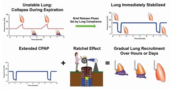
  • APRV is an Open Lung Approach (OLA) and, thus, is similar to those OLA using conventional ventilation methods that have not shown reduced mortality. Indeed, the whole point of this proof-of-concept manuscript is that TCAV is not a typical OLA, but rather a novel stabilize and then gradually recruit method, as suggested in the title. We review the current OLA strategies in Section 2.2. Open Lung Approach. Using the TCAV method, the brief expiratory time will very quickly stabilize alveoli, minimizing RACE-induced atelectrauma, a major VILI mechanism. The lung can then be gradually and safely reopened over an extended period of time (hours or days). Lung reopening is accomplished using the inflation and brake ratcheting open of tissue with each breath, combined with an extended CPAP Phase that recruits alveoli with long-opening time constants. Contrast this with a conventional low VT and PEEP method with recruitment maneuvers designed to open the lung in minutes or seconds, which does not afford durable lung recruitment [69]. It is true that the TCAV method will open the lung, but the method and timing are completely different from the methods used with conventional ventilation methods.

6. Conclusions

The current methods of protective mechanical lung ventilation have not substantially reduced the mortality associated with ARDS. TCAV is a personalized and adaptive approach designed to stabilize alveoli rapidly, reduce progressive lung collapse, and avoid a spiral into the VILI Vortex. Although often used as a rescue mode, TCAV is simply CPAP with a release and can be applied as soon as the patient is intubated so the lung never enters the Vortex. After immediately reducing atelectrauma, collapsed lung tissue can be gradually and safely reopened over an extended period of time (hours or days) while simultaneously avoiding excessive dynamic strain or mechanical power. TCAV is the most clinically applied and scientifically studied method to set and adjust the settings of the APRV modality. TCAV results in stable and homogeneous lung inflation, maintains end-expiratory lung volume, minimizes loss of surfactant function, and reduces inflammation and histopathologic markers of lung injury. The mechanisms of TCAV efficacy include adjusting inspiratory and expiratory durations, first to stabilize and then to gradually open collapsed lung tissue and prevent re-collapse. This treatment sequence of stabilization followed by progressive recruitment is a paradigm shift in protective ventilation and is crucial since regional alveolar instability and stress concentrators are the primary mechanisms of VILI. Using such an approach to unshrink the baby lung may be an effective strategy for improved outcomes in ARDS. Of course, there needs to be successful RCT testing TCAV against the current standard of care before this method can become a primary ventilation strategy on a large scale. The key to this RCT is that untested methods to set and adjust APRV cannot be used. TCAV is based on sound physiological principles and is the most studied protocol to set and adjust the APRV mode. As we know, using the volume assist/control mode, a change in tidal volume from 6 cc/kg to 12 cc/kg resulted in a significant mortality increase. The conclusion was not that the volume assist/control mode was ‘bad’, but, rather, that one method to set and adjust the mode was better. After all, It’s not the Wand, It’s the Wizard.

Supplementary Materials

The following supporting information can be downloaded at: https://www.mdpi.com/article/10.3390/jcm12144633/s1, Video S1: Hexagon model of interdependent alveoli with a central area of instability. Acute lung injury can cause pulmonary edema, which deactivates pulmonary surfactant, resulting in alveolar instability. ‘Alveoli’ in the model’s center have become unstable following simulated surfactant deactivation and collapse and re-open with each breath. These alveoli are subjected to atelectrauma by excessive ‘peeling’ stress as the alveolar wall in apposition peel apart. Collapsed alveoli at expiration act as a stress-multiplier on the surrounding normal alveoli with shared walls causing overdistension and excessive strain [99]; Video S2: Hexagon model of interdependent alveoli with an area of collapse acting as a stress multiplier during INSPIRATION and EXPIRATION. Note the overdistension and excessive dynamic strain on the normal alveoli surrounding the collapsed or edema-filled tissue acting as a stress multiplier. Overdistension is greatest during expiration near the collapsed area but extends further into the normal tissue on inspiration [99]; Video S3: Subpleural alveoli (spherical objects with bright borders) in a rat with acute lung injury caused by surfactant deactivation. Large areas of atelectasis (consolidated red areas) are present at atmospheric pressure. An airway pressure (40 cmH2O) was applied and held for 40 s. Note that there is a delay of ~2 s after the applied airway pressure before alveoli begin to recruit (spherical objects with bright borders appear in the dark red areas), and recruitment continues over the entire 40 s period without increasing pressure [81]; Video S4: Rat lung with acute injury by surfactant deactivation. Darker red areas are atelectatic, and the pink area is inflated tissue. Airway pressure (40 cmH2O) was applied and held for 40 s. Note that there is a delay of ~2 s after the applied airway pressure before lung tissue recruitment (dark red areas turning pink) and recruitment continues over the entire 40 s period without increasing pressure [81].

Author Contributions

Conceptualization, G.F.N., D.W.K. and N.M.H.; writing—original draft preparation, G.F.N.; writing—review and editing, D.W.K., P.L.A., A.G., H.A.-K., L.C., J.S., J.H. and N.M.H.; funding acquisition, G.F.N. and D.W.K. All authors have read and agreed to the published version of the manuscript.

Funding

This work was supported by the Office of the Assistant Secretary of Defense for Health Affairs, through the Peer Reviewed Medical Research Program under Award Nos. W81XWH-20-1-0696 and W81XWH-21-1-0507. Opinions, interpretations, conclusions, and recommendations are those of the authors and are not necessarily endorsed by the Department of Defense. This work was also supported by the National Institutes of Health award R01 HL142702.

Institutional Review Board Statement

Not applicable.

Informed Consent Statement

Not applicable.

Data Availability Statement

Not applicable.

Conflicts of Interest

GFN received a research grant (2019) and equipment donations (refurbished ventilators and neonatal incubators in 2015 and a Perseus Anesthesia Workstation in 2021) from Drager Medical Systems, Inc. GFN has presented and received honoraria and/or travel reimbursement at event(s) sponsored by Drager Medical Systems, Inc., with the most recent being in 2018, outside of the published work. GFN has lectured for Intensive Care Online Network, Inc. (ICON). NMH is the founder of ICON, of which PLA is an employee. NMH holds patents on a method of initiating, managing, and/or weaning airway pressure release ventilation, as well as controlling a ventilator in accordance with the same., DWK and JH are co-founders and shareholders of OscillaVent, Inc., and are co-inventors on a patent involving multi-frequency oscillatory ventilation. DWK and JH also receive research support from ZOLL Medical Corporation. The authors maintain that the industry had no role in the design and conduct of the study; the collection, management, analysis, or interpretation of the data; or the preparation, review, or approval of the manuscript.

References

  1. Matthay, M.A.; Zemans, R.L.; Zimmerman, G.A.; Arabi, Y.M.; Beitler, J.R.; Mercat, A.; Herridge, M.; Randolph, A.G.; Calfee, C.S. Acute respiratory distress syndrome. Nat. Rev. Dis. Primers 2019, 5, 18. [Google Scholar] [CrossRef]
  2. Slutsky, A.S.; Ranieri, V.M. Ventilator-induced lung injury. N. Engl. J. Med. 2013, 369, 2126–2136. [Google Scholar] [CrossRef] [Green Version]
  3. Caser, E.B.; Zandonade, E.; Pereira, E.; Gama, A.M.; Barbas, C.S. Impact of distinct definitions of acute lung injury on its incidence and outcomes in Brazilian ICUs: Prospective evaluation of 7133 patients*. Crit. Care Med. 2014, 42, 574–582. [Google Scholar] [CrossRef] [PubMed]
  4. Laffey, J.G.; Bellani, G.; Pham, T.; Fan, E.; Madotto, F.; Bajwa, E.K.; Brochard, L.; Clarkson, K.; Esteban, A.; Gattinoni, L.; et al. Potentially modifiable factors contributing to outcome from acute respiratory distress syndrome: The LUNG SAFE study. Intensive Care Med. 2016, 42, 1865–1876. [Google Scholar] [CrossRef] [PubMed]
  5. Fan, E.; Brodie, D.; Slutsky, A.S. Acute Respiratory Distress Syndrome: Advances in Diagnosis and Treatment. JAMA 2018, 319, 698–710. [Google Scholar] [CrossRef]
  6. Bellani, G.; Laffey, J.G.; Pham, T.; Fan, E.; Brochard, L.; Esteban, A.; Gattinoni, L.; van Haren, F.; Larsson, A.; McAuley, D.F.; et al. Epidemiology, patterns of care, and mortality for patients with acute respiratory distress syndrome in intensive care units in 50 countries. JAMA 2016, 315, 788–800. [Google Scholar] [CrossRef]
  7. Bates, J.H.T.; Smith, B.J. Ventilator-induced lung injury and lung mechanics. Ann. Transl. Med. 2018, 6, 378. [Google Scholar] [CrossRef]
  8. Cressoni, M.; Chiurazzi, C.; Gotti, M.; Amini, M.; Brioni, M.; Algieri, I.; Cammaroto, A.; Rovati, C.; Massari, D.; di Castiglione, C.B.; et al. Lung inhomogeneities and time course of ventilator-induced mechanical injuries. Anesthesiology 2015, 123, 618–627. [Google Scholar] [CrossRef]
  9. Cereda, M.; Xin, Y. Alveolar recruitment and lung injury: An issue of timing and location? Crit. Care Med. 2013, 41, 2837–2838. [Google Scholar] [CrossRef]
  10. Cereda, M.; Xin, Y.; Meeder, N.; Zeng, J.; Jiang, Y.; Hamedani, H.; Profka, H.; Kadlecek, S.; Clapp, J.; Deshpande, C.G.; et al. Visualizing the Propagation of Acute Lung Injury. Anesthesiology 2016, 124, 121–131. [Google Scholar] [CrossRef] [PubMed] [Green Version]
  11. Albert, K.; Krischer, J.M.; Pfaffenroth, A.; Wilde, S.; Lopez-Rodriguez, E.; Braun, A.; Smith, B.J.; Knudsen, L. Hidden Microatelectases Increase Vulnerability to Ventilation-Induced Lung Injury. Front. Physiol. 2020, 11, 530485. [Google Scholar] [CrossRef] [PubMed]
  12. Gattinoni, L.; Pesenti, A. The concept of “baby lung”. Intensive Care Med. 2005, 31, 776–784. [Google Scholar] [CrossRef]
  13. Marini, J.J.; Gattinoni, L. Time Course of Evolving Ventilator-Induced Lung Injury: The “Shrinking Baby Lung”. Crit. Care Med. 2020, 48, 1203–1209. [Google Scholar] [CrossRef] [PubMed]
  14. Writing Group for the Alveolar Recruitment for Acute Respiratory Distress Syndrome Trial (ART) Investigators; Cavalcanti, A.B.; Suzumura, E.A.; Laranjeira, L.N.; Paisani, D.M.; Damiani, L.P.; Guimaraes, H.P.; Romano, E.R.; Regenga, M.M.; Taniguchi, L.N.T.; et al. Effect of Lung Recruitment and Titrated Positive End-Expiratory Pressure (PEEP) vs Low PEEP on Mortality in Patients with Acute Respiratory Distress Syndrome: A Randomized Clinical Trial. JAMA 2017, 318, 1335–1345. [Google Scholar] [CrossRef] [PubMed]
  15. Brower, R.G.; Lanken, P.N.; MacIntyre, N.; Matthay, M.A.; Morris, A.; Ancukiewicz, M.; Schoenfeld, D.; Thompson, B.T.; The National Heart, Lung, and Blood Institute ARDS Clinical Trials Network. Higher versus lower positive end-expiratory pressures in patients with the acute respiratory distress syndrome. N. Engl. J. Med. 2004, 351, 327–336. [Google Scholar]
  16. Meade, M.O.; Cook, D.J.; Guyatt, G.H.; Slutsky, A.S.; Arabi, Y.M.; Cooper, D.J.; Davies, A.R.; Hand, L.E.; Zhou, Q.; Thabane, L.; et al. Ventilation strategy using low tidal volumes, recruitment maneuvers, and high positive end-expiratory pressure for acute lung injury and acute respiratory distress syndrome: A randomized controlled trial. JAMA 2008, 299, 637–645. [Google Scholar] [CrossRef] [Green Version]
  17. Mercat, A.; Richard, J.C.; Vielle, B.; Jaber, S.; Osman, D.; Diehl, J.L.; Lefrant, J.Y.; Prat, G.; Richecoeur, J.; Nieszkowska, A.; et al. Positive end-expiratory pressure setting in adults with acute lung injury and acute respiratory distress syndrome: A randomized controlled trial. JAMA 2008, 299, 646–655. [Google Scholar] [CrossRef] [Green Version]
  18. Nieman, G.F.; Andrews, P.; Satalin, J.; Wilcox, K.; Kollisch-Singule, M.; Madden, M.; Aiash, H.; Blair, S.J.; Gatto, L.A.; Habashi, N.M. Acute lung injury: How to stabilize a broken lung. Crit. Care 2018, 22, 136. [Google Scholar] [CrossRef] [Green Version]
  19. Parker, J.C.; Townsley, M.I. Evaluation of lung injury in rats and mice. Am. J. Physiol. Lung Cell. Mol. Physiol. 2004, 286, L231–L246. [Google Scholar] [CrossRef]
  20. Taeusch, H.W.; Bernardino de la Serna, J.; Perez-Gil, J.; Alonso, C.; Zasadzinski, J.A. Inactivation of pulmonary surfactant due to serum-inhibited adsorption and reversal by hydrophilic polymers: Experimental. Biophys. J. 2005, 89, 1769–1779. [Google Scholar] [CrossRef] [Green Version]
  21. Albert, R.K. The role of ventilation-induced surfactant dysfunction and atelectasis in causing acute respiratory distress syndrome. Am. J. Respir. Crit. Care Med. 2012, 185, 702–708. [Google Scholar] [CrossRef]
  22. Nieman, G.F.; Bredenberg, C.E. High surface tension pulmonary edema induced by detergent aerosol. J. Appl. Physiol. 1985, 58, 129–136. [Google Scholar] [CrossRef] [PubMed]
  23. Perlman, C.E.; Lederer, D.J.; Bhattacharya, J. Micromechanics of alveolar edema. Am. J. Respir. Cell. Mol. Biol. 2011, 44, 34–39. [Google Scholar] [CrossRef] [PubMed]
  24. Chen, Z.L.; Chen, Y.Z.; Hu, Z.Y. A micromechanical model for estimating alveolar wall strain in mechanically ventilated edematous lungs. J. Appl. Physiol. 2014, 117, 586–592. [Google Scholar] [CrossRef] [PubMed] [Green Version]
  25. Makiyama, A.M.; Gibson, L.J.; Harris, R.S.; Venegas, J.G. Stress concentration around an atelectatic region: A finite element model. Respir. Physiol. Neurobiol. 2014, 201, 101–110. [Google Scholar] [CrossRef]
  26. Retamal, J.; Bergamini, B.C.; Carvalho, A.R.; Bozza, F.A.; Borzone, G.; Borges, J.B.; Larsson, A.; Hedenstierna, G.; Bugedo, G.; Bruhn, A. Non-lobar atelectasis generates inflammation and structural alveolar injury in the surrounding healthy tissue during mechanical ventilation. Crit. Care 2014, 18, 505. [Google Scholar] [CrossRef]
  27. Acute Respiratory Distress Syndrome Network; Brower, R.G.; Matthay, M.A.; Morris, A.; Schoenfeld, D.; Thompson, B.T.; Wheeler, A. Ventilation with lower tidal volumes as compared with traditional tidal volumes for acute lung injury and the acute respiratory distress syndrome. N. Engl. J. Med. 2000, 342, 1301–1308. [Google Scholar] [CrossRef] [Green Version]
  28. Gattinoni, L.; Pesenti, A.; Avalli, L.; Rossi, F.; Bombino, M. Pressure-volume curve of total respiratory system in acute respiratory failure. Computed tomographic scan study. Am. Rev. Respir. Dis. 1987, 136, 730–736. [Google Scholar] [CrossRef] [PubMed]
  29. Deans, K.J.; Minneci, P.C.; Cui, X.; Banks, S.M.; Natanson, C.; Eichacker, P.Q. Mechanical ventilation in ARDS: One size does not fit all. Crit. Care Med. 2005, 33, 1141–1143. [Google Scholar] [CrossRef]
  30. Nieman, G.F.; Gatto, L.A.; Andrews, P.; Satalin, J.; Camporota, L.; Daxon, B.; Blair, S.J.; Al-Khalisy, H.; Madden, M.; Kollisch-Singule, M.; et al. Prevention and treatment of acute lung injury with time-controlled adaptive ventilation: Physiologically informed modification of airway pressure release ventilation. Ann. Intensive Care 2020, 10, 3. [Google Scholar] [CrossRef] [Green Version]
  31. Amato, M.B.; Meade, M.O.; Slutsky, A.S.; Brochard, L.; Costa, E.L.; Schoenfeld, D.A.; Stewart, T.E.; Briel, M.; Talmor, D.; Mercat, A.; et al. Driving pressure and survival in the acute respiratory distress syndrome. N. Engl. J. Med. 2015, 372, 747–755. [Google Scholar] [CrossRef] [Green Version]
  32. Yehya, N.; Hodgson, C.L.; Amato, M.B.P.; Richard, J.C.; Brochard, L.J.; Mercat, A.; Goligher, E.C. Response to Ventilator Adjustments for Predicting Acute Respiratory Distress Syndrome Mortality. Driving Pressure versus Oxygenation. Ann. Am. Thorac. Soc. 2021, 18, 857–864. [Google Scholar] [CrossRef]
  33. Costa, E.L.V.; Slutsky, A.; Brochard, L.J.; Brower, R.; Serpa-Neto, A.; Cavalcanti, A.B.; Mercat, A.; Meade, M.; Morais, C.C.A.; Goligher, E.; et al. Ventilatory Variables and Mechanical Power in Patients with Acute Respiratory Distress Syndrome. Am. J. Respir. Crit. Care Med. 2021, 204, 303–311. [Google Scholar] [CrossRef] [PubMed]
  34. Aoyama, H.; Pettenuzzo, T.; Aoyama, K.; Pinto, R.; Englesakis, M.; Fan, E. Association of Driving Pressure with Mortality Among Ventilated Patients with Acute Respiratory Distress Syndrome: A Systematic Review and Meta-Analysis. Crit. Care Med. 2018, 46, 300–306. [Google Scholar] [CrossRef]
  35. Chen, Z.; Wei, X.; Liu, G.; Tai, Q.; Zheng, D.; Xie, W.; Chen, L.; Wang, G.; Sun, J.Q.; Wang, S.; et al. Higher vs. Lower DP for Ventilated Patients with Acute Respiratory Distress Syndrome: A Systematic Review and Meta-Analysis. Emerg. Med. Int. 2019, 2019, 4654705. [Google Scholar] [CrossRef]
  36. Goligher, E.C.; Costa, E.L.V.; Yarnell, C.J.; Brochard, L.J.; Stewart, T.E.; Tomlinson, G.; Brower, R.G.; Slutsky, A.S.; Amato, M.P.B. Effect of Lowering Tidal Volume on Mortality in ARDS Varies with Respiratory System Elastance. Am. J. Respir. Crit. Care Med. 2021, 203, 1378–1385. [Google Scholar] [CrossRef]
  37. Protti, A.; Andreis, D.T.; Monti, M.; Santini, A.; Sparacino, C.C.; Langer, T.; Votta, E.; Gatti, S.; Lombardi, L.; Leopardi, O.; et al. Lung stress and strain during mechanical ventilation: Any difference between statics and dynamics? Crit. Care Med. 2013, 41, 1046–1055. [Google Scholar] [CrossRef] [Green Version]
  38. Protti, A.; Andreis, D.T.; Iapichino, G.E.; Monti, M.; Comini, B.; Milesi, M.; Zani, L.; Gatti, S.; Lombardi, L.; Gattinoni, L. High positive end-expiratory pressure: Only a dam against oedema formation? Crit. Care 2013, 17, R131. [Google Scholar] [CrossRef] [Green Version]
  39. Protti, A.; Maraffi, T.; Milesi, M.; Votta, E.; Santini, A.; Pugni, P.; Andreis, D.T.; Nicosia, F.; Zannin, E.; Gatti, S.; et al. Role of Strain Rate in the Pathogenesis of Ventilator-Induced Lung Edema. Crit. Care Med. 2016, 44, e838–e845. [Google Scholar] [CrossRef] [PubMed]
  40. Jain, S.V.; Kollisch-Singule, M.; Satalin, J.; Searles, Q.; Dombert, L.; Abdel-Razek, O.; Yepuri, N.; Leonard, A.; Gruessner, A.; Andrews, P.; et al. The role of high airway pressure and dynamic strain on ventilator-induced lung injury in a heterogeneous acute lung injury model. Intensive Care Med. Exp. 2017, 5, 25. [Google Scholar] [CrossRef] [PubMed] [Green Version]
  41. Seah, A.S.; Grant, K.A.; Aliyeva, M.; Allen, G.B.; Bates, J.H.T. Quantifying the roles of tidal volume and PEEP in the pathogenesis of ventilator-induced lung injury. Ann. Biomed. Eng. 2011, 39, 1505–1516. [Google Scholar] [CrossRef] [PubMed]
  42. Wilson, M.R.; Patel, B.V.; Takata, M. Ventilation with “clinically relevant” high tidal volumes does not promote stretch-induced injury in the lungs of healthy mice. Crit. Care Med. 2012, 40, 2850–2857. [Google Scholar] [CrossRef] [PubMed] [Green Version]
  43. Syring, R.S.; Otto, C.M.; Spivack, R.E.; Markstaller, K.; Baumgardner, J.E. Maintenance of end-expiratory recruitment with increased respiratory rate after saline-lavage lung injury. J. Appl. Physiol. 2007, 102, 331–339. [Google Scholar] [CrossRef] [Green Version]
  44. Hamlington, K.L.; Smith, B.J.; Dunn, C.M.; Charlebois, C.M.; Roy, G.S.; Bates, J.H.T. Linking lung function to structural damage of alveolar epithelium in ventilator-induced lung injury. Respir. Physiol. Neurobiol. 2018, 255, 22–29. [Google Scholar] [CrossRef] [PubMed]
  45. Otto, C.M.; Markstaller, K.; Kajikawa, O.; Karmrodt, J.; Syring, R.S.; Pfeiffer, B.; Good, V.P.; Frevert, C.W.; Baumgardner, J.E. Spatial and temporal heterogeneity of ventilator-associated lung injury after surfactant depletion. J. Appl. Physiol. 2008, 104, 1485–1494. [Google Scholar] [CrossRef]
  46. Schiller, H.J.; McCann, U.G., 2nd; Carney, D.E.; Gatto, L.A.; Steinberg, J.M.; Nieman, G.F. Altered alveolar mechanics in the acutely injured lung. Crit. Care Med. 2001, 29, 1049–1055. [Google Scholar] [CrossRef]
  47. Cereda, M.; Emami, K.; Kadlecek, S.; Xin, Y.; Mongkolwisetwara, P.; Profka, H.; Barulic, A.; Pickup, S.; Mansson, S.; Wollmer, P.; et al. Quantitative imaging of alveolar recruitment with hyperpolarized gas MRI during mechanical ventilation. J. Appl. Physiol. 2011, 110, 499–511. [Google Scholar] [CrossRef] [Green Version]
  48. Gaver, D.P., 3rd; Nieman, G.F.; Gatto, L.A.; Cereda, M.; Habashi, N.M.; Bates, J.H.T. The POOR get POORer: A hypothesis for the pathogenesis of ventilator-induced lung injury. Am. J. Respir. Crit. Care Med. 2020, 202, 1081–1087. [Google Scholar] [CrossRef]
  49. Cereda, M.; Xin, Y.; Goffi, A.; Herrmann, J.; Kaczka, D.W.; Kavanagh, B.P.; Perchiazzi, G.; Yoshida, T.; Rizi, R.R. Imaging the Injured Lung: Mechanisms of Action and Clinical Use. Anesthesiology 2019, 131, 716–749. [Google Scholar] [CrossRef]
  50. Cabrera-Benitez, N.E.; Laffey, J.G.; Parotto, M.; Spieth, P.M.; Villar, J.; Zhang, H.; Slutsky, A.S. Mechanical ventilation-associated lung fibrosis in acute respiratory distress syndrome: A significant contributor to poor outcome. Anesthesiology 2014, 121, 189–198. [Google Scholar] [CrossRef]
  51. Burkhardt, A. Alveolitis and collapse in the pathogenesis of pulmonary fibrosis. Am. Rev. Respir. Dis. 1989, 140, 513–524. [Google Scholar] [CrossRef] [PubMed]
  52. Knudsen, L.; Ruppert, C.; Ochs, M. Tissue remodelling in pulmonary fibrosis. Cell. Tissue Res. 2017, 367, 607–626. [Google Scholar] [CrossRef] [PubMed]
  53. Lutz, D.; Gazdhar, A.; Lopez-Rodriguez, E.; Ruppert, C.; Mahavadi, P.; Gunther, A.; Klepetko, W.; Bates, J.H.; Smith, B.; Geiser, T.; et al. Alveolar derecruitment and collapse induration as crucial mechanisms in lung injury and fibrosis. Am. J. Respir. Cell. Mol. Biol. 2015, 52, 232–243. [Google Scholar] [CrossRef] [PubMed]
  54. Duggan, M.; Kavanagh, B.P. Pulmonary atelectasis: A pathogenic perioperative entity. Anesthesiology 2005, 102, 838–854. [Google Scholar] [CrossRef]
  55. Spinelli, E.; Mauri, T.; Beitler, J.R.; Pesenti, A.; Brodie, D. Respiratory drive in the acute respiratory distress syndrome: Pathophysiology, monitoring, and therapeutic interventions. Intensive Care Med. 2020, 46, 606–618. [Google Scholar] [CrossRef]
  56. Petersson, J.; Glenny, R.W. Gas exchange and ventilation-perfusion relationships in the lung. Eur. Respir. J. 2014, 44, 1023–1041. [Google Scholar] [CrossRef] [Green Version]
  57. Andrews, P.L.; Sadowitz, B.; Kollisch-Singule, M.; Satalin, J.; Roy, S.; Snyder, K.; Gatto, L.A.; Nieman, G.F.; Habashi, N.M. Alveolar instability (atelectrauma) is not identified by arterial oxygenation predisposing the development of an occult ventilator-induced lung injury. Intensive Care Med. Exp. 2015, 3, 54. [Google Scholar] [CrossRef] [Green Version]
  58. Pavone, L.; Albert, S.; DiRocco, J.; Gatto, L.; Nieman, G. Alveolar instability caused by mechanical ventilation initially damages the nondependent normal lung. Crit. Care 2007, 11, R104. [Google Scholar] [CrossRef] [Green Version]
  59. Williams, E.M.; Viale, J.P.; Hamilton, R.M.; McPeak, H.; Sutton, L.; Hahn, C.E. Within-breath arterial PO2 oscillations in an experimental model of acute respiratory distress syndrome. Br. J. Anaesth. 2000, 85, 456–459. [Google Scholar] [CrossRef] [Green Version]
  60. Duggan, M.; McCaul, C.L.; McNamara, P.J.; Engelberts, D.; Ackerley, C.; Kavanagh, B.P. Atelectasis causes vascular leak and lethal right ventricular failure in uninjured rat lungs. Am. J. Respir. Crit. Care Med. 2003, 167, 1633–1640. [Google Scholar] [CrossRef]
  61. Sipmann, F.S.; Santos, A.; Tusman, G. Heart-lung interactions in acute respiratory distress syndrome: Pathophysiology, detection and management strategies. Ann. Transl. Med. 2018, 6, 27. [Google Scholar] [CrossRef]
  62. Pearse, D.B.; Searcy, R.M.; Mitzner, W.; Permutt, S.; Sylvester, J.T. Effects of tidal volume and respiratory frequency on lung lymph flow. J. Appl. Physiol. 2005, 99, 556–563. [Google Scholar] [CrossRef] [PubMed]
  63. Majumdar, A.; Arold, S.P.; Bartolak-Suki, E.; Parameswaran, H.; Suki, B. Jamming dynamics of stretch-induced surfactant release by alveolar type II cells. J. Appl. Physiol. 2012, 112, 824–831. [Google Scholar] [CrossRef] [Green Version]
  64. Nieman, G.; Kollisch-Singule, M.; Ramcharran, H.; Satalin, J.; Blair, S.; Gatto, L.A.; Andrews, P.; Ghosh, A.; Kaczka, D.W.; Gaver, D.; et al. Unshrinking the baby lung to calm the VILI vortex. Crit. Care 2022, 26, 242. [Google Scholar] [CrossRef]
  65. Ferguson, N.D.; Cook, D.J.; Guyatt, G.H.; Mehta, S.; Hand, L.; Austin, P.; Zhou, Q.; Matte, A.; Walter, S.D.; Lamontagne, F.; et al. High-frequency oscillation in early acute respiratory distress syndrome. N. Engl. J. Med. 2013, 368, 795–805. [Google Scholar] [CrossRef] [PubMed] [Green Version]
  66. Young, D.; Lamb, S.E.; Shah, S.; MacKenzie, I.; Tunnicliffe, W.; Lall, R.; Rowan, K.; Cuthbertson, B.H.; Group, O.S. High-frequency oscillation for acute respiratory distress syndrome. N. Engl. J. Med. 2013, 368, 806–813. [Google Scholar] [CrossRef] [Green Version]
  67. Malhotra, A.; Drazen, J.M. High-frequency oscillatory ventilation on shaky ground. N. Engl. J. Med. 2013, 368, 863–865. [Google Scholar] [CrossRef] [Green Version]
  68. van der Zee, P.; Gommers, D. Recruitment Maneuvers and Higher PEEP, the So-Called Open Lung Concept, in Patients with ARDS. Crit. Care 2019, 23, 73. [Google Scholar] [CrossRef] [PubMed] [Green Version]
  69. Grasso, S.; Fanelli, V.; Cafarelli, A.; Anaclerio, R.; Amabile, M.; Ancona, G.; Fiore, T. Effects of high versus low positive end-expiratory pressures in acute respiratory distress syndrome. Am. J. Respir. Crit. Care Med. 2005, 171, 1002–1008. [Google Scholar] [CrossRef]
  70. Angriman, F.; Ferreyro, B.L.; Donaldson, L.; Cuthbertson, B.H.; Ferguson, N.D.; Bollen, C.W.; Bachman, T.E.; Lamontagne, F.; Adhikari, N.K.J. The harm of high-frequency oscillatory ventilation (HFOV) in ARDS is not related to a high baseline risk of acute cor pulmonale or short-term changes in hemodynamics. Intensive Care Med. 2020, 46, 132–134. [Google Scholar] [CrossRef]
  71. Kaczka, D.W. Oscillatory ventilation redux: Alternative perspectives on ventilator-induced lung injury in the acute respiratory distress syndrome. Curr. Opin. Physiol. 2021, 21, 36–43. [Google Scholar] [CrossRef] [PubMed]
  72. Villar, J.; Blanco, J.; Anon, J.M.; Santos-Bouza, A.; Blanch, L.; Ambros, A.; Gandia, F.; Carriedo, D.; Mosteiro, F.; Basaldua, S.; et al. The ALIEN study: Incidence and outcome of acute respiratory distress syndrome in the era of lung protective ventilation. Intensive Care Med. 2011, 37, 1932–1941. [Google Scholar] [CrossRef] [PubMed] [Green Version]
  73. Shen, Y.; Cai, G.; Gong, S.; Dong, L.; Yan, J.; Cai, W. Interaction between low tidal volume ventilation strategy and severity of acute respiratory distress syndrome: A retrospective cohort study. Crit. Care 2019, 23, 254. [Google Scholar] [CrossRef] [PubMed] [Green Version]
  74. Parcha, V.; Kalra, R.; Bhatt, S.P.; Berra, L.; Arora, G.; Arora, P. Trends and Geographic Variation in Acute Respiratory Failure and ARDS Mortality in the United States. Chest 2021, 159, 1460–1472. [Google Scholar] [CrossRef] [PubMed]
  75. Raschke, R.A.; Stoffer, B.; Assar, S.; Fountain, S.; Olsen, K.; Heise, C.W.; Gallo, T.; Padilla-Jones, A.; Gerkin, R.; Parthasarathy, S.; et al. The relationship of tidal volume and driving pressure with mortality in hypoxic patients receiving mechanical ventilation. PLoS ONE 2021, 16, e0255812. [Google Scholar] [CrossRef]
  76. Sahetya, S.K.; Brower, R.G. Lung Recruitment and Titrated PEEP in Moderate to Severe ARDS: Is the Door Closing on the Open Lung? JAMA 2017, 318, 1327–1329. [Google Scholar] [CrossRef]
  77. Cereda, M.; Xin, Y.; Emami, K.; Huang, J.; Rajaei, J.; Profka, H.; Han, B.; Mongkolwisetwara, P.; Kadlecek, S.; Kuzma, N.N.; et al. Positive end-expiratory pressure increments during anesthesia in normal lung result in hysteresis and greater numbers of smaller aerated airspaces. Anesthesiology 2013, 119, 1402–1409. [Google Scholar] [CrossRef] [Green Version]
  78. Dianti, J.; Tisminetzky, M.; Ferreyro, B.L.; Englesakis, M.; Del Sorbo, L.; Sud, S.; Talmor, D.; Ball, L.; Meade, M.; Hodgson, C.; et al. Association of Positive End-Expiratory Pressure and Lung Recruitment Selection Strategies with Mortality in Acute Respiratory Distress Syndrome: A Systematic Review and Network Meta-analysis. Am. J. Respir. Crit. Care Med. 2022, 205, 1300–1310. [Google Scholar] [CrossRef]
  79. Habashi, N.M. Other approaches to open-lung ventilation: Airway pressure release ventilation. Crit. Care Med. 2005, 33, S228–S240. [Google Scholar] [CrossRef]
  80. Andrews, P.L.; Shiber, J.R.; Jaruga-Killeen, E.; Roy, S.; Sadowitz, B.; O’Toole, R.V.; Gatto, L.A.; Nieman, G.F.; Scalea, T.; Habashi, N.M. Early application of airway pressure release ventilation may reduce mortality in high-risk trauma patients: A systematic review of observational trauma ARDS literature. J. Trauma. Acute Care Surg. 2013, 75, 635–641. [Google Scholar] [CrossRef] [Green Version]
  81. Albert, S.P.; DiRocco, J.; Allen, G.B.; Bates, J.H.; Lafollette, R.; Kubiak, B.D.; Fischer, J.; Maroney, S.; Nieman, G.F. The role of time and pressure on alveolar recruitment. J. Appl. Physiol. 2009, 106, 757–765. [Google Scholar] [CrossRef] [Green Version]
  82. Emr, B.; Gatto, L.A.; Roy, S.; Satalin, J.; Ghosh, A.; Snyder, K.; Andrews, P.; Habashi, N.; Marx, W.; Ge, L.; et al. Airway pressure release ventilation prevents ventilator-induced lung injury in normal lungs. JAMA Surg. 2013, 148, 1005–1012. [Google Scholar] [CrossRef] [Green Version]
  83. Kollisch-Singule, M.; Emr, B.; Smith, B.; Roy, S.; Jain, S.; Satalin, J.; Snyder, K.; Andrews, P.; Habashi, N.; Bates, J.; et al. Mechanical breath profile of airway pressure release ventilation: The effect on alveolar recruitment and microstrain in acute lung injury. JAMA Surg. 2014, 149, 1138–1145. [Google Scholar] [CrossRef] [Green Version]
  84. Kollisch-Singule, M.; Emr, B.; Smith, B.; Ruiz, C.; Roy, S.; Meng, Q.; Jain, S.; Satalin, J.; Snyder, K.; Ghosh, A.; et al. Airway pressure release ventilation reduces conducting airway micro-strain in lung injury. J. Am. Coll. Surg. 2014, 219, 968–976. [Google Scholar] [CrossRef] [PubMed] [Green Version]
  85. Kollisch-Singule, M.; Jain, S.; Andrews, P.; Smith, B.J.; Hamlington-Smith, K.L.; Roy, S.; DiStefano, D.; Nuss, E.; Satalin, J.; Meng, Q.; et al. Effect of Airway Pressure Release Ventilation on Dynamic Alveolar Heterogeneity. JAMA Surg. 2015, 151, 64–72. [Google Scholar] [CrossRef] [PubMed] [Green Version]
  86. Kollisch-Singule, M.; Emr, B.; Jain, S.V.; Andrews, P.; Satalin, J.; Liu, J.; Porcellio, E.; Kenyon, V.; Wang, G.; Marx, W.; et al. The effects of airway pressure release ventilation on respiratory mechanics in extrapulmonary lung injury. Intensive Care Med. Exp. 2015, 3, 35. [Google Scholar] [CrossRef] [PubMed] [Green Version]
  87. Smith, B.J.; Lundblad, L.K.; Kollisch-Singule, M.; Satalin, J.; Nieman, G.; Habashi, N.; Bates, J.H. Predicting the response of the injured lung to the mechanical breath profile. J. Appl. Physiol. 2015, 118, 932–940. [Google Scholar] [CrossRef] [Green Version]
  88. Kollisch-Singule, M.; Jain, S.V.; Satalin, J.; Andrews, P.; Searles, Q.; Liu, Z.; Zhou, Y.; Wang, G.; Meier, A.H.; Gatto, L.A.; et al. Limiting ventilator-associated lung injury in a preterm porcine neonatal model. J. Pediatr. Surg. 2017, 52, 50–55. [Google Scholar] [CrossRef]
  89. Jain, S.V.; Kollisch-Singule, M.; Sadowitz, B.; Dombert, L.; Satalin, J.; Andrews, P.; Gatto, L.A.; Nieman, G.F.; Habashi, N.M. The 30-year evolution of airway pressure release ventilation (APRV). Intensive Care Med. Exp. 2016, 4, 11. [Google Scholar] [CrossRef] [PubMed] [Green Version]
  90. Andrews, P.; Shiber, J.; Madden, M.; Nieman, G.F.; Camporota, L.; Habashi, N.M. Myths and Misconceptions of Airway Pressure Release Ventilation: Getting Past the Noise and on to the Signal. Front. Physiol. 2022, 13, 928562. [Google Scholar] [CrossRef]
  91. Boehme, S.; Bentley, A.H.; Hartmann, E.K.; Chang, S.; Erdoes, G.; Prinzing, A.; Hagmann, M.; Baumgardner, J.E.; Ullrich, R.; Markstaller, K.; et al. Influence of inspiration to expiration ratio on cyclic recruitment and derecruitment of atelectasis in a saline lavage model of acute respiratory distress syndrome. Crit. Care Med. 2015, 43, e65–e74. [Google Scholar] [CrossRef] [PubMed] [Green Version]
  92. Henderson, W.R.; Dominelli, P.B.; Molgat-Seon, Y.; Lipson, R.; Griesdale, D.E.; Sekhon, M.; Ayas, N.; Sheel, A.W. Effect of tidal volume and positive end-expiratory pressure on expiratory time constants in experimental lung injury. Physiol. Rep. 2016, 4, e12737. [Google Scholar] [CrossRef] [PubMed] [Green Version]
  93. Neumann, P.; Berglund, J.E.; Fernandez Mondejar, E.; Magnusson, A.; Hedenstierna, G. Dynamics of lung collapse and recruitment during prolonged breathing in porcine lung injury. J. Appl. Physiol. 1998, 85, 1533–1543. [Google Scholar] [CrossRef] [PubMed]
  94. Herrmann, J.; Gerard, S.E.; Shao, W.; Xin, Y.; Cereda, M.; Reinhardt, J.M.; Christensen, G.E.; Hoffman, E.A.; Kaczka, D.W. Effects of Lung Injury on Regional Aeration and Expiratory Time Constants: Insights From Four-Dimensional Computed Tomography Image Registration. Front. Physiol. 2021, 12, 707119. [Google Scholar] [CrossRef]
  95. Bates, J.H.; Irvin, C.G. Time dependence of recruitment and derecruitment in the lung: A theoretical model. J. Appl. Physiol. 2002, 93, 705–713. [Google Scholar] [CrossRef] [Green Version]
  96. Markstaller, K.; Eberle, B.; Kauczor, H.U.; Scholz, A.; Bink, A.; Thelen, M.; Heinrichs, W.; Weiler, N. Temporal dynamics of lung aeration determined by dynamic CT in a porcine model of ARDS. Br. J. Anaesth. 2001, 87, 459–468. [Google Scholar] [CrossRef] [Green Version]
  97. Lachmann, B. Open up the lung and keep the lung open. Intensive Care Med. 1992, 18, 319–321. [Google Scholar] [CrossRef] [Green Version]
  98. Roy, S.K.; Emr, B.; Sadowitz, B.; Gatto, L.A.; Ghosh, A.; Satalin, J.M.; Snyder, K.P.; Ge, L.; Wang, G.; Marx, W.; et al. Preemptive application of airway pressure release ventilation prevents development of acute respiratory distress syndrome in a rat traumatic hemorrhagic shock model. Shock 2013, 40, 210–216. [Google Scholar] [CrossRef] [Green Version]
  99. Nieman, G.F.; Satalin, J.; Kollisch-Singule, M.; Andrews, P.; Aiash, H.; Habashi, N.M.; Gatto, L.A. Physiology in Medicine: Understanding dynamic alveolar physiology to minimize ventilator-induced lung injury. J. Appl. Physiol. 2017, 122, 1516–1522. [Google Scholar] [CrossRef]
  100. Mead, J.; Takishima, T.; Leith, D. Stress distribution in lungs: A model of pulmonary elasticity. J. Appl. Physiol. 1970, 28, 596–608. [Google Scholar] [CrossRef]
  101. Suki, B.; Stamenovic, D.; Hubmayr, R. Lung parenchymal mechanics. Compr. Physiol. 2011, 1, 1317–1351. [Google Scholar] [CrossRef] [PubMed] [Green Version]
  102. Paré, P.D.; Mitzner, W. Airway-parenchymal interdependence. Compr. Physiol. 2012, 2, 1921–1935. [Google Scholar]
  103. Ryans, J.M.; Fujioka, H.; Gaver, D.P., 3rd. Micro-scale to Meso-scale Analysis of Parenchymal Tethering: The Effect of Heterogeneous Alveolar Pressures on the Pulmonary Mechanics of Compliant Airways. J. Appl. Physiol. 2019, 126, 1204–1213. [Google Scholar] [CrossRef]
  104. Fujioka, H.; Halpern, D.; Gaver, D.P., 3rd. A model of surfactant-induced surface tension effects on the parenchymal tethering of pulmonary airways. J. Biomech. 2013, 46, 319–328. [Google Scholar] [CrossRef] [PubMed]
  105. Kollisch-Singule, M.C.; Jain, S.V.; Andrews, P.L.; Satalin, J.; Gatto, L.A.; Villar, J.; De Backer, D.; Gattinoni, L.; Nieman, G.F.; Habashi, N.M. Looking beyond macroventilatory parameters and rethinking ventilator-induced lung injury. J. Appl. Physiol. (1985) 2018, 124, 1214–1218. [Google Scholar] [CrossRef] [PubMed] [Green Version]
  106. Marshall, R.; Widdicombe, J.G. Stress relaxation of the human lung. Clin. Sci. 1960, 20, 19–31. [Google Scholar]
  107. Suki, B.; Barabasi, A.L.; Lutchen, K.R. Lung tissue viscoelasticity: A mathematical framework and its molecular basis. J. Appl. Physiol. 1994, 76, 2749–2759. [Google Scholar] [CrossRef]
  108. Coughlin, M.F.; Suki, B.; Stamenovic, D. Dynamic behavior of lung parenchyma in shear. J. Appl. Physiol. 1996, 80, 1880–1890. [Google Scholar] [CrossRef]
  109. Sugihara, T.; Hildebrandt, J.; Martin, C.J. Viscoelastic properties of alveolar wall. J. Appl. Physiol. 1972, 33, 93–98. [Google Scholar] [CrossRef]
  110. Bates, J.H.T.; Gaver, D.P.; Habashi, N.M.; Nieman, G.F. Atelectrauma Versus Volutrauma: A Tale of Two Time-Constants. Crit. Care Explor. 2020, 2, e0299. [Google Scholar] [CrossRef]
  111. Parhar, K.K.S.; Doig, C. Caution-Do Not Attempt This at Home. Airway Pressure Release Ventilation Should Not Routinely Be Used in Patients with or at Risk of Acute Respiratory Distress Syndrome Outside of a Clinical Trial. Crit. Care Med. 2023. ahead of print. [Google Scholar] [CrossRef] [PubMed]
  112. Carsetti, A.; Damiani, E.; Domizi, R.; Scorcella, C.; Pantanetti, S.; Falcetta, S.; Donati, A.; Adrario, E. Airway pressure release ventilation during acute hypoxemic respiratory failure: A systematic review and meta-analysis of randomized controlled trials. Ann. Intensive Care 2019, 9, 44. [Google Scholar] [CrossRef]
  113. Chen, C.; Zhen, J.; Gong, S.; Yan, J.; Li, L. Efficacy of airway pressure release ventilation for acute respiratory distress syndrome: A systematic review with meta-analysis. Ann. Palliat. Med. 2021, 10, 10349–10359. [Google Scholar] [CrossRef]
  114. Lim, J.; Litton, E. Airway Pressure Release Ventilation in Adult Patients with Acute Hypoxemic Respiratory Failure: A Systematic Review and Meta-Analysis. Crit. Care Med. 2019, 47, 1794–1799. [Google Scholar] [CrossRef] [PubMed]
  115. Othman, F.; Alsagami, N.; Alharbi, R.; Almuammer, Y.; Alshahrani, S.; Ismaeil, T. The efficacy of airway pressure release ventilation in acute respiratory distress syndrome adult patients: A meta-analysis of clinical trials. Ann. Thorac. Med. 2021, 16, 245–252. [Google Scholar] [CrossRef]
  116. Roshdy, A.; Elsayed, A.S.; Saleh, A.S. Airway Pressure Release Ventilation for Acute Respiratory Failure Due to Coronavirus Disease 2019: A Systematic Review and Meta-Analysis. J. Intensive Care Med. 2023, 38, 160–168. [Google Scholar] [CrossRef] [PubMed]
  117. Sun, X.; Liu, Y.; Li, N.; You, D.; Zhao, Y. The safety and efficacy of airway pressure release ventilation in acute respiratory distress syndrome patients: A PRISMA-compliant systematic review and meta-analysis. Medicine 2020, 99, e18586. [Google Scholar] [CrossRef]
  118. Zhong, X.; Wu, Q.; Yang, H.; Dong, W.; Wang, B.; Zhang, Z.; Liang, G. Airway pressure release ventilation versus low tidal volume ventilation for patients with acute respiratory distress syndrome/acute lung injury: A meta-analysis of randomized clinical trials. Ann. Transl. Med. 2020, 8, 1641. [Google Scholar] [CrossRef]
  119. Nieman, G.F.; Satalin, J.; Andrews, P.; Aiash, H.; Habashi, N.M.; Gatto, L.A. Personalizing mechanical ventilation according to physiologic parameters to stabilize alveoli and minimize ventilator induced lung injury (VILI). Intensive Care Med. Exp. 2017, 5, 8. [Google Scholar] [CrossRef] [Green Version]
  120. Nieman, G.F.; Gatto, L.A.; Habashi, N.M. Impact of mechanical ventilation on the pathophysiology of progressive acute lung injury. J. Appl. Physiol. 2015, 119, 1245–1261. [Google Scholar] [CrossRef] [Green Version]
  121. Silva, P.L.; Cruz, F.F.; Samary, C.D.S.; Moraes, L.; de Magalhaes, R.F.; Fernandes, M.V.S.; Bose, R.; Pelegati, V.B.; Carvalho, H.F.; Capelozzi, V.L.; et al. Biological Response to Time-Controlled Adaptive Ventilation Depends on Acute Respiratory Distress Syndrome Etiology. Crit. Care Med. 2018, 46, e609–e617. [Google Scholar] [CrossRef] [PubMed]
  122. Roy, S.; Sadowitz, B.; Andrews, P.; Gatto, L.A.; Marx, W.; Ge, L.; Wang, G.; Lin, X.; Dean, D.A.; Kuhn, M.; et al. Early stabilizing alveolar ventilation prevents acute respiratory distress syndrome: A novel timing-based ventilatory intervention to avert lung injury. J. Trauma Acute Care Surg. 2012, 73, 391–400. [Google Scholar] [CrossRef] [PubMed] [Green Version]
  123. Roy, S.; Habashi, N.; Sadowitz, B.; Andrews, P.; Ge, L.; Wang, G.; Roy, P.; Ghosh, A.; Kuhn, M.; Satalin, J.; et al. Early airway pressure release ventilation prevents ARDS-a novel preventive approach to lung injury. Shock 2013, 39, 28–38. [Google Scholar] [CrossRef] [PubMed] [Green Version]
  124. Nieman, G.F.; Al-Khalisy, H.; Kollisch-Singule, M.; Satalin, J.; Blair, S.; Trikha, G.; Andrews, P.; Madden, M.; Gatto, L.A.; Habashi, N.M. A Physiologically Informed Strategy to Effectively Open, Stabilize, and Protect the Acutely Injured Lung. Front. Physiol. 2020, 11, 227. [Google Scholar] [CrossRef] [PubMed] [Green Version]
  125. Kollisch-Singule, M.; Satalin, J.; Blair, S.J.; Andrews, P.L.; Gatto, L.A.; Nieman, G.F.; Habashi, N.M. Mechanical Ventilation Lessons Learned From Alveolar Micromechanics. Front. Physiol. 2020, 11, 233. [Google Scholar] [CrossRef] [Green Version]
  126. Hirshberg, E.L.; Lanspa, M.J.; Peterson, J.; Carpenter, L.; Wilson, E.L.; Brown, S.M.; Dean, N.C.; Orme, J.; Grissom, C.K. Randomized Feasibility Trial of a Low Tidal Volume-Airway Pressure Release Ventilation Protocol Compared with Traditional Airway Pressure Release Ventilation and Volume Control Ventilation Protocols. Crit. Care Med. 2018, 46, 1943–1952. [Google Scholar] [CrossRef]
  127. Lalgudi Ganesan, S.; Jayashree, M.; Chandra Singhi, S.; Bansal, A. Airway Pressure Release Ventilation in Pediatric Acute Respiratory Distress Syndrome. A Randomized Controlled Trial. Am. J. Respir. Crit. Care Med. 2018, 198, 1199–1207. [Google Scholar] [CrossRef]
  128. Ibarra-Estrada, M.A.; Garcia-Salas, Y.; Mireles-Cabodevila, E.; Lopez-Pulgarin, J.A.; Chavez-Pena, Q.; Garcia-Salcido, R.; Mijangos-Mendez, J.C.; Aguirre-Avalos, G. Use of Airway Pressure Release Ventilation in Patients with Acute Respiratory Failure Due to COVID-19: Results of a Single-Center Randomized Controlled Trial. Crit. Care Med. 2022, 50, 586–594. [Google Scholar] [CrossRef]
Figure 1. Acute respiratory distress syndrome (ARDS) tetrad of pathologic components [18]. Pulmonary surfactant forms a complete monolayer (blue layer) on the alveolar walls. Pulmonary capillaries (red ovals) run through the alveolar walls. ARDS is a syndrome and can be caused by multiple mechanisms, including severe trauma, hemorrhagic shock, sepsis, and viral or bacterial pneumonia (including that caused by SARS-CoV-2). These injuries initiate systemic inflammatory response syndrome (SIRS), increasing microvascular permeability and causing Endothelial Leakage of plasma (black arrows) from the pulmonary capillaries (red ovals). The increased permeability allows pulmonary edema to move into the alveolus (Endothelial Leakage—tan edema blebs and black arrows) [19]. As edema expands, the monolayer is disrupted, causing Surfactant Deactivation (blue surfactant sluffed into the alveolus). If unchecked, pulmonary edema will eventually flood the entire alveoli (Alveolar Edema—tan area within alveolus) [20]. Improperly set mechanical ventilation can exacerbate surfactant disruption [21] initially caused by edema leaking into the alveolus (Surfactant Deactivation). High alveolar surface tension can independently increase edema [22], setting up a vicious cycle of increased vascular permeability → alveolar flooding → improper mechanical ventilation → surfactant deactivation → elevated alveolar surface tension → more edema. Alveoli flood in a heterogeneous manner such that there are edema-filled alveoli directly adjacent to air-filled alveoli. The stress of ventilation is concentrated in areas between collapsed or flooded and open alveoli and is known as a stress multiplier. These locations are subjected to stress failure, as alveolar walls are bent toward the edema-filled alveolus (Alveolar Edema—green arrow) [23]. Alveolar Edema will eventually cause CO2 retention and hypoxemia, necessitating the use of mechanical ventilation; however, if inappropriately set, this can cause an unintentional ventilator-induced lung injury (VILI). The mechanisms of VILI include stress multipliers causing volutrauma in adjacent inflated alveoli (green arrow) [23,24,25,26] and unstable alveoli that collapse and reopen with every breath (Repetitive Alveolar Collapse and Expansion—RACE), causing atelectrauma. Both volutrauma and atelectrauma exacerbate endothelial leak (Endothelial Leakage), accelerating lung damage. Reproduced from Reference [18], under terms of the Creative Commons Attribution 4.0 International License.
Jcm 12 04633 g001
Figure 2. Pressure/Time and Flow/Time curves for the mechanical breath profile (MBP) generated by the ARDSnet method to set and adjust the Volume Assist-Control (VAC) and airway pressure release ventilation (APRV) mode, set using the time-controlled adaptive ventilation (TCAV) method. The MBP contains all airway pressures, volumes, flows, rates, and the time they are applied at both inspiration and expiration. (A) Pressure/Time and Flow/Time curves for the MBP generated with the VAC mode. Key features of the VAC mode include an inspiratory/expiratory ratio of 1:3, generating a short inspiratory and long expiratory time. There is no extended plateau pressure, so peak inspiratory pressure is very brief. A set positive end-expiratory pressure (Set-PEEP) and FiO2 adjusted using oxygenation as the trigger for change [27]. (B) Pressure/Time and Flow/Time curves for the MBP generated with the TCAV method to set and adjust the APRV mode. Key features include an inspiratory/expiratory ratio of ~12:1, generating a long inspiratory and short expiratory time. The continuous positive airway pressure (CPAP) Phase is ~90% of each breath. A tidal volume (VT), which is measured as the volume of gas released (VR) during the Release Phase (brown arrow), is not set but is influenced by changes in, (i) respiratory system compliance (CRS), (ii) the CPAP Phase pressure, and (iii) the duration of the Release Phase. The Release Phase is determined by the Slope of the Expiratory Flow Curve (SlopeEF, red arrow), which is a breath-to-breath measure of CRS. The lower the CRS, the faster the lung recoil, the steeper the SlopeEF, and the shorter the Release Phase, further reducing VT. Thus, the VT will always be low in a non-compliant injured lung and will increase in size only when the lung recruits and CRS increases. Since a change in CRS directs the Release Phase duration, which in turn adjusts the VT and the time-controlled PEEP (TC-PEEP), the TCAV method is both personalized and adaptive as the patient’s lung becomes better or worse: small VT and higher TC-PEEP in the injured lung and larger VT and lower TC-PEEP in the normal lung, always keeping the Driving Pressure (∆P = Vt/CRS) in the safe range. Reproduced from Reference [30], under terms of the Creative Commons Attribution 4.0 International License.
Jcm 12 04633 g002
Figure 3. In patients with acute respiratory distress syndrome (ARDS), the evolution of ventilator-induced lung injury (VILI) can be described as an ever-shrinking normal ‘baby’ lung, resulting in a ‘VILI Vortex’. A ventilation strategy that does not prevent progressive lung collapse fuels the VILI Vortex. If unchecked, lung injury will progress into severe ARDS, at which point rescue methods such as extracorporeal membrane oxygenation (ECMO) may be necessary. In order to circumvent this VILI Vortex, methods to quickly stabilize and then gradually reopen collapsed lung tissue must be developed. We hypothesize that a stabilize the lung approach (SLA) that first stabilizes alveoli and then gradually reopens collapsed tissue can be accomplished using the time-controlled adaptive ventilation (TCAV) method to set and adjust the airway pressure release ventilation (APRV) mode. If our hypothesis is correct, this will be a paradigm shift in the way medicine is practiced from open-the-lung first and stabilize-the-lung second using the Open Lung Approach (OLA) to reversing this order of treatment (stabilize and then gradually recruit) using the Stabilize Lung Approach. Reproduced from Reference [64], under terms of the Creative Commons Attribution 4.0 International License.
Jcm 12 04633 g003
Figure 4. The effect of tracheal tidal volume (tVT solid line, measured by the ventilator) and alveolar tidal volume (aVT dashed line, measured using in vivo microscopy of subpleural alveoli, as the difference in alveolar size at inspiration and expiration) with increasing PEEP or decreasing Expiratory Duration (A → C). Low PEEP or a Prolonged Expiration (A) result in alveolar collapse (fewer open blue alveoli during expiration) and fewer alveoli open during inspiration. Applying High PEEP (C) increases the number of open alveoli available to accommodate the tidal volume delivered by the ventilator and, thus, even though the tVT remains constant (6 cc/kg—dashed line), the aVT decreases (~0.12% to 0.04%—solid line). As Expiratory Duration is reduced, both tVT—dashed line—and aVT—solid line—decrease. In addition, more alveoli remain open at end-expiration (C—blue subpleural alveoli). Using a Brief Expiration, even the presence of tVT~12 cc/kg (C—dashed line) results in a lower aVT as compared with that with High PEEP (C, ~0.04% vs. 0.02%—solid lines). Subpleural alveoli were filmed using in vivo microscopy and color-coded blue using computer image analysis. Alveolar tidal volume was measured as the percent change (%) in the area of the photomicrograph occupied by alveoli (blue) from inspiration to expiration. With Permission [105].
Jcm 12 04633 g004
Figure 5. Lung tissue and alveolar recruitment over time without an increase in airway pressure. An airway pressure of 40 cmH2O was applied at T0 and held for 40 s (T40). Bottom panel shows subpleural alveoli (black circles), middle panel shows lung tissue (dark red = atelectasis; pink = inflated tissue) at T0, T2, and T40 after airway pressure was applied. Top panel is the Percent Recruitment/Time curve with alveolar recruitment measures as the percent of the photomicrograph occupied by inflated alveoli and, for the whole lung, the percent of the entire lung surface occupied by inflated (pink) tissue. There was a slight pause following the application of airway pressure followed by rapid alveolar recruitment by T2. The lung continued to recruit over 40 s without an increase in airway pressure (Videos S3 and S4). With Permission [81].
Jcm 12 04633 g005
Figure 6. The time−controlled adaptive ventilation (TCAV) method to set and adjust the airway pressure release ventilation (APRV) mode delivers a personalized tidal volume (VT) and time-controlled positive end-expiratory pressure (TC-PEEP) directed by lung physiology (Figure 2B, TCAV Method). (A) In a healthy patient, lung recoil is small (thin spring), resulting in slow expiratory gas flow. This can be identified on the ventilator monitor as a Shallow Slope of the expiratory flow curve (SlopeEF). The slow gas flow results in a relatively extended expiratory duration because it takes longer to reach the termination of expiratory flow (TEF) point (0.5 s). Using the TCAV method TEF is determined by multiplying the peak expiratory flow (PEF) by 75% (PEF L/min × 75% = TEF L/min). In a healthy patient, the VT is larger (many ‘V’ leaving the lung) and the lung has a longer time to depressurize (many ‘P’ leaving the lung), lowering the TC-PEEP, because of the longer expiratory time. (B) Lung collapse, pulmonary edema, and surfactant deactivation in an ARDS patient’s lung greatly increase lung recoil (thick spring), causing a rapid expiratory gas flow seen as a steep SlopeEF on the ventilator monitor. The rapid expiratory flow results in the TEF being reached very quickly (0.35 s). The very brief expiratory duration allows only a small volume of gas to be exhaled. This significantly reduces VT (few ‘V’ leaving the lung) and does not allow time for the lung to depressurize (few ‘P’ leaving the lung), maintaining a higher TC-PEEP and end-expiratory lung volume (EELV). Using the TCAV method, the expiratory duration (TLow) determines both the VT and TC-PEEP levels, which are personalized and adaptive as the lung becomes better or worse. ‘V’ = Volume; ‘P’ = Pressure.
Jcm 12 04633 g006
Figure 7. The impact of expiratory and inspiratory time on alveolar collapse and recruitment. ((A) Top) A graph of the tracheal tidal volume (tVT, dotted line) measured by the ventilator, and subpleural alveolar tidal volume (aVT, solid line) measured using in vivo microscopy at three Release Phase durations: (a) Prolonged, (b) Moderate, and (c) Very Brief. Both tVT and aVT decrease as the Release Phase is shortened. Alveolar tidal volume is very small (c 0.02%—solid line) with a very brief Release Phase (green box), even with a large tVT (12 cc/kg—dashed line) [105]. A large alveolar tidal volume (aVT—solid line) is a direct measurement of repetitive alveolar collapse and expansion (RACE). ((A) Bottom) Subpleural alveoli (Blue circles) measured using in vivo microscopy during the CPAP Phase and Release Phase using the APRV mode. With a prolonged Release Phase (a, b), alveoli collapsed and did not all reinflate during the CPAP Phase (large white areas between blue alveoli). With a brief Release Phase set by the TCAV method (c—green box), alveoli were open and stable (i.e., no RACE) throughout the ventilatory cycle (green box, blue alveoli fill the photomicrographs at both the CPAP and Release phases). With Permission [105]. ((B) Top) A graph showing whole lung and alveolar recruitment over time with static applied pressure. The Percent Recruitment/Time curve following 40 cmH2O airway pressure on both subpleural alveoli (black line) and the gross lung surface (gray line). Following the application of airway pressure, there was a very slight delay in opening, followed by short rapid recruitment, and then continual recruitment as long as the airway pressure was applied. With Permission [81]. ((B) bottom) In vivo microscopy showing progressive subpleural alveoli recruitment (circled in black) (Video S3) and the progressive recruitment of lung tissue (red tissue turning pink) (Video S4) over 40 s at a constant airway pressure. Lung tissue and alveoli demonstrate viscoelastic behavior and continue to recruit throughout the CPAP Phase without an increase in airway pressure [81]. With an extended CPAP Phase set by the TCAV method (yellow box), alveoli (black circles) and lung tissue (red tissue turning pink) are gradually and continually recruited over 40 s without an increase in airway pressure (green box). (C) Ventilator monitor showing Pressure/ Flow and Volume/Time curves set using the TCAV method. The very short Release Phase stabilizes alveoli, preventing collapse (green boxes). The extended CPAP Phase recruits alveoli for the entire time the pressure is applied (yellow boxes). Subpleural alveoli were filmed using in vivo microscopy and color-coded blue using computer image analysis. Alveolar tidal volume was measured as the percent change (%) in the area of the photomicrograph occupied by alveoli (blue) from inspiration to expiration [105]. Percent Recruitment for alveoli was measured as the percent of the photomicrograph occupied by inflated alveoli and, for the whole lung, the percent of the entire lung surface occupied by inflated (pink) tissue.
Jcm 12 04633 g007
Figure 8. Ratchet concept of breath-by-breath gradual lung recruitment. A ratchet is a mechanical device (ventilator) that allows a continuous linear motion in only one direction (lung inflation), while preventing motion in the opposite direction (lung re-collapse). Ratchet example: (AC) An individual pulls the lever and turns the ratchet wheel (yellow arrow) to progressively lift the bucket with each stroke up the well shaft (Open Fraction, red arrow). The pawl acts as a brake to prevent the bucket from moving back into the well. TCAV Method Breath-by-Breath Recruitment: (A) When TCAV is first applied, the end-expiration lung volume (EELV) is below normal, depicted as the densely colored tissue within the fully inflated lung shadow. Areas of atelectasis are shown as heterogeneous dark regions on the lung surface (Figure 6, ARDS Patient, EELV below normal). (B) With each breath, a small amount of tissue is opened, increasing the Open Fraction (Red arrow) until the lung is fully recruited (C). TCAV Flow/Time Curve Inflate and Brake Ratchet: The TCAV Gas Flow/Time Curve (Figure 2B). (AC) Inspiration—Rapid lung inflation (red line) inflates a small portion of lung tissue with each breath. Expiration—The very brief expiratory duration (red line and arrowhead) acts as a brake to keep the newly open lung tissue from re-collapsing. Together, this inflate and brake system will progressively ratchet open lung tissue, over hours or days.
Jcm 12 04633 g008
Table 1. Problems with the low tidal volume (LVt) Protective Lung Approach (PLA) [27]. Although protecting normal tissue from over-distension and ‘resting’ the collapsed lung sounds protective, the lung is a dynamic organ designed to inflate and deflate continually. Long-term atelectasis (resting) generates multiple pathophysiologic problems including.
Problem 1: Reducing VT in patients with higher compliance can increase mortality [29].
Problem 2: Using LVT and airway pressure results in atelectasis-induced loss of lung volume, resulting in more stress and strain applied to the remaining normal tissue during ventilation (‘VILI Vortex’) [13].
Problem 3: Areas of atelectasis that remain collapsed, known as collapse induration, can become permanently dysfunctional and fibrotic [50,51,52,53].
Problem 4: Atelectasis shrinks perfusion surface area, causing hypercapnia and hypoxemia resulting in higher FiO2 requirements, which in turn can cause oxygen toxicity and adsorption atelectasis, exacerbating all problems associated with lung collapse. Also, atelectasis is independently associated with loss of surfactant function and carries an increased risk of pneumonia [21,54].
Problem 5: Hypoxemia, hypercapnia, and stretch receptors in the atelectatic tissue cause dyspnea, resulting in patient–ventilator asynchrony [55,56].
Problem 6: To prevent repetitive alveolar collapse and expansion (RACE)-induced atelectrauma, the LVT approach uses changes in oxygenation to guide the settings and adjustments of PEEP. However, oxygenation is a very poor indicator of alveolar instability [57,58,59].
Problem 7: End-expiratory lung volume (EELV) decreases as atelectasis increases, which increases the pulmonary vascular resistance (PVR) [60]; this can lead to right heart failure requiring vasoactive therapy [61].
Problem 8: Low VT and respiratory rate decrease lung lymph, which could exacerbate pulmonary edema accumulation [62].
Problem 9: Lung stretch during ventilation is needed to stimulate exogenous surfactant release [63].
Disclaimer/Publisher’s Note: The statements, opinions and data contained in all publications are solely those of the individual author(s) and contributor(s) and not of MDPI and/or the editor(s). MDPI and/or the editor(s) disclaim responsibility for any injury to people or property resulting from any ideas, methods, instructions or products referred to in the content.

