

Hepatocellular Carcinoma with Bile Duct Tumor Thrombus: A Case Report and Literature Review of 890 Patients Affected by Uncommon Primary Liver Tumor Presentation
 , , ,
, , ,
Abstract
:1. Introduction
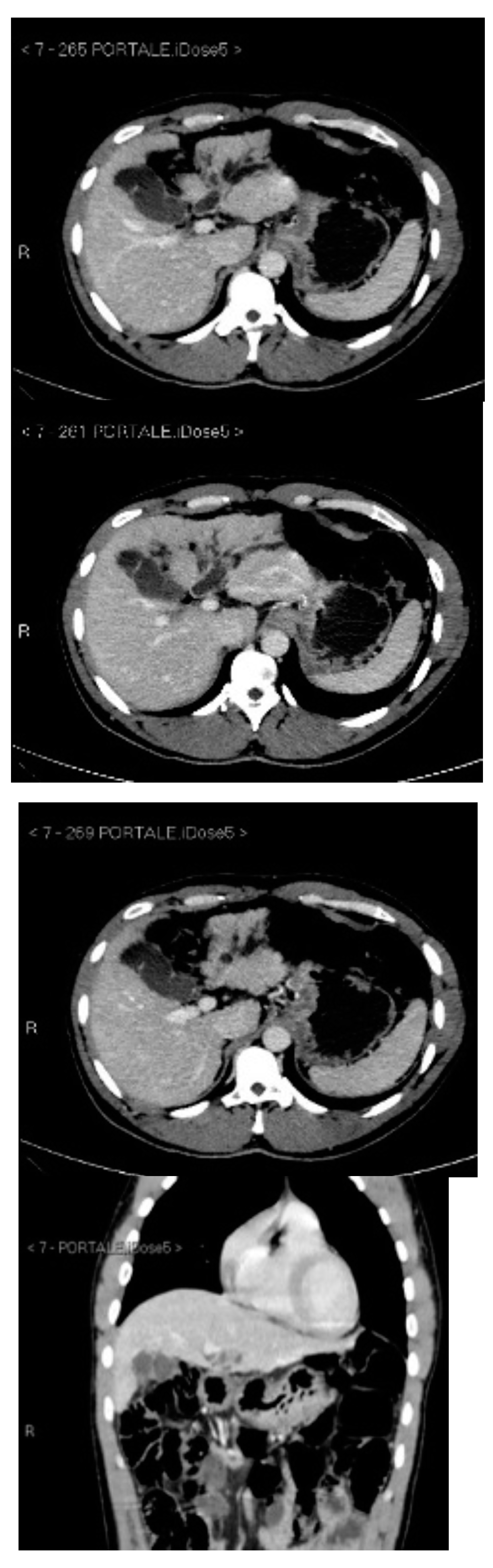
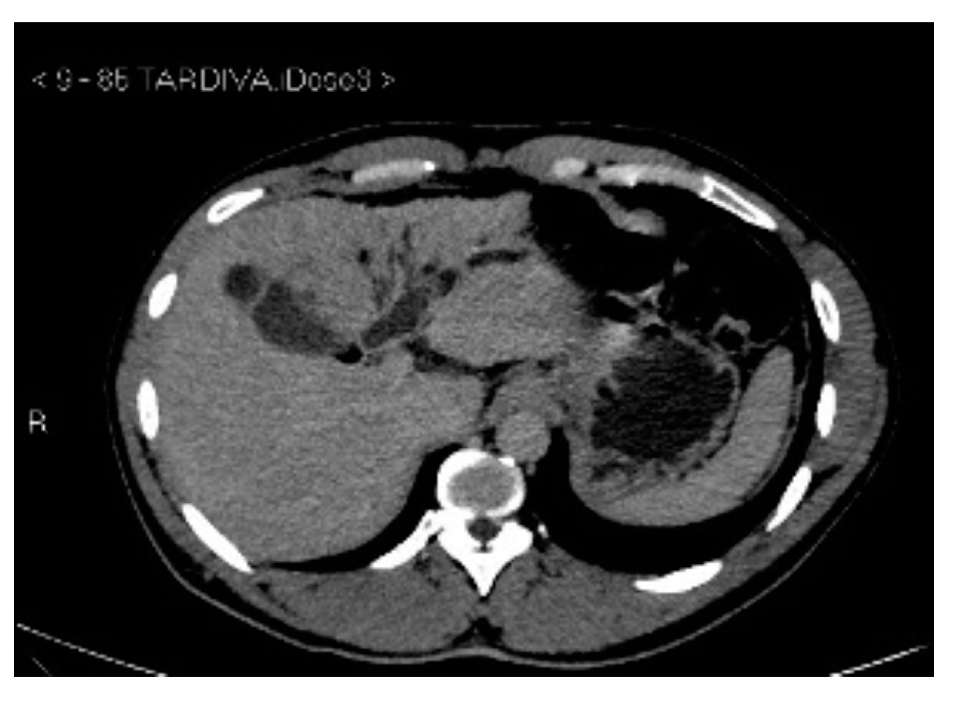
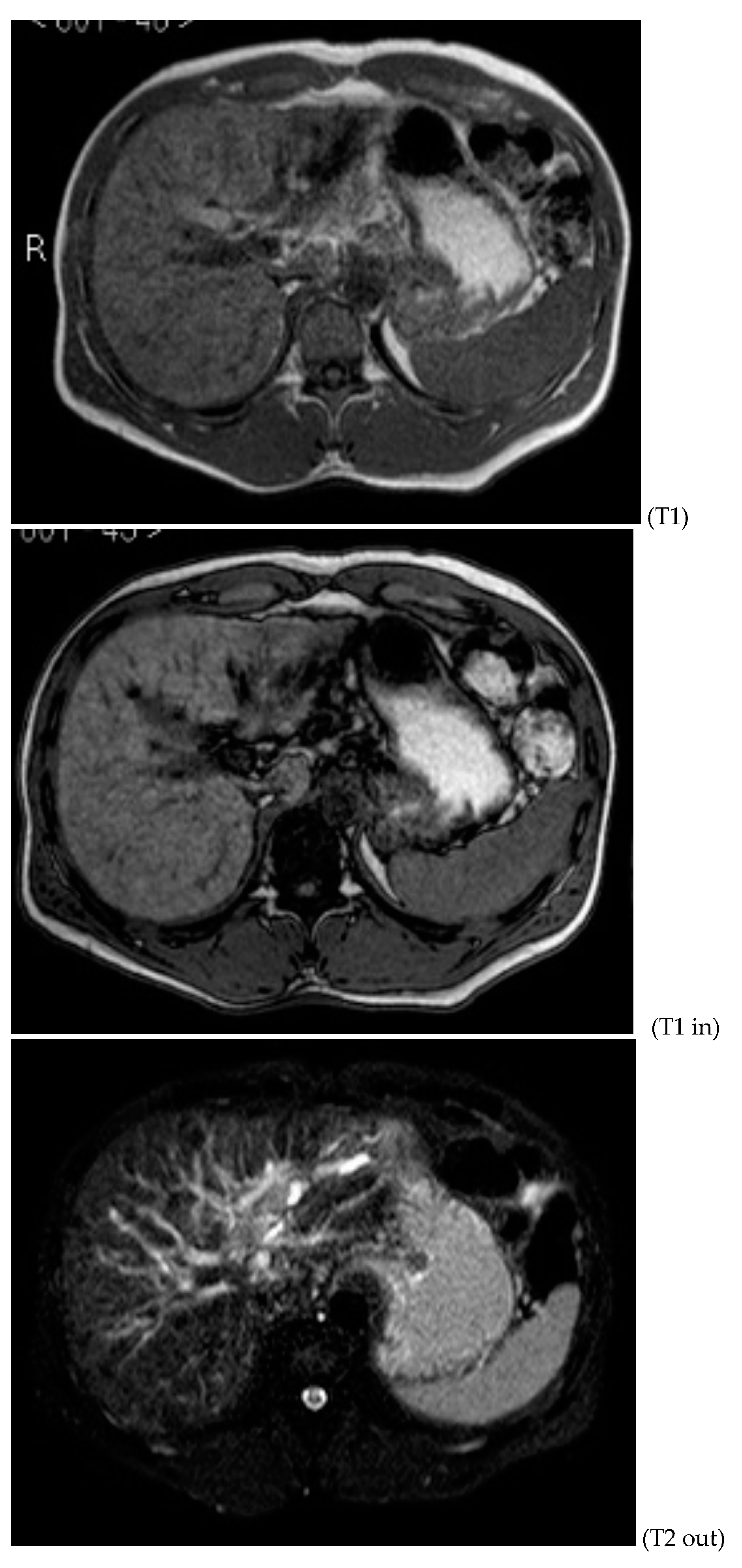
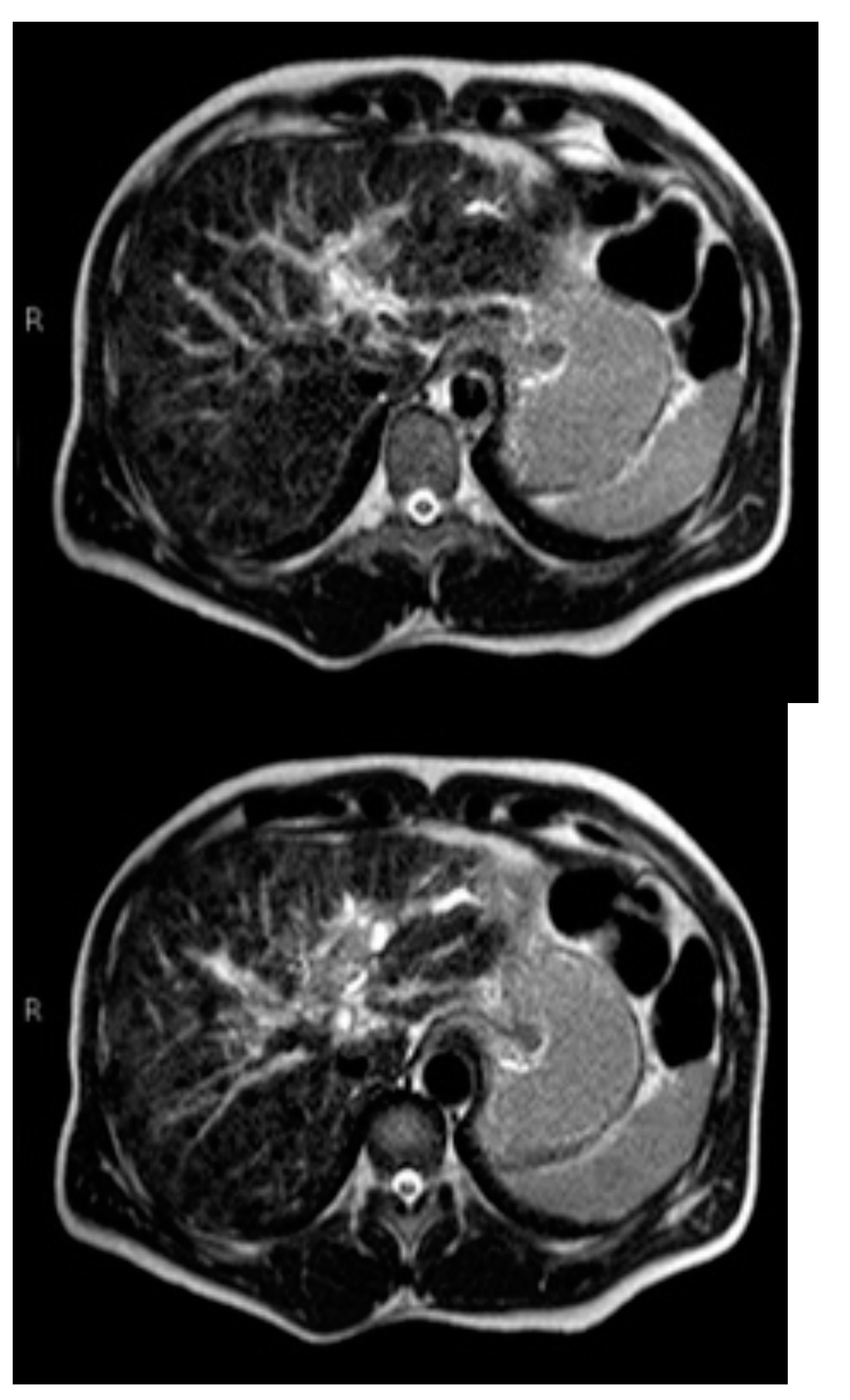
2. Case Report
3. Discussion
4. Conclusions
Author Contributions
Funding
Institutional Review Board Statement
Informed Consent Statement
Data Availability Statement
Conflicts of Interest
References
- Peng, Y.; Chen, K.; Li, B.; Xu, H.; Wei, Y.; Liu, F. Laparoscopic versus open liver resection for resectable HCC with BCLC stage B: A propensity score-matched analysis. Updates Surg. 2022, 74, 1291–1297. [Google Scholar] [CrossRef] [PubMed]
- Ceccarelli, G.; Andolfi, E.; Fontani, A.; Calise, F.; Rocca, A.; Giuliani, A. Robot-assisted liver surgery in a general surgery unit with a “Referral Centre Hub&Spoke Learning Program”. Early outcomes after our first 70 consecutive patients. Minerva Chir. 2018, 73, 460–468. [Google Scholar] [CrossRef] [PubMed]
- Rocca, A.; Cipriani, F.; Belli, G.; Berti, S.; Boggi, U.; Bottino, V.; Cillo, U.; Cescon, M.; Cimino, M.; Corcione, F.; et al. The Italian Consensus on minimally invasive simultaneous resections for synchronous liver metastasis and primary colorectal cancer: A Delphi methodology. Updates Surg. 2021, 73, 1247–1265. [Google Scholar] [CrossRef]
- Rocca, A.; Scacchi, A.; Cappuccio, M.; Avella, P.; Bugiantella, W.; De Rosa, M.; Costa, G.; Polistena, A.; Codacci-Pisanelli, M.; Amato, B.; et al. Robotic surgery for colorectal liver metastases resection: A systematic review. Int. J. Med. Robot. 2021, 17, e2330. [Google Scholar] [CrossRef] [PubMed]
- Rocca, A.; Brunese, M.C.; Santone, A.; Avella, P.; Bianco, P.; Scacchi, A.; Scaglione, M.; Bellifemine, F.; Danzi, R.; Varriano, G.; et al. Early Diagnosis of Liver Metastases from Colorectal Cancer through CT Radiomics and Formal Methods: A Pilot Study. J. Clin. Med. 2021, 11, 31. [Google Scholar] [CrossRef]
- Loffredo, D.; Marvaso, A.; Ceraso, S.; Cinelli, N.; Rocca, A.; Vitale, M.; Rossi, M.; Genovese, E.; Amato, B.; Cinelli, M. Minimal invasive surgery in treatment of liver metastases from colorectal carcinomas: Case studies and survival rates. BMC Surg. 2013, 13 (Suppl. S2), S45. [Google Scholar] [CrossRef] [PubMed] [Green Version]
- Liu, Y.W.; Yong, C.C.; Lin, C.C.; Wang, C.C.; Chen, C.L.; Cheng, Y.F.; Wang, J.H.; Yen, Y.H. Liver resection in elderly patients with hepatocellular carcinoma: Age does matter. Updates Surg. 2021, 73, 1371–1380. [Google Scholar] [CrossRef] [PubMed]
- Wu, J.Y.; Huang, L.M.; Bai, Y.N.; Wei, Y.G.; Zhang, Z.B.; Yan, M.L. Imaging Features of Hepatocellular Carcinoma with Bile Duct Tumor Thrombus: A Multicenter Study. Front. Oncol. 2021, 11, 723455. [Google Scholar] [CrossRef]
- Zhou, D.; Hu, G.F.; Gao, W.C.; Zhang, X.Y.; Guan, W.B.; Wang, J.D.; Ma, F. Hepatocellular carcinoma with tumor thrombus in bile duct: A proposal of new classification according to resectability of primary lesion. World J. Gastroenterol. 2020, 26, 7005–7021. [Google Scholar] [CrossRef]
- Wu, J.Y.; Sun, J.X.; Wu, J.Y.; Huang, X.X.; Bai, Y.N.; Zeng, Y.Y.; Zhang, Z.B.; Cheng, S.Q.; Yan, M.L. A nomogram based on combining systemic and hepatic inflammation markers for predicting microscopic bile duct tumor thrombus in hepatocellular carcinoma. BMC Cancer 2021, 21, 272. [Google Scholar] [CrossRef]
- Rompianesi, G.; Pegoraro, F.; Ceresa, C.D.; Montalti, R.; Troisi, R.I. Artificial intelligence in the diagnosis and management of colorectal cancer liver metastases. World J. Gastroenterol. 2022, 28, 108–122. [Google Scholar] [CrossRef] [PubMed]
- Viganò, L.; Jayakody Arachchige, V.S.; Fiz, F. Is precision medicine for colorectal liver metastases still a utopia? New perspectives by modern biomarkers, radiomics, and artificial intelligence. World J. Gastroenterol. 2022, 28, 608–623. [Google Scholar] [CrossRef] [PubMed]
- Ji, G.W.; Wang, K.; Xia, Y.X.; Li, X.C.; Wang, X.H. Application and challenge of radiomics technique in the era of precision medicine for hepatobiliary disease. Chin. J. Surg. 2020, 58, 749–753. [Google Scholar] [CrossRef] [PubMed]
- Altini, N.; Prencipe, B.; Cascarano, G.D.; Brunetti, A.; Brunetti, G.; Triggiani, V.; Carnimeo, L.; Marino, F.; Guerriero, A.; Villani, L.; et al. Liver, kidney and spleen segmentation from CT scans and MRI with deep learning: A survey. Neurocomputing 2022, 490, 30–53. [Google Scholar] [CrossRef]
- Amato, B.; Compagna, R.; Rocca, A.; Bianco, T.; Milone, M.; Sivero, L.; Vigliotti, G.; Amato, M.; Danzi, M.; Aprea, G.; et al. Fondaparinux vs warfarin for the treatment of unsuspected pulmonary embolism in cancer patients. Drug Des. Devel. Ther. 2016, 10, 2041–2046. [Google Scholar] [CrossRef] [Green Version]
- Komici, K.; Cappuccio, M.; Scacchi, A.; Vaschetti, R.; Delli Carpini, G.; Picerno, V.; Avella, P.; Brunese, M.C.; Rengo, G.; Guerra, G.; et al. The Prevalence and the Impact of Frailty in Hepato-Biliary Pancreatic Cancers: A Systematic Review and Meta-Analysis. J. Clin. Med. 2022, 11, 1116. [Google Scholar] [CrossRef] [PubMed]
- Zhou, X.; Wang, J.; Tang, M.; Huang, M.; Xu, L.; Peng, Z.; Li, Z.P.; Feng, S.T. Hepatocellular carcinoma with hilar bile duct tumor thrombus versus hilar Cholangiocarcinoma on enhanced computed tomography: A diagnostic challenge. BMC Cancer 2020, 20, 54. [Google Scholar] [CrossRef] [Green Version]
- Rocca, A.; Brunese, M.C.; Cappuccio, M.; Scacchi, A.; Martucci, G.; Buondonno, A.; Perrotta, F.M.; Quarto, G.; Avella, P.; Amato, B. Impact of Physical Activity on Disability Risk in Elderly Patients Hospitalized for Mild Acute Diverticulitis and Diverticular Bleeding Undergone Conservative Management. Medicina 2021, 57, 360. [Google Scholar] [CrossRef]
- Kim, A.Y.; Jeong, W.K. Intraductal malignant tumors in the liver mimicking cholangiocarcinoma: Imaging features for differential diagnosis. Clin. Mol. Hepatol. 2016, 22, 192–197. [Google Scholar] [CrossRef]
- Banales, J.M.; Marin, J.J.; Lamarca, A.; Rodrigues, P.M.; Khan, S.A.; Roberts, L.R.; Cardinale, V.; Carpino, G.; Andersen, J.B.; Braconi, C.; et al. Cholangiocarcinoma 2020: The next horizon in mechanisms and management. Nat. Rev. Gastroenterol. Hepatol. 2020, 17, 557–588. [Google Scholar] [CrossRef]
- Kim, H.S.; Han, Y.; Kang, J.S.; Kang, Y.H.; Lee, M.; Sohn, H.J.; Kim, H.; Kwon, W.; Jang, J.Y. Serum carcinoembryonic antigen and carbohydrate antigen 19-9 as preoperative diagnostic biomarkers of extrahepatic bile duct cancer. BJS Open 2021, 5, zrab127. [Google Scholar] [CrossRef] [PubMed]
- Bracale, U.; Podda, M.; Castiglioni, S.; Peltrini, R.; Sartori, A.; Arezzo, A.; Corcione, F.; Agresta, F. Changes in surgicaL behaviOrs dUring the COVID-19 pandemic. The SICE CLOUD19 Study. Updates Surg. 2021, 73, 731–744. [Google Scholar] [CrossRef] [PubMed]
- Gallo, G.; Picciariello, A.; Di Tanna, G.L.; Santoro, G.A.; Perinotti, R.; Grossi, U. E-consensus on telemedicine in colorectal surgery: A RAND/UCLA-modified study. Updates Surg. 2022, 74, 163–170. [Google Scholar] [CrossRef] [PubMed]
- Aldrighetti, L.; Boggi, U.; Falconi, M.; Giuliante, F.; Cipriani, F.; Ratti, F.; Torzilli, G. Perspectives from Italy during the COVID-19 pandemic: Nationwide survey-based focus on minimally invasive HPB surgery. Updates Surg. 2020, 72, 241–247. [Google Scholar] [CrossRef] [PubMed]
- Cavaliere, D.; Parini, D.; Marano, L.; Cipriani, F.; Di Marzo, F.; Macrì, A.; D’Ugo, D.; Roviello, F.; Gronchi, A. Surgical management of oncologic patient during and after the COVID-19 outbreak: Practical recommendations from the Italian society of Surgical Oncology. Updates Surg. 2021, 73, 321–329. [Google Scholar] [CrossRef] [PubMed]
- Giuliani, A.; Avella, P.; Segreto, A.L.; Izzo, M.L.; Buondonno, A.; Coluzzi, M.; Cappuccio, M.; Brunese, M.C.; Vaschetti, R.; Scacchi, A.; et al. Postoperative Outcomes Analysis After Pancreatic Duct Occlusion: A Safe Option to Treat the Pancreatic Stump after Pancreaticoduodenectomy in Low-Volume Centers. Front. Surg. 2021, 8, 804675. [Google Scholar] [CrossRef]
- Luciani, C.; Scacchi, A.; Vaschetti, R.; Di Marzo, G.; Fatica, I.; Cappuccio, M.; Guerra, G.; Ceccarelli, G.; Avella, P.; Rocca, A. The uniportal VATS in the treatment of stage II pleural empyema: A safe and effective approach for adults and elderly patients-a single-center experience and literature review. World J. Emerg. Surg. 2022, 17, 46. [Google Scholar] [CrossRef]
- Ceccarelli, G.; Rocca, A.; De Rosa, M.; Fontani, A.; Ermili, F.; Andolfi, E.; Bugiantella, W.; Levi Sandri, G.B. Minimally invasive robotic-assisted combined colorectal and liver excision surgery: Feasibility, safety and surgical technique in a pilot series. Updates Surg. 2021, 73, 1015–1022. [Google Scholar] [CrossRef]
- Marte, G.; Scuderi, V.; Rocca, A.; Surfaro, G.; Migliaccio, C.; Ceriello, A. Laparoscopic splenectomy: A single center experience. Unusual cases and expanded inclusion criteria for laparoscopic approach. Updates Surg. 2013, 65, 115–119. [Google Scholar] [CrossRef]
- Buondonno, A.; Avella, P.; Cappuccio, M.; Scacchi, A.; Vaschetti, R.; Di Marzo, G.; Maida, P.; Luciani, C.; Amato, B.; Brunese, M.C.; et al. A Hub and Spoke Learning Program in Bariatric Surgery in a Small Region of Italy. Front. Surg. 2022, 9, 855527. [Google Scholar] [CrossRef] [PubMed]
- Granata, V.; Fusco, R.; Bicchierai, G.; Cozzi, D.; Grazzini, G.; Danti, G.; De Muzio, F.; Maggialetti, N.; Smorchkova, O.; D’Elia, M.; et al. Diagnostic protocols in oncology: Workup and treatment planning. Part 1: The optimitation of CT protocol. Eur. Rev. Med. Pharmacol. Sci. 2021, 25, 6972–6994. [Google Scholar] [CrossRef] [PubMed]
- Granata, V.; Bicchierai, G.; Fusco, R.; Cozzi, D.; Grazzini, G.; Danti, G.; De Muzio, F.; Maggialetti, N.; Smorchkova, O.; D’Elia, M. Diagnostic protocols in oncology: Workup and treatment planning. Part 2: Abbreviated MR protocol. Eur. Rev. Med. Pharmacol. Sci. 2021, 25, 6499–6528. [Google Scholar] [CrossRef] [PubMed]
- Bevilacqua, V.; Brunetti, A.; Trotta, G.F.; Dimauro, G.; Elez, K.; Alberotanza, V.; Scardapane, A. A novel approach for Hepatocellular Carcinoma detection and classification based on triphasic CT Protocol. In Proceedings of the 2017 IEEE Congress on Evolutionary Computation (CEC), Donostia, Spain, 5–8 June 2017; IEEE: Manhattan, NY, USA, 2017. [Google Scholar]
- Wong, T.C.; Cheung, T.T.; Chok, K.S.; Chan, A.C.; Dai, W.C.; Chan, S.C.; Poon, R.T.; Fan, S.T.; Lo, C.M. Outcomes of hepatectomy for hepatocellular carcinoma with bile duct tumour thrombus. HPB 2015, 17, 401–408. [Google Scholar] [CrossRef] [PubMed] [Green Version]
- Ha, T.Y.; Hwang, S.; Moon, D.B.; Ahn, C.S.; Kim, K.H.; Song, G.W.; Jung, D.H.; Park, G.C.; Park, H.W.; Park, Y.H.; et al. Long-term survival analysis of liver transplantation for hepatocellular carcinoma with bile duct tumor thrombus. Transplant. Proc. 2014, 46, 774–777. [Google Scholar] [CrossRef] [PubMed]
- Kim, D.S.; Kim, B.W.; Hatano, E.; Hwang, S.; Hasegawa, K.; Kudo, A.; Ariizumi, S.; Kaibori, M.; Fukumoto, T.; Baba, H.; et al. Surgical Outcomes of Hepatocellular Carcinoma with Bile Duct Tumor Thrombus: A Korea-Japan Multicenter Study. Ann. Surg. 2020, 271, 913–921. [Google Scholar] [CrossRef]
- Satoh, S.; Ikai, I.; Honda, G.; Okabe, H.; Takeyama, O.; Yamamoto, Y.; Yamamoto, N.; Iimuro, Y.; Shimahara, Y.; Yamaoka, Y. Clinicopathologic evaluation of hepatocellular carcinoma with bile duct thrombi. Surgery 2000, 128, 779–783. [Google Scholar] [CrossRef] [PubMed]
- Shiomi, M.; Kamiya, J.; Nagino, M.; Uesaka, K.; Sano, T.; Hayakawa, N.; Kanai, M.; Yamamoto, H.; Nimura, Y. Hepatocellular carcinoma with biliary tumor thrombi: Aggressive operative approach after appropriate preoperative management. Surgery 2001, 129, 692–698. [Google Scholar] [CrossRef]
- Peng, S.Y.; Wang, J.W.; Liu, Y.B.; Cai, X.J.; Deng, G.L.; Xu, B.; Li, H.J. Surgical intervention for obstructive jaundice due to biliary tumor thrombus in hepatocellular carcinoma. World J. Surg. 2004, 28, 43–46. [Google Scholar] [CrossRef]
- Esaki, M.; Shimada, K.; Sano, T.; Sakamoto, Y.; Kosuge, T.; Ojima, H. Surgical results for hepatocellular carcinoma with bile duct invasion: A clinicopathologic comparison between macroscopic and microscopic tumor thrombus. J. Surg. Oncol. 2005, 90, 226–232. [Google Scholar] [CrossRef]
- Shao, W.; Sui, C.; Liu, Z.; Yang, J.; Zhou, Y. Surgical outcome of hepatocellular carcinoma patients with biliary tumor thrombi. World J. Surg. Oncol. 2011, 9, 2. [Google Scholar] [CrossRef] [PubMed]
- Yu, X.H.; Xu, L.B.; Liu, C.; Zhang, R.; Wang, J. Clinicopathological characteristics of 20 cases of hepatocellular carcinoma with bile duct tumor thrombi. Dig. Dis. Sci. 2011, 56, 252–259. [Google Scholar] [CrossRef] [PubMed]
- Noda, T.; Nagano, H.; Tomimaru, Y.; Murakami, M.; Wada, H.; Kobayashi, S.; Marubashi, S.; Eguchi, H.; Takeda, Y.; Tanemura, M.; et al. Prognosis of hepatocellular carcinoma with biliary tumor thrombi after liver surgery. Surgery 2011, 149, 371–377. [Google Scholar] [CrossRef] [PubMed]
- Moon, D.B.; Hwang, S.; Wang, H.J.; Yun, S.S.; Kim, K.S.; Lee, Y.J.; Kim, K.H.; Park, Y.K.; Xu, W.; Kim, B.W.; et al. Surgical outcomes of hepatocellular carcinoma with bile duct tumor thrombus: A Korean multicenter study. World J. Surg. 2013, 37, 443–451. [Google Scholar] [CrossRef] [PubMed]
- Oba, A.; Takahashi, S.; Kato, Y.; Gotohda, N.; Kinoshita, T.; Shibasaki, H.; Ikeda, M.; Konishi, M. Usefulness of resection for hepatocellular carcinoma with macroscopic bile duct tumor thrombus. Anticancer Res. 2014, 34, 4367–4372. [Google Scholar]
- Rammohan, A.; Sathyanesan, J.; Rajendran, K.; Pitchaimuthu, A.; Perumal, S.K.; Balaraman, K.; Ramasamy, R.; Palaniappan, R.; Govindan, M. Bile duct thrombi in hepatocellular carcinoma: Is aggressive surgery worthwhile? HPB 2015, 17, 508–513. [Google Scholar] [CrossRef] [Green Version]
- Kasai, Y.; Hatano, E.; Seo, S.; Taura, K.; Yasuchika, K.; Uemoto, S. Hepatocellular carcinoma with bile duct tumor thrombus: Surgical outcomes and the prognostic impact of concomitant major vascular invasion. World J. Surg. 2015, 39, 1485–1493. [Google Scholar] [CrossRef]
- Chotirosniramit, A.; Liwattanakun, A.; Lapisatepun, W.; Ko-Iam, W.; Sandhu, T.; Junrungsee, S. A single institution report of 19 hepatocellular carcinoma patients with bile duct tumor thrombus. J. Hepatocell. Carcinoma 2017, 4, 41–47. [Google Scholar] [CrossRef] [Green Version]
- Lin, Z.; Han, M.; Zhou, Z. Prognosis for patients with hepatocellular carcinoma (HCC) with bile duct tumor thrombus (BDTT) after surgical treatment. Biosci. Trends 2019, 13, 77–85. [Google Scholar] [CrossRef] [Green Version]
- Sun, J.; Wu, J.; Shi, J.; Liu, C.; Wei, Y.; Zhou, J.; Zhang, Z.; Yan, M.; Cheng, S. Thrombus-First Surgery for Hepatocellular Carcinoma with Bile Duct Tumor Thrombus. J. Gastrointest. Surg. 2021, 25, 1973–1979. [Google Scholar] [CrossRef]
- Navadgi, S.; Chang, C.C.; Bartlett, A.; McCall, J.; Pandanaboyana, S. Systematic review and meta-analysis of outcomes after liver resection in patients with hepatocellular carcinoma (HCC) with and without bile duct thrombus. HPB 2016, 18, 312–316. [Google Scholar] [CrossRef] [Green Version]
- Reig, M.; Forner, A.; Rimola, J.; Ferrer-Fàbrega, J.; Burrel, M.; Garcia-Criado, Á.; Kelley, R.K.; Galle, P.R.; Mazzaferro, V.; Salem, R.; et al. BCLC strategy for prognosis prediction and treatment recommendation: The 2022 update. J. Hepatol. 2022, 76, 681–693. [Google Scholar] [CrossRef]
- Chun, Y.S.; Pawlik, T.M.; Vauthey, J.N. 8th Edition of the AJCC Cancer Staging Manual: Pancreas and Hepatobiliary Cancers. Ann. Surg. Oncol. 2018, 4, 845–847. [Google Scholar] [CrossRef]
- Lu, W.P.; Tang, H.W.; Yang, Z.Y.; Jiang, K.; Chen, Y.L.; Lu, S.C. A proposed modification for the Barcelona Clinic Liver Cancer staging system: Adding bile duct tumor thrombus status in patients with hepatocellular carcinoma. Am. J. Surg. 2020, 220, 965–971. [Google Scholar] [CrossRef]
- Huang, Q.; Chen, Y.; Lin, K.; Sun, C.; Zheng, S.; Chen, J.; Wang, Y.; Zhou, Y.; Zhou, W.; Liu, J.; et al. Redefining Hepatocellular Carcinoma Staging Systems Based on the Bile Duct Invasion Status: A Multicenter Study. Front. Oncol. 2021, 11, 673285. [Google Scholar] [CrossRef]
- Associazione Italiana di Oncologia Medica (AIOM). Linee Guida “Tumori delle Vie Biliari”. 2019. Available online: https://www.aiom.it/wp-content/uploads/2019/10/2019_LG_AIOM_Vie_biliari.pdf (accessed on 2 November 2022).
- Machairas, N.; Kostakis, I.D.; Schizas, D.; Kykalos, S.; Nikiteas, N.; Sotiropoulos, G.C. Meta-analysis of laparoscopic versus open liver resection for intrahepatic cholangiocarcinoma. Updates Surg. 2021, 73, 59–68. [Google Scholar] [CrossRef]
- Gruttadauria, S.; Barbara, M.; Liotta, R. Liver transplantation for unresectable intrahepatic cholangiocarcinoma: An Italian experience. Updates Surg. 2021, 4, 1587–1588. [Google Scholar] [CrossRef]
- Peng, B.G.; Liang, L.J.; Li, S.Q.; Zhou, F.; Hua, Y.P.; Luo, S.M. Surgical treatment of hepatocellular carcinoma with bile duct tumor thrombi. World J. Gastroenterol. 2005, 11, 3966–3969. [Google Scholar] [CrossRef]


| Author | Year | Study Type | N. of Cases | Age, Years | Sex, M/F | Symptoms | Jaundice | HBV Positive | Diagnosis |
|---|---|---|---|---|---|---|---|---|---|
| Satoh et al. [31] | 2000 | Retrospective cohort study | 17 | 58.18 ± 8.94 | 15 (88.24)/2 (11.76) | Jaundice | 9 (52.94) | 5 (29.4) | Ultrasonography; CT |
| Shiomi et al. [38] | 2001 | Retrospective cohort study | 17 | 58.8 ± 2 | 15 (88.24)/2 (11.76) | Jaundice, abdominal pain, poor appetite, general fatigue, or fever | 10 (58.82) | 7 of 14 (50) | Ultrasonography; CT |
| Peng et al. [39] | 2004 | Retrospective cohort study | 8 | 51.75 ± 8.15 | 7 (87.5)/1 (12.5) | Jaundice | 8 (100) | 6 (75) | Ultrasonography; CT; MRI |
| Esaki et al. [40] | 2005 | Retrospective cohort study | 19 | 59.79 ± 11.26 | 19 (100)/0 (0) | Jaundice, fever, or abdominal pain | NA | 8 (42.10) | Ultrasonography; CT; MRI; angiography |
| Shao et al. [41] | 2011 | Retrospective cohort study | 27 | 47.1 ± 10.5 | 23 (85.18)/4 (14.81) | NA | NA | 8 (42.10) | Chest XR; abdominal ultrasonography; CT; CPRE |
| Yu et al. [42] | 2011 | Retrospective cohort study | 20 | 50.6 ± 2.4 | 17 (85)/3 (15) | Obstructive jaundice and upper abdominal pain | 14 (70) | 16 (80) | NA |
| Noda et al. [43] | 2011 | Retrospective cohort study | 22 | 45% were ≤60 y; 55% were >60 y | 21 (95)/1 (5) | NA | 8 (36.36) | 15 (68.18) | Ultrasonography; CT; angiography; ERCP or MR cholangiopancreatography |
| Moon et al. [44] | 2012 | Retrospective cohort study | 73 | 54.2 ± 11.1 | 52 (71.23)/21 (28.77) | Jaundice | 34 (46.60) | 59 (80.82) | CT; MRI |
| Oba et al. [45] | 2014 | Retrospective cohort study | 13 | 60.85 ± 8.64 | 12 (92.31)/1 (7.69) | NA | NA | 4 (30.77) | Ultrasonography; CT; MRI |
| Wong et al. [34] | 2014 | Retrospective cohort study | 37 | 56.75 ± 14.75 | 29 (78.38)/8 (21.62) | NA | NA | 30 (81.1) | CT; MRI |
| Rammohan et al. [46] | 2014 | Retrospective cohort study | 39 | 52.1 ± 10.9 | 28 (71.80)/11 (28.20) | Jaundice | 18 (46.10) | 7 (17.9) | Abdominal ultrasonography; abdominal CT |
| Ha et al. [35] | 2014 | Retrospective cohort study | 14 | 54.6 ± 5.6 | 10 (71.43)/4 (28.57) | Jaundice | 9 (64.29) | 11 (78.57) | NA |
| Kasai et al. [47] | 2015 | Retrospective cohort study | 44 | 64 ± 9.1 | 35 (79.5) /9 (20.5) | Jaundice | 27 (61.36) | 8 (18.2) | NA |
| Chotirosniramit et al. [48] | 2017 | Retrospective cohort study | 19 | 51.1 ± 11.5 | 15 (78.95)/4 (21.05) | Jaundice or cholangitis | 14 (73.68) | 16 (84.2) | Abdominal CT |
| Kim et al. [36] | 2018 | Retrospective cohort study | 257 | 61 ± 11.6 | 210 (81.71)/47 (18.29) | Jaundice | 120 (46.70) | 115 (44.75) | NA |
| Lin et al. [49] | 2019 | Retrospective cohort study | 49 | 55.51 ± 13.09 | 43 (87.75)/6 (12.25) | NA | NA | NA | Abdominal ultrasonography; abdominal CT; hepatic angiography; MR cholangiopancreatography |
| Zhou et al. [9] | 2020 | Retrospective cohort study | 7 | 66 ± 6.24 | 6 (85.71)/1 (14.28) | Jaundice | 7 (100) | 4 (57.14) | CT; MRI; |
| Zhou et al. [17] | 2020 | Retrospective cohort study | 58 | 49.84 ± 10.23 | 51 (87.93)/7 (2.07) | Jaundice and upper abdominal pain | 39 (67.24) | 42 (72.41) | CT |
| Sun et al. [50] | 2020 | Retrospective multicenter study | 120 | 50.55 ± 11.35 | 106 (88.33)/14 (11.67) | NA | NA | 95 (79.17) | NA |
| Wu et al. [8] | 2021 | Multicenter study | 30 | 48.5 ± 13.04 | 23 (76.67)/7 (23.33) | NA | NA | 29 (96.67) | CT; MRI |
| Conticchio et al. | 2022 | Case report | 1 | 47 | 1 (100)/0 (0) | Abdominal pain | 0 (0) | 1 (100) | Ultrasonography; CT; MRI |
| Author | AFP (>400 ng/mL), n. | AFP, Mean ± SD | Total Bilirubin, Mean ± SD | Tumor Size, cm | Surgical Procedure | Mortality, n | Overall Survival, % | ||
|---|---|---|---|---|---|---|---|---|---|
| 1-Year | 3-Year | 5-Year | |||||||
| Satoh et al. [31] | NA | NA | NA | NA | NA | 12 (70.59) | NA | NA | NA |
| Shiomi et al. [38] | NA | 73.87 ± 72.89 | 6.9 ± 1.8 mg/dL | 6.1 ± 1.2 | 1 right hepatic trisegmentectomy with caudate lobectomy; 5 right hepatic lobectomies with caudate lobectomy; 6 left hepatic lobectomies with caudate lobectomy; 1 right anterior segmentectomy; 1 right anterior segmentectomy with caudate lobectomy; 1 segmentectomy S5; 1 S4; 1 S1 | 11 (64.70) | NA | 47 | 28 |
| Peng et al. [39] | 7 (87.5) | NA | Ra 68.4–436.4 umol/L | Ra 2–9 | 3 hepatectomies with removal of the tumor thrombus; 1 hepatectomy combined with extrahepatic bile duct resection; 3 thrombectomies through a choledochotomy; 1 orthotopic liver transplantation | 7 (87.5) | 62.5 | 37.5 | NA |
| Esaki et al. [40] | NA | NA | 5 ± 7.3 mg/dL | NA | 7 left hepatectomies; 1 lateral segmentectomy; 6 right hepatectomies; 1 central bisegmentectomy; 3 medial segmentectomies; 1 anterior segmentectomy | NA | 79 | 45 | 33 |
| Shao et al. [41] | 16 (59.3) | NA | 116.4 ± 135.4 umol/L | NA | 1 right anterior resection; 2 right posterior resections; 4 right hepatectomies; 8 left hepatectomies; 1 left hepatectomy with caudate lobectomy; 3 left lateral resections; 2 left medial resections; 6 partial resections | 1 (3.70) | NA | NA | NA |
| Yu et al. [42] | 9 (45) | 2651.85 ± 6135.32 | 123.25 ± 142.06 mol/L | 3.65 ± 2.4 | 5 hepatectomies with thrombectomy; 7 hepatectomies with thrombectomy and T-tube drainage; 6 hepatectomies with resection of the common bile duct and hepaticojejunostomy; 2 liver transplantations | 6 (30) | 73.1 | 20.6 | NA |
| Noda et al. [43] | 12 (55) | NA | NA | 59% were ≤5 cm; 41% were >5 cm | 16 lobectomies; 6 surgically noncurative procedures | 0 (0) | 62 | 30 | 30 |
| Moon et al. [44] | NA | 25,280.10 ± 109,395.40 | 5.7 ± 5.9 mg/dL | 5.8 ± 3.7 | 25 right hemihepatectomies ± caudate lobectomy; 4 right trisectionectomies ± caudate lobectomy; 29 left hemihepatectomies ± caudate lobectomy; 1 posterior sectionectomies; 2 anterior sectionectomies; 4 lateral sectionectomies; 2 central bisectionectomies; 1 S5-S6 bisegmentectomy; 1 isolated caudate lobectomy; 4 nonsystematic hepatectomies; 2 partial hepatectomies; 1 partial caudate lobectomy; 1 S8 subsegmentectomy | 3 (4.11) | 76.5 | 41.4 | 32 |
| Oba et al. [45] | NA | 2193.25 ± 2815.06 | NA | 6.37 ± 4.01 | 4 left hepatectomies; 4 right hepatectomies; 1 right hepatectomy and segment 2/3 limited resection; 1 central bisegmentectomy; 1 right trisegmentectomy; 1 anterior segmentectomy; 1 posterior segmentectomy; 6 bile duct resections and bilioenteric anastomosis | 7 (53.85) | 92 | 77 | 48 |
| Wong et al. [34] | NA | 50 (Ra 2–63,320) | 29.05 ± 15 umol/L | 9.5 ± 6 | 34 major hepatectomies; 3 left lateral sectionectomies | 1 (2.7) | 69.4 | 54.3 | 38.5 |
| Rammohan et al. [46] | 28 (71.7) | NA | 6.1 ± 5.1 mg/dL | 5.6 ± 3.2 | 16 right hepatectomies with thrombectomy; 10 extended right hepatectomies with extrahepatic bile duct excision; 9 left hepatectomies; 2 extended left hepatectomies; 2 left lateral segmentectomies | 2 (5.1) | 82 | 48 | 10 |
| Ha et al. [35] | 14 (100) | 2043.1 ± 5528.6 | 5.1 ± 5.2 mg/dL | 3.9 ± 1.9 | 13 living-donor transplantations; 1 deceased-donor transplantation | 1 (7.14) | 92.9 | 57.1 | 50 |
| Kasai et al. [47] | NA | 5.31 ± 13.02 | 1.2 ± 0.8 mg/dL | 5.8 ± 3.5 | 41 bisectionectomies; 3 monosectionectomies; 7 combined BDRs | 2 (4.54) | NA | NA | 31 |
| Chotirosniramit et al. [48] | NA | 12,673.82 ± 12,499.87 | 11.3 ± 6.45 mg/dL | 8.2 ± 4.2 | 2 right trisectionectomies + bile duct resection + caudate resection; 1 left trisectionectomy + bile duct resection + caudate resection; 1 right hepatectomy + bile duct resection; 4 left hepatectomies + bile duct resection; 3 right hepatectomies; 4 left hepatectomies + CBD exploration to remove BDTT; 1 left hepatectomy; 2 CBD explorations to remove BDTT and palliative biliary drainage; 1 no operation; | 0 (0) | NA | 60 | NA |
| Kim et al. [36] | NA | 754.25 ± 451.5 | 2.85 ± 1.03 | NA | 121 right hemihepatectomies; 7 right trisectionectomy; 81 left hemihepatectomies; 2 left trisectionectomies; 5 posterior sectionectomies; 12 anterior sectionectomies; 8 left lateral sectionectomies; 3 left medial sectionectomies; 6 central bisectionectomies; 10 nonsystematic resections; 2 liver transplantations | NA | 74.5 | 52.9 | 43.6 |
| Lin et al. [49] | NA | NA | NA | NA | 25 radical resections; 7 thrombectomies through a choledochotomy; 17 palliative internal and external bile duct drainages | NA | 42.86 | 18.37 | 12.24 |
| Zhou et al. [9] | 2 (28.57) | 1497.03 ± 3503.20 | 91.36 ± 80.92 umol/L | NA | NA | NA | NA | NA | NA |
| Zhou et al. [17] | 39 (67.24) | NA | NA | 4.60 ± 1.02 | 36 simple hepatectomies; 11 hepatectomies plus bile duct excision | NA | NA | NA | NA |
| Sun et al. [50] | 46 (38.33) | NA | 149.4 ± 129.75 | 5.05 ± 2.75 | 19 right hepatectomies; 35 left hepatectomies; 6 left lateral sectionectomies; 13 right sectionectomies; 47 non-anatomic resections; | 3 (2.5) | NA | NA | NA |
| Wu et al. [8] | 27 (90) | NA | 15.9 ± 4.15 umol/L | 7.4 ± 3.05 | NA | NA | NA | NA | NA |
| Conticchio et al. | 1 (100) | 2157 | 0.9 mg/dL | 1.8 | Segmentectomy S3 | 0 (0) | NA | NA | NA |
Disclaimer/Publisher’s Note: The statements, opinions and data contained in all publications are solely those of the individual author(s) and contributor(s) and not of MDPI and/or the editor(s). MDPI and/or the editor(s) disclaim responsibility for any injury to people or property resulting from any ideas, methods, instructions or products referred to in the content. |
© 2023 by the authors. Licensee MDPI, Basel, Switzerland. This article is an open access article distributed under the terms and conditions of the Creative Commons Attribution (CC BY) license (https://creativecommons.org/licenses/by/4.0/).
Share and Cite
Conticchio, M.; Maggialetti, N.; Rescigno, M.; Brunese, M.C.; Vaschetti, R.; Inchingolo, R.; Calbi, R.; Ferraro, V.; Tedeschi, M.; Fantozzi, M.R.; et al. Hepatocellular Carcinoma with Bile Duct Tumor Thrombus: A Case Report and Literature Review of 890 Patients Affected by Uncommon Primary Liver Tumor Presentation. J. Clin. Med. 2023, 12, 423. https://doi.org/10.3390/jcm12020423
Conticchio M, Maggialetti N, Rescigno M, Brunese MC, Vaschetti R, Inchingolo R, Calbi R, Ferraro V, Tedeschi M, Fantozzi MR, et al. Hepatocellular Carcinoma with Bile Duct Tumor Thrombus: A Case Report and Literature Review of 890 Patients Affected by Uncommon Primary Liver Tumor Presentation. Journal of Clinical Medicine. 2023; 12(2):423. https://doi.org/10.3390/jcm12020423
Chicago/Turabian StyleConticchio, Maria, Nicola Maggialetti, Marco Rescigno, Maria Chiara Brunese, Roberto Vaschetti, Riccardo Inchingolo, Roberto Calbi, Valentina Ferraro, Michele Tedeschi, Maria Rita Fantozzi, and et al. 2023. "Hepatocellular Carcinoma with Bile Duct Tumor Thrombus: A Case Report and Literature Review of 890 Patients Affected by Uncommon Primary Liver Tumor Presentation" Journal of Clinical Medicine 12, no. 2: 423. https://doi.org/10.3390/jcm12020423