
Pathogenesis, Diagnosis and Management of Obstetric Antiphospholipid Syndrome: A Comprehensive Review

 , ,
, ,
Abstract
:1. Introduction
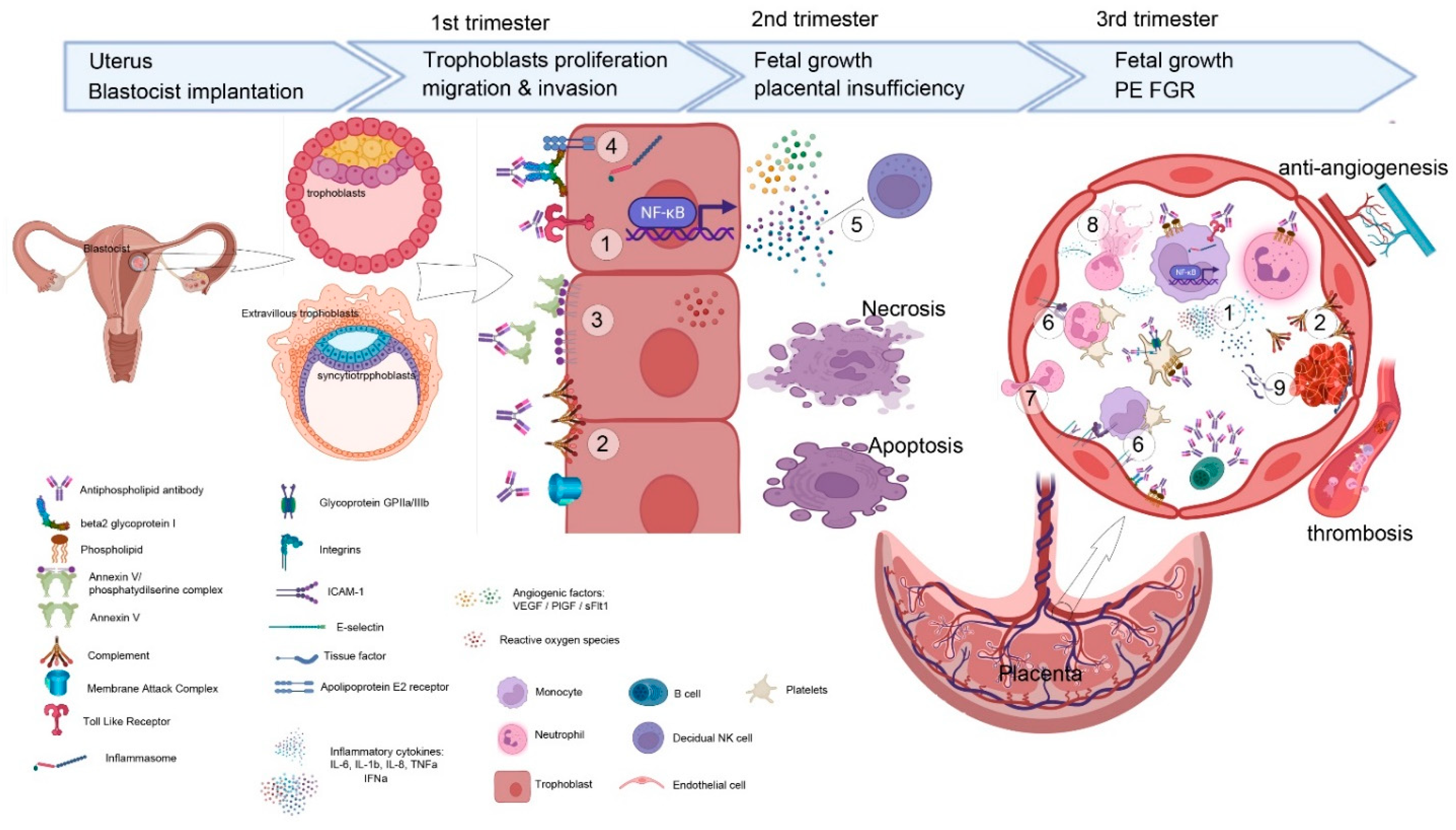
2. Pathophysiology of OAPS
2.1. aPL Affects Implantation and Trophoblasts Growth
2.2. aPL Activates Complement in the Pathophysiology of OAPS
2.3. Thrombosis versus Complementopathy
2.4. Different aPL with Different Mechanisms of Action
3. OAPS: Clinical Manifestations and Classification Criteria
3.1. Clinical Phenotypes of OAPS
3.1.1. Recurrent Miscarriage and Fetal Loss
3.1.2. Stillbirths
3.1.3. Placental Insufficiency: Prematurity or Stillbirth Related to Early PE and/or Fetal Growth Retardation
4. OMAPS and NC-OAPS: Diagnostic Concerns
4.1. NC-OAPS
4.2. OMAPS
5. Catastrophic Antiphospholipid Syndrome
6. Risk Profiles and Risk Scores in OAPS Patients
7. Clinical Management
7.1. Preconception Counselling
7.2. Complementary Tests during Pregnancy
7.3. General Therapeutic Measures
7.4. Gold Standard Treatment
7.5. Refractory Cases
7.5.1. Hydroxychloroquine
7.5.2. Corticosteroids
7.5.3. Intravenous Immunoglobulins
7.5.4. Biologic Therapy: TNFα-Targeted Therapies
7.6. Other Drugs Tested in the Treatment of OAPS
7.7. Antithrombotic Drug Management during Delivery and Early Puerperium
8. Conclusions
Author Contributions
Funding
Institutional Review Board Statement
Informed Consent Statement
Data Availability Statement
Acknowledgments
Conflicts of Interest
References
- Cervera, R.; Piette, J.-C.C.; Font, J.; Khamashta, M.A.; Shoenfeld, Y.; Camps, M.T.; Jacobsen, S.; Lakos, G.; Tincani, A.; Kontopoulou-Griva, I.; et al. Antiphospholipid syndrome: Clinical and immunologic manifestations and patterns of disease expression in a cohort of 1000 patients. Arthritis Rheum. 2002, 46, 1019–1027. [Google Scholar] [CrossRef]
- Miyakis, S.; Lockshin, M.D.; Atsumi, T.; Branch, D.W.; Brey, R.L.; Cervera, R.; Derksen, R.H.W.M.; De Groot, P.G.; Koike, T.; Meroni, P.L.; et al. International consensus statement on an update of the classification criteria for definite antiphospholipid syndrome (APS). J. Thromb. Haemost. 2006, 4, 295–306. [Google Scholar] [CrossRef]
- Gómez-Puerta, J.A.; Cervera, R. Diagnosis and classification of the antiphospholipid syndrome. J. Autoimmun. 2014, 48–49, 20–25. [Google Scholar] [CrossRef]
- Ruiz-Irastorza, G.; Crowther, M.; Branch, W.; Khamashta, M.A. Antiphospholipid syndrome. Lancet 2010, 376, 1498–1509. [Google Scholar] [CrossRef] [Green Version]
- Rodríguez-Pintó, I.; Moitinho, M.; Santacreu, I.; Shoenfeld, Y.; Erkan, D.; Espinosa, G.; Cervera, R. Catastrophic antiphospholipid syndrome (CAPS): Descriptive analysis of 500 patients from the International CAPS Registry. Autoimmun. Rev. 2016, 15, 1120–1124. [Google Scholar] [CrossRef]
- Esteve-Valverde, E.; Ferrer-Oliveras, R.; Alijotas-Reig, J. Obstetric antiphospholipid syndrome. Rev. Clin. Esp. 2016, 216, 135–145. [Google Scholar] [CrossRef]
- Alijotas-Reig, J.; Esteve-Valverde, E.; Ferrer-Oliveras, R.; Sáez-Comet, L.; Lefkou, E.; Mekinian, A.; Belizna, C.; Ruffatti, A.; Hoxha, A.; Tincani, A.; et al. Comparative study of obstetric antiphospholipid syndrome (OAPS) and non-criteria obstetric APS (NC-OAPS): Report of 1640 cases from the EUROAPS registry. Rheumatology 2019, 59, 1306–1314. [Google Scholar] [CrossRef]
- Pires da Rosa, G.; Bettencourt, P.; Rodríguez-Pintó, I.; Cervera, R.; Espinosa, G. “Non-criteria” antiphospholipid syndrome: A nomenclature proposal. Autoimmun. Rev. 2020, 19, 102689. [Google Scholar] [CrossRef]
- Alijotas-Reig, J.; Esteve-Valverde, E.; Ferrer-Oliveras, R.; LLurba, E.; Ruffatti, A.; Tincani, A.; Lefkou, E.; Bertero, M.T.; Espinosa, G.; de Carolis, S.; et al. Comparative study between obstetric antiphospholipid syndrome and obstetric morbidity related with antiphospholipid antibodies. Med. Clin. 2018, 151, 215–222. [Google Scholar] [CrossRef]
- Conti, F.; Capozzi, A.; Truglia, S.; Lococo, E.; Longo, A.; Misasi, R.; Alessandri, C.; Valesini, G.; Sorice, M. The mosaic of “seronegative” antiphospholipid syndrome. J. Immunol. Res. 2014, 2014, 389601. [Google Scholar] [CrossRef] [Green Version]
- Arachchillage, D.R.J.; Machin, S.J.; Mackie, I.J.; Cohen, H. Diagnosis and management of non-criteria obstetric antiphospholipid syndrome. Thromb. Haemost. 2015, 113, 13–19. [Google Scholar]
- Zohoury, N.; Bertolaccini, M.L.; Rodriguez-Garcia, J.L.; Shums, Z.; Ateka-Barrutia, O.; Sorice, M.; Norman, G.L.; Khamashta, M. Closing the Serological Gap in the Antiphospholipid Syndrome: The Value of “Non-criteria” Antiphospholipid Antibodies. J. Rheumatol. 2017, 44, 1597–1602. [Google Scholar] [CrossRef] [Green Version]
- Misasi, R.; Longo, A.; Recalchi, S.; Caissutti, D.; Riitano, G.; Manganelli, V.; Garofalo, T.; Sorice, M.; Capozzi, A. Molecular Mechanisms of “Antiphospholipid Antibodies” and Their Paradoxical Role in the Pathogenesis of “Seronegative APS”. Int. J. Mol. Sci. 2020, 21, 8411. [Google Scholar] [CrossRef]
- Lackner, K.J.; Müller-Calleja, N. Pathogenesis of antiphospholipid syndrome: Recent insights and emerging concepts. Expert Rev. Clin. Immunol. 2019, 15, 199–209. [Google Scholar] [CrossRef]
- Radic, M.; Pattanaik, D. Cellular and molecular mechanisms of anti-phospholipid syndrome. Front. Immunol. 2018, 9, 969. [Google Scholar] [CrossRef] [Green Version]
- Roggenbuck, D.; Borghi, M.O.; Somma, V.; Büttner, T.; Schierack, P.; Hanack, K.; Grossi, C.; Bodio, C.; Macor, P.; von Landenberg, P.; et al. Antiphospholipid antibodies detected by line immunoassay differentiate among patients with antiphospholipid syndrome, with infections and asymptomatic carriers. Arthritis Res. Ther. 2016, 18, 1–14. [Google Scholar] [CrossRef] [Green Version]
- Ruiz-Irastorza, G.; Egurbide, M.V.; Ugalde, J.; Aguirre, C. High impact of antiphospholipid syndrome on irreversible organ damage and survival of patients with systemic lupus erythematosus. Arch. Intern. Med. 2004, 164, 77–82. [Google Scholar] [CrossRef] [Green Version]
- Alijotas-Reig, J.; Esteve-Valverde, E.; Ferrer-Oliveras, R.; Sáez-Comet, L.; Lefkou, E.; Mekinian, A.; Belizna, C.; Ruffatti, A.; Tincani, A.; Marozio, L.; et al. The European Registry on Obstetric Antiphospholipid Syndrome (EUROAPS): A survey of 1000 consecutive cases. Autoimmun. Rev. 2019, 18, 406–414. [Google Scholar] [CrossRef] [Green Version]
- Chaturvedi, S.; Braunstein, E.M.; Yuan, X.; Yu, J.; Alexander, A.; Chen, H.; Gavriilaki, E.; Alluri, R.; Streiff, M.B.; Petri, M.; et al. Complement activity and complement regulatory gene mutations are associated with thrombosis in APS and CAPS. Blood 2020, 135, 239–251. [Google Scholar] [CrossRef]
- Chaturvedi, S.; Braunstein, E.M.; Brodsky, R.A. Antiphospholipid syndrome: Complement activation, complement gene mutations, and therapeutic implications. J. Thromb. Haemost. 2021, 19, 607–616. [Google Scholar] [CrossRef]
- Sorice, M.; Longo, A.; Capozzi, A.; Garofalo, T.; Misasi, R.; Alessandri, C.; Conti, F.; Buttari, B.; Riganò, R.; Ortona, E.; et al. Anti-β2-glycoprotein I antibodies induce monocyte release of tumor necrosis factor α and tissue factor by signal transduction pathways involving lipid rafts. Arthritis Rheum. 2007, 56, 2687–2697. [Google Scholar] [CrossRef] [PubMed]
- Tektonidou, M.G.; Andreoli, L.; Limper, M.; Amoura, Z.; Cervera, R.; Costedoat-Chalumeau, N.; Cuadrado, M.J.; Dörner, T.; Ferrer-Oliveras, R.; Hambly, K.; et al. EULAR recommendations for the management of antiphospholipid syndrome in adults. Ann. Rheum. Dis. 2019, 78, 1296–1304. [Google Scholar] [CrossRef] [PubMed]
- Hamulyák, E.N.; Scheres, L.J.J.; Marijnen, M.C.; Goddijn, M.; Middeldorp, S. Aspirin or heparin or both for improving pregnancy outcomes in women with persistent antiphospholipid antibodies and recurrent pregnancy loss. Cochrane Database Syst. Rev. 2020, 5, CD012852. [Google Scholar] [CrossRef] [PubMed]
- Yang, Z.; Shen, X.; Zhou, C.; Wang, M.; Liu, Y.; Zhou, L. Prevention of recurrent miscarriage in women with antiphospholipid syndrome: A systematic review and network meta-analysis. Lupus 2021, 30, 70–79. [Google Scholar] [CrossRef]
- Sebire, N.J.; Backos, M.; El Gaddal, S.; Goldin, R.D.; Regan, L. Placental pathology, antiphospholipid antibodies, and pregnancy outcome in recurrent miscarriage patients. Obstet. Gynecol. 2003, 101, 258–263. [Google Scholar]
- Viall, C.A.; Chamley, L.W. Histopathology in the placentae of women with antiphospholipid antibodies: A systematic review of the literature. Autoimmun. Rev. 2015, 14, 446–471. [Google Scholar] [CrossRef]
- Out, H.J.; Kooijman, C.D.; Bruinse, H.W.; Derksen, R.H.W.M. Histopathological findings in placentae from patients with intra-uterine fetal death and anti-phospholipid antibodies. Eur. J. Obstet. Gynecol. Reprod. Biol. 1991, 41, 179–186. [Google Scholar] [CrossRef]
- Simopoulou, M.; Sfakianoudis, K.; Maziotis, E.; Grigoriadis, S.; Giannelou, P.; Rapani, A.; Tsioulou, P.; Pantou, A.; Kalampokas, T.; Vlahos, N.; et al. The impact of autoantibodies on ivf treatment and outcome: A systematic review. Int. J. Mol. Sci. 2019, 20, 892. [Google Scholar] [CrossRef] [Green Version]
- Stern, C.; Chamley, L. Antiphospholipid antibodies and coagulation defects in women with implantation failure after IVF and recurrent miscarriage. Reprod. BioMedicine Online 2006, 13, 29–37. [Google Scholar] [CrossRef]
- Schreiber, K.; Radin, M.; Sciascia, S. Current insights in obstetric antiphospholipid syndrome. Curr. Opin. Obstet. Gynecol. 2017, 29, 397–403. [Google Scholar] [CrossRef]
- Garcia, D.; Erkan, D. Diagnosis and Management of the Antiphospholipid Syndrome. N. Engl. J. Med. 2018, 378, 2010–2021. [Google Scholar] [CrossRef] [PubMed]
- Van Horn, J.T.; Craven, C.; Ward, K.; Branch, D.W.; Silver, R.M. Histologic features of placentas and abortion specimens from women with antiphospholipid and antiphospholipis-like syndromes. Placenta 2004, 25, 642–648. [Google Scholar] [CrossRef] [PubMed]
- Velayuthaprabhu, S.; Matsubayashi, H.; Sugi, T.; Nakamura, M.; Ohnishi, Y.; Ogura, T.; Archunan, G. Expression of apoptosis in placenta of experimental antiphospholipid syndrome mouse. Am. J. Reprod. Immunol. 2013, 69, 486–494. [Google Scholar] [CrossRef] [PubMed]
- Stone, S.; Pijnenborg, R.; Vercruysse, L.; Poston, R.; Khamashta, M.A.; Hunt, B.J.; Poston, L. The placental bed in pregnancies complicated by primary antiphospholipid syndrome. Placenta 2006, 27, 457–467. [Google Scholar] [CrossRef]
- Beeksma, F.A.; Erwich, J.J.H.M.; Khong, T.Y. Placental fetal vascular thrombosis lesions and maternal thrombophilia. Pathology 2012, 44, 24–28. [Google Scholar] [CrossRef] [Green Version]
- Shamonki, J.M.; Salmon, J.E.; Hyjek, E.; Baergen, R.N. Excessive complement activation is associated with placental injury in patients with antiphospholipid antibodies. Am. J. Obstet. Gynecol. 2007, 196, 167.e1–167.e5. [Google Scholar] [CrossRef] [Green Version]
- Cohen, D.; Buurma, A.; Goemaere, N.N.; Girardi, G.; Le Cessie, S.; Scherjon, S.; Bloemenkamp, K.W.M.; De Heer, E.; Bruijn, J.A.; Bajema, I.M. Classical complement activation as a footprint for murine and human antiphospholipid antibody-induced fetal loss. J. Pathol. 2011, 225, 502–511. [Google Scholar] [CrossRef]
- Ackerman, J.; Gilbert-Barness, E.F. An immunological study of the placenta in maternal connective tissue disease disease. Pediatr. Pathol. Lab. Med. 1999, 2, 19–24. [Google Scholar]
- Bettiol, A.; Emmi, G.; Finocchi, M.; Silvestri, E.; Urban, M.L.; Mattioli, I.; Scalera, A.; Lupoli, R.; Vannacci, A.; Di Minno, M.N.D.; et al. Obstetric antiphospholipid syndrome is not associated with an increased risk of subclinical atherosclerosis. Rheumatology 2021, 59, 3709–3716. [Google Scholar] [CrossRef]
- Lambrianides, A.; Carroll, C.J.; Pierangeli, S.S.; Pericleous, C.; Branch, W.; Rice, J.; Latchman, D.S.; Townsend, P.; Isenberg, D.A.; Rahman, A.; et al. Effects of Polyclonal IgG Derived from Patients with Different Clinical Types of the Antiphospholipid Syndrome on Monocyte Signaling Pathways. J. Immunol. 2010, 184, 6622–6628. [Google Scholar] [CrossRef]
- Poulton, K.; Ripoll, V.M.; Pericleous, C.; Meroni, P.L.; Gerosa, M.; Ioannou, Y.; Rahman, A.; Giles, I.P. Purified IgG from patients with obstetric but not IgG from non-obstetric antiphospholipid syndrome inhibit trophoblast invasion. Am. J. Reprod. Immunol. 2015, 73, 390–401. [Google Scholar] [CrossRef] [PubMed] [Green Version]
- Ripoll, V.M.; Pregnolato, F.; Mazza, S.; Bodio, C.; Grossi, C.; McDonnell, T.; Pericleous, C.; Meroni, P.L.; Isenberg, D.A.; Rahman, A.; et al. Gene expression profiling identifies distinct molecular signatures in thrombotic and obstetric antiphospholipid syndrome. J. Autoimmun. 2018, 93, 114–123. [Google Scholar] [CrossRef] [PubMed]
- Erlebacher, A. Immunology of the Maternal-Fetal Interface. Annu. Rev. Immunol. 2013, 31, 387–411. [Google Scholar] [CrossRef] [PubMed]
- Carroll, T.Y.; Mulla, M.J.; Han, C.S.; Brosens, J.J.; Chamley, L.W.; Giles, I.; Pericleous, C.; Rahman, A.; Sfakianaki, A.K.; Paidas, M.J.; et al. Modulation of trophoblast angiogenic factor secretion by antiphospholipid antibodies is not reversed by heparin. Am. J. Reprod. Immunol. 2011, 66, 286–296. [Google Scholar] [CrossRef]
- Di Simone, N.; Di Nicuolo, F.; D’Ippolito, S.; Castellani, R.; Tersigni, C.; Caruso, A.; Meroni, P.; Marana, R. Antiphospholipid antibodies affect human endometrial angiogenesis. Biol. Reprod. 2010, 83, 212–219. [Google Scholar] [CrossRef]
- Di Simone, N.; D’Ippolito, S.; Marana, R.; Di Nicuolo, F.; Castellani, R.; Pierangeli, S.S.; Chen, P.; Tersigni, C.; Scambia, G.; Meroni, P.L. Antiphospholipid Antibodies Affect Human Endometrial Angiogenesis: Protective Effect of a Synthetic Peptide (TIFI) Mimicking the Phospholipid Binding Site of β2glycoprotein I. Am. J. Reprod. Immunol. 2013, 70, 299–308. [Google Scholar] [CrossRef]
- Quenby, S.; Mountfield, S.; Cartwright, J.E.; Whitley, G.S.J.; Chamley, L.; Vince, G. Antiphospholipid antibodies prevent extravillous trophoblast differentiation. Fertil. Steril. 2005, 83, 691–698. [Google Scholar] [CrossRef]
- Viall, C.A.; Chen, Q.; Liu, B.; Hickey, A.; Snowise, S.; Salmon, J.E.; Stone, P.R.; Chamley, L.W. Antiphospholipid antibodies internalised by human syncytiotrophoblast cause aberrant cell death and the release of necrotic trophoblast debris. J. Autoimmun. 2013, 47, 45–57. [Google Scholar] [CrossRef]
- Viall, C.A.; Chen, Q.; Stone, P.R.; Chamley, L.W. Human extravillous trophoblasts bind but do not internalize antiphospholipid antibodies. Placenta 2016, 42, 9–16. [Google Scholar] [CrossRef] [PubMed]
- Mulla, M.J.; Brosens, J.J.; Chamley, L.W.; Giles, I.; Pericleous, C.; Rahman, A.; Joyce, S.K.; Panda, B.; Paidas, M.J.; Abrahams, V.M. Antiphospholipid antibodies induce a pro-inflammatory response in first trimester trophoblast via the TLR4/MyD88 pathway. Am. J. Reprod. Immunol. 2009, 62, 96–111. [Google Scholar] [CrossRef] [Green Version]
- Mulla, M.J.; Salmon, J.E.; Chamley, L.W.; Brosens, J.J.; Boeras, C.M.; Kavathas, P.B.; Abrahams, V.M. A Role for Uric Acid and the Nalp3 Inflammasome in Antiphospholipid Antibody-Induced IL-1β Production by Human First Trimester Trophoblast. PLoS ONE 2013, 8, 2–9. [Google Scholar] [CrossRef] [PubMed]
- Gysler, S.M.; Mulla, M.J.; Guerra, M.; Brosens, J.J.; Salmon, J.E.; Chamley, L.W.; Abrahams, V.M. Antiphospholipid antibody-induced miR-146a-3p drives trophoblast interleukin-8 secretion through activation of Toll-like receptor 8. Mol. Hum. Reprod. 2016, 22, 465–474. [Google Scholar] [CrossRef] [PubMed] [Green Version]
- Mulla, M.J.; Weel, I.C.; Potter, J.A.; Gysler, S.M.; Salmon, J.E.; Peraçoli, M.T.S.; Rothlin, C.V.; Chamley, L.W.; Abrahams, V.M. Antiphospholipid Antibodies Inhibit Trophoblast Toll-Like Receptor and Inflammasome Negative Regulators. Arthritis Rheumatol. 2018, 70, 891–902. [Google Scholar] [CrossRef] [PubMed] [Green Version]
- Tan, H.X.; Yang, S.L.; Li, M.Q.; Wang, H.Y. Autophagy suppression of trophoblast cells induces pregnancy loss by activating decidual NK cytotoxicity and inhibiting trophoblast invasion. Cell Commun. Signal. 2020, 18, 1–16. [Google Scholar] [CrossRef]
- Vento-Tormo, R.; Efremova, M.; Botting, R.A.; Turco, M.Y.; Vento-Tormo, M.; Meyer, K.B.; Park, J.E.; Stephenson, E.; Polański, K.; Goncalves, A.; et al. Single-cell reconstruction of the early maternal–fetal interface in humans. Nature 2018, 563, 347–353. [Google Scholar] [CrossRef] [Green Version]
- Zhang, J.; Dunk, C.E.; Shynlova, O.; Caniggia, I.; Lye, S.J. TGFb1 suppresses the activation of distinct dNK subpopulations in preeclampsia. EBioMedicine 2019, 39, 531–539. [Google Scholar] [CrossRef] [Green Version]
- Lefkou, E.; Mamopoulos, A.; Dagklis, T.; Vosnakis, C.; Rousso, D.; Girardi, G. Pravastatin improves pregnancy outcomes in obstetric antiphospholipid syndrome refractory to antithrombotic therapy. J. Clin. Investig. 2016, 126, 2933–2940. [Google Scholar] [CrossRef]
- Lefkou, E.; Varoudi, K.; Pombo, J.; Jurisic, A.; Jurisic, Z.; Contento, G.; Girardi, G. Triple therapy with pravastatin, low molecular weight heparin and low dose aspirin improves placental haemodynamics and pregnancy outcomes in obstetric antiphospholipid syndrome in mice and women through a nitric oxide-dependent mechanism. Biochem. Pharmacol. 2020, 182, 114217. [Google Scholar] [CrossRef]
- Ramesh, S.; Morrell, C.N.; Tarango, C.; Thomas, G.D.; Yuhanna, I.S.; Girardi, G.; Herz, J.; Urbanus, R.T.; De Groot, P.G.; Thorpe, P.E.; et al. Antiphospholipid antibodies promote leukocyte-endothelial cell adhesion and thrombosis in mice by antagonizing eNOS via β2GPI and apoER2. J. Clin. Investig. 2011, 121, 120–131. [Google Scholar] [CrossRef] [Green Version]
- Alijotas-Reig, J.; Esteve-Valverde, E.; Llurba, E.; Gris, J.M. Treatment of refractory poor aPL-related obstetric outcomes with TNF-alpha blockers: Maternal-fetal outcomes in a series of 18 cases. Semin. Arthritis Rheum. 2019, 49, 314–318. [Google Scholar] [CrossRef]
- De Carolis, S.; Vitucci, A.; Garofalo, S.; Del Sordo, G.; Rella, R. Role of the complement in pregnancy with antiphospholipid syndrome: Mechanisms of pathogenesis and clinical aspects. Int. J. Clin. Rheumatol. 2013, 8, 399–405. [Google Scholar] [CrossRef]
- Li, X.; Deng, X.; Duan, H.; Zeng, L.; Zhou, J.; Liu, C.; Guo, X.; Liu, X. Clinical features associated with pregnancy outcomes in women with positive antiphospholipid antibodies and previous adverse pregnancy outcomes: A real-world prospective study. Clin. Rheumatol. 2021, 40, 193–204. [Google Scholar] [CrossRef]
- Kim, M.Y.; Guerra, M.M.; Kaplowitz, E.; Laskin, C.A.; Petri, M.; Branch, D.W.; Lockshin, M.D.; Sammaritano, L.R.; Merrill, J.T.; Porter, T.F.; et al. Complement activation predicts adverse pregnancy outcome in patients with systemic lupus erythematosus and/or antiphospholipid antibodies. Ann. Rheum. Dis. 2018, 77, 549–555. [Google Scholar] [CrossRef] [PubMed]
- Girardi, G.; Berman, J.; Redecha, P.; Spruce, L.; Thurman, J.M.; Kraus, D.; Hollmann, T.J.; Casali, P.; Caroll, M.C.; Wetsel, R.A.; et al. Complement C5a receptors and neutrophils mediate fetal injury in the antiphospholipid syndrome. J. Clin. Investig. 2003, 112, 1644–1654. [Google Scholar] [CrossRef] [PubMed] [Green Version]
- Holers, M.V.; Girardi, G.; Mo, L.; Guthridge, J.M.; Molina, H.; Pierangeli, S.S.; Espinola, R.; Xiaowei, L.E.; Mao, D.; Vialpando, C.G.; et al. Complement C3 activation is required for antiphospholipid antibody-induced fetal loss. J. Exp. Med. 2002, 195, 211–220. [Google Scholar] [CrossRef] [Green Version]
- Berman, J.; Girardi, G.; Salmon, J.E. TNF-α Is a Critical Effector and a Target for Therapy in Antiphospholipid Antibody-Induced Pregnancy Loss. J. Immunol. 2005, 174, 485–490. [Google Scholar] [CrossRef] [Green Version]
- Fischetti, F.; Durigutto, P.; Pellis, V.; Debeus, A.; Macor, P.; Bulla, R.; Bossi, F.; Ziller, F.; Sblattero, D.; Meroni, P.; et al. Thrombus formation induced by antibodies to β2-glycoprotein I is complement dependent and requires a priming factor. Blood 2005, 106, 2340–2346. [Google Scholar] [CrossRef] [Green Version]
- Pierangeli, S.S.; Girardi, G.; Vega-Ostertag, M.; Liu, X.; Espinola, R.G.; Salmon, J. Requirement of activation of complement C3 and C5 for antiphospholipid antibody-mediated thrombophilia. Arthritis Rheum. 2005, 52, 2120–2124. [Google Scholar] [CrossRef]
- Rampersad, R.; Barton, A.; Sadovsky, Y.; Nelson, D.M. The C5b-9 Membrane Attack Complex of Complement Activation Localizes to Villous Trophoblast Injury in vivo and Modulates Human Trophoblast Function in vitro. Placenta 2008, 29, 855–861. [Google Scholar] [CrossRef] [Green Version]
- Chaturvedi, S.; Brodsky, R.A.; McCrae, K.R. Complement in the pathophysiology of the antiphospholipid syndrome. Front. Immunol. 2019, 10, 1–9. [Google Scholar] [CrossRef] [Green Version]
- Redecha, P.; Franzke, C.W.; Ruf, W.; Mackman, N.; Girardi, G. Neutrophil activation by the tissue factor/Factor VIIa/PAR2 axis mediates fetal death in a mouse model of antiphospholipid syndrome. J. Clin. Investig. 2008, 118, 3453–3461. [Google Scholar] [CrossRef] [PubMed] [Green Version]
- Redecha, P.; Tilley, R.; Tencati, M.; Salmon, J.E.; Kirchhofer, D.; Mackman, N.; Girardi, G. Tissue factor: A link between C5a and neutrophil activation in antiphospholipid antibody-induced fetal injury. Blood 2007, 110, 2423–2431. [Google Scholar] [CrossRef] [PubMed] [Green Version]
- Thomas, A.M.; Gerogianni, A.; McAdam, M.B.; Fløisand, Y.; Lau, C.; Espevik, T.; Nilsson, P.H.; Mollnes, T.E.; Barratt-Due, A. Complement Component C5 and TLR Molecule CD14 Mediate Heme-Induced Thromboinflammation in Human Blood. J. Immunol. 2019, 203, 1571–1578. [Google Scholar] [CrossRef] [PubMed]
- Girardi, G.; Redecha, P.; Salmon, J.E. Heparin prevents antiphospholipid antibody-induced fetal loss by inhibiting complement activation. Nat. Med. 2004, 10, 1222–1226. [Google Scholar] [CrossRef] [PubMed]
- Rand, J.H.; Wu, X.-X.; Andree, H.; Lockwood, C.; Guller, S.; Scher, J.; Harpel, P. Pregnancy Loss in the Antiphospholipid-antibody Syndrome. A possible Thrombogenic Mechanism. N. Engl. J. Med. 1997, 337, 154–160. [Google Scholar] [CrossRef]
- Rand, J.H.; Wu, X.X.; Quinn, A.S.; Ashton, A.W.; Chen, P.P.; Hathcock, J.J.; Andree, H.A.M.; Taatjes, D.J. Hydroxychloroquine protects the annexinA5 anticoagulant shield from disruption by antiphospholipid antibodies: Evidence for a novel effect for an old antimalarial drug. Blood 2010, 115, 2292–2299. [Google Scholar] [CrossRef] [Green Version]
- Edwards, M.H.; Pierangeli, S.; Liu, X.; Barker, J.H.; Anderson, G.; Nigel Harris, E. Hydroxychloroquine reverses thrombogenic properties of antiphospholipid antibodies in mice. Circulation 1997, 96, 4380–4384. [Google Scholar] [CrossRef]
- Urbanus, R.T.; Pennings, M.T.T.; Derksen, R.H.W.M.; de Groot, P.G. Platelet activation by dimeric β2-glycoprotein I requires signaling via both glycoprotein Ibα and apolipoprotein E receptor 2′. J. Thromb. Haemost. 2008, 6, 1405–1412. [Google Scholar] [CrossRef]
- Romay-Penabad, Z.; Aguilar-Valenzuela, R.; Urbanus, R.T.; Derksen, R.H.W.M.; Pennings, M.T.T.; Papalardo, E.; Shilagard, T.; Vargas, G.; Hwang, Y.; De Groot, P.G.; et al. Apolipoprotein E receptor 2 is involved in the thrombotic complications in a murine model of the antiphospholipid syndrome. Blood 2011, 117, 1408–1414. [Google Scholar] [CrossRef] [Green Version]
- Capozzi, A.; Riitano, G.; Recalchi, S.; Manganelli, V.; Costi, R.; Saccoliti, F.; Pulcinelli, F.; Garofalo, T.; Misasi, R.; Longo, A.; et al. Effect of heparanase inhibitor on tissue factor overexpression in platelets and endothelial cells induced by anti-β2-GPI antibodies. J. Thromb. Haemost. 2021, 19, 2302–2313. [Google Scholar] [CrossRef]
- Zhou, Q.; Lian, Y.; Zhang, Y.; Li, L.; Li, H.; Shen, D.; Zhou, Y.; Zhang, M.; Lu, Y.; Liu, J.; et al. Platelet-derived microparticles from recurrent miscarriage associated with antiphospholipid antibody syndrome influence behaviours of trophoblast and endothelial cells. Mol. Hum. Reprod. 2019, 25, 483–494. [Google Scholar] [CrossRef] [PubMed]
- Das, A.; Varma, S.S.; Mularczyk, C.; Meling, D.D. Functional Investigations of Thromboxane Synthase (CYP5A1) in Lipid Bilayers of Nanodiscs. ChemBioChem 2014, 15, 892–899. [Google Scholar] [CrossRef] [PubMed]
- Devaraj, S.; Jialal, I. Biochemistry, Apolipoprotein B; StatPearls Publishing: Treasure Island, FL, USA, 2019. [Google Scholar]
- Lockshin, M.D.; Kim, M.; Laskin, C.A.; Guerra, M.; Branch, D.W.; Merrill, J.; Petri, M.; Porter, T.F.; Sammaritano, L.; Stephenson, M.D.; et al. Prediction of adverse pregnancy outcome by the presence of lupus anticoagulant, but not anticardiolipin antibody, in patients with antiphospholipid antibodies. Arthritis Rheum. 2012, 64, 2311–2318. [Google Scholar] [CrossRef] [PubMed]
- Bowman, Z.S.; Wünsche, V.; Porter, T.F.; Silver, R.M.; Branch, D.W. Prevalence of antiphospholipid antibodies and risk of subsequent adverse obstetric outcomes in women with prior pregnancy loss. J. Reprod. Immunol. 2015, 107, 59–63. [Google Scholar] [CrossRef] [PubMed]
- Pengo, V.; Ruffatti, A.; Del Ross, T.; Tonello, M.; Cuffaro, S.; Hoxha, A.; Banzato, A.; Bison, E.; Denas, G.; Bracco, A.; et al. Confirmation of initial antiphospholipid antibody positivity depends on the antiphospholipid antibody profile. J. Thromb. Haemost. 2013, 11, 1527–1531. [Google Scholar] [CrossRef] [PubMed]
- Clark, C.A.; Davidovits, J.; Spitzer, K.A.; Laskin, C.A. The lupus anticoagulant: Results from 2257 patients attending a high-risk pregnancy clinic. Blood 2013, 122, 341–347. [Google Scholar] [CrossRef] [PubMed] [Green Version]
- Cohn, D.M.; Goddijn, M.; Middeldorp, S.; Korevaar, J.C.; Dawood, F.; Farquharson, R.G. Recurrent miscarriage and antiphospholipid antibodies: Prognosis of subsequent pregnancy. J. Thromb Haemost 2010, 8, 2208–2213. [Google Scholar] [CrossRef] [Green Version]
- Shi, W.; Krilis, S.A.; Chong, B.H.; Gordon, S.; Chesterman, C.N. Prevalence of lupus anticoagulant and anticardiolipin antibodies in a healthy population. Aust. N. Z. J. Med. 1990, 20, 231–236. [Google Scholar] [CrossRef]
- Herrera, C.A.; Heuser, C.C.; Branch, D.W. Stillbirth: The impact of antiphospholipid syndrome? Lupus 2017, 26, 237–239. [Google Scholar] [CrossRef]
- Silver, R.M.; Parker, C.B.; Reddy, U.M.; Goldenberg, R.; Coustan, D.; Dudley, D.J.; Saade, G.R.; Stoll, B.; Koch, M.A.; Conway, D.; et al. Antiphospholipid antibodies in stillbirth. Obstet. Gynecol. 2013, 122, 641–657. [Google Scholar] [CrossRef]
- Belhocine, M.; Coutte, L.; Martin Silva, N.; Morel, N.; Guettrot-Imbert, G.; Paule, R.; Le Jeunne, C.; Fredi, M.; Dreyfus, M.; Piette, J.C.; et al. Intrauterine fetal deaths related to antiphospholipid syndrome: A descriptive study of 65 women. Arthritis Res. Ther. 2018, 20, 249. [Google Scholar] [CrossRef] [PubMed] [Green Version]
- Belhomme, N.; Le Noir de Carlan, M.; Lescoat, A.; Le Gallou, T.; Rouget, F.; Loget, P.; Jego, P. Investigating in utero fetal death: Outcome of internal medicine consultation. Int. J. Rheum. Dis. 2018, 21, 381–386. [Google Scholar] [CrossRef] [PubMed]
- Helgadottir, L.B.; Skjeldestad, F.E.; Jacobsen, A.F.; Sandset, P.M.; Jacobsen, E.M. The association of antiphospholipid antibodies with intrauterine fetal death: A case-control study. Thromb. Res. 2012, 130, 32–37. [Google Scholar] [CrossRef] [PubMed]
- Hughes, D.S.; Magann, E.F.; Whittington, J.R.; Wendel, M.P.; Sandlin, A.T.; Ounpraseuth, S.T. Accuracy of the Ultrasound Estimate of the Amniotic Fluid Volume (Amniotic Fluid Index and Single Deepest Pocket) to Identify Actual Low, Normal, and High Amniotic Fluid Volumes as Determined by Quantile Regression. J. Ultrasound Med. 2020, 39, 373–378. [Google Scholar] [CrossRef]
- Ives, C.W.; Sinkey, R.; Rajapreyar, I.; Tita, A.T.N.; Oparil, S. Preeclampsia-Pathophysiology and Clinical Presentations: JACC State-of-the-Art Review. J. Am. Coll. Cardiol. 2020, 76, 1690–1702. [Google Scholar] [CrossRef]
- Poon, L.C.; Shennan, A.; Hyett, J.A.; Kapur, A.; Hadar, E.; Divakar, H.; McAuliffe, F.; da Silva Costa, F.; von Dadelszen, P.; McIntyre, H.D.; et al. The International Federation of Gynecology and Obstetrics (FIGO) initiative on pre-eclampsia: A pragmatic guide for first-trimester screening and prevention. Int. J. Gynaecol. Obstet. 2019, 145, 1–33. [Google Scholar] [CrossRef] [Green Version]
- Staff, A.C. The two-stage placental model of preeclampsia: An update. J. Reprod. Immunol. 2019, 134–135, 1–10. [Google Scholar] [CrossRef]
- Gestational Hypertension and Preeclampsia: ACOG Practice Bulletin Summary, Number 222. Obstet. Gynecol. 2020, 135, 1492–1495. [CrossRef]
- Herraiz, I.; Llurba, E.; Verlohren, S.; Galindo, A.; Bartha, J.L.; De La Calle, M.; Delgado, J.L.; De Paco, C.; Escudero, A.I.; Moreno, F.; et al. Update on the Diagnosis and Prognosis of Preeclampsia with the Aid of the sFlt-1/ PlGF Ratio in Singleton Pregnancies. Fetal Diagn. Ther. 2018, 43, 81–89. [Google Scholar] [CrossRef] [Green Version]
- Bartsch, E.; Medcalf, K.E.; Park, A.L.; Ray, J.G.; Al-Rubaie, Z.T.A.; Askie, L.M.; Berger, H.; Blake, J.; Graves, L.; Kingdom, J.C.; et al. Clinical risk factors for pre-eclampsia determined in early pregnancy: Systematic review and meta-analysis of large cohort studies. BMJ 2016, 353, i1753. [Google Scholar] [CrossRef] [Green Version]
- Bouvier, S.; Cochery-Nouvellon, É.; Lavigne-Lissalde, G.; Mercier, É.; Marchetti, T.; Balducchi, J.P.; Marès, P.; Gris, J.C. Comparative incidence of pregnancy outcomes in treated obstetric antiphospholipid syndrome: The NOH-APS observational study. Blood 2014, 123, 404–413. [Google Scholar] [CrossRef] [PubMed]
- Ferrer-Oliveras, R.; Llurba, E.; Cabero-Roura, L.; Alijotas-Reig, J. Prevalence and clinical usefulness of antiphospholipid and anticofactor antibodies in different Spanish preeclampsia subsets. Lupus 2012, 21, 257–263. [Google Scholar] [CrossRef] [PubMed]
- Asherson, R.A.; Galarza-Maldonado, C.; Sanin-Blair, J. The HELLP syndrome, antiphospholipid antibodies, and syndromes. Clin. Rheumatol. 2008, 27, 1–4. [Google Scholar] [CrossRef] [PubMed]
- Gibbins, K.J.; Tebo, A.E.; Nielsen, S.K.; Branch, D.W. Antiphospholipid antibodies in women with severe preeclampsia and placental insufficiency: A case-control study. Lupus 2018, 27, 1903–1910. [Google Scholar] [CrossRef] [PubMed]
- Nayfe, R.; Uthman, I.; Aoun, J.; Aldin, E.S.; Merashli, M.; Khamashta, M.A. Seronegative antiphospholipid syndrome. Rheumatology 2013, 52, 1358–1367. [Google Scholar] [CrossRef] [PubMed] [Green Version]
- Truglia, S.; Mancuso, S.; Capozzi, A.; Recalchi, S.; Riitano, G.; Longo, A.; De Carolis, S.; Spinelli, F.R.; Alessandri, C.; Ceccarelli, F.; et al. “Non-criteria antiphospholipid antibodies”: Bridging the gap between seropositive and seronegative Antiphospholipid Syndrome. Rheumatology 2021. [Google Scholar] [CrossRef]
- Sciascia, S.; Radin, M.; Bazzan, M.; Roccatello, D. Novel diagnostic and therapeutic frontiers in thrombotic anti-phospholipid syndrome. Intern. Emerg. Med. 2017, 12, 1–7. [Google Scholar] [CrossRef]
- Abreu, M.M.; Danowski, A.; Wahl, D.G.; Amigo, M.C.; Tektonidou, M.; Pacheco, M.S.; Fleming, N.; Domingues, V.; Sciascia, S.; Lyra, J.O.; et al. The relevance of “non-criteria” clinical manifestations of antiphospholipid syndrome: 14th International Congress on Antiphospholipid Antibodies Technical Task Force Report on Antiphospholipid Syndrome Clinical Features. Autoimmun. Rev. 2015, 14, 401–414. [Google Scholar] [CrossRef] [Green Version]
- Rodriguez-Garcia, J.L.; Bertolaccini, M.L.; Cuadrado, M.J.; Sanna, G.; Ateka-Barrutia, O.; Khamashta, M.A. Clinical manifestations of antiphospholipid syndrome (APS) with and without antiphospholipid antibodies (the so-called ’seronegative APS’). Ann. Rheum. Dis. 2012, 71, 242–244. [Google Scholar] [CrossRef]
- Litvinova, E.; Darnige, L.; Kirilovsky, A.; Burnel, Y.; de Luna, G.; Dragon-Durey, M.A. Prevalence and Significance of Non-conventional Antiphospholipid Antibodies in Patients With Clinical APS Criteria. Front. Immunol. 2018, 9, 2971. [Google Scholar] [CrossRef]
- Lynch, A.; Silver, R.; Emlen, W. Antiphospholipid antibodies in healthy pregnant women. Rheum. Dis. Clin. N. Am. 1997, 23, 55–70. [Google Scholar] [CrossRef]
- Donohoe, S.; Mackie, I.; Machin, S.; Quenby, S.; Farquharson, R.; Panal, G.; Kingdom, J.; Malia, R. Fluctuations in levels of antiphospholipid antibodies and increased coagulation activation markers in normal and heparin-treated antiphospholipid syndrome pregnancies. Lupus 2002, 11, 11–20. [Google Scholar] [CrossRef] [PubMed]
- Topping, J.; Quenby, S.; Farquharson, R.; Malia, R.; Greaves, M. Marked variation in antiphospholipid antibodies during pregnancy: Relationships to pregnancy outcome. Hum. Reprod. 1999, 14, 224–228. [Google Scholar] [CrossRef] [PubMed] [Green Version]
- Kwak, J.Y.H.; Barini, R.; Gilman-Sachs, A.; Beaman, K.D.; Beer, A.E. Down-regulation of maternal antiphospholipid antibodies during early pregnancy and pregnancy outcome. Am. J. Obstet. Gynecol. 1994, 171, 239–246. [Google Scholar] [CrossRef]
- Masamoto, H.; Toma, T.; Sakumoto, K.; Kanazawa, K. Clearance of antiphospholipid antibodies in pregnancies treated with heparin. Obstet. Gynecol. 2001, 97, 394–398. [Google Scholar]
- Franklin, R.D.; Kutteh, W.H. Effects of unfractionated and low molecular weight heparin on antiphospholipid antibody binding in vitro. Obstet. Gynecol. 2003, 101, 455–462. [Google Scholar]
- Ermel, L.D.; Marshburn, P.B.; Kutteh, W.H. Interaction of heparin with antiphospholipid antibodies (APA) from the sera of women with recurrent pregnancy loss (RPL). Am. J. Reprod. Immunol. 1995, 33, 14–20. [Google Scholar] [CrossRef]
- Mekinian, A.; Carbillon, L.; Nicaise-Roland, P.; Rousseau, H.; Lachassinne, E.; Motta, M.; Vicaut, E.; Boinot, C.; Avcin, T.; De Carolis, S.; et al. Mothers’ antiphospholipid antibodies during pregnancy and the relation to offspring outcome. Clin. Exp. Rheumatol. 2014, 32, 446. [Google Scholar]
- Gardiner, C.; Hills, J.; MacHin, S.J.; Cohen, H. Diagnosis of Antiphospholipid Syndrome in routine clinical practice. Lupus 2013, 22, 18–25. [Google Scholar] [CrossRef]
- Abisror, N.; Nguyen, Y.; Marozio, L.; Esteve Valverde, E.; Udry, S.; Pleguezuelo, D.E.; Billoir, P.; Mayer-Pickel, K.; Urbanski, G.; Zigon, P.; et al. Obstetrical outcome and treatments in seronegative primary APS: Data from European retrospective study. RMD Open 2020, 6, 1–7. [Google Scholar] [CrossRef]
- Alijotas-Reig, J. Does incomplete obstetric antiphospholipid syndrome really exist? Med. Clin. 2021, 156, 515–519. [Google Scholar] [CrossRef] [PubMed]
- Conti, F.; Andreoli, L.; Crisafulli, F.; Mancuso, S.; Truglia, S.; Tektonidou, M.G. Does seronegative obstetric APS exist? “pro” and “cons”. Autoimmun. Rev. 2019, 18, 102407. [Google Scholar] [CrossRef]
- Ortona, E.; Capozzi, A.; Colasanti, T.; Conti, F.; Alessandri, C.; Longo, A.; Garofalo, T.; Margutti, P.; Misasi, R.; Khamashta, M.A.; et al. Vimentin/cardiolipin complex as a new antigenic target of the antiphospholipid syndrome. Blood 2010, 116, 2960–2967. [Google Scholar] [CrossRef] [PubMed] [Green Version]
- Pengo, V.; Tripodi, A.; Reber, G.; Rand, J.H.; Ortel, T.L.; Galli, M.; De Groot, P.G. Update of the guidelines for lupus anticoagulant detection. J. Thromb. Haemost. 2009, 7, 1737–1740. [Google Scholar] [CrossRef] [PubMed]
- Keeling, D.; Mackie, I.; Moore, G.W.; Greer, I.A.; Greaves, M. Guidelines on the investigation and management of antiphospholipid syndrome. Br. J. Haematol. 2012, 157, 47–58. [Google Scholar] [CrossRef]
- Chighizola, C.B.; Raschi, E.; Banzato, A.; Borghi, M.O.; Pengo, V.; Meroni, P.L. The challenges of lupus anticoagulants. Expert Rev. Hematol. 2016, 9, 389–400. [Google Scholar] [CrossRef] [PubMed]
- Ruiz-García, R.; Serrano, M.; Ángel Martínez-Flores, J.; Mora, S.; Morillas, L.; Martín-Mola, M.Á.; Morales, J.M.; Paz-Artal, E.; Serrano, A. Isolated IgA anti- β2 glycoprotein I antibodies in patients with clinical criteria for antiphospholipid syndrome. J. Immunol. Res. 2014, 2014, 704395. [Google Scholar] [CrossRef] [Green Version]
- Cousins, L.; Pericleous, C.; Khamashta, M.; Bertolaccini, M.L.; Ioannou, Y.; Giles, I.; Rahman, A. Antibodies to domain I of β-2-glycoprotein I and IgA antiphospholipid antibodies in patients with “seronegative” antiphospholipid syndrome. Ann. Rheum. Dis. 2015, 74, 317–319. [Google Scholar] [CrossRef] [Green Version]
- Sciascia, S.; Sanna, G.; Murru, V.; Roccatello, D.; Khamashta, M.A.; Bertolaccini, M.L. Anti-prothrombin (aPT) and anti-phosphatidylserine/prothrombin (aPS/PT) antibodies and the risk of thrombosis in the antiphospholipid syndrome. A systematic review. Thromb. Haemost. 2014, 111, 354–364. [Google Scholar] [CrossRef]
- Shi, H.; Zheng, H.; Yin, Y.F.; Hu, Q.Y.; Teng, J.L.; Sun, Y.; Liu, H.L.; Cheng, X.B.; Ye, J.N.; Su, Y.T.; et al. Antiphosphatidylserine/prothrombin antibodies (aPS/PT) as potential diagnostic markers and risk predictors of venous thrombosis and obstetric complications in antiphospholipid syndrome. Clin. Chem. Lab. Med. 2018, 56, 614–624. [Google Scholar] [CrossRef] [Green Version]
- De Jesus, G.R.; Agmon-Levin, N.; Andrade, C.A.; Andreoli, L.; Chighizola, C.B.; Flint Porter, T.; Salmon, J.; Silver, R.M.; Tincani, A.; Ware Branch, D. 14th International Congress on Antiphospholipid Antibodies Task Force Report on Obstetric Antiphospholipid Syndrome. Autoimmun. Rev. 2014, 13, 795–813. [Google Scholar] [CrossRef] [PubMed]
- Asherson, R.A.; Cervera, R.; Piette, J.C.; Font, J.; Lie, J.T.; Burcoglu, A.; Lim, K.; Muñoz-Rodríguez, F.J.; Levy, R.A.; Boué, F.; et al. Catastrophic antiphospholipid syndrome. Clinical and laboratory features of 50 patients. Medicine 1998, 77, 195–207. [Google Scholar] [CrossRef] [PubMed]
- Cervera, R.; Rodríguez-Pintó, I.; Espinosa, G. The diagnosis and clinical management of the catastrophic antiphospholipid syndrome: A comprehensive review. J. Autoimmun. 2018, 92, 1–11. [Google Scholar] [CrossRef] [PubMed]
- Costedoat-Chalumeau, N.; Coutte, L.; Le Guern, V.; Morel, N.; Leroux, G.; Paule, R.; Mouthon, L.; Piette, J.C. 2016 review on catastrophic antiphospholipid syndrome. Presse Med. 2016, 45, 1089–1092. [Google Scholar]
- Cervera, R.; Font, J.; Gómez-Puerta, J.A.; Espinosa, G.; Cucho, M.; Bucciarelli, S.; Ramos-Casals, M.; Ingelmo, M.; Piette, J.; Shoenfeld, Y.; et al. Validation of the preliminary criteria for the classification of catastrophic antiphospholipid syndrome. Ann. Rheum. Dis. 2005, 64, 1205–1209. [Google Scholar] [CrossRef] [PubMed] [Green Version]
- Espinosa, G.; Rodríguez-Pintó, I.; Cervera, R. Catastrophic antiphospholipid syndrome: An update. Panminerva Med. 2017, 59, 254–268. [Google Scholar] [CrossRef] [PubMed]
- Kazzaz, N.M.; Joseph McCune, W.; Knight, J.S. Treatment of catastrophic antiphospholipid syndrome. Curr. Opin. Rheumatol. 2016, 28, 218–227. [Google Scholar] [CrossRef]
- Unlu, O.; Erkan, D. Catastrophic Antiphospholipid Syndrome: Candidate Therapies for a Potentially Lethal Disease. Annu. Rev. Med. 2017, 68, 287–296. [Google Scholar] [CrossRef]
- Andreoli, L.; Chighizola, C.B.; Banzato, A.; Pons-Estel, G.J.; De Jesus, G.R.; Erkan, D. Estimated frequency of antiphospholipid antibodies in patients with pregnancy morbidity, stroke, myocardial infarction, and deep vein thrombosis: A critical review of the literature. Arthritis Care Res. 2013, 65, 1869–1873. [Google Scholar] [CrossRef]
- Branch, D.W. What’s new in obstetric antiphospholipid syndrome. Hematol. Am. Soc. Hematol. Educ. Progr. 2019, 2019, 421–425. [Google Scholar] [CrossRef]
- Sciascia, S.; Cuadrado, M.J.; Sanna, G.; Murru, V.; Roccatello, D.; Khamashta, M.A.; Bertolaccini, M.L. Thrombotic risk assessment in systemic lupus erythematosus: Validation of the global antiphospholipid syndrome score in a prospective cohort. Arthritis Care Res. 2014, 66, 1915–1920. [Google Scholar] [CrossRef] [PubMed]
- Sciascia, S.; Cosseddu, D.; Montaruli, B.; Kuzenko, A.; Bertero, M.T. Risk Scale for the diagnosis of antiphospholipid syndrome. Ann. Rheum. Dis. 2011, 70, 1517–1518. [Google Scholar] [CrossRef] [PubMed] [Green Version]
- Otomo, K.; Atsumi, T.; Amengual, O.; Fujieda, Y.; Kato, M.; Oku, K.; Horita, T.; Yasuda, S.; Koike, T. Efficacy of the antiphospholipid score for the diagnosis of antiphospholipid syndrome and its predictive value for thrombotic events. Arthritis Rheum. 2012, 64, 504–512. [Google Scholar] [CrossRef] [PubMed]
- Oku, K.; Amengual, O.; Bohgaki, T.; Horita, T.; Yasuda, S.; Atsumi, T. An independent validation of the Global Anti-Phospholipid Syndrome Score in a Japanese cohort of patients with autoimmune diseases. Lupus 2015, 24, 774–775. [Google Scholar] [CrossRef]
- Fernandez Mosteirin, N.; Saez Comet, L.; Salvador Osuna, C.; Calvo Villas, J.M.; Velilla Marco, J. Independent validation of the adjusted GAPSS: Role of thrombotic risk assessment in the real-life setting. Lupus 2017, 26, 1328–1332. [Google Scholar] [CrossRef]
- Sciascia, S.; Sanna, G.; Murru, V.; Roccatello, D.; Khamashta, M.A.; Bertolaccini, M.L. GAPSS: The Global Anti-Phospholipid Syndrome Score. Rheumatology 2013, 52, 1397–1403. [Google Scholar] [CrossRef] [Green Version]
- Alijotas-Reig, J. Treatment of refractory obstetric antiphospholipid syndrome: The state of the art and new trends in the therapeutic management. Lupus 2013, 22, 6–17. [Google Scholar] [CrossRef]
- Drozdinsky, G.; Hadar, E.; Shmueli, A.; Gabbay-Benziv, R.; Shiber, S. Obstetric antiphospholipid syndrome and long term arterial thrombosis risk. J. Thromb. Thrombolysis 2017, 44, 371–375. [Google Scholar] [CrossRef]
- Gris, J.C.; Bouvier, S.; Molinari, N.; Galanaud, J.P.; Cochery-Nouvellon, É.; Mercier, É.; Fabbro-Peray, P.; Balducchi, J.P.; Marès, P.; Quéré, I.; et al. Comparative incidence of a first thrombotic event in purely obstetric antiphospholipid syndrome with pregnancy loss: The NOH-APS observational study. Blood 2012, 119, 2624–2632. [Google Scholar] [CrossRef] [Green Version]
- Ho, K.T.; Ahn, C.W.; Alarcón, G.S.; Baethge, B.A.; Tan, F.K.; Roseman, J.; Bastian, H.M.; Fessler, B.J.; McGwin, G.; Vilá, L.M.; et al. Systemic lupus erythematosus in a multiethnic cohort (LUMINA): XXVIII. Factors predictive of thrombotic events. Rheumatology 2005, 44, 1303–1307. [Google Scholar] [CrossRef] [Green Version]
- Saccone, G.; Berghella, V.; Maruotti, G.M.; Ghi, T.; Rizzo, G.; Simonazzi, G.; Rizzo, N.; Facchinetti, F.; Dall’Asta, A.; Visentin, S.; et al. Antiphospholipid antibody profile based obstetric outcomes of primary antiphospholipid syndrome: The PREGNANTS study. Am. J. Obstet. Gynecol. 2017, 216, 525.e1–525.e12. [Google Scholar] [CrossRef] [PubMed]
- Udry, S.; Morales-Perez, S.; Belizna, C.; Aranda, F.; Esteve-Valverde, E.; Perés-Wingeyer, S.; Fernández-Romero, D.; Latino, O.J.; de Larrañaga, G.; Alijotas-Reig, J. Clinical and therapeutic value of the adjusted Global Antiphospholipid Syndrome Score in primary obstetric antiphospholipid syndrome. Lupus 2021. accepted. [Google Scholar]
- De Jesús, G.R.; Sciascia, S.; Andrade, D.; Barbhaiya, M.; Tektonidou, M.; Banzato, A.; Pengo, V.; Ji, L.; Meroni, P.L.; Ugarte, A.; et al. Factors associated with first thrombosis in patients presenting with obstetric antiphospholipid syndrome (APS) in the APS Alliance for Clinical Trials and International Networking Clinical Database and Repository: A retrospective study. BJOG 2019, 126, 656–661. [Google Scholar] [PubMed]
- Pregnolato, F.; Gerosa, M.; Raimondo, M.G.; Comerio, C.; Bartoli, F.; Lonati, P.A.; Borghi, M.O.; Acaia, B.; Ossola, M.W.; Ferrazzi, E.; et al. EUREKA algorithm predicts obstetric risk and response to treatment in women with different subsets of anti-phospholipid antibodies. Rheumatology 2021, 60, 1114–1124. [Google Scholar] [CrossRef]
- Andreoli, L.; Bertsias, G.K.; Agmon-Levin, N.; Brown, S.; Cervera, R.; Costedoat-Chalumeau, N.; Doria, A.; Fischer-Betz, R.; Forger, F.; Moraes-Fontes, M.F.; et al. EULAR recommendations for women’s health and the management of family planning, assisted reproduction, pregnancy and menopause in patients with systemic lupus erythematosus and/or antiphospholipid syndrome. Ann. Rheum. Dis. 2017, 76, 476–485. [Google Scholar] [CrossRef] [Green Version]
- Cervera, R. Antiphospholipid syndrome. Thromb. Res. 2017, 151, S43–S47. [Google Scholar] [CrossRef]
- Cowchock, F.S.; Reece, E.A.; Balaban, D.; Branch, D.W.; Plouffe, L. Repeated fetal losses associated with antiphospholipid antibodies: A collaborative randomized trial comparing prednisone with low-dose heparin treatment. Am. J. Obstet. Gynecol. 1992, 166, 1318–1323. [Google Scholar] [CrossRef]
- Alijotas-Reig, J.; Vilardell-Tarres, M. Is obstetric antiphospholipid syndrome a primary nonthrombotic, proinflammatory, complement-mediated disorder related to antiphospholipid antibodies? Obstet. Gynecol. Surv. 2010, 65, 39–45. [Google Scholar] [CrossRef]
- Girardi, G. Heparin treatment in pregnancy loss: Potential therapeutic benefits beyond anticoagulation. J. Reprod. Immunol. 2005, 66, 45–51. [Google Scholar] [CrossRef]
- Rao, L.V.M.; Rapaport, S.I.; Hoang, A.D. Binding of Factor VIIa to Tissue Factor Permits Rapid Antithrombin III/Heparin Inhibition of Factor VIIa. Blood 1993, 81, 2600–2607. [Google Scholar] [CrossRef] [Green Version]
- Petri, M. Pregnancy and Systemic Lupus Erythematosus. Best Pract. Res. Clin. Obstet. Gynaecol. 2020, 64, 24–30. [Google Scholar] [CrossRef] [PubMed]
- Rand, J.H.; Wu, X.X.; Quinn, A.S.; Taatjes, D.J. The annexin A5-mediated pathogenic mechanism in the antiphospholipid syndrome: Role in pregnancy losses and thrombosis. Lupus 2010, 19, 460–469. [Google Scholar] [CrossRef] [PubMed]
- Wu, X.X.; Guller, S.; Rand, J.H. Hydroxychloroquine reduces binding of antiphospholipid antibodies to syncitiotrophoblasts and restores annexin A5 expression. Am. J. Obstet. Gynecol. 2011, 205, 576.e7. [Google Scholar] [CrossRef] [PubMed] [Green Version]
- Dos Reis Neto, E.T.; Kakehasi, A.M.; de Medeiros Pinheiro, M.; Ferreira, G.A.; Lopes Marques, C.D.; da Mota, L.M.H.; dos Santos Paiva, E.; Salviato Pileggi, G.C.; Sato, E.I.; Gomides Reis, A.P.M.; et al. Revisiting hydroxychloroquine and chloroquine for patients with chronic immunity-mediated inflammatory rheumatic diseases. Adv. Rheumatol. 2020, 60, 32. [Google Scholar] [CrossRef] [PubMed]
- Arachchillage, D.R.J.; Laffan, M. Pathogenesis and management of antiphospholipid syndrome. Br. J. Haematol. 2017, 178, 181–195. [Google Scholar] [CrossRef] [PubMed] [Green Version]
- Birru Talabi, M.; Clowse, M.E.B. Antirheumatic medications in pregnancy and breastfeeding. Curr. Opin. Rheumatol. 2020, 32, 238–246. [Google Scholar] [CrossRef]
- Mekinian, A.; Cohen, J.; Alijotas-Reig, J.; Carbillon, L.; Nicaise-Roland, P.; Kayem, G.; Daraï, E.; Fain, O.; Bornes, M. Unexplained Recurrent Miscarriage and Recurrent Implantation Failure: Is There a Place for Immunomodulation? Am. J. Reprod. Immunol. 2016, 76, 8–28. [Google Scholar] [CrossRef]
- Sciascia, S.; Branch, D.W.; Levy, R.A.; Middeldorp, S.; Pavord, S.; Roccatello, D.; Ruiz-Irastorza, G.; Tincani, A.; Khamashta, M.; Schreiber, K.; et al. The efficacy of hydroxychloroquine in altering pregnancy outcome in women with antiphospholipid antibodies. Evidence and clinical judgment. Thromb. Haemost. 2016, 115, 285–290. [Google Scholar] [CrossRef]
- Ruffatti, A.; Tonello, M.; Favaro, M.; Del Ross, T.; Calligaro, A.; Ruffatti, A.T.; Gervasi, M.T.; Hoxha, A. The efficacy and safety of second-line treatments of refractory and/or high risk pregnant antiphospholipid syndrome patients. A systematic literature review analyzing 313 pregnancies. Semin. Arthritis Rheum. 2021, 51, 28–35. [Google Scholar] [CrossRef]
- Schreiber, K.; Breen, K.; Cohen, H.; Jacobsen, S.; Middeldorp, S.; Pavord, S.; Regan, L.; Roccatello, D.; Robinson, S.E.; Sciascia, S.; et al. HYdroxychloroquine to Improve Pregnancy Outcome in Women with AnTIphospholipid Antibodies (HYPATIA) Protocol: A Multinational Randomized Controlled Trial of Hydroxychloroquine versus Placebo in Addition to Standard Treatment in Pregnant Women with Antipho. Semin. Thromb. Hemost. 2017, 43, 562–571. [Google Scholar] [CrossRef] [Green Version]
- Belizna, C.; Pregnolato, F.; Abad, S.; Alijotas-Reig, J.; Amital, H.; Amoura, Z.; Andreoli, L.; Andres, E.; Aouba, A.; Apras Bilgen, S.; et al. HIBISCUS: Hydroxychloroquine for the secondary prevention of thrombotic and obstetrical events in primary antiphospholipid syndrome. Autoimmun. Rev. 2018, 17, 1153–1168. [Google Scholar] [CrossRef] [PubMed]
- Mekinian, A.; Vicaut, E.; Cohen, J.; Bornes, M.; Kayem, G.; Fain, O. [Hydroxychloroquine to obtain pregnancy without adverse obstetrical events in primary antiphospholipid syndrome: French phase II multicenter randomized trial, HYDROSAPL]. Gynecol. Obstet. Fertil. Senol. 2018, 46, 598–604. [Google Scholar] [PubMed]
- Gerde, M.; Ibarra, E.; Mac Kenzie, R.; Fernandez Suarez, C.; Heer, C.; Alvarez, R.; Iglesias, M.; Balparda, J.; Beruti, E.; Rubinstein, F. The impact of hydroxychloroquine on obstetric outcomes in refractory obstetric antiphospholipid syndrome. Thromb. Res. 2021, 206, 104–110. [Google Scholar] [CrossRef] [PubMed]
- Eswaran, K.; Rosen, S.W. Recurrent abortions, thromboses, and a circulating anticoagulant. Am. J. Obstet. Gynecol. 1985, 151, 751–752. [Google Scholar] [CrossRef]
- Huang, L.; Ho, H.; Hwang, J.; Hsieh, C.; Shen, M. Recurrent fetal loss with circulating lupus anticoagulant: Report of 2 cases. Taiwan Yi Xue Hui Za Zhi 1989, 88, 1056–1059. [Google Scholar] [PubMed]
- Quenby, S.; Farquharson, R.; Young, M.; Vince, G. Successful pregnancy outcome following 19 consecutive miscarriages: Case report. Hum. Reprod. 2003, 18, 2562–2564. [Google Scholar] [CrossRef] [Green Version]
- Das, A.; Rana, S. The role of human C5a as a non-genomic target in corticosteroid therapy for management of severe COVID19. Comput. Biol. Chem. 2021, 92, 107482. [Google Scholar] [CrossRef]
- Sorrells, S.F.; Sapolsky, R.M. An inflammatory review of glucocorticoid actions in the CNS. Brain. Behav. Immun. 2007, 21, 259–272. [Google Scholar] [CrossRef] [Green Version]
- Michael, A.E.; Papageorghiou, A.T. Potential significance of physiological and pharmacological glucocorticoids in early pregnancy. Hum. Reprod. Update 2008, 14, 497–517. [Google Scholar] [CrossRef] [Green Version]
- Quenby, S.; Kalumbi, C.; Bates, M.; Farquharson, R.; Vince, G. Prednisolone reduces preconceptual endometrial natural killer cells in women with recurrent miscarriage. Fertil. Steril. 2005, 84, 980–984. [Google Scholar] [CrossRef]
- Bramham, K.; Thomas, M.; Nelson-Piercy, C.; Khamashta, M.; Hunt, B.J. First-trimester low-dose prednisolone in refractory antiphospholipid antibody–related pregnancy loss. Blood 2011, 117, 6948–6951. [Google Scholar] [CrossRef] [PubMed]
- Palmsten, K.; Rolland, M.; Hebert, M.F.; Clowse, M.E.B.; Schatz, M.; Xu, R.; Chambers, C.D. Patterns of prednisone use during pregnancy in women with rheumatoid arthritis: Daily and cumulative dose. Pharmacoepidemiol. Drug Saf. 2018, 27, 430–438. [Google Scholar] [CrossRef] [PubMed]
- Palmsten, K.; Palmsten, K.; Bandoli, G.; Bandoli, G.; Vazquez-Benitez, G.; Xi, M.; Johnson, D.L.; Xu, R.; Xu, R.; Chambers, C.D.; et al. Oral corticosteroid use during pregnancy and risk of preterm birth. Rheumatology 2020, 59, 1262–1271. [Google Scholar] [CrossRef] [PubMed]
- Benediktsson, R.; Calder, A.A.; Edwards, C.R.W.; Seckl, J.R. Placental 11β-hydroxysteroid dehydrogenase: A key regulator of fetal glucocorticoid exposure. Clin. Endocrinol. 1997, 46, 161–166. [Google Scholar] [CrossRef] [PubMed]
- Galeotti, C.; Kaveri, S.V.; Bayry, J. IVIG-mediated effector functions in autoimmune and inflammatory diseases. Int. Immunol. 2017, 29, 491–498. [Google Scholar] [CrossRef]
- Perino, A.; Vassiliadis, A.; Vucetich, A.; Colacurci, N.; Menato, G.; Cignitti, M.; Semprini, A.E. Short-term therapy for recurrent abortion using intravenous immunoglobulins: Results of a double-blind placebo-controlled Italian study. Hum. Reprod. 1997, 12, 2388–2392. [Google Scholar] [CrossRef] [Green Version]
- Jablonowska, B.; Selbing, A.; Palfi, M.; Ernerudh, J.; Kjellberg, S.; Lindton, B. Prevention of recurrent spontaneous abortion by intravenous immunoglobulin: A double-blind placebo-controlled study. Hum. Reprod. 1999, 14, 838–841. [Google Scholar] [CrossRef] [Green Version]
- Christiansen, O.B.; Pedersen, B.; Rosgaard, A.; Husth, M. A randomized, double-blind, placebo-controlled trial of intravenous immunoglobulin in the prevention of recurrent miscarriage: Evidence for a therapeutic effect in women with secondary recurrent miscarriage. Hum. Reprod. 2002, 17, 809–816. [Google Scholar] [CrossRef] [Green Version]
- Triolo, G.; Ferrante, A.; Ciccia, F.; Accardo-Palumbo, A.; Perino, A.; Castelli, A.; Giarratano, A.; Licata, G. Randomized study of subcutaneous low molecular weight heparin plus aspirin versus intravenous immunoglobulin in the treatment of recurrent fetal loss associated with antiphospholipid antibodies. Arthritis Rheum. 2003, 48, 728–731. [Google Scholar] [CrossRef]
- Mahmoud, F.; Diejomaoh, M.; Omu, A.; Abul, H.; Haines, D. Effect of IgG Therapy on Lymphocyte Subpopulations in the Peripheral Blood of Kuwaiti Women Experiencing Recurrent Pregnancy Loss. Gynecol. Obstet. Investig. 2004, 58, 77–83. [Google Scholar] [CrossRef]
- Dendrinos, S.; Sakkas, E.; Makrakis, E. Low-molecular-weight heparin versus intravenous immunoglobulin for recurrent abortion associated with antiphospholipid antibody syndrome. Int. J. Gynecol. Obstet. 2009, 104, 223–225. [Google Scholar] [CrossRef] [PubMed]
- Stephenson, M.D.; Dreher, K.; Houlihan, E.; Wu, V. Prevention of unexplained recurrent spontaneous abortion using intravenous immunoglobulin: A prospective, randomized, double-blinded, placebo-controlled trial. Am. J. Reprod. Immunol. 1998, 39, 82–88. [Google Scholar] [CrossRef] [PubMed]
- Stephenson, M.D.; Kutteh, W.H.; Purkiss, S.; Librach, C.; Schultz, P.; Houlihan, E.; Liao, C. Intravenous immunoglobulin and idiopathic secondary recurrent miscarriage: A multicentered randomized placebo-controlled trial. Hum. Reprod. 2010, 25, 2203. [Google Scholar] [CrossRef] [PubMed] [Green Version]
- Christiansen, O.B.; Larsen, E.C.; Egerup, P.; Lunoee, L.; Egestad, L.; Nielsen, H.S. Intravenous immunoglobulin treatment for secondary recurrent miscarriage: A randomised, double-blind, placebo-controlled trial. BJOG Int. J. Obstet. Gynaecol. 2015, 122, 500–508. [Google Scholar] [CrossRef] [PubMed]
- Urban, M.L.; Bettiol, A.; Serena, C.; Comito, C.; Turrini, I.; Fruttuoso, S.; Silvestri, E.; Vannacci, A.; Ravaldi, C.; Petraglia, F.; et al. Intravenous immunoglobulin for the secondary prevention of stillbirth in obstetric antiphospholipid syndrome: A case series and systematic review of literature. Autoimmun. Rev. 2020, 19, 102620. [Google Scholar] [CrossRef]
- Ruffatti, A.; Tonello, M.; Hoxha, A.; Sciascia, S.; Cuadrado, M.J.; Latino, J.O.; Udry, S.; Reshetnyak, T.; Costedoat-Chalumeau, N.; Morel, N.; et al. Effect of Additional Treatments Combined with Conventional Therapies in Pregnant Patients with High-Risk Antiphospholipid Syndrome: A Multicentre Study. Thromb. Haemost. 2018, 118, 639–646. [Google Scholar]
- Chatzantoni, K.; Mouzaki, A. Anti-TNF-alpha antibody therapies in autoimmune diseases. Curr. Top. Med. Chem. 2006, 6, 1707–1714. [Google Scholar] [CrossRef]
- Hemperly, A.; Vande Casteele, N. Clinical Pharmacokinetics and Pharmacodynamics of Infliximab in the Treatment of Inflammatory Bowel Disease. Clin. Pharmacokinet. 2018, 57, 929–942. [Google Scholar] [CrossRef]
- Zhao, S.; Mysler, E.; Moots, R.J. Etanercept for the treatment of rheumatoid arthritis. Immunotherapy 2018, 10, 433–445. [Google Scholar] [CrossRef]
- LaMattina, K.C.; Goldstein, D.A. Adalimumab for the treatment of uveitis. Expert Rev. Clin. Immunol. 2017, 13, 181–188. [Google Scholar] [CrossRef]
- Esposito, M.; Carubbi, F.; Giunta, A.; Alunno, A.; Giacomelli, R.; Fargnoli, M.C. Certolizumab pegol for the treatment of psoriatic arthritis and plaque psoriasis. Expert Rev. Clin. Immunol. 2020, 16, 119–128. [Google Scholar] [CrossRef] [PubMed]
- Alijotas-Reig, J.; Esteve-Valverde, E.; Ferrer-Oliveras, R.; Llurba, E.; Gris, J.M. Tumor Necrosis Factor-Alpha and Pregnancy: Focus on Biologics. An Updated and Comprehensive Review. Clin. Rev. Allergy Immunol. 2017, 53, 40–53. [Google Scholar] [CrossRef] [PubMed]
- Agmon-Levin, N.; Blank, M.; Zandman-Goddard, G.; Orbach, H.; Meroni, P.L.; Tincani, A.; Doria, A.; Cervera, R.; Miesbach, W.; Stojanovich, L.; et al. Vitamin D: An instrumental factor in the anti-phospholipid syndrome by inhibition of tissue factor expression. Ann. Rheum. Dis. 2011, 70, 145–150. [Google Scholar] [CrossRef] [PubMed]
- Esteve-Valverde, E.; Ferrer-Oliveras, R.; Gil-Aliberas, N.; Baraldès-Farré, A.; Llurba, E.; Alijotas-Reig, J. Pravastatin for Preventing and Treating Preeclampsia: A Systematic Review. Obstet. Gynecol. Surv. 2018, 73, 40–55. [Google Scholar] [CrossRef] [PubMed]
- Rovere-Querini, P.; Canti, V.; Erra, R.; Bianchi, E.; Slaviero, G.; D’Angelo, A.; Rosa, S.; Candiani, M.; Castiglioni, M.T. Eculizumab in a pregnant patient with laboratory onset of catastrophic antiphospholipid syndrome: A case report. Medicine 2018, 97, e12584. [Google Scholar] [CrossRef] [PubMed]
- Gerardi, M.C.; Fernandes, M.A.; Tincani, A.; Andreoli, L. Obstetric Anti-phospholipid Syndrome: State of the Art. Curr. Rheumatol. Rep. 2018, 20, 59. [Google Scholar] [CrossRef]
- Marshall, A.L. Diagnosis, treatment, and prevention of venous thromboembolism in pregnancy. Postgrad. Med. 2014, 126, 25–34. [Google Scholar] [CrossRef]
- Alijotas-Reig, J.; Esteve-Valverde, E.; Ferrer-Oliveras, R.; Sáez-Comet, L.; Lefkou, E.; Mekinian, A.; Belizna, C.; Ruffatti, A.; Tincani, A.; Pardos-Gea, J.; et al. Bleeding and antithrombotic therapy during pregnancy in women with poor aPL-related obstetric outcomes: A survey of 1075 cases from EUROAPS registry. Eur. J. Anaesthesiol. 2021, 38, 916–922. [Google Scholar] [CrossRef]


| OAPS | Clinical Criteria |
| 1. ≥3 consecutive miscarriages before week 10 of gestation | |
| 2. At least one fetal loss after week 10 of gestation | |
| 3. At least one premature birth before week 34 of gestation due to PE/eclampsia or placental insufficiency | |
| Laboratory criteria | |
| 1. Two LA positive tests at least 12 weeks apart | |
| 2. Two IgG or IgM aCL positive tests at least 12 weeks apart | |
| 3. Two IgG or IgM aβ2GPI positive tests at least 12 weeks apart | |
| OMAPS | Clinical criteria |
| 1. Two consecutive unexplained miscarriages of well-formed embryos | |
| 2. Three or more non-consecutive miscarriages of well-formed embryos | |
| 3. PE/eclampsia after week 34 of gestation or at puerperium | |
| 4. Placental abruption | |
| 5. Late premature birth | |
| 6. Premature rupture of membranes | |
| 7. Unexplained recurrent implantation failure in in vitro fertilization * | |
| Laboratory criteria | |
| Fulfil the laboratory criteria described for OAPS | |
| NC-OAPS | Clinical criteria |
| Fulfil the clinical criteria described for OAPS | |
| Laboratory criteria | |
| 1. Positivity for LA, aCL or aβ2GPI only detected once | |
| 2. Low positive IgG/IgM aCL or aβ2GPI titers | |
| 3. Persistent positivity for non-criteria aPL, including IgA-aCL and aβ2GPI | |
| 4. Resistance to Annexin A5 anticoagulant activity | |
| 5. Thrombocytopenia |
| Tissue Affectation | Histologic Features aPL-Related | References | Histologic Features aPL Non-Related | References |
|---|---|---|---|---|
| Decidua | Necrosis | [32,33] | ||
| Acute Inflammation | [32] | |||
| Chronic Inflammation | [32] | |||
| Partial Remodeling spiral arteries | [34] | Complete remodeling spiral arteries | [32,34] | |
| Vessel thrombus | [32,35] | |||
| Placenta | Villous infarcts | [27,32] | Placental infarction | [25] |
| Hydropic villi | [32] | Intervillous fibrin | [32] | |
| Stromal fibrosis | [27,32] | Hemorrhagic endovasculitis | [32] | |
| Syncytial knots | [32] | Syncytial knots | [34] | |
| Complement deposition C4d | [36,37] | Complement deposition C3 | [38] | |
| Intervillous thrombus | [27,32,35] | Intervillous thrombus | [25] |
| Treatment | aPL Titers | Refs. | |
|---|---|---|---|
| Fluctuated | Cleared | ||
| prednisone plus LDA | Yes | No | [115] |
| LMWH ± LDA | Yes | Yes | [114] |
| no treatment * | Yes | Yes | [112] |
| unfractionated heparin | Yes | Yes | [116] |
| LMWH plus LDA | Yes | No | [119] |
| LMWH | Yes | No | [113] |
| LMWH | Yes | Yes | [118] |
| LMWH | aPL-PL reduction binding | [118] | |
| LMWH | aPL-PL reduction binding | [117] | |
| Feature | GAPSS | aGAPSS |
|---|---|---|
| aCL | 5 | 5 |
| aβ2GPI | 4 | 4 |
| LA | 4 | 4 |
| aPS/PT complex | 3 | - |
| Hyperlipidemia | 3 | 3 |
| Arterial hypertension | 1 | 1 |
| Total | 20 | 17 |
| Gold Standard therapy in spontaneous pregnancy loss: recurrent miscarriage/fetal loss | LMWH 0.4–06 mg/kg/day (“prophylactic” dose) since the positive pregnancy test combined with preconception daily LDA at least one month before starting attempts for a new pregnancy. |
| Gold Standard Therapy in assisted reproductive techniques (ART) | LMWH 0.4–0.6 mg/kg/day since estrogens are started in the substituted cycle (or 14 days prior to the transfer, if not), combined with preconception LDA, at least one month before starting ART |
| Women with a previous history of thrombotic APS or thrombosis that appeared during pregnancy | LMWH 1 mg/kg/12 h since the thrombotic event, combined with LDA. |
| Presence of severe thrombocytopenia (less than 20,000 platelets) or presence of mild-moderate bleeding | Stop LDA LMWH 0.2 mg/kg/day in the case of OAPS LMWH 1 mg/kg/day in thrombotic APS Monitor total platelet count Monitor anti-factor Xa activity |
| Presence of mild-moderate renal failure (GFR 15–45 mL/min) | Reduce the LMWH dose that was administered and discontinue aspirin. Monitor anti-factor Xa activity monthly |
| Presence of extreme weights (less than 40 kg or greater than 120 Kg) | LMWH 0.2 to 0.8 mg/kg/day (prophylactic dose adjusted to body weight), since positive pregnancy test combined with preconception LD., Monitoring anti-factor Xa activity monthly. |
| Step 1 | ASA 100 mg started 4 weeks before gestation. Prophylactic LMWH from the time of knowing pregnancy |
| Step 2 | Step 1 + HCQ 200–400 mg/day, according to body weight |
| Step 3 | Step 2 + low-dose prednisone 10 to 15 mg/d (started as soon as pregnancy is known) or increasing LMWH dose. |
| Step 4 | Step 3 + add IVIGs and/or perform plasma exchange |
| Step 5 | Step 4 + add anti-TNF (infliximab, etanercept, but better use adalimumab or certolizumab). |
| Step 6 | Consider adding hydrophilic statins (pravastatin 20–40 mg/day), or eculizumab. |
Publisher’s Note: MDPI stays neutral with regard to jurisdictional claims in published maps and institutional affiliations. |
© 2022 by the authors. Licensee MDPI, Basel, Switzerland. This article is an open access article distributed under the terms and conditions of the Creative Commons Attribution (CC BY) license (https://creativecommons.org/licenses/by/4.0/).
Share and Cite
Alijotas-Reig, J.; Esteve-Valverde, E.; Anunciación-Llunell, A.; Marques-Soares, J.; Pardos-Gea, J.; Miró-Mur, F. Pathogenesis, Diagnosis and Management of Obstetric Antiphospholipid Syndrome: A Comprehensive Review. J. Clin. Med. 2022, 11, 675. https://doi.org/10.3390/jcm11030675
Alijotas-Reig J, Esteve-Valverde E, Anunciación-Llunell A, Marques-Soares J, Pardos-Gea J, Miró-Mur F. Pathogenesis, Diagnosis and Management of Obstetric Antiphospholipid Syndrome: A Comprehensive Review. Journal of Clinical Medicine. 2022; 11(3):675. https://doi.org/10.3390/jcm11030675
Chicago/Turabian StyleAlijotas-Reig, Jaume, Enrique Esteve-Valverde, Ariadna Anunciación-Llunell, Joana Marques-Soares, Josep Pardos-Gea, and Francesc Miró-Mur. 2022. "Pathogenesis, Diagnosis and Management of Obstetric Antiphospholipid Syndrome: A Comprehensive Review" Journal of Clinical Medicine 11, no. 3: 675. https://doi.org/10.3390/jcm11030675