
Gender-Dependent Associations between Serum Betatrophin Levels and Lipoprotein Subfractions in Diabetic and Nondiabetic Obese Patients

 , , ,
, , ,
Abstract
:1. Introduction
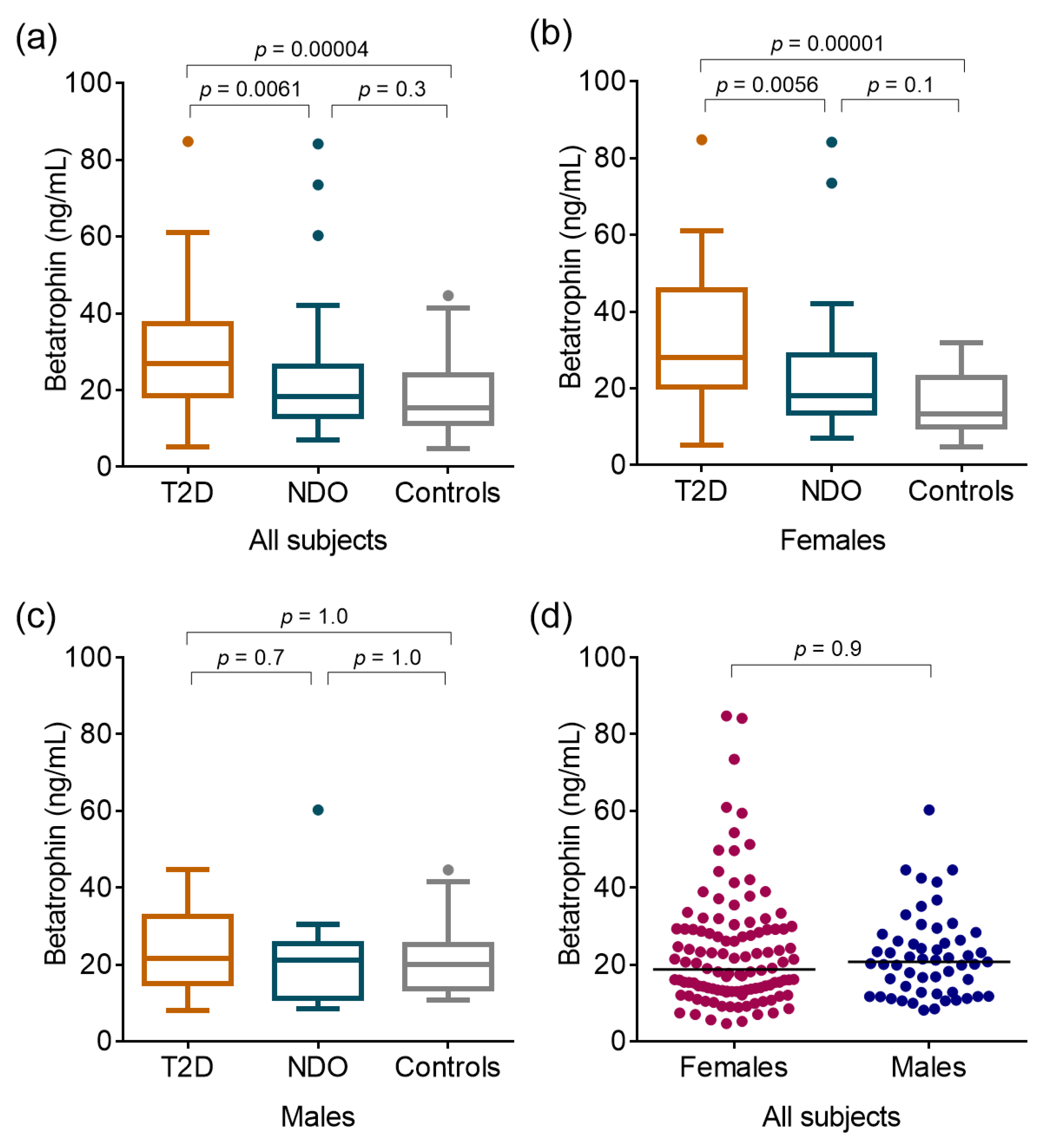
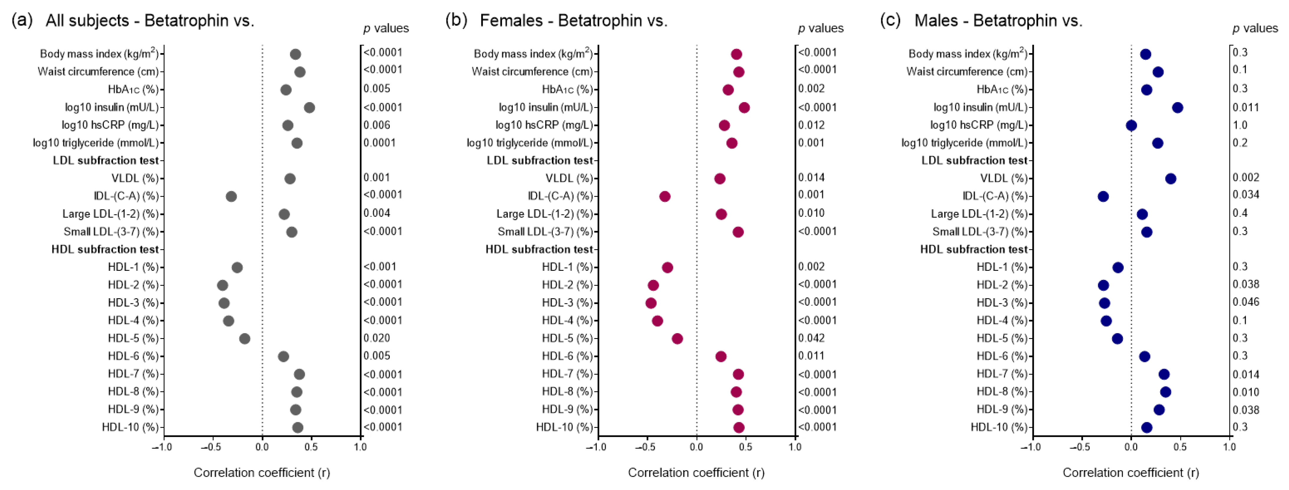
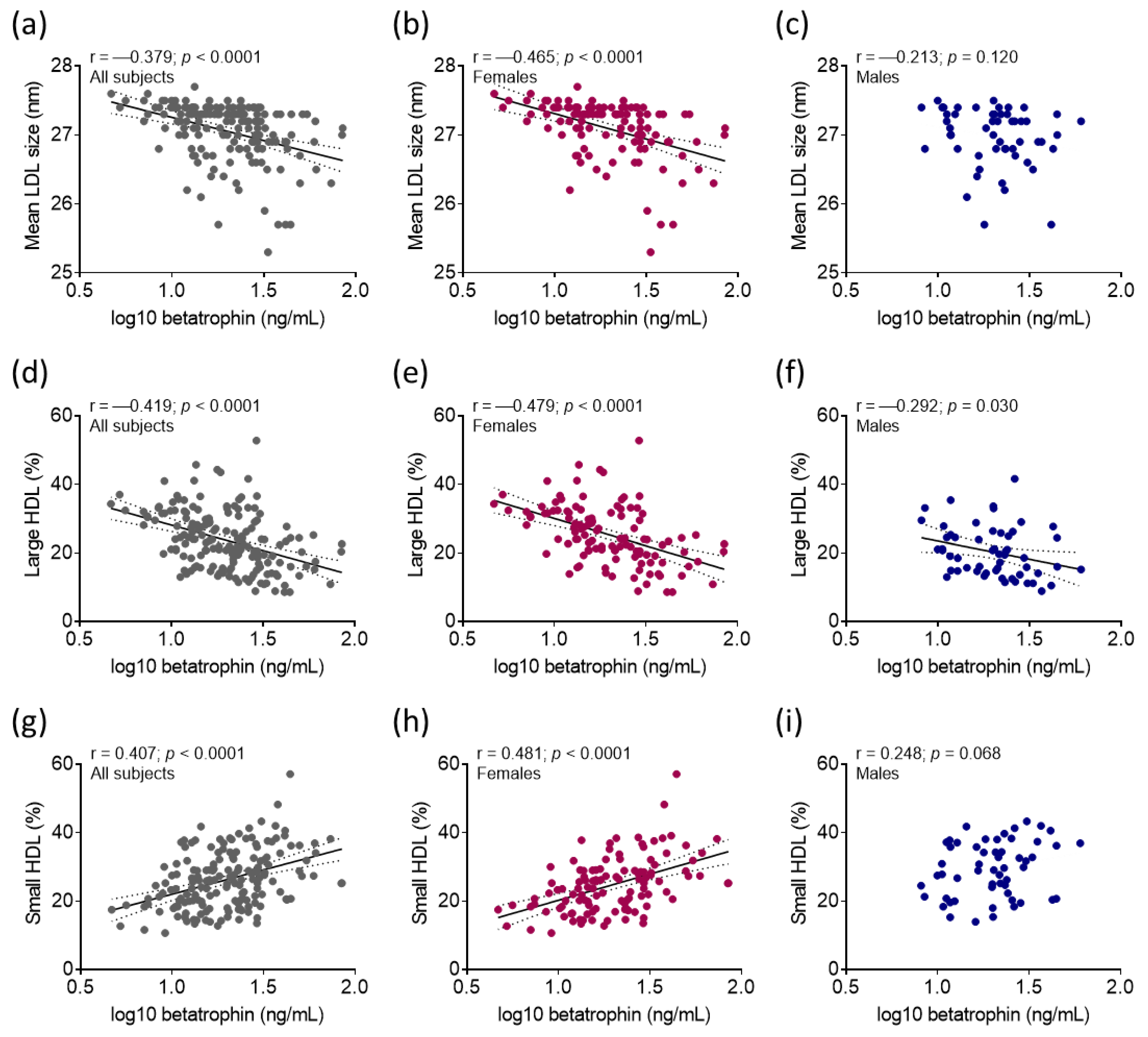
2. Results
3. Discussion
4. Materials and Methods
4.1. Enrolment of Study Participants
4.2. Determination of Routine Laboratory Parameters
4.3. Determination of Serum Betatrophin
4.4. Determination of Lipoprotein Subfractions
4.5. Statistical Analyses
5. Conclusions
Supplementary Materials
Author Contributions
Funding
Institutional Review Board Statement
Informed Consent Statement
Data Availability Statement
Acknowledgments
Conflicts of Interest
References
- Zhang, R. Lipasin, a novel nutritionally-regulated liver-enriched factor that regulates serum triglyceride levels. Biochem. Biophys. Res. Commun. 2012, 424, 786–792. [Google Scholar] [CrossRef]
- Tseng, Y.H.; Yeh, Y.H.; Chen, W.J.; Lin, K.H. Emerging regulation and function of betatrophin. Int. J. Mol. Sci. 2014, 15, 23640–23657. [Google Scholar] [CrossRef] [PubMed]
- Gómez-Ambrosi, J.; Pascual-Corrales, E.; Catalán, V.; Rodríguez, A.; Ramírez, B.; Romero, S.; Vila, N.; Ibáñez, P.; Margall, M.A.; Silva, C.; et al. Altered Concentrations in Dyslipidemia Evidence a Role for ANGPTL8/Betatrophin in Lipid Metabolism in Humans. J. Clin. Endocrinol. Metab. 2016, 101, 3803–3811. [Google Scholar] [CrossRef]
- Perdomo, C.M.; Gómez-Ambrosi, J.; Becerril, S.; Valentí, V.; Moncada, R.; Fernández-Sáez, E.M.; Méndez-Giménez, L.; Ezquerro, S.; Catalán, V.; Silva, C.; et al. Role of ANGPTL8 in NAFLD Improvement after Bariatric Surgery in Experimental and Human Obesity. Int. J. Mol. Sci. 2021, 22, 2945. [Google Scholar] [CrossRef]
- Lőrincz, H.; Somodi, S.; Ratku, B.; Harangi, M.; Paragh, G. Crucial Regulatory Role of Organokines in Relation to Metabolic Changes in Non-Diabetic Obesity. Metabolites 2023, 13, 270. [Google Scholar] [CrossRef] [PubMed]
- Lee, M.J.; Wu, Y.; Fried, S.K. Adipose tissue heterogeneity: Implication of depot differences in adipose tissue for obesity complications. Mol. Aspects Med. 2013, 34, 1–11. [Google Scholar] [CrossRef] [PubMed]
- Lőrincz, H.; Ratku, B.; Csiha, S.; Seres, I.; Szabó, Z.; Paragh, G.; Harangi, M.; Somodi, S. Impaired Organokine Regulation in Non-Diabetic Obese Subjects: Halfway to the Cardiometabolic Danger Zone. Int. J. Mol. Sci. 2023, 24, 4115. [Google Scholar] [CrossRef]
- Mayoral, L.P.; Andrade, G.M.; Mayoral, E.P.; Huerta, T.H.; Canseco, S.P.; Rodal Canales, F.J.; Cabrera-Fuentes, H.A.; Cruz, M.M.; Pérez Santiago, A.D.; Alpuche, J.J.; et al. Obesity subtypes, related biomarkers & heterogeneity. Indian. J. Med. Res. 2020, 151, 11–21. [Google Scholar] [CrossRef]
- Vekic, J.; Zeljkovic, A.; Stefanovic, A.; Jelic-Ivanovic, Z.; Spasojevic-Kalimanovska, V. Obesity and dyslipidemia. Metabolism 2019, 92, 71–81. [Google Scholar] [CrossRef] [PubMed]
- Szentpéteri, A.; Lőrincz, H.; Somodi, S.; Varga, V.E.; Paragh, G.; Seres, I.; Harangi, M. Serum obestatin level strongly correlates with lipoprotein subfractions in non-diabetic obese patients. Lipids Health Dis. 2018, 17, 39. [Google Scholar] [CrossRef]
- Abu-Farha, M.; Abubaker, J.; Al-Khairi, I.; Cherian, P.; Noronha, F.; Hu, F.B.; Behbehani, K.; Elkum, N. Higher plasma betatrophin/ANGPTL8 level in Type 2 Diabetes subjects does not correlate with blood glucose or insulin resistance. Sci. Rep. 2015, 5, 10949. [Google Scholar] [CrossRef]
- Abu-Farha, M.; Al Madhoun, A.; Abubaker, J. The Rise and the Fall of Betatrophin/ANGPTL8 as an Inducer of. J. Diabetes Res. 2016, 2016, 4860595. [Google Scholar] [CrossRef]
- Espes, D.; Martinell, M.; Carlsson, P.O. Increased circulating betatrophin concentrations in patients with type 2 diabetes. Int. J. Endocrinol. 2014, 2014, 323407. [Google Scholar] [CrossRef] [PubMed]
- Gómez-Ambrosi, J.; Pascual, E.; Catalán, V.; Rodríguez, A.; Ramírez, B.; Silva, C.; Gil, M.J.; Salvador, J.; Frühbeck, G. Circulating betatrophin concentrations are decreased in human obesity and type 2 diabetes. J. Clin. Endocrinol. Metab. 2014, 99, E2004–E2009. [Google Scholar] [CrossRef]
- Murawska, K.; Krintus, M.; Kuligowska-Prusinska, M.; Szternel, L.; Stefanska, A.; Sypniewska, G. Relationship between Serum Angiopoietin-like Proteins 3 and 8 and Atherogenic Lipid Biomarkers in Non-Diabetic Adults Depends on Gender and Obesity. Nutrients 2021, 13, 4339. [Google Scholar] [CrossRef] [PubMed]
- Saruhan, E.; Ispir, E.; Basaran, Y.; Akbaba, E.; Tosun, K.; Aslan, M. Relationship between serum Betatrophin, GPIHBP1, and LDL subfractions in patients with gestational diabetes mellitus. Clin. Biochem. 2023, 118, 110592. [Google Scholar] [CrossRef] [PubMed]
- Sylvers-Davie, K.L.; Davies, B.S.J. Regulation of lipoprotein metabolism by ANGPTL3, ANGPTL4, and ANGPTL8. Am. J. Physiol. Endocrinol. Metab. 2021, 321, E493–E508. [Google Scholar] [CrossRef] [PubMed]
- Guo, Q.; Cao, S.; Wang, X. Betatrophin and Insulin Resistance. Metabolites 2022, 12, 925. [Google Scholar] [CrossRef] [PubMed]
- Zou, H.; Xu, Y.; Chen, X.; Yin, P.; Li, D.; Li, W.; Xie, J.; Shao, S.; Liu, L.; Yu, X. Predictive values of ANGPTL8 on risk of all-cause mortality in diabetic patients: Results from the REACTION Study. Cardiovasc. Diabetol. 2020, 19, 121. [Google Scholar] [CrossRef] [PubMed]
- Chi, X.; Britt, E.C.; Shows, H.W.; Hjelmaas, A.J.; Shetty, S.K.; Cushing, E.M.; Li, W.; Dou, A.; Zhang, R.; Davies, B.S.J. ANGPTL8 promotes the ability of ANGPTL3 to bind and inhibit lipoprotein lipase. Mol. Metab. 2017, 6, 1137–1149. [Google Scholar] [CrossRef]
- Chen, Y.Q.; Pottanat, T.G.; Siegel, R.W.; Ehsani, M.; Qian, Y.W.; Zhen, E.Y.; Regmi, A.; Roell, W.C.; Guo, H.; Luo, M.J.; et al. Angiopoietin-like protein 8 differentially regulates ANGPTL3 and ANGPTL4 during postprandial partitioning of fatty acids. J. Lipid Res. 2020, 61, 1203–1220. [Google Scholar] [CrossRef] [PubMed]
- Zhang, R.; Zhang, K. An updated ANGPTL3-4-8 model as a mechanism of triglyceride partitioning between fat and oxidative tissues. Prog. Lipid Res. 2022, 85, 101140. [Google Scholar] [CrossRef] [PubMed]
- Yi, M.; Chen, R.P.; Yang, R.; Guo, X.F.; Zhang, J.C.; Chen, H. Betatrophin Acts as a Diagnostic Biomarker in Type 2 Diabetes Mellitus and Is Negatively Associated with HDL-Cholesterol. Int. J. Endocrinol. 2015, 2015, 479157. [Google Scholar] [CrossRef] [PubMed]
- Ghasemi, H.; Tavilani, H.; Khodadadi, I.; Saidijam, M.; Karimi, J. Circulating Betatrophin Levels Are Associated with the Lipid Profile in Type 2 Diabetes. Chonnam Med. J. 2015, 51, 115–119. [Google Scholar] [CrossRef]
- Horn, P.; Radtke, S.; Metzing, U.B.; Steidl, R.; Sponholz, C.; Sommerfeld, O.; Roth, J.; Claus, R.A.; Birkenfeld, A.L.; Settmacher, U.; et al. Associations of Betatrophin/ANGPTL8 with Septic Dyslipidemia in Human Peritonitis: An Explorative Analysis. Biomedicines 2022, 10, 3151. [Google Scholar] [CrossRef]
- Ideishi, A.; Suematsu, Y.; Tashiro, K.; Morita, H.; Kumagai-Koyanagi, N.; Kuwano, T.; Miura, S.I. Changes in serum levels of angiopoietin-like protein-8 and glycosylphosphatidylinositol-anchored high-density lipoprotein binding protein 1 after ezetimibe therapy in patients with dyslipidemia. Clin. Chim. Acta 2020, 510, 675–680. [Google Scholar] [CrossRef]
- Subramanian, S.; Chait, A. Hypertriglyceridemia secondary to obesity and diabetes. Biochim. Biophys. Acta 2012, 1821, 819–825. [Google Scholar] [CrossRef]
- Rashid, S.; Watanabe, T.; Sakaue, T.; Lewis, G.F. Mechanisms of HDL lowering in insulin resistant, hypertriglyceridemic states: The combined effect of HDL triglyceride enrichment and elevated hepatic lipase activity. Clin. Biochem. 2003, 36, 421–429. [Google Scholar] [CrossRef]
- Krauss, R.M. Lipoprotein subfractions and cardiovascular disease risk. Curr. Opin. Lipidol. 2010, 21, 305–311. [Google Scholar] [CrossRef]
- Ahsan, L.; Zheng, W.Q.; Kaur, G.; Kadakuntla, A.; Remaley, A.T.; Sampson, M.; Feustel, P.; Nappi, A.; Mookherjee, S.; Lyubarova, R. Association of Lipoprotein Subfractions With Presence and Severity of Coronary Artery Disease in Patients Referred for Coronary Angiography. Am. J. Cardiol. 2023, 203, 212–218. [Google Scholar] [CrossRef]
- Stanciulescu, L.A.; Scafa-Udriste, A.; Dorobantu, M. Exploring the Association between Low-Density Lipoprotein Subfractions and Major Adverse Cardiovascular Outcomes-A Comprehensive Review. Int. J. Mol. Sci. 2023, 24, 6669. [Google Scholar] [CrossRef]
- Zhang, Y.; Luo, S.; Gao, Y.; Tong, W.; Sun, S. High-Density Lipoprotein Subfractions Remodeling: A Critical Process for the Treatment of Atherosclerotic Cardiovascular Diseases. Angiology 2023, 33197231157473, Advance online publication. [Google Scholar] [CrossRef] [PubMed]
- Agarwala, A.; Michos, E.D.; Samad, Z.; Ballantyne, C.M.; Virani, S.S. The Use of Sex-Specific Factors in the Assessment of Women’s Cardiovascular Risk. Circulation 2020, 141, 592–599. [Google Scholar] [CrossRef] [PubMed]
- Palmisano, B.T.; Zhu, L.; Eckel, R.H.; Stafford, J.M. Sex differences in lipid and lipoprotein metabolism. Mol. Metab. 2018, 15, 45–55. [Google Scholar] [CrossRef]
- Hodis, H.N.; Mack, W.J. Menopausal Hormone Replacement Therapy and Reduction of All-Cause Mortality and Cardiovascular Disease: It Is About Time and Timing. Cancer J. 2022, 28, 208–223. [Google Scholar] [CrossRef] [PubMed]
- Ng, D.S. Evolving ANGPTL-based lipid-lowering strategies and beyond. Curr. Opin. Lipidol. 2021, 32, 271–272. [Google Scholar] [CrossRef] [PubMed]


| T2D (n = 50) | NDO (n = 70) | Controls (n = 49) | |
|---|---|---|---|
| Anthropometric parameters | |||
| Male/Female (n) | 20/30 | 19/51 | 16/33 |
| Age (yrs) | 47.6 ± 8.1 | 44.8 ± 12.0 | 43.2 ± 9.1 |
| Waist circumference (cm) | 127.1 ± 18.5 * | 125.5 ± 17.9 § | 85.2 ± 12.3 |
| Body mass index (kg/m2) | 43.1 ± 9.3 * | 43.3 ± 7.9 § | 24.7 ± 2.8 |
| Main medication | |||
| Metformin (n; %) | 37; 74.0 | 8; 11.4 | 0; 0.0 |
| Insulin (n; %) | 12; 24.0 | 0; 0.0 | 0; 0.0 |
| GLP-1 RA (n; %) | 9; 18.0 | 0; 0.0 | 0; 0.0 |
| Statin (n; %) | 23; 46.0 | 8; 11.4 | 0; 0.0 |
| ACEI/ARB (n; %) | 24; 48.0 | 23; 32.9 | 1; 2.0 |
| CCB (n; %) | 13; 26.0 | 6; 8.6 | 1; 2.0 |
| Diuretics (n; %) | 8; 16.0 | 18; 25.7 | 0; 0.0 |
| Laboratory parameters | |||
| Total cholesterol (mmol/L) | 5.0 ± 1.2 | 5.0 ± 0.9 | 5.0 ± 0.8 |
| HDL-C (mmol/L) | 1.2 ± 0.3 * | 1.3 ± 0.3 § | 1.5 ± 0.4 |
| LDL-C (mmol/L) | 3.0 ± 0.9 | 3.2 ± 0.8 | 3.0 ± 0.5 |
| ApolipoproteinA-I (g/L) | 1.4 ± 0.2 * | 1.5 ± 0.2 | 1.6 ± 0.3 |
| Triglyceride (mmol/L) | 1.7 (1.2–2.7) * | 1.4 (1.0–1.9) | 1.1 (0.9–1.5) |
| hsCRP (mg/L) | 6.6 (3.1–13.7) * | 7.8 (3.2–13.9) § | 1.3 (0.6–2.5) |
| Fasting glucose (mmol/L) | 6.3 (5.5–10.5) *# | 5.2 (4.9–5.8) § | 4.8 (4.5–5.1) |
| HbA1C (%) | 7.2 ± 1.6 *# | 5.6 ± 0.9 | 5.1 ± 0.3 |
| Insulin (mU/L) | 25.4 (14.1–31.4) (n = 16) * | 15 (11.7–21.8) (n = 59) § | 10.9 (6.6–12.9) (n = 16) |
| GFR (mL/1.73 m2) | 90 (90–90) | 90 (90–90) | 90 (90–90) |
| AST (U/L) | 25.0 (17.0–30.5) | 20.0 (17.0–27.0) | 19.0 (17.0–24.0) |
| sTSH (mU/L) | 2.3 (1.2–15.2) (n = 31) * | 2.1 (1.6–2.8) | 1.7 (1.2–2.1) |
| T2D (n = 50) | NDO (n = 70) | Controls (n = 49) | |
|---|---|---|---|
| LDL subfraction test | |||
| VLDL (%) | 21.03 ± 5.47 *# | 18.30 ± 3.81 | 17.69 ± 3.21 |
| (mmol/L) | 1.07 ± 0.51 *# | 0.91 ± 0.23 | 0.89 ± 0.19 |
| IDL (%) | 24.62 ± 3.66 | 26.14 ± 4.00 | 26.56 ± 6.34 |
| (mmol/L) | 1.23 ± 0.33 | 1.28 ± 0.30 | 1.34 ± 0.41 |
| Large LDL (%) | 26.58 ± 5.68 * | 28.68 ± 5.01 § | 23.18 ± 6.10 |
| (mmol/L) | 1.32 ± 0.42 | 1.45 ± 0.38 § | 1.17 ± 0.38 |
| Small LDL (%) | 1.50 (0.0–3.30) | 0.00 (0.00–1.80) | 0.60 (0.00–1.90) |
| (mmol/L) | 0.067 (0.000–0.202) | 0.028 (0.000–0.103) | 0.032 (0.093–0.173) |
| Mean LDL size (nm) | 26.85 (26.60–27.30) * | 27.20 (26.90–27.40) § | 27.30 (27.00–27.40) |
| HDL subfraction test | |||
| Large HDL (%) | 19.06 ± 6.55 *# | 23.47 ± 7.49 § | 28.98 ± 8.67 |
| (mmol/L) | 0.24 ± 0.12 * | 0.30 ± 0.12 § | 0.46 ± 0.26 |
| Intermediate HDL (%) | 49.02 ± 4.02 | 50.49 ± 3.96 | 50.35 ± 4.59 |
| (mmol/L) | 0.58 ± 0.15 *# | 0.66 ± 0.17 § | 0.73 ± 0.17 |
| Small HDL (%) | 31.91 ± 7.97 *# | 26.04 ± 7.15 § | 20.67 ± 6.29 |
| (mmol/L) | 0.37 ± 0.10 * | 0.34 ± 0.12 § | 0.29 ± 0.07 |
| Predictor | β | p | |
|---|---|---|---|
| Model 1 | |||
| All subjects | Small HDL (%) | 0.446 | <0.0001 |
| Model 2 | |||
| Small LDL (%) | 0.378 | <0.0001 | |
| Females | Large HDL (%) | −0.330 | <0.0001 |
| Model 3 | |||
| Males | VLDL (%) | 0.425 | 0.006 |
| Model 4 | |||
| T2D | Large HDL (%) | −0.430 | 0.006 |
| Model 5 | |||
| NDO | Small LDL (%) | 0.451 | 0.002 |
| Model 6 | |||
| Controls | Small HDL (%) | 0.448 | 0.007 |
Disclaimer/Publisher’s Note: The statements, opinions and data contained in all publications are solely those of the individual author(s) and contributor(s) and not of MDPI and/or the editor(s). MDPI and/or the editor(s) disclaim responsibility for any injury to people or property resulting from any ideas, methods, instructions or products referred to in the content. |
© 2023 by the authors. Licensee MDPI, Basel, Switzerland. This article is an open access article distributed under the terms and conditions of the Creative Commons Attribution (CC BY) license (https://creativecommons.org/licenses/by/4.0/).
Share and Cite
Lőrincz, H.; Csiha, S.; Ratku, B.; Somodi, S.; Sztanek, F.; Seres, I.; Paragh, G.; Harangi, M. Gender-Dependent Associations between Serum Betatrophin Levels and Lipoprotein Subfractions in Diabetic and Nondiabetic Obese Patients. Int. J. Mol. Sci. 2023, 24, 16504. https://doi.org/10.3390/ijms242216504
Lőrincz H, Csiha S, Ratku B, Somodi S, Sztanek F, Seres I, Paragh G, Harangi M. Gender-Dependent Associations between Serum Betatrophin Levels and Lipoprotein Subfractions in Diabetic and Nondiabetic Obese Patients. International Journal of Molecular Sciences. 2023; 24(22):16504. https://doi.org/10.3390/ijms242216504
Chicago/Turabian StyleLőrincz, Hajnalka, Sára Csiha, Balázs Ratku, Sándor Somodi, Ferenc Sztanek, Ildikó Seres, György Paragh, and Mariann Harangi. 2023. "Gender-Dependent Associations between Serum Betatrophin Levels and Lipoprotein Subfractions in Diabetic and Nondiabetic Obese Patients" International Journal of Molecular Sciences 24, no. 22: 16504. https://doi.org/10.3390/ijms242216504