
Peripheral Inflammatory Markers in Autism Spectrum Disorder and Attention Deficit/Hyperactivity Disorder at Adolescent Age
 , ,
, ,
Abstract
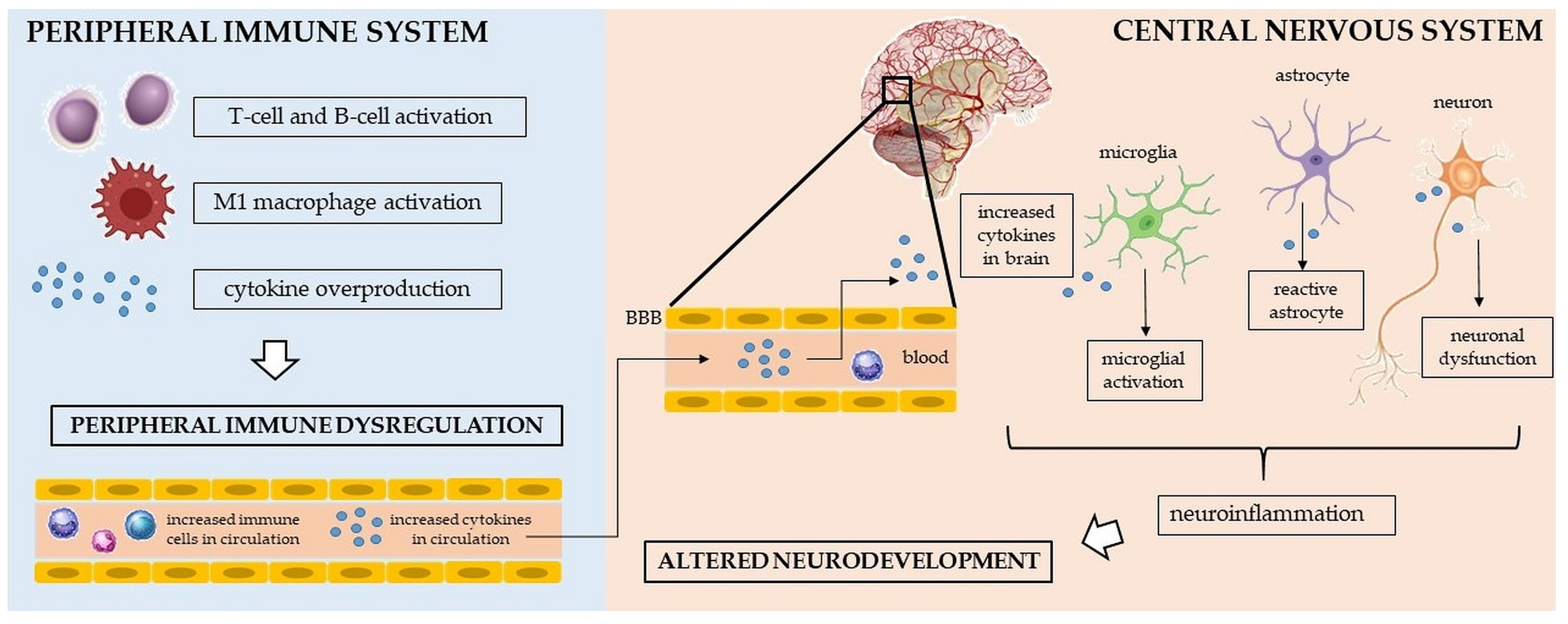
:1. Introduction
2. Results
2.1. Between-Group, between-Sex, and Mixed-Group × Sex Comparison of the Complete Blood Count Parameters and Selected Ratios
2.2. Between-Group, between-Sex, and Mixed-Group × Sex Comparison of the Cytokines, Cytokine Profiles, and Cytokine Ratios
2.3. ROC Curve Analyses
2.3.1. ROC Curve Analysis for Whole ASD Group
2.3.2. ROC Curve Analysis for ASD Females
2.3.3. ROC Curve Analysis for ASD Males
2.3.4. ROC Curve Analysis for Whole ADHD Group
2.3.5. ROC Curve Analysis for ADHD Females
2.3.6. ROC Curve Analysis for ADHD Males
3. Discussion
4. Materials and Methods
4.1. Subjects
4.1.1. ASD Diagnosis
4.1.2. ADHD Diagnosis
4.1.3. Exclusion Criteria
4.2. Blood Analysis
4.3. Statistical Analysis
5. Conclusions
Author Contributions
Funding
Institutional Review Board Statement
Informed Consent Statement
Data Availability Statement
Acknowledgments
Conflicts of Interest
References
- Xu, G.; Strathearn, L.; Liu, B.; O’Brien, M.; Kopelman, T.G.; Zhu, J.; Snetselaar, L.G.; Bao, W. Prevalence and Treatment Patterns of Autism Spectrum Disorder in the United States, 2016. JAMA Pediatr. 2019, 173, 153–159. [Google Scholar] [CrossRef] [PubMed] [Green Version]
- Faraone, S.V.; Asherson, P.; Banaschewski, T.; Biederman, J.; Buitelaar, J.K.; Ramos-Quiroga, J.A.; Rohde, L.A.; Sonuga-Barke, E.J.S.; Tannock, R.; Franke, B. Attention-deficit/hyperactivity disorder. Nat. Rev. Dis. Prim. 2015, 1, 15020. [Google Scholar] [CrossRef]
- Nylander, L.; Holmqvist, M.; Gustafson, L.; Gillberg, C. Attention-deficit/hyperactivity disorder (ADHD) and autism spectrum disorder (ASD) in adult psychiatry. A 20-year register study. Nord. J. Psychiatry 2013, 67, 344–350. [Google Scholar] [CrossRef]
- American Psychiatric Association. Diagnostic and Statistical Manual of Mental Disorders (DSM–5), 5th ed.; American Psychiatric Association: Arlington, TX, USA, 2013. [Google Scholar]
- Rommelse, N.N.J.; Geurts, H.M.; Franke, B.; Buitelaar, J.K.; Hartman, C.A. A review on cognitive and brain endophenotypes that may be common in autism spectrum disorder and attention-deficit/hyperactivity disorder and facilitate the search for pleiotropic genes. Neurosci. Biobehav. Rev. 2011, 35, 1363–1396. [Google Scholar] [CrossRef]
- Truedsson, E.; Bohlin, G.; Wåhlstedt, C. The Specificity and Independent Contribution of Inhibition, Working Memory, and Reaction Time Variability in Relation to Symptoms of ADHD and ASD. J. Atten. Disord. 2015, 24, 1266–1275. [Google Scholar] [CrossRef] [PubMed]
- Van Der Meer, J.M.J.; Oerlemans, A.M.; Van Steijn, D.J.; Lappenschaar, M.G.A.; De Sonneville, L.M.J.; Buitelaar, J.K.; Rommelse, N.N.J. Are autism spectrum disorder and attention-deficit/hyperactivity disorder different manifestations of one overarching disorder? Cognitive and symptom evidence from a clinical and population-based sample. J. Am. Acad. Child Adolesc. Psychiatry 2012, 51, 1160–1172.e3. [Google Scholar] [CrossRef]
- Van der Meer, J.M.J.; Lappenschaar, M.G.A.; Hartman, C.A.; Greven, C.U.; Buitelaar, J.K.; Rommelse, N.N.J. Homogeneous Combinations of ASD–ADHD Traits and Their Cognitive and Behavioral Correlates in a Population-Based Sample. J. Atten. Disord. 2017, 21, 753–763. [Google Scholar] [CrossRef] [PubMed]
- Hoogman, M.; van Rooij, D.; Klein, M.; Boedhoe, P.; Ilioska, I.; Li, T.; Patel, Y.; Postema, M.C.; Zhang-James, Y.; Anagnostou, E.; et al. Consortium neuroscience of attention deficit/hyperactivity disorder and autism spectrum disorder: The ENIGMA adventure. Hum. Brain Mapp. 2022, 43, 37–55. [Google Scholar] [CrossRef]
- Robinson-Agramonte, M.d.l.A.; García, E.N.; Guerra, J.F.; Hurtado, Y.V.; Antonucci, N.; Semprún-Hernández, N.; Schultz, S.; Siniscalco, D. Immune Dysregulation in Autism Spectrum Disorder: What Do We Know about It? Int. J. Mol. Sci. 2022, 23, 3033. [Google Scholar] [CrossRef]
- Leffa, D.T.; Torres, I.L.S.; Rohde, L.A. A review on the role of inflammation in attention-deficit/hyperactivity disorder. Neuroimmunomodulation 2019, 25, 328–333. [Google Scholar] [CrossRef]
- Erbescu, A.; Papuc, S.M.; Budisteanu, M.; Arghir, A.; Neagu, M. Re-emerging concepts of immune dysregulation in autism spectrum disorders. Front. Psychiatry 2022, 13, 1006612. [Google Scholar] [CrossRef] [PubMed]
- Dunn, G.A.; Nigg, J.T.; Sullivan, E.L. Neuroinflammation as a risk factor for attention deficit hyperactivity disorder. Pharmacol. Biochem. Behav. 2019, 182, 22–34. [Google Scholar] [CrossRef] [PubMed]
- George-Gay, B.; Parker, K. Understanding the complete blood count with differential. J. Perianesthesia Nurs. 2003, 18, 96–117. [Google Scholar] [CrossRef]
- Pogorzelska, K.; Krętowska, A.; Krawczuk-Rybak, M.; Sawicka-Żukowska, M. Characteristics of platelet indices and their prognostic significance in selected medical condition—A systematic review. Adv. Med. Sci. 2020, 65, 310–315. [Google Scholar] [CrossRef]
- Kounis, N.G.; Soufras, G.D.; Tsigkas, G.; Hahalis, G. White blood cell counts, leukocyte ratios, and eosinophils as inflammatory markers in patients with coronary artery disease. Clin. Appl. Thromb. 2015, 21, 139–143. [Google Scholar] [CrossRef]
- Mohamed, O.S.D.; Azmy, G.J.; Elfadl, E.M.A. Clinical significance of red blood cell distribution width in systemic lupus erythematosus patients. Egypt. Rheumatol. Rehabil. 2020, 47, 38. [Google Scholar] [CrossRef]
- Korniluk, A.; Koper-Lenkiewicz, O.M.; Kamińska, J.; Kemona, H.; Dymicka-Piekarska, V. Mean platelet volume (MPV): New perspectives for an old marker in the course and prognosis of inflammatory conditions. Mediators Inflamm. 2019, 2019, 9213074. [Google Scholar] [CrossRef] [PubMed] [Green Version]
- Qin, B.; Ma, N.; Tang, Q.; Wei, T.; Yang, M.; Fu, H.; Hu, Z.; Liang, Y.; Yang, Z.; Zhong, R. Neutrophil to lymphocyte ratio (NLR) and platelet to lymphocyte ratio (PLR) were useful markers in assessment of inflammatory response and disease activity in SLE patients. Mod. Rheumatol. 2016, 26, 372–376. [Google Scholar] [CrossRef]
- Liu, J.; Huang, X.; Yue, S.; Wang, J.; Ye, E.; Huang, J.; Zhao, Y.; Niu, D.; Hou, X.; Wu, J. Association of Red Cell Distribution Width-to-Platelet Ratio and Mortality in Patients with Sepsis. Mediators Inflamm. 2022, 2022, 4915887. [Google Scholar] [CrossRef]
- Bahrami, A.; Bahrami-Taghanaki, H.; Khorasanchi, Z.; Tayefi, M.; Ferns, G.A.; Sadeghnia, H.R.; Ghayour-Mobarhan, M. The Association Between Neuropsychological Function with Serum Vitamins A, D, and E and hs-CRP Concentrations. J. Mol. Neurosci. 2019, 68, 243–250. [Google Scholar] [CrossRef]
- Ates, S.; Oksuz, H.; Dogu, B.; Bozkus, F.; Ucmak, H.; Yanıt, F. Can mean platelet volume and mean platelet volume/platelet count ratio be used as a diagnostic marker for sepsis and systemic inflammatory response syndrome? Saudi Med. J. 2015, 36, 1186–1190. [Google Scholar] [CrossRef] [PubMed]
- Bozlu, G. Role of mean platelet volume-to-lymphocyte ratio in the diagnosis of childhood appendicitis. Arch. Argent. Pediatr. 2019, 117, 375–380. [Google Scholar] [CrossRef] [PubMed]
- Balta, S.; Demirer, Z.; Aparci, M.; Yildirim, A.O.; Ozturk, C. The lymphocyte-monocyte ratio in clinical practice. J. Clin. Pathol. 2016, 69, 88–89. [Google Scholar] [CrossRef] [PubMed]
- Nanava, N.; Betaneli, M.; Giorgobiani, G.; Chikovani, T.; Janikashvili, N. Complete blood count derived inflammatory biomarkers in patients with hematologic malignancies. Georgian Med. News 2020, 302, 39–44. [Google Scholar]
- Sweeten, T.L.; Posey, D.J.; McDougle, C.J. High blood monocyte counts and neopterin levels in children with autistic disorder. Am. J. Psychiatry 2003, 160, 1691–1693. [Google Scholar] [CrossRef]
- Kutlu, A.; Binici, N.C. Kutlu and Cevher Binici 607 Does increased neutrophil-lymphocyte ratio predict autism spectrum disorder? Anatol. J. Psychiatry 2018, 19, 607–614. [Google Scholar] [CrossRef]
- Kulaksizoglu, S.; Koparan, C. High neutrophil to lymphocyte ratio and low mean platelet volume level in autism spectrum disorders. Ann. Med. Res. 2019, 26, 2382–2385. [Google Scholar] [CrossRef]
- Tonhajzerova, I.; Ondrejka, I.; Mestanik, M.; Mikolka, P.; Hrtanek, I.; Mestanikova, A.; Bujnakova, I.; Mokra, D. Inflammatory activity in autism spectrum disorder. Adv. Exp. Med. Biol. 2015, 861, 93–98. [Google Scholar] [CrossRef]
- Pardo, C.A.; Farmer, C.A.; Thurm, A.; Shebl, F.M.; Ilieva, J.; Kalra, S.; Swedo, S. Serum and cerebrospinal fluid immune mediators in children with autistic disorder: A longitudinal study. Mol. Autism 2017, 8, 1. [Google Scholar] [CrossRef] [Green Version]
- Topal, Z.; Tufan, A.E.; Karadag, M.; Gokcen, C.; Akkaya, C.; Sarp, A.S.; Bahsi, I.; Kilinc, M. Evaluation of peripheral inflammatory markers, serum B12, folate, ferritin levels and clinical correlations in children with autism spectrum disorder (ASD) and attention deficit hyperactivity disorder (ADHD). Nord. J. Psychiatry 2022, 76, 150–157. [Google Scholar] [CrossRef]
- Avcil, S. Evaluation of the neutrophil/lymphocyte ratio, platelet/lymphocyte ratio, and mean platelet volume as inflammatory markers in children with attention-deficit hyperactivity disorder. Psychiatry Clin. Neurosci. 2018, 72, 522–530. [Google Scholar] [CrossRef] [PubMed] [Green Version]
- Cevher Binici, N.; Kutlu, A. Is ADHD an inflammation-related disorder? Anadolu Psikiyatr. Derg. 2019, 20, 313–320. [Google Scholar] [CrossRef]
- Tural Hesapcioglu, S.; Kasak, M.; Cıtak Kurt, A.N.; Ceylan, M.F. High monocyte level and low lymphocyte to monocyte ratio in autism spectrum disorders. Int. J. Dev. Disabil. 2019, 65, 73–81. [Google Scholar] [CrossRef]
- Arteaga-Henríquez, G.; Lugo-Marín, J.; Gisbert, L.; Setién-Ramos, I.; Martínez-Gallo, M.; Pujol-Borrell, R.; Ramos-Quiroga, J.A. Activation of the Monocyte/Macrophage System and Abnormal Blood Levels of Lymphocyte Subpopulations in Individuals with Autism Spectrum Disorder: A Systematic Review and Meta-Analysis. Int. J. Mol. Sci. 2022, 23, 14329. [Google Scholar] [CrossRef]
- Akıncı, M.A.; Uzun, N. Evaluation of hematological inflammatory markers in children and adolescents with attention deficit/hyperactivity disorder. Bratislava Med. J. 2021, 122, 256–262. [Google Scholar] [CrossRef] [PubMed]
- Yorbik, O.; Mutlu, C.; Tanju, I.A.; Celik, D.; Ozcan, O. Mean platelet volume in children with attention deficit hyperactivity disorder. Med. Hypotheses 2014, 82, 341–345. [Google Scholar] [CrossRef]
- Guler, G.; Dağ, P. Evaluation of the indicators of inflammation in children and adolescents with attention deficit and hyperactivity disorder: Effect of sex and subtype. Duzce Med. J. 2020, 22, 84–90. [Google Scholar] [CrossRef]
- Önder, A.; Gizli Çoban, Ö.; Sürer Adanır, A. Elevated neutrophil-to-lymphocyte ratio in children and adolescents with attention-deficit/hyperactivity disorder. Int. J. Psychiatry Clin. Pract. 2021, 25, 43–48. [Google Scholar] [CrossRef]
- Ceyhun, H.A.; Gürbüzer, N. New Hematological Parameters as Inflammatory Biomarkers: Systemic Immune Inflammation Index, Platerethritis, and Platelet Distribution Width in Patients with Adult Attention Deficit Hyperactivity Disorder. Adv. Neurodev. Disord. 2022, 6, 211–223. [Google Scholar] [CrossRef]
- Deverman, B.E.; Patterson, P.H. Cytokines and CNS Development. Neuron 2009, 64, 61–78. [Google Scholar] [CrossRef] [Green Version]
- Masi, A.; Glozier, N.; Dale, R.; Guastella, A.J. The Immune System, Cytokines, and Biomarkers in Autism Spectrum Disorder. Neurosci. Bull. 2017, 33, 194–204. [Google Scholar] [CrossRef] [Green Version]
- Kennedy, R.H.; Silver, R. Neuroimmune Signaling: Cytokines and the CNS. In Neuroscience in the 21st Century; Springer: New York, NY, USA, 2015; pp. 1–41. [Google Scholar]
- Siniscalco, D.; Schultz, S.; Brigida, A.L.; Antonucci, N. Inflammation and neuro-immune dysregulations in autism spectrum disorders. Pharmaceuticals 2018, 11, 56. [Google Scholar] [CrossRef] [Green Version]
- González, H.; Elgueta, D.; Montoya, A.; Pacheco, R. Neuroimmune regulation of microglial activity involved in neuroinflammation and neurodegenerative diseases. J. Neuroimmunol. 2014, 274, 1–13. [Google Scholar] [CrossRef]
- Lobo-Silva, D.; Carriche, G.M.; Castro, A.G.; Roque, S.; Saraiva, M. Balancing the immune response in the brain: IL-10 and its regulation. J. Neuroinflamm. 2016, 13, 297. [Google Scholar] [CrossRef] [Green Version]
- Ashwood, P.; Krakowiak, P.; Hertz-Picciotto, I.; Hansen, R.; Pessah, I.; Van de Water, J. Elevated plasma cytokines in autism spectrum disorders provide evidence of immune dysfunction and are associated with impaired behavioral outcome. Brain. Behav. Immun. 2011, 25, 40–45. [Google Scholar] [CrossRef] [Green Version]
- Jácome, M.C.I.; Chacòn, L.M.M.; Cuesta, H.V.; Rizo, C.M.; Santiesteban, M.W.; Hernandez, L.R.; García, E.N.; Fraguela, M.E.G.; Verdecia, C.I.F.; Hurtado, Y.V.; et al. Peripheral inflammatory markers contributing to comorbidities in autism. Behav. Sci. 2016, 6, 29. [Google Scholar] [CrossRef] [PubMed] [Green Version]
- Eftekharian, M.M.; Ghafouri-Fard, S.; Noroozi, R.; Omrani, M.D.; Arsang-jang, S.; Ganji, M.; Gharzi, V.; Noroozi, H.; Komaki, A.; Mazdeh, M.; et al. Cytokine profile in autistic patients. Cytokine 2018, 108, 120–126. [Google Scholar] [CrossRef] [PubMed]
- Wei, H.; Mori, S.; Hua, K.; Li, X. Alteration of brain volume in IL-6 overexpressing mice related to autism. Int. J. Dev. Neurosci. 2012, 30, 554–559. [Google Scholar] [CrossRef] [PubMed]
- Wei, H.; Chadman, K.K.; McCloskey, D.P.; Sheikh, A.M.; Malik, M.; Brown, W.T.; Li, X. Brain IL-6 elevation causes neuronal circuitry imbalances and mediates autism-like behaviors. Biochim. Biophys. Acta Mol. Basis Dis. 2012, 1822, 831–842. [Google Scholar] [CrossRef] [Green Version]
- Li, X.; Chauhan, A.; Sheikh, A.M.; Patil, S.; Chauhan, V.; Li, X.M.; Ji, L.; Brown, T.; Malik, M. Elevated immune response in the brain of autistic patients. J. Neuroimmunol. 2009, 207, 111–116. [Google Scholar] [CrossRef] [PubMed] [Green Version]
- Masi, A.; Quintana, D.S.; Glozier, N.; Lloyd, A.R.; Hickie, I.B.; Guastella, A.J. Cytokine aberrations in autism spectrum disorder: A systematic review and meta-analysis. Mol. Psychiatry 2015, 20, 440–446. [Google Scholar] [CrossRef] [PubMed]
- Vargas, D.L.; Nascimbene, C.; Krishnan, C.; Zimmerman, A.W.; Pardo, C.A. Neuroglial activation and neuroinflammation in the brain of patients with autism. Ann. Neurol. 2005, 57, 67–81. [Google Scholar] [CrossRef] [PubMed]
- Estes, M.L.; McAllister, A.K. Immune mediators in the brain and peripheral tissues in autism spectrum disorder. Nat. Rev. Neurosci. 2015, 16, 469–486. [Google Scholar] [CrossRef] [PubMed] [Green Version]
- Gottfried, C.; Bambini-Junior, V.; Francis, F.; Riesgo, R.; Savino, W. The impact of neuroimmune alterations in autism spectrum disorder. Front. Psychiatry 2015, 6, 121. [Google Scholar] [CrossRef] [PubMed] [Green Version]
- Donfrancesco, R.; Nativio, P.; Borrelli, E.; Giua, E.; Andriola, E.; Villa, M.P.; Ditrani, M. Serum cytokines in pediatric neuropsychiatric syndromes: Focus on Attention Deficit Hyperactivity Disorder. Minerva Pediatr. 2021, 73, 398–404. [Google Scholar] [CrossRef]
- Darwish, A.H.; Elgohary, T.M.; Nosair, N.A. Serum Interleukin-6 Level in Children with Attention-Deficit Hyperactivity Disorder (ADHD). J. Child Neurol. 2019, 34, 61–67. [Google Scholar] [CrossRef]
- Elsadek, A.E.; Al-Shokary, A.H.; Abdelghani, W.E.; Kamal, N.M.; Ibrahim, A.O.; El-Shorbagy, H.H.; Suliman, H.A.; Barseem, N.F.; Maksoud, Y.H.A.; Azab, S.M.; et al. Serum levels of interleukin-6 and tumor necrosis factor alpha in children with attention-deficit hyperactivity disorder. J. Pediatr. Neurosci. 2020, 15, 402–408. [Google Scholar] [CrossRef]
- Mohamed Mahmoud, I.; Taha El-Keiy, M.; Soliman Hammad, K.; Ibrahim Mostafa, A. Serum interleukin-6 level in children with attention-deficit hyperactivity disorder. Al-Azhar J. Ped. 2020, 23, 1315–1332. [Google Scholar]
- Dursun, S.; Demirci, E.; Kilic, E.; Ozmen, S. A different view on the etiopathogenesis of attention-deficit hyperactivity disorder from an inflammation perspective. Clin. Psychopharmacol. Neurosci. 2021, 19, 145–154. [Google Scholar] [CrossRef]
- Puzino, K.; Bourchtein, E.; Calhoun, S.L.; He, F.; Vgontzas, A.N.; Liao, D.; Bixler, E.O.; Fernandez-Mendoza, J. Behavioral, neurocognitive, polysomnographic and cardiometabolic profiles associated with obstructive sleep apnea in adolescents with ADHD. J. Child Psychol. Psychiatry Allied Discip. 2022, 63, 544–552. [Google Scholar] [CrossRef]
- Verlaet, A.A.J.; Breynaert, A.; Ceulemans, B.; De Bruyne, T.; Fransen, E.; Pieters, L.; Savelkoul, H.F.J.; Hermans, N. Oxidative stress and immune aberrancies in attention-deficit/hyperactivity disorder (ADHD): A case–control comparison. Eur. Child Adolesc. Psychiatry 2019, 28, 719–729. [Google Scholar] [CrossRef]
- Chang, J.P.C.; Su, K.P.; Mondelli, V.; Pariante, C.M. Cortisol and inflammatory biomarker levels in youths with attention deficit hyperactivity disorder (ADHD): Evidence from a systematic review with meta-analysis. Transl. Psychiatry 2021, 11, 430. [Google Scholar] [CrossRef]
- Misiak, B.; Wójta-Kempa, M.; Samochowiec, J.; Schiweck, C.; Aichholzer, M.; Reif, A.; Samochowiec, A.; Stańczykiewicz, B. Peripheral blood inflammatory markers in patients with attention deficit/hyperactivity disorder (ADHD): A systematic review and meta-analysis. Prog. Neuropsychopharmacol. Biol. Psychiatry 2022, 118, 110581. [Google Scholar] [CrossRef] [PubMed]
- Xu, N.; Li, X.; Zhong, Y. Inflammatory cytokines: Potential biomarkers of immunologic dysfunction in autism spectrum disorders. Mediators Inflamm. 2015, 2015, 531518. [Google Scholar] [CrossRef] [PubMed] [Green Version]
- Maes, M.; Carvalho, A.F. The Compensatory Immune-Regulatory Reflex System (CIRS) in Depression and Bipolar Disorder. Mol. Neurobiol. 2018, 55, 8885–8903. [Google Scholar] [CrossRef] [PubMed]
- Saghazadeh, A.; Ataeinia, B.; Keynejad, K.; Abdolalizadeh, A.; Hirbod-Mobarakeh, A.; Rezaei, N. Anti-inflammatory cytokines in autism spectrum disorders: A systematic review and meta-analysis. Cytokine 2019, 123, 154740. [Google Scholar] [CrossRef]
- Zablotsky, B.; Black, L.I.; Blumberg, S.J. Estimated Prevalence of Children with Diagnosed Developmental Disabilities in the United States, 2014–2016. NCHS Data Brief. 2017, 291, 1–8. [Google Scholar]
- Danielson, M.L.; Bitsko, R.H.; Ghandour, R.M.; Holbrook, J.R.; Kogan, M.D.; Blumberg, S.J. Prevalence of Parent-Reported ADHD Diagnosis and Associated Treatment Among U.S. Children and Adolescents. J. Clin. Child Adolesc. Psychol. 2018, 47, 199–212. [Google Scholar] [CrossRef]
- Giarelli, E.; Wiggins, L.D.; Rice, C.E.; Levy, S.E.; Kirby, R.S.; Pinto-Martin, J.; Mandell, D. Sex differences in the evaluation and diagnosis of autism spectrum disorders among children. Disabil. Health J. 2010, 3, 107–116. [Google Scholar] [CrossRef] [PubMed] [Green Version]
- Mandy, W.; Chilvers, R.; Chowdhury, U.; Salter, G.; Seigal, A.; Skuse, D. Sex differences in autism spectrum disorder: Evidence from a large sample of children and adolescents. J. Autism Dev. Disord. 2012, 42, 1304–1313. [Google Scholar] [CrossRef]
- Gershon, J. A meta-analytic review of gender differences in ADHD. J. Atten. Disord. 2002, 5, 143–154. [Google Scholar] [CrossRef] [PubMed]
- Breach, M.R.; Lenz, K.M. Sex Differences in Neurodevelopmental Disorders: A Key Role for the Immune System. Curr. Top. Behav. Neurosci. 2023, 62, 165–206. [Google Scholar] [CrossRef] [PubMed]
- Ferri, S.L.; Abel, T.; Brodkin, E.S. Sex Differences in Autism Spectrum Disorder: A Review. Curr. Psychiatry Rep. 2018, 20, 9. [Google Scholar] [CrossRef] [PubMed]
- Ardalan, M.; Chumak, T.; Vexler, Z.; Mallard, C. Sex-dependent effects of perinatal inflammation on the brain: Implication for neuro-psychiatric disorders. Int. J. Mol. Sci. 2019, 20, 2270. [Google Scholar] [CrossRef] [PubMed] [Green Version]
- Masi, A.; Breen, E.J.; Alvares, G.A.; Glozier, N.; Hickie, I.B.; Hunt, A.; Hui, J.; Beilby, J.; Ravine, D.; Wray, J.; et al. Cytokine levels and associations with symptom severity in male and female children with autism spectrum disorder. Mol. Autism 2017, 8, 63. [Google Scholar] [CrossRef] [Green Version]
- Schwarz, E.; Guest, P.C.; Rahmoune, H.; Wang, L.; Levin, Y.; Ingudomnukul, E.; Ruta, L.; Kent, L.; Spain, M.; Baron-Cohen, S.; et al. Sex-specific serum biomarker patterns in adults with Asperger’s syndrome. Mol. Psychiatry 2011, 16, 1213–1220. [Google Scholar] [CrossRef]
- Werling, D.M.; Parikshak, N.N.; Geschwind, D.H. Gene expression in human brain implicates sexually dimorphic pathways in autism spectrum disorders. Nat. Commun. 2016, 7, 10717. [Google Scholar] [CrossRef]
- Chang, S.J.; Kuo, H.C.; Chou, W.J.; Tsai, C.S.; Lee, S.Y.; Wang, L.J. Cytokine Levels and Neuropsychological Function among Patients with Attention-Deficit/Hyperactivity Disorder and Atopic Diseases. J. Pers. Med. 2022, 12, 1155. [Google Scholar] [CrossRef]
- Abruzzo, P.M.; Ghezzo, A.; Bolotta, A.; Ferreri, C.; Minguzzi, R.; Vignini, A.; Visconti, P.; Marini, M. Perspective Biological Markers for Autism Spectrum Disorders: Advantages of the Use of Receiver Operating Characteristic Curves in Evaluating Marker Sensitivity and Specificity. Dis. Markers 2015, 2015, 329607. [Google Scholar] [CrossRef] [Green Version]
- Chen, X.; Yao, T.; Cai, J.; Fu, X.; Li, H.; Wu, J. Systemic inflammatory regulators and 7 major psychiatric disorders: A two-sample Mendelian randomization study. Prog. Neuropsychopharmacol. Biol. Psychiatry 2022, 116, 110534. [Google Scholar] [CrossRef]
- Tigner, A.; Ibrahim, S.A.; Murray, I. Histology, White Blood Cell; StatPearls Publishing: Treasure Island, FL, USA, 2023; pp. 1–7. [Google Scholar]
- Wright, H.L.; Moots, R.J.; Bucknall, R.C.; Edwards, S.W. Neutrophil function in inflammation and inflammatory diseases. Rheumatology 2010, 49, 1618–1631. [Google Scholar] [CrossRef] [PubMed] [Green Version]
- Wiwanitkit, V. Plateletcrit, Mean Platelet Volume, Platelet Distribution Width: Its Expected Values and Correlation with Parallel Red Blood Cell Parameters. Clin. Appl. Thromb. 2004, 10, 175–178. [Google Scholar] [CrossRef]
- Vagdatli, E.; Gounari, E.; Lazaridou, E.; Katsibourlia, E.; Tsikopoulou, F.; Labrianou, I. Platelet distribution width: A simple, practical and specific marker of activation of coagulation. Hippokratia 2010, 14, 28–32. [Google Scholar] [PubMed]
- Margetic, S. Inflammation and haemostasis. Biochem. Med. 2012, 22, 49–62. [Google Scholar] [CrossRef]
- Sayit, A.T.; Gunbey, P.H.; Terzi, Y. Is the mean platelet volume in patients with acute cholecystitis an inflammatory marker? J. Clin. Diagnostic Res. 2015, 9, TC05–TC07. [Google Scholar] [CrossRef] [PubMed]
- Wang, F.; Meng, Z.; Li, S.; Zhang, Y.; Wu, H. Platelet Distribution Width Levels Can Be a Predictor in the Diagnosis of Persistent Organ Failure in Acute Pancreatitis. Gastroenterol. Res. Pract. 2017, 2017, 8374215. [Google Scholar] [CrossRef]
- Turner, M.D.; Nedjai, B.; Hurst, T.; Pennington, D.J. Cytokines and chemokines: At the crossroads of cell signalling and inflammatory disease. Biochim. Biophys. Acta Mol. Cell Res. 2014, 1843, 2563–2582. [Google Scholar] [CrossRef] [PubMed] [Green Version]
- Saad, K.; Abdallah, A.E.M.; Abdel-Rahman, A.A.; Al-Atram, A.A.; Abdel-Raheem, Y.F.; Gad, E.F.; Abo-Elela, M.G.M.; Elserogy, Y.M.; Elhoufey, A.; Nigm, D.A.; et al. Polymorphism of interleukin-1β and interleukin-1 receptor antagonist genes in children with autism spectrum disorders. Prog. Neuropsychopharmacol. Biol. Psychiatry 2020, 103, 109999. [Google Scholar] [CrossRef]
- Mazahery, H.; Conlon, C.A.; Beck, K.L.; Mugridge, O.; Kruger, M.C.; Stonehouse, W.; Camargo, C.A.; Meyer, B.J.; Tsang, B.; von Hurst, P.R. Inflammation (IL-1β) Modifies the Effect of Vitamin D and Omega-3 Long Chain Polyunsaturated Fatty Acids on Core Symptoms of Autism Spectrum Disorder—An Exploratory Pilot Study. Nutrients 2020, 12, 661. [Google Scholar] [CrossRef] [Green Version]
- García-Juárez, M.; Camacho-Morales, A. Defining the Role of Anti- and Pro-inflammatory Outcomes of Interleukin-6 in Mental Health. Neuroscience 2022, 492, 32–46. [Google Scholar] [CrossRef]
- Hunter, C.A.; Jones, S.A. IL-6 as a keystone cytokine in health and disease. Nat. Immunol. 2015, 16, 448–457. [Google Scholar] [CrossRef]
- Erta, M.; Quintana, A.; Hidalgo, J. Interleukin-6, a major cytokine in the central nervous system. Int. J. Biol. Sci. 2012, 8, 1254–1266. [Google Scholar] [CrossRef]
- Rohleder, N.; Aringer, M.; Boentert, M. Role of interleukin-6 in stress, sleep, and fatigue. Ann. N. Y. Acad. Sci. 2012, 1261, 88–96. [Google Scholar] [CrossRef]
- Pan, W.; Kastin, A.J. Tumor necrosis factor and stroke: Role of the blood-brain barrier. Prog. Neurobiol. 2007, 83, 363–374. [Google Scholar] [CrossRef] [Green Version]
- Medeiros, R.; Figueiredo, C.P.; Pandolfo, P.; Duarte, F.S.; Prediger, R.D.S.; Passos, G.F.; Calixto, J.B. The role of TNF-signaling pathway on COX-2 upregulation and cognitive decline induced by-amyloid peptide. Behav. Brain Res. 2010, 209, 165–173. [Google Scholar] [CrossRef] [PubMed]
- McAfoose, J.; Koerner, H.; Baune, B.T. The effects of TNF deficiency on age-related cognitive performance. Psychoneuroendocrinology 2009, 34, 615–619. [Google Scholar] [CrossRef] [PubMed]
- Hayley, S.; Wall, P.; Anisman, H. Sensitization to the neuroendocrine, central monoamine and behavioural effects of murine tumor necrosis factor-α: Peripheral and central mechanisms. Eur. J. Neurosci. 2002, 15, 1061–1076. [Google Scholar] [CrossRef] [PubMed]
- Réus, G.Z.; Fries, G.R.; Stertz, L.; Badawy, M.; Passos, I.C.; Barichello, T.; Kapczinski, F.; Quevedo, J. The role of inflammation and microglial activation in the pathophysiology of psychiatric disorders. Neuroscience 2015, 300, 141–154. [Google Scholar] [CrossRef]
- Belmadani, A.; Tran, P.B.; Ren, D.; Miller, R.J. Chemokines regulate the migration of neural progenitors to sites of neuroinflammation. J. Neurosci. 2006, 26, 3182–3191. [Google Scholar] [CrossRef] [Green Version]
- Kronfol, Z.; Remick, D.G. Cytokines and the brain: Implications for clinical psychiatry. Am. J. Psychiatry 2000, 157, 683–694. [Google Scholar] [CrossRef] [PubMed]
- Hassan, W.; Noreen, H.; Castro-Gomes, V.; Mohammadzai, I.; Batista Teixeira da Rocha, J.; Landeira-Fernandez, J. Association of Oxidative Stress with Psychiatric Disorders. Curr. Pharm. Des. 2016, 22, 2960–2974. [Google Scholar] [CrossRef] [PubMed]
- Jiang, H.Y.; Xu, L.L.; Shao, L.; Xia, R.M.; Yu, Z.H.; Ling, Z.X.; Yang, F.; Deng, M.; Ruan, B. Maternal infection during pregnancy and risk of autism spectrum disorders: A systematic review and meta-analysis. Brain. Behav. Immun. 2016, 58, 165–172. [Google Scholar] [CrossRef] [PubMed]
- Jones, K.L.; Croen, L.A.; Yoshida, C.K.; Heuer, L.; Hansen, R.; Zerbo, O.; Delorenze, G.N.; Kharrazi, M.; Yolken, R.; Ashwood, P.; et al. Autism with intellectual disability is associated with increased levels of maternal cytokines and chemokines during gestation. Mol. Psychiatry 2017, 22, 273–279. [Google Scholar] [CrossRef] [PubMed] [Green Version]
- Fiorentino, M.; Sapone, A.; Senger, S.; Camhi, S.S.; Kadzielski, S.M.; Buie, T.M.; Kelly, D.L.; Cascella, N.; Fasano, A. Blood-brain barrier and intestinal epithelial barrier alterations in autism spectrum disorders. Mol. Autism 2016, 7, 49. [Google Scholar] [CrossRef] [PubMed] [Green Version]
- Mohammadi, M.R.; Yadegari, N.; Hassanzadeh, E.; Farokhnia, M.; Yekehtaz, H.; Mirshafiee, O.; Akhondzadeh, S. Double-blind, placebo-controlled trial of risperidone plus amantadine in children with autism: A 10-week randomized study. Clin. Neuropharmacol. 2013, 36, 179–184. [Google Scholar] [CrossRef] [PubMed]
- Duffy, F.H.; Shankardass, A.; McAnulty, G.B.; Eksioglu, Y.Z.; Coulter, D.; Rotenberg, A.; Als, H. Corticosteroid therapy in regressive autism: A retrospective study of effects on the Frequency Modulated Auditory Evoked Response (FMAER), language, and behavior. BMC Neurol. 2014, 14, 70. [Google Scholar] [CrossRef] [PubMed] [Green Version]
- Pardo, C.A.; Buckley, A.; Thurm, A.; Lee, L.C.; Azhagiri, A.; Neville, D.M.; Swedo, S.E. A pilot open-label trial of minocycline in patients with autism and regressive features. J. Neurodev. Disord. 2013, 5, 9. [Google Scholar] [CrossRef] [Green Version]
- Asadabadi, M.; Mohammadi, M.R.; Ghanizadeh, A.; Modabbernia, A.; Ashrafi, M.; Hassanzadeh, E.; Forghani, S.; Akhondzadeh, S. Celecoxib as adjunctive treatment to risperidone in children with autistic disorder: A randomized, double-blind, placebo-controlled trial. Psychopharmacology 2013, 225, 51–59. [Google Scholar] [CrossRef]
- Lu, S.; Nasrallah, H.A. The use of memantine in neuropsychiatric disorders: An overview. Ann. Clin. Psychiatry 2018, 30, 234–248. [Google Scholar]
- Taliou, A.; Zintzaras, E.; Lykouras, L.; Francis, K. An open-label pilot study of a formulation containing the anti-inflammatory flavonoid luteolin and its effects on behavior in children with autism spectrum disorders. Clin. Ther. 2013, 35, 592–602. [Google Scholar] [CrossRef]
- Zawadzka, A.; Cieślik, M.; Adamczyk, A. The role of maternal immune activation in the pathogenesis of autism: A review of the evidence, proposed mechanisms and implications for treatment. Int. J. Mol. Sci. 2021, 22, 11516. [Google Scholar] [CrossRef] [PubMed]
- Singh, R.; Kisku, A.; Kungumaraj, H.; Nagaraj, V.; Pal, A.; Kumar, S.; Sulakhiya, K. Autism Spectrum Disorders: A Recent Update on Targeting Inflammatory Pathways with Natural Anti-Inflammatory Agents. Biomedicines 2023, 11, 115. [Google Scholar] [CrossRef]
- Hafizi, S.; Tabatabaei, D.; Lai, M.C. Review of Clinical Studies Targeting Inflammatory Pathways for Individuals with Autism. Front. Psychiatry 2019, 10, 849. [Google Scholar] [CrossRef] [PubMed]
- Arteaga-Henríquez, G.; Gisbert, L.; Ramos-Quiroga, J.A. Immunoregulatory and/or Anti-inflammatory Agents for the Management of Core and Associated Symptoms in Individuals with Autism Spectrum Disorder: A Narrative Review of Randomized, Placebo-Controlled Trials. CNS Drugs 2023, 37, 215–229. [Google Scholar] [CrossRef]
- Alvarez-Arellano, L.; González-García, N.; Salazar-García, M.; Corona, J.C. Antioxidants as a potential target against inflammation and oxidative stress in attention-deficit/hyperactivity disorder. Antioxidants 2020, 9, 176. [Google Scholar] [CrossRef] [PubMed] [Green Version]
- Jiang, N.M.; Cowan, M.; Moonah, S.N.; Petri, W.A. The Impact of Systemic Inflammation on Neurodevelopment. Trends Mol. Med. 2018, 24, 794–804. [Google Scholar] [CrossRef] [PubMed]
- Mosser, D.M.; Zhang, X. Interleukin-10: New perspectives on an old cytokine. Immunol. Rev. 2008, 226, 205–218. [Google Scholar] [CrossRef] [PubMed] [Green Version]
- Couper, K.N.; Blount, D.G.; Riley, E.M. IL-10: The Master Regulator of Immunity to Infection. J. Immunol. 2008, 180, 5771–5777. [Google Scholar] [CrossRef] [PubMed] [Green Version]
- O’Garra, A.; Vieira, P. TH1 cells control themselves by producing interleukin-10. Nat. Rev. Immunol. 2007, 7, 425–428. [Google Scholar] [CrossRef]
- Luzina, I.G.; Keegan, A.D.; Heller, N.M.; Rook, G.A.W.; Shea-Donohue, T.; Atamas, S.P. Regulation of inflammation by interleukin-4: A review of “alternatives”. J. Leukoc. Biol. 2012, 92, 753–764. [Google Scholar] [CrossRef] [Green Version]
- Gadani, S.P.; Cronk, J.C.; Norris, G.T.; Kipnis, J. IL-4 in the Brain: A Cytokine to Remember. J. Immunol. 2012, 189, 4213–4219. [Google Scholar] [CrossRef] [PubMed] [Green Version]
- Xie, J.; Huang, L.; Li, X.; Li, H.; Zhou, Y.; Zhu, H.; Pan, T.; Kendrick, K.M.; Xu, W. Immunological cytokine profiling identifies TNF-α as a key molecule dysregulated in autistic children. Oncotarget 2017, 8, 82390–82398. [Google Scholar] [CrossRef] [PubMed] [Green Version]
- Tostes, M.H.F.S.; Teixeira, H.C.; Gattaz, W.F.; Brandão, M.A.F.; Raposo, N.R.B. Altered neurotrophin, neuropeptide, cytokines and nitric oxide levels in autism. Pharmacopsychiatry 2012, 45, 241–243. [Google Scholar] [CrossRef]
- Suzuki, K.; Matsuzaki, H.; Iwata, K.; Kameno, Y.; Shimmura, C.; Kawai, S.; Yoshihara, Y.; Wakuda, T.; Takebayashi, K.; Takagai, S.; et al. Plasma cytokine profiles in subjects with high-functioning autism spectrum disorders. PLoS ONE 2011, 6, e20470. [Google Scholar] [CrossRef] [Green Version]
- Bryn, V.; Aass, H.C.D.; Skjeldal, O.H.; Isaksen, J.; Saugstad, O.D.; Ormstad, H. Cytokine Profile in Autism Spectrum Disorders in Children. J. Mol. Neurosci. 2017, 61, 1–7. [Google Scholar] [CrossRef]
- El-Ansary, A.; Al-Ayadhi, L. Neuroinflammation in autism spectrum disorders. J. Neuroinflamm. 2012, 9, 265. [Google Scholar] [CrossRef] [PubMed] [Green Version]
- Duke, S.A.; Balzer, B.W.R.; Steinbeck, K.S. Testosterone and its effects on human male adolescent mood and behavior: A systematic review. J. Adolesc. Health 2014, 55, 315–322. [Google Scholar] [CrossRef]
- Liva, S.M.; Voskuhl, R.R. Testosterone Acts Directly on CD4 + T Lymphocytes to Increase IL-10 Production. J. Immunol. 2001, 167, 2060–2067. [Google Scholar] [CrossRef]
- Mohamad, N.V.; Wong, S.K.; Wan Hasan, W.N.; Jolly, J.J.; Nur-Farhana, M.F.; Ima-Nirwana, S.; Chin, K.Y. The relationship between circulating testosterone and inflammatory cytokines in men. Aging Male 2019, 22, 129–140. [Google Scholar] [CrossRef]
- Muscatello, R.A.; Rafatjoo, E.; Mirpuri, K.K.; Kim, A.; Vandekar, S.; Corbett, B.A. Salivary testosterone in male and female youth with and without autism spectrum disorder: Considerations of development, sex, and diagnosis. Mol. Autism 2022, 13, 37. [Google Scholar] [CrossRef]
- Nie, Z.Q.; Han, D.; Zhang, K.; Li, M.; Kwon, H.K.; Im, S.H.; Xu, L.; Yang, J.C.; Li, Z.W.; Huang, X.W.; et al. TH1/Treg ratio may be a marker of autism in children with immune dysfunction. Res. Autism Spectr. Disord. 2023, 101, 102085. [Google Scholar] [CrossRef]
- Zhao, H.; Zhang, H.; Liu, S.; Luo, W.; Jiang, Y.; Gao, J. Association of Peripheral Blood Levels of Cytokines with Autism Spectrum Disorder: A Meta-Analysis. Front. Psychiatry 2021, 12, 1006. [Google Scholar] [CrossRef] [PubMed]
- Kordulewska, N.K.; Kostyra, E.; Piskorz-Ogórek, K.; Moszyńska, M.; Cieślińska, A.; Fiedorowicz, E.; Jarmołowska, B. Serum cytokine levels in children with spectrum autism disorder: Differences in pro- and anti-inflammatory balance. J. Neuroimmunol. 2019, 337, 577066. [Google Scholar] [CrossRef] [PubMed]
- Andrade, C. Z Scores, Standard Scores, and Composite Test Scores Explained. Indian J. Psychol. Med. 2021, 43, 555–557. [Google Scholar] [CrossRef] [PubMed]
- Guimarães, P.M.; Scavuzzi, B.M.; Stadtlober, N.P.; Franchi Santos, L.F.D.R.; Lozovoy, M.A.B.; Iriyoda, T.M.V.; Costa, N.T.; Reiche, E.M.V.; Maes, M.; Dichi, I.; et al. Cytokines in systemic lupus erythematosus: Far beyond Th1/Th2 dualism lupus: Cytokine profiles. Immunol. Cell Biol. 2017, 95, 824–831. [Google Scholar] [CrossRef]
- Ferencova, N.; Visnovcova, Z.; Kelcikova, S.; Tonhajzerova, I.; Ondrejka, I.; Funakova, D.; Hrtanek, I. Evaluation of Inflammatory Response System (IRS) and Compensatory Immune Response System (CIRS) in Adolescent Major Depression. J. Inflamm. Res. 2022, 15, 5959–5976. [Google Scholar] [CrossRef]
- Hajian-Tilaki, K. Receiver operating characteristic (ROC) curve analysis for medical diagnostic test evaluation. Casp. J. Intern. Med. 2013, 4, 627–635. [Google Scholar]


| Parameter | Controls a (n = 20) | Control Females b (n = 5) | Control Males c (n = 15) | ASD d (n = 20) | ASD Females e (n = 5) | ASD Males f (n = 15) | ADHD g (n = 20) | ADHD Females h (n = 5) | ADHD Males i (n = 15) |
|---|---|---|---|---|---|---|---|---|---|
| WBC (109/L) | 6.18 ± 0.97 | 6.15 ± 0.46 | 6.19 ± 0.26 | 7.37 ± 1.89 | 6.64 ± 0.72 | 7.63 ± 0.53 | 7.21 ± 1.42 | 7.02 ± 0.33 | 7.28 ± 0.43 |
| Neu (109/L) | 2.78 ± 0.73 | 3.12 ± 0.40 | 2.66 ± 0.17 | 3.46 ± 1.19 | 3.13 ± 0.40 | 3.58 ± 0.34 | 3.50 ± 0.99 | 2.94 ± 0.41 | 3.66 ± 0.27 |
| Lym (109/L) | 2.56 (2.20, 3.01) | 2.21 (2.11, 2.45) | 2.62 (2.40, 3.13) | 2.63 (2.25, 3.59) | 2.46 (2.27, 2.73) | 2.65 (2.28, 3.59) | 2.90 (1.91, 3.73) | 3.28 (2.87, 4.36) | 2.71 (1.90, 3.40) |
| Mon (109/L) | 0.47 (0.40, 0.53) | 0.59 (0.50, 0.62) | 0.46 (0.39, 0.48) | 0.58 (0.49, 0.70) | 0.50 (0.41, 0.61) | 0.62 (0.50, 0.76) | 0.52 (0.42, 0.68) | 0.41 (0.38, 0.44) | 0.56 (0.45, 0.72) |
| PLT (109/L) | 277 (241, 297) | 277 (276, 300) | 266 (241, 295) | 313 (255, 345) | 331 (297, 365) | 296 (251, 344) | 302 (263, 348) | 288 (265, 331) | 303 (274, 352) |
| MPV (fL) | 8.55 ± 0.71 | 8.66 ± 0.43 | 8.52 ± 0.17 | 8.34 ± 0.81 | 7.68 ± 0.33 | 8.58 ± 0.19 | 8.61 ± 0.68 | 8.66 ± 0.32 | 8.59 ± 0.18 |
| PDW (%) | 16.60 ± 0.38 | 16.70 ± 0.10 | 16.60 ± 0.11 | 17.00 ± 0.83 | 16.90 ± 0.52 | 17.00 ± 0.20 | 17.30 ± 0.87 | 17.10 ± 0.47 | 17.30 ± 0.22 |
| NLR | 0.97 (0.80, 1.37) | 1.39 (1.36, 1.41) | 0.91 (0.80, 1.10) | 1.15 (1.02, 1.43) | 1.07 (0.98, 1.23) | 1.15 (1.07, 1.45) | 1.21 (0.91, 1.73) | 0.75 (0.57, 1.03) | 1.30 (0.98, 1.84) |
| PLR | 103.0 (85.4, 113.0) | 114.0 (113.0, 136.0) | 101.0 (83.1, 104.0) | 113.0 (99.7, 119.0) | 96.3 (85.9, 129.0) | 114.0 (110.0, 119.0) | 102.0 (72.1, 125.0) | 70.0 (66.1, 101.0) | 115.0 (84.8, 140.0) |
| LMR | 5.23 (4.56, 6.45) | 4.21 (3.58, 4.42) | 5.71 (4.88, 6.67) | 4.92 (3.64, 6.52) | 5.54 (4.92, 6.06) | 4.30 (3.34, 6.59) | 5.22 (4.17, 7.05) | 8.55 (7.00, 8.63) | 4.96 (3.91, 5.96) |
| MLR | 0.19 (0.16, 0.22) | 0.24 (0.23, 0.28) | 0.18 (0.15, 0.21) | 0.20 (0.15, 0.28) | 0.18 (0.17, 0.20) | 0.23 (0.15, 0.30) | 0.19 (0.14, 0.24) | 0.12 (0.12, 0.14) | 0.20 (0.17, 0.26) |
| PMR | 531 (466, 688) | 476 (445, 600) | 539 (493, 704) | 409 (370, 590) | 543 (454, 669) | 408 (372, 579) | 583 (415, 700) | 602 (565, 846) | 487 (404, 692) |
| MPVLR | −0.06 (−1.07, 2.82) | −1.01 (−1.23, −0.31) | 0.25 (−0.58, 2.88) | −0.59 (−1.36, 0.86) | −0.59 (−2.57, 1.58) | −0.78 (−1.29, 0.66) | −0.23 (−0.59, 0.59) | −0.20 (−0.25, 0.58) | −0.29 (−1.41, 0.47) |
| MPVPR | −1.28 (−1.95, 0.19) | −1.26 (−5.68, −0.74) | −1.29 (−1.95, 0.20) | −0.84 (−1.45, −0.49) | −1.37 (−1.41, −1.13) | −0.78 (−1.69, −0.25) | −0.10 (−1.13, 0.61) | 0.78 (0.28, 1.35) | −0.67 (−1.33, 0.57) |
| Parameter | p-Value/ Cohen’s d a vs. d | p-Value/ Cohen’s d a vs. g | p-Value/ Cohen’s d d vs. g | p-Value/ Cohen’s d b vs. e | p-Value/ Cohen’s d b vs. h | p-Value/ Cohen’s d e vs. h | p-Value/ Cohen’s d c vs. f | p-Value/ Cohen’s d c vs. i | p-Value/ Cohen’s d f vs. i | p-Value/ Cohen’s d b vs. c | p-Value/ Cohen’s d e vs. f | p-Value/ Cohen’s d h vs. i |
|---|---|---|---|---|---|---|---|---|---|---|---|---|
| WBC (109/L) | 0.042/0.812 | 0.099/0.700 | 0.999/−0.112 | 0.999/0.332 | 0.999/0.585 | 0.999/0.253 | 0.175/0.971 | 0.818/0.731 | 0.999/−0.241 | 0.999/−0.027 | 0.999/−0.667 | 0.999/−0.174 |
| Neu (109/L) | 0.104/0.694 | 0.082/0.737 | 0.999/0.042 | 0.999/0.012 | 0.999/−0.186 | 0.999/−0.198 | 0.231/0.931 | 0.126/1.019 | 0.999/0.087 | 0.999/0.466 | 0.999/−0.454 | 0.999/−0.740 |
| Lym (109/L) | 0.785/0.321 | 0.793/0.393 | 0.998/0.071 | 0.861/0.536 | 0.621/1.076 | 0.936/0.540 | 0.999/0.245 | 0.999/0.159 | 0.994/−0.086 | 0.444/−0.485 | 0.999/−0.194 | 0.987/0.432 |
| Mon (109/L) | 0.047/0.790 | 0.482/0.557 | 0.694/−0.233 | 0.998/−0.172 | 0.608/−0.737 | 0.960/−0.565 | 0.033/1.184 | 0.235/1.035 | 0.998/−0.149 | 0.429/0.625 | 0.895/−0.731 | 0.206/−1.146 |
| PLT (109/L) | 0.180/0.741 | 0.064/0.556 | 0.999/−0.185 | 0.901/0.831 | 0.999/−0.160 | 0.991/−0.990 | 0.614/0.752 | 0.201/0.789 | 0.998/0.037 | 0.997/0.352 | 0.987/0.431 | 0.942/−0.597 |
| MPV (fL) | 0.999/−0.290 | 0.999/0.075 | 0.780/0.365 | 0.526/−1.368 | 0.999/0.000 | 0.526/1.368 | 0.999/0.082 | 0.999/0.102 | 0.999/0.021 | 0.999/0.120 | 0.293/−1.255 | 0.999/0.093 |
| PDW (%) | 0.319/0.540 | 0.015/0.938 | 0.678/0.398 | 0.999/0.268 | 0.999/0.589 | 0.999/0.321 | 0.999/0.624 | 0.118/1.029 | 0.999/0.405 | 0.999/0.182 | 0.999/−0.175 | 0.999/−0.259 |
| NLR | 0.416/0.275 | 0.600/0.446 | 0.995/0.171 | 0.936/−0.412 | 0.825/−1.017 | 0.825/−0.605 | 0.364/0.519 | 0.268/0.908 | 0.988/0.389 | 0.589/0.713 | 0.987/−0.218 | 0.462/−1.212 |
| PLR | 0.420/0.123 | 0.996/0.283 | 0.954/0.160 | 0.976/−0.348 | 0.352/−1.071 | 0.903/−0.723 | 0.229/0.296 | 0.758/0.747 | 0.999/0.451 | 0.083/0.736 | 0.999/0.093 | 0.589/−1.082 |
| LMR | 0.591/−0.333 | 0.979/−0.109 | 0.779/0.224 | 0.753/0.607 | 0.241/1.756 | 0.621/1.149 | 0.441/−0.705 | 0.445/−0.761 | 0.999/−0.055 | 0.154/−1.062 | 0.962/0.251 | 0.226/1.455 |
| MLR | 0.591/0.349 | 0.979/0.289 | 0.779/−0.060 | 0.753/−0.689 | 0.241/−1.297 | 0.621/−0.608 | 0.441/0.748 | 0.445/0.857 | 0.999/0.110 | 0.154/0.953 | 0.962/−0.483 | 0.226/−1.201 |
| PMR | 0.519/−0.241 | 0.989/−0.059 | 0.670/0.181 | 0.999/0.167 | 0.936/0.476 | 0.976/0.309 | 0.840/−0.358 | 0.970/−0.242 | 0.982/0.116 | 0.977/−0.339 | 0.999/0.186 | 0.977/0.379 |
| MPVLR | 0.763/−0.243 | 0.851/−0.067 | 0.877/0.176 | 0.999/0.029 | 0.621/1.206 | 0.903/1.176 | 0.902/−0.350 | 0.708/−0.495 | 0.999/−0.144 | 0.848/−0.433 | 0.999/−0.053 | 0.943/1.267 |
| MPVPR | 0.999/0.120 | 0.229/0.674 | 0.143/0.554 | 0.999/−0.374 | 0.684/0.394 | 0.276/0.768 | 0.999/−0.526 | 0.958/−1.105 | 0.943/−0.579 | 0.994/−0.899 | 0.916/−0.746 | 0.727/0.601 |
| Parameter | Controls a (n = 20) | Control Females b (n = 5) | Control Males c (n = 15) | ASD d (n = 20) | ASD Females e (n = 5) | ASD Males f (n = 15) | ADHD g (n = 20) | ADHD Females h (n = 5) | ADHD Males i (n = 15) |
|---|---|---|---|---|---|---|---|---|---|
| Cytokines | |||||||||
| IL-1α (pg/mL) | 0.21 (0.18, 0.22) | 0.21 (0.19, 0.22) | 0.21 (0.18, 0.22) | 0.25 (0.21, 0.30) | 0.70 (0.57, 0.74) | 0.24 (0.21, 0.28) | 0.13 (0.11, 0.21) | 0.21 (0.08, 0.37) | 0.13 (0.11, 0.17) |
| IL-1β (pg/mL) | 0.88 (0.68, 0.97) | 0.64 (0.62, 0.91) | 0.88 (0.75, 1.05) | 1.19 (0.93, 1.69) | 1.08 (0.93, 2.06) | 1.21 (0.97, 1.61) | 1.28 (1.02, 2.83) | 1.13 (1.11, 1.31) | 1.80 (0.94, 2.97) |
| IL-2 (pg/mL) | 1.68 (1.26, 2.21) | 1.40 (1.01, 1.74) | 1.88 (1.33, 2.38) | 2.25 (1.70, 4.97) | 5.12 (2.55, 7.66) | 2.19 (1.70, 3.92) | 1.19 (0.92, 1.68) | 1.78 (1.28, 1.88) | 1.07 (0.86, 1.28) |
| IL-4 (pg/mL) | 1.89 ± 0.37 | 1.92 ± 0.13 | 1.88 ± 0.11 | 2.37 ± 0.72 | 2.61 ± 0.41 | 2.31 ± 0.18 | 1.69 ± 0.40 | 1.66 ± 0.15 | 1.70 ± 0.12 |
| IL-6 (pg/mL) | 0.52 (0.37, 0.63) | 0.53 (0.38, 0.68) | 0.52 (0.37, 0.61) | 0.81 (0.66, 1.43) | 1.35 (1.00, 1.85) | 0.74 (0.65, 1.00) | 1.17 (1.06, 1.45) | 1.15 (1.10, 1.69) | 1.21 (0.98, 1.43) |
| IL-8 (pg/mL) | 3.38 (3.06, 3.88) | 3.29 (3.19, 3.48) | 3.70 (2.98, 4.36) | 4.48 (3.90, 5.42) | 5.85 (5.36, 6.29) | 4.27 (3.83, 5.06) | 3.05 (2.54, 3.44) | 2.96 (2.67, 3.31) | 3.08 (2.49, 3.46) |
| IL-10 (pg/mL) | 0.61 (0.51, 0.70) | 0.66 (0.52, 0.72) | 0.58 (0.51, 0.67) | 0.96 (0.70, 1.76) | 1.77 (0.79, 1.99) | 0.80 (0.68, 1.32) | 0.54 (0.50, 0.82) | 0.52 (0.50, 0.62) | 0.55 (0.50, 0.85) |
| IFN-γ (pg/mL) | 0.29 (0.19, 0.42) | 0.31 (0.16, 0.37) | 0.28 (0.19, 0.46) | 0.28 (0.22, 0.36) | 0.37 (0.34, 0.44) | 0.26 (0.16, 0.30) | 0.43 (0.28, 0.55) | 0.29 (0.18, 0.54) | 0.44 (0.26, 0.53) |
| TNF-α (pg/mL) | 2.22 ± 0.84 | 1.74 ± 0.14 | 2.38 ± 0.23 | 2.57 ± 0.81 | 3.04 ± 0.51 | 2.42 ± 0.17 | 3.73 ± 0.98 | 4.06 ± 0.47 | 3.62 ± 0.25 |
| Cytokine profiles | |||||||||
| Th1 profile | −0.53 ± 1.22 | −0.95 ± 0.46 | −0.40 ± 0.33 | 0.10 ± 1.51 | 1.14 ± 1.20 | −0.16 ± 0.38 | −0.09 ± 1.30 | 0.55 ± 0.69 | −0.33 ± 0.33 |
| M1 profile | −1.88 (−2.68, −0.72) | −2.51 (−3.57, −1.70) | −1.76 (−2.67, −0.47) | −0.31 (−1.48, 0.58) | −1.47 (−1.47, 1.38) | −0.25 (−1.53, 0.52) | 1.85 (1.13, 3.27) | 2.20 (1.74, 3.23) | 1.83 (0.39, 3.25) |
| proinflammatory profile | −2.44 ± 3.31 | −4.15 ± 0.99 | −1.83 ± 0.93 | −0.14 ± 2.53 | 1.93 ± 1.70 | −0.52 ± 0.74 | 1.01 ± 2.60 | 1.70 ± 1.39 | 0.84 ± 0.79 |
| anti-inflammatory profile | −0.35 (−1.55, 0.18) | −0.30 (−0.36, −0.04) | −0.74 (−1.81, 0.20) | 0.84 (−0.32, 2.93) | 1.93 (0.28, 3.63) | 0.84 (−0.38, 2.52) | −0.91 (−1.78, −0.34) | −0.97 (−1.05, −0.72) | −0.85 (−1.92, −0.19) |
| Cytokine ratios | |||||||||
| Th1/Th2 ratio | −0.23 ± 1.24 | −0.93 ± 0.33 | 0.01 ± 0.34 | −0.43 ± 0.95 | 0.03 ± 0.76 | −0.51 ± 0.28 | 0.46 ± 1.27 | 1.11 ± 0.82 | 0.21 ± 0.27 |
| Th1/Treg ratio | −0.03 ± 1.00 | −0.55 ± 0.56 | 0.15 ± 0.23 | −0.36 ± 1.08 | 0.32 ± 0.58 | −0.52 ± 0.31 | 0.33 ± 1.44 | 1.09 ± 0.72 | 0.01 ± 0.38 |
| Th1/Th2+ Treg ratio | 0.28 ± 1.21 | −0.54 ± 0.52 | 0.55 ± 0.29 | −0.75 ± 0.98 | 0.01 ± 0.63 | −0.87 ± 0.28 | 0.95 ± 1.33 | 1.65 ± 0.81 | 0.66 ± 0.30 |
| Proinflammatory/ anti-inflammatory ratio | −1.78 ± 2.82 | −3.74 ± 1.02 | −1.07 ± 0.73 | −0.59 ± 1.82 | 1.85 ± 1.30 | −1.03 ± 0.45 | 1.66 ± 2.52 | 2.49 ± 1.38 | 1.43 ± 0.79 |
| Parameter | p-Value/ Cohen’s d a vs. d | p-Value/ Cohen’s d a vs. g | p-Value/ Cohen’s d d vs. g | p-Value/ Cohen’s d b vs. e | p-Value/ Cohen’s d b vs. h | p-Value/ Cohen’s d e vs. h | p-Value/ Cohen’s d c vs. f | p-Value/ Cohen’s d c vs. i | p-Value/ Cohen’s d f vs. i | p-Value/ Cohen’s d b vs. c | p-Value/ Cohen’s d e vs. f | p-Value/ Cohen’s d h vs. i |
|---|---|---|---|---|---|---|---|---|---|---|---|---|
| Cytokines | ||||||||||||
| IL-1α (pg/mL) | 0.025/0.862 | 0.121/−0.089 | 0.010/−0.951 | 0.221/4.734 | 0.999/0.636 | 0.372/−4.098 | 0.477/0.446 | 0.145/−0.393 | 0.051/−0.839 | 0.999/−0.098 | 0.109/4.190 | 0.987/0.931 |
| IL-1β (pg/mL) | 0.005/0.776 | <0.001/1.278 | 0.571/0.502 | 0.572/1.076 | 0.095/1.014 | 0.976/−0.062 | 0.080/0.683 | 0.034/1.338 | 0.890/0.654 | 0.803/−0.169 | 0.999/0.224 | 0.999/−0.492 |
| IL-2 (pg/mL) | 0.043/1.118 | 0.086/−0.342 | <0.001/−1.459 | 0.522/2.770 | 0.936/0.256 | 0.522/−2.515 | 0.695/0.750 | 0.056/−0.588 | 0.002/−1.339 | 0.755/−0.348 | 0.901/1.671 | 0.275/0.495 |
| IL-4 (pg/mL) | 0.021/0.921 | 0.752/−0.382 | <0.001/−1.303 | 0.883/1.297 | 0.999/−0.484 | 0.159/−1.781 | 0.579/0.805 | 0.999/−0.334 | 0.052/−1.390 | 0.999/0.075 | 0.999/0.567 | 0.999/−0.075 |
| IL-6 (pg/mL) | <0.001/1.242 | <0.001/1.638 | 0.152/0.396 | 0.241/2.351 | 0.221/2.092 | 0.999/−0.259 | 0.021/0.942 | <0.001/1.668 | 0.257/0.726 | 0.987/0.120 | 0.676/1.528 | 0.999/0.544 |
| IL-8 (pg/mL) | 0.010/0.951 | 0.562/−0.205 | 0.002/−1.157 | 0.140/2.157 | 0.995/0.226 | 0.684/−1.932 | 0.516/0.618 | 0.921/−0.363 | 0.048/−0.980 | 0.993/−0.353 | 0.180/1.187 | 0.999/0.236 |
| IL-10 (pg/mL) | <0.001/1.421 | 0.999/0.242 | 0.007/−1.179 | 0.352/2.169 | 0.989/−0.088 | 0.241/−2.257 | 0.028/1.201 | 0.999/0.371 | 0.314/−0.830 | 0.995/0.089 | 0.755/1.058 | 0.994/−0.369 |
| IFN-γ (pg/mL) | 0.935/−0.094 | 0.230/0.645 | 0.149/0.739 | 0.925/0.578 | 0.999/0.299 | 0.997/−0.279 | 0.939/−0.318 | 0.642/0.762 | 0.106/1.080 | 0.995/−0.193 | 0.452/0.703 | 0.990/−0.656 |
| TNF-α (pg/mL) | 0.629/0.401 | <0.001/1.717 | <0.001/1.316 | 0.302/1.515 | 0.001/2.687 | 0.999/1.172 | 0.108/0.039 | 0.004/1.435 | 0.005/1.395 | 0.999/−0.750 | 0.999/0.726 | 0.999/0.503 |
| Cytokine profiles | ||||||||||||
| Th1 profile | 0.513/0.475 | 0.934/0.332 | 0.999/−0.142 | 0.523/1.587 | 0.999/1.138 | 0.999/−0.448 | 0.999/0.180 | 0.999/0.046 | 0.999/−0.134 | 0.999/−0.419 | 0.999/0.987 | 0.999/0.673 |
| M1 profile | 0.011/1.210 | <0.001/2.300 | 0.011/1.090 | 0.221/1.703 | 0.221/2.865 | 0.885/1.162 | 0.226/1.044 | <0.001/2.097 | 0.082/1.054 | 0.917/−0.381 | 0.999/0.278 | 0.975/0.386 |
| proinflammatory profile | 0.098/0.795 | 0.004/1.193 | 0.896/0.399 | 0.226/2.123 | 0.116/2.045 | 0.999/−0.078 | 0.999/0.458 | 0.341/0.931 | 0.999/0.474 | 0.999/−0.811 | 0.999/0.854 | 0.999/0.302 |
| anti-inflammatory profile | 0.002/1.471 | 0.763/−0.056 | <0.001/−1.527 | 0.684/1.645 | 0.621/−0.473 | 0.140/−2.118 | 0.032/1.422 | 0.999/0.091 | 0.053/−1.331 | 0.987/0.368 | 0.947/0.591 | 0.999/−0.197 |
| Cytokine ratios | ||||||||||||
| Th1/Th2 ratio | 0.999/−0.173 | 0.249/0.575 | 0.123/0.748 | 0.999/0.828 | 0.121/1.752 | 0.999/0.924 | 0.999/−0.445 | 0.999/0.169 | 0.999/0.614 | 0.999/−0.807 | 0.999/0.467 | 0.999/0.777 |
| Th1/Treg ratio | 0.999/−0.276 | 0.999/0.299 | 0.334/0.575 | 0.999/0.759 | 0.436/1.425 | 0.999/0.666 | 0.999/−0.582 | 0.999/−0.121 | 0.999/0.462 | 0.999/−0.607 | 0.999/0.734 | 0.999/0.939 |
| Th1/Th2+Treg ratio | 0.053/−0.857 | 0.288/0.560 | <0.001/1.417 | 0.999/0.478 | 0.065/1.900 | 0.999/1.421 | 0.038/−1.239 | 0.999/0.093 | 0.032/1.332 | 0.999/−0.945 | 0.999/0.772 | 0.999/0.862 |
| Proinflammatory/anti-inflammatory ratio | 0.569/0.480 | <0.001/1.383 | 0.071/0.903 | 0.107/2.372 | 0.012/2.646 | 0.999/0.273 | 0.999/0.020 | 0.178/1.022 | 0.285/1.043 | 0.534/−1.133 | 0.999/1.220 | 0.999/0.451 |
| Parameter | Cut-Off Values | Sensitivity (%) | Specificity (%) | AUC |
|---|---|---|---|---|
| WBC | 7.64 | 47.37 | 95.00 | 0.680 |
| Mon | 0.66 | 42.11 | 100.00 | 0.724 |
| IL-1α | 0.24 | 61.11 | 94.44 | 0.753 |
| IL-1β | 1.03 | 72.22 | 80.00 | 0.797 |
| IL-2 | 2.95 | 42.11 | 95.00 | 0.725 |
| IL-4 | 2.06 | 63.16 | 72.22 | 0.687 |
| IL-6 | 0.69 | 73.68 | 90.00 | 0.857 |
| IL-8 | 3.89 | 77.78 | 75.00 | 0.776 |
| IL-10 | 0.70 | 80.00 | 75.00 | 0.839 |
| M1 profile | −0.617 | 76.47 | 70.00 | 0.800 |
| anti-inflammatory cytokine profile | 0.023 | 68.42 | 90.00 | 0.808 |
| combination of all significantly changed | −1.476 | 88.24 | 85.00 | 0.909 |
| Parameter | Cut-Off Values | Sensitivity (%) | Specificity (%) | AUC |
|---|---|---|---|---|
| Mon | 0.58 | 64.29 | 92.86 | 0.832 |
| IL-6 | 0.69 | 71.43 | 93.33 | 0.843 |
| IL-10 | 0.70 | 73.33 | 80.00 | 0.827 |
| anti-inflammatory cytokine profile | 0.67 | 60.00 | 100.00 | 0.822 |
| Th1/Th2+Treg ratio | −2.96 | 100.00 | 0.00 | 0.144 |
| combination of all significantly changed | −0.32 | 68.06 | 64.00 | 0.682 |
| combination of significantly changed without Th1/Th2+Treg ratio | −0.32 | 75.00 | 76.67 | 0.802 |
| Parameter | Cut-Off Values | Sensitivity (%) | Specificity (%) | AUC |
|---|---|---|---|---|
| PDW | 17.2 | 65 | 100 | 0.774 |
| IL-1β | 1.25 | 60 | 100 | 0.851 |
| IL-6 | 0.86 | 94.12 | 95 | 0.988 |
| TNF-α | 2.89 | 85 | 80 | 0.893 |
| M1 profile | 0.187 | 94.12 | 95 | 0.985 |
| proinflammatory profile | −0.007 | 73.33 | 68.42 | 0.761 |
| proinflammatory/anti-inflammatory ratio | −0.667 | 85.71 | 68.42 | 0.782 |
| combination of all significantly changed | 0.193 | 92.68 | 73.68 | 0.887 |
| Parameter | Cut-Off Values | Sensitivity (%) | Specificity (%) | AUC |
|---|---|---|---|---|
| TNF-α | 2.97 | 100 | 100 | 1 |
| proinflammatory/anti-inflammatory ratio | 0.02 | 100 | 100 | 1 |
| combination of all significantly changed | 0.02 | 100 | 100 | 1 |
| Parameter | Cut-Off Values | Sensitivity (%) | Specificity (%) | AUC |
|---|---|---|---|---|
| IL-1β | 1.25 | 66.67 | 100 | 0.820 |
| IL-6 | 0.67 | 100 | 93.33 | 0.990 |
| TNF-α | 3.23 | 73.33 | 86.67 | 0.836 |
| M1 profile | 0.03 | 92.86 | 93.33 | 0.971 |
| combination of all significantly changed parameters | 0.03 | 83.05 | 83.33 | 0.913 |
| Evaluated Parameters | ASD Adolescents | ASD Females | ASD Males | ADHD Adolescents | ADHD Females | ADHD Males |
|---|---|---|---|---|---|---|
| WBC | ↑ | = | = | = | = | = |
| Neu | = | = | = | = | = | = |
| Lym | = | = | = | = | = | = |
| Mon | ↑ | = | ↑ | = | = | = |
| PLT | = | = | = | = | = | = |
| MPV | = | = | = | = | = | = |
| PDW | = | = | = | ↑ | = | = |
| NLR | = | = | = | = | = | = |
| PLR | = | = | = | = | = | = |
| LMR | = | = | = | = | = | = |
| MLR | = | = | = | = | = | = |
| PMR | = | = | = | = | = | = |
| MPVLR | = | = | = | = | = | = |
| MPVPR | = | = | = | = | = | = |
| IL-1α | ↑ | = | = | = | = | = |
| IL-1β | ↑ | = | = | ↑ | = | ↑ |
| IL-2 | ↑ | = | = | = | = | = |
| IL-4 | ↑ | = | = | = | = | = |
| IL-6 | ↑ | = | ↑ | ↑ | = | ↑ |
| IL-8 | ↑ | = | = | = | = | = |
| IL-10 | ↑ | = | ↑ | = | = | = |
| TNF-α | = | = | = | ↑ | ↑ | ↑ |
| IFN-γ | = | = | = | = | = | = |
| Th1 profile | = | = | = | = | = | = |
| M1 profile | ↑ | = | = | ↑ | = | ↑ |
| Proinflammatory cytokine profile | = | = | = | ↑ | = | = |
| Anti-inflammatory cytokine profile | ↑ | = | ↑ | = | = | = |
| Th1/Th2 ratio | = | = | = | = | = | = |
| Th1/Treg ratio | = | = | = | = | = | = |
| Th1/Th2+Treg ratio | = | = | ↓ | = | = | = |
| Proinflammatory/anti-inflammatory ratio | = | = | = | ↑ | ↑ | = |
Disclaimer/Publisher’s Note: The statements, opinions and data contained in all publications are solely those of the individual author(s) and contributor(s) and not of MDPI and/or the editor(s). MDPI and/or the editor(s) disclaim responsibility for any injury to people or property resulting from any ideas, methods, instructions or products referred to in the content. |
© 2023 by the authors. Licensee MDPI, Basel, Switzerland. This article is an open access article distributed under the terms and conditions of the Creative Commons Attribution (CC BY) license (https://creativecommons.org/licenses/by/4.0/).
Share and Cite
Ferencova, N.; Visnovcova, Z.; Ondrejka, I.; Hrtanek, I.; Bujnakova, I.; Kovacova, V.; Macejova, A.; Tonhajzerova, I. Peripheral Inflammatory Markers in Autism Spectrum Disorder and Attention Deficit/Hyperactivity Disorder at Adolescent Age. Int. J. Mol. Sci. 2023, 24, 11710. https://doi.org/10.3390/ijms241411710
Ferencova N, Visnovcova Z, Ondrejka I, Hrtanek I, Bujnakova I, Kovacova V, Macejova A, Tonhajzerova I. Peripheral Inflammatory Markers in Autism Spectrum Disorder and Attention Deficit/Hyperactivity Disorder at Adolescent Age. International Journal of Molecular Sciences. 2023; 24(14):11710. https://doi.org/10.3390/ijms241411710
Chicago/Turabian StyleFerencova, Nikola, Zuzana Visnovcova, Igor Ondrejka, Igor Hrtanek, Iveta Bujnakova, Veronika Kovacova, Andrea Macejova, and Ingrid Tonhajzerova. 2023. "Peripheral Inflammatory Markers in Autism Spectrum Disorder and Attention Deficit/Hyperactivity Disorder at Adolescent Age" International Journal of Molecular Sciences 24, no. 14: 11710. https://doi.org/10.3390/ijms241411710