
Molecular Management of High-Grade Serous Ovarian Carcinoma
by
 , ,
, ,
Paula Punzón-Jiménez
1,
Victor Lago
2,3,
Santiago Domingo
2,4,
Carlos Simón
1,4,5,6 and
Aymara Mas
1,* 1
Carlos Simon Foundation, INCLIVA Health Research Institute, 46010 Valencia, Spain
2
Department of Gynecologic Oncology, La Fe University and Polytechnic Hospital, 46026 Valencia, Spain
3
Department of Obstetrics and Gynecology, CEU Cardenal Herrera University, 46115 Valencia, Spain
4
Department of Pediatrics, Obstetrics and Gynecology, Universidad de Valencia, 46010 Valencia, Spain
5
Department of Pediatrics, Obstetrics and Gynecology, Beth Israel Deaconess Medical Center, Harvard University, Boston, MA 02215, USA
6
Department of Obstetrics and Gynecology, Baylor College of Medicine, Houston, TX 77030, USA
*
Author to whom correspondence should be addressed.
Int. J. Mol. Sci. 2022, 23(22), 13777; https://doi.org/10.3390/ijms232213777
Submission received: 24 October 2022
/
Revised: 4 November 2022
/
Accepted: 7 November 2022
/
Published: 9 November 2022
(This article belongs to the Special Issue Molecular Advances in Cancer Genetics 2.0)
Abstract
:High-grade serous ovarian carcinoma (HGSOC) represents the most common form of epithelial ovarian carcinoma. The absence of specific symptoms leads to late-stage diagnosis, making HGSOC one of the gynecological cancers with the worst prognosis. The cellular origin of HGSOC and the role of reproductive hormones, genetic traits (such as alterations in P53 and DNA-repair mechanisms), chromosomal instability, or dysregulation of crucial signaling pathways have been considered when evaluating prognosis and response to therapy in HGSOC patients. However, the detection of HGSOC is still based on traditional methods such as carbohydrate antigen 125 (CA125) detection and ultrasound, and the combined use of these methods has yet to support significant reductions in overall mortality rates. The current paradigm for HGSOC management has moved towards early diagnosis via the non-invasive detection of molecular markers through liquid biopsies. This review presents an integrated view of the relevant cellular and molecular aspects involved in the etiopathogenesis of HGSOC and brings together studies that consider new horizons for the possible early detection of this gynecological cancer.
1. Introduction
Ovarian cancer (OC), situated among the most aggressive and deadly gynecological malignancies, is the fifth leading cause of cancer-related death in females in developed countries, with a total of 313,959 new cases diagnosed in 2020 worldwide (66,693 in European countries) and 12,810 predicted annual deaths and 19,880 predicted diagnoses in 2022 in the US [1,2,3]. OC refers to a heterogeneous set of neoplasms subdivided according to genetics, histological evaluations, and tissue of origin [4,5,6]. The main subtypes include sex-cord stromal OC, germ-cell OC, and epithelial ovarian carcinoma (EOC) as the most common subtype, accounting for up to 95% of all cases [7,8]. EOC can be subdivided into five histological subtypes: mucinous, endometrioid, clear-cell, low-grade (LGSOC), and high-grade (HGSOC). HGSOC, the most common histological subtype, constitutes 70% of diagnosed EOC cases [9,10] and is often first diagnosed at advanced stages (III and IV), where the tumor has spread to the abdomen or outside the abdominal cavity [11,12], due to the lack of specific symptoms. A late diagnosis dramatically reduces therapeutic responses and overall survival rates in affected patients [13]; therefore, a strong, urgent rationale supports the design of early detection strategies, as tumor detection at stage I (i.e., confined to the ovaries) or II (i.e., confined to the pelvic area) could improve overall five-year survival rates [4,11,12,14]. On the other hand, the differential diagnosis of ovarian masses represents a challenge for clinicians [15]. A diagnosis can be made intraoperatively or weeks after surgery, which leads to a poor optimization of the hospital’s surgical resources and the need for re-interventions, with consequent associated costs to the health system [16,17].
Current clinical approaches that guide physicians in managing EOC remain primarily based on imaging, histological evaluation, serum markers such as CA125, or predictive models, which do not display sufficient sensitivity and sensibility [18,19]; however, a growing trend exists in exploring novel/alternative molecular tools [20]. In this review, we discuss the cellular origin of EOC and the role of hormones, genetic and epigenetic processes, and disrupted signaling pathways, and explore the diagnosis, prognosis, and response to therapy of HGSOC. Finally, we summarize current research aims regarding the identification of new biomarkers based on liquid biopsy and next-generation sequencing (NGS) to offer an integrated view of the progress made in the last decade toward the early and non-invasive diagnosis of EOC.
2. Current Molecular Approaches for the Diagnosis and Prognosis of HGSOC
Difficulties in the early detection of HGSOC, before the disease develops to advanced stages, can be attributed to the lack of specific symptoms, which are usually missed or attributed to other pathologies [21]. In clinical practice, the diagnosis of EOC is based on four main techniques: pelvic palpation examination (PPE), imaging (which includes transvaginal ultrasound or sonography, magnetic resonance imaging, computed tomography, and positron-emission computed tomography [22,23,24]), serum levels of specific proteins, and surgery (either laparoscopic or laparotomic) [25]; however, there is an urgent need to develop alternative techniques for early-stage HGSOC identification. Accordingly, new diagnostic methods based on cellular techniques or molecular approaches—such as gene expression profiling via NGS—are under development.
2.1. Molecular Markers and Algorithm Decisions for the Diagnosis of HGSOC: Carbohydrate Antigen 125 (CA125), Alone or in Combination with Other Imaging Techniques or Biomarkers
Initial studies found elevated levels of CA125—a transmembrane cell-surface protein encoded by the MUC16 gene—in HGSOC compared to healthy ovarian tissue [26,27]. CA125 remains the most-used molecular marker; however, the use of CA125 suffers from several drawbacks. Elevated CA125 levels occur in only 23–50% of stage I-II cases, and CA125 cannot be detected in all advanced HGSOC cases [28,29,30,31,32]. Moreover, female patients who smoke or develop inflammatory processes, as well as those with physiological or benign conditions (e.g., menstruation, pregnancy, uterine fibroids), might display altered serum CA125 levels, thereby increasing false positive rates and HGSOC misdiagnoses [33,34,35,36,37,38,39,40,41,42,43,44]. This fact highlights the need to conduct further studies to elucidate the relationship between these variables and CA125 levels. Therefore, CA125 levels alone cannot discriminate early HGSOC cases with sufficient sensitivity and specificity. To solve this problem, clinical trials and triage algorithms have explored the power of combining CA125 with imaging techniques for the early diagnosis of HGSOC. Additionally, distinct predictive indices based on biomarkers and ultrasound have been developed to differentiate the nature of adnexal masses, which have the advantage of eliminating inter- and intra-observer variability, estimating the probability of mass malignancy, and increasing efficacy and efficiency [45].
The Risk of Malignancy Index (RMI) algorithm combines CA125 levels, ultrasound results, and menopausal status [46]; however, despite the proposal of several combinations of the formula [47], the RMI possesses lower sensitivity than transvaginal sonography (TVS) alone [32,48]. For this reason, clinical trials such as the Prostate, Lung, Colorectal, and Ovarian (PLCO) Cancer Screening Trial evaluated CA125 levels combined with TVS; however, the results failed to provide evidence of improvements in mortality rates or early-stage HGSOC detection rates [30,31,49,50].
Other strategies based on the Risk of Ovarian Cancer Algorithm (ROCA) [51] aimed to stratify risk according to CA125 levels; however, the use of ROCA in the Normal Risk Ovarian Screening Study (NROSS) [52] or the United Kingdom Collaborative Trial of Ovarian Cancer Screening (UKCTOCS) [53] clinical trials failed to suffice for the early detection of OC due to a high number of false positives and the associated failure to reduce HGSOC-associated mortality rates [14,54]. Conversely, a study by Srivastava et al. that combined CA125 with additional protein biomarkers suggested some clinical impact [20]. The glycoprotein HE4 (human epididymis 4) displays elevated levels in HGSOC and endometrioid EOCs, presumably at advanced tumor stages [55,56]. Meanwhile, other biomarkers examined in combination with CA125 and HE4 include mesothelin [57], CEA and VCAM-1 [58], glycodelin [59], IL-6 and E-cadherin [60], and transthyretin [61]. The Assessment of Different Neoplasias in the Adnexa (ADNEX) model represents an alternative strategy that considers six ultrasound and three clinical predictors (including CA125 levels) to discriminate between benign, borderline, invasive, and metastatic ovarian tumors [62].
Alternative strategies such as the Risk of Ovarian Malignancy Algorithm (ROMA), OVA1™, or OVERA™ deserve special consideration given their U.S. Food and Drug Administration (FDA) approval and their relevance to routine clinical practice. ROMA, which displays high performance in menopausal patients, evaluates HE4 and CA125 levels and stratifies patients with adnexal masses into low or high risk of malignancy [63,64]. HE4 levels are not usually modified by benign pathologies or external factors, and they display greater specificity in differentiating between malignant and benign tumors; furthermore, HE4 combined with CA125 outperforms the specificity and sensitivity in detecting cases missed when using CA125 alone [32,65,66,67,68]. OVA1™ comprises a multivariate index assay (MIA) test that examines five biomarkers (i.e., transthyretin, apolipoprotein A-1, 2-microglobulin, transferrin, and CA125) and scores the likelihood of malignancy of a pre-detected adnexal mass prior to surgical intervention [69,70,71]. OVERA™ examines levels of CA125, HE4, apolipoprotein A-1, FSH (follicle-stimulating hormone), and transferrin, achieving a sensitivity of 91% and specificity of 61% for HGSOC screening [72]. Other recently proposed protein panels have included CA125, vitamin-K-dependent protein Z, C-reactive protein, and LCAT [73]; CA125, HE4, CA72-4, and MMP-7 [74]; or CA125, HE4, FOLR1, KLK11, WISP1, MDK, CXCL13, MSLN, and ADAM8 [75]. The most recent strategies rely on the detection of autoantibody levels in combination with CA125 [76,77] and involve the detection of anti-TP53 [78], anti-HSF1, or anti-CCDC155 [79].
2.2. Gene Expression Profiling and Gene Panels
While ongoing gene expression profiling studies have suggested that HGSOC represents a highly heterogeneous pathology [6], expression analyses can differentiate between HGSCO and other types of EOC. For example, Sallum et al. reported a differential WT1, TP53, and P16 expression profile that distinguishes HGSCO from LGSOC [80], while Li et al. found that 11 differentially expressed genes (DEGs) could discriminate between borderline cases and HGSOC tumors [81].
Different gene expression profiles reported by studies such as the Cancer Genome Atlas (TCGA) project [82,83] can be associated with pathological outcomes [84]. For instance, high expression of HOX, FAP (myofibroblast markers), and ANGPTL1/2 (markers of microvascular pericytes) or high expression of HMGA2, SOX11 (transcription factors), MCM2, and PCNA (proliferation markers) is correlated with worse prognosis [85,86,87]. Additionally, those studies linked improved survival rates and patient prognoses to MUC gene (MUC16) expression or CXCL11, CXCL10 (chemokine ligands), and CXCR3 (receptor) expression [85,86,87].
Gene expression panels such as NR5A1, GATA4, FOXL2, TP53, and BMP7 possess different profiles when comparing primary OC tumors and their metastases, and could eventually be used to predict patient survival [88]. Additionally, the expression of homologous recombination repair (HRR) genes (e.g., BRCA1, ATR, FANCD2, BRIP1, BARD1, and RAD51) is associated with a better prognosis in HGSOC cases, whereas the expression of epithelial-to-mesenchymal genes (e.g., GATA4, GATA6, FOXC2, KLF6, and TWIST2) is associated with a worse prognosis [89].
Studies have also assessed differential gene expression in HGSOC to guide the optimal therapeutic choice and measure expected responses [84]. A meta-analysis by Matondo et al. that included 1020 patients identified a prognostic signature regulated by HIF1α and TP53 in therapy-unresponsive patients as an indicator of a worse overall prognosis [90]. Lee et al. also identified several DEGs in patients who underwent complete gross resection or neoadjuvant chemotherapy with either positive or negative responses [91]. Most recently, Buttarelli et al. identified a 10-gene signature (including genes such as CTNNBL1, CKB, GNG11, IGFBP7, and PLCG2) for classifying wild-type BRCA HGSOC patients into sensitive- and resistant-to-therapy groups [92].
Novel bioinformatic approaches are currently attempting to refine gene expression signatures that predict therapeutic responses [93] and differentiate between HGSOC and other cancer types [94]. For example, co-expression network analyses have identified UBE2Q1 as a prognostic biomarker associated with poor relapse-free overall survival in HGSOC patients [95].
Overall, using DEG analysis in early diagnosis has the potential to improve the management and survival of HGSOC patients.
3. Novel Trends in the Understanding of the Origin of HGSOC
3.1. Cell Origin
Scientists and physicians have attempted to elucidate the possible cellular origin of HGSOC for over a century; unfortunately, a clear origin has yet to be established, since no specific precancerous lesion has been recognized, which represents a significant obstacle to early diagnosis approaches [6,96,97].
The first theory for HGSOC’s cell of origin points to the ovarian surface epithelium (OSE) (Figure 1). Fathalla (1971) suggested a putative relationship between ovulation and the development of ovarian neoplasms [98], giving rise to other studies assessing this link [99,100,101]. While the pro-inflammatory and pro-oxidative environment generated in the OSE due to ovulation-induced tissue rupture may result in cell and DNA damage [102,103], the induction of so-called cortical inclusion cysts (CICs—sections of the OSE that invaginate and remain trapped beneath the ovarian surface during ovulation cycles [104]) is also regarded as a potential origin of HGSOC.
The observation of genetically and phenotypically altered cells (i.e., protoneoplastic lesions) in the ciliated end of the fallopian tube in female patients carrying BRCA1 or BRCA2 mutations undergoing salpingectomies [105,106] provided support for the fallopian tube epithelium (FTE) as the principal site for the origin of HGSOC [107,108,109,110,111] (Figure 1). These lesions were later described as “serous tubular intra-epithelial carcinomas” (STICs) and might arise due to the influence of the pro-inflammatory and pro-oxidative factors released during ovulation [102,112,113]. STICs and HGSOC share several features [6,96], including the presence of BRCA gene mutations [114,115], identical P53 gene mutations, and a strong correlation in CCNE1 copy number amplification [116,117]. STICs also display a so-called p53 signature: secretory cells in the distal fallopian tube characterized by intense TP53 staining (proof of p53-mutated protein), positive γ-H2AX staining (a DNA damage marker), and the absence of Ki-67 staining (minimal proliferation activity) [116,118,119]. Indeed, a p53 signature may highlight precursor lesions to STICs, as they also display genomic instability (a genomic feature of HGSOC) [97] and telomere shortage (an early sign of HGSOC onset) [120,121]; therefore, the p53 signature may be necessary but not sufficient for the development of HGSOC [122,123]. Lately, a more complex theory has suggested that secretory epithelial cells from the distal fallopian tube (where STICs are found) can implant into the OSE, thereby resembling CICs of tubal epithelial origin [124], which would explain cases where HGSOC does not arise directly on the fallopian tube [13].
Although recent evidence suggests the likelihood of a tubular origin of HGSOC precursor lesions, a possible dual origin (OSE or FTE) for this tumor is also currently accepted [125,126,127,128]. Thus, future research efforts may focus on designing novel studies that establish biomarkers to distinguish between FTE and OSE origin as a potential preliminary step to achieve differential and early diagnoses.
3.2. Hormones
The heterogeneous expression of estrogen and the estrogen receptor (ER) alters depending on tumor stage and subcellular location and even changes with different prognoses in HGSOC patients [7,129,130]; however, hormones such as FSH, luteinizing hormone (LH), androgens, and their respective receptors are associated with disrupted cell proliferation and serous EOC development [129,130,131,132,133]. Conversely, studies have highlighted the protective nature of progesterone (P4) [134,135]. In this regard, two theories are considered:
- (a) Gonadotropin hypothesis: The risk of serous EOC increases due to the excessive ovarian tissue uptake of FSH and LH [136,137]. The FSH-mediated proliferation and migration of EOC cells via SphK [138] and the FSH-R/LH-R-mediated cell migration and invasiveness via COX2 [139,140] or ERBB-2 [141] support this theory.
- (b) Androgen/progestin hypothesis: This dual hypothesis acknowledges high androgen levels (usually linked to polycystic ovarian syndrome (PCOS) or obesity) as an EOC risk factor [142,143,144] and P4 as a protective factor [132,133]. High expression of AR [145,146] in HGSOC [147,148] and FTE is associated with the onset of serous EOC [149]. Conversely, PR expression is associated with a favorable prognosis and a reduced metastatic risk [134,150].
In line with these two hypotheses, differing physiological conditions could be considered to be risk factors (e.g., ovulation and menopause) or protective factors (e.g., pregnancy, breastfeeding, oral contraceptives). During ovulation and menopause, elevated FSH and LH flux from the pituitary gland to the ovarian epithelium increases the risk of tumor development [151]. Indeed, a recent meta-analysis concluded that a higher risk of serous EOC is associated with more lifetime ovulatory years [152], while another publication linked the administration of hormone replacement therapy (HRT) to increased risk of serous EOC during menopause [153]. The link between factors suppressing ovulation and the putative reduction in an individual′s risk of developing the disease has been evaluated by several authors [154]. Pregnancy could exert a protective effect against EOC due to the anovulation period, the strong negative regulation of FSH and LH secretion, and increased P4 secretion [151]. The association between parity and a trend toward a reduced risk of serous EOC further supports this protective role [155,156]. Such protection against EOC has also been attributed to factors such as breastfeeding and the consumption of oral contraceptive drugs [157]. Breastfeeding is associated with a reduction in the risk of OC, which may persist for several years depending on the number of breastfeeding episodes and the age at first feeding [158,159]. The protective role exerted by oral contraceptives has been found to increase with the length of time they are used [160,161].
3.3. Genetic Traits
The complex genomic landscape of HGSOC, which arises from a combination of inherited and/or somatic mutations and chromosomal abnormalities, epigenetic alterations, and signaling pathway dysregulation (Table 1), has been studied in the search for traits that predict patient prognosis [163] or response to therapy [164]. The location of the most frequent genetic alterations and an understanding of the contribution of each mutation to pathology may be significant; however, not all reported mutations affect HGSOC’s malignant transformation equally [165]. Thus, more in-depth knowledge regarding tumor mutational burden may contribute to defining biomarkers that support the early diagnosis of HGSOC.
3.3.1. Inherited Mutations
HGSOC possesses a robust heritable component involving germline mutations that contribute to the development of disease in 25% of cases [183]. Germline mutations in the BRCA1 and BRCA2 genes, present in 13–22% of HGSOC cases [184,185,186], are tightly correlated with the onset of OC, given their detection in patients who developed HGSOC from precancerous FTE lesions [97]. Other reported germline mutations include RAD51, BRIP1, PALB2, CHEK2, MRE11A, RAD50, and ATM, which belong to the Fanconi anemia–BRCA pathway and play crucial roles in the HRR system for double-stranded DNA breaks [187,188,189,190]. Mutations affecting genes involved in HRR may also trigger HRR pathway deficiency (HRD), thereby increasing DNA errors, genomic instability, and the risk of HGSOC [82,191,192]. In this sense, HRD testing provides a new tool for the stratification of HGSOC that might simplify physicians’ decisions when managing and monitoring responses to therapy in affected patients [193,194]. New HRD testing approaches have assessed the performance of biomarkers such as RAD51 [195] or calculated the levels of genomic instability based on sequence variants in the BRCA genes [196].
Genome-wide association studies (GWASs) have revealed associations between single-nucleotide polymorphisms (SNPs) and pathogenic phenotypes within the population [197] or the incidence and survival rates among ethnicities [198]. Studies have reported an additional percentage of estimated heritability for HGSOC from SNP characterization at susceptible loci (e.g., 3q28, 4q32.3, 8q21.11, 10q24.33, 18q11.2, and 22q12) [166,199].
As most SNPs occur in non-coding regulatory regions, transcriptome-wide association studies (TWASs) and expression quantitative trait locus (eQTL) analyses have sought to overcome this drawback when studying disease predisposition. Lawrenson et al. described three statistically significant eQTL associations for HOXD9, CDC42, and CDA8 [167], while Gusev et al. reported an additional 23 candidate genes [168], indicating their putative role in early-stage HGSOC. A study combining breast and ovarian cancer GWAS datasets and transcriptomic data reported 11 candidate susceptibility genes, including CCNE1, CPNE1, HEATR3, and STRCP1 [169]. Novel analyses integrating candidate genes and gene regulation have encountered additional genes linked to an increased risk of HGSOC [200]. These data emphasize the importance of genome elements in non-coding regions, even though the precise mechanisms underlying these associations remain unclear.
3.3.2. Somatic Mutations
TP53, the most frequently mutated gene associated with HGSOC, functions as “the guardian of the genome” by regulating upstream or downstream genes to control tumor suppression and maintain cellular homeostasis [201,202]. The TCGA project and subsequent studies have reported TP53 mutations in around 93–96% of HGSOC patients [82,172,201,203]. A study by Cheng et al. that compared early and late HGSOC mutational landscapes reported TP53 mutation rates of 100% in late-stage cases and 82% in early-stage cases [170], consistent with previous results showing the very frequent but not global TP53 mutational load in HGSOC patients [204]. TP53 mutations are mostly missense (60.52%) but can also be frameshift (15.24%), splice-site (10.53%), nonsense (10.73%), and in-frame (3.22%) mutations [82]; however, a recent study by Park at al. reported even higher rates of missense mutations (62.5% and 95.8%, analyzed by NGS and immunohistochemistry (IHC), respectively) [205]. R273, R248, R175, and Y220 represent the most reported mutation hotspots in the DNA-binding domain of the TP53 sequence [205,206,207] and may define differential features between early and late stages of HGSOC [170,203,208,209,210,211,212]. HRR genes such as BRCA1/2 and RAD51D may also undergo somatic mutations [183,213,214]. According to the TCGA project and succeeding studies, other mutated genes with a role in HGSOC include CSMD3 and FAT3 (tumor-suppressor genes), MLH3 (with known roles in DNA mismatch repair), and CDK12 (RNA splicing regulation) [82,91,171].
Corona et al. described several point mutations that converge on TEAD4/PAX8-binding sites during OC’s progression [215], highlighting the role of non-coding elements in the development of OC. Moreover, Ni et al. reported several mitochondrial genes with heteroplasmic somatic DNA mutations that may confer selective advantages to tumor cells [216]. These data may help to stratify different HGSOC cases—a promising strategy to consider when seeking early diagnostic tools [217].
3.3.3. Chromosomal Aberrations
Chromosomal instability (CIN) leads to aberrant gene expression patterns and protein functions related to primary tumor growth/development and metastatic burden in several cancers [218]. Specifically, gains in chromosomes 1, 3, 7, 8, 12, and 20 and losses in chromosomes 5, 6, 11, 16, 17, 18, 19, and 22 characterize HGSOC [82,173]. Identifying such patterns may help to predict tumor behavior and define early diagnostic strategies (as reported by Drews et al. [219]). Chromosomal aberrations in HGSOC may be solely due to ubiquitous TP53 mutations [220], cell-cycle imbalances promoted by mutations in HRR genes, or alterations in regulator proteins such as AURKA or CNNE1 [221]. CIN affects genes located in focal amplification areas (e.g., CNNE1, MYC, and MECOM) and focal deletion areas (e.g., PTEN, RB1, and NF1) [82,172]; consequently, patients harboring these aberrations usually evade therapy and display tumor recurrence, as occurs for CNNE1 amplification [222] or PTEN loss [223]. Interestingly, focal amplifications, deletions, and copy number signatures display different profiles in early-stage and late-stage HGSOC, thereby representing a promising option when seeking strategies for early diagnosis [170].
Chromothripsis, a well-reported form of CIN with a critical role in cancer [224], is correlated with poor responses to therapy in HGSOC [91]. Interestingly, Engqvist et al. reported 46 chromothripsis-like patterns (CLPs) for HGSOC stages I and II, which affected cancer-related genes such as MLF1, BRCA1, CCNE1, TP53, ARID1A, MYC, and PIK3CA [174]. Finally, whole-genome duplication (WGD), which leads to CIN, is frequently encountered in late-stage HGSOC due to a relationship with TP53 mutations, CNNE1 amplification, or RB1 loss [170,225].
3.4. Epigenetics
Epigenetic mechanisms involving DNA methylation (5-methylcytosine; 5 mC), histone modifications, or non-coding RNAs (ncRNAs) can regulate gene expression, participate in the onset of HGSOC, and influence therapeutic outcomes [4,226,227].
DNA methylation is generally associated with decreased gene expression and can affect the promoters of tumor-suppressor genes. For instance, BRCA1 promoter hypermethylation in families affected by HGSOC without BRCA1/2 germline mutations is correlated with allelic BRCA1 loss [175]. Hypomethylation also generates genomic instability through the enhanced transcription of oncogenes [176,177]. Moreover, decreased levels of 5-hydroxymethylcytosine (5-hmC—an oxidized form of 5 mC) represent a hallmark indicator of malignancy and tumor progression in HGSOC patients [228].
Histone modifications carried out by methyltransferases (HMTs) or deacetylases (HDACs) have also been implicated in the development of HGSOC [229]. Specifically, overexpression of the CARM1 protein arginine methyltransferase (PRMT) occurs in breast cancer and HGSOC cases [230], leading to the transcriptional repression of tumor-suppressor genes such as NOXA [178]. Furthermore, overexpression of HDAC in advanced OC stages (i.e., III and IV) compared to early stages (i.e., I and II) may represent a predictive factor and a target to prevent OC progression [179,231].
The expression of ncRNAs—specifically, microRNAs (miRNAs) and long non-coding RNAs (lncRNAs)—represents an additional epigenetic mechanism with importance in the pathophysiology of cancer [4,232]. A study including 894 EOC cases found 16 and 19 miRNAs to be associated with better and worse prognoses, respectively [233]. Overexpression of miR-1290 occurs in HGSOC patients compared to healthy controls [180], while miR-27-a-3p expression in HGSOC patients is correlated with a worse prognosis [181]. Other ncRNAs—such as miR23a, the miR200 family, and miR205—participate in cell proliferation, migration, apoptosis, invasion, or metastasis in OC cases [182,234]. Finally, Wang et al. observed the differential expression of 633 lncRNAs in malignant EOC compared with benign and normal conditions [235]. In a specific example, Liu et al. described the upregulation of the CTBP1-DT lncRNA and its link to the malignant behavior of HGSOC cells and disease progression, suggesting this ncRNA as a possible biomarker for early diagnosis or a therapeutic target [236].
3.5. Target Signaling Pathways in HGSOC
Relevant signaling pathways involved in HGSOC include RB (67% of cases), PI3K/RAS (45% of cases), NOTCH (22% of cases), and FOXM1 (85% of cases) [82,172,237] (Figure 2).
The RB pathway controls the G1-to-S phase transition in mammalian cells [238,239]. Overactivation of the RB pathway prompts the increased activity of the E2F transcription factor compared to normal tissues, leading to exacerbated cell proliferation due to an uncontrolled cell cycle [240]. Additionally, genomic conditions involving RB1 loss combined with HRD are associated with unusually long survival rates after chemotherapy, providing opportunities for more precise stratification of HGSOC [241].
Alterations in the PI3K/Akt/mTOR pathway in HGSOC mainly derive from genomic amplification of PIK3CA (20% of cases) or AKT isoforms (15–20% of cases) [242,243]. None of the tested inhibitors of this pathway are currently used in clinical practice, due to the lack of successful results in previous studies [244,245]. Ras/Raf/MEK pathway alterations observed in HGSOC cases derive from copy number changes such as KRAS oncogene amplifications (11% of cases), MAPK amplification (20% of cases), NF1 loss (8% of cases), and other less frequent changes in NRAS or BRAF [82].
Interestingly, BRCA1/2 promoters represent downstream targets of the RB, PI3K, and RAS signaling pathways, offering opportunities for therapeutic interventions using CDK, RTK, and MAPK inhibitors such as cediranib, nindetanib or pazopanib (which unfortunately have not demonstrated efficacy in terms of improving disease-free interval or survival [246,247]), or PARP inhibitors such as niraparib (first-line treatment for these tumors) or olaparib and rucaparib (specifically targeting BRCA-positive tumors) [239,248]. Other drugs currently used for treating HGSOC include bevacizumab, directed against the vascular endothelial growth factor (VEGF) [249,250].
Different molecular traits, such as E2F copy number gain and amplification or the overexpression/inactivation of FOXM1 upstream transcription factors such as ERBB2 (tyrosine kinase receptor) or TP53, might cause overexpression of FOXM1 in HGSOC. Abnormal FOXM1 overactivation, in addition to copy number gains on this gene, coincides with abnormal cell-cycle processes that promote cell proliferation, cancer stemness, genomic instability, and poor prognosis [251,252].
Notch family members are transmembrane receptors that, upon binding and cleavage, migrate to the nucleus and target the promoters of genes such as CCND, p21CIP1, NF-κβ, and c-MYC [237]. Other factors related to Notch signaling disruption include CXCR4/SDF1α signaling dysregulation [253] or DK1 overexpression (non-canonical Notch ligand) [254], with both being related to tumor progression, higher epithelial-to-mesenchymal transition, and worse prognosis in HGSOC.
4. Ongoing Clinical Trials
The upward trend in the number of clinical trials focused on OC—and on HGSOC in particular—has translated into an increase in survival and positive outcomes when comparing the last five years with the early 2000s [255].
Up to 949 clinical trials have been registered in the clinicaltrials.gov portal of the U.S. National Institutes of Health (NIH) within the last five years [256], with 19 related to early diagnosis. Most clinical trials involve the detection of different genetic traits in Pap smears and/or uterine lavage or blood (so-called “liquid biopsies”) that might guide the diagnosis of HGSOC. The latest advances include the detection of differential DNA methylation levels by NGS (NCT0362238, NCT04651946), tumor-educated platelets (TEPs) and circulating tumor DNA (ctDNA) (NCT04022863, NCT04971421), biomolecules via plasma spectroscopy (NCT04817449), blood proteins (NCT04794322), cell-free DNA (cfDNA) (NCT04261972, NCT04511988), circulating (ct)RNAs (e.g., miRNAs and lncRNAs) (NCT03738319), and other tumor-associated changes in circulating glycoproteins (NCT03837327).
5. Liquid Biopsy as a Non-Invasive Tool for the Early Diagnosis of HGSOC
Liquid biopsies, based on the non-invasive detection of molecular biomarkers (mainly through NGS) released from tumor cells into the bloodstream, represent an alternative approach to the early detection of HGSOC [260]. Here, we review current evidence regarding the detection of circulating tumor cells (CTCs), ctDNA, ctRNA, and extracellular vesicles (EVs) [261] (Figure 3).
5.1. Circulating Tumor Cells
CTCs are released from a primary or metastatic tumor and shed into the peripheral blood, where they can be isolated by size selection [262] or by targeting surface proteins such as EpCAM, MUC1, or HER2 [263]. Benign masses release lower CTC counts than EOC tumors [264,265], as corroborated by studies reporting that patients with stage I-II and stage III-IV are 8.4 and 16.9 times more likely, respectively, to harbor CTCs than patients with benign adnexal masses [266,267]. Recent bioinformatic approaches have explored the International Federation of Gynecology and Obstetrics (FIGO) stages of preoperative EOC patients based on CTCs [268]. Although CTCs possess a greater predictive value than CA125 for early tumor stages (IA-IB) [263], they display low survival rates in blood circulation, making them more suitable for monitoring disease progression or metastasis [269,270] than for early detection [271].
5.2. Cell-Free DNA and Circulating DNA
ctDNA is the fraction of total cfDNA released from tumor cells, mainly through necrosis and/or apoptosis [272]. ctDNA levels in healthy patients or patients with benign conditions remain low [273,274,275] but increase proportionally according to tumor burden and stage (i.e., higher ctDNA levels when advancing from stage III to IV), reaching maximum levels when metastasis develops [276,277,278].
Early detection strategies using ctDNA rely on evaluating methylation patterns and detecting somatic mutations or aberrant fragmentation patterns [260,279,280]. Promoter hypermethylation patterns in genes such as OPCML, RASSF1A, RUNX3, and APC [281,282,283], COL23A1, C2CD4D, and WNT6 [284], or HOXA9 and HIC1 [285] have been detected in ctDNA from early-stage HGSOC cases. TP53 mutations and other somatic variants have also been detected in ctDNA from stage I and II HGSOC cases [286,287,288]. ctDNA analyses in combination with other markers also deserve consideration [289,290], as evidenced in a study by Cohen et al. that described a combination of a 61-amplicon panel with serum protein biomarkers for the early diagnosis of EOC [291]. Finally, ctDNA fragmentation patterns, which display irregularity in cancer patients compared to healthy individuals, are currently regarded as a novel strategy for predicting the risk of OC [280,292].
5.3. Cell-Free RNA and Circulating RNA
Detection of ctRNA, as a fraction of the total cfRNA released from tumor cells, represents a promising approach in liquid biopsies, since this strategy offers a snapshot of the transcriptional landscape and mirrors potential pathogenic processes [293]. ctRNA has mostly been reported as a prognostic maker but has been explored in detecting lung or breast cancers [294,295], suggesting that early diagnosis of HGSOC could take advantage of ctRNA levels. Currently, the few diagnostic markers contemplated for early diagnosis are miRNAs (e.g., miR-1246, miR-595, and miR-2278) [296,297] overexpressed in HGSOC tumors [298] or lncRNAs (e.g., MIR4435-2HG [299] and CASC11 [300]) upregulated in OC. It should be noted that further research will need to determine the diagnostic sensitivity and specificity of ctRNA for early tumor detection and standardize protocols for isolation and analysis [301].
5.4. Exosomes
Exosomes are a subtype of EVs that are released from tumors and extravasate into the bloodstream, where they can avoid the immune system [302], promote metastasis in sites distant from the primary tumor [260,303], and promote the development of chemotherapy resistance [304]. Several components of the exosomal “cargo” can be isolated when seeking tumor-specific features [305]. A panel of eight miRNAs (miR-141, miR-21, miR-200a, miR-200b, miR-200c, miR-214, miR-205, and miR-203) [306], miR-200c, miR-93, and miR-145 [307], and miR-34-a [308] have shown potential as diagnostic biomarkers for early OC detection. Furthermore, proteins such as LBP, GSN, GFA, and FGG have also been considered as potential biomarkers [309]. The latest research evaluating exosomal proteomes from FIGO stage I and II HGSOC cases confirms the potential of proteins enclosed in exosomes in early-stage OC screening [310].
6. Conclusions
Ranked as one of the most aggressive and deadly forms of gynecological cancer, HGSOC is currently considered to be a public healthcare issue that significantly impacts female patients’ quality of life. Current challenges arise from the difficulty in establishing an early and effective diagnosis in patients with adnexal masses. HGSOC is mostly diagnosed in advanced FIGO stages (III-IV) due to the lack of symptoms and the use of traditional diagnostic methods. The performance of CA125—the only available molecular serum biomarker—alone or in combination with other biomarkers or TVS, has yet to support significant reductions in mortality rates.
Other strategies have focused on the possible dual origin of HGSOC (the FTE or OSE) and the influence of hormones in promoting or protecting against the development of disease. Furthermore, HGSOC is characterized by extensive genomic instability, promoted by almost universal mutations in the TP53 gene, genes belonging to the HRR system, and CIN. Furthermore, alterations in multiple signaling pathways and epigenomic mechanisms have also been described.
Research in this field is rapidly moving forward, allowing the molecular management of HGSOC by encompassing multiple disease-associated features in high-throughput, personalized approaches. Proof of such advances includes the putative gene panels and expression analyses intended for the stratification of HGSOC, the establishment of treatment-guided decisions, and the monitoring of disease progression, which can be extended to early detection.
Most recently, liquid biopsies have gained momentum as an emerging strategy in the field of gynecological oncology, providing a non-invasive approach with promising applications from several clinical standpoints, including early diagnosis of adnexal masses or monitoring of HGSOC patients’ prognosis and treatment effectiveness.
This review sought to provide an update of those studies concerning the molecular and cellular characterization of HGSOC tumors, which may serve as a starting point for the design of new strategies aimed at improving management of this subtype of cancer.
Author Contributions
Conceptualization, A.M.; writing—original draft preparation, A.M. and P.P.-J.; writing—review and editing, A.M., V.L., S.D. and C.S.; supervision, A.M. and C.S.; project administration, A.M. and C.S.; funding acquisition, A.M. and P.P.-J. All authors have read and agreed to the published version of the manuscript.
Funding
This work was supported by a Miguel Servet Spanish Program Grant CP19/00162 and Health Research Funds PI20/00942 from the Carlos III Institute, Spain (A.M.), as well as an ACIF/2021/348 Ph.D. Training Grant for Valencian Entities (P.P.-J.).
Institutional Review Board Statement
Not applicable.
Informed Consent Statement
Not applicable.
Data Availability Statement
Not applicable.
Acknowledgments
The authors wish to thank Marta Gálvez-Viedma and Alba Machado-Lopez for their support.
Conflicts of Interest
The authors declare no conflict of interest.
Abbreviations
| Abbreviation | Whole Name |
| 5-hmc | 5-Hydroxymethylcytosine |
| 5-mc | 5-Methylcytosine |
| ADNEX | Assessment of Different Neoplasias in the Adnexa |
| AUC | Area under the curve |
| CA125 | Carbohydrate antigen 125 |
| CICs | Cortical inclusion cysts |
| CIN | Chromosomal instability |
| CLP | Chromothripsis-like pattern |
| CTCs | Circulating tumor cells |
| ctDNA | Circulating tumor DNA |
| ctRNA | Circulating tumor RNA |
| EOC | Epithelial ovarian carcinoma |
| eQTL | Expression quantitative trait locus |
| EVs | Extracellular vesicles |
| FDA | Food and Drug Administration |
| FIGO | International Federation of Gynecology and Obstetrics |
| FTE | Fallopian tube epithelium |
| FSH | Follicle-stimulating hormone |
| GWASs | Genome-wide association studies |
| HDACs | Deacetylases |
| HGSOC | High-grade serous ovarian carcinoma |
| HMTs | Histone methyl transferases |
| HPO | Hypothalamic–pituitary–ovarian |
| HRD | HRR pathway deficiency |
| HRR | Homologous recombination repair |
| IHC | Immunohistochemistry |
| LGSOC | Low-grade serous ovarian carcinoma |
| LH | Luteinizing hormone |
| lncRNAs | Long non-coding RNAs |
| MIA | Multivariate index assay |
| miRNAs | MicroRNAs |
| NACT | Neoadjuvant chemotherapy |
| ncRNAs | Non-coding RNAs |
| NGS | Next-generation sequencing |
| NIH | National Institutes of Health |
| NROSS | Normal Risk Ovarian Screening Study |
| OC | Ovarian cancer |
| OSE | Ovarian surface epithelium |
| P4 | Progesterone |
| PCOS | Polycystic ovarian syndrome |
| PLCO | Prostate, Lung, Ovarian, Colorectal |
| PPE | Pelvic palpation examination |
| PRMT | Protein arginine methyltransferase |
| ROCA | Risk of Ovarian Cancer Algorithm |
| ROMA | Risk of Ovarian Malignancy Algorithm |
| ROS | Reactive oxygen species |
| RMI | Risk of Malignancy Index |
| SNPs | Single-nucleotide polymorphisms |
| STICs | Serous tubular intra-epithelial carcinomas |
| TCGA | The Cancer Genome Atlas |
| TEPs | Tumor-educated platelets |
| TVS | Transvaginal ultrasound/transvaginal sonography |
| TWAS | Transcriptome-wide association studies |
| UKCTOCS | United Kingdom Collaborative Trial of Ovarian Cancer Screening |
| WGD | Whole-genome duplication |
| Gene Symbol | Gene Name |
| ANGPTL1/2 | Angiopoietin Like 1/2 |
| APC | APC regulator of WNT signaling pathway |
| ARID1A | AT-rich interaction domain 1A |
| ATM | ATM serine/threonine kinase |
| ATR | ATR serine/threonine kinase |
| BARD1 | BRCA1-associated RING domain 1 |
| BMP7 | Bone morphogenetic protein 7 |
| BRAF | B-Raf proto-oncogene, serine/threonine kinase |
| BRCA1/2 | BRCA1/2 DNA-repair-associated |
| BRIP1 | BRCA1-interacting helicase 1 |
| C2CD4D | C2 calcium-dependent domain containing 4D |
| CASC11 | Cancer susceptibility 11 |
| CCNE1 | Cyclin E1 |
| CDK12 | Cyclin-dependent kinase 12 |
| CHEK2 | Checkpoint kinase 2 |
| CKB | Creatine kinase B |
| COL23A1 | Collagen type XXIII alpha 1 chain |
| CPNE1 | Copine 1 |
| CSMD3 | CUB and Sushi multiple domains 3 |
| CTBP1-DT | C-terminal binding protein 1-divergent transcript |
| CTNNBL1 | Catenin beta-like 1 |
| CXCL10 | C-X-C motif chemokine ligand 10 |
| CXCL11 | C-X-C motif chemokine ligand 11 |
| CXCR3 | C-X-C motif chemokine receptor 3 |
| CCND | Cyclin D |
| FANCD2 | FA complementation group D2 |
| FAP | Fibroblast activation protein alpha |
| FAT3 | FAT atypical cadherin 3 |
| FOXC2 | Forkhead box C2 |
| FOXL2 | Forkhead box L2 |
| FOXM1 | Forkhead box M1 |
| GATA4 | GATA-binding protein 4 |
| GATA6 | GATA-binding protein 6 |
| GNG11 | G protein subunit gamma 11 |
| HEATR3 | HEAT repeat-containing 3 |
| HIC1 | HIC ZBTB transcriptional repressor 1 |
| HIF1α | Hypoxia-inducible factor 1 subunit alpha |
| HMGA2 | High mobility group AT-hook 2 |
| HOX | Homeobox family |
| HOXA9 | Homeobox A9 |
| IGFBP7 | Insulin-like growth-factor-binding protein 7 |
| KLF6 | Kruppel-like factor 6 |
| KRAS | KRAS proto-oncogene, GTPase |
| MCM2 | Minichromosome maintenance complex component 2 |
| MECOM | MDS1 and EVI1 complex locus |
| miR-1246 | MicroRNA 1246 |
| miR-1290 | MicroRNA 1290 |
| miR-141 | MicroRNA 141 |
| miR-145 | MicroRNA 145 |
| miR-200 | MicroRNA 200 |
| miR-200a/b/c | MicroRNA 200a/b/c |
| miR-203 | MicroRNA 203 |
| miR-205 | MicroRNA 205 |
| miR-21 | MicroRNA 21 |
| miR-214 | MicroRNA 214 |
| miR-2278 | MicroRNA 2278 |
| miR-23a | MicroRNA 23a |
| miR-27-a-3p | MicroRNA 27-a-3p |
| miR-34-a | MicroRNA 34-a |
| MIR4435-2HG | MIR4435-2 Host Gene |
| miR-595 | MicroRNA 595 |
| miR-93 | MicroRNA 93 |
| MLF1 | Myeloid leukemia factor 1 |
| MLH3 | MutL homolog 3 |
| MRE11A | MRE11 homolog, double-strand break repair nuclease |
| MUC16 | Mucin 16, cell-surface-associated |
| MYC | MYC proto-oncogene, bHLH transcription factor |
| NF1 | Neurofibromin 1 |
| NF-κβ | Nuclear factor kappa B |
| NOXA | Phorbol-12-myristate-13-acetate-induced protein 1 |
| NR5A1 | Nuclear receptor subfamily 5 group A member 1 |
| OPCML | Opioid-binding protein/cell adhesion molecule-like |
| P16 (CCDKN2A) | Cyclin-dependent kinase inhibitor 2A |
| P21cip1 (CCDKN1A) | Cyclin-dependent kinase inhibitor 1A |
| PALB2 | Partner and localizer of BRCA2 |
| PCNA | Proliferating cell nuclear antigen |
| PIK3CA | Phosphatidylinositol-4,5-bisphosphate 3-kinase catalytic subunit alpha |
| PLCG2 | Phospholipase C gamma 2 |
| PTEN | Phosphatase and tensin homolog |
| RAD50 | RAD50 double-strand break repair protein |
| RAD51 | RAD51 recombinase |
| RAD51D | RAD51 paralog D |
| RASSF1A | Ras association domain family member 1 |
| RB1 | RB transcriptional corepressor 1 |
| RUNX3 | RUNX family transcription factor 3 |
| SOX11 | SRY-box transcription factor 11 |
| STRCP1 | Stereocilin pseudogene 1 |
| TP53 | Tumor protein p53 |
| TWIST2 | Twist family bHLH transcription factor 2 |
| UBE2Q1 | Ubiquitin-conjugating enzyme E2 Q1 |
| WNT6 | Wnt family member 6 |
| WT1 | WT1 transcription factor |
| Protein Symbol | Protein Name |
| ADAM8 | ADAM metallopeptidase domain 8 |
| anti-HSF1 | Anti-heat-shock transcription factor 1 |
| AR | Androgen receptor |
| AURKA | Aurora kinase A |
| CA125 | Carbohydrate antigen 125 |
| CA72-4 | Cancer antigen 72-4 |
| CARM1 | Coactivator-associated arginine methyltransferase 1 |
| CCDC155 | KASH domain-containing 5 (KASH5) |
| CDKs | Cyclin-dependent kinases |
| CEA | Carcinoembryonic antigen |
| CNNE1 | G1/S-specific cyclin-E1 |
| COX2 | Cytochrome c oxidase subunit 2 |
| CXCL13 | C-X-C motif chemokine ligand 13 |
| E2F | E2F transcription factor family |
| ER | Estrogen receptor |
| ERBB2 | Receptor tyrosine-protein kinase erbB-2 |
| EZH2 | Enhancer of zeste 2 polycomb repressive complex 2 subunit |
| FGG | Fibrinogen gamma chain |
| FOLR1 | Folate receptor alpha |
| FSH-R | Follicle-stimulating hormone receptor |
| GFA | Glutamine–fructose-6-phosphate transaminase 1 (GFPT1) |
| GPCR | G-protein-coupled receptor |
| GSN | Gelsolin |
| HE4 | Human epididymis 4 |
| IL-6 | Interleukin-6 |
| KLK11 | Kallikrein-related peptidase 11 |
| LBP | Lipopolysaccharide-binding protein |
| LCAT | Lecithin cholesterol acyltransferase |
| LH-R | Luteinizing hormone receptor |
| MAPKs | Mitogen-activated protein kinases |
| MDK | Midkine |
| MMP-7 | Matrilysin |
| MSLN | Mesothelin |
| P53 | Cellular tumor antigen p53 |
| PARP | Poly [ADP-ribose] polymerase |
| PR | Progesterone receptor |
| RAF | RAF proto-oncogene serine/threonine-protein kinase |
| RAS | RAS protein |
| RB | Retinoblastoma-associated protein |
| RTKs | Receptor tyrosine kinases |
| SphK | Sphingosine kinase |
| TKR | Tyrosine kinase receptor |
| VCAM-1 | Vascular cell adhesion protein 1 |
| VEGF | Vascular endothelial growth factor |
| WISP1 | Cellular communication network factor 4 (CCN4) |
| γ-H2AX | γ-Histone H2AX |
References
- Common Cancer Sites—Cancer Stat Facts. Available online: https://seer.cancer.gov/statfacts/html/common.html (accessed on 10 September 2022).
- Siegel, R.L.; Miller, K.D.; Fuchs, H.E.; Jemal, A. Cancer Statistics, 2021. CA Cancer J. Clin. 2021, 71, 7–33. [Google Scholar] [CrossRef] [PubMed]
- Sung, H.; Ferlay, J.; Siegel, R.L.; Laversanne, M.; Soerjomataram, I.; Jemal, A.; Bray, F. Global Cancer Statistics 2020: GLOBOCAN Estimates of Incidence and Mortality Worldwide for 36 Cancers in 185 Countries. CA Cancer J. Clin. 2021, 71, 209–249. [Google Scholar] [CrossRef] [PubMed]
- Xie, W.; Sun, H.; Li, X.; Lin, F.; Wang, Z.; Wang, X. Ovarian Cancer: Epigenetics, Drug Resistance, and Progression. Cancer Cell Int. 2021, 21, 434. [Google Scholar] [CrossRef]
- Köbel, M.; Kang, E.Y. The Evolution of Ovarian Carcinoma Subclassification. Cancers 2022, 14, 416. [Google Scholar] [CrossRef]
- Lisio, M.A.; Fu, L.; Goyeneche, A.; Gao, Z.H.; Telleria, C. High-Grade Serous Ovarian Cancer: Basic Sciences, Clinical and Therapeutic Standpoints. Int. J. Mol. Sci. 2019, 20, 952. [Google Scholar] [CrossRef] [Green Version]
- Lheureux, S.; Braunstein, M.; Oza, A.M. Epithelial Ovarian Cancer: Evolution of Management in the Era of Precision Medicine. CA Cancer J. Clin. 2019, 69, 280–304. [Google Scholar] [CrossRef] [PubMed] [Green Version]
- Torre, L.A.; Trabert, B.; DeSantis, C.E.; Miller, K.D.; Samimi, G.; Runowicz, C.D.; Gaudet, M.M.; Jemal, A.; Siegel, R.L. Ovarian Cancer Statistics, 2018. CA Cancer J. Clin. 2018, 68, 284–296. [Google Scholar] [CrossRef] [PubMed] [Green Version]
- Hirst, J.; Crow, J.; Godwin, A. Ovarian Cancer Genetics: Subtypes and Risk Factors. In Ovarian Cancer—From Pathogenesis to Treatment; IntechOpen: London, UK, 2018. [Google Scholar]
- Prat, J. Ovarian Carcinomas: Five Distinct Diseases with Different Origins, Genetic Alterations, and Clinicopathological Features. Virchows Archiv 2012, 460, 237–249. [Google Scholar] [CrossRef]
- Berek, J.S.; Renz, M.; Kehoe, S.; Kumar, L.; Friedlander, M. Cancer of the Ovary, Fallopian Tube, and Peritoneum: 2021 Update. Int. J. Gynaecol. Obstet. 2021, 155, 61–85. [Google Scholar] [CrossRef]
- Peres, L.C.; Cushing-Haugen, K.L.; Köbel, M.; Harris, H.R.; Berchuck, A.; Rossing, M.A.; Schildkraut, J.M.; Doherty, J.A. Invasive Epithelial Ovarian Cancer Survival by Histotype and Disease Stage. J. Natl. Cancer Inst. 2019, 111, 60–68. [Google Scholar] [CrossRef] [Green Version]
- Bowtell, D.D.; Böhm, S.; Ahmed, A.A.; Aspuria, P.J.; Bast, R.C.; Beral, V.; Berek, J.S.; Birrer, M.J.; Blagden, S.; Bookman, M.A.; et al. Rethinking Ovarian Cancer II: Reducing Mortality from High-Grade Serous Ovarian Cancer. Nat. Rev. Cancer 2015, 15, 668–679. [Google Scholar] [CrossRef] [PubMed] [Green Version]
- Bast, R.C.; Lu, Z.; Han, C.Y.; Lu, K.H.; Anderson, K.S.; Drescher, C.W.; Skates, S.J. Biomarkers and Strategies for Early Detection of Ovarian Cancer. Cancer Epidemiol. Biomark. Prev. 2020, 29, 2504–2512. [Google Scholar] [CrossRef] [PubMed]
- Chacón, E.; Dasí, J.; Caballero, C.; Alcázar, J.L. Risk of Ovarian Malignancy Algorithm versus Risk Malignancy Index-I for Preoperative Assessment of Adnexal Masses: A Systematic Review and Meta-Analysis. Gynecol. Obstet. Investig. 2019, 84, 591–598. [Google Scholar] [CrossRef]
- Ratnavelu, N.D.; Brown, A.P.; Mallett, S.; Scholten, R.J.; Patel, A.; Founta, C.; Galaal, K.; Cross, P.; Naik, R. Intraoperative Frozen Section Analysis for the Diagnosis of Early Stage Ovarian Cancer in Suspicious Pelvic Masses. Cochrane Database Syst. Rev. CDS 2016, 2016, CD010360. [Google Scholar] [CrossRef]
- Querleu, D.; Planchamp, F.; Chiva, L.; Fotopoulou, C.; Barton, D.; Cibula, D.; Aletti, G.; Carinelli, S.; Creutzberg, C.; Davidson, B.; et al. European Society of Gynaecological Oncology (ESGO) Guidelines for Ovarian Cancer Surgery. Int. J. Gynecol. Cancer 2017, 27, 1534–1542. [Google Scholar] [CrossRef] [PubMed]
- Zhang, M.; Cheng, S.; Jin, Y.; Zhao, Y.; Wang, Y. Roles of CA125 in Diagnosis, Prediction, and Oncogenesis of Ovarian Cancer. Biochim. Biophys. Acta Rev. Cancer 2021, 1875, 188503. [Google Scholar] [CrossRef] [PubMed]
- Charkhchi, P.; Cybulski, C.; Gronwald, J.; Wong, F.O.; Narod, S.A.; Akbari, M.R. CA125 and Ovarian Cancer: A Comprehensive Review. Cancers 2020, 12, 3730. [Google Scholar] [CrossRef]
- Srivastava, A.; Gupta, A.; Patidar, S. Review of Biomarker Systems as an Alternative for Early Diagnosis of Ovarian Carcinoma. Clin. Transl. Oncol. 2021, 23, 1967–1978. [Google Scholar] [CrossRef]
- Matulonis, U.A.; Sood, A.K.; Fallowfield, L.; Howitt, B.E.; Sehouli, J.; Karlan, B.Y. Ovarian Cancer. Nat. Rev. Dis. Primers 2016, 2, 16061. [Google Scholar] [CrossRef]
- Shinagare, A.B.; Sadowski, E.A.; Park, H.; Brook, O.R.; Forstner, R.; Wallace, S.K.; Horowitz, J.M.; Horowitz, N.; Javitt, M.; Jha, P.; et al. Ovarian Cancer Reporting Lexicon for Computed Tomography (CT) and Magnetic Resonance (MR) Imaging Developed by the SAR Uterine and Ovarian Cancer Disease-Focused Panel and the ESUR Female Pelvic Imaging Working Group. Eur. Radiol. 2022, 32, 3220–3235. [Google Scholar] [CrossRef]
- Sokalska, A.; Timmerman, D.; Testa, A.C.; van Holsbeke, C.; Lissoni, A.A.; Leone, F.P.G.; Jurkovic, D.; Valentin, L. Diagnostic Accuracy of Transvaginal Ultrasound Examination for Assigning a Specific Diagnosis to Adnexal Masses. Ultrasound Obstet. Gynecol. 2009, 34, 462–470. [Google Scholar] [CrossRef] [PubMed]
- Jung, S.E.; Lee, J.M.; Rha, S.E.; Byun, J.Y.; Jung, J.I.; Hahn, S.T. CT and MR Imaging of Ovarian Tumors with Emphasis on Differential Diagnosis. Radiographics 2002, 22, 1305–1325. [Google Scholar] [CrossRef] [PubMed] [Green Version]
- Liberto, J.M.; Chen, S.-Y.; Shih, I.-M.; Wang, T.-H.; Wang, T.-L.; Pisanic, T.R. Current and Emerging Methods for Ovarian Cancer Screening and Diagnostics: A Comprehensive Review. Cancers 2022, 14, 2885. [Google Scholar] [CrossRef]
- Bast, R.C.; Feeney, M.; Lazarus, H.; Nadler, L.M.; Colvin, R.B.; Knapp, R.C. Reactivity of a Monoclonal Antibody with Human Ovarian Carcinoma. J. Clin. Investig. 1981, 68, 1331–1337. [Google Scholar] [CrossRef] [PubMed] [Green Version]
- Köbel, M.; Kalloger, S.E.; Boyd, N.; McKinney, S.; Mehl, E.; Palmer, C.; Leung, S.; Bowen, N.J.; Ionescu, D.N.; Rajput, A.; et al. Ovarian Carcinoma Subtypes Are Different Diseases: Implications for Biomarker Studies. PLoS Med. 2008, 5, 1749–1760. [Google Scholar] [CrossRef] [Green Version]
- Van Haaften-day, C.; Shen, Y.; Xu, F.; Yu, Y.; Berchuck, A.; Havrilesky, L.J.; De Bruijn, H.W.A.; Hacker, N.F. OVX1, Macrophague-Colony Stimulating Factor, and CA-125-II as Tumor Markers for Epithelial. A Critical Appraisal. Cancer 2001, 92, 2837–2844. [Google Scholar] [CrossRef]
- Urban, N.; McIntosh, M.W.; Andersen, M.; Karlan, B.Y. Ovarian Cancer Screening. Hematol. Oncol. Clin. N. Am. 2003, 17, 989–1005. [Google Scholar] [CrossRef]
- Kobayashi, H.; Yamada, Y.; Sado, T.; Sakata, M.; Yoshida, S.; Kawaguchi, R.; Kanayama, S.; Shigetomi, H.; Haruta, S.; Tsuji, Y.; et al. A Randomized Study of Screening for Ovarian Cancer: A Multicenter Study in Japan. Int. J. Gynecol. Cancer 2008, 18, 414–420. [Google Scholar] [CrossRef]
- Jacobs, I.J.; Skates, S.J.; MacDonald, N.; Menon, U.; Rosenthal, A.N.; Davies, A.P.; Woolas, R.; Jeyarajah, A.R.; Sibley, K.; Lowe, D.G.; et al. Screening for Ovarian Cancer: A Pilot Randomised Controlled Trial. Lancet 1999, 353, 1207–1210. [Google Scholar] [CrossRef]
- Dochez, V.; Caillon, H.; Vaucel, E.; Dimet, J.; Winer, N.; Ducarme, G. Biomarkers and Algorithms for Diagnosis of Ovarian Cancer: CA125, HE4, RMI and ROMA, a Review. J. Ovarian Res. 2019, 12, 28. [Google Scholar] [CrossRef]
- Kafali, H.; Artuc, H.; Demir, N. Use of CA125 Fluctuation during the Menstrual Cycle as a Tool in the Clinical Diagnosis of Endometriosis; a Preliminary Report. Eur. J. Obstet. Gynecol. Reprod. Biol. 2004, 116, 85–88. [Google Scholar] [CrossRef] [PubMed]
- Kokot, I.; Piwowar, A.; Jędryka, M.; Sołkiewicz, K.; Kratz, E.M. Diagnostic Significance of Selected Serum Inflammatory Markers in Women with Advanced Endometriosis. Int. J. Mol. Sci. 2021, 22, 2295. [Google Scholar] [CrossRef] [PubMed]
- Wang, Z.; Zhou, F.; Xiao, X.; Ying, C. Serum Levels of Human Epididymis Protein 4 Are More Stable than Cancer Antigen 125 in Early and Mid-Term Pregnancy. J. Obstet. Gynaecol. Res. 2018, 44, 2053–2058. [Google Scholar] [CrossRef] [PubMed]
- Hirsch, M.; Duffy, J.M.N.; Davis, C.J.; Nieves Plana, M.; Khan, K.S. Diagnostic Accuracy of Cancer Antigen 125 for Endometriosis: A Systematic Review and Meta-Analysis. BJOG 2016, 123, 1761–1768. [Google Scholar] [CrossRef]
- Szecsi, P.B.; Andersen, M.R.; Bjørngaard, B.; Hedengran, K.K.; Stender, S. Cancer Antigen 125 after Delivery in Women with a Normal Pregnancy: A Prospective Cohort Study. Acta Obstet. Gynecol. Scand. 2014, 93, 1295–1301. [Google Scholar] [CrossRef]
- Machado-Lopez, A.; Simón, C.; Mas, A. Molecular and Cellular Insights into the Development of Uterine Fibroids. Int. J. Mol. Sci. 2021, 22, 8483. [Google Scholar] [CrossRef]
- Hu, X.; Zhang, J.; Cao, Y. Factors Associated with Serum CA125 Level in Women without Ovarian Cancer in the United States: A Population-Based Study. BMC Cancer 2022, 22, 544. [Google Scholar] [CrossRef]
- Ataseven, H.; Öztürk, Z.A.; Arhan, M.; Yüksel, O.; Köklü, S.; Ibiş, M.; Başar, Ö.; Yilmaz, F.M.; Yüksel, I. Cancer Antigen 125 Levels in Inflammatory Bowel Diseases. J. Clin. Lab. Anal. 2009, 23, 244–248. [Google Scholar] [CrossRef]
- Johnson, C.C.; Kessel, B.; Riley, T.L.; Ragard, L.R.; Williams, C.R.; Xu, J.L.; Buys, S.S. The Epidemiology of CA-125 in Women without Evidence of Ovarian Cancer in the Prostate, Lung, Colorectal and Ovarian Cancer (PLCO) Screening Trial. Gynecol. Oncol. 2008, 110, 383–389. [Google Scholar] [CrossRef] [Green Version]
- Fortner, R.T.; Vitonis, A.F.; Schock, H.; Hüsing, A.; Johnson, T.; Fichorova, R.N.; Fashemi, T.; Yamamoto, H.S.; Tjønneland, A.; Hansen, L.; et al. Correlates of Circulating Ovarian Cancer Early Detection Markers and Their Contribution to Discrimination of Early Detection Models: Results from the EPIC Cohort. J. Ovarian Res. 2017, 10, 20. [Google Scholar] [CrossRef]
- Sasamoto, N.; Babic, A.; Rosner, B.A.; Fortner, R.T.; Vitonis, A.F.; Yamamoto, H.; Fichorova, R.N.; Titus, L.J.; Tjønneland, A.; Hansen, L.; et al. Development and Validation of Circulating CA125 Prediction Models in Postmenopausal Women. J. Ovarian Res. 2019, 12, 116. [Google Scholar] [CrossRef] [PubMed]
- Pauler, D.K.; Menon, U.; McIntosh, M.; Symecko, H.L.; Skates, S.J.; Jacobs, I.J. Factors Influencing Serum CA125II Levels in Healthy Postmenopausal Women. Cancer Epidemiol. Biomark. Prev. 2001, 10, 489–493. [Google Scholar]
- Lycke, M.; Kristjansdottir, B.; Sundfeldt, K. A Multicenter Clinical Trial Validating the Performance of HE4, CA125, Risk of Ovarian Malignancy Algorithm and Risk of Malignancy Index. Gynecol. Oncol. 2018, 151, 159–165. [Google Scholar] [CrossRef] [PubMed]
- Jacobs, I.; Oram, D.; Fairbanks, J.; Turner, J.; Frost, C.; Grudzinskas, J.G. A Risk of Malignancy Index Incorporating CA 125, Ultrasound and Menopausal Status for the Accurate Preoperative Diagnosis of Ovarian Cancer. Maturitas 1991, 13, 177. [Google Scholar] [CrossRef]
- Campos, C.; Sarian, L.O.; Jales, R.M.; Hartman, C.; Araújo, K.G.; Pitta, D.; Yoshida, A.; Andrade, L.; Derchain, S. Performance of the Risk of Malignancy Index for Discriminating Malignant Tumors in Women with Adnexal Masses. J. Med. Ultrasound 2016, 35, 143–152. [Google Scholar] [CrossRef] [PubMed] [Green Version]
- Meys, E.M.J.; Kaijser, J.; Kruitwagen, R.F.P.M.; Slangen, B.F.M.; van Calster, B.; Aertgeerts, B.; Verbakel, J.Y.; Timmerman, D.; van Gorp, T. Subjective Assessment versus Ultrasound Models to Diagnose Ovarian Cancer: A Systematic Review and Meta-Analysis. Eur. J. Cancer 2016, 58, 17–29. [Google Scholar] [CrossRef]
- Buys, S.S.; Partridge, E.; Black, A.; Johnson, C.C.; Lamerato, L.; Isaacs, C.; Reding, D.J.; Greenlee, R.T.; Yokochi, L.A.; Kessel, B.; et al. Effect of Screening on Ovarian Cancer Mortality: The Prostate, Lung, Colorectal and Ovarian (PLCO) Cancer Screening Randomized Controlled Trial. JAMA 2011, 305, 2295–2302. [Google Scholar] [CrossRef] [Green Version]
- Pinsky, P.F.; Yu, K.; Kramer, B.S.; Black, A.; Buys, S.S.; Partridge, E.; Gohagan, J.; Berg, C.D.; Prorok, P.C. Extended Mortality Results for Ovarian Cancer Screening in the PLCO Trial with Median 15 Years Follow-Up. Gynecol. Oncol. 2016, 143, 270–275. [Google Scholar] [CrossRef] [PubMed] [Green Version]
- Skates, S.J. OCS: Development of the Risk of Ovarian Cancer Algorithm (ROCA) and ROCA Screening Trials. Int. J. Gynecol. Cancer 2012, 22, S24–S26. [Google Scholar] [CrossRef] [Green Version]
- Lu, K.H.; Skates, S.; Hernandez, M.A.; Bedi, D.; Bevers, T.; Leeds, L.; Moore, R.; Granai, C.; Harris, S.; Newland, W.; et al. A 2-Stage Ovarian Cancer Screening Strategy Using the Risk of Ovarian Cancer Algorithm (ROCA) Identifies Early-Stage Incident Cancers and Demonstrates High Positive Predictive Value. Cancer 2013, 119, 3454–3461. [Google Scholar] [CrossRef] [PubMed]
- Jacobs, I.J.; Menon, U.; Ryan, A.; Gentry-Maharaj, A.; Burnell, M.; Kalsi, J.K.; Amso, N.N.; Apostolidou, S.; Benjamin, E.; Cruickshank, D.; et al. Ovarian Cancer Screening and Mortality in the UK Collaborative Trial of Ovarian Cancer Screening (UKCTOCS): A Randomised Controlled Trial. Lancet 2016, 387, 945–956. [Google Scholar] [CrossRef]
- Henderson, J.T.; Webber, E.M.; Sawaya, G.F. Screening for Ovarian Cancer Updated Evidence Report and Systematic Review for the US Preventive Services Task Force. JAMA 2018, 319, 595–606. [Google Scholar] [CrossRef] [Green Version]
- Blackman, A.; Mitchell, J.; Rowswell-Turner, R.; Singh, R.; Kim, K.K.; Eklund, E.; Skates, S.; Bast, R.C.; Messerlian, G.; Miller, M.C.; et al. Analysis of Serum HE4 Levels in Various Histologic Subtypes of Epithelial Ovarian Cancer and Other Malignant Tumors. Tumour Biol. 2021, 43, 355–365. [Google Scholar] [CrossRef]
- Drapkin, R.; von Horsten, H.H.; Lin, Y.; Mok, S.C.; Crum, C.P.; Welch, W.R.; Hecht, J.L. Human Epididymis Protein 4 (HE4) Is a Secreted Glycoprotein That Is Overexpressed by Serous and Endometrioid Ovarian Carcinomas. Cancer Res. 2005, 65, 2162–2169. [Google Scholar] [CrossRef] [PubMed] [Green Version]
- Anderson, G.L.; McIntosh, M.; Wu, L.; Barnett, M.; Goodman, G.; Thorpe, J.D.; Bergan, L.; Thornquist, M.D.; Scholler, N.; Kim, N.; et al. Assessing Lead Time of Selected Ovarian Cancer Biomarkers: A Nested Case-Control Study. J. Natl. Cancer Inst. 2010, 102, 26–38. [Google Scholar] [CrossRef] [Green Version]
- Yurkovetsky, Z.; Skates, S.; Lomakin, A.; Nolen, B.; Pulsipher, T.; Modugno, F.; Marks, J.; Godwin, A.; Gorelik, E.; Jacobs, I.; et al. Development of a Multimarker Assay for Early Detection of Ovarian Cancer. J. Clin. Oncol. 2010, 28, 2159–2166. [Google Scholar] [CrossRef] [PubMed]
- Blyuss, O.; Gentry-Maharaj, A.; Fourkala, E.O.; Ryan, A.; Zaikin, A.; Menon, U.; Jacobs, I.; Timms, J.F. Serial Patterns of Ovarian Cancer Biomarkers in a Prediagnosis Longitudinal Dataset. Biomed. Res. Int. 2015, 2015, 681416. [Google Scholar] [CrossRef] [PubMed] [Green Version]
- Chanhee, H.; Bellone, S.; Siegel, E.R.; Altwerger, G.; Menderes, G.; Bonazzoli, E.; Takata, T.; Petinella, F.; Bianchi, A.; Riccio, F.; et al. A Novel Multiple Biomarker Panel for the Early Detection of High-Grade Serous Ovarian Carcinoma. Gynecol. Oncol. 2018, 149, 585–591. [Google Scholar] [CrossRef]
- Zheng, X.; Chen, S.; Li, L.; Liu, X.; Liu, X.; Dai, S.; Zhang, P.; Lu, H.; Lin, Z.; Yu, Y.; et al. Evaluation of HE4 and TTR for Diagnosis of Ovarian Cancer: Comparison with CA-125. J. Gynecol. Obstet. Hum. Reprod. 2018, 47, 227–230. [Google Scholar] [CrossRef]
- van Calster, B.; van Hoorde, K.; Valentin, L.; Testa, A.C.; Fischerova, D.; van Holsbeke, C.; Savelli, L.; Franchi, D.; Epstein, E.; Kaijser, J.; et al. Evaluating the Risk of Ovarian Cancer before Surgery Using the ADNEX Model to Differentiate between Benign, Borderline, Early and Advanced Stage Invasive, and Secondary Metastatic Tumours: Prospective Multicentre Diagnostic Study. BMJ 2014, 349, g5920. [Google Scholar] [CrossRef] [Green Version]
- Moore, R.G.; McMeekin, D.S.; Brown, A.K.; DiSilvestro, P.; Miller, M.C.; Allard, W.J.; Gajewski, W.; Kurman, R.; Bast, R.C.; Skates, S.J. A Novel Multiple Marker Bioassay Utilizing HE4 and CA125 for the Prediction of Ovarian Cancer in Patients with a Pelvic Mass. Gynecol. Oncol. 2009, 112, 40–46. [Google Scholar] [CrossRef]
- Cui, R.; Wang, Y.; Li, Y.; Li, Y. Clinical Value of ROMA Index in Diagnosis of Ovarian Cancer: Meta-Analysis. Cancer Manag. Res. 2019, 11, 2545–2551. [Google Scholar] [CrossRef] [Green Version]
- Elorriaga, M.Á.; Neyro, J.L.; Mieza, J.; Cristóbal, I.; Llueca, A. Biomarkers in Ovarian Pathology: From Screening to Diagnosis. Review of the Literature. J. Pers. Med. 2021, 11, 1115. [Google Scholar] [CrossRef] [PubMed]
- Goff, B.A.; Agnew, K.; Neradilek, M.B.; Gray, H.J.; Liao, J.B.; Urban, R.R. Combining a Symptom Index, CA125 and HE4 (Triple Screen) to Detect Ovarian Cancer in Women with a Pelvic Mass. Gynecol. Oncol. 2017, 147, 291–295. [Google Scholar] [CrossRef]
- Furrer, D.; Grégoire, J.; Turcotte, S.; Plante, M.; Bachvarov, D.; Trudel, D.; Têtu, B.; Douville, P.; Bairati, I. Performance of Preoperative Plasma HE4 and CA-125 Levels in Predicting Ovarian Cancer Mortality in Women with Epithelial Ovarian Cancer (EOC). PLoS ONE 2019, 14, e0218621. [Google Scholar] [CrossRef]
- Qing, X.; Liu, L.; Mao, X. A Clinical Diagnostic Value Analysis of Serum CA125, CA199, and HE4 in Women with Early Ovarian Cancer: Systematic Review and Meta-Analysis. Comput. Math. Methods Med. 2022, 2022, 9339325. [Google Scholar] [CrossRef] [PubMed]
- Bristow, R.E.; Smith, A.; Zhang, Z.; Chan, D.W.; Crutcher, G.; Fung, E.T.; Munroe, D.G. Ovarian Malignancy Risk Stratification of the Adnexal Mass Using a Multivariate Index Assay. Gynecol. Oncol. 2013, 128, 252–259. [Google Scholar] [CrossRef] [PubMed]
- Fung, E.T. A Recipe for Proteomics Diagnostic Test Development: The OVA1 Test, from Biomarker Discovery to FDA Clearance. Clin. Chem. 2010, 56, 327–329. [Google Scholar] [CrossRef]
- Ueland, F.R.; Desimone, C.P.; Seamon, L.G.; Miller, R.A.; Goodrich, S.; Podzielinski, I.; Sokoll, L.; Smith, A.; van Nagell, J.R.; Zhang, Z. Effectiveness of a Multivariate Index Assay in the Preoperative Assessment of Ovarian Tumors. Obstet. Gynecol. 2011, 117, 1289–1297. [Google Scholar] [CrossRef]
- Coleman, R.L.; Herzog, T.J.; Chan, D.W.; Munroe, D.G.; Pappas, T.C.; Smith, A.; Zhang, Z.; Wolf, J. Validation of a Second-Generation Multivariate Index Assay for Malignancy Risk of Adnexal Masses. Am. J. Obstet. Gynecol. 2016, 215, 82.e1–82.e11. [Google Scholar] [CrossRef] [Green Version]
- Russell, M.R.; Graham, C.; D’Amato, A.; Gentry-Maharaj, A.; Ryan, A.; Kalsi, J.K.; Whetton, A.D.; Menon, U.; Jacobs, I.; Graham, R.L.J. Diagnosis of Epithelial Ovarian Cancer Using a Combined Protein Biomarker Panel. Br. J. Cancer 2019, 121, 483–489. [Google Scholar] [CrossRef]
- Simmons, A.R.; Fourkala, E.O.; Gentry-Maharaj, A.; Ryan, A.; Sutton, M.N.; Baggerly, K.; Zheng, H.; Lu, K.H.; Jacobs, I.; Skates, S.; et al. Complementary Longitudinal Serum Biomarkers to CA125 for Early Detection of Ovarian Cancer. Cancer Prev. Res. 2019, 12, 391–399. [Google Scholar] [CrossRef] [Green Version]
- Mukama, T.; Fortner, R.T.; Katzke, V.; Hynes, L.C.; Petrera, A.; Hauck, S.M.; Johnson, T.; Schulze, M.; Schiborn, C.; Rostgaard-Hansen, A.L.; et al. Prospective Evaluation of 92 Serum Protein Biomarkers for Early Detection of Ovarian Cancer. Br. J. Cancer 2022, 126, 1301–1309. [Google Scholar] [CrossRef]
- Ma, Y.; Wang, X.; Qiu, C.; Qin, J.; Wang, K.; Sun, G.; Jiang, D.; Li, J.; Wang, L.; Shi, J.; et al. Using Protein Microarray to Identify and Evaluate Autoantibodies to Tumor-Associated Antigens in Ovarian Cancer. Cancer Sci. 2021, 112, 537–549. [Google Scholar] [CrossRef]
- Nebgen, D.R.; Lu, K.H.; Bast, R.C. Novel Approaches to Ovarian Cancer Screening. Curr. Oncol. Rep. 2019, 21, 75. [Google Scholar] [CrossRef]
- Yang, W.L.; Gentry-Maharaj, A.; Simmons, A.; Ryan, A.; Fourkala, E.O.; Lu, Z.; Baggerly, K.A.; Zhao, Y.; Lu, K.H.; Bowtell, D.; et al. Elevation of TP53 Autoantibody before CA125 in Preclinical Invasive Epithelial Ovarian Cancer. Clin. Cancer Res. 2017, 23, 5912–5922. [Google Scholar] [CrossRef] [Green Version]
- Wilson, A.L.; Moffitt, L.R.; Duffield, N.; Rainczuk, A.; Jobling, T.W.; Plebanski, M.; Stephens, A.N. Autoantibodies against HSF1 and CCDC155 as Biomarkers of Early-Stage, High-Grade Serous Ovarian Cancer. Cancer Epidemiol. Biomark. Prev. 2018, 27, 183–192. [Google Scholar] [CrossRef] [PubMed] [Green Version]
- Sallum, L.F.; Andrade, L.; Ramalho, S.; Ferracini, A.C.; de Andrade Natal, R.; Borsarelli, A.; Brito, C.; Sarian, L.O.; Derchain, S. WT1, P53 and P16 Expression in the Diagnosis of Low-and High-Grade Serous Ovarian Carcinomas and Their Relation to Prognosis. Oncotarget 2018, 9, 15818–15827. [Google Scholar] [CrossRef] [PubMed] [Green Version]
- Li, Y.; Jaiswal, S.K.; Kaur, R.; Alsaadi, D.; Liang, X.; Drews, F.; DeLoia, J.A.; Krivak, T.; Petrykowska, H.M.; Gotea, V.; et al. Differential Gene Expression Identifies a Transcriptional Regulatory Network Involving ER-Alpha and PITX1 in Invasive Epithelial Ovarian Cancer. BMC Cancer 2021, 21, 768. [Google Scholar] [CrossRef] [PubMed]
- Bell, D.; Berchuck, A.; Birrer, M.; Chien, J.; Cramer, D.W.; Dao, F.; Dhir, R.; Disaia, P.; Gabra, H.; Glenn, P.; et al. Integrated Genomic Analyses of Ovarian Carcinoma. Nature 2011, 474, 609–615. [Google Scholar] [CrossRef] [Green Version]
- Tothill, R.W.; Tinker, A.V.; George, J.; Brown, R.; Fox, S.B.; Lade, S.; Johnson, D.S.; Trivett, M.K.; Etemadmoghadam, D.; Locandro, B.; et al. Novel Molecular Subtypes of Serous and Endometrioid Ovarian Cancer Linked to Clinical Outcome. Clin. Cancer Res. 2008, 14, 5198–5208. [Google Scholar] [CrossRef] [PubMed]
- Testa, U.; Petrucci, E.; Pasquini, L.; Castelli, G.; Pelosi, E. Ovarian Cancers: Genetic Abnormalities, Tumor Heterogeneity and Progression, Clonal Evolution and Cancer Stem Cells. Medicines 2018, 5, 16. [Google Scholar] [CrossRef] [PubMed] [Green Version]
- Verhaak, R.G.W.; Tamayo, P.; Yang, J.Y.; Hubbard, D.; Zhang, H.; Creighton, C.J.; Fereday, S.; Lawrence, M.; Carter, S.L.; Mermel, C.H.; et al. Prognostically Relevant Gene Signatures of High-Grade Serous Ovarian Carcinoma. J. Clin. Investig. 2013, 123, 517–525. [Google Scholar] [CrossRef] [PubMed]
- Konecny, G.E.; Wang, C.; Hamidi, H.; Winterhoff, B.; Kalli, K.R.; Dering, J.; Ginther, C.; Chen, H.W.; Dowdy, S.; Cliby, W.; et al. Prognostic and Therapeutic Relevance of Molecular Subtypes in High-Grade Serous Ovarian Cancer. J. Natl. Cancer Inst. 2014, 106, dju249. [Google Scholar] [CrossRef]
- Shilpi, A.; Kandpal, M.; Ji, Y.; Seagle, B.L.; Shahabi, S.; Davuluri, R.V. Platform-Independent Classification System to Predict Molecular Subtypes of High-Grade Serous Ovarian Carcinoma. JCO Clin. Cancer Inform. 2019, 3, 1–9. [Google Scholar] [CrossRef]
- Sallinen, H.; Janhonen, S.; Pölönen, P.; Niskanen, H.; Liu, O.H.; Kivelä, A.; Hartikainen, J.M.; Anttila, M.; Heinäniemi, M.; Ylä-Herttuala, S.; et al. Comparative Transcriptome Analysis of Matched Primary and Distant Metastatic Ovarian Carcinoma. BMC Cancer 2019, 19, 1121. [Google Scholar] [CrossRef]
- Sohn, M.H.; Kim, S.I.; Shin, J.Y.; Kim, H.S.; Chung, H.H.; Kim, J.W.; Lee, M.; Seo, J.S. Classification of High-Grade Serous Ovarian Carcinoma by Epithelial-to-Mesenchymal Transition Signature and Homologous Recombination Repair Genes. Genes 2021, 12, 1103. [Google Scholar] [CrossRef]
- Matondo, A.; Jo, Y.H.; Shahid, M.; Choi, T.G.; Nguyen, M.N.; Nguyen, N.N.Y.; Akter, S.; Kang, I.; Ha, J.; Maeng, C.H.; et al. The Prognostic 97 Chemoresponse Gene Signature in Ovarian Cancer. Sci. Rep. 2017, 7, 9689. [Google Scholar] [CrossRef] [Green Version]
- Lee, S.; Zhao, L.; Rojas, C.; Bateman, N.W.; Yao, H.; Lara, O.D.; Celestino, J.; Morgan, M.B.; Nguyen, T.V.; Conrads, K.A.; et al. Molecular Analysis of Clinically Defined Subsets of High-Grade Serous Ovarian Cancer. Cell Rep. 2020, 31, 107502. [Google Scholar] [CrossRef]
- Buttarelli, M.; Ciucci, A.; Palluzzi, F.; Raspaglio, G.; Marchetti, C.; Perrone, E.; Minucci, A.; Giacò, L.; Fagotti, A.; Scambia, G.; et al. Identification of a Novel Gene Signature Predicting Response to First-Line Chemotherapy in BRCA Wild-Type High-Grade Serous Ovarian Cancer Patients. J. Exp. Clin. Cancer Res. 2022, 41, 50. [Google Scholar] [CrossRef]
- McGrail, D.J.; Lin, C.C.J.; Garnett, J.; Liu, Q.; Mo, W.; Dai, H.; Lu, Y.; Yu, Q.; Ju, Z.; Yin, J.; et al. Improved Prediction of PARP Inhibitor Response and Identification of Synergizing Agents through Use of a Novel Gene Expression Signature Generation Algorithm. NPJ Syst. Biol. Appl. 2017, 3, 8. [Google Scholar] [CrossRef]
- Talhouk, A.; George, J.; Wang, C.; Budden, T.; Tan, T.Z.; Chiu, D.S.; Kommoss, S.; Leong, H.S.; Chen, S.; Intermaggio, M.P.; et al. Development and Validation of the Gene Expression Predictor of High-Grade Serous Ovarian Carcinoma Molecular SubTYPE (PrOTYPE). Clin. Cancer Res. 2020, 26, 5411–5423. [Google Scholar] [CrossRef] [PubMed]
- Topno, R.; Singh, I.; Kumar, M.; Agarwal, P. Integrated Bioinformatic Analysis Identifies UBE2Q1 as a Potential Prognostic Marker for High Grade Serous Ovarian Cancer. BMC Cancer 2021, 21, 220. [Google Scholar] [CrossRef] [PubMed]
- Kim, J.; Park, E.Y.; Kim, O.; Schilder, J.M.; Coffey, D.M.; Cho, C.H.; Bast, R.C. Cell Origins of High-Grade Serous Ovarian Cancer. Cancers 2018, 10, 433. [Google Scholar] [CrossRef] [Green Version]
- Labidi-Galy, S.I.; Papp, E.; Hallberg, D.; Niknafs, N.; Adleff, V.; Noe, M.; Bhattacharya, R.; Novak, M.; Jones, S.; Phallen, J.; et al. High Grade Serous Ovarian Carcinomas Originate in the Fallopian Tube. Nat. Commun. 2017, 8, 1093. [Google Scholar] [CrossRef] [Green Version]
- Fathalla, M.F. Incessant Ovulation-a Factor in Ovarian Neoplasia? Lancet 1971, 298, 163. [Google Scholar] [CrossRef]
- Banet, N.; Kurman, R.J. Two Types of Ovarian Cortical Inclusion Cysts: Proposed Origin and Possible Role in Ovarian Serous Carcinogenesis. Int. J. Gynecol. Pathol. 2015, 34, 3–8. [Google Scholar] [CrossRef] [PubMed]
- Choi, P.W.; So, W.W.; Yang, J.; Liu, S.; Tong, K.K.; Kwan, K.M.; Kwok, J.S.L.; Tsui, S.K.W.; Ng, S.K.; Hales, K.H.; et al. MicroRNA-200 Family Governs Ovarian Inclusion Cyst Formation and Mode of Ovarian Cancer Spread. Oncogene 2020, 39, 4045–4060. [Google Scholar] [CrossRef]
- Fleszar, A.J.; Walker, A.; Porubsky, V.; Flanigan, W.; James, D.; Campagnola, P.J.; Weisman, P.S.; Kreeger, P.K. The Extracellular Matrix of Ovarian Cortical Inclusion Cysts Modulates Invasion of Fallopian Tube Epithelial Cells. APL Bioeng. 2018, 2, 031902. [Google Scholar] [CrossRef]
- Ahmed, N.; Abubaker, K.; Findlay, J.; Quinn, M. Cancerous Ovarian Stem Cells: Obscure Targets for Therapy but Relevant to Chemoresistance. J. Cell Biochem. 2013, 114, 21–34. [Google Scholar] [CrossRef]
- Klotz, D.M.; Wimberger, P. Cells of Origin of Ovarian Cancer: Ovarian Surface Epithelium or Fallopian Tube? Arch. Gynecol. Obstet. 2017, 296, 1055–1062. [Google Scholar] [CrossRef] [PubMed]
- Auersperg, N.; Wong, A.S.T.; Choi, K.C.; Kang, S.K.; Leung, P.C.K. Ovarian Surface Epithelium: Biology, Endocrinology, and Pathology. Endocr. Rev. 2001, 22, 255–288. [Google Scholar] [CrossRef] [PubMed]
- King, M.C.; Marks, J.H.; Mandell, J.B. Breast and Ovarian Cancer Risks Due to Inherited Mutations in BRCA1 and BRCA2. Science 2003, 302, 643–646. [Google Scholar] [CrossRef] [PubMed]
- Roy, R.; Chun, J.; Powell, S.N. BRCA1 and BRCA2: Different Roles in a Common Pathway of Genome Protection. Nat. Rev. Cancer 2012, 12, 68–78. [Google Scholar] [CrossRef] [PubMed] [Green Version]
- Piek, J.M.J.; van Diest, P.J.; Zweemer, R.P.; Jansen, J.W.; Poort-Keesom, R.J.J.; Menko, F.H.; Gille, J.J.P.; Jongsma, A.P.M.; Pals, G.; Kenemans, P.; et al. Dysplastic Changes in Prophylactically Removed Fallopian Tubes of Women Predisposed to Developing Ovarian Cancer. J. Pathol. 2001, 195, 451–456. [Google Scholar] [CrossRef]
- Kuhn, E.; Kurman, R.J.; Shih, I.-M. Ovarian Cancer Is an Imported Disease: Fact or Fiction? Curr. Obstet. Gynecol. Rep. 2012, 1, 1–9. [Google Scholar] [CrossRef] [Green Version]
- Finch, A.P.M.; Lubinski, J.; Møller, P.; Singer, C.F.; Karlan, B.; Senter, L.; Rosen, B.; Maehle, L.; Ghadirian, P.; Cybulski, C.; et al. Impact of Oophorectomy on Cancer Incidence and Mortality in Women with a BRCA1 or BRCA2 Mutation. J. Clin. Oncol. 2014, 32, 1547–1553. [Google Scholar] [CrossRef] [Green Version]
- Hartmann, L.C.; Lindor, N.M. The Role of Risk-Reducing Surgery in Hereditary Breast and Ovarian Cancer. N. Engl. J. Med. 2016, 374, 454–468. [Google Scholar] [CrossRef]
- Kurman, R.J.; Shih, I.M. The Dualistic Model of Ovarian Carcinogenesis Revisited, Revised, and Expanded. Am. J. Pathol. 2016, 186, 733–747. [Google Scholar] [CrossRef] [Green Version]
- Jarboe, E.; Folkins, A.; Nucci, M.R.; Kindelberger, D.; Drapkin, R.; Miron, A.; Lee, Y.; Crum, C.P. Serous Carcinogenesis in the Fallopian Tube: A Descriptive Classification. Int. J. Gynecol. Pathol. 2008, 27, 1–9. [Google Scholar] [CrossRef] [Green Version]
- Visvanathan, K.; Vang, R.; Shaw, P.; Gross, A.; Soslow, R.; Parkash, V.; Shih, I.M.; Kurman, R.J. Diagnosis of Serous Tubal Intraepithelial Carcinoma Based on Morphologic and Immunohistochemical Features: A Reproducibility Study. Am. J. Surg. Pathol. 2011, 35, 1766–1775. [Google Scholar] [CrossRef] [PubMed]
- Wethington, S.L.; Park, K.J.; Soslow, R.A.; Kauff, N.D.; Brown, C.L.; Dao, F.; Otegbeye, E.; Sonoda, Y.; Abu-Rustum, N.R.; Barakat, R.R.; et al. Clinical Outcome of Isolated Serous Tubal Intraepithelial Carcinomas (STIC). Int. J. Gynecol. Cancer 2013, 23, 1603–1611. [Google Scholar] [CrossRef] [PubMed] [Green Version]
- Howitt, B.E.; Hanamornroongruang, S.; Lin, D.I.; Conner, J.E.; Schulte, S.; Horowitz, N.; Crum, C.P.; Meserve, E.E. Evidence for a Dualistic Model of High-Grade Serous Carcinoma: BRCA Mutation Status, Histology, and Tubal Intraepithelial Carcinoma. Am. J. Surg. Pathol. 2015, 39, 287–293. [Google Scholar] [CrossRef] [PubMed]
- Kuhn, E.; Kurman, R.J.; Vang, R.; Sehdev, A.S.; Han, G.; Soslow, R.; Wang, T.L.; Shih, I.M. TP53 Mutations in Serous Tubal Intraepithelial Carcinoma and Concurrent Pelvic High-Grade Serous Carcinoma-Evidence Supporting the Clonal Relationship of the Two Lesions. J. Pathol. 2012, 226, 421–426. [Google Scholar] [CrossRef] [Green Version]
- Kuhn, E.; Wang, T.L.; Doberstein, K.; Bahadirli-Talbott, A.; Ayhan, A.; Sehdev, A.S.; Drapkin, R.; Kurman, R.J.; Shih, I.M. CCNE1 Amplification and Centrosome Number Abnormality in Serous Tubal Intraepithelial Carcinoma: Further Evidence Supporting Its Role as a Precursor of Ovarian High-Grade Serous Carcinoma. Mod. Pathol. 2016, 29, 1254–1261. [Google Scholar] [CrossRef]
- Kindelberger, D.W.; Lee, Y.; Miron, A.; Hirsch, M.S.; Feltmate, C.; Medeiros, F.; Callahan, M.J.; Garner, E.O.; Gordon, R.W.; Birch, C.; et al. Intraepithelial Carcinoma of the Fimbria and Pelvic Serous Carcinoma: Evidence for a Causal Relationship. Am. J. Surg. Pathol. 2007, 31, 161–169. [Google Scholar] [CrossRef] [Green Version]
- Lengyel, E. Ovarian Cancer Development and Metastasis. Am. J. Pathol. 2010, 177, 1053–1064. [Google Scholar] [CrossRef]
- Asaka, S.; Davis, C.; Lin, S.F.; Wang, T.L.; Heaphy, C.M.; Shih, I.M. Analysis of Telomere Lengths in P53 Signatures and Incidental Serous Tubal Intraepithelial Carcinomas Without Concurrent Ovarian Cancer. Am. J. Surg. Pathol. 2019, 43, 1083–1091. [Google Scholar] [CrossRef]
- Kuhn, E.; Meeker, A.; Wang, T.L.; Sehdev, A.S.; Kurman, R.J.; Shih, I.M. Shortened Telomeres in Serous Tubal Intraepithelial Carcinoma: An Early Event in Ovarian High-Grade Serous Carcinogenesis. Am. J. Surg. Pathol. 2010, 34, 829–836. [Google Scholar] [CrossRef] [Green Version]
- Akahane, T.; Masuda, K.; Hirasawa, A.; Kobayashi, Y.; Ueki, A.; Kawaida, M.; Misu, K.; Nakamura, K.; Nagai, S.; Chiyoda, T.; et al. TP53 Variants in P53 Signatures and the Clonality of STICs in RRSO Samples. J. Gynaecol. Oncol. 2022, 33, e50. [Google Scholar] [CrossRef]
- Kyo, S.; Ishikawa, N.; Nakamura, K.; Nakayama, K. The Fallopian Tube as Origin of Ovarian Cancer: Change of Diagnostic and Preventive Strategies. Cancer Med. 2020, 9, 421–431. [Google Scholar] [CrossRef] [PubMed]
- Karnezis, A.N.; Cho, K.R.; Gilks, C.B.; Pearce, C.L.; Huntsman, D.G. The Disparate Origins of Ovarian Cancers: Pathogenesis and Prevention Strategies. Nat. Rev. Cancer 2017, 17, 65–74. [Google Scholar] [CrossRef] [PubMed]
- Lawrenson, K.; Fonseca, M.A.S.; Liu, A.Y.; Segato Dezem, F.; Lee, J.M.; Lin, X.; Corona, R.I.; Abbasi, F.; Vavra, K.C.; Dinh, H.Q.; et al. A Study of High-Grade Serous Ovarian Cancer Origins Implicates the SOX18 Transcription Factor in Tumor Development. Cell Rep. 2019, 29, 3726–3735.e4. [Google Scholar] [CrossRef] [PubMed] [Green Version]
- Ducie, J.; Dao, F.; Considine, M.; Olvera, N.; Shaw, P.A.; Kurman, R.J.; Shih, I.M.; Soslow, R.A.; Cope, L.; Levine, D.A. Molecular Analysis of High-Grade Serous Ovarian Carcinoma with and without Associated Serous Tubal Intra-Epithelial Carcinoma. Nat. Commun. 2017, 8, 990. [Google Scholar] [CrossRef] [PubMed] [Green Version]
- Zhang, S.; Dolgalev, I.; Zhang, T.; Ran, H.; Levine, D.A.; Neel, B.G. Both Fallopian Tube and Ovarian Surface Epithelium Are Cells-of-Origin for High-Grade Serous Ovarian Carcinoma. Nat. Commun. 2019, 10, 5367. [Google Scholar] [CrossRef] [Green Version]
- Lõhmussaar, K.; Kopper, O.; Korving, J.; Begthel, H.; Vreuls, C.P.H.; van Es, J.H.; Clevers, H. Assessing the Origin of High-Grade Serous Ovarian Cancer Using CRISPR-Modification of Mouse Organoids. Nat. Commun. 2020, 11, 2660. [Google Scholar] [CrossRef]
- Chan, K.K.L.; Siu, M.K.Y.; Jiang, Y.X.; Wang, J.J.; Wang, Y.; Leung, T.H.Y.; Liu, S.S.; Cheung, A.N.Y.; Ngan, H.Y.S. Differential Expression of Estrogen Receptor Subtypes and Variants in Ovarian Cancer: Effects on Cell Invasion, Proliferation and Prognosis. BMC Cancer 2017, 17, 606. [Google Scholar] [CrossRef] [Green Version]
- Pavlič, R.; Gjorgoska, M.; Rižner, T.L. Model Cell Lines and Tissues of Different HGSOC Subtypes Differ in Local Estrogen Biosynthesis. Cancers 2022, 14, 2583. [Google Scholar] [CrossRef]
- Li, H.; Liu, Y.; Wang, Y.; Zhao, X.; Qi, X. Hormone Therapy for Ovarian Cancer: Emphasis on Mechanisms and Applications. Oncol. Rep. 2021, 46, 223. [Google Scholar] [CrossRef]
- Gibson, D.A.; Simitsidellis, I.; Collins, F.; Saunders, P.T.K. Evidence of Androgen Action in Endometrial and Ovarian Cancers. Endocr. Relat. Cancer 2014, 21, 203–218. [Google Scholar] [CrossRef]
- Mizushima, T.; Miyamoto, H. The Role of Androgen Receptor Signaling in Ovarian Cancer. Cells 2019, 8, 176. [Google Scholar] [CrossRef] [PubMed]
- Luo, H.; Li, S.; Zhao, M.; Sheng, B.; Zhu, H.; Zhu, X. Prognostic Value of Progesterone Receptor in Ovarian Cancer: A Meta-Analysis. Chin. J. Evid.-Based Med. 2017, 17, 327–333. [Google Scholar] [CrossRef] [PubMed] [Green Version]
- Schüler, S.; Ponnath, M.; Engel, J.; Ortmann, O. Ovarian Epithelial Tumors and Reproductive Factors: A Systematic Review. Arch. Gynecol. Obstet. 2013, 287, 1187–1204. [Google Scholar] [CrossRef] [PubMed]
- Rodriguez, C.; Patel, A.V.; Calle, E.E.; Jacob, E.J.; Thun, M.J. Estrogen Replacement Therapy and Ovarian Cancer Mortality in a Large Prospective Study of US Women. J. Am. Med. Assoc. 2001, 285, 1460–1465. [Google Scholar] [CrossRef] [PubMed] [Green Version]
- Cramer, D.W.; Welch, W.R. Determinants of Ovarian Cancer Risk. II. Inferences Regarding Pathogenesis. J. Natl. Cancer Inst. 1983, 71, 717–721. [Google Scholar]
- Song, K.; Dai, L.; Long, X.; Wang, W.; Di, W. Follicle-Stimulating Hormone Promotes the Proliferation of Epithelial Ovarian Cancer Cells by Activating Sphingosine Kinase. Sci. Rep. 2020, 10, 13834. [Google Scholar] [CrossRef]
- Lau, M.T.; Wong, A.S.T.; Leung, P.C.K. Gonadotropins Induce Tumor Cell Migration and Invasion by Increasing Cyclooxygenases Expression and Prostaglandin E2 Production in Human Ovarian Cancer Cells. Endocrinology 2010, 151, 2985–2993. [Google Scholar] [CrossRef] [Green Version]
- Feng, D.; Zhao, T.; Yan, K.; Liang, H.; Liang, J.; Zhou, Y.; Zhao, W.; Ling, B. Gonadotropins Promote Human Ovarian Cancer Cell Migration and Invasion via a Cyclooxygenase 2-Dependent Pathway. Oncol. Rep. 2017, 38, 1091–1098. [Google Scholar] [CrossRef] [Green Version]
- Warrenfeltz, S.W.; Lott, S.A.; Palmer, T.M.; Gray, J.C.; Puett, D. Luteinizing Hormone-Induced up-Regulation of ErbB-2 Is Insufficient Stimulant of Growth and Invasion in Ovarian Cancer Cells. Mol. Cancer Res. 2008, 6, 1775–1785. [Google Scholar] [CrossRef] [Green Version]
- Zhu, H.; Zhu, X.; Zheng, L.; Hu, X.; Sun, L.Z.; Zhu, X. The Role of the Androgen Receptor in Ovarian Cancer Carcinogenesis and Its Clinical Implications. Oncotarget 2017, 8, 29395–29405. [Google Scholar] [CrossRef] [Green Version]
- Rahim, B.; O’Regan, R. AR Signaling in Breast Cancer. Cancers 2017, 9, 21. [Google Scholar] [CrossRef] [PubMed] [Green Version]
- Calvillo-Robledo, A.; Pedernera, E.; Morales-Vásquez, F.; Pérez-Montiel, D.; Gómora, M.J.; Almaraz, M.Á.; de Alba Graue, P.G.; Rendón, E.; López-Basave, H.N.; Quintanar-Stephano, A.; et al. Simultaneous Expression of Steroid Sulfatase and Androgen Receptor Reduced Overall Survival of Patients with Epithelial Ovarian Tumors. J. Ovarian Res. 2021, 14, 98. [Google Scholar] [CrossRef] [PubMed]
- Elattar, A.; Warburton, K.G.; Mukhopadhyay, A.; Freer, R.M.; Shaheen, F.; Cross, P.; Plummer, E.R.; Robson, C.N.; Edmondson, R.J. Androgen Receptor Expression Is a Biological Marker for Androgen Sensitivity in High Grade Serous Epithelial Ovarian Cancer. Gynecol. Oncol. 2012, 124, 142–147. [Google Scholar] [CrossRef] [PubMed]
- de Toledo, M.C.S.; Sarian, L.O.; Sallum, L.F.; Andrade, L.L.A.; Vassallo, J.; de Paiva Silva, G.R.; Pinto, G.A.; Soares, F.A.; Fonseca, C.D.P.P.; Derchain, S.F.M. Analysis of the Contribution of Immunologically-Detectable HER2, Steroid Receptors and of the “Triple-Negative” Tumor Status to Disease-Free and Overall Survival of Women with Epithelial Ovarian Cancer. Acta Histochem 2014, 116, 440–447. [Google Scholar] [CrossRef]
- Feng, Z.; Wen, H.; Bi, R.; Ju, X.; Chen, X.; Yang, W.; Wu, X. A Clinically Applicable Molecular Classification for High-Grade Serous Ovarian Cancer Based on Hormone Receptor Expression. Sci. Rep. 2016, 6, 25408. [Google Scholar] [CrossRef] [Green Version]
- Feng, Z.; Wen, H.; Ju, X.; Bi, R.; Chen, X.; Yang, W.; Wu, X. Hormone Receptor Expression Profiles Differ between Primary and Recurrent High-Grade Serous Ovarian Cancers. Oncotarget 2017, 8, 32848–32855. [Google Scholar] [CrossRef]
- Maclean, A.; Bunni, E.; Makrydima, S.; Withington, A.; Kamal, A.M.; Valentijn, A.J.; Hapangama, D.K. Fallopian Tube Epithelial Cells Express Androgen Receptor and Have a Distinct Hormonal Responsiveness When Compared with Endometrial Epithelium. Hum. Reprod. 2020, 35, 2097–2106. [Google Scholar] [CrossRef]
- Chen, S.; Dai, X.; Gao, Y.; Shen, F.; Ding, J.; Chen, Q. The Positivity of Estrogen Receptor and Progesterone Receptor May Not Be Associated with Metastasis and Recurrence in Epithelial Ovarian Cancer. Sci. Rep. 2017, 7, 16922. [Google Scholar] [CrossRef] [Green Version]
- Troisi, R.; Bjørge, T.; Gissler, M.; Grotmol, T.; Kitahara, C.M.; Myrtveit Sæther, S.M.; Ording, A.G.; Sköld, C.; Sørensen, H.T.; Trabert, B.; et al. The Role of Pregnancy, Perinatal Factors and Hormones in Maternal Cancer Risk: A Review of the Evidence. J. Intern. Med. 2018, 283, 430–445. [Google Scholar] [CrossRef] [Green Version]
- Fu, Z.; Taylor, S.; Modugno, F. Lifetime Ovulations and Epithelial Ovarian Cancer Risk and Survival: A Systematic Review and Meta-Analysis. Gynecol. Oncol. 2022, 165, 650–663. [Google Scholar] [CrossRef]
- Liu, Y.; Ma, L.; Yang, X.; Bie, J.; Li, D.; Sun, C.; Zhang, J.; Meng, Y.; Lin, J. Menopausal Hormone Replacement Therapy and the Risk of Ovarian Cancer: A Meta-Analysis. Front. Endocrinol. 2019, 10, 16922. [Google Scholar] [CrossRef] [PubMed] [Green Version]
- Trabert, B.; Tworoger, S.S.; O’Brien, K.M.; Townsend, M.K.; Fortner, R.T.; Iversen, E.S.; Hartge, P.; White, E.; Amiano, P.; Arslan, A.A.; et al. The Risk of Ovarian Cancer Increases with an Increase in the Lifetime Number of Ovulatory Cycles: An Analysis from the Ovarian Cancer Cohort Consortium (OC3). Cancer Res. 2020, 80, 1210–1218. [Google Scholar] [CrossRef] [PubMed]
- Toufakis, V.; Katuwal, S.; Pukkala, E.; Tapanainen, J.S. Impact of Parity on the Incidence of Ovarian Cancer Subtypes: A Population-Based Case–Control Study. Acta Oncol. 2021, 60, 850–855. [Google Scholar] [CrossRef] [PubMed]
- Hou, X.; Zhai, Y.; Hu, K.; Liu, C.J.; Udager, A.; Pearce, C.L.; Fearon, E.R.; Cho, K.R. Aging Accelerates While Multiparity Delays Tumorigenesis in Mouse Models of High-Grade Serous Carcinoma. Gynecol. Oncol. 2022, 165, 552–559. [Google Scholar] [CrossRef] [PubMed]
- Kotsopoulos, J.; Lubinski, J.; Gronwald, J.; Cybulski, C.; Demsky, R.; Neuhausen, S.L.; Kim-Sing, C.; Tung, N.; Friedman, S.; Senter, L.; et al. Factors Influencing Ovulation and the Risk of Ovarian Cancer in BRCA1 and BRCA2 Mutation Carriers. Int. J. Cancer 2015, 137, 1136–1146. [Google Scholar] [CrossRef] [PubMed] [Green Version]
- Modugno, F.; Goughnour, S.L.; Wallack, D.; Edwards, R.P.; Odunsi, K.; Kelley, J.L.; Moysich, K.; Ness, R.B.; Brooks, M.M. Breastfeeding Factors and Risk of Epithelial Ovarian Cancer. Gynecol. Oncol. 2019, 153, 116–122. [Google Scholar] [CrossRef]
- Babic, A.; Sasamoto, N.; Rosner, B.A.; Tworoger, S.S.; Jordan, S.J.; Risch, H.A.; Harris, H.R.; Rossing, M.A.; Doherty, J.A.; Fortner, R.T.; et al. Association between Breastfeeding and Ovarian Cancer Risk. JAMA Oncol. 2020, 6, e200421. [Google Scholar] [CrossRef]
- Michels, K.A.; Pfeiffer, R.M.; Brinton, L.A.; Trabert, B. Modification of the Associations between Duration of Oral Contraceptive Use and Ovarian, Endometrial, Breast, and Colorectal Cancers. JAMA Oncol. 2018, 4, 516–521. [Google Scholar] [CrossRef]
- Havrilesky, L.J.; Moorman, P.G.; Lowery, W.J.; Gierisch, J.M.; Coeytaux, R.R.; Urrutia, R.P.; Dinan, M.; McBroom, A.J.; Hasselblad, V.; Sanders, G.D.; et al. Oral Contraceptive Pills as Primary Prevention for Ovarian Cancer: A Systematic Review and Meta-Analysis. Obstet. Gynecol. 2013, 122, 139–147. [Google Scholar] [CrossRef] [Green Version]
- Tan, J.; Song, C.; Wang, D.; Hu, Y.; Liu, D.; Ma, D.; Gao, Q. Expression of Hormone Receptors Predicts Survival and Platinum Sensitivity of High-Grade Serous Ovarian Cancer. BioSci. Rep. 2021, 41, BSR20210478. [Google Scholar] [CrossRef]
- Millstein, J.; Budden, T.; Goode, E.L.; Anglesio, M.S.; Talhouk, A.; Intermaggio, M.P.; Leong, H.S.; Chen, S.; Elatre, W.; Gilks, B.; et al. Prognostic Gene Expression Signature for High-Grade Serous Ovarian Cancer. Ann. Oncol. 2020, 31, 1240–1250. [Google Scholar] [CrossRef] [PubMed]
- Yang, S.; Lheureux, S.; Karakasis, K.; Burnier, J.V.; Bruce, J.P.; Clouthier, D.L.; Danesh, A.; Quevedo, R.; Dowar, M.; Hanna, Y.; et al. Landscape of Genomic Alterations in High-Grade Serous Ovarian Cancer from Exceptional Long- and Short-Term Survivors. Genome Med. 2018, 10, 81. [Google Scholar] [CrossRef] [PubMed]
- Yamulla, R.J.; Nalubola, S.; Flesken-Nikitin, A.; Nikitin, A.Y.; Schimenti, J.C. Most Commonly Mutated Genes in High-Grade Serous Ovarian Carcinoma Are Nonessential for Ovarian Surface Epithelial Stem Cell Transformation. Cell Rep. 2020, 32, 108086. [Google Scholar] [CrossRef] [PubMed]
- Phelan, C.M.; Kuchenbaecker, K.B.; Tyrer, J.P.; Kar, S.P.; Lawrenson, K.; Winham, S.J.; Dennis, J.; Pirie, A.; Riggan, M.J.; Chornokur, G.; et al. Identification of 12 New Susceptibility Loci for Different Histotypes of Epithelial Ovarian Cancer. Nat. Genet. 2017, 49, 680–691. [Google Scholar] [CrossRef] [Green Version]
- Lawrenson, K.; Li, Q.; Kar, S.; Seo, J.H.; Tyrer, J.; Spindler, T.J.; Lee, J.; Chen, Y.; Karst, A.; Drapkin, R.; et al. Cis-EQTL Analysis and Functional Validation of Candidate Susceptibility Genes for High-Grade Serous Ovarian Cancer. Nat. Commun. 2015, 6, 8234. [Google Scholar] [CrossRef]
- Gusev, A.; Lawrenson, K.; Lin, X.; Lyra, P.C.; Kar, S.; Vavra, K.C.; Segato, F.; Fonseca, M.A.S.; Lee, J.M.; Pejovic, T.; et al. A Transcriptome-Wide Association Study of High-Grade Serous Epithelial Ovarian Cancer Identifies New Susceptibility Genes and Splice Variants. Nat. Genet. 2019, 51, 815–823. [Google Scholar] [CrossRef]
- Kar, S.P.; Considine, D.P.C.; Tyrer, J.P.; Plummer, J.T.; Chen, S.; Dezem, F.S.; Barbeira, A.N.; Rajagopal, P.S.; Rosenow, W.T.; Moreno, F.; et al. Pleiotropy-Guided Transcriptome Imputation from Normal and Tumor Tissues Identifies Candidate Susceptibility Genes for Breast and Ovarian Cancer. Hum. Genet. Genom. Adv. 2021, 2, 100042. [Google Scholar] [CrossRef]
- Cheng, Z.; Mirza, H.; Ennis, D.P.; Smith, P.; Morrill Gavarró, L.; Sokota, C.; Giannone, G.; Goranova, T.; Bradley, T.; Piskorz, A.; et al. The Genomic Landscape of Early-Stage Ovarian High-Grade Serous Carcinoma. Clin. Cancer Res. 2022, 28, 2911–2922. [Google Scholar] [CrossRef]
- Longacre, M.; Snyder, N.A.; Housman, G.; Leary, M.; Lapinska, K.; Heerboth, S.; Willbanks, A.; Sarkar, S. A Comparative Analysis of Genetic and Epigenetic Events of Breast and Ovarian Cancer Related to Tumorigenesis. Int. J. Mol. Sci. 2016, 17, 759. [Google Scholar] [CrossRef] [Green Version]
- Patch, A.M.; Christie, E.L.; Etemadmoghadam, D.; Garsed, D.W.; George, J.; Fereday, S.; Nones, K.; Cowin, P.; Alsop, K.; Bailey, P.J.; et al. Whole-Genome Characterization of Chemoresistant Ovarian Cancer. Nature 2015, 521, 489–494. [Google Scholar] [CrossRef]
- Micci, F.; Haugom, L.; Abeler, V.M.; Davidson, B.; Tropé, C.G.; Heim, S. Genomic Profile of Ovarian Carcinomas. BMC Cancer 2014, 14, 315. [Google Scholar] [CrossRef] [PubMed] [Green Version]
- Engqvist, H.; Parris, T.Z.; Biermann, J.; Rönnerman, E.W.; Larsson, P.; Sundfeldt, K.; Kovács, A.; Karlsson, P.; Helou, K. Integrative Genomics Approach Identifies Molecular Features Associated with Early-Stage Ovarian Carcinoma Histotypes. Sci. Rep. 2020, 10, 7946. [Google Scholar] [CrossRef] [PubMed]
- Evans, D.G.R.; van Veen, E.M.; Byers, H.J.; Wallace, A.J.; Ellingford, J.M.; Beaman, G.; Santoyo-Lopez, J.; Aitman, T.J.; Eccles, D.M.; Lalloo, F.I.; et al. A Dominantly Inherited 5′ UTR Variant Causing Methylation-Associated Silencing of BRCA1 as a Cause of Breast and Ovarian Cancer. Am. J. Hum. Genet. 2018, 103, 213–220. [Google Scholar] [CrossRef] [PubMed] [Green Version]
- Esteller, M. Epigenetics in Cancer. N. Engl. J. Med. 2008, 358, 1148–1159. [Google Scholar] [CrossRef]
- Baylin, S.B.; Jones, P.A. Epigenetic Determinants of Cancer. Cold Spring Harb. Perspect. Biol. 2016, 8, 1–35. [Google Scholar] [CrossRef] [Green Version]
- Karakashev, S.; Zhu, H.; Wu, S.; Yokoyama, Y.; Bitler, B.G.; Park, P.H.; Lee, J.H.; Kossenkov, A.V.; Gaonkar, K.S.; Yan, H.; et al. CARM1-Expressing Ovarian Cancer Depends on the Histone Methyltransferase EZH2 Activity. Nat. Commun. 2018, 9, 631. [Google Scholar] [CrossRef] [Green Version]
- Khabele, D. The Therapeutic Potential of Class I Selective Histone Deacetylase Inhibitors in Ovarian Cancer. Front. Oncol. 2014, 4, 111. [Google Scholar] [CrossRef] [Green Version]
- Kobayashi, M.; Sawada, K.; Nakamura, K.; Yoshimura, A.; Miyamoto, M.; Shimizu, A.; Ishida, K.; Nakatsuka, E.; Kodama, M.; Hashimoto, K.; et al. Exosomal MiR-1290 Is a Potential Biomarker of High-Grade Serous Ovarian Carcinoma and Can Discriminate Patients from Those with Malignancies of Other Histological Types. J. Ovarian Res. 2018, 11, 81. [Google Scholar] [CrossRef] [Green Version]
- Li, R.; Wu, H.; Jiang, H.; Wang, Q.; Dou, Z.; Ma, H.; Yan, S.; Yuan, C.; Yang, N.; Kong, B. FBLN5 Is Targeted by MicroRNA-27a-3p and Suppresses Tumorigenesis and Progression in High-Grade Serous Ovarian Carcinoma. Oncol. Rep. 2020, 44, 2143–2151. [Google Scholar] [CrossRef]
- Mirahmadi, Y.; Nabavi, R.; Taheri, F.; Samadian, M.M.; Ghale-Noie, Z.N.; Farjami, M.; Samadi-Khouzani, A.; Yousefi, M.; Azhdari, S.; Salmaninejad, A.; et al. MicroRNAs as Biomarkers for Early Diagnosis, Prognosis, and Therapeutic Targeting of Ovarian Cancer. J. Oncol. 2021, 2021, 3408937. [Google Scholar] [CrossRef]
- Frugtniet, B.; Morgan, S.; Murray, A.; Palmer-Smith, S.; White, R.; Jones, R.; Hanna, L.; Fuller, C.; Hudson, E.; Mullard, A.; et al. The Detection of Germline and Somatic BRCA1/2 Genetic Variants through Parallel Testing of Patients with High-Grade Serous Ovarian Cancer: A National Retrospective Audit. BJOG 2022, 129, 433–442. [Google Scholar] [CrossRef] [PubMed]
- Walsh, T.; Casadei, S.; Lee, M.K.; Pennil, C.C.; Nord, A.S.; Thornton, A.M.; Roeb, W.; Agnew, K.J.; Stray, S.M.; Wickramanayake, A.; et al. Mutations in 12 Genes for Inherited Ovarian, Fallopian Tube, and Peritoneal Carcinoma Identified by Massively Parallel Sequencing. Proc. Natl. Acad. Sci. USA 2011, 108, 18032–18037. [Google Scholar] [CrossRef] [PubMed]
- Hauke, J.; Hahnen, E.; Schneider, S.; Reuss, A.; Richters, L.; Kommoss, S.; Heimbach, A.; Marmé, F.; Schmidt, S.; Prieske, K.; et al. Deleterious Somatic Variants in 473 Consecutive Individuals with Ovarian Cancer: Results of the Observational AGO-TR1 Study (NCT02222883). J. Med. Genet. 2019, 56, 574–580. [Google Scholar] [CrossRef] [PubMed]
- Risch, H.A.; McLaughlin, J.R.; Cole, D.E.C.; Rosen, B.; Bradley, L.; Fan, I.; Tang, J.; Li, S.; Zhang, S.; Shaw, P.A.; et al. Population BRCA1 and BRCA2 Mutation Frequencies and Cancer Penetrances: A Kin-Cohort Study in Ontario, Canada. J. Natl. Cancer Inst. 2006, 98, 1694–1706. [Google Scholar] [CrossRef] [PubMed]
- Norquist, B.M.; Harrell, M.I.; Brady, M.F.; Walsh, T.; Lee, M.K.; Gulsuner, S.; Bernards, S.S.; Casadei, S.; Yi, Q.; Burger, R.A.; et al. Inherited Mutations in Women with Ovarian Carcinoma. JAMA Oncol. 2016, 2, 482–490. [Google Scholar] [CrossRef] [Green Version]
- Song, H.; Dicks, E.; Ramus, S.J.; Tyrer, J.P.; Intermaggio, M.P.; Hayward, J.; Edlund, C.K.; Conti, D.; Harrington, P.; Fraser, L.; et al. Contribution of Germline Mutations in the RAD51B, RAD51C, and RAD51D Genes to Ovarian Cancer in the Population. J. Clin. Oncol. 2015, 33, 2901–2907. [Google Scholar] [CrossRef] [Green Version]
- Turchiano, A.; Loconte, D.C.; De Nola, R.; Arezzo, F.; Chiarello, G.; Pantaleo, A.; Iacoviello, M.; Bagnulo, R.; De Luisi, A.; Perrelli, S.; et al. Beyond Brca1/2: Homologous Recombination Repair Genetic Profile in A Large Cohort of Apulian Ovarian Cancers. Cancers 2022, 14, 365. [Google Scholar] [CrossRef]
- Weiss, A.S.; Swisher, E.; Pennington, K.P.; Radke, M.; Khasnavis, N.; Garcia, R.L.; Kilgore, M.R.; Lee, M.K.; Norquist, B.M. Inherited Mutations in Fallopian Tube, Ovarian and Primary Peritoneal Carcinoma: Changes in Diagnoses and Mutational Frequency over 20 Years. Gynecol. Oncol. 2020, 159, 214–220. [Google Scholar] [CrossRef]
- Holloman, W.K. Unraveling the Mechanism of BRCA2 in HR. Nat. Struct. Mol. Biol. 2013, 18, 748–754. [Google Scholar] [CrossRef] [Green Version]
- Prakash, R.; Zhang, Y.; Feng, W.; Jasin, M. Homologous Recombination and Human Health: The Roles of BRCA1, BRCA2, and Associated Proteins. Cold Spring Harb. Perspect. Biol. 2015, 7, 1–27. [Google Scholar] [CrossRef] [Green Version]
- Takaya, H.; Nakai, H.; Takamatsu, S.; Mandai, M.; Matsumura, N. Homologous Recombination Deficiency Status-Based Classification of High-Grade Serous Ovarian Carcinoma. Sci. Rep. 2020, 10, 2757. [Google Scholar] [CrossRef] [Green Version]
- Creeden, J.F.; Nanavaty, N.S.; Einloth, K.R.; Gillman, C.E.; Stanbery, L.; Hamouda, D.M.; Dworkin, L.; Nemunaitis, J. Homologous Recombination Proficiency in Ovarian and Breast Cancer Patients. BMC Cancer 2021, 21, 1154. [Google Scholar] [CrossRef] [PubMed]
- Pellegrino, B.; Herencia-Roper, A.; Llop-Guevar, A.; Pedretti, F.; Moles-Fernández, A.; Viaplana, C.; Villacampa, G.; Guzmán, M.; Rodríguez, O.; Grueso, J.; et al. Preclinical In Vivo Validation of the RAD51 Test for Identification of Homologous Recombination-Deficient Tumors and Patient Stratification. Cancer Res. 2022, 82, 1646–1657. [Google Scholar] [CrossRef] [PubMed]
- Liontos, M. Testing of Newly Diagnosed Advanced High Grade Ovarian Cancer (OC) Patients with the Myriad Genetics MyChoice CDx Plus next Generation Sequencing-Based in Vitro Diagnostic Test Emphasizes the Need for Public Insurance Coverage of Genetic Testing: Results of a National Program by the Hellenic Society of Medical Oncology (HeSMO). J. Clin. Oncol. 2022, 40, e18520. [Google Scholar] [CrossRef]
- Uffelmann, E.; Huang, Q.Q.; Munung, N.S.; de Vries, J.; Okada, Y.; Martin, A.R.; Martin, H.C.; Lappalainen, T.; Posthuma, D. Genome-Wide Association Studies. Nat. Rev. Methods Prim. 2021, 1, 59. [Google Scholar] [CrossRef]
- Manichaikul, A.; Peres, L.C.; Wang, X.Q.; Barnard, M.E.; Chyn, D.; Sheng, X.; Du, Z.; Tyrer, J.; Dennis, J.; Schwartz, A.G.; et al. Identification of Novel Epithelial Ovarian Cancer Loci in Women of African Ancestry. Int. J. Cancer 2019, 176, 139–148. [Google Scholar] [CrossRef]
- Flaum, N.; Crosbie, E.J.; Edmondson, R.J.; Smith, M.J.; Evans, D.G. Epithelial Ovarian Cancer Risk: A Review of the Current Genetic Landscape. Clin. Genet. 2020, 97, 54–63. [Google Scholar] [CrossRef] [PubMed] [Green Version]
- Nameki, R.; Shetty, A.; Dareng, E.; Tyrer, J.; Lin, X.; Pharoah, P.; Corona, R.I.; Kar, S.; Lawrenson, K. ChromMAGMA: Regulatory Element-Centric Interrogation of Risk Variants. bioRxiv 2022. [Google Scholar] [CrossRef]
- Schaafsma, E.; Takacs, E.M.; Kaur, S.; Cheng, C.; Kurokawa, M. Predicting Clinical Outcomes of Cancer Patients with a P53 Deficiency Gene Signature. Sci. Rep. 2022, 12, 1317. [Google Scholar] [CrossRef]
- Lacroix, M.; Riscal, R.; Arena, G.; Linares, L.K.; Le Cam, L. Metabolic Functions of the Tumor Suppressor P53: Implications in Normal Physiology, Metabolic Disorders, and Cancer. Mol. Metab. 2020, 33, 2–22. [Google Scholar] [CrossRef]
- Cole, A.J.; Dwight, T.; Gill, A.J.; Dickson, K.A.; Zhu, Y.; Clarkson, A.; Gard, G.B.; Maidens, J.; Valmadre, S.; Clifton-Bligh, R.; et al. Assessing Mutant P53 in Primary High-Grade Serous Ovarian Cancer Using Immunohistochemistry and Massively Parallel Sequencing. Sci. Rep. 2016, 6, 26191. [Google Scholar] [CrossRef] [PubMed] [Green Version]
- Chien, J.; Sicotte, H.; Fan, J.B.; Humphray, S.; Cunningham, J.M.; Kalli, K.R.; Oberg, A.L.; Hart, S.N.; Li, Y.; Davila, J.I.; et al. TP53 Mutations, Tetraploidy and Homologous Recombination Repair Defects in Early Stage High-Grade Serous Ovarian Cancer. Nucleic Acids Res. 2015, 43, 6945–6958. [Google Scholar] [CrossRef] [PubMed]
- Park, E.; Han, H.; Choi, S.E.; Park, H.; Woo, H.Y.; Jang, M.; Shim, H.S.; Hwang, S.; Kang, H.; Cho, N.H. P53 Immunohistochemistry and Mutation Types Mismatching in High-Grade Serous Ovarian Cancer. Diagnostics 2022, 12, 579. [Google Scholar] [CrossRef] [PubMed]
- Duffy, M.J.; Synnott, N.C.; Crown, J. Mutant P53 as a Target for Cancer Treatment. Eur. J. Cancer 2017, 83, 258–265. [Google Scholar] [CrossRef] [PubMed]
- Guo, T.; Dong, X.; Xie, S.; Zhang, L.; Zeng, P.; Zhang, L. Cellular Mechanism of Gene Mutations and Potential Therapeutic Targets in Ovarian Cancer. Cancer Manag. Res. 2021, 13, 3081–3100. [Google Scholar] [CrossRef] [PubMed]
- Chiang, Y.T.; Chien, Y.C.; Lin, Y.H.; Wu, H.H.; Lee, D.F.; Yu, Y.L. The Function of the Mutant P53-R175h in Cancer. Cancers 2021, 13, 4088. [Google Scholar] [CrossRef]
- Klemke, L.; Fehlau, C.F.; Winkler, N.; Toboll, F.; Singh, S.K.; Moll, U.M.; Schulz-Heddergott, R. The Gain-of-Function P53 R248W Mutant Promotes Migration by STAT3 Deregulation in Human Pancreatic Cancer Cells. Front. Oncol. 2021, 11, 642603. [Google Scholar] [CrossRef]
- Li, L.; Li, X.; Tang, Y.; Lao, Z.; Lei, J.; Wei, G. Common Cancer Mutations R175H and R273H Drive the P53 DNA-Binding Domain towards Aggregation-Prone Conformations. Phys. Chem. Chem. Phys. 2020, 22, 9225–9232. [Google Scholar] [CrossRef]
- Mandilaras, V.; Garg, S.; Cabanero, M.; Tan, Q.; Pastrello, C.; Burnier, J.; Karakasis, K.; Wang, L.; Dhani, N.C.; Butler, M.O.; et al. TP53 Mutations in High Grade Serous Ovarian Cancer and Impact on Clinical Outcomes: A Comparison of next Generation Sequencing and Bioinformatics Analyses. Int. J. Gynecol. Cancer 2019, 29, 346–352. [Google Scholar] [CrossRef]
- Tuna, M.; Ju, Z.; Yoshihara, K.; Amos, C.I.; Tanyi, J.L.; Mills, G.B. Clinical Relevance of TP53 Hotspot Mutations in High-Grade Serous Ovarian Cancers. Br. J. Cancer 2020, 122, 405–412. [Google Scholar] [CrossRef]
- Eoh, K.J.; Kim, H.M.; Lee, J.Y.; Kim, S.; Kim, S.W.; Kim, Y.T.; Nam, E.J. Mutation Landscape of Germline and Somatic BRCA1/2 in Patients with High-Grade Serous Ovarian Cancer. BMC Cancer 2020, 20, 204. [Google Scholar] [CrossRef] [PubMed]
- Ji, G.; Yao, Q.; Bao, L.; Zhang, J.; Bai, Q.; Zhu, X.; Tu, X.; Bi, R.; Zhou, X. Germline and Tumor BRCA1/2 Mutations in Chinese High Grade Serous Ovarian Cancer Patients. Ann. Transl. Med. 2021, 9, 453. [Google Scholar] [CrossRef] [PubMed]
- Corona, R.I.; Seo, J.H.; Lin, X.; Hazelett, D.J.; Reddy, J.; Fonseca, M.A.S.; Abassi, F.; Lin, Y.G.; Mhawech-Fauceglia, P.Y.; Shah, S.P.; et al. Non-Coding Somatic Mutations Converge on the PAX8 Pathway in Ovarian Cancer. Nat. Commun. 2020, 11, 2020. [Google Scholar] [CrossRef] [Green Version]
- Ni, J.; Wang, Y.; Cheng, X.; Teng, F.; Wang, C.; Han, S.; Chen, X.; Guo, W. Pathogenic Heteroplasmic Somatic Mitochondrial DNA Mutation Confers Platinum-Resistance and Recurrence of High-Grade Serous Ovarian Cancer. Cancer Manag. Res. 2020, 12, 11085–11093. [Google Scholar] [CrossRef]
- Serio, P.A.D.M.P.; de Lima Pereira, G.F.; Katayama, M.L.H.; Roela, R.A.; Maistro, S.; Folgueira, M.A.A.K. Somatic Mutational Profile of High-Grade Serous Ovarian Carcinoma and Triple-Negative Breast Carcinoma in Young and Elderly Patients: Similarities and Divergences. Cells 2021, 10, 3586. [Google Scholar] [CrossRef]
- Nguyen, B.; Fong, C.; Luthra, A.; Smith, S.A.; DiNatale, R.G.; Nandakumar, S.; Walch, H.; Chatila, W.K.; Madupuri, R.; Kundra, R.; et al. Genomic Characterization of Metastatic Patterns from Prospective Clinical Sequencing of 25,000 Patients. Cell 2022, 185, 563–575.e11. [Google Scholar] [CrossRef] [PubMed]
- Drews, R.M.; Hernando, B.; Tarabichi, M.; Haase, K.; Lesluyes, T.; Philip, S.; Brenton, D.; Van Loo, P.; Macintyre, G.; Markowetz, F. A Pan-Cancer Compendium of Chromosomal Instability. Nature 2022, 606, 976–983. [Google Scholar] [CrossRef]
- Bronder, D.; Tighe, A.; Wangsa, D.; Zong, D.; Meyer, T.J.; Wardenaar, R.; Minshall, P.; Hirsch, D.; Heselmeyer-Haddad, K.; Nelson, L.; et al. TP53 Loss Initiates Chromosomal Instability in Fallopian Tube Epithelial Cells. Dis. Model. Mech. 2021, 14, dmm049001. [Google Scholar] [CrossRef]
- Ertych, N.; Stolz, A.; Valerius, O.; Braus, G.H.; Bastians, O. CHK2-BRCA1 Tumor-Suppressor Axis Restrains Oncogenic Aurora-A Kinase to Ensure Proper Mitotic Microtubule Assembly. Proc. Natl. Acad. Sci. USA 2016, 113, 1817–1822. [Google Scholar] [CrossRef] [Green Version]
- Gallo, D.; Young, J.T.F.; Fourtounis, J.; Martino, G.; Álvarez-Quilón, A.; Bernier, C.; Duffy, N.M.; Papp, R.; Roulston, A.; Stocco, R.; et al. CCNE1 Amplification Is Synthetic Lethal with PKMYT1 Kinase Inhibition. Nature 2022, 604, 749–756. [Google Scholar] [CrossRef]
- Chui, M.H.; Doodnauth, S.A.; Erdmann, N.; Tiedemann, R.E.; Sircoulomb, F.; Drapkin, R.; Shaw, P.; Rottapel, R. Chromosomal Instability and MTORC1 Activation through PTEN Loss Contribute to Proteotoxic Stress in Ovarian Carcinoma. Cancer Res. 2019, 79, 5536–5549. [Google Scholar] [CrossRef] [PubMed] [Green Version]
- Cortés-Ciriano, I.; Lee, J.J.K.; Xi, R.; Jain, D.; Jung, Y.L.; Yang, L.; Gordenin, D.; Klimczak, L.J.; Zhang, C.Z.; Pellman, D.S.; et al. Comprehensive Analysis of Chromothripsis in 2,658 Human Cancers Using Whole-Genome Sequencing. Nat. Genet. 2020, 52, 331–341. [Google Scholar] [CrossRef]
- Bielski, C.M.; Zehir, A.; Penson, A.V.; Donoghue, M.T.A.; Chatila, W.; Armenia, J.; Chang, M.T.; Schram, A.M.; Jonsson, P.; Bandlamudi, C.; et al. Genome Doubling Shapes the Evolution and Prognosis of Advanced Cancers. Nat. Genet. 2018, 50, 1189–1195. [Google Scholar] [CrossRef]
- Martinez-Useros, J.; Martin-Galan, M.; Florez-Cespedes, M.; Garcia-Foncillas, J. Epigenetics of Most Aggressive Solid Tumors: Pathways, Targets and Treatments. Cancers 2021, 13, 3209. [Google Scholar] [CrossRef] [PubMed]
- Matthews, B.G.; Bowden, N.A.; Wong-Brown, M.W. Epigenetic Mechanisms and Therapeutic Targets in Chemoresistant High-Grade Serous Ovarian Cancer. Cancers 2021, 13, 5993. [Google Scholar] [CrossRef] [PubMed]
- Tucker, D.W.; Getchell, C.R.; McCarthy, E.T.; Ohman, A.W.; Sasamoto, N.; Xu, S.; Ko, J.Y.; Gupta, M.; Shafrir, A.; Medina, J.E.; et al. Epigenetic Reprogramming Strategies to Reverse Global Loss of 5-Hydroxymethylcytosine, a Prognostic Factor for Poor Survival in High-Grade Serous Ovarian Cancer. Clin. Cancer Res. 2018, 24, 1389–1401. [Google Scholar] [CrossRef] [Green Version]
- Coughlan, A.Y.; Testa, G. Exploiting Epigenetic Dependencies in Ovarian Cancer Therapy. Int. J. Cancer 2021, 149, 1732–1743. [Google Scholar] [CrossRef]
- Yang, Y.; Bedford, M.T. Protein Arginine Methyltransferases and Cancer. Nat. Rev. Cancer 2013, 13, 37–50. [Google Scholar] [CrossRef]
- Gu, Y.; Yang, P.; Shao, Q.; Liu, X.; Xia, S.; Zhang, M.; Xu, H.; Shao, Q. Investigation of the Expression Patterns and Correlation of DNA Methyltransferases and Class I Histone Deacetylases in Ovarian Cancer Tissues. Oncol. Lett. 2013, 5, 452–458. [Google Scholar] [CrossRef] [Green Version]
- Slack, F.J.; Chinnaiyan, A.M. The Role of Non-Coding RNAs in Oncology. Cell 2019, 179, 1033–1055. [Google Scholar] [CrossRef] [PubMed]
- Bagnoli, M.; Canevari, S.; Califano, D.; Losito, S.; Maio, M.D.; Raspagliesi, F.; Carcangiu, M.L.; Toffoli, G.; Cecchin, E.; Sorio, R.; et al. Development and Validation of a MicroRNA-Based Signature (MiROvaR) to Predict Early Relapse or Progression of Epithelial Ovarian Cancer: A Cohort Study. Lancet Oncol. 2016, 17, 1137–1146. [Google Scholar] [CrossRef]
- Leskelä, S.; Leandro-García, L.J.; Mendiola, M.; Barriuso, J.; Inglada-Pérez, L.; Muñoz, I.; Martínez-Delgado, B.; Redondo, A.; de Santiago, J.; Robledo, M.; et al. The MiR-200 Family Controls β-Tubulin III Expression and Is Associated with Paclitaxel-Based Treatment Response and Progression-Free Survival in Ovarian Cancer Patients. Endocr. Relat. Cancer 2011, 18, 85–95. [Google Scholar] [CrossRef]
- Wang, H.; Fu, Z.; Dai, C.; Cao, J.; Liu, X.; Xu, J.; Lv, M.; Gu, Y.; Zhang, J.; Hua, X.; et al. LncRNAs Expression Profiling in Normal Ovary, Benign Ovarian Cyst and Malignant Epithelial Ovarian Cancer. Sci. Rep. 2016, 6, 38983. [Google Scholar] [CrossRef] [Green Version]
- Liu, P.; Fu, R.; Chen, K.; Zhang, L.; Wang, S.; Liang, W.; Zou, H.; Tao, L.; Jia, W. ETV5-Mediated Upregulation of LncRNA CTBP1-DT as a CeRNA Facilitates HGSOC Progression by Regulating MiR-188-5p/MAP3K3 Axis. Cell Death Dis. 2021, 12, 1146. [Google Scholar] [CrossRef]
- López-Reig, R.; López-Guerrero, J.A. The Hallmarks of Ovarian Cancer: Proliferation and Cell Growth. Eur. J. Cancer Suppl. 2020, 15, 27–37. [Google Scholar] [CrossRef]
- Giacinti, C.; Giordano, A. RB and Cell Cycle Progression. Oncogene 2006, 25, 5220–5227. [Google Scholar] [CrossRef] [Green Version]
- Wiedemeyer, W.R.; Beach, J.A.; Karlan, B.Y. Reversing Platinum Resistance in High-Grade Serous Ovarian Carcinoma: Targeting BRCA and the Homologous Recombination System. Front. Oncol. 2014, 4, 34. [Google Scholar] [CrossRef] [PubMed] [Green Version]
- Wang, A.; Schneider-Broussard, R.; Kumar, A.P.; MacLeod, M.C.; Johnson, D.G. Regulation of BRCA1 Expression by the Rb-E2F Pathway. J. Biol. Chem. 2000, 275, 4532–4536. [Google Scholar] [CrossRef] [PubMed] [Green Version]
- Garsed, D.W.; Alsop, K.; Fereday, S.; Emmanuel, C.; Kennedy, C.J.; Etemadmoghadam, D.; Gao, B.; Gebski, V.; Gares, V.; Christie, E.L.; et al. Homologous Recombination DNA Repair Pathway Disruption and Retinoblastoma Protein Loss Are Associated with Exceptional Survival in High-Grade Serous Ovarian Cancer. Clin. Cancer Res. 2018, 24, 569–580. [Google Scholar] [CrossRef] [Green Version]
- Ediriweera, M.K.; Tennekoon, K.H.; Samarakoon, S.R. Role of the PI3K/AKT/MTOR Signaling Pathway in Ovarian Cancer: Biological and Therapeutic Significance. Semin. Cancer Biol. 2019, 59, 147–160. [Google Scholar] [CrossRef] [PubMed]
- Rinne, N.; Christie, E.L.; Ardasheva, A.; Kwok, C.H.; Demchenko, N.; Low, C.; Tralau-Stewart, C.; Fotopoulou, C.; Cunnea, P. Targeting the PI3K/AKT/MTOR Pathway in Epithelial Ovarian Cancer, Therapeutic Treatment Options for Platinum-Resistant Ovarian Cancer. Cancer Drug Resist. 2021, 4, 573–595. [Google Scholar] [CrossRef]
- van der Ploeg, P.; Uittenboogaard, A.; Thijs, A.M.J.; Westgeest, H.M.; Boere, I.A.; Lambrechts, S.; van de Stolpe, A.; Bekkers, R.L.M.; Piek, J.M.J. The Effectiveness of Monotherapy with PI3K/AKT/MTOR Pathway Inhibitors in Ovarian Cancer: A Meta-Analysis. Gynecol. Oncol. 2021, 163, 433–444. [Google Scholar] [CrossRef] [PubMed]
- Huang, T.T.; Lampert, E.J.; Coots, C.; Lee, J.M. Targeting the PI3K Pathway and DNA Damage Response as a Therapeutic Strategy in Ovarian Cancer. Cancer Treat. Rev. 2020, 86, 102021. [Google Scholar] [CrossRef] [PubMed]
- Rendell, A.; Thomas-Bland, I.; McCuish, L.; Taylor, C.; Binju, M.; Yu, Y. Targeting Tyrosine Kinases in Ovarian Cancer: Small Molecule Inhibitor and Monoclonal Antibody, Where Are We Now? Biomedicines 2022, 10, 2113. [Google Scholar] [CrossRef]
- Klempner, S.J.; Myers, A.P.; Mills, G.B.; Westin, S.N. Clinical Investigation of Receptor and Non-Receptor Tyrosine Kinase Inhibitors for the Treatment of Epithelial Ovarian Cancer. Expert Opin. Pharmacother. 2013, 14, 2171–2182. [Google Scholar] [CrossRef] [Green Version]
- Knudsen, E.S.; Wang, J.Y.J. Targeting the RB-Pathway in Cancer Therapy. Clin. Cancer Res. 2010, 16, 1094–1099. [Google Scholar] [CrossRef] [PubMed] [Green Version]
- Nakai, H.; Matsumura, N. The Roles and Limitations of Bevacizumab in the Treatment of Ovarian Cancer. Int. J. Clin. Oncol. 2022, 27, 1120–1126. [Google Scholar] [CrossRef]
- Burger, R.A.; Brady, M.F.; Bookman, M.A.; Fleming, G.F.; Monk, B.J.; Huang, H.; Mannel, R.S.; Homesley, H.D.; Fowler, J.; Greer, B.E.; et al. Incorporation of Bevacizumab in the Primary Treatment of Ovarian Cancer. N. Engl. J. Med. 2011, 365, 2473–2483. [Google Scholar] [CrossRef] [Green Version]
- Liu, C.; Barger, C.J.; Karpf, A.R. Foxm1: A Multifunctional Oncoprotein and Emerging Therapeutic Target in Ovarian Cancer. Cancers 2021, 13, 3065. [Google Scholar] [CrossRef]
- Oturkar, C.C.; Gandhi, N.; Rao, P.; Eng, K.H.; Miller, A.; Singh, P.K.; Zsiros, E.; Odunsi, K.O.; Das, G.M. Estrogen Receptor-Beta2 (ERβ2)–Mutant P53–FOXM1 Axis: A Novel Driver of Proliferation, Chemoresistance, and Disease Progression in High Grade Serous Ovarian Cancer (HGSOC). Cancers 2022, 14, 1120. [Google Scholar] [CrossRef]
- Chiaramonte, R.; Colombo, M.; Bulfamante, G.; Falleni, M.; Tosi, D.; Garavelli, S.; de Simone, D.; Vigolo, E.; Todoerti, K.; Neri, A.; et al. Notch Pathway Promotes Ovarian Cancer Growth and Migration via CXCR4/SDF1α Chemokine System. Int. J. Biochem. Cell Biol. 2015, 66, 134–140. [Google Scholar] [CrossRef]
- Huang, C.C.; Cheng, S.H.; Wu, C.H.; Li, W.Y.; Wang, J.S.; Kung, M.L.; Chu, T.H.; Huang, S.T.; Feng, C.T.; Huang, S.C.; et al. Delta-like 1 Homologue Promotes Tumorigenesis and Epithelial-Mesenchymal Transition of Ovarian High-Grade Serous Carcinoma through Activation of Notch Signaling. Oncogene 2019, 38, 3201–3215. [Google Scholar] [CrossRef] [PubMed]
- de Felice, F.; Vertechy, L.; Giudice, E.; Ergasti, R.; Boccia, S.; Fagotti, A.; Scambia, G.; Marchetti, C. Evolution of Clinical Trials in Ovarian Cancer Management over the Past 20 Years: Never Settle Down, Always Go Beyond. J. Oncol. 2021, 2021, 1682532. [Google Scholar] [CrossRef]
- Home—ClinicalTrials.Gov. Available online: https://clinicaltrials.gov/ct2/home (accessed on 11 August 2022).
- Lawton, F.G.; Pavlik, E.J. Perspectives on Ovarian Cancer 1809 to 2022 and Beyond. Diagnostics 2022, 12, 791. [Google Scholar] [CrossRef]
- Menon, U.; Karpinskyj, C.; Gentry-Maharaj, A. Ovarian Cancer Prevention and Screening. Obstet. Gynecol. 2018, 131, 909–927. [Google Scholar] [CrossRef] [PubMed] [Green Version]
- Stewart, C.; Ralyea, C.; Lockwood, S. Ovarian Cancer: An Integrated Review. Semin. Oncol. Nurs. 2019, 35, 151–156. [Google Scholar] [CrossRef]
- Zhu, J.W.; Charkhchi, P.; Akbari, M.R. Potential Clinical Utility of Liquid Biopsies in Ovarian Cancer. Mol. Cancer 2022, 21, 114. [Google Scholar] [CrossRef]
- Heitzer, E.; Haque, I.S.; Roberts, C.E.S.; Speicher, M.R. Current and Future Perspectives of Liquid Biopsies in Genomics-Driven Oncology. Nat. Rev. Genet. 2019, 20, 71–88. [Google Scholar] [CrossRef]
- Yang, J.; Ma, J.; Jin, Y.; Cheng, S.; Huang, S.; Zhang, N.; Wang, Y. Development and Validation for Prognostic Nomogram of Epithelial Ovarian Cancer Recurrence Based on Circulating Tumor Cells and Epithelial–Mesenchymal Transition. Sci. Rep. 2021, 11, 6540. [Google Scholar] [CrossRef]
- Zhang, X.; Li, H.; Yu, X.; Li, S.; Lei, Z.; Li, C.; Zhang, Q.; Han, Q.; Li, Y.; Zhang, K.; et al. Analysis of Circulating Tumor Cells in Ovarian Cancer and Their Clinical Value as a Biomarker. Cell. Physiol. Biochem. 2018, 48, 1983–1994. [Google Scholar] [CrossRef] [PubMed]
- Yang, J.; Cheng, S.; Zhang, N.; Jin, Y.; Wang, Y. Liquid Biopsy for Ovarian Cancer Using Circulating Tumor Cells: Recent Advances on the Path to Precision Medicine. Biochim. Biophys. Acta Rev. Cancer 2022, 1877, 188660. [Google Scholar] [CrossRef] [PubMed]
- Guo, Y.-X.; Hong Neoh, K.; Chang, X.-H.; Sun, Y.; Cheng, H.-Y.; Ye, X.; Ma, R.-Q.; Han, R.P.; Cui, H. Diagnostic Value of HE4+ Circulating Tumor Cells in Patients with Suspicious Ovarian Cancer. Oncotarget 2018, 9, 7522–7533. [Google Scholar] [CrossRef]
- Pearl, M.L.; Zhao, Q.; Yang, J.; Dong, H.; Tulley, S.; Zhang, Q.; Golightly, M.; Zucker, S.; Chen, W.T. Prognostic Analysis of Invasive Circulating Tumor Cells (ICTCs) in Epithelial Ovarian Cancer. Gynecol. Oncol. 2014, 134, 581–590. [Google Scholar] [CrossRef] [PubMed] [Green Version]
- Lou, E.; Vogel, R.I.; Teoh, D.; Hoostal, S.; Grad, A.; Gerber, M.; Monu, M.; Lukaszewski, T.; Deshpande, J.; Linden, M.A.; et al. Assessment of Circulating Tumor Cells as a Predictive Biomarker of Histology in Women with Suspected Ovarian Cancer. Lab. Med. 2018, 49, 134–139. [Google Scholar] [CrossRef] [PubMed]
- Ma, J.; Yang, J.; Jin, Y.; Cheng, S.; Huang, S.; Zhang, N.; Wang, Y. Artificial Intelligence Based on Blood Biomarkers Including Ctcs Predicts Outcomes in Epithelial Ovarian Cancer: A Prospective Study. OncoTargets Ther. 2021, 14, 3267–3280. [Google Scholar] [CrossRef] [PubMed]
- Schuster, E.; Taftaf, R.; Reduzzi, C.; Albert, M.K.; Romero-Calvo, I.; Liu, H. Better Together: Circulating Tumor Cell Clustering in Metastatic Cancer. Trends Cancer 2021, 7, 1020–1032. [Google Scholar] [CrossRef]
- Zhou, Y.; Bian, B.; Yuan, X.; Xie, G.; Ma, Y.; Shen, L. Prognostic Value of Circulating Tumor Cells in Ovarian Cancer: A Meta-Analysis. PLoS ONE 2015, 10, e130873. [Google Scholar] [CrossRef] [PubMed] [Green Version]
- Trinidad, C.V.; Tetlow, A.L.; Bantis, L.E.; Godwin, A.K. Reducing Ovarian Cancer Mortality through Early Detection: Approaches Using Circulating Biomarkers. Cancer Prev. Res. 2020, 13, 241–252. [Google Scholar] [CrossRef] [Green Version]
- Keller, L.; Belloum, Y.; Wikman, H.; Pantel, K. Clinical Relevance of Blood-Based CtDNA Analysis: Mutation Detection and Beyond. Br. J. Cancer 2021, 124, 345–358. [Google Scholar] [CrossRef]
- Barbosa, A.; Peixoto, A.; Pinto, P.; Pinheiro, M.; Teixeira, M.R. Potential Clinical Applications of Circulating Cell-Free DNA in Ovarian Cancer Patients. Expert Rev. Mol. Med. 2018, 20, e6. [Google Scholar] [CrossRef]
- Mari, R.; Mamessier, E.; Lambaudie, E.; Provansal, M.; Birnbaum, D.; Bertucci, F.; Sabatier, R. Liquid Biopsies for Ovarian Carcinoma: How Blood Tests May Improve the Clinical Management of a Deadly Disease. Cancers 2019, 11, 774. [Google Scholar] [CrossRef] [PubMed] [Green Version]
- Li, B.; Pu, K.; Ge, L.; Wu, X. Diagnostic Significance Assessment of the Circulating Cell-Free DNA in Ovarian Cancer: An Updated Meta-Analysis. Gene 2019, 714, 143993. [Google Scholar] [CrossRef] [PubMed]
- Zill, O.A.; Banks, K.C.; Fairclough, S.R.; Mortimer, S.A.; Vowles, J.V.; Mokhtari, R.; Gandara, D.R.; Mack, P.C.; Odegaard, J.I.; Nagy, R.J.; et al. The Landscape of Actionable Genomic Alterations in Cell-Free Circulating Tumor DNA from 21,807 Advanced Cancer Patients. Clin. Cancer Res. 2018, 24, 3528–3538. [Google Scholar] [CrossRef] [Green Version]
- Parkinson, C.A.; Gale, D.; Piskorz, A.M.; Biggs, H.; Hodgkin, C.; Addley, H.; Freeman, S.; Moyle, P.; Sala, E.; Sayal, K.; et al. Exploratory Analysis of TP53 Mutations in Circulating Tumour DNA as Biomarkers of Treatment Response for Patients with Relapsed High-Grade Serous Ovarian Carcinoma: A Retrospective Study. PLoS Med. 2016, 13, e1002198. [Google Scholar] [CrossRef] [PubMed] [Green Version]
- Bettegowda, C.; Sausen, M.; Leary, R.J.; Kinde, I.; Wang, Y.; Agrawal, N.; Bartlett, B.R.; Wang, H.; Luber, B.; Alani, R.M.; et al. Detection of Circulating Tumor DNA in Early-and Late-Stage Human Malignancies. Sci. Transl. Med. 2014, 6, 224ra24. [Google Scholar] [CrossRef] [Green Version]
- Elias, K.M.; Guo, J.; Bast, R.C. Early Detection of Ovarian Cancer. Hematol. Oncol. Clin. N. Am. 2018, 32, 903–914. [Google Scholar] [CrossRef] [Green Version]
- Cristiano, S.; Leal, A.; Phallen, J.; Fiksel, J.; Adleff, V.; Bruhm, D.C.; Jensen, S.Ø.; Medina, J.E.; Hruban, C.; White, J.R.; et al. Genome-Wide Cell-Free DNA Fragmentation in Patients with Cancer. Nature 2019, 570, 385–389. [Google Scholar] [CrossRef]
- Wang, B.; Yu, L.; Luo, X.; Huang, L.; Li, Q.S.; Shao, X.S.; Liu, Y.; Fan, Y.; Yang, G.Z. Detection of OPCML Methylation, a Possible Epigenetic Marker, from Free Serum Circulating DNA to Improve the Diagnosis of Early-Stage Ovarian Epithelial Cancer. Oncol. Lett. 2017, 14, 217–223. [Google Scholar] [CrossRef] [Green Version]
- Zhang, Q.; Hu, G.; Yang, Q.; Dong, R.; Xie, X.; Ma, D.; Shen, K.; Kong, B. A Multiplex Methylation-Specific PCR Assay for the Detection of Early-Stage Ovarian Cancer Using Cell-Free Serum DNA. Gynecol. Oncol. 2013, 130, 132–139. [Google Scholar] [CrossRef]
- Giannopoulou, L.; Chebouti, I.; Pavlakis, K.; Kasimir-Bauer, S.; Lianidou, E.S. RASSF1A Promoter Methylation in High-Grade Serous Ovarian Cancer: A Direct Comparison Study in Primary Tumors, Adjacent Morphologically Tumor Cell-Free Tissues and Paired Circulating Tumor DNA. Oncotarget 2017, 8, 21429–21443. [Google Scholar] [CrossRef] [Green Version]
- Widschwendter, M.; Zikan, M.; Wahl, B.; Lempiäinen, H.; Paprotka, T.; Evans, I.; Jones, A.; Ghazali, S.; Reisel, D.; Eichner, J.; et al. The Potential of Circulating Tumor DNA Methylation Analysis for the Early Detection and Management of Ovarian Cancer. Genome Med. 2017, 9, 116. [Google Scholar] [CrossRef] [PubMed]
- Singh, A.; Gupta, S.; Badarukhiya, J.A.; Sachan, M. Detection of Aberrant Methylation of HOXA9 and HIC1 through Multiplex MethyLight Assay in Serum DNA for the Early Detection of Epithelial Ovarian Cancer. Int. J. Cancer 2020, 147, 1740–1752. [Google Scholar] [CrossRef]
- Asante, D.B.; Calapre, L.; Ziman, M.; Meniawy, T.M.; Gray, E.S. Liquid Biopsy in Ovarian Cancer Using Circulating Tumor DNA and Cells: Ready for Prime Time? Cancer Lett. 2020, 468, 59–71. [Google Scholar] [CrossRef]
- Barbosa, A.; Pinto, P.; Peixoto, A.; Guerra, J.; Pinheiro, M.; Santos, C.; Pinto, C.; Escudeiro, C.; Bartosch, C.; Santos, R.; et al. Next Generation Sequencing of Tumor and Matched Plasma Samples: Identification of Somatic Variants in CtDNA From Ovarian Cancer Patients. Front. Oncol. 2021, 11, 754094. [Google Scholar] [CrossRef]
- Phallen, J.; Sausen, M.; Adleff, V.; Leal, A.; Hruban, C.; White, J.; Anagnostou, V.; Fiksel, J.; Cristiano, S.; Papp, E.; et al. Direct Detection of Early-Stage Cancers Using Circulating Tumor DNA. Sci. Transl. Med. 2017, 9, eaan2415. [Google Scholar] [CrossRef] [Green Version]
- Zhou, Q.; Li, W.; Leng, B.; Zheng, W.; He, Z.; Zuo, M.; Chen, A. Circulating Cell Free DNA as the Diagnostic Marker for Ovarian Cancer: A Systematic Review and Meta-Analysis. PLoS ONE 2016, 11, e155495. [Google Scholar] [CrossRef] [PubMed] [Green Version]
- Thusgaard, C.F.; Korsholm, M.; Koldby, K.M.; Kruse, T.A.; Thomassen, M.; Jochumsen, K.M. Epithelial Ovarian Cancer and the Use of Circulating Tumor DNA: A Systematic Review. Gynecol. Oncol. 2021, 161, 884–895. [Google Scholar] [CrossRef] [PubMed]
- Cohen, J.D.; Li, L.; Wang, Y.; Thoburn, C.; Afsari, B.; Danilova, L.; Douville, C.; Javed, A.A.; Wong, F.; Mattox, A.; et al. Detection and Localization of Surgically Resectable Cancers with a Multi-Analyte Blood Test. Science 2018, 359, 926–930. [Google Scholar] [CrossRef] [PubMed] [Green Version]
- Mathios, D.; Johansen, J.S.; Cristiano, S.; Medina, J.E.; Phallen, J.; Larsen, K.R.; Bruhm, D.C.; Niknafs, N.; Ferreira, L.; Adleff, V.; et al. Detection and Characterization of Lung Cancer Using Cell-Free DNA Fragmentomes. Nat. Commun. 2021, 12, 5060. [Google Scholar] [CrossRef]
- Drag, M.H.; Kilpeläinen, T.O. Cell-Free Dna and Rna—Measurement and Applications in Clinical Diagnostics with Focus on Metabolic Disorders. Physiol Genom. 2021, 53, 33–46. [Google Scholar] [CrossRef]
- Joosse, S.A.; Müller, V.; Steinbach, B.; Pantel, K.; Schwarzenbach, H. Circulating Cell-Free Cancer-Testis MAGE-A RNA, BORIS RNA, Let-7b and MiR-202 in the Blood of Patients with Breast Cancer and Benign Breast Diseases. Br. J. Cancer 2014, 111, 909–917. [Google Scholar] [CrossRef] [PubMed] [Green Version]
- Larson, M.H.; Pan, W.; Kim, H.J.; Mauntz, R.E.; Stuart, S.M.; Pimentel, M.; Zhou, Y.; Knudsgaard, P.; Demas, V.; Aravanis, A.M.; et al. A Comprehensive Characterization of the Cell-Free Transcriptome Reveals Tissue- and Subtype-Specific Biomarkers for Cancer Detection. Nat. Commun. 2021, 12, 2357. [Google Scholar] [CrossRef] [PubMed]
- Yokoi, A.; Yoshioka, Y.; Hirakawa, A.; Yamamoto, Y.; Ishikawa, M.; Ikeda, S.-I.; Kato, T.; Niimi, K.; Kajiyama, H.; Kikkawa, F.; et al. A Combination of Circulating MiRNAs for the Early Detection of Ovarian Cancer. Oncotarget 2017, 8, 89811–89823. [Google Scholar] [CrossRef] [Green Version]
- Alshamrani, A.A. Roles of MicroRNAs in Ovarian Cancer Tumorigenesis: Two Decades Later, What Have We Learned? Front. Oncol. 2020, 10, 1084. [Google Scholar] [CrossRef] [PubMed]
- Todeschini, P.; Salviato, E.; Paracchini, L.; Ferracin, M.; Petrillo, M.; Zanotti, L.; Tognon, G.; Gambino, A.; Calura, E.; Caratti, G.; et al. Circulating MiRNA Landscape Identifies MiR-1246 as Promising Diagnostic Biomarker in High-Grade Serous Ovarian Carcinoma: A Validation across Two Independent Cohorts. Cancer Lett. 2017, 388, 320–327. [Google Scholar] [CrossRef]
- Gong, J.; Xu, X.; Zhang, X.; Zhou, Y. LncRNA MIR4435-2HG Is a Potential Early Diagnostic Marker for Ovarian Carcinoma. Acta Biochim. Biophys. Sin. 2019, 51, 953–959. [Google Scholar] [CrossRef]
- Cui, Y.; Shen, G.; Zhou, D.; Wu, F. CASC11 Overexpression Predicts Poor Prognosis and Regulates Cell Proliferation and Apoptosis in Ovarian Carcinoma. Cancer Manag. Res. 2020, 12, 523–529. [Google Scholar] [CrossRef] [Green Version]
- Hulstaert, E.; Morlion, A.; Levanon, K.; Vandesompele, J.; Mestdagh, P. Candidate RNA Biomarkers in Biofluids for Early Diagnosis of Ovarian Cancer: A Systematic Review. Gynecol. Oncol. 2021, 160, 633–642. [Google Scholar] [CrossRef]
- Ye, S.-B.; Li, Z.-L.; Luo, D.-H.; Huang, B.-J.; Chen, Y.-S.; Zhang, X.-S.; Cui, J.; Zeng, Y.-X.; Li, J. Tumor-Derived Exosomes Promote Tumor Progression and T-Cell Dysfunction through the Regulation of Enriched Exosomal MicroRNAs in Human Nasopharyngeal Carcinoma. Oncotarget 2014, 5, 5439. [Google Scholar] [CrossRef] [Green Version]
- Feng, W.; Dean, D.C.; Hornicek, F.J.; Shi, H.; Duan, Z. Exosomes Promote Pre-Metastatic Niche Formation in Ovarian Cancer. Mol. Cancer 2019, 18, 124. [Google Scholar] [CrossRef] [PubMed] [Green Version]
- Au Yeung, C.L.; Co, N.N.; Tsuruga, T.; Yeung, T.L.; Kwan, S.Y.; Leung, C.S.; Li, Y.; Lu, E.S.; Kwan, K.; Wong, K.K.; et al. Exosomal Transfer of Stroma-Derived MiR21 Confers Paclitaxel Resistance in Ovarian Cancer Cells through Targeting APAF1. Nat. Commun. 2016, 7, 11150. [Google Scholar] [CrossRef] [Green Version]
- Croft, P.K.D.; Sharma, S.; Godbole, N.; Rice, G.E.; Salomon, C. Ovarian-Cancer-Associated Extracellular Vesicles: Microenvironmental Regulation and Potential Clinical Applications. Cells 2021, 10, 2272. [Google Scholar] [CrossRef]
- Taylor, D.D.; Gercel-Taylor, C. MicroRNA Signatures of Tumor-Derived Exosomes as Diagnostic Biomarkers of Ovarian Cancer. Gynecol. Oncol. 2008, 110, 13–21. [Google Scholar] [CrossRef] [PubMed]
- Kim, S.; Choi, M.C.; Jeong, J.Y.; Hwang, S.; Jung, S.G.; Joo, W.D.; Park, H.; Song, S.H.; Lee, C.; Kim, T.H.; et al. Serum Exosomal MiRNA-145 and MiRNA-200c as Promising Biomarkers for Preoperative Diagnosis of Ovarian Carcinomas. J. Cancer 2019, 10, 1958–1967. [Google Scholar] [CrossRef] [PubMed] [Green Version]
- Maeda, K.; Sasaki, H.; Ueda, S.; Miyamoto, S.; Terada, S.; Konishi, H.; Kogata, Y.; Ashihara, K.; Fujiwara, S.; Tanaka, Y.; et al. Serum Exosomal MicroRNA-34a as a Potential Biomarker in Epithelial Ovarian Cancer. J. Ovarian Res. 2020, 13, 47. [Google Scholar] [CrossRef] [PubMed] [Green Version]
- Zhang, W.; Ou, X.; Wu, X. Proteomics Profiling of Plasma Exosomes in Epithelial Ovarian Cancer: A Potential Role in the Coagulation Cascade, Diagnosis and Prognosis. Int. J. Oncol. 2019, 54, 1719–1733. [Google Scholar] [CrossRef] [Green Version]
- Hinestrosa, J.P.; Kurzrock, R.; Lewis, J.M.; Schork, N.J.; Schroeder, G.; Kamat, A.M.; Lowy, A.M.; Eskander, R.N.; Perrera, O.; Searson, D.; et al. Early-Stage Multi-Cancer Detection Using an Extracellular Vesicle Protein-Based Blood Test. Commun. Med. 2022, 2, 29. [Google Scholar] [CrossRef]
Figure 1.
Two main theories of the cellular origin of high-grade serous ovarian carcinoma (HGSOC): (1) Reactive oxygen species (ROS), inflammatory molecules (IL-6, IL-8, and cytokines), and hormones (FSH, LH, and estrogens) released during ovulation induce constant tissue damage and repair cycles, leading to HGSOC in the ovarian epithelial surface (OSE), which might also be triggered by the formation of cortical inclusion cysts (CICs). (2) Alternatively, malignant transformation triggered by the pro-inflammatory and pro-oxidative environment generates serous tubular intra-epithelial carcinoma (STIC) lesions in the fallopian tube epithelium (FTE), which give rise to HGSOC after migrating and invaginating in the OSE as CICs.

Figure 2.
Frequently altered signaling pathways in HGSOC: Different mutations and genomic aberrations affect the PI3K/Akt, Ras/Raf, Rb, FOXM1, and Notch pathways, resulting in the dysregulation of downstream target gene expression, which can subsequently promote genomic instability, cell-cycle dysregulation, enhanced cell proliferation, cancer stemness, and tumor progression.

Figure 3.
Liquid biopsy strategies for the early detection of HGSOC: Liquid biopsies rely on isolating CTCs, ctDNA, ctRNAs, or exosomes released by tumors that enter the bloodstream. Different analytical techniques support the identification of putative biomarkers for the early diagnosis of HGSOC.
Figure 3.
Liquid biopsy strategies for the early detection of HGSOC: Liquid biopsies rely on isolating CTCs, ctDNA, ctRNAs, or exosomes released by tumors that enter the bloodstream. Different analytical techniques support the identification of putative biomarkers for the early diagnosis of HGSOC.


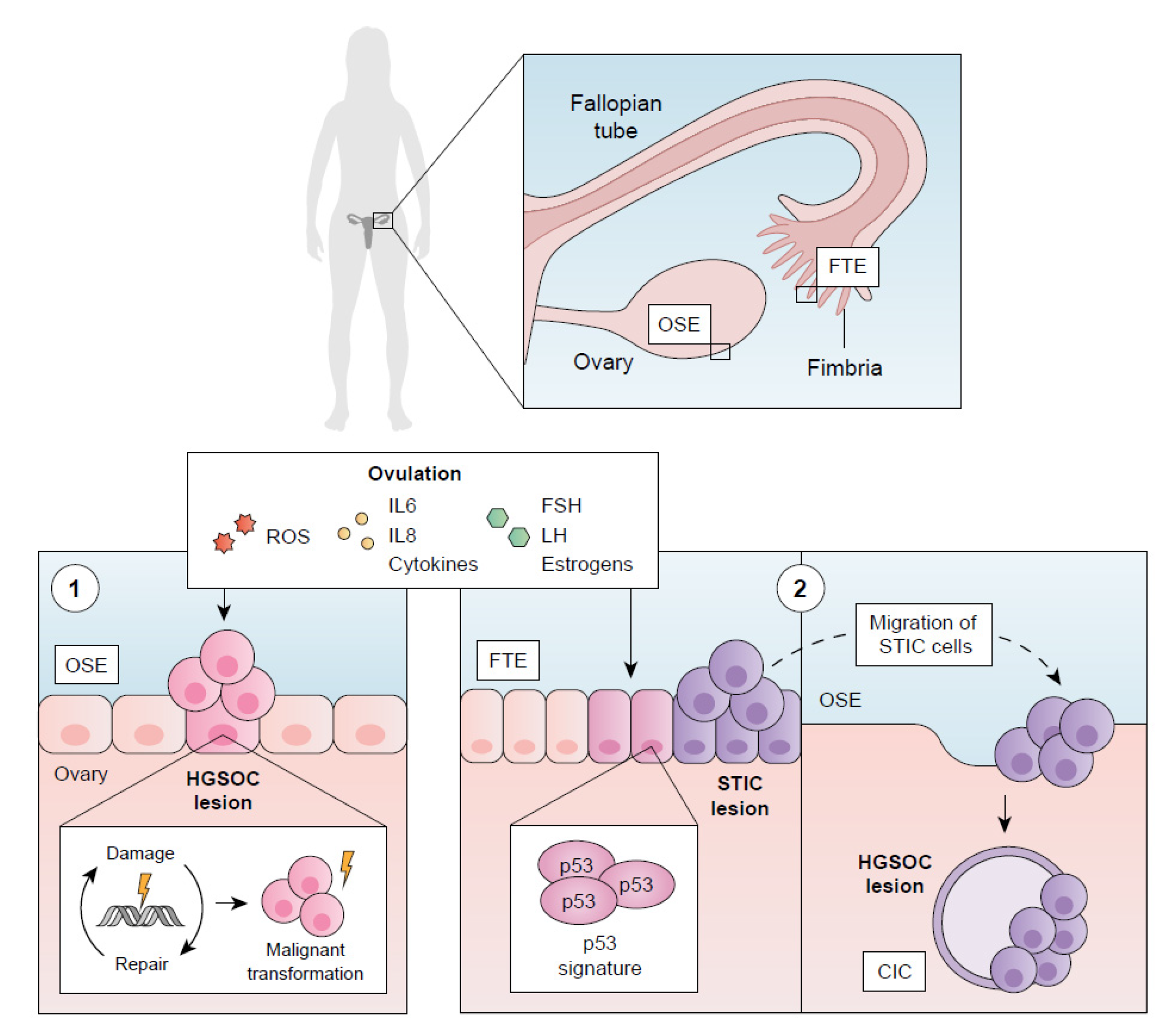
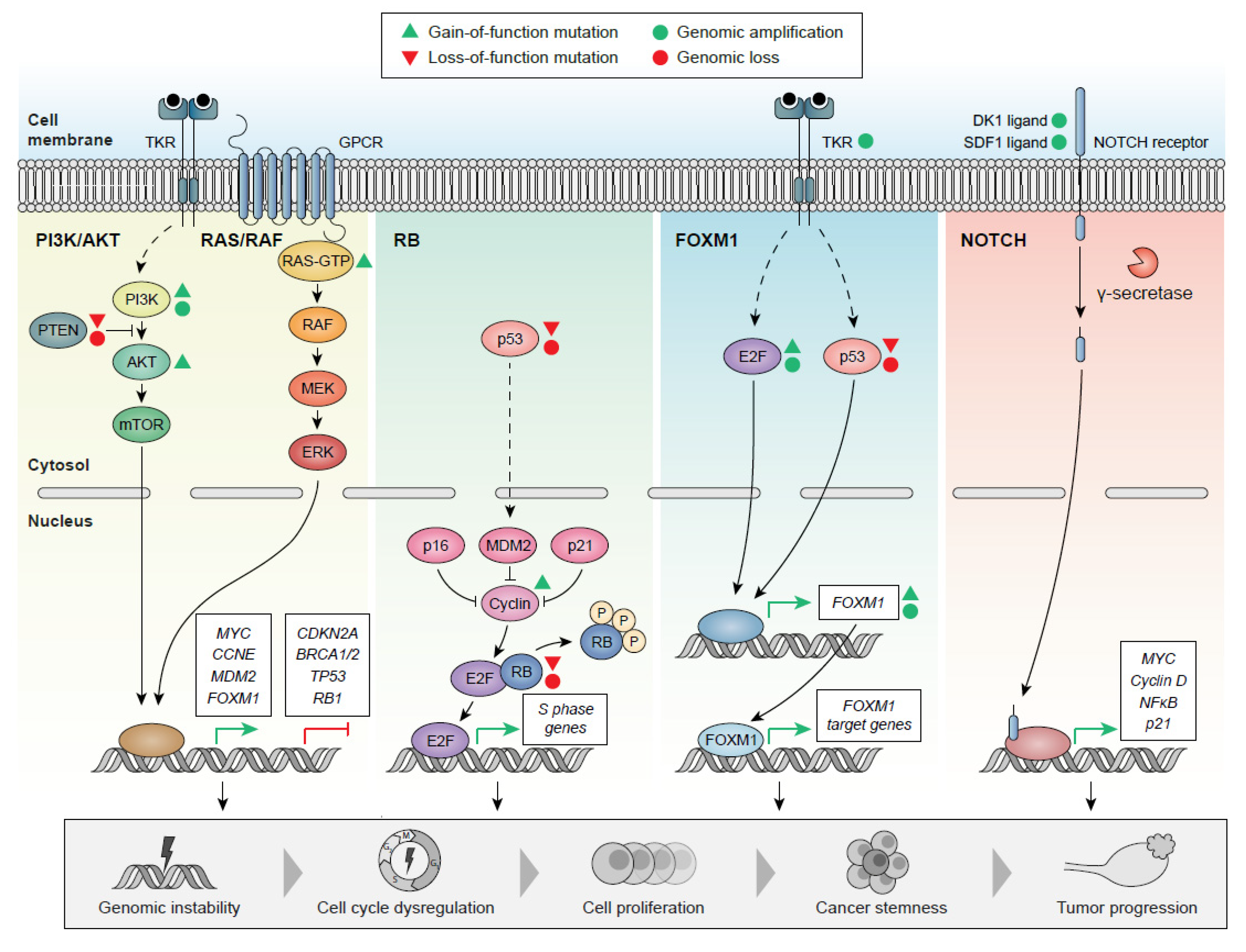
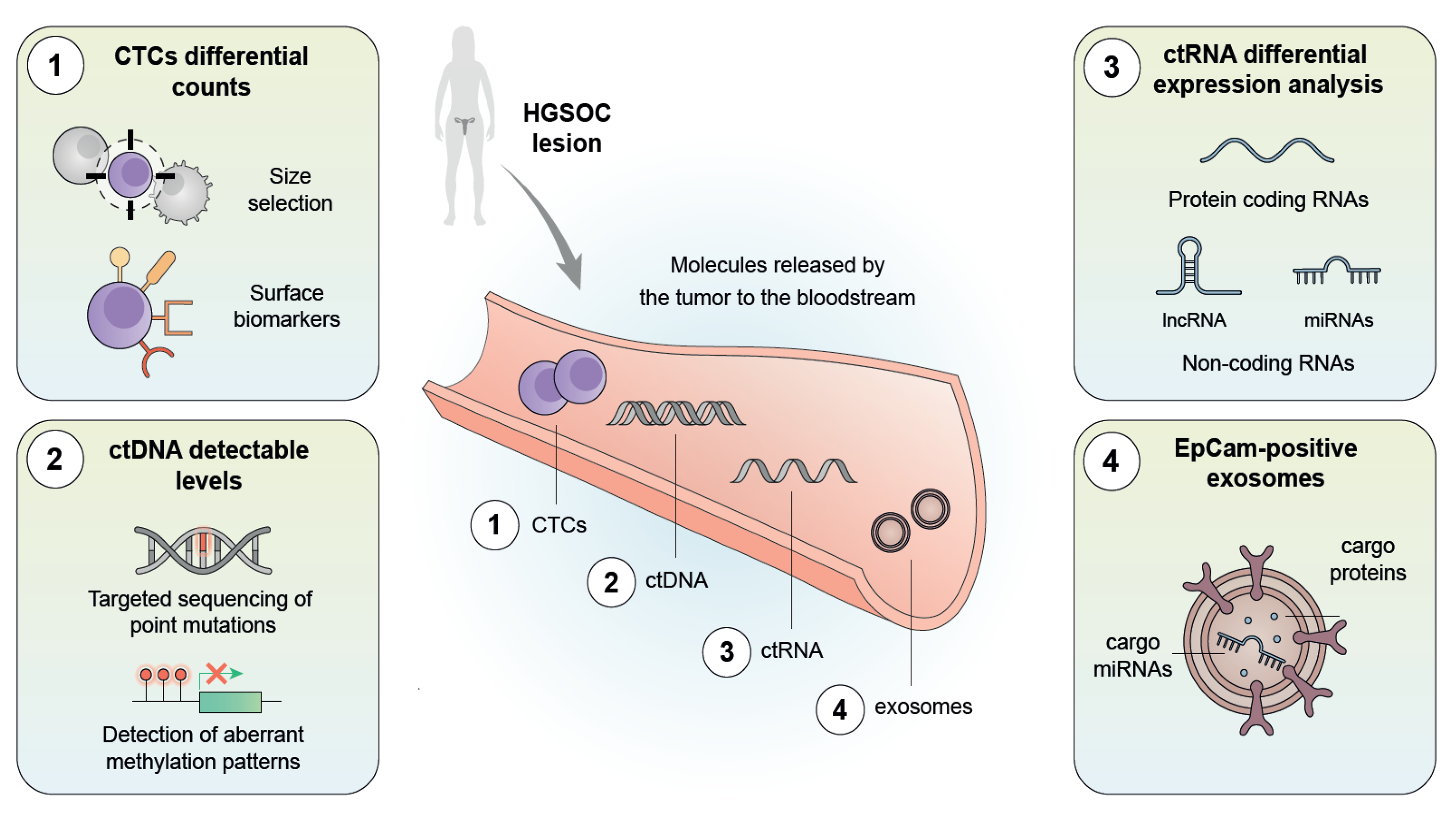
Table 1.
Genetic and epigenetic traits involved in the etiopathogenesis of HGSOC. Major genetic alterations include inherited mutations, somatic mutations, chromosomal aberrations, and epigenetic alterations, which may be considered for the early diagnosis, prognosis, and follow-up evaluation of HGSOC.
| Inherited Mutations | Mutations | Susceptible Loci | eQTL Associations | [82,166,167,168,169] | |||
| BRCA1/2, CHEK2, RAD51, MRE11A, BRIP1, RAD50, PALB2, ATM | 3q28, 10q24.33, 4q32.3, 18q11.2, 8q21.11, 22q12 | HOXD9, STRCP1, CDC42, CPNE1, CDA8, HEATR3, CCNE1 | |||||
| Somatic Mutations | Universal Mutation | Other Mutations | [82,91,170,171] | ||||
| TP53, in almost all cases | PTEN, KRAS, FAT3, CSDM, MLH3, RB1 | ||||||
| Chromosomal Aberrations | Chromosomal Gains | Chromosomal Losses | Focal Amplification Areas | Focal Deletion Areas | CLP | [82,172,173,174] | |
| Chr 1, 3, 7, 8, 12, 20 | Chr 5, 6, 11, 16, 17, 18, 19 | CNNE1, MYC, MECOM | PTEN, RB1, NF1 | MLF1, ARID1A, BRCA1, MYC, CCNE1, PIK3CA, TP53 | |||
| Epigenetic Alterations | DNA methylation | Histone Modifications | miRNAs | lncRNAs | [175,176,177,178,179,180,181,182] | ||
| Hypermethylation: ↓ transcription BRCA1 promoter | Methyltransferase overexpression (CARM1) | miR-1290, miR27-a-3, miR23a, miR200 family, miR025 | CTBP1-DT | ||||
| Hypomethylation: ↑ oncogene transcription | Histone deacetylase overexpression | ||||||
Chr: chromosome; CLP: chromothripsis-like patterns; eQTL: expression quantitative trait locus. (↑) indicates increase in transcription and (↓) indicates decrease in transcription.
Publisher’s Note: MDPI stays neutral with regard to jurisdictional claims in published maps and institutional affiliations. |
© 2022 by the authors. Licensee MDPI, Basel, Switzerland. This article is an open access article distributed under the terms and conditions of the Creative Commons Attribution (CC BY) license (https://creativecommons.org/licenses/by/4.0/).
Share and Cite
MDPI and ACS Style
Punzón-Jiménez, P.; Lago, V.; Domingo, S.; Simón, C.; Mas, A. Molecular Management of High-Grade Serous Ovarian Carcinoma. Int. J. Mol. Sci. 2022, 23, 13777. https://doi.org/10.3390/ijms232213777
AMA Style
Punzón-Jiménez P, Lago V, Domingo S, Simón C, Mas A. Molecular Management of High-Grade Serous Ovarian Carcinoma. International Journal of Molecular Sciences. 2022; 23(22):13777. https://doi.org/10.3390/ijms232213777
Chicago/Turabian StylePunzón-Jiménez, Paula, Victor Lago, Santiago Domingo, Carlos Simón, and Aymara Mas. 2022. "Molecular Management of High-Grade Serous Ovarian Carcinoma" International Journal of Molecular Sciences 23, no. 22: 13777. https://doi.org/10.3390/ijms232213777
Note that from the first issue of 2016, this journal uses article numbers instead of page numbers. See further details here.