Review

The Yin/Yang Balance of Communication between Sensory Neurons and Macrophages in Traumatic Peripheral Neuropathic Pain

by
Roxana-Olimpia Gheorghe
,
Andreea Violeta Grosu
,
Melania Bica-Popi
and
Violeta Ristoiu
*
Department of Anatomy, Animal Physiology and Biophysics, Faculty of Biology, University of Bucharest, 91-95 Splaiul Independentei, District 5, 050095 Bucharest, Romania
*
Author to whom correspondence should be addressed.
Int. J. Mol. Sci. 2022, 23(20), 12389; https://doi.org/10.3390/ijms232012389
Submission received: 5 September 2022 / Revised: 7 October 2022 / Accepted: 11 October 2022 / Published: 16 October 2022
(This article belongs to the Special Issue Ion Channels of Nociception 2.0)

Abstract

:
Traumatic peripheral neuropathic pain is a complex syndrome caused by a primary lesion or dysfunction of the peripheral nervous system. Secondary to the lesion, resident or infiltrating macrophages proliferate and initiate a cross-talk with the sensory neurons, at the level of peripheral nerves and sensory ganglia. The neuron–macrophage interaction, which starts very early after the lesion, is very important for promoting pain development and for initiating changes that will facilitate the chronicization of pain, but it also has the potential to facilitate the resolution of injury-induced changes and, consequently, promote the reduction of pain. This review is an overview of the unique characteristics of nerve-associated macrophages in the peripheral nerves and sensory ganglia and of the molecules and signaling pathways involved in the neuro-immune cross-talk after a traumatic lesion, with the final aim of better understanding how the balance between pro- and anti-nociceptive dialogue between neurons and macrophages may be modulated for new therapeutic approaches.

1. Introduction: Neuropathic Pain Associated with Traumatic Peripheral Nerve Injuries

Peripheral nerve fibers are prone to being damaged easily by compression, crush, or transection, which results in nerve injuries of different degrees of severity. Peripheral nerve injuries are considered major health issues, given their higher prevalence and their deleterious impact on the motor activity and on the sensations and perceptions from the skin and joints, which frequently results in lifelong disability of an individual [1]. Depending on the degree of demyelination, the extent of damage to the axons and to the connective tissue, peripheral nerve injuries have been classified into several types according to Seddon [2] and expanded by Sunderland [3] (Figure 1): Neurapraxia is the mildest type of nerve injury, caused by local ischemia, traction, mild crush or compression. It is defined by a temporary blockage of nerve conduction caused by a segmental demyelination. Axonotmesis is caused by a nerve crush (due to a bat, a surgical clamp, or other crushing object) that does not result in a transection. It is characterized by a loss of axonal continuity, but the connective tissue surrounding the nerve is preserved. Neurotmesis is the most severe. It is caused by a nerve crush (due to laceration from a knife, gunshot, glass, etc.), which determines a full transection of the axons and connective tissue layers, with a complete loss of axonal continuity. A more extensive characterization of these types of peripheral nerve injuries has been reviewed elsewhere [4,5].
In axonotmesis and neurotmesis, axon regeneration is the primary goal. For a full recovery, the nerve must undergo three main processes: Wallerian degeneration (WD) (the clearing process of the remaining debris), axonal regeneration and end-organ reinnervation. WD takes place within the first 2 weeks after injury and includes: (1) fragmentation and degeneration of the axons distal to the lesion and (2) phagocytosis of the myelin sheath by Schwann cells and macrophages with the purpose of removing the debris, cleaning the area and allowing the peripheral nerve to regrow/regenerate from the segment proximal to the injury [6]. As the events associated with WD progress, more and more resident or hematogenous macrophages are recruited to the lesion site, which emphasizes their critical role for the WD and subsequent regeneration [7]. However, their contribution comes with a price, and this price is pain.
The pain that develops after an extensive nerve injury is usually chronic (persists for 3 months after trauma) [8] and neuropathic in nature. Chronic pain represents a major cause of human suffering worldwide (~20% of adults in developed nations) [9,10,11,12] especially because effective, specific and safe therapies have yet to be developed. Many of the changes that occur in response to nerve injury are adaptive, promoting repairing and healing, such as: removal of cell and myelin debris, changes in ionic channels and receptors to balance the loss of input, recruitment of antiapoptotic survival mechanisms to prevent neuronal death, induction of axon growth and sprouting, synaptic modelling and remyelination [13]. There are, however, many maladaptive changes, such as: abnormal stimulus thresholds and sensitivity, ectopic impulse generation, conduction slowing or block, reduced inhibition, inappropriate connectivity, abortive growth, neuronal loss and glial scarring [14,15]. These maladaptive changes induce an increase in pain sensitivity along the pain pathway, both in the peripheral component of the pain pathway, in a process known as peripheral sensitization, and in the central component of the pain pathway, in a process known as central sensitization [16,17]. Peripheral sensitization is characterized by an increased responsiveness and reduced spiking threshold of peripheral nociceptive neurons to stimulation of their receptive fields, induced by the local release of primary mediators either from primary sensory neurons or from the immune cells [18]. Central sensitization is characterized by an increased responsiveness of nociceptive neurons in the central nervous system (CNS) to their normal or subthreshold afferent input, induced by the release of secondary mediators from primary afferent terminals in the spinal dorsal horn, or tertiary mediators from microglia or astrocytes in the spinal cord [18]. Peripheral and central sensitization are frequently accompanied by allodynia (painful response to non-noxious stimuli), hyperalgesia (exaggerated response to noxious stimuli), or spontaneous pain [14], which significantly reduce the quality of life of patients and require therapeutic interventions. To understand the pathogenesis of traumatic neuropathic pain, many rodent surrogate models have been developed, such as: sciatic nerve transection (SNT), partial sciatic nerve ligation (PSNL), spinal nerve ligation (SNL), spared nerve injury (SNI) and chronic constriction injury (CCI) [19,20]. Even though these models are not always very good predictors of the involvement of particular targets or processes in human neuropathic pain and are incomplete because the subjective component of pain cannot be evaluated, they have great utility in exploring the cellular and molecular processes responsible for pain.
Neuropathic pain is mainly the result of a neuronal dysfunction [14,15,17,21]. However, there is a growing body of evidence indicating that the cause of neuropathic pain is not restricted to changes in neuronal activity, but may involve numerous neuro-immune interactions among neurons, immune cells and immune-like glial cells in the periphery and CNS, mediated by inflammatory cytokines and chemokines [22,23,24,25] and facilitated by ion channels expressed in either neurons or immune cells. Among the immune cells involved in these neuro-immune interactions, macrophages, which are the primary sensors of danger in the host, seem to play an important role. In this review, we will outline the molecules that mediate the interactions between macrophages and sensory neurons from the peripheral nervous system and facilitate the development and maintenance of neuropathic pain, by integrating data mainly from animal models of traumatic peripheral neuropathic pain.

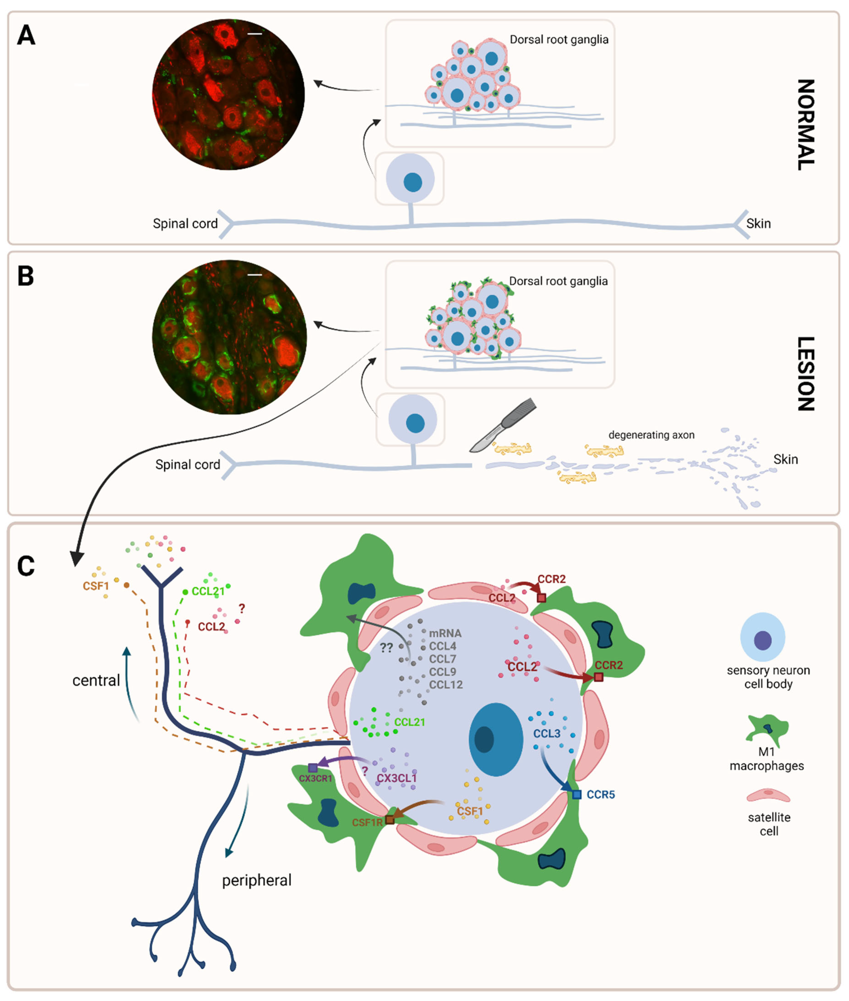
2. Macrophages in the Peripheral Nervous System: A Distinct Population

Macrophages are present in all tissues and have a chameleon-like phenotype, changing their physiology easily as a response to environmental stimuli [26]. They act as house-keeping cells, performing immune surveillance in order to remove, repair and replace cells or matrices lost through senescence or following wounding. They do these by processes such as phagocytosis and cytokine/growth factor secretion, which all belong to the innate immune response, or by antigen presentation through which the connection with the adaptive immune system is made [26]. At a given moment, depending on the signal from the environment, macrophages can exist in an M0 (resting) phenotype from which they switch to M1 (pro-inflammatory) state or M2 (anti-inflammatory) state, with many intermediary states in between [27,28].
In the peripheral nervous system, macrophages are localized in the nerves at the epineurium level, where they are typically described near blood vessels and in the endoneurial space where they represent 2–9% of all endoneurial cells, or in the sensory ganglia scattered between neuronal cell bodies [29]. In the last years, the transcriptional identity and functions of macrophages in the peripheral nervous system were intensively studied, and new information about their origin [30] and features [29,31] have been obtained.
In the nerve, studies of single-cell RNA sequencing have revealed the presence of two major subsets of macrophages inside the mouse sciatic nerve, with distinct locations: one located in the endoneurium (RelmαMgl1Iba1+ snMac1 or R1 population) and one in the epineurium (Relmα+Mgl1+Iba1+ snMac2 or R2 population) [31]. Additional transcriptomic analysis has revealed that the endoneurial R1 population expresses genes specific to microglia (the equivalent of macrophages in the CNS), such as CX3CR1, CCL2, or Trem2, and that, compared to the epineurial R2 population, it expresses more immediate early response genes, such as Atf3, Fos or Jun [31]. These data suggest that the R1 population, which resides within the nerve fascicles and is in close contact with neurons and Schwann cells, is intrinsically more responsive. Differences have been observed not only inside a nerve, but also between nerves in mice: a transcriptomic study identified 24 genes enriched in sciatic nerve macrophages, 23 genes enriched in cutaneous intercostal (fascial) nerve macrophages, and 12 genes enriched in vagus nerve macrophages [29]. Interestingly, the upregulated genes in mouse sciatic nerve-associated macrophages resemble genes expressed in macrophages isolated from sympathetic nerve fibers associated with visceral fat and genes expressed by macrophages derived from cutaneous and visceral fat [32,33].
In the dorsal root ganglia (DRG) (more commonly investigated than other sensory ganglia), macrophages have been identified as belonging to the same two distinct CD11b (+)/Ly6G (−) myeloid subpopulations as in the nerve, that is MHCII (+) and MHCII (−)/Ly6C (−) [33], likely equivalent to CX3CR1 (+) resident populations described elsewhere [29]. However, in comparison with nerve-resident macrophages, macrophages residing within the mouse DRG were found to express 79 upregulated genes and 52 downregulated genes [29]. Interestingly, the upregulated genes in mouse DRG-associated macrophages resemble genes expressed in macrophages isolated from sympathetic ganglia and genes commonly expressed by resident microglia [32,33,34].
These data suggest that although macrophages in the peripheral nervous system (PNS) are transcriptionally similar, significant differences exist between those adjacent to axons and those residing close to neuronal cell bodies, which is conceivable given the fact that tissue-resident macrophage populations are distinct from one another, and that their development and phenotype are dependent on local environmental cues [35,36].

3. Macrophages Are Quickly Activated after a Traumatic Peripheral Nerve Lesion

After a traumatic injury at the peripheral nerve level in mice, MHCII (+) and MHCII (−)/Ly6C (−) resident macrophages are joined by Ly6C (+) and MHCII (+)/Ly6C (+) infiltrating macrophages [33], which accumulate at the injury site at the nerve level, but also around the neuronal cell bodies in the sensory ganglia, and switch to the M1 phenotype. At the nerve level, 1 and 5 days after crush injury on the sciatic nerve in mice, resident macrophages (epineurial and endoneurial) represented only 5–10% of the total population, while the rest (90–95%) were infiltrating macrophages, most of them appearing at the injury site 1 day after the lesion [31]. Among the resident macrophages, it has been shown that the epineurial macrophages did not significantly react to the nerve injury, while endoneurial macrophages reacted by producing monocyte chemoattractants [31]. In the first 5 days after crush injury in mice, the recruited macrophages adopted the resident PNS macrophage signature (i.e., lost Ly6C expression and gained CD64 expression) and remained in the PNS during the repairing process, such that 1 month after the injury, macrophages were still predominantly derived from recruited circulating precursors and did not revert to the original resident macrophage population [31]. In contrast, after a partial sciatic nerve injury in mice, both resident (MHCII (+) and MHCII (−)/Ly6C (−)) and infiltrating (Ly6C (+) and MHCII (+)/Ly6C (+)) macrophages were significantly upregulated 1 to 3 days and 2, 10, and 14 weeks after injury [33]. These data suggest that after a traumatic injury, although both resident and infiltrating macrophages are recruited to the nerve lesion site, how they will respond depends largely on the type of injury.
After a traumatic injury at the sensory ganglia level, in particular DRG, macrophages form ring-like clusters around neuronal bodies, as it has been described for several traumatic neuropathic pain models: 5 days after spinal nerve ligation [37], 7 days after spared nerve injury [38], 7 days after sciatic nerve transection [39] or 7 days after sciatic nerve ligation and transection [40]. However, macrophages do not always behave like that inside DRG, as it has also been previously reviewed [41]. For example, in rats with diabetic neuropathy, macrophages do not cluster around neuronal bodies, but remain scattered between neuronal cell bodies, although the number of macrophages is increased from the moment of induction of diabetes [37], which reconfirms again that macrophages’ responses are dependent on local environmental cues.
To separate resident from infiltrating macrophages at the DRG level, studies on MAFIA (macrophage Fas-induced apoptosis) transgenic mice proved very useful [42]. More specifically, MAFIA mice carry a suicide gene expressed in macrophages, which allows a targeted, specific killing of peripheral macrophages by a specific compound, AP20187, which, unlike other drugs, fails to cross the blood–brain barrier. Mice in which a specific depletion of resident macrophages from DRG was thus performed developed mechanical allodynia 7 days after SNI, compared with 24 h later in non-treated animals, and interestingly, showed no sign of hematogenous macrophages infiltration in the meantime [42]. These data suggest that, at least for this neuropathic pain model and in contrast with the response at the nerve level, resident macrophages rather than infiltrating ones seem to be more involved in the first days after the lesion.
In the first hours after a traumatic peripheral nerve lesion, at the nerve lesion site, Schwann cells, fibroblasts and endoneurial macrophages activate, releasing chemoattractant factors which will recruit hematogenous macrophages starting 2–3 days after the lesion, with a peak at about 7 days [6,31]. In the first hours (5–10 h) after a nerve lesion, Schwann cells secrete pro-inflammatory cytokines (i.e., TNFα, IL-1α, IL-1β), which will induce in an autocrine manner their own secretion of chemoattractant chemokines (i.e., CCL2, CCL3) [43,44,45] and in a paracrine manner, in nearby resident fibroblasts, the secretion of IL-6 and GM-CSF, which downstream will act as an algesic agent and inducer of macrophages’ polarization towards the M1 phenotype (see below) [6] (Figure 2). Independently or activated by the cytokines released by Schwann cells (not clear yet), endoneurial macrophages start to release monocyte-macrophage chemoattractants (i.e., CCL6, CCL7, CCL8, and CCL12) on day 1 after injury [31] (Figure 2). Recruitment is further aided by TNFα-dependent induction of the matrix metalloproteinase MMP-9 that Schwann cells produce [46] and by complement [47].
At the sensory ganglia level, in particular DRG, macrophages are attracted around DRG neurons by different chemoattractant molecules.
The CCL2/MCP1 chemokine (Figure 3) is upregulated in DRG neurons after SNL [48], SNI [49], PSNL [50], sciatic nerve ligation and transection [40] or CCI [51], and facilitates clustering of macrophages around DRG neurons since this clustering does not happen anymore in CCL2/ animals [40]. The same clustering effect was obtained by just overexpressing CCL2 in mouse L5 DRG using viral transfection methods [52]. However, CCR2 (+) (the CCL2 receptor) macrophages have been also identified in L4/L5 DRG of uninjured CSF1R-GFP+/ & CCR2-RFP+/ mice, suggesting that there might be an endogenous secretion of CCL2 to which resident macrophages can respond [42].
CSF1/M-CSF (Figure 3) is a cytokine secreted at the mRNA level by DRG neurons after SNI as soon as 2 days after the lesion [53], which stays up 3–7 days after SNI in mouse DRG neurons specifically surrounded by rings of GFP (+) macrophages [42]. Conditional deletion of the CSF1 gene abrogated macrophage proliferation and clustering at PO4 after SNI in the ipsilateral axotomized DRG, but these events were not compromised in mice globally lacking CCL2, which suggest that the macrophages’ attraction around DRG neurons is a CSF1-dependent and CCL2-independent process [42]. Considering that the inhibition of the CSF1 receptor also prevented the proliferation of resident macrophages in rats [54], it can be concluded that the ring of the macrophages formed around DRG neurons is the result of a combination of chemotaxis and local proliferation CSF1-dependent, at least after SNI.
In addition, some other chemokines might also contribute to the ring formation, although additional experimental data are still necessary (Figure 3). Thus, injections of CCL3 and CX3CL1 directly into the L5 rat DRG leads to a macrophage accumulation in the ganglion comparable to that which occurred after sciatic nerve lesion or after injection of CCL2 [40]. In addition, after L5 spinal nerve transection injury in mice, CCL3, whose expression increases at significant levels at 8 h after the lesion, to constantly decrease at 24 and 48 h later, is, at least partly and in a TLR2-dependent manner, responsible for the macrophage infiltration in the DRG [55]. On the other hand, CX3CL1 is expressed in all rat DRG neurons regardless of size in control conditions, but no increase was noticed after CCI [56], similar to the sciatic nerve, where no increase was found after axotomy in mice [57]. These data put CX3CL1′s contribution to the ring formation under a question mark, although its role in neuropathic pain development at the level of the spinal cord is very well documented [58].
In an RNA-seq study on mice, increased levels for mRNA for CCL4, CCL7, CCL9, and CCL12 in the DRG were reported after injury, but the effects of these molecules on macrophage infiltration and a possible ring formation around DRG neurons in vivo remain to be tested [59] (Figure 3). These chemokines may also act as secondary mediators between DRG neurons and the dorsal horn of the spinal cord, where they will activate spinal microglia, causing them to further release tertiary mediators which will activate spinal neurons [58]. In this way, spinal microglia can detect and respond to peripheral nerve injury and participate in the central sensitization processes mentioned above. The following chemokines are well documented as secondary mediators: (1) CSF1: it can be released from primary afferent terminals and activates microglia [53]; (2) CCL21: vesicles containing CCL21 are preferentially transported into axons [60], and it can be released from terminals of injured neurons [61] and affects microglia functioning [62]; (3) CCL2: it is known that it is expressed in vesicles in DRG neurons, and it is released in a Ca2+-dependent manner [63], but it was not identified in the primary afferent terminals ending in the dorsal horn [64]. Therefore, rather than functioning as a secondary mediator between primary afferents and spinal microglia, CCL2 may fulfill an autocrine or paracrine function within the DRG.

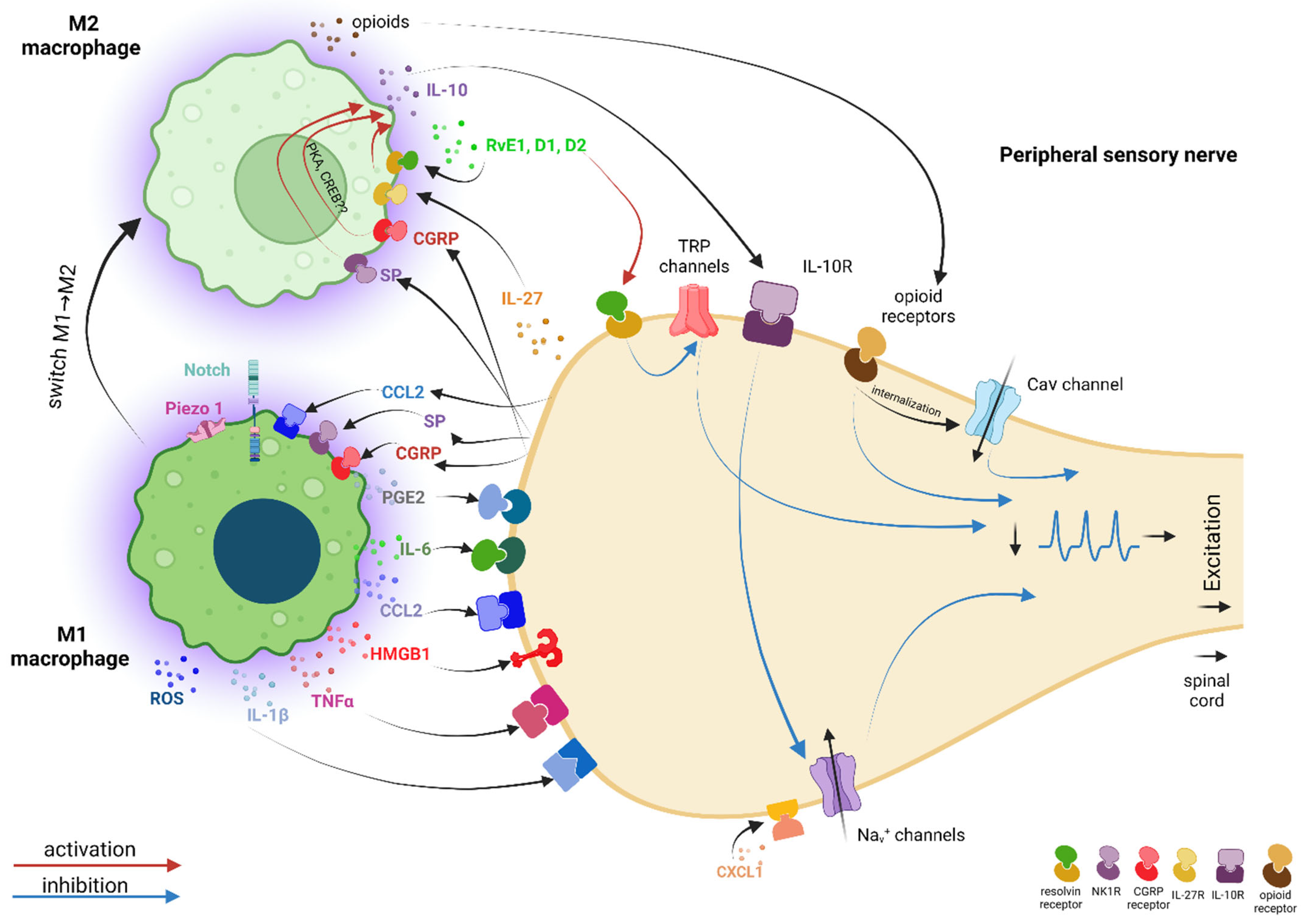
4. There Is an Intense Cross-Talk between Macrophages and Sensory Neurons Facilitated by Pro-Inflammatory Mediators, in Which Ion Channels Are Important Players

As mentioned above, macrophages that accumulate at the injury site at the nerve level or around the neuronal cell bodies in the sensory ganglia switch to the M1 phenotype. This switch is facilitated by some of the chemoattractant molecules that may have more than a chemotactic role. Thus, direct stimulation of macrophages with CCL2 favors M1 polarization, causing upregulation of TNFα in the macrophage cell line RAW264.7 [65], increased TNFα expression in murine peritoneal macrophages [66], upregulation of IL-1β, iNOS, and IL-6 mRNA levels in murine alveolar macrophages and BMDMs [67], and activation of NOX2-dependent oxidative burst in macrophages infiltrated in the mouse sciatic nerve after PSNL, further on associated with mechanical allodynia development via TRPA1 activation in Schwann cells and DRG neurons [43]. In contrast, on human macrophages, CCL2 seemed to rather facilitate the acquisition of an M2, anti-inflammatory phenotype, by modulating the expression of functionally relevant and polarization-associated genes [68]. GM-CSF secreted at the peripheral nerve level by the fibroblasts facilitated the M1 polarization as well [69], while CSF1, a powerful chemoattractant at the DRG level seemed to be, surprisingly, associated with M2 polarization [70]. TNF-α, a pro-inflammatory cytokine, was shown to also stimulate M1 polarization [71], possibly facilitated by the iron accumulation in macrophages, as it was shown in the case of macrophages infiltrated in the spinal cord after spinal cord injury in mice [72].
The close vicinity between M1 macrophages and sensory neurons facilitates an intense bidirectional communication via different primary mediators, which contributes to the establishment of a peripheral sensitization process as part of the traumatic neuropathic pain development process [73,74].
Many of the primary mediators released by macrophages have as a final target neuronal ion channels, whose altered properties contribute to increased neuronal excitability.
The IL-1β and TNFα cytokines, whose implications in neuropathic pain pathogenesis are extensively documented [75], act directly on primary sensory neurons and substantially increase their excitability by augmenting TTX-resistant sodium current via p38 MAP kinase in rats [76] (Figure 4).
The IL-6 cytokine, strongly related to neuropathic pain pathogenesis [77] and specifically associated with macrophages after PSNL injury [78], contributes to an increased neuronal excitability by the upregulation of CaV3.2 T-type channel expression and function through the IL-6/sIL-6R trans-signaling pathway via a JAK/STAT signaling in rats [79] (Figure 4).
PGE2, a pro-inflammatory prostanoid specifically associated with macrophages and traumatic neuropathic pain [80,81], causes pain by direct or indirect actions on nociceptors. Directly, PGE2 acting on specific receptors and via PKC and PKA signaling pathways sensitizes the TRPV1 receptor in mice [82] and bradykinin receptors in rats [83], facilitating a faster response of nociceptors to subsequent molecules and, therefore, the generation of an increased number of action potentials. Indirectly, PGE2 stimulates the release of pain-related neuropeptides, such as: (1) SP and CGRP via PKC and PKA signaling pathways [84]; (2) CCL2 chemokine via a not-clear-yet signaling pathway, which is downstream of the EP3 receptor [85]; (3) IL-6 cytokine via EP4/PKA/PKC/ERK signaling pathways [80]; and (4) BDNF via EP1&EP4/PKA/CREB and EP1&EP4/ERK/CREB signaling pathways [80]. Macrophages, which do not express BDNF, are nevertheless required for the nerve injury-induced upregulation of BDNF in mouse axotomized sensory neurons [42] (Figure 4).
CCL2 is secreted not only by sensory neurons, but also by macrophages [86]. Directly, via CCR2, CCL2 has excitatory effects on rat primary sensory neurons in a chronic compression of the DRG (CCD) neuropathic pain model [87], due to the activation of a non-voltage-dependent depolarizing current with characteristics similar to a non-selective cation conductance, possibly TRP channels [63], and inhibition of a voltage-dependent outward current [88] (Figure 4).
CXCL1 is another chemokine that significantly increases in rats after traumatic peripheral nerve injury [89], but its origin under these circumstances (i.e., either satellite cells or macrophages) was not clearly established [90]. When released locally, via CXCR2, CXCL1 amplifies the peripheral neuronal excitability by increasing Nav 1.1 and 1.7 currents in both IB4 (+) (nonpeptidergic sensory neurons) and IB4 (−) (peptidergic sensory neurons) DRG neurons without altering their voltage dependence or channel-opening kinetics [91]. This effect was further on correlated with a marked increase in excitability (i.e., resting potential depolarization, decreased rheobase, lower action potential threshold) and a striking ability of neurons to fire repetitively (Figure 4). In addition, opposite effects were noticed on IB4 (+) and IB4 (−) neurons upon CXCL1 stimulation, even though CXCR2 are expressed on both types of neurons. More specifically, it was shown an increase in the transient and sustained potassium currents only in IB4 (−) neurons after overnight incubation, which may facilitate repetitive firing [92], while the voltage-activated K+ current of IB4 (+) neurons was unchanged (Figure 4).
In 2010, it was reported that the high-mobility group box 1 (HMGB1) protein was upregulated in rat primary afferent neurons and satellite glial cells in the DRG and in the Schwann cells of the spinal nerve after SNL, providing the first evidence for its role in neuropathic pain pathogenesis [93]. Later on, increased HMGB1 expression was detected also in mice, in both infiltrating macrophages and proliferating Schwann cells in the sciatic nerve 14 days following PSNL [94], confirming HMGB1 as a new mediator which facilitates the communication between macrophages and sensory neurons. Interestingly, mechanical hyperalgesia induced by HMGB1 intraplantar administration in mice proved to be dependent of RAGE and TLR4 [95], which downstream induced early growth response 1 (Egr-1) transcription factor upregulation via the RAGE pathway, and further down the CaV3.2 overexpression in L4 DRG after L5 spinal nerve cutting in rats [96] (Figure 4).
Reactive oxygen species (ROS) released by macrophages were specifically related to TRPA1-mediated mechanical allodynia and neuroinflammation in a PSNL neuropathic pain model performed on mice [43]. The authors proposed a comprehensive mechanism in which ROS released by either macrophages or nociceptors maintain, in a spatially confined manner, macrophage infiltration into the injured nerve and send paracrine signals to activate TRPA1 of Schwann cells surrounding the nociceptors, to sustain mechanical allodynia [43]. More specifically, 10 days after surgery, CCL2 released at the site of the injury would promote the extravasation of hematogenous monocytes/macrophages, which would generate a rapid burst of ROS (via the NOX2-dependent oxidative pathway), which would further activate the TRPA1 channel localized in Schwann cells. TRPA1 activation in Schwann cells will evoke a Ca2+-dependent, NOX1-mediated prolonged H2O2 generation with a dual function: the outward H2O2 release would produce a space-scaled gradient that would determine the final macrophage influx to the injured nerve trunk, whereas the inward H2O2 release would target the nociceptor TRPA1 to produce mechanical allodynia [43] (Figure 4). Although not specifically related to a traumatic neuropathic pain model, it was shown that ROS released by activated macrophages upon intraplantar administration of angiotensin II further on activated TRPA1 on mouse and human DRG sensory neurons via cysteine modification of the channel [97]. Interestingly, intrathecal administration of angiotensin II in mice did not induce mechanical hypersensitivity.
Sensory neurons respond to the signals received from macrophages, not only by increasing their excitability, but also by secreting other primary mediators, which, in a positive feedback loop, act on macrophages, further activating them, thus reinforcing peripheral sensitization and pain development. Ion channels in the macrophages can be the target of this neuron–macrophage back communication.
As mentioned above, CCL2 released by injured neurons not only acts as a chemoattractant molecule, but also promotes the activation of NOX2-dependent oxidative burst in macrophages and further on ROS-dependent TRPA1 activation in Schwann cells and DRG neurons [43]. The CCL2-dependent recruitment of macrophages to the nerve injury site is most likely initiated by the mechanically activated cationic channel Piezo1, as it was suggested for macrophages involved in renal fibrosis [98]. In their study, the authors showed that Piezo1 gene deletion in the myeloid lineage of genetically engineered mice prevented the development of renal fibrosis by decreasing macrophage recruitment to the injured kidney, most likely by regulating CCL2–CCR2 signaling via Notch activity, and by restraining macrophage activation and secretion of pro-inflammatory cytokines (IL-1β, TNF-α, IL-6) via a calpain-dependent signaling pathway (Figure 4). After a traumatic nerve injury, the mechanical properties of the nerve and of the surrounding tissues, including stiffness, architecture, and molecular and cellular composition, change dramatically. There are no clear data whether Piezo1, putatively expressed by nerve-associated macrophages too, upon activation by the nerve injury-induced mechanical stress, is involved in initiating the NOX2-dependent oxidative burst in macrophages after PSNL as mentioned above or whether other signaling pathways are activated downstream. However, since physical factors strongly coregulate macrophages’ plasticity and phagocytosis [99] and Piezo1 seems to be very important in modulating macrophages’ polarization and stiffness sensing [100], it is highly likely that this ion channel is strongly involved in the dialogue macrophages initiate with neurons after a nerve injury. As also mentioned above, PGE2 secreted by injury-activated macrophages may stimulate in DRG neurons the release of CCL2, and thus, the CCL2-mediated communication loop between neurons and macrophages is complete.
Upon PGE2 activation, DRG neurons may secrete other primary mediators as well, some of which further increase the positive feedback loop and increase neuronal excitability, others facilitate the M1 to M2 switch of macrophages and initiate an anti-nociceptive dialogue between macrophages and DRG neurons, and others may act as secondary mediators and facilitate communication further up at the spinal cord level. Substance P, CGRP, IL-6, and BDNF induced by PGE2 may have one or more of these roles.
Substance P (SP) was shown to enhance the secretion of the inflammatory chemokines MIP-2 and CCL2 by murine macrophages via ERK/p38 MAPK-mediated NF-kappaB activation [101] or of the TNF-α pro-inflammatory cytokine via the JNK and p38 MAPK pathway [102], all specifics to the M1 pro-inflammatory macrophages (Figure 4). Interestingly, SP can also directly induce M2 polarization and IL-10 secretion in rat macrophages, even in the presence of IFNγ, a potent M1-skewing cytokine [103] (Figure 5). This switch is possible because macrophages have a unique, flexible metabolism, able to generate customized responses, even antagonistic ones to the same metabolite, like in the case of substance P. This may explain how, during WD, at around 7 days after a nerve injury, macrophages start to switch towards M2 anti-inflammatory phenotype and begin to secrete anti-inflammatory cytokines, such as IL-10, which will help in the resolution of inflammation and pain. In parallel to this process, axons’ regeneration is promoted by Schwann cells, which form Büngner bands to guide the regrowing of axons from the proximal end. Therefore, even though they may be “bathed” in a pro-inflammatory environment, macrophages have the potential to gradually switch to a healing phenotype due to their unique metabolism, in a very delicate Yin/Yang balance.
CGRP was strongly related to neuropathic pain, where it was shown to support both peripheral and central sensitization, without eliciting nociceptive signals alone to [104]. After a nerve injury, at DRG the level, CGRP may act in an autocrine manner and enhance TTX-resistant Na+ currents in small- and medium-diameter cultured DRG neurons [105] (Figure 4), while at the spinal cord level, upon release from the primary afferent terminals, it was shown to be able to activate signaling cascades that sensitize the NMDA receptor [106]. Interestingly, CGRP may act on macrophages as well, promoting the M2 phenotype in two ways: (1) by acting as a costimulus in parallel with other TLR4 agonists (i.e., LPS) to enhance the secretion of the anti-inflammatory cytokine IL-10 via the CGRP receptor and a PKA signaling pathway associated with a prolonged activation of CREB [107] (Figure 5), and (2) by stimulating phagocytosis [108]. These M2 pathways were not specifically associated with a traumatic pain model. Instead, after PSNL, M1-specific effects were described: increased levels of CGRP in injured neuroma and invading macrophages were associated with the upregulation of IL-6 in rat macrophages and with the maintenance of neuropathic pain [109] (Figure 4). Therefore, although not proven for traumatic neuropathic pain, neurons may have the potential to not only activate macrophages via CGRP, but also to induce their M2 switch and initiate the healing and pain resolution.
IL-6 secreted by the DRG neurons upon PGE2 activation may act in an autocrine manner to increase neuronal excitability at the DRG level, but it may also act as a secondary mediator serving as an “off signal” to ensure the transient nature of injury-induced neuroinflammation by promoting a desensitized phenotype of microglia at the spinal cord level [110] (Figure 4). BDNF secreted by the DRG neurons upon PGE2 activation acts as a secondary mediator at the level of the spinal cord, mediating the transition from acute to chronic pain and contributing to the establishment of a central sensitization process as part of the traumatic neuropathic pain development process [111,112] (Figure 4).

5. In Parallel with the “Pro-nociceptive” Dialogue between Primary Sensory Neurons and Macrophages, There Is Also an “Anti-nociceptive” Dialogue with M2 Macrophages That Helps to Reduce Pain

As mentioned in Section 4, SP and CGRP have the potential to facilitate the M2 switch of macrophages while also sustaining pro-inflammatory activities, as a sort of primary buds of anti-nociceptive communication between neurons and macrophages. In addition to that, other substances that promote the M2 switch of macrophages and have an anti-nociceptive effect have been described. Their release confirms that, in fact, macrophages located closer to neurons are there to heal them after injury, but until this happens, the neurons have to pay a toll in increased excitability and pain.
It was recently shown that 7 to 14 days after SNI, mouse dorsal root ganglia neurons release increased amounts of IL-27, which, via the WSX-1 receptor expressed mainly in macrophages, stimulates macrophages to release at their turn increased amounts of the anti-inflammatory cytokine IL-10 [113]. SNI-induced neuropathic pain was enhanced in IL-27-deficient mice, whereas nociceptive pain was similar to that of wild-type mice, indicating that IL-27 signaling is important for controlling the development of neuropathic pain, but it is not involved in the detection of mechanical, thermal, and chemical nociception in basal conditions. Interestingly, IL-10 production was not abrogated in IL-27-deficient mice, suggesting that other mechanisms might be involved in the induction of IL-10, such as SP and CGRP mentioned in Section 4. The anti-nociceptive effect of IL-10 is via IL-10 receptors, which were shown to be expressed on rat primary sensory neurons, and is due to a reduction of TTX-sensitive and Nav1.8 currents and to a reverse of the increased sodium currents induced by TNF-α [114] (Figure 5).
In addition, M2 macrophages may attenuate neuropathic pain by releasing opioid peptides (Met-enkephalin, dynorphin and β-endorphin), which bind to opioid receptors on nociceptors. It was shown that perineural transplantation of M2 macrophages (induced in vitro by treatment with IL-4) resulted in the amelioration of CCI-induced mechanical hypersensitivity even if the injections were performed on days 14 and 15 after injury [115]. Compared with M0 and M1 macrophages, M2 macrophages contained and secreted significantly higher levels of opioids, and their anti-neuropathic pain effect was compromised by opioid receptor blocking. β-endorphin and enkephalins are anti-nociceptive peptides, linking µ (mu) and δ (delta) opioid receptors, while dynorphins can mediate not only anti-nociceptive effects, via κ (kappa) opioid receptors, but also pro-nociceptive effects via NMDA receptors [116]. Opioid receptors are G-protein-coupled receptors expressed in the central and peripheral nervous systems, in neuroendocrine and immune tissues and cells [117]. In DRG neurons, N-type calcium channels along with opioid receptors can be co-internalized following prolonged agonist exposure, which may further reduce neurotransmitter release and the transmission of pain signals to CNS [118] (Figure 5).
A special category of compounds, the specialized pro-resolving lipid mediators (SPMs), may also contribute to the anti-nociceptive environment in the DRG, not necessarily in the first days after the lesion, but in the later stages of injury-induced inflammation. Recent preclinical and clinical studies have described potent anti-nociceptive effects of SPMs in different pain pathologies, either inflammatory pain (i.e., carrageenan-elicited pain, complete Freud’s adjuvant-induced pain, osteoarthritis pain, temporomandibular joint inflammatory pain), traumatic neuropathic pain (i.e., CCI, spinal cord injury, chronic compression of the dorsal root ganglia, chronic post-thoracotomy pain, postoperative pain induced by tibial bone fracture), or chemotherapy-induced pain [119]. SPMs are synthesized from polyunsaturated fatty acids (PUFA) (i.e., lipoxins (LX) derive from arachidonic acid, an omega-6 PUFA, and protectins (PD), maresins (MaR) and resolvins (Rv) derive from omega-3 fatty acids) and can be secreted by different types of cells, such as endothelial cells, leukocytes, monocytes, T cells and macrophages [119]. Interestingly, even though macrophages may secrete SPMs, they also express specific receptors for SPMs, which, either in an autocrine manner or directly if SPMs are coming from other source, may facilitate M2 polarization of macrophages and enhance their phagocytic activity to resolve inflammation [120,121]. Specifically for the traumatic neuropathic pain models discussed in this review, intrathecal administration of resolvin E1 and aspirin-triggered lipoxin A4 was associated with reduced CCI-neuropathic pain mainly by reducing microgliosis in the rat spinal cord, either as a pre-treatment, at 7 days after CCI or at 3 weeks after CCI [122,123]. However, local delivery of neuroprotectin D1/protectin D1 (NPD1/PD1) to the injury site of the sciatic nerve, at the time of surgery, was able to also completely prevent CCI-induced mechanical allodynia for 4 weeks and suppress the injury-induced infiltration of inflammatory macrophages inside DRG [124]. The analgesic effect of SPMs might also be due to blocking TRP channels on DRG neurons, via specific GPCR receptors: RvE1 inhibits TRPV1 via ChemR23 co-expressed in small DRG neurons [125], while RvD1 and D2 inhibit TRPV1 and TRPA1 via GPR18 (at least for RvD2) [126,127] (Figure 5). It is not very clear how much of the SPMs secreted at the lesion site or inside a DRG after a traumatic nerve lesion are coming from macrophages or from other cells, but the anti-nociceptive results are compelling, and more data about their role in neuron–macrophage communication are required.

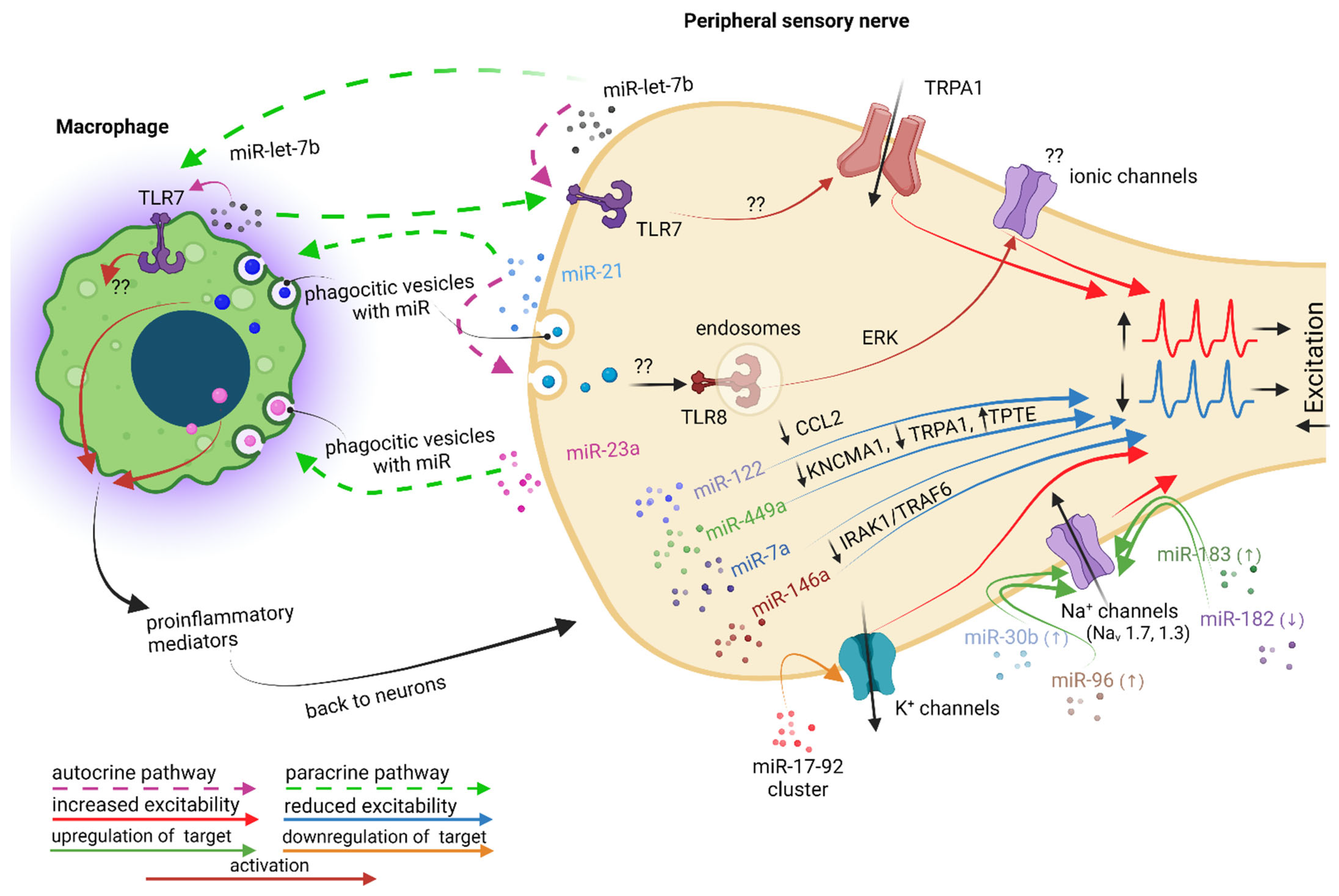
6. MicroRNAs May Also Facilitate Pro- and Anti-nociceptive Effects on Primary Sensory Neurons, with Macrophage Involvement More or Less Well Documented

In the last years, miRNAs, small noncoding RNAs of 19–25 nucleotides that bind the 3‘ UTRs of mRNAs to regulate gene expression post-transcriptionally, have started to be considered potential new targets for pain therapy, given their implication in neuropathic pain development and/or maintenance both in humans and in animal neuropathic pain models, as comprehensively reviewed by Kalpachidou et al. [128]. Generated inside the cells (i.e., nucleus or cytosol), miRNAs are also detectable in exosomes, the extracellular vesicles that both immune cells and neurons can shed in their extracellular environment and in the body fluids (i.e., CSF, blood plasma or saliva), where they can be exploited for diagnostic purposes. In this review, we will concentrate on the pro- or anti-nociceptive effects of those miRNAs, which were specifically associated with traumatic neuropathic pain in PNS.
miR-let-7b was shown to be an important pain mediator in the peripheral nervous system, given the fact that it is able to induce, in a sequence-dependent manner requiring GUUGUGU motif, rapid inward currents and action potentials only in DRG nociceptors co-expressing TLR7 and TRPA1, and that intraplantar injection of miR-let-7b elicited persistent mechanical allodynia, which was abolished in mice lacking TLR7 or TRPA1 [129]. Notably, the authors showed that miR-let-7b is highly expressed in the DRG tissue and can be released from DRG neurons in an activity-dependent manner. Knowing that miR-let-7b can activate murine microglia and macrophages through TLR7 [130] and that it can also be released by human macrophages [131], it is highly possible that this miRNA may facilitate the cross-talk between macrophages and neurons after traumatic injuries, even though, so far, it was not associated with a particular traumatic neuropathic pain model (Figure 6).
In contrast, the contribution of miR-21 to traumatic neuropathic pain is much better documented. Thus, 7 days after SNI, it was shown that the expression of miR-21 was upregulated in mouse DRG neurons with pro-nociceptive effects: the intrathecal delivery of a miR-21 antagomir and the conditional deletion of miR-21 in sensory neurons reduced neuropathic hypersensitivity and macrophage infiltration in the DRG [132]. The authors also confirmed the action mechanism: upon noxious stimulation, TRPV1 receptors are activated, which facilitates the release of miR-21 packaged in exosomes by DRG neuronal cell bodies, which are next readily engulfed by macrophages. Inside macrophages, miR-21 induces polarization towards a pro-inflammatory, pro-nociceptive M1 phenotype, which will further on facilitate pain signaling following peripheral nerve injury (Figure 6). An increase in miR-21 inside all mouse DRG neurons was also confirmed 10 days after SNL, where it was proposed that it acts in an autocrine manner via TLR8 receptors only in small and medium-sized neurons, which are the only types of DRG neurons that coexpress miR-21 and TLR8 receptors [133]. After SNL injury, miR-21 could be released by large neurons as well, and be taken up by small and medium neurons to reach endosomal TLR8. TLR8 is not expressed in the membrane of DRG neurons, indicating that it cannot directly control plasma membrane excitability. Instead, TLR8 is expressed in the subcellular endoplasmic reticulum, endosomes and lysosomes of DRG neurons, similar to its location in monocytes and macrophages [133]. Intrathecal injection of miR-21 decreases the paw withdrawal threshold, increases p-ERK expression in the DRG in WT mice but not in TLR8/ mice, and further increases the number of action potentials in the whole-mount DRGs after incubation with miR-21 in the DRG neurons of WT mice but not TLR8/ mice. Even though ionic channel targets are not clear in this case, all these data confirm that miR-21 is involved in neuropathic pain pathogenesis (Figure 6).
A similar polarizing effect towards an M1 phenotype was shown for miR-23a, upregulated in mice DRG neurons after SNI [134]. Capsaicin stimulation of DRG neurons in culture facilitated the release of extracellular vesicles enriched with miR-23a, which were further on taken up in macrophages where, by interaction with the A20 gene, facilitated the macrophages’ switch towards a pro-inflammatory phenotype, as confirmed by increased Nos2 mRNA (M1 marker) and decreased Mrc1 mRNA (M2 marker) expression (Figure 6). A20 is a gene that encodes a ubiquitin-editing protein that is involved in the negative feedback regulation of NF-κB signaling. The effect was additionally confirmed by the fact that the intrathecal delivery of a miR-23a antagomir attenuated neuropathic hypersensitivity and reduced the number of M1 macrophages in injured DRGs [134].
The miR-17-92 cluster, a microRNA cluster with six distinct members, was shown to be upregulated in L5 rat DRG at 14 days after SNL, which was associated with significant mechanical allodynia, but not thermal hyperalgesia [135]. Through the overexpression of miR-17-92 members in L5 DRG using AAV vector injection, it was confirmed that of the six members of the cluster, only miR-18a, miR-19a, miR-19b or miR-92a were associated with mechanical allodynia. The upregulation of these miRNAs was associated with a downregulation of A-type K+ currents (Kv1.4, Kv3.4, and Kv4.3) only in small, nociceptive DRG neurons, while non-A-type K+ currents were not significantly affected (Figure 6). Since the suppression of K+ channels promotes increased neuronal excitability, the miR-17-92 cluster appears to perpetuate mechanical allodynia [135]. However, in this study, the macrophages’ contribution was not explored.
Increased levels of miR-30b, miR-96, and miR-183 were correlated with increased expression in rat DRG neurons of different subtypes of NaV channels and subsequent increased pain sensitivity: NaV1.7 after SNI [136], and NaV1.3 after CCI [137] and SNL [138], respectively. In contrast, a down-regulated level of miR-182 was associated with the increased expression of NaV1.7 in L4-L6 rat DRGs from 3 days until 14 days after SNI [139] (Figure 6). The overexpression of miR-182 via microinjection reversed the abnormal increase of NaV1.7 at both the mRNA and protein level and significantly attenuated the mechanical hypersensitivity, while the administration of miR-182 antagomir enhanced the NaV1.7 expression at both the mRNA and protein level in L4-L6 DRG and, consequently, enhanced mechanical sensitivity in naive rats [139]. Again, in these studies, the macrophages’ contribution was not explored.
Several miRNAs were proved to have anti-nociceptive effects, alleviating neuropathic pain through different mechanisms (Figure 6). An upregulated level of miR-122 was shown to be responsible for the analgesic effect of GCSF by suppressing CCL2 expression in rat DRG neurons 7 days after CCI [140]. miR-449a alleviates mouse SNI-induced pain by decreasing the level of calcium-activated K+ channel subunit α-1 (KCNMA1) and TRPA1 and by increasing the level of transmembrane phosphatase with tension homology (TPTE) in DRG neurons [141]. miR-7a ameliorates rat SNL-induced pain via the neurofilament light polypeptide-dependent signal transducer and activator of the transcription signaling pathway [142], and miR-146a ameliorates rat CCI-induced pain through the suppression of the IRAK1/TRAF6 signaling pathway in L4-L6 DRG [143].
For these miRNA molecules, which were proven as most promising candidates to facilitate either neuropathic pain development, but also pain resolution, a number of questions remain. What is the origin of these miRNAs, neurons or macrophages? Which is the initial trigger for their production and release: is it a mechanism inside the cell (like after a nerve injury) or an external pro-inflammatory mediator? Given their influence on neuronal excitability, what ionic channels are their final target, and which is the receptor that initiates the signaling pathway towards those putative ionic channels? Is there an injury-specific pattern of miRNAs? If yes, could it be that a specific miRNA is the most important, or more miRNAs are involved, in which case the network effect (some miRNAs increased; some miRNA decreased) is more important for the development/resolution of pain? Modulating the ion channels involved in increased neuronal excitability after injury would be more efficient if it would be made via miRNAs than by blocking other pro-inflammatory induced signaling pathways? These issues require further investigation.
There is still a debate out there on which macrophages are more important for the traumatic neuropathic pain development: those from the peripheral nerve or those from the sensory ganglia?
Studies on mice have shown that in a PSNL pain model, clodronate depletion of infiltrating monocytes in injured peripheral nerve significantly attenuated macrophage infiltration and allodynia [43,144], similar to an SNI pain model on MAFIA mice where chemogenic depletion of circulating monocytes and macrophages at the nerve injury site reduced the nerve-injury-induced mechanical hypersensitivity [145]. Even more, as these authors found that DRG macrophages were spared, they concluded that peripheral macrophages, but not those in the DRG, are the critical contributors to nerve-injury-induced neuropathic pain [145]. These data are in contrast with previous studies showing that selective depletion of peripheral monocytes/macrophages had limited impact on neuropathic pain development after spinal nerve transection in mice [146] or after SNL in rats [147], which raises the possibility of a type of injury-dependent mechanism for macrophage response at the peripheral nerve.
At the sensory ganglia level, in particular DRG, Yu et al. showed not only that in MAFIA mice resident macrophages from DRG seem to be more important than infiltrating ones in the development of SNI-induced neuropathic pain, but also that DRG macrophages and not the peripheral macrophages are critical for the initiation and persistence of SNI-induced neuropathic pain [42]. Similarly, clodronate-treated mice, which lost DRG macrophages after SNI, showed reduced neuropathic tactile allodynia and cold hypersensitivity [59]. The genetic analysis identified 36 transcripts whose expression closely correlated with the onset of mechanical sensitivity and were functionally related to immune function, and 137 transcripts closely correlated with the onset of cold sensitivity, which were functionally related to neuronal function, supporting the idea that resident macrophages in the DRG are crucial to the development and persistence of tactile, but not cold allodynia, at least after SNI.

7. Conclusions

The bidirectional communication established between activated macrophages clustered close to the traumatically lesioned peripheral nerves is very complex, involving both classic primary mediators, but also new miRNa-type primary mediators, released by both immune cells and neurons, with both pro- and anti-nociceptive effects. The fact that neurons are able to secrete substances similar to the ones released by macrophages in pathological conditions, illustrates how intricate the neuro-immune communication is. Since macrophages are the first sensors of defense activated after a lesion, understanding the dialogue they carry with neurons would allow the development of new therapeutic approaches for neuropathic pain, before peripheral sensitization or central sensitization develop.

Author Contributions

Conceptualization, R.-O.G., A.V.G., M.B.-P. and V.R.; writing—original draft preparation, R.-O.G. and V.R.; writing—review and editing, R.-O.G., A.V.G., M.B.-P. and V.R.; visualization, A.V.G. and M.B.-P.; supervision, R.-O.G. and V.R. All authors have read and agreed to the published version of the manuscript.

Funding

This research was funded by the Executive Agency for Higher Education, Research, Development, and Innovation Funding (UEFISCDI), grant number PN-III-P2-2.1-PED-2019-3103 and by the University of Bucharest, grant number C1.2.PFE-CDI.2021-587, Contract no. 41PFE/30.12.2021.

Institutional Review Board Statement

Not applicable.

Informed Consent Statement

Not applicable.

Data Availability Statement

Not applicable.

Acknowledgments

All figures were created with BioRender.com.

Conflicts of Interest

The authors declare that they have no conflicts of interest.

Abbreviations

AAVadeno-associated virus
Atf3activating transcription factor 3
BDNFbrain-derived neurotrophic factor
BMDMsbone marrow-derived macrophages
Cav3.2 T-typevoltage-gated calcium channels isoform 3.2
CCDchronic compression of the DRG
CCIchronic constriction injury
CCL12chemokine (C-C motif) ligand 12
CCL2/MCP1chemokine (C-C motif) ligand 2/monocyte chemoattractant protein-1
CCL21chemokine (C-C motif) ligand 21
CCL3chemokine (C-C motif) ligand 3
CCL4chemokine (C-C motif) ligand 4
CCL6chemokine (C-C motif) ligand 6
CCL7chemokine (C-C motif) ligand 7
CCL8chemokine (C-C motif) ligand 8
CCL9chemokine (C-C motif) ligand 9
CCR2C-C chemokine receptor type 2
CD11bcluster of differentiation molecule 11B
CD64cluster of differentiation 64
CGRPcalcitonin gene-related peptide
CNScentral nervous system
CREBcyclic adenosine monophosphate (cAMP) response element-binding protein
CSF1/M-CSFcolony-stimulating factor 1/macrophage colony-stimulating factor
CSF1Rcolony-stimulating factor 1 receptor
CX3CL1chemokine (C-X3-C motif) ligand 1
CX3CR1C-X3-C motif chemokine receptor 1
CXCR2C-X-C chemokine receptor 2
DRGdorsal root ganglia
Egr-1early growth response factor 1
EP1E-prostanoid 1
EP3E-prostanoid 3
EP4E-prostanoid 4
ERKextracellular signal-regulated protein kinase
Fosfos proto-oncogene, AP-1 transcription factor subunit
GFPgreen fluorescent protein
GM-CSFgranulocyte-macrophage colony-stimulating factor
GPCRG-protein-coupled receptors
HMGB1high-mobility group box 1
IASPInternational Association for the Study of Pain
IB4isolectin B4
Iba1ionized calcium-binding adapter molecule 1
IFNγinterferon gamma
IL-10interleukin 10
IL-1αinterleukin 1 α
IL-1βinterleukin 1 β
IL-27interleukin 27
IL-4interleukin 4
IL-6interleukin 6
iNOSinducible nitric oxide synthase
IRAKIL-1R-associated kinase
JAK-STAT-Janus-kinase (JAK)signal transducers and activators of transcription (STAT)
JNKc-Jun N-terminal kinases
JunJun proto-oncogene, AP-1 transcription factor subunit
KCNMA1calcium-activated K+ channel subunit alpha-1
KV1.4voltage-gated K+ channels isoform 1.4
KV3.4voltage-gated K+ channels isoform 3.4
KV4.3voltage-gated K+ channels isoform 4.3
L4/L5/L6lumbar 4/5/6
LPSlipopolysaccharide
Ly6Clymphocyte antigen 6 complex, locus C
Ly6Glymphocyte antigen 6 complex locus G6
MAFIAmacrophage Fas-induced apoptosis
Mgl1macrophage galactose-type lectin 1
MHCIImajor histocompatibility complex class II molecules
MIP-2macrophage inflammatory protein-2
miRNAs or miRsmicroRNAs
MMP-9matrix metalloproteinase-9
mRNAmessenger RNA
NaV1.1voltage-gated Na+ channels isoform 1.1
NaV1.3voltage-gated Na+ channels isoform 1.3
NaV1.7voltage-gated Na+ channels isoform 1.7
NaV1.8voltage-gated Na+ channels isoform 1.8
NF-κBnuclear factor κB
NMDAN-methyl-d-aspartate
NOX1nicotinamide adenine dinucleotide phosphate (NADPH) oxidase isoform 1
NOX2nicotinamide adenine dinucleotide phosphate (NADPH) oxidase isoform 2
p38 MAPKp38 mitogen-activated protein kinase
p-ERKphospho-ERK
PGE2prostaglandin E2
PKAcyclic adenosine monophosphate (cAMP)-dependent protein kinase
PKCprotein kinase C
PNSperipheral nervous system
PSNLpartial sciatic nerve ligation
RAGEreceptor for advanced glycation end-products
Relmαresistin-like molecule alpha
RFPred fluorescent protein
RNAribonucleic acid
RNAseqRNA sequencing
ROSreactive oxygen species
Rv E1, D1, D2resolvins E1, D1, and D2
sIL-6Rsoluble IL-6 receptor
SNIspared nerve injury
SNLspinal nerve ligation
SNTsciatic nerve transection
SPsubstance P
TLR2, 4, 7, 8toll-like receptor isoform 2, 4, 7, or 8
TNF-αtumor necrosis factor alpha
TPTEtransmembrane phosphatase with tension homology
TRAF6tumor necrosis factor receptor (TNFR)-associated factor 6
Trem2triggering receptor expressed on myeloid cells 2
TRPtransient receptor potential
TRPA1transient receptor potential ankyrin 1
TRPV1transient receptor potential vanilloid type-1
TTXtetrodotoxin
WDWallerian degeneration
WSX-1receptor for IL-27

References

  1. Kouyoumdjian, J.A.; Graca, C.R.; Ferreira, V.F.M. Peripheral nerve injuries: A retrospective survey of 1124 cases. Neurol. India 2017, 65, 551–555. [Google Scholar] [CrossRef]
  2. Seddon, H. Three types of nerve injury. Brain J. Neurol. 1943, 66, 237–288. [Google Scholar] [CrossRef]
  3. Sunderland, S. A classification of peripheral nerve injuries producing loss of function. Brain J. Neurol. 1951, 74, 491–516. [Google Scholar] [CrossRef]
  4. Hussain, G.; Wang, J.; Rasul, A.; Anwar, H.; Qasim, M.; Zafar, S.; Aziz, N.; Razzaq, A.; Hussain, R.; de Aguilar, J.G.; et al. Current Status of Therapeutic Approaches against Peripheral Nerve Injuries: A Detailed Story from Injury to Recovery. Int. J. Biol. Sci. 2020, 16, 116–134. [Google Scholar] [CrossRef] [PubMed] [Green Version]
  5. Menorca, R.M.; Fussell, T.S.; Elfar, J.C. Nerve physiology: Mechanisms of injury and recovery. Hand Clin. 2013, 29, 317–330. [Google Scholar] [CrossRef] [PubMed] [Green Version]
  6. Rotshenker, S. Wallerian degeneration: The innate-immune response to traumatic nerve injury. J. Neuroinflamm. 2011, 8, 109. [Google Scholar] [CrossRef] [PubMed] [Green Version]
  7. Chen, P.; Piao, X.; Bonaldo, P. Role of macrophages in Wallerian degeneration and axonal regeneration after peripheral nerve injury. Acta Neuropathol. 2015, 130, 605–618. [Google Scholar] [CrossRef] [PubMed]
  8. Treede, R.D.; Rief, W.; Barke, A.; Aziz, Q.; Bennett, M.I.; Benoliel, R.; Cohen, M.; Evers, S.; Finnerup, N.B.; First, M.B.; et al. A classification of chronic pain for ICD-11. Pain 2015, 156, 1003–1007. [Google Scholar] [CrossRef] [Green Version]
  9. Breivik, H.; Collett, B.; Ventafridda, V.; Cohen, R.; Gallacher, D. Survey of chronic pain in Europe: Prevalence, impact on daily life, and treatment. Eur. J. Pain 2006, 10, 287–333. [Google Scholar] [CrossRef] [PubMed]
  10. Yong, R.J.; Mullins, P.M.; Bhattacharyya, N. Prevalence of chronic pain among adults in the United States. Pain 2022, 163, e328–e332. [Google Scholar] [CrossRef] [PubMed]
  11. Chronic Pain in Australia; Australian Institute of Health and Welfare: Canberra, Australia, 2020.
  12. Fayaz, A.; Croft, P.; Langford, R.M.; Donaldson, L.J.; Jones, G.T. Prevalence of chronic pain in the UK: A systematic review and meta-analysis of population studies. BMJ Open 2016, 6, e010364. [Google Scholar] [CrossRef] [PubMed]
  13. Benn, S.C.; Woolf, C.J. Adult neuron survival strategies--slamming on the brakes. Nat. Rev. Neurosci. 2004, 5, 686–700. [Google Scholar] [CrossRef]
  14. Costigan, M.; Scholz, J.; Woolf, C.J. Neuropathic pain: A maladaptive response of the nervous system to damage. Annu. Rev. Neurosci. 2009, 32, 1–32. [Google Scholar] [CrossRef] [PubMed] [Green Version]
  15. von Hehn, C.A.; Baron, R.; Woolf, C.J. Deconstructing the neuropathic pain phenotype to reveal neural mechanisms. Neuron 2012, 73, 638–652. [Google Scholar] [CrossRef] [PubMed] [Green Version]
  16. Gangadharan, V.; Kuner, R. Pain hypersensitivity mechanisms at a glance. Dis. Model. Mech. 2013, 6, 889–895. [Google Scholar] [CrossRef] [PubMed] [Green Version]
  17. Kuner, R.; Flor, H. Structural plasticity and reorganisation in chronic pain. Nat. Rev. Neurosci. 2017, 18, 113. [Google Scholar] [CrossRef] [PubMed] [Green Version]
  18. IASP, International Association for the Study of Pain (IASP) Terminology. International Association for the Study of Pain. 2017. Available online: https://www.iasp-pain.org/terminology?navItemNumber=576 (accessed on 10 October 2022).
  19. Dowdall, T.; Robinson, I.; Meert, T.F. Comparison of five different rat models of peripheral nerve injury. Pharmacol. Biochem. Behav. 2005, 80, 93–108. [Google Scholar] [CrossRef]
  20. Sorkin, L.S.; Yaksh, T.L. Behavioral models of pain states evoked by physical injury to the peripheral nerve. Neurother. J. Am. Soc. Exp. Neurother. 2009, 6, 609–619. [Google Scholar] [CrossRef] [Green Version]
  21. Gangadharan, V.; Zheng, H.; Taberner, F.J.; Landry, J.; Nees, T.A.; Pistolic, J.; Agarwal, N.; Mannich, D.; Benes, V.; Helmstaedter, M.; et al. Neuropathic pain caused by miswiring and abnormal end organ targeting. Nature 2022, 606, 137–145. [Google Scholar] [CrossRef]
  22. Raoof, R.; Willemen, H.; Eijkelkamp, N. Divergent roles of immune cells and their mediators in pain. Rheumatology 2018, 57, 429–440. [Google Scholar] [CrossRef]
  23. Lim, J.S.; Kam, P.C. Neuroimmune mechanisms of pain: Basic science and potential therapeutic modulators. Anaesth. Intensive Care 2020, 48, 167–178. [Google Scholar] [CrossRef]
  24. Calvo, M.; Dawes, J.M.; Bennett, D.L. The role of the immune system in the generation of neuropathic pain. Lancet Neurol. 2012, 11, 629–642. [Google Scholar] [CrossRef]
  25. Grace, P.M.; Hutchinson, M.R.; Maier, S.F.; Watkins, L.R. Pathological pain and the neuroimmune interface. Nat. Rev. Immunol. 2014, 14, 217–231. [Google Scholar] [CrossRef] [Green Version]
  26. Wynn, T.A.; Chawla, A.; Pollard, J.W. Macrophage biology in development, homeostasis and disease. Nature 2013, 496, 445–455. [Google Scholar] [CrossRef] [Green Version]
  27. Mosser, D.M.; Edwards, J.P. Exploring the full spectrum of macrophage activation. Nat. Rev. Immunol. 2008, 8, 958–969. [Google Scholar] [CrossRef]
  28. Italiani, P.; Boraschi, D. From Monocytes to M1/M2 Macrophages: Phenotypical vs. Functional Differentiation. Front. Immunol. 2014, 5, 514. [Google Scholar] [CrossRef] [PubMed] [Green Version]
  29. Wang, P.L.; Yim, A.K.Y.; Kim, K.W.; Avey, D.; Czepielewski, R.S.; Colonna, M.; Milbrandt, J.; Randolph, G.J. Peripheral nerve resident macrophages share tissue-specific programming and features of activated microglia. Nat. Commun. 2020, 11, 2552. [Google Scholar] [CrossRef] [PubMed]
  30. Kolter, J.; Kierdorf, K.; Henneke, P. Origin and Differentiation of Nerve-Associated Macrophages. J. Immunol. 2020, 204, 271–279. [Google Scholar] [CrossRef] [PubMed]
  31. Ydens, E.; Amann, L.; Asselbergh, B.; Scott, C.L.; Martens, L.; Sichien, D.; Mossad, O.; Blank, T.; De Prijck, S.; Low, D.; et al. Profiling peripheral nerve macrophages reveals two macrophage subsets with distinct localization, transcriptome and response to injury. Nat. Neurosci. 2020, 23, 676–689. [Google Scholar] [CrossRef] [PubMed]
  32. Pirzgalska, R.M.; Seixas, E.; Seidman, J.S.; Link, V.M.; Sanchez, N.M.; Mahu, I.; Mendes, R.; Gres, V.; Kubasova, N.; Morris, I.; et al. Sympathetic neuron-associated macrophages contribute to obesity by importing and metabolizing norepinephrine. Nat. Med. 2017, 23, 1309–1318. [Google Scholar] [CrossRef] [PubMed]
  33. Liang, Z.; Hore, Z.; Harley, P.; Uchenna Stanley, F.; Michrowska, A.; Dahiya, M.; La Russa, F.; Jager, S.E.; Villa-Hernandez, S.; Denk, F. A transcriptional toolbox for exploring peripheral neuroimmune interactions. Pain 2020, 161, 2089–2106. [Google Scholar] [CrossRef] [PubMed]
  34. Krasemann, S.; Madore, C.; Cialic, R.; Baufeld, C.; Calcagno, N.; El Fatimy, R.; Beckers, L.; O’Loughlin, E.; Xu, Y.; Fanek, Z.; et al. The TREM2-APOE Pathway Drives the Transcriptional Phenotype of Dysfunctional Microglia in Neurodegenerative Diseases. Immunity 2017, 47, 566–581.e9. [Google Scholar] [CrossRef] [PubMed] [Green Version]
  35. Lavin, Y.; Winter, D.; Blecher-Gonen, R.; David, E.; Keren-Shaul, H.; Merad, M.; Jung, S.; Amit, I. Tissue-resident macrophage enhancer landscapes are shaped by the local microenvironment. Cell 2014, 159, 1312–1326. [Google Scholar] [CrossRef] [Green Version]
  36. Gosselin, D.; Link, V.M.; Romanoski, C.E.; Fonseca, G.J.; Eichenfield, D.Z.; Spann, N.J.; Stender, J.D.; Chun, H.B.; Garner, H.; Geissmann, F.; et al. Environment drives selection and function of enhancers controlling tissue-specific macrophage identities. Cell 2014, 159, 1327–1340. [Google Scholar] [CrossRef] [PubMed]
  37. Ton, B.H.; Chen, Q.; Gaina, G.; Tucureanu, C.; Georgescu, A.; Strungaru, C.; Flonta, M.L.; Sah, D.; Ristoiu, V. Activation profile of dorsal root ganglia Iba-1 (+) macrophages varies with the type of lesion in rats. Acta Histochem. 2013, 115, 840–850. [Google Scholar] [CrossRef]
  38. Vega-Avelaira, D.; Geranton, S.M.; Fitzgerald, M. Differential regulation of immune responses and macrophage/neuron interactions in the dorsal root ganglion in young and adult rats following nerve injury. Mol. Pain 2009, 5, 70. [Google Scholar] [CrossRef] [Green Version]
  39. Kwon, M.J.; Kim, J.; Shin, H.; Jeong, S.R.; Kang, Y.M.; Choi, J.Y.; Hwang, D.H.; Kim, B.G. Contribution of macrophages to enhanced regenerative capacity of dorsal root ganglia sensory neurons by conditioning injury. J. Neurosci. 2013, 33, 15095–15108. [Google Scholar] [CrossRef] [Green Version]
  40. Kwon, M.J.; Shin, H.Y.; Cui, Y.; Kim, H.; Thi, A.H.; Choi, J.Y.; Kim, E.Y.; Hwang, D.H.; Kim, B.G. CCL2 Mediates Neuron-Macrophage Interactions to Drive Proregenerative Macrophage Activation Following Preconditioning Injury. J. Neurosci. 2015, 35, 15934–15947. [Google Scholar] [CrossRef] [Green Version]
  41. Ristoiu, V. Contribution of macrophages to peripheral neuropathic pain pathogenesis. Life Sci. 2013, 93, 870–881. [Google Scholar] [CrossRef]
  42. Yu, X.; Liu, H.; Hamel, K.A.; Morvan, M.G.; Yu, S.; Leff, J.; Guan, Z.; Braz, J.M.; Basbaum, A.I. Dorsal root ganglion macrophages contribute to both the initiation and persistence of neuropathic pain. Nat. Commun. 2020, 11, 264. [Google Scholar] [CrossRef]
  43. De Logu, F.; Nassini, R.; Materazzi, S.; Carvalho Goncalves, M.; Nosi, D.; Rossi Degl’Innocenti, D.; Marone, I.M.; Ferreira, J.; Li Puma, S.; Benemei, S.; et al. Schwann cell TRPA1 mediates neuroinflammation that sustains macrophage-dependent neuropathic pain in mice. Nat. Commun. 2017, 8, 1887. [Google Scholar] [CrossRef] [PubMed] [Green Version]
  44. Martini, R.; Fischer, S.; Lopez-Vales, R.; David, S. Interactions between Schwann cells and macrophages in injury and inherited demyelinating disease. Glia 2008, 56, 1566–1577. [Google Scholar] [CrossRef] [PubMed]
  45. Perrin, F.E.; Lacroix, S.; Aviles-Trigueros, M.; David, S. Involvement of monocyte chemoattractant protein-1, macrophage inflammatory protein-1alpha and interleukin-1beta in Wallerian degeneration. Brain J. Neurol. 2005, 128, 854–866. [Google Scholar] [CrossRef] [PubMed] [Green Version]
  46. Shubayev, V.I.; Angert, M.; Dolkas, J.; Campana, W.M.; Palenscar, K.; Myers, R.R. TNFalpha-induced MMP-9 promotes macrophage recruitment into injured peripheral nerve. Mol. Cell. Neurosci. 2006, 31, 407–415. [Google Scholar] [CrossRef] [PubMed] [Green Version]
  47. Dailey, A.T.; Avellino, A.M.; Benthem, L.; Silver, J.; Kliot, M. Complement depletion reduces macrophage infiltration and activation during Wallerian degeneration and axonal regeneration. J. Neurosci. 1998, 18, 6713–6722. [Google Scholar] [CrossRef] [PubMed] [Green Version]
  48. Jeon, S.M.; Lee, K.M.; Cho, H.J. Expression of monocyte chemoattractant protein-1 in rat dorsal root ganglia and spinal cord in experimental models of neuropathic pain. Brain Res. 2009, 1251, 103–111. [Google Scholar] [CrossRef]
  49. Bravo-Caparros, I.; Ruiz-Cantero, M.C.; Perazzoli, G.; Cronin, S.J.F.; Vela, J.M.; Hamed, M.F.; Penninger, J.M.; Baeyens, J.M.; Cobos, E.J.; Nieto, F.R. Sigma-1 receptors control neuropathic pain and macrophage infiltration into the dorsal root ganglion after peripheral nerve injury. FASEB J. 2020, 34, 5951–5966. [Google Scholar] [CrossRef] [Green Version]
  50. Tanaka, T.; Minami, M.; Nakagawa, T.; Satoh, M. Enhanced production of monocyte chemoattractant protein-1 in the dorsal root ganglia in a rat model of neuropathic pain: Possible involvement in the development of neuropathic pain. Neurosci. Res. 2004, 48, 463–469. [Google Scholar] [CrossRef]
  51. Zhang, J.; De Koninck, Y. Spatial and temporal relationship between monocyte chemoattractant protein-1 expression and spinal glial activation following peripheral nerve injury. J. Neurochem. 2006, 97, 772–783. [Google Scholar] [CrossRef]
  52. Niemi, J.P.; DeFrancesco-Lisowitz, A.; Cregg, J.M.; Howarth, M.; Zigmond, R.E. Overexpression of the monocyte chemokine CCL2 in dorsal root ganglion neurons causes a conditioning-like increase in neurite outgrowth and does so via a STAT3 dependent mechanism. Exp. Neurol. 2016, 275 Pt 1, 25–37. [Google Scholar] [CrossRef]
  53. Okubo, M.; Yamanaka, H.; Kobayashi, K.; Dai, Y.; Kanda, H.; Yagi, H.; Noguchi, K. Macrophage-Colony Stimulating Factor Derived from Injured Primary Afferent Induces Proliferation of Spinal Microglia and Neuropathic Pain in Rats. PLoS ONE 2016, 11, e0153375. [Google Scholar] [CrossRef] [PubMed] [Green Version]
  54. Krishnan, A.; Bhavanam, S.; Zochodne, D. An Intimate Role for Adult Dorsal Root Ganglia Resident Cycling Cells in the Generation of Local Macrophages and Satellite Glial Cells. J. Neuropathol. Exp. Neurol. 2018, 77, 929–941. [Google Scholar] [CrossRef] [Green Version]
  55. Kim, D.; You, B.; Lim, H.; Lee, S.J. Toll-like receptor 2 contributes to chemokine gene expression and macrophage infiltration in the dorsal root ganglia after peripheral nerve injury. Mol. Pain 2011, 7, 74. [Google Scholar] [CrossRef] [PubMed] [Green Version]
  56. Verge, G.M.; Milligan, E.D.; Maier, S.F.; Watkins, L.R.; Naeve, G.S.; Foster, A.C. Fractalkine (CX3CL1) and fractalkine receptor (CX3CR1) distribution in spinal cord and dorsal root ganglia under basal and neuropathic pain conditions. Eur. J. Neurosci. 2004, 20, 1150–1160. [Google Scholar] [CrossRef]
  57. Lindborg, J.A.; Mack, M.; Zigmond, R.E. Neutrophils Are Critical for Myelin Removal in a Peripheral Nerve Injury Model of Wallerian Degeneration. J. Neurosci. 2017, 37, 10258–10277. [Google Scholar] [CrossRef] [PubMed] [Green Version]
  58. Boakye, P.A.; Tang, S.J.; Smith, P.A. Mediators of Neuropathic Pain; Focus on Spinal Microglia, CSF-1, BDNF, CCL21, TNF-alpha, Wnt Ligands, and Interleukin 1beta. Front. Pain Res. 2021, 2, 698157. [Google Scholar] [CrossRef] [PubMed]
  59. Cobos, E.J.; Nickerson, C.A.; Gao, F.; Chandran, V.; Bravo-Caparros, I.; Gonzalez-Cano, R.; Riva, P.; Andrews, N.A.; Latremoliere, A.; Seehus, C.R.; et al. Mechanistic Differences in Neuropathic Pain Modalities Revealed by Correlating Behavior with Global Expression Profiling. Cell Rep. 2018, 22, 1301–1312. [Google Scholar] [CrossRef] [Green Version]
  60. de Jong, E.K.; Vinet, J.; Stanulovic, V.S.; Meijer, M.; Wesseling, E.; Sjollema, K.; Boddeke, H.W.; Biber, K. Expression, transport, and axonal sorting of neuronal CCL21 in large dense-core vesicles. FASEB J. 2008, 22, 4136–4145. [Google Scholar] [CrossRef] [Green Version]
  61. de Jong, E.K.; Dijkstra, I.M.; Hensens, M.; Brouwer, N.; van Amerongen, M.; Liem, R.S.; Boddeke, H.W.; Biber, K. Vesicle-mediated transport and release of CCL21 in endangered neurons: A possible explanation for microglia activation remote from a primary lesion. J. Neurosci. 2005, 25, 7548–7557. [Google Scholar] [CrossRef] [Green Version]
  62. Honjoh, K.; Nakajima, H.; Hirai, T.; Watanabe, S.; Matsumine, A. Relationship of Inflammatory Cytokines From M1-Type Microglia/Macrophages at the Injured Site and Lumbar Enlargement With Neuropathic Pain After Spinal Cord Injury in the CCL21 Knockout (plt) Mouse. Front. Cell. Neurosci. 2019, 13, 525. [Google Scholar] [CrossRef]
  63. Jung, H.; Toth, P.T.; White, F.A.; Miller, R.J. Monocyte chemoattractant protein-1 functions as a neuromodulator in dorsal root ganglia neurons. J. Neurochem. 2008, 104, 254–263. [Google Scholar] [CrossRef] [Green Version]
  64. Jung, H.; Bhangoo, S.; Banisadr, G.; Freitag, C.; Ren, D.; White, F.A.; Miller, R.J. Visualization of chemokine receptor activation in transgenic mice reveals peripheral activation of CCR2 receptors in states of neuropathic pain. J. Neurosci. 2009, 29, 8051–8062. [Google Scholar] [CrossRef] [PubMed] [Green Version]
  65. Wang, Q.; Ren, J.; Morgan, S.; Liu, Z.; Dou, C.; Liu, B. Monocyte chemoattractant protein-1 (MCP-1) regulates macrophage cytotoxicity in abdominal aortic aneurysm. PLoS ONE 2014, 9, e92053. [Google Scholar] [CrossRef]
  66. Sodhi, A.; Biswas, S.K. Monocyte chemoattractant protein-1-induced activation of p42/44 MAPK and c-Jun in murine peritoneal macrophages: A potential pathway for macrophage activation. J. Interferon Cytokine Res. 2002, 22, 517–526. [Google Scholar] [CrossRef]
  67. Mu, J. RhoA signaling in CCL2-induced macrophage polarization. J. Allergy Clin. Immunol. 2018, 141, AB114. [Google Scholar] [CrossRef]
  68. Sierra-Filardi, E.; Nieto, C.; Dominguez-Soto, A.; Barroso, R.; Sanchez-Mateos, P.; Puig-Kroger, A.; Lopez-Bravo, M.; Joven, J.; Ardavin, C.; Rodriguez-Fernandez, J.L.; et al. CCL2 shapes macrophage polarization by GM-CSF and M-CSF: Identification of CCL2/CCR2-dependent gene expression profile. J. Immunol. 2014, 192, 3858–3867. [Google Scholar] [CrossRef] [Green Version]
  69. Fleetwood, A.J.; Lawrence, T.; Hamilton, J.A.; Cook, A.D. Granulocyte-macrophage colony-stimulating factor (CSF) and macrophage CSF-dependent macrophage phenotypes display differences in cytokine profiles and transcription factor activities: Implications for CSF blockade in inflammation. J. Immunol. 2007, 178, 5245–5252. [Google Scholar] [CrossRef] [PubMed] [Green Version]
  70. Jones, C.V.; Ricardo, S.D. Macrophages and CSF-1: Implications for development and beyond. Organogenesis 2013, 9, 249–260. [Google Scholar] [CrossRef] [Green Version]
  71. Liu, P.; Peng, J.; Han, G.H.; Ding, X.; Wei, S.; Gao, G.; Huang, K.; Chang, F.; Wang, Y. Role of macrophages in peripheral nerve injury and repair. Neural Regen. Res. 2019, 14, 1335–1342. [Google Scholar] [CrossRef] [PubMed]
  72. Kroner, A.; Greenhalgh, A.D.; Zarruk, J.G.; Passos Dos Santos, R.; Gaestel, M.; David, S. TNF and increased intracellular iron alter macrophage polarization to a detrimental M1 phenotype in the injured spinal cord. Neuron 2014, 83, 1098–1116. [Google Scholar] [CrossRef]
  73. Silva, C.E.A.; Guimaraes, R.M.; Cunha, T.M. Sensory neuron-associated macrophages as novel modulators of neuropathic pain. Pain Rep. 2021, 6, e873. [Google Scholar] [CrossRef]
  74. Domoto, R.; Sekiguchi, F.; Tsubota, M.; Kawabata, A. Macrophage as a Peripheral Pain Regulator. Cells 2021, 10, 1881. [Google Scholar] [CrossRef] [PubMed]
  75. Verri, W.A., Jr.; Cunha, T.M.; Parada, C.A.; Poole, S.; Cunha, F.Q.; Ferreira, S.H. Hypernociceptive role of cytokines and chemokines: Targets for analgesic drug development? Pharmacol. Ther. 2006, 112, 116–138. [Google Scholar] [CrossRef]
  76. Binshtok, A.M.; Wang, H.; Zimmermann, K.; Amaya, F.; Vardeh, D.; Shi, L.; Brenner, G.J.; Ji, R.R.; Bean, B.P.; Woolf, C.J.; et al. Nociceptors are interleukin-1beta sensors. J. Neurosci. 2008, 28, 14062–14073. [Google Scholar] [CrossRef] [Green Version]
  77. Zhou, Y.Q.; Liu, Z.; Liu, Z.H.; Chen, S.P.; Li, M.; Shahveranov, A.; Ye, D.W.; Tian, Y.K. Interleukin-6: An emerging regulator of pathological pain. J. Neuroinflamm. 2016, 13, 141. [Google Scholar] [CrossRef] [Green Version]
  78. Ma, W.; Quirion, R. Up-regulation of interleukin-6 induced by prostaglandin E from invading macrophages following nerve injury: An in vivo and in vitro study. J. Neurochem. 2005, 93, 664–673. [Google Scholar] [CrossRef] [PubMed]
  79. Liu, Q.; Chen, W.; Fan, X.; Wang, J.; Fu, S.; Cui, S.; Liao, F.; Cai, J.; Wang, X.; Huang, Y.; et al. Upregulation of interleukin-6 on Cav3.2 T-type calcium channels in dorsal root ganglion neurons contributes to neuropathic pain in rats with spinal nerve ligation. Exp. Neurol. 2019, 317, 226–243. [Google Scholar] [CrossRef]
  80. Ma, W.; St-Jacques, B.; Duarte, P.C. Targeting pain mediators induced by injured nerve-derived COX2 and PGE2 to treat neuropathic pain. Expert Opin. Ther. Targets 2012, 16, 527–540. [Google Scholar] [CrossRef]
  81. Ma, W.; Eisenach, J.C. Morphological and pharmacological evidence for the role of peripheral prostaglandins in the pathogenesis of neuropathic pain. Eur. J. Neurosci. 2002, 15, 1037–1047. [Google Scholar] [CrossRef] [PubMed]
  82. Moriyama, T.; Higashi, T.; Togashi, K.; Iida, T.; Segi, E.; Sugimoto, Y.; Tominaga, T.; Narumiya, S.; Tominaga, M. Sensitization of TRPV1 by EP1 and IP reveals peripheral nociceptive mechanism of prostaglandins. Mol. Pain 2005, 1, 3. [Google Scholar] [CrossRef] [PubMed]
  83. Smith, J.A.; Davis, C.L.; Burgess, G.M. Prostaglandin E2-induced sensitization of bradykinin-evoked responses in rat dorsal root ganglion neurons is mediated by cAMP-dependent protein kinase A. Eur. J. Neurosci. 2000, 12, 3250–3258. [Google Scholar] [CrossRef] [PubMed]
  84. Ma, W. Chronic prostaglandin E2 treatment induces the synthesis of the pain-related peptide substance P and calcitonin gene-related peptide in cultured sensory ganglion explants. J. Neurochem. 2010, 115, 363–372. [Google Scholar] [CrossRef] [PubMed]
  85. Treutlein, E.M.; Kern, K.; Weigert, A.; Tarighi, N.; Schuh, C.D.; Nusing, R.M.; Schreiber, Y.; Ferreiros, N.; Brune, B.; Geisslinger, G.; et al. The prostaglandin E2 receptor EP3 controls CC-chemokine ligand 2-mediated neuropathic pain induced by mechanical nerve damage. J. Biol. Chem. 2018, 293, 9685–9695. [Google Scholar] [CrossRef] [PubMed] [Green Version]
  86. Gschwandtner, M.; Derler, R.; Midwood, K.S. More Than Just Attractive: How CCL2 Influences Myeloid Cell Behavior Beyond Chemotaxis. Front. Immunol. 2019, 10, 2759. [Google Scholar] [CrossRef] [PubMed] [Green Version]
  87. White, F.A.; Sun, J.; Waters, S.M.; Ma, C.; Ren, D.; Ripsch, M.; Steflik, J.; Cortright, D.N.; Lamotte, R.H.; Miller, R.J. Excitatory monocyte chemoattractant protein-1 signaling is up-regulated in sensory neurons after chronic compression of the dorsal root ganglion. Proc. Natl. Acad. Sci. USA 2005, 102, 14092–14097. [Google Scholar] [CrossRef] [PubMed] [Green Version]
  88. Sun, J.H.; Yang, B.; Donnelly, D.F.; Ma, C.; LaMotte, R.H. MCP-1 enhances excitability of nociceptive neurons in chronically compressed dorsal root ganglia. J. Neurophysiol. 2006, 96, 2189–2199. [Google Scholar] [CrossRef] [Green Version]
  89. Li, H.; Xie, W.; Strong, J.A.; Zhang, J.M. Systemic anti-inflammatory corticosteroid reduces mechanical pain behavior, sympathetic sprouting, and elevation of pro-inflammatory cytokines in a rat model of neuropathic pain. Anesthesiology 2007, 107, 469–477. [Google Scholar] [CrossRef] [Green Version]
  90. Silva, R.L.; Lopes, A.H.; Guimaraes, R.M.; Cunha, T.M. CXCL1/CXCR2 signaling in pathological pain: Role in peripheral and central sensitization. Neurobiol. Dis. 2017, 105, 109–116. [Google Scholar] [CrossRef] [PubMed]
  91. Wang, J.G.; Strong, J.A.; Xie, W.; Yang, R.H.; Coyle, D.E.; Wick, D.M.; Dorsey, E.D.; Zhang, J.M. The chemokine CXCL1/growth related oncogene increases sodium currents and neuronal excitability in small diameter sensory neurons. Mol. Pain 2008, 4, 38. [Google Scholar] [CrossRef] [Green Version]
  92. Yang, R.H.; Strong, J.A.; Zhang, J.M. NF-kappaB mediated enhancement of potassium currents by the chemokine CXCL1/growth related oncogene in small diameter rat sensory neurons. Mol. Pain 2009, 5, 26. [Google Scholar] [CrossRef]
  93. Shibasaki, M.; Sasaki, M.; Miura, M.; Mizukoshi, K.; Ueno, H.; Hashimoto, S.; Tanaka, Y.; Amaya, F. Induction of high mobility group box-1 in dorsal root ganglion contributes to pain hypersensitivity after peripheral nerve injury. Pain 2010, 149, 514–521. [Google Scholar] [CrossRef] [PubMed]
  94. Zhang, F.F.; Morioka, N.; Harano, S.; Nakamura, Y.; Liu, K.; Nishibori, M.; Hisaoka-Nakashima, K.; Nakata, Y. Perineural expression of high-mobility group box-1 contributes to long-lasting mechanical hypersensitivity via matrix metalloprotease-9 up-regulation in mice with painful peripheral neuropathy. J. Neurochem. 2016, 136, 837–850. [Google Scholar] [CrossRef] [Green Version]
  95. Yamasoba, D.; Tsubota, M.; Domoto, R.; Sekiguchi, F.; Nishikawa, H.; Liu, K.; Nishibori, M.; Ishikura, H.; Yamamoto, T.; Taga, A.; et al. Peripheral HMGB1-induced hyperalgesia in mice: Redox state-dependent distinct roles of RAGE and TLR4. J. Pharmacol. Sci. 2016, 130, 139–142. [Google Scholar] [CrossRef] [Green Version]
  96. Tomita, S.; Sekiguchi, F.; Kasanami, Y.; Naoe, K.; Tsubota, M.; Wake, H.; Nishibori, M.; Kawabata, A. Cav3.2 overexpression in L4 dorsal root ganglion neurons after L5 spinal nerve cutting involves Egr-1, USP5 and HMGB1 in rats: An emerging signaling pathway for neuropathic pain. Eur. J. Pharmacol. 2020, 888, 173587. [Google Scholar] [CrossRef] [PubMed]
  97. Shepherd, A.J.; Copits, B.A.; Mickle, A.D.; Karlsson, P.; Kadunganattil, S.; Haroutounian, S.; Tadinada, S.M.; de Kloet, A.D.; Valtcheva, M.V.; McIlvried, L.A.; et al. Angiotensin II Triggers Peripheral Macrophage-to-Sensory Neuron Redox Crosstalk to Elicit Pain. J. Neurosci. 2018, 38, 7032–7057. [Google Scholar] [CrossRef] [Green Version]
  98. He, Y.; Deng, B.; Liu, S.; Luo, S.; Ning, Y.; Pan, X.; Wan, R.; Chen, Y.; Zhang, Z.; Jiang, J.; et al. Myeloid Piezo1 Deletion Protects Renal Fibrosis by Restraining Macrophage Infiltration and Activation. Hypertension 2022, 79, 918–931. [Google Scholar] [CrossRef]
  99. Jain, N.; Moeller, J.; Vogel, V. Mechanobiology of Macrophages: How Physical Factors Coregulate Macrophage Plasticity and Phagocytosis. Annu. Rev. Biomed. Eng. 2019, 21, 267–297. [Google Scholar] [CrossRef]
  100. Atcha, H.; Jairaman, A.; Holt, J.R.; Meli, V.S.; Nagalla, R.R.; Veerasubramanian, P.K.; Brumm, K.T.; Lim, H.E.; Othy, S.; Cahalan, M.D.; et al. Mechanically activated ion channel Piezo1 modulates macrophage polarization and stiffness sensing. Nat. Commun. 2021, 12, 3256. [Google Scholar] [CrossRef] [PubMed]
  101. Sun, J.; Ramnath, R.D.; Zhi, L.; Tamizhselvi, R.; Bhatia, M. Substance P enhances NF-kappaB transactivation and chemokine response in murine macrophages via ERK1/2 and p38 MAPK signaling pathways. Am. J. Physiol. Cell Physiol. 2008, 294, C1586–C1596. [Google Scholar] [CrossRef]
  102. Lucrezi, J.D.; Burns, T.J.; Matesic, D.F.; Oldham, C.D.; May, S.W. Inhibition of JNK and p38 MAPK phosphorylation by 5-(acetylamino)-4-oxo-6-phenyl-2-hexenoic acid methyl ester and 4-phenyl-butenoic acid decreases substance P-induced TNF-alpha upregulation in macrophages. Int. Immunopharmacol. 2014, 21, 44–50. [Google Scholar] [CrossRef]
  103. Lim, J.E.; Chung, E.; Son, Y. A neuropeptide, Substance-P, directly induces tissue-repairing M2 like macrophages by activating the PI3K/Akt/mTOR pathway even in the presence of IFNgamma. Sci. Rep. 2017, 7, 9417. [Google Scholar] [CrossRef] [Green Version]
  104. Iyengar, S.; Ossipov, M.H.; Johnson, K.W. The role of calcitonin gene-related peptide in peripheral and central pain mechanisms including migraine. Pain 2017, 158, 543–559. [Google Scholar] [CrossRef] [Green Version]
  105. Natura, G.; von Banchet, G.S.; Schaible, H.G. Calcitonin gene-related peptide enhances TTX-resistant sodium currents in cultured dorsal root ganglion neurons from adult rats. Pain 2005, 116, 194–204. [Google Scholar] [CrossRef] [PubMed]
  106. Benarroch, E.E. CGRP: Sensory neuropeptide with multiple neurologic implications. Neurology 2011, 77, 281–287. [Google Scholar] [CrossRef] [PubMed]
  107. Baliu-Pique, M.; Jusek, G.; Holzmann, B. Neuroimmunological communication via CGRP promotes the development of a regulatory phenotype in TLR4-stimulated macrophages. Eur. J. Immunol. 2014, 44, 3708–3716. [Google Scholar] [CrossRef]
  108. Ichinose, M.; Sawada, M. Enhancement of phagocytosis by calcitonin gene-related peptide (CGRP) in cultured mouse peritoneal macrophages. Peptides 1996, 17, 1405–1414. [Google Scholar] [CrossRef]
  109. Ma, W.; Quirion, R. Increased calcitonin gene-related peptide in neuroma and invading macrophages is involved in the up-regulation of interleukin-6 and thermal hyperalgesia in a rat model of mononeuropathy. J. Neurochem. 2006, 98, 180–192. [Google Scholar] [CrossRef] [PubMed]
  110. Recasens, M.; Almolda, B.; Perez-Clausell, J.; Campbell, I.L.; Gonzalez, B.; Castellano, B. Chronic exposure to IL-6 induces a desensitized phenotype of the microglia. J. Neuroinflamm. 2021, 18, 31. [Google Scholar] [CrossRef] [PubMed]
  111. Sikandar, S.; Minett, M.S.; Millet, Q.; Santana-Varela, S.; Lau, J.; Wood, J.N.; Zhao, J. Brain-derived neurotrophic factor derived from sensory neurons plays a critical role in chronic pain. Brain J. Neurol. 2018, 141, 1028–1039. [Google Scholar] [CrossRef] [Green Version]
  112. Lever, I.J.; Bradbury, E.J.; Cunningham, J.R.; Adelson, D.W.; Jones, M.G.; McMahon, S.B.; Marvizon, J.C.; Malcangio, M. Brain-derived neurotrophic factor is released in the dorsal horn by distinctive patterns of afferent fiber stimulation. J. Neurosci. 2001, 21, 4469–4477. [Google Scholar] [CrossRef] [PubMed] [Green Version]
  113. Fonseca, M.M.; Davoli-Ferreira, M.; Santa-Cecilia, F.; Guimaraes, R.M.; Oliveira, F.F.B.; Kusuda, R.; Ferreira, D.W.; Alves-Filho, J.C.; Cunha, F.Q.; Cunha, T.M. IL-27 Counteracts Neuropathic Pain Development Through Induction of IL-10. Front. Immunol. 2019, 10, 3059. [Google Scholar] [CrossRef]
  114. Shen, K.F.; Zhu, H.Q.; Wei, X.H.; Wang, J.; Li, Y.Y.; Pang, R.P.; Liu, X.G. Interleukin-10 down-regulates voltage gated sodium channels in rat dorsal root ganglion neurons. Exp. Neurol. 2013, 247, 466–475. [Google Scholar] [CrossRef]
  115. Pannell, M.; Labuz, D.; Celik, M.O.; Keye, J.; Batra, A.; Siegmund, B.; Machelska, H. Adoptive transfer of M2 macrophages reduces neuropathic pain via opioid peptides. J. Neuroinflamm. 2016, 13, 262. [Google Scholar] [CrossRef] [Green Version]
  116. Malafoglia, V.; Ilari, S.; Vitiello, L.; Tenti, M.; Balzani, E.; Muscoli, C.; Raffaeli, W.; Bonci, A. The Interplay between Chronic Pain, Opioids, and the Immune System. Neurosci. Rev. J. Bringing Neurobiol. Neurol. Psychiatry 2021, 10738584211030493. [Google Scholar] [CrossRef] [PubMed]
  117. Corder, G.; Castro, D.C.; Bruchas, M.R.; Scherrer, G. Endogenous and Exogenous Opioids in Pain. Annu. Rev. Neurosci. 2018, 41, 453–473. [Google Scholar] [CrossRef]
  118. Altier, C.; Khosravani, H.; Evans, R.M.; Hameed, S.; Peloquin, J.B.; Vartian, B.A.; Chen, L.; Beedle, A.M.; Ferguson, S.S.; Mezghrani, A.; et al. ORL1 receptor-mediated internalization of N-type calcium channels. Nat. Neurosci. 2006, 9, 31–40. [Google Scholar] [CrossRef] [PubMed]
  119. Chavez-Castillo, M.; Ortega, A.; Cudris-Torres, L.; Duran, P.; Rojas, M.; Manzano, A.; Garrido, B.; Salazar, J.; Silva, A.; Rojas-Gomez, D.M.; et al. Specialized Pro-Resolving Lipid Mediators: The Future of Chronic Pain Therapy? Int. J. Mol. Sci. 2021, 22, 10370. [Google Scholar] [CrossRef]
  120. Ohira, T.; Arita, M.; Omori, K.; Recchiuti, A.; Van Dyke, T.E.; Serhan, C.N. Resolvin E1 receptor activation signals phosphorylation and phagocytosis. J. Biol. Chem. 2010, 285, 3451–3461. [Google Scholar] [CrossRef] [Green Version]
  121. Schmid, M.; Gemperle, C.; Rimann, N.; Hersberger, M. Resolvin D1 Polarizes Primary Human Macrophages toward a Proresolution Phenotype through GPR32. J. Immunol. 2016, 196, 3429–3437. [Google Scholar] [CrossRef] [PubMed] [Green Version]
  122. Wang, Z.F.; Li, Q.; Liu, S.B.; Mi, W.L.; Hu, S.; Zhao, J.; Tian, Y.; Mao-Ying, Q.L.; Jiang, J.W.; Ma, H.J.; et al. Aspirin-triggered Lipoxin A4 attenuates mechanical allodynia in association with inhibiting spinal JAK2/STAT3 signaling in neuropathic pain in rats. Neuroscience 2014, 273, 65–78. [Google Scholar] [CrossRef] [PubMed]
  123. Xu, Z.Z.; Berta, T.; Ji, R.R. Resolvin E1 inhibits neuropathic pain and spinal cord microglial activation following peripheral nerve injury. J. Neuroimmune Pharmacol. 2013, 8, 37–41. [Google Scholar] [CrossRef] [Green Version]
  124. Xu, Z.Z.; Liu, X.J.; Berta, T.; Park, C.K.; Lu, N.; Serhan, C.N.; Ji, R.R. Neuroprotectin/protectin D1 protects against neuropathic pain in mice after nerve trauma. Ann. Neurol. 2013, 74, 490–495. [Google Scholar] [CrossRef] [PubMed] [Green Version]
  125. Jo, Y.Y.; Lee, J.Y.; Park, C.K. Resolvin E1 Inhibits Substance P-Induced Potentiation of TRPV1 in Primary Sensory Neurons. Mediat. Inflamm. 2016, 2016, 5259321. [Google Scholar] [CrossRef] [PubMed] [Green Version]
  126. Perna, E.; Aguilera-Lizarraga, J.; Florens, M.V.; Jain, P.; Theofanous, S.A.; Hanning, N.; De Man, J.G.; Berg, M.; De Winter, B.; Alpizar, Y.A.; et al. Effect of resolvins on sensitisation of TRPV1 and visceral hypersensitivity in IBS. Gut 2021, 70, 1275–1286. [Google Scholar] [CrossRef] [PubMed]
  127. Park, C.K.; Xu, Z.Z.; Liu, T.; Lu, N.; Serhan, C.N.; Ji, R.R. Resolvin D2 is a potent endogenous inhibitor for transient receptor potential subtype V1/A1, inflammatory pain, and spinal cord synaptic plasticity in mice: Distinct roles of resolvin D1, D2, and E1. J. Neurosci. 2011, 31, 18433–18438. [Google Scholar] [CrossRef] [PubMed] [Green Version]
  128. Kalpachidou, T.; Kummer, K.K.; Kress, M. Non-coding RNAs in neuropathic pain. Neuronal Signal. 2020, 4, NS20190099. [Google Scholar] [CrossRef] [Green Version]
  129. Park, C.K.; Xu, Z.Z.; Berta, T.; Han, Q.; Chen, G.; Liu, X.J.; Ji, R.R. Extracellular microRNAs activate nociceptor neurons to elicit pain via TLR7 and TRPA1. Neuron 2014, 82, 47–54. [Google Scholar] [CrossRef] [PubMed] [Green Version]
  130. Lehmann, S.M.; Kruger, C.; Park, B.; Derkow, K.; Rosenberger, K.; Baumgart, J.; Trimbuch, T.; Eom, G.; Hinz, M.; Kaul, D.; et al. An unconventional role for miRNA: Let-7 activates Toll-like receptor 7 and causes neurodegeneration. Nat. Neurosci. 2012, 15, 827–835. [Google Scholar] [CrossRef]
  131. Cobos Jimenez, V.; Bradley, E.J.; Willemsen, A.M.; van Kampen, A.H.; Baas, F.; Kootstra, N.A. Next-generation sequencing of microRNAs uncovers expression signatures in polarized macrophages. Physiol. Genom. 2014, 46, 91–103. [Google Scholar] [CrossRef] [Green Version]
  132. Simeoli, R.; Montague, K.; Jones, H.R.; Castaldi, L.; Chambers, D.; Kelleher, J.H.; Vacca, V.; Pitcher, T.; Grist, J.; Al-Ahdal, H.; et al. Exosomal cargo including microRNA regulates sensory neuron to macrophage communication after nerve trauma. Nat. Commun. 2017, 8, 1778. [Google Scholar] [CrossRef] [PubMed]
  133. Zhang, Z.J.; Guo, J.S.; Li, S.S.; Wu, X.B.; Cao, D.L.; Jiang, B.C.; Jing, P.B.; Bai, X.Q.; Li, C.H.; Wu, Z.H.; et al. TLR8 and its endogenous ligand miR-21 contribute to neuropathic pain in murine DRG. J. Exp. Med. 2018, 215, 3019–3037. [Google Scholar] [CrossRef] [PubMed] [Green Version]
  134. Zhang, Y.; Liu, J.; Wang, X.; Zhang, J.; Xie, C. Extracellular vesicle-encapsulated microRNA-23a from dorsal root ganglia neurons binds to A20 and promotes inflammatory macrophage polarization following peripheral nerve injury. Aging 2021, 13, 6752–6764. [Google Scholar] [CrossRef] [PubMed]
  135. Sakai, A.; Saitow, F.; Maruyama, M.; Miyake, N.; Miyake, K.; Shimada, T.; Okada, T.; Suzuki, H. MicroRNA cluster miR-17-92 regulates multiple functionally related voltage-gated potassium channels in chronic neuropathic pain. Nat. Commun. 2017, 8, 16079. [Google Scholar] [CrossRef] [PubMed] [Green Version]
  136. Shao, J.; Cao, J.; Wang, J.; Ren, X.; Su, S.; Li, M.; Li, Z.; Zhao, Q.; Zang, W. MicroRNA-30b regulates expression of the sodium channel Nav1.7 in nerve injury-induced neuropathic pain in the rat. Mol. Pain 2016, 12, 1744806916671523. [Google Scholar] [CrossRef]
  137. Chen, H.P.; Zhou, W.; Kang, L.M.; Yan, H.; Zhang, L.; Xu, B.H.; Cai, W.H. Intrathecal miR-96 inhibits Nav1.3 expression and alleviates neuropathic pain in rat following chronic construction injury. Neurochem. Res. 2014, 39, 76–83. [Google Scholar] [CrossRef]
  138. Lin, C.R.; Chen, K.H.; Yang, C.H.; Huang, H.W.; Sheen-Chen, S.M. Intrathecal miR-183 delivery suppresses mechanical allodynia in mononeuropathic rats. Eur. J. Neurosci. 2014, 39, 1682–1689. [Google Scholar] [CrossRef]
  139. Cai, W.; Zhao, Q.; Shao, J.; Zhang, J.; Li, L.; Ren, X.; Su, S.; Bai, Q.; Li, M.; Chen, X.; et al. MicroRNA-182 Alleviates Neuropathic Pain by Regulating Nav1.7 Following Spared Nerve Injury in Rats. Sci. Rep. 2018, 8, 16750. [Google Scholar] [CrossRef] [Green Version]
  140. Liao, M.F.; Hsu, J.L.; Lu, K.T.; Chao, P.K.; Cheng, M.Y.; Hsu, H.C.; Lo, A.L.; Lee, Y.L.; Hung, Y.H.; Lyu, R.K.; et al. Granulocyte Colony Stimulating Factor (GCSF) Can Attenuate Neuropathic Pain by Suppressing Monocyte Chemoattractant Protein-1 (MCP-1) Expression, through Upregulating the Early MicroRNA-122 Expression in the Dorsal Root Ganglia. Cells 2020, 9, 1669. [Google Scholar] [CrossRef]
  141. Lu, S.; Ma, S.; Wang, Y.; Huang, T.; Zhu, Z.; Zhao, G. Mus musculus-microRNA-449a ameliorates neuropathic pain by decreasing the level of KCNMA1 and TRPA1, and increasing the level of TPTE. Mol. Med. Rep. 2017, 16, 353–360. [Google Scholar] [CrossRef] [Green Version]
  142. Yang, F.R.; Chen, J.; Yi, H.; Peng, L.Y.; Hu, X.L.; Guo, Q.L. MicroRNA-7a ameliorates neuropathic pain in a rat model of spinal nerve ligation via the neurofilament light polypeptide-dependent signal transducer and activator of transcription signaling pathway. Mol. Pain 2019, 15, 1744806919842464. [Google Scholar] [CrossRef]
  143. Wang, Z.; Liu, F.; Wei, M.; Qiu, Y.; Ma, C.; Shen, L.; Huang, Y. Chronic constriction injury-induced microRNA-146a-5p alleviates neuropathic pain through suppression of IRAK1/TRAF6 signaling pathway. J. Neuroinflamm. 2018, 15, 179. [Google Scholar] [CrossRef] [PubMed] [Green Version]
  144. Liu, T.; van Rooijen, N.; Tracey, D.J. Depletion of macrophages reduces axonal degeneration and hyperalgesia following nerve injury. Pain 2000, 86, 25–32. [Google Scholar] [CrossRef]
  145. Shepherd, A.J.; Mickle, A.D.; Golden, J.P.; Mack, M.R.; Halabi, C.M.; de Kloet, A.D.; Samineni, V.K.; Kim, B.S.; Krause, E.G.; Gereau, R.W.t.; et al. Macrophage angiotensin II type 2 receptor triggers neuropathic pain. Proc. Natl. Acad. Sci. USA 2018, 115, E8057–E8066. [Google Scholar] [CrossRef] [PubMed] [Green Version]
  146. Peng, J.; Gu, N.; Zhou, L.; BEyo, U.; Murugan, M.; Gan, W.B.; Wu, L.J. Microglia and monocytes synergistically promote the transition from acute to chronic pain after nerve injury. Nat. Commun. 2016, 7, 12029. [Google Scholar] [CrossRef] [PubMed]
  147. Rutkowski, M.D.; Pahl, J.L.; Sweitzer, S.; van Rooijen, N.; DeLeo, J.A. Limited role of macrophages in generation of nerve injury-induced mechanical allodynia. Physiol. Behav. 2000, 71, 225–235. [Google Scholar] [CrossRef]
Figure 1. Different types of peripheral nerve lesions. Seddon (1943) classified them into neurapraxia, axonotmesis and neurotmesis, and Sunderland (1951) expanded on this classification to distinguish the extent of damage in the connective tissue: Grade I and Grade V correspond with Seddon’s neuropraxia and neurotmesis, respectively, while Grades II–IV are all forms of axonotmesis with increasing amounts of connective tissue damage. L—lesion; D—demyelination after neurapraxia or Wallerian degeneration after axonotmesis and neurotmesis; R—regeneration.
Ijms 23 12389 g001
Figure 2. After a peripheral nerve lesion, macrophages are attracted to the lesion site. (A) Graphic representation of a normal DRG. Peripheral nerve consists of nerve fibers of different diameters and degrees of myelination, with elongated macrophages oriented along the longitudinal axis and scattered between nerve fibers. In normal conditions, few macrophages are scattered between DRG neurons as well. To the left, DRG immunostaining showing a small number of Iba1 (+) macrophages (green) scattered among large NF200 (+) neurons (red). Scale bar—20 μm. (B) Graphic representation of the peripheral nerve lesion site, where numerous endoneurial and hematogenous macrophages are attracted. (C) Signaling pathways that attract macrophages to the peripheral nerve lesion site. Lesioned Schwann cells release cytokines that in an autocrine manner induce the secretion of chemoattractant chemokines (CCL2, CCL3), which further on recruit hematogenous macrophages to the lesion site. Endoneurial macrophages, activated after the lesion, also release chemoattractant chemokines, which further increase the recruitment of hematogenous macrophages to the lesion site. IL-6 and GM-CSF secreted by activated fibroblasts do not directly participate in the recruitment of macrophages. Legend: DRG—dorsal root ganglia; Iba1—ionized calcium-binding adaptor molecule 1; NF200—neurofilament 200; CCL2, -3—chemokine (C-C motif) ligand 2, -3; IL-6—interleukin 6; GM-CSF—granulocyte-macrophage colony-stimulating factor.
Ijms 23 12389 g002
Figure 3. After a peripheral nerve lesion, macrophages are attracted around the DRG neurons. (A) Graphic representation of a normal DRG, with few macrophages scattered between DRG neurons. To the left, DRG immunostaining showing a small number of Iba1 (+) macrophages (green) scattered among large NF200 (+) neurons (red). Scale bar—20 μm. (B) Graphic representation of the DRG after the peripheral nerve lesion, with numerous endoneurial and hematogenous macrophages attracted around neurons. To the left, DRG immunostaining showing an increased number of Iba1 (+) macrophages (green) around large NF200 (+) neurons (red). Scale bar—20 μm. (C) Signaling pathways that attract macrophages around DRG neurons. CCL2, CSF1, and CCL3 are experimentally proven to contribute to clustering of macrophages around DRG neurons. For CX3CL1, CCL4, CCL7, CCL9, and CCL12, more experimental data are required to confirm their chemoattractant effects. CCL2, CSF1 and CCL21 may also act as secondary mediators at the spinal cord level. Legend: DRG—dorsal root ganglia; Iba1—ionized calcium-binding adaptor molecule 1; NF200—neurofilament 200; CCL2, -3, -4, -7, -9, -12, -21—chemokine (C-C motif) ligand 2, -3, -4, -7, -9, -12, -21; CX3CL1—chemokine (C-X3-C motif) ligand 1; CSF1—colony-stimulating factor 1.
Ijms 23 12389 g003
Figure 4. Illustration of the pro-nociceptive dialogue between macrophages and peripheral sensory neurons after traumatic peripheral nerve injury. Macrophages attracted in the vicinity of lesioned neurons switch to M1 phenotype and initiate with the neurons a pro-nociceptive dialogue mediated by specific primary mediators, which increases neuronal excitability. Some of the primary mediators released by macrophages act directly on neurons via specific receptors (i.e., PGE2, IL-6, CCL2, HMGB1, TNF-α, IL-1β) or indirectly via Schwann cells (i.e., ROS) to upregulate/activate ionic channels (i.e., Nav channels, TRPA1, TRPV1, Cav3.2 channels), downregulate ionic channels (i.e., Kv channels), or sensitize specific receptors (BK, TRPV1), with the final end results of more action potentials generated. These action potentials will propagate to the brain via the spinal cord, where they will be interpreted as pain. In response, lesioned primary sensory neurons will release other primary mediators, which in a positive feedback loop will act on macrophages to further activate them (i.e., SP, CGRP, CCL2), or will act in an autocrine manner to activate neurons (i.e., IL-6, CGRP). Some of the substances secreted by the lesioned neurons as a response to communication with the macrophages may also act as secondary mediators, further up at the spinal cord, where they can either facilitate the activation of microglia (= more pain) (i.e., BDNF) or desensitize microglia (= less pain) (i.e., IL-6). Legend: PGE2—prostaglandin E2; IL-6—interleukin 6; CCL2—chemokine (C-C motif) ligand 2; HMGB1—high-mobility group box 1; TNF-α—tumor necrosis factor alpha; IL-1β—interleukin 1 beta; ROS—reactive oxygen species; TRPA1—transient receptor potential ankyrin 1; TRPV1—transient receptor potential vanilloid type-1; BK—bradykinin; SP—substance P; CGRP—calcitonin gene related peptide; BDNF—brain derived neurotrophic factor.
Ijms 23 12389 g004
Figure 5. Illustration of the anti-nociceptive dialogue between macrophages and peripheral sensory neurons after traumatic peripheral nerve injury. Some of the primary mediators released by lesioned neurons as part of the dialogue with activated M1 macrophages not only further activate macrophages, but also initiate the secretion of anti-inflammatory cytokine IL-10 specific to the M2 anti-inflammatory phenotype of macrophages (i.e., SP, CGRP). In addition, lesioned neurons may also secrete IL-27, which also stimulates the IL-10 release by macrophages, further augmenting their switch to the M2 phenotype. IL-10 acts back on neurons, where it inhibits Nav channels, decreasing their excitability and reducing pain. In the M2 state, macrophages additionally release opioids (met-enkephalin, dynorphin and β-endorphin), which act back on neurons where, via specific opioid receptors, they may induce the internalization of Cav channels which decreases neuronal excitability and reduces pain. Additionally, M2 macrophages may secrete resolvins (E1, D1 or D2), which back on neurons may inhibit TRP channels via GPCR receptors, or may promote the M2 phenotype in an autocrine manner. Legend: SP—substance P; CGRP—calcitonin gene-related peptide; IL-10—interleukin 10; IL-27—interleukin 10; Rv E1, D1, D2—resolvins E1, D1 and D2; GPCR—G-protein-coupled receptors.
Ijms 23 12389 g005
Figure 6. Illustration of the pro- and anti-nociceptive dialogue between macrophages and peripheral sensory neurons mediated (or putatively mediated) by miRNAs after traumatic peripheral nerve injury. Traumatically lesioned neurons release miRNAs that have pro-nociceptive effects, exerted via macrophages or in an autocrine manner, back on neurons. miR-21 and miR-23 released by lesioned neurons are engulfed my neighboring macrophages, at the level of which promote the M1 switch and downstream secretion of pro-inflammatory primary mediators, which act back on neurons to increase their excitability. The miR-17-92 cluster and miRs-39b, -96 and -183 released by lesioned neurons seem to act in an autocrine manner on neurons, where they induce increased excitability via the downregulation of Kv ion channels or the upregulation of Nav ion channels, respectively. A similar effect on Nav ion channels is induced by a reduced expression of miR-182. miR-let-7b is the only one proved to be secreted both by neurons and macrophages, but its role in traumatic induced neuro-immune communication is putative, since there are not enough experimental data. Lesioned neurons also secrete anti-nociceptive miRNAs (i.e., miR-122, miR-449a, miR-7a and miR-146a) for which the analgesic effects were proved together with the signaling pathways they activate, although the final molecular target is not yet clear.
Ijms 23 12389 g006
Publisher’s Note: MDPI stays neutral with regard to jurisdictional claims in published maps and institutional affiliations.

