Article

Peripheral Serotonin Deficiency Affects Anxiety-like Behavior and the Molecular Response to an Acute Challenge in Rats

1
Department of Pharmacological and Biomolecular Sciences, Università degli Studi di Milano, 20133 Milan, Italy
2
Department of Cognitive Neuroscience, Donders Institute for Brain, Cognition and Behavior, Radboud University Nijmegen Medical Centre, 6525 EN Nijmegen, The Netherlands
3
Max-Delbrück-Center for Molecular Medicine (MDC), 13125 Berlin, Germany
4
Charite-University Medicine, 10117 Berlin, Germany
5
DZHK (German Center for Cardiovascular Research), Partner Site Berlin, 10115 Berlin, Germany
6
Institute for Biology, University of Lübeck, 23562 Lubeck, Germany
*
Author to whom correspondence should be addressed.
Int. J. Mol. Sci. 2022, 23(9), 4941; https://doi.org/10.3390/ijms23094941
Submission received: 2 March 2022 / Revised: 22 April 2022 / Accepted: 26 April 2022 / Published: 29 April 2022

Abstract

:
Serotonin is synthetized through the action of tryptophan hydroxylase (TPH) enzymes. While the TPH2 isoform is responsible for the production of serotonin in the brain, TPH1 is expressed in peripheral organs. Interestingly, despite its peripheral localization, alterations of the gene coding for TPH1 have been related to stress sensitivity and an increased susceptibility for psychiatric pathologies. On these bases, we took advantage of newly generated TPH1−/− rats, and we evaluated the impact of the lack of peripheral serotonin on the behavior and expression of brain plasticity-related genes under basal conditions and in response to stress. At a behavioral level, TPH1−/− rats displayed reduced anxiety-like behavior. Moreover, we found that neuronal activation, quantified by the expression of Bdnf and the immediate early gene Arc and transcription of glucocorticoid responsive genes after 1 h of acute restraint stress, was blunted in TPH1−/− rats in comparison to TPH1+/+ animals. Overall, we provided evidence for the influence of peripheral serotonin levels in modulating brain functions under basal and dynamic situations.

1. Introduction

In humans, about 95% of whole-body serotonin is localized in the peripheral compartments and is involved in the control of smooth muscle contraction, blood pressure, hemostasis, insulin secretion, and energy metabolism [1,2,3,4,5].
Serotonin is synthetized from the essential amino acid, tryptophan (Trp). In the periphery, the first, rate-limiting step of its synthesis is driven by tryptophan hydroxylase 1, (TPH1) which is selectively expressed in the enterochromaffin cells, in the thymus, in the spleen and in the pineal gland [6,7,8,9,10].
Despite the peripheral localization of this enzyme, some single-nucleotide polymorphisms, both in the coding and in the non-coding regions of the human TPH1 gene, have been associated with an increased susceptibility to depression, post-traumatic stress disorders, and alcohol abuse [11,12,13]. Moreover, low levels of monoamines have been found in the peripheral blood of people suffering from depression [14,15]. However, the mechanisms through which peripheral serotonin could influence brain functionality are not clear, also considering the inability of the serotonin to pass through the blood–brain barrier during adulthood [16].
Mice with a genetic deletion of Tph1 were bred by several laboratories. These mice were instrumental to unraveling the role of peripheral serotonin in platelet function, liver regeneration, the regulation of metabolic processes and adipocyte differentiation, lactation, erythropoiesis, heart development, and inflammatory and fibrotic diseases of gut, pancreas, lung, and liver (reviewed in [17]). However, the impact of Tph1 deletion on brain function was addressed only in very few studies. Moreover, slight alterations in gait dynamics [18] and no changes in basic behavior were reported in these mice [7,19], whereas the molecular aspects of brain function were not investigated so far.
This study aimed to clarify if the lack of peripheral serotonin affected animal behavior and gene expression in the central nervous system. Although mice were a preferred model in preclinical research during previous decades because of the availability of tools to manipulate their genomes, these tools are now also available for rats [20]. Since rats exhibit a more extensive behavioral repertoire compared to mice, the availability of transgenic rats enriches our arsenal of experimental models in (neuro)science research [20]. Therefore, in this study we employed TPH1-deficient (TPH1−/−) rats newly generated at the Gene Editing Rat Resource Center of the Medical College of Wisconsin. As a first step, we measured serotonin and tryptophan levels in different peripheral organs to confirm the lack of TPH1 activity. Next, we exposed TPH1+/+ and TPH1−/− rats to the elevated plus maze test to evaluate their anxiety-like behavior, a common feature of a wide range of different psychopathologies linked to TPH1 polymorphisms [21]. Finally, considering the relevance of the interaction between the genetic background and environment in the development of psychiatric symptoms [22,23,24,25,26], we evaluated if and how TPH1−/− rats responded to an acute challenge by exposing these animals to a single session of acute restraint stress (RS). At the molecular level, we focused on the expression of the neurotrophin brain-derived neurotrophic factors (Bdnf) and other immediate early genes, such as Arc and Fos, as well as the expression of different glucocorticoid responsive genes that were involved in anxiety-phenotype and responded to stress exposure [27,28,29,30].
Molecular analyses aimed to dissect the possible mechanisms that are physiologically activated under baseline conditions and in response to stress [25,26,30] and may be impaired in TPH1−/− rats. Gene expression measurements were conducted in the prefrontal cortex (PFC), a brain region that is sensitive to environmental changes [24,25,30] and known to interact with the hypothalamus–pituitary–adrenal (HPA) axis [31,32,33] and the ventral hippocampus (VHip), another region implicated in this stress response [34]. We discovered that peripheral serotonin contributed to the modulation of brain function in rats under basal and stressful conditions.

2. Results

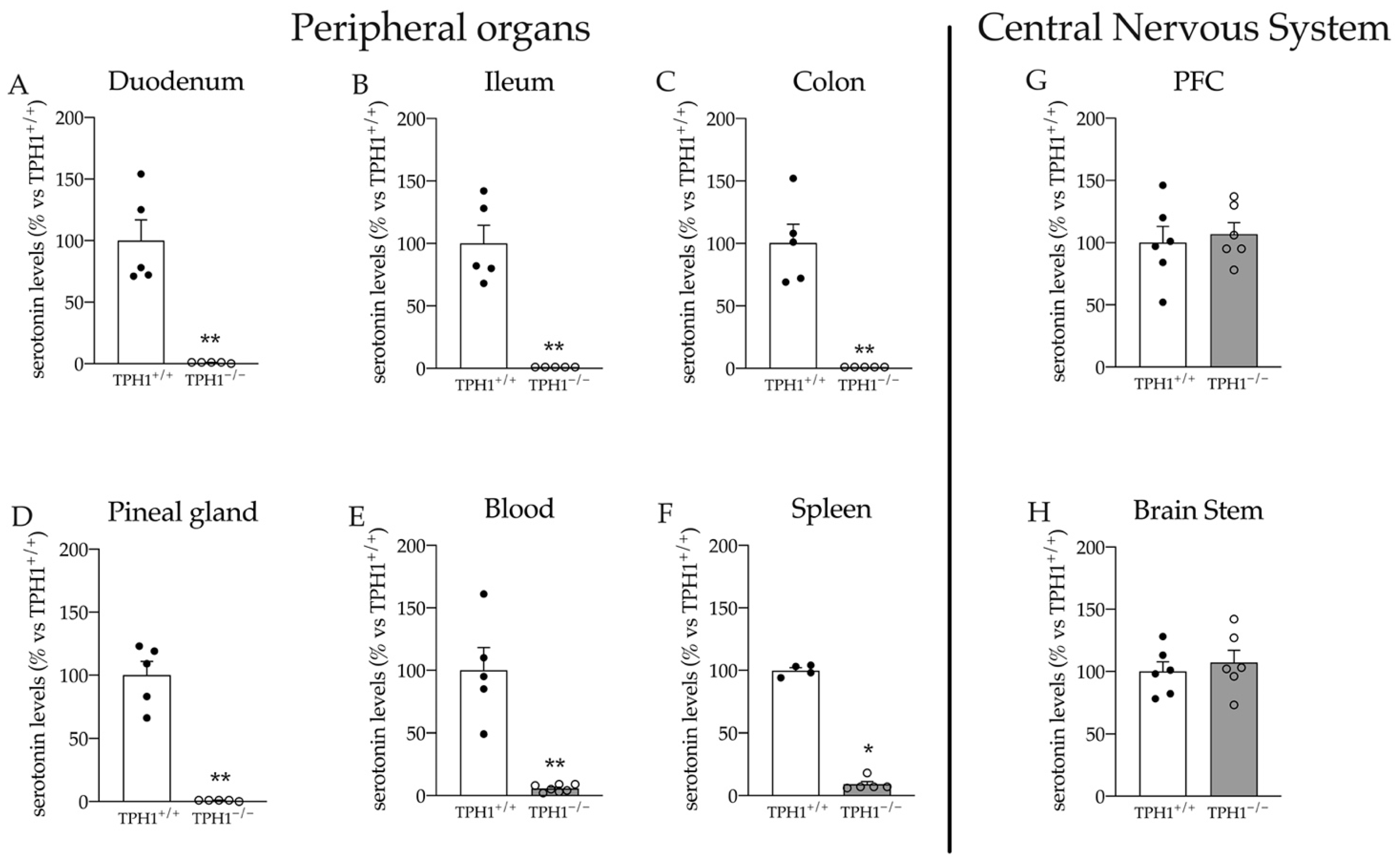
2.1. Serotonin Levels Are Dramatically Reduced in Peripheral Organs of TPH1−/− Rats

First, we analyzed serotonin levels in TPH1−/− and TPH1+/+ rats. As expected, and in line with data previously obtained in TPH1−/− mice [6,20,35,36], serotonin was almost undetectable in the serotonin-producing tissues of TPH1−/− rats (duodenum: −99% p < 0.01 vs. TPH1+/+; ileum: −99% p < 0.01 vs. TPH1+/+; colon: −99% p < 0.01 vs. TPH1+/+; pineal gland: −99% p < 0.01 vs. TPH1+/+; Student’s t-test) (Figure 1A–D). Although a decrease was observed in blood and spleen, about 5 to 10% of wild-type serotonin levels were still detectable in these tissues (blood: −94% p < 0.01 vs. TPH1+/+ spleen: −91% p < 0.001 vs. TPH1+/+, Student’s t-test), confirming previous TPH1−/− mouse data (26, 27) (Figure 1E,F). We also evaluated serotonin levels in the PFC and brainstem. As shown in Figure 1G,H, no differences between genotypes were observed in these brain regions, confirming the preservation of the brain serotonin system in the absence of peripheral serotonin, similar to previous data obtained in mice [37,38].

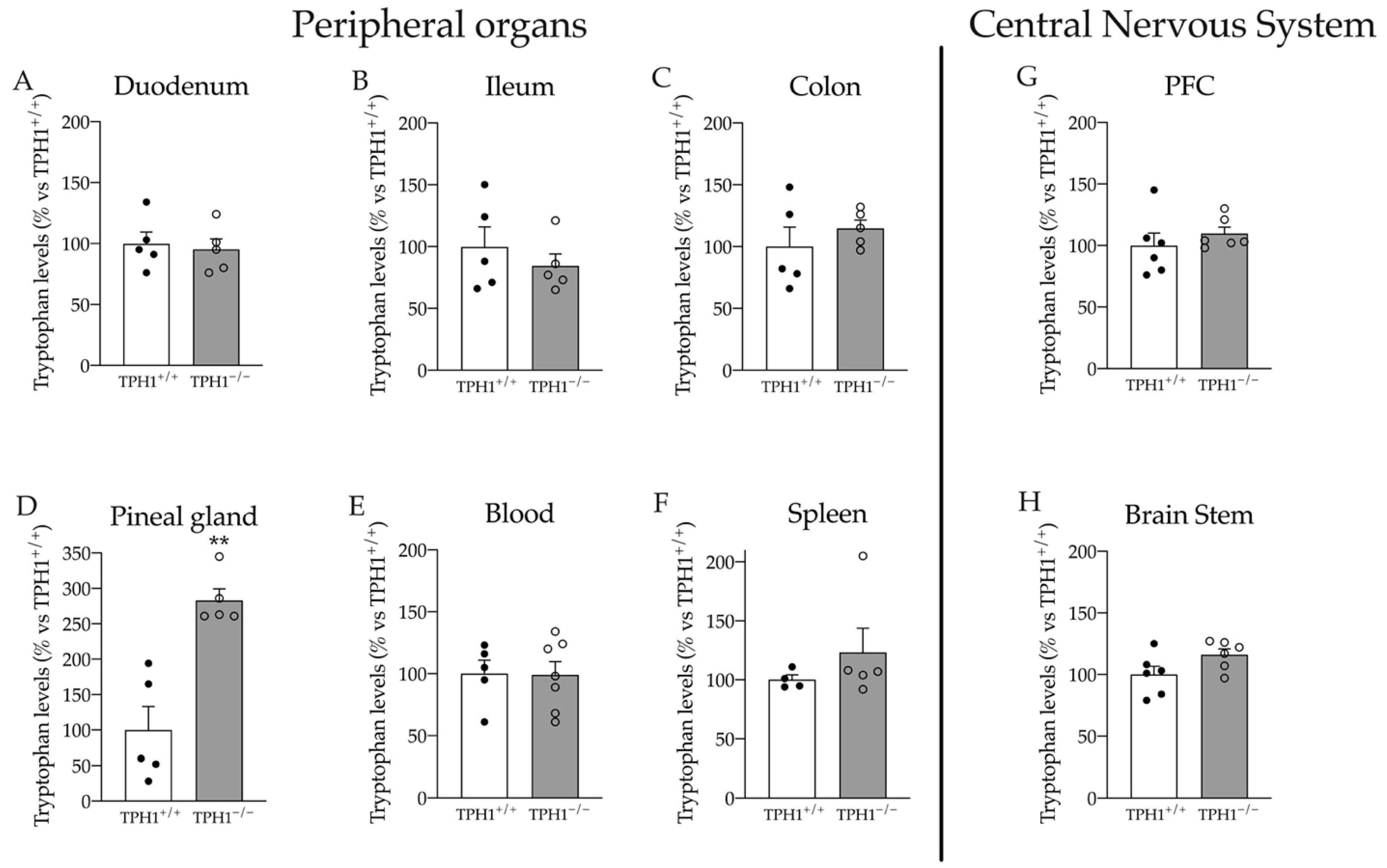
2.2. Tryptophan Levels Are Modulated in the Pineal Gland of TPH1−/− Rats

While serotonin concentration was homogenously reduced in peripheral organs, we found that the levels of its precursor tryptophan were increased by almost three times specifically in the pineal glands (+183% p < 0.01 vs. TPH1+/+; Student’s t-test) (Figure 2A). Conversely, we did not find any alteration in Trp levels in the blood, spleen, different gut compartments and brain parts (Figure 2B–H).

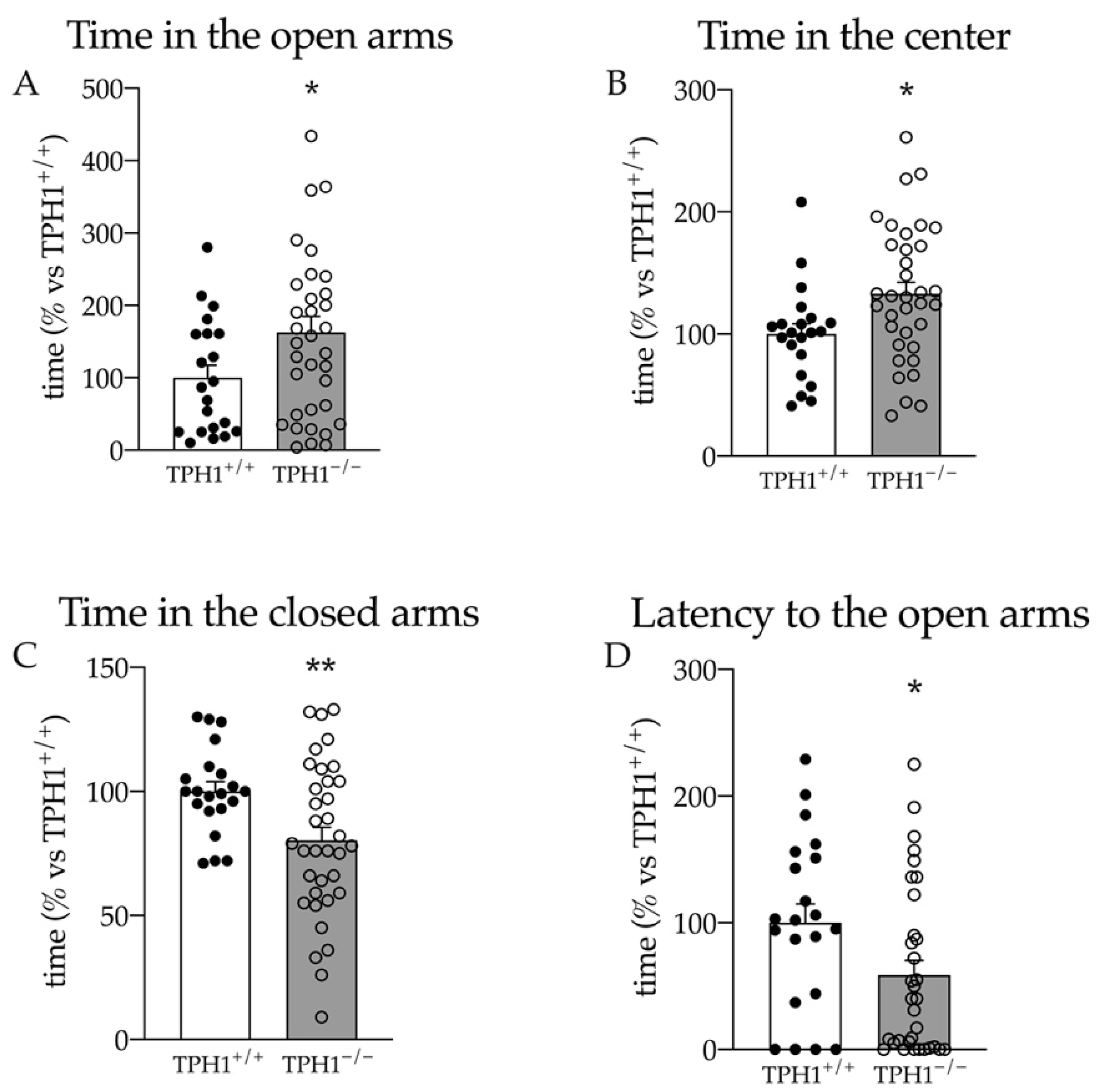
2.3. TPH1−/− Rats Show Reduced Anxiety in the Elevated Plus Maze Test

Anxiety is a common feature of different psychopathologies that have been linked to human TPH1 polymorphisms [21]. Hence, here we analyzed the anxiety levels of TPH1+/+ and TPH1−/− rats by exposing the animals to the elevated plus maze test.
Interestingly, the lack of peripheral serotonin induced a reduction in anxiety behavior. TPH1−/− rats spent more time in the open arms (+63% vs. TPH1+/+ p < 0.05; Student’s t-test; Figure 3A) and in the center of the maze (+33% p < 0.05 vs. TPH1+/+ p < 0.05; Student’s t-test; Figure 3B), while they spent less time in the closed arms (−20% p < 0.01 vs. TPH1+/+ p < 0.01; Student’s t-test; Figure 3C). Moreover, as shown in Figure 3D, the rats showed a reduced latency to the first entry in the open arms (−41% vs. TPH1+/+ p < 0.05; Student’s t-test).

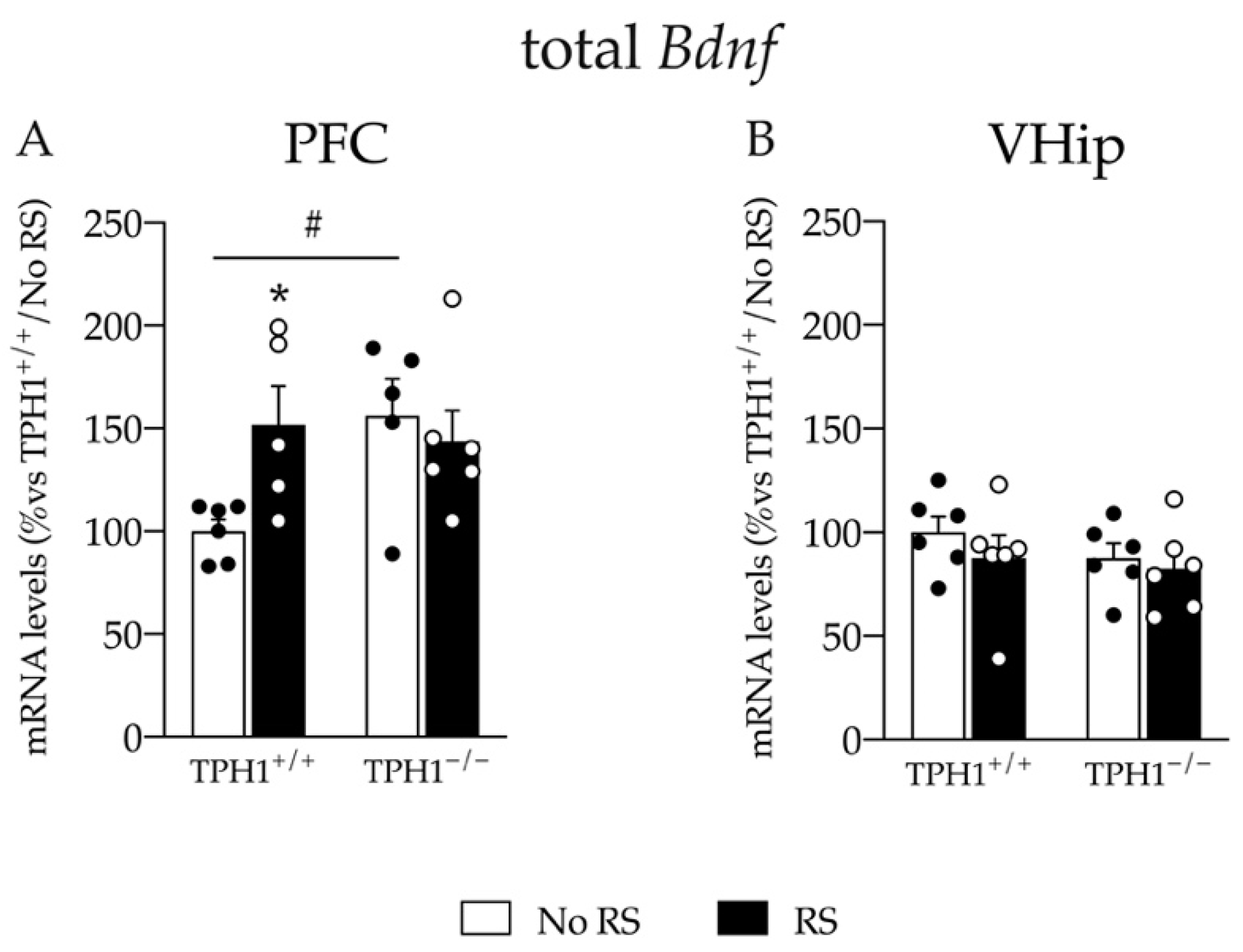
2.4. Baseline Bdnf mRNA Levels Are Increased and Induction of Its Expression Is Impaired after RS in TPH1−/− Rats Specifically in the Prefrontal Cortex

To evaluate if the change in anxiety is paralleled by changes in brain plasticity in TPH1−/− rats, we measured the levels of Bdnf in the PFC and in the VHip, both at basal level and after one single session of RS.
We found a significant genotype X RS interaction for total Bdnf mRNA levels (F(1–21) = 4.806 p < 0.05; two-way ANOVA). In line with this, we found an upregulation of the total neurotrophin expression at basal level in TPH1−/− rats compared to TPH1+/+ animals (+56% p < 0.05 vs. TPH1+/+/No RS; Fisher’s PLSD). However, total Bdnf mRNA levels were increased after RS in TPH1+/+ rats (+52% p < 0.05 vs. TPH1+/+/No RS; Fisher’s PLSD) but not in TPH1−/− rats (Figure 4A).
On the contrary, as shown in Figure 4B, we did not find changes in the expression of total Bdnf in the VHip, neither due to the genotype nor due to RS, suggesting a specific effect of both factors in the PFC.

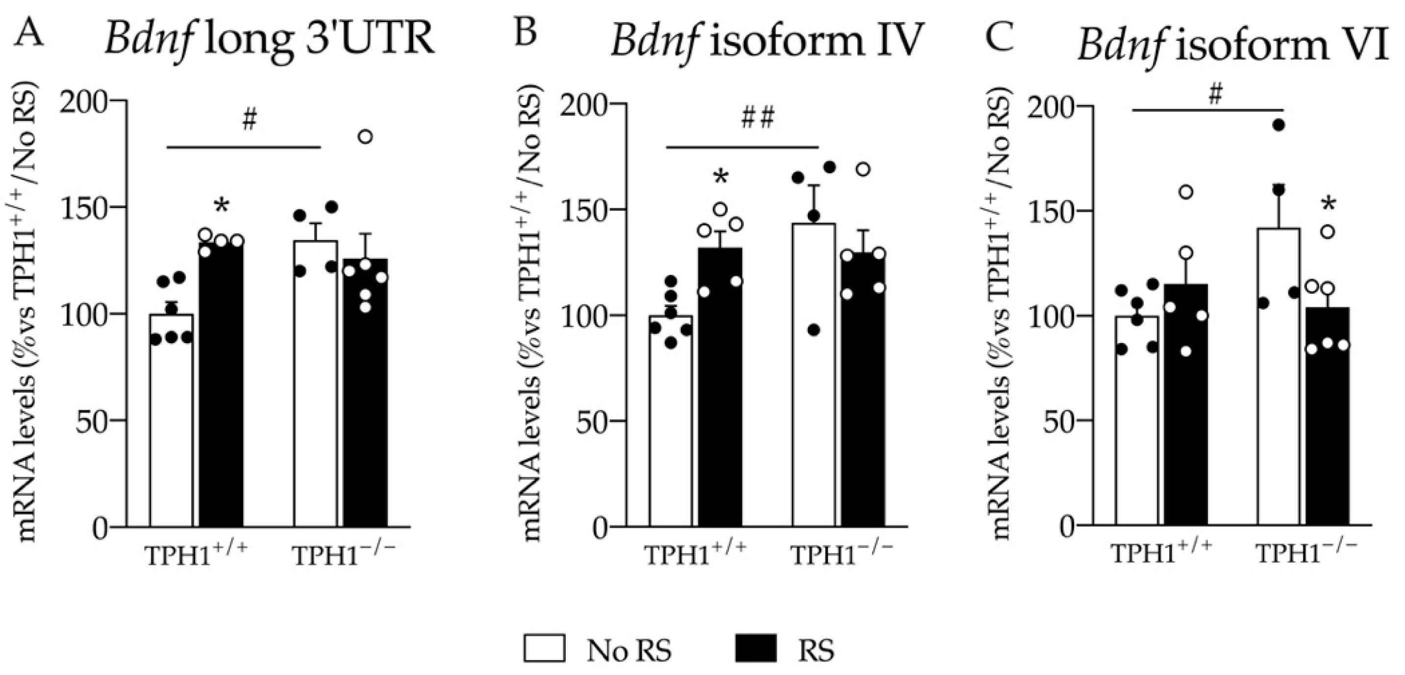
2.5. The Changes in Total Bdnf Expression in the PFC Are Sustained by Bdnf Long 3′UTR, Bdnf Isoform IV and VI

To further dissect the effect observed for Bdnf transcription, we measured the expression of Bdnf long 3′UTR and of the two most abundant isoforms in the brain, the isoforms IV and VI, in the PFC.
As shown in Figure 5A,B, we found a similar expression pattern for Bdnf long 3′UTR and Bdnf isoform IV (Bdnf long 3′UTR: genotype X RS interaction: F(1–19) = 5.817 p < 0.05; Bdnf isoform IV: genotype X RS interaction: F(1–19) = 5.345 p < 0.05; two-way ANOVA). Indeed, the lack of peripheral serotonin induced an increase in their mRNA levels in TPH1−/− animals (Bdnf long 3′UTR: +35% p < 0.05; Bdnf isoform IV: +32% p < 0.01 vs. TPH1+/+/No RS; Fisher’s PLSD), and one hour of RS upregulated the pool of the long transcripts and the isoform IV mRNA levels in wild-type rats (Bdnf long 3′UTR: +33% p < 0.05; Bdnf isoform IV: +43% p < 0.05 vs. TPH1+/+/No RS; Fisher’s PLSD), while we did not find any further increase for TPH1−/− RS rats.
Finally, we observed a genotype X RS interaction for Bdnf isoform VI (F(1–20) = 5.165 p < 0.05; two-way ANOVA) with an upregulation of its expression in TPH1−/− No RS animals (+42% p < 0.05 vs. TPH1+/+/No RS; Fisher’s PLSD), which was reduced after RS (−27% p < 0.05 vs. TPH1+/+/No RS; Fisher’s PLSD) (Figure 5C).

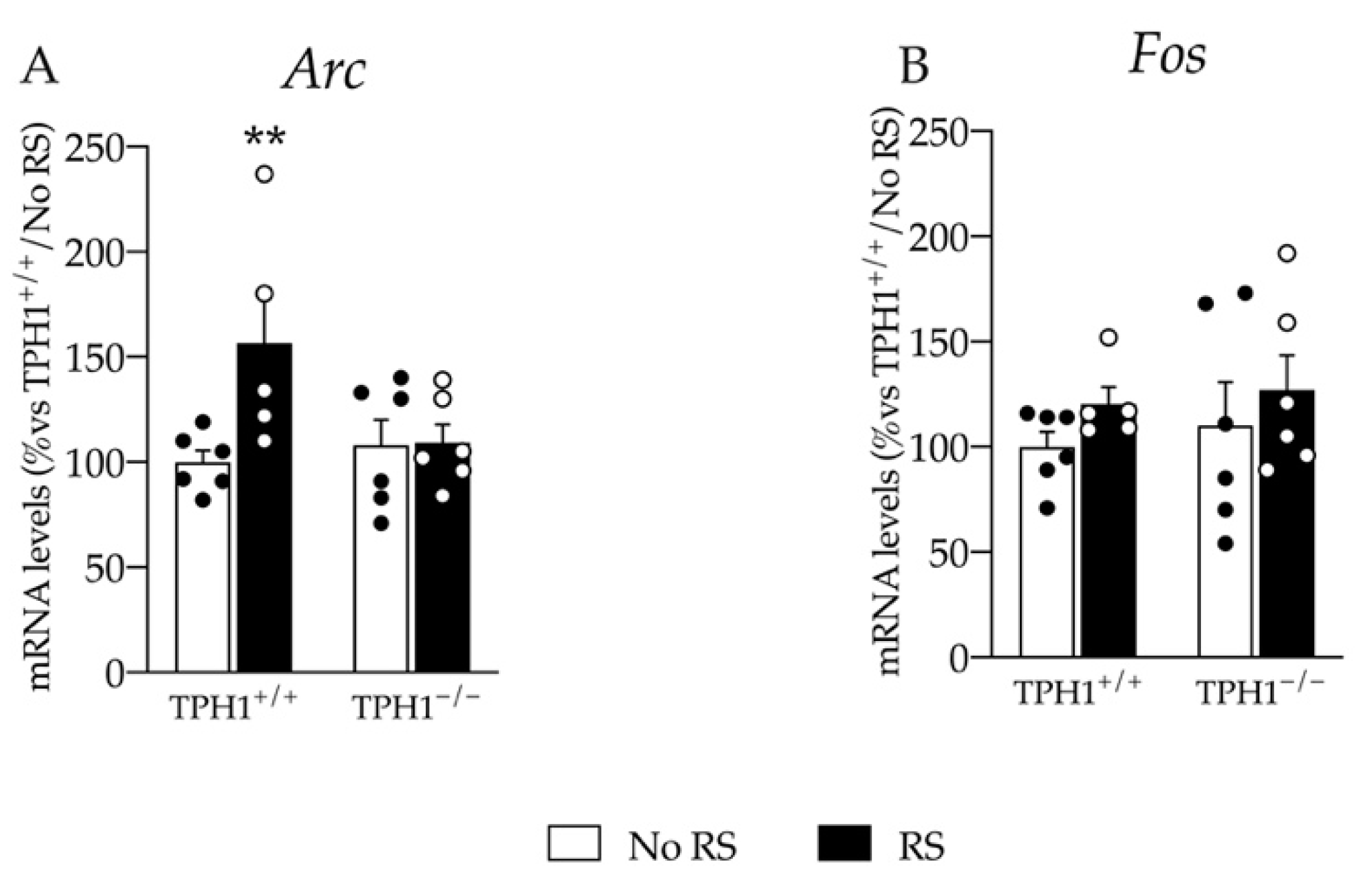
2.6. The Enhancement in the Immediate Early Gene Arc mRNA Levels Due to RS Exposure Is Blunted in TPH1−/− Rats

Given that the response to RS in TPH1−/− rats was blunted in terms of Bdnf gene expression, we further evaluated the expression of the two immediate early genes Arc and Fos that were normally upregulated after acute stimuli [26,30].
Interestingly, we found an effect of RS (F(1–22) = 4.963 p < 0.05; two-way ANOVA) and a genotype X RS interaction (F(1–22) = 4.518 p < 0.05; two-way ANOVA) on Arc expression. Indeed, as shown in Figure 6A, we found an upregulation of its mRNA levels specifically in TPH1+/+/RS rats (+57% p < 0.01 vs. TPH1+/+/No RS; Fisher’s PLSD) with no changes in the TPH1-deficient counterpart.
On the contrary, Fos expression was neither affected by RS nor by the genotype (Figure 6B).

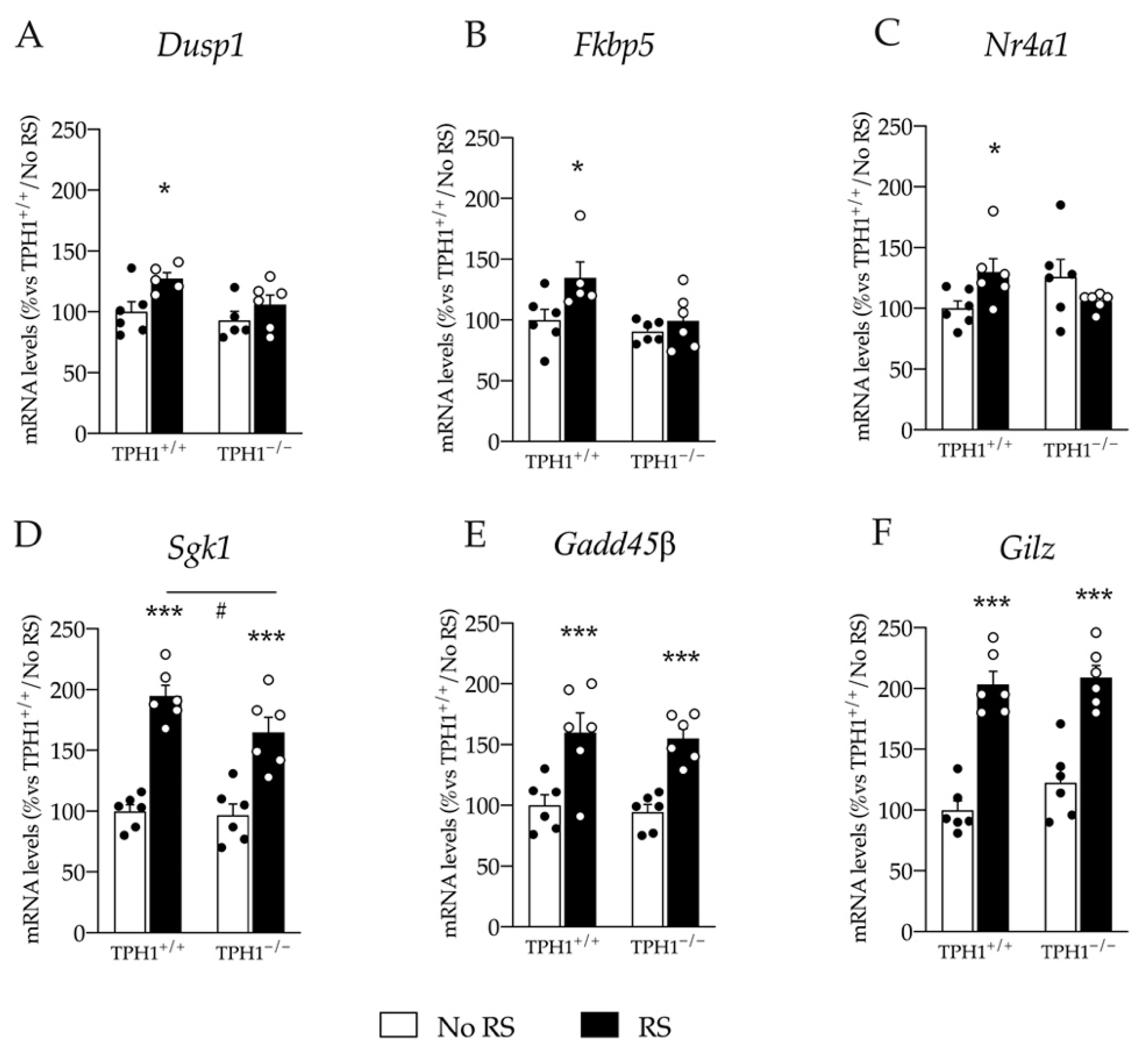
2.7. Upregulation of Glucocorticoid Responsive Genes Expression Induced by RS Exposure Is Blunted in TPH1−/− Rats

Glucocorticoids play a crucial role in stress response, inducing transcriptional activation of glucocorticoid-dependent genes [25]. Recently, we evaluated the expression pattern of several glucocorticoid-dependent genes in the PFC in response to acute stress [25]. Hence, we chose highly reactive genes for evaluation after RS in the PFC of the TPH1−/− rats.
We found an effect of RS on Dusp1 expression (F(1–21) = 7.478 p < 0.05; two-way ANOVA). Indeed, RS upregulated Dusp1 expression specifically in TPH1+/+ rats (+27% p < 0.05 vs. TPH1+/+/No RS: Fisher’s PLSD) but not in TPH1−/− rats (Figure 7A).
Similarly, we found an effect of genotype and of RS on Fkbp5 mRNA levels (genotype: F(1–22) = 6.241 p < 0.05; RS: F(1–22) = 5.874 p <0.05 two-way ANOVA) and a significant genotype X RS interaction on Nr4a1 expression (F(1–23) = 6.569 p < 0.05; two-way ANOVA). Indeed, we found a specific upregulation of both Fkbp5 and Nr4a1 after RS in TPH1+/+ rats (Fkbp5: +35% p < 0.05; Nr4a1: +30% p < 0.05 vs. TPH1+/+/No RS; Fisher’s PLSD) but no changes in TPH1−/− rats (Figure 7B,C).
Moreover, Sgk1 expression was modulated by RS (F(1–23) = 76.873 p < 0.001 two-way ANOVA) with an upregulation of its expression both in TPH1+/+ (+95% p < 0.001 vs. TPH1+/+/No RS; Fisher’s PLSD) and TPH1−/− rats (+71% p < 0.001 vs. TPH1−/−/No RS; Fisher’s PLSD). However, this increase was more robust in TPH1+/+ than in TPH1−/− RS rats (+18% p < 0.05 vs. TPH1+/+/RS; Fisher’s PLSD) (Figure 7D).
Finally, as shown in Figure 7E,F, we observed an effect of RS on Gadd45β and Gilz expression (Gadd45β: F(1–23) = 33.405 p < 0.001; Gilz: F(1–23) = 86.627 p < 0.001; two-way ANOVA) with a similar upregulation of their expression in TPH1+/+ rats (Gadd45β: +60% p < 0.001; Gilz: +103% p < 0.001 vs. TPH1+/+/No RS; Fisher’s PLSD) and in TPH1−/− rats (Gadd45β: +65% p < 0.001; Gilz: +71% p < 0.001 vs. TPH1−/−/No RS; Fisher’s PLSD).

3. Discussion

In this study, we confirmed our initial hypothesis that peripheral serotonin can modulate brain functions. Specifically, we found that Tph1-deletion in rats led to a dramatic decrease in peripheral, but not central, serotonin in these animals. Furthermore, this life-long depletion of peripheral serotonin in rats affected anxiety-like behavior and expression of genes involved in brain plasticity and stress response under basal conditions and after the exposure to RS.
Among the animal models that have been developed to study the role of peripheral serotonin, TPH1-deficient mice exhibit low levels of serotonin in peripheral organs, while, in line with the selective distribution of the TPH1 isoform in the periphery, the levels of serotonin in the central nervous system remain unchanged [6,19,38]. Accordingly, TPH1-deficient mice are a useful tool for studying the role of peripheral serotonin in cardiac function, metabolism, and hemostasis [3,35,36,38,39]. However, for cognitive and molecular neuroscience, rats are the preferred model [20]. Here, for the first time, we characterized TPH1-deficient rats and investigated if a lack of TPH1 had an impact on behavior and brain plasticity.
In line with previous mouse data [7,19,37,38], TPH1-deficient rats showed a reduction in serotonin levels in the periphery. Indeed, the deletion of the Tph1 gene in rats resulted in a dramatic decrease in serotonin levels, which were almost undetectable in Tph1-expressing organs, such as pineal glands and intestinal tissues. In mammals, besides sites of serotonin synthesis, this monoamine is also present in high amounts in platelets, which take it up via serotonin transporter (SERT). Serotonin levels in blood and spleen, the main platelet-containing tissues, were also drastically reduced in TPH1−/− rats, but still accounted for 5–10% of the wild type. The same phenotype was previously observed in TPH1-deficient mice, arguing for the existence of a conserved mechanism to maintain serotonin homeostasis in the absence of TPH1 enzyme, probably through phenylalanine hydroxylase [37].
Similar to TPH1-deficient mice [37], we did not find any alterations in serotonin levels in the PFC and brain stem of TPH1−/− rats, suggesting that the central serotonergic system is rather unaffected by the lack of peripheral serotonin. Hence, the observed central phenotypes must be mediated by other, indirect mechanisms.
Interestingly, while levels of the serotonin precursor Trp were not altered in peripheral tissues, such as blood, spleen, and gut, we found an increase in Trp in the pineal gland. Trp is an essential amino acid, and most dietary Trp is metabolized through the kynurenine pathway, whereas only a small portion (around 1–2%) is utilized for the serotonin production (reviewed in [40]). In the pineal gland, melatonin synthesis from Trp with serotonin as an intermediate product is a main metabolic pathway of Trp. Evidently, the kynurenine pathway [41] is not able to remove the excessive Trp from the pineals in the absence of TPH1, leading to its accumulation in this organ.
Since several studies revealed a link between single-nucleotide polymorphisms in the human TPH1 gene and an increased susceptibility to developing psychiatric conditions, including depression and anxiety-related disorders [11,12,13,42], we evaluated the anxiety-like behavior of TPH1-deficient rats by exposing them to the elevated plus maze test. The results from this behavioral test indicate that TPH1−/− rats are less anxious when compared to TPH1+/+ rats. Indeed, they spent more time in the open arms and in the center of the apparatus, while spending less time in the closed arms, where rats are supposed to feel safer. Moreover, they showed a reduced latency to the first entry in the open arms. Although it has been shown that the lack of peripheral serotonin is able to alter locomotor activity in mice [18], no study to date has evaluated whether the altered function of TPH1 could modulate other behaviors, including those related to psychiatric pathologies. Our study suggests that the lack of peripheral serotonin in TPH1−/− rats has a positive impact on anxiety-like behavior at basal level. Here, we showed that the levels of Bdnf are increased under basal conditions in the PFC of TPH1−/− rats, suggesting an interplay between serotonin and Bdnf expression in modulating the behavioral phenotype observed in TPH1−/− rats. Yet, the mechanistic links between low serotonin in the periphery, Bdnf expression and anxiety-related behavior are to be identified.
In the context of psychiatric illnesses, the genetic background often acts as a risk factor and different molecular alterations can be unmasked only after an environmental stimulus. For example, in our previous studies, we showed that the lack of central serotonin impaired the activation of different systems only after an acute stress, but not under basal conditions [25,26]. Hence, here we exposed TPH1+/+ and TPH1−/− rats to a single session of RS to evaluate whether reduced peripheral serotonin levels could interfere with the activation of stress response. We firstly focused on neurotrophic mechanisms and found that the total Bdnf mRNA levels were upregulated at basal level in TPH1−/− rats and that this increase was sustained by the expression of the pool containing long transcripts of the neurotrophin and by Bdnf isoforms IV and VI. However, while, as expected, TPH1+/+ RS rats showed an activation of the system with an increase in total Bdnf mRNA levels, Bdnf long 3′UTR and Bdnf isoform IV, we did not find any further increase in rats lacking peripheral serotonin. Thus, we could hypothesize that peripheral serotonin was able to interfere with the activation of the brain Bdnf machinery after RS exposure. In line with these results, by measuring the expression of the immediate early genes Arc and Fos, which are implicated in acute stress responses [43,44], we found an upregulation of Arc specifically in TPH1+/+ rats exposed to RS, while a lack of serotonin in peripheral compartments blunted its activation.
To further support this evidence, we also evaluated the transcriptional activation induced by glucocorticoid receptors, which is normally boosted after acute stress exposure due to the activation of the HPA axis and subsequent release of corticosterone [45,46]. We found that the expressions of glucocorticoid-responsive genes—Dusp1, Fkbp5, and Nr4a1—were upregulated specifically in wild-type rats exposed to RS while their transcription remained unchanged in knockout animals. Moreover, Sgk1 was upregulated in both genotypes, but this increase was higher in TPH1+/+ rats. Finally, Gadd45β and Gilz were equally upregulated after RS both in TPH1+/+ and TPH1−/− rats. Although other analyses should confirm this hypothesis, we can speculate here that the absence of peripheral serotonin could interfere with corticosterone release, consequently inhibiting the whole downstream pathway. In line with this, the adrenal gland, from which corticosterone is released, expresses serotonin receptors, and their agonism stimulates the release of this hormone [47]. Moreover, serotonin can stimulate the release of the adrenocorticotropic hormone (ACTH) from the pituitary gland, a central region lacking the blood–brain barrier and exposed to peripheral serotonin [48,49]. Unfortunately, we were not able to measure corticosterone and ACTH release to confirm these hypotheses, which is a limitation of our study. Moreover, not all glucocorticoid-responsive genes show the same activation pattern in TPH1−/− rats, which could be due to other stress-related factors involved in the activation of their transcription [50,51].
While the results obtained in this work argue for a link between peripheral serotonin levels and brain functionality under resting conditions and in response to environmental stimuli, the underlying mechanisms and the direct causality between the lack of peripheral serotonin and brain functions have yet to be fully understood. In particular, ontogenetic analyses at different developmental time points could clarify if the crosstalk between peripheral serotonin and brain wiring starts to occur during development. Indeed, the blood–brain barrier represents a clear boundary at adulthood, but during the first stages of life it is still immature and different molecules, including serotonin, can cross it [52]. Therefore, the reduction in serotonin amounts reaching the brain during the first period of life could interfere with the correct formation of neuronal circuits and inhibit their correct functioning [53]. Moreover, the analysis of the gut microbiome and the gut–brain axis represents another important factor to be studied in TPH1-deficient animals [54]. The evaluation of these aspects may uncover mechanisms linking the lack of peripheral serotonin and behavior and brain plasticity alterations observed in TPH1−/− rats in this study.

4. Materials and Methods

4.1. Animals

TPH1−/− rats were obtained at the Gene Editing Rat Resource Center of The Medical College Of Wisconsin by CRISPR/Cas9 technology. A Cas9 protein was injected together with a guide RNA, targeting the sequence AGATGTCATTCAGCTGTTCTCGG in the TPH1 gene, into zygotes of Wistar Kyoto rats, producing one base pair deletion in the exon 4 and thereby a frameshift mutation.
The breeding was conducted at the Central Animal Laboratory of the Radboud University Medical Center in Nijmegen, the Netherlands. From birth, the animals were housed under standardized conditions involving a 12/12 h light/dark cycle, at 22 °C and around 80% of humidity with access to food and water ad libitum.
Heterozygous females were bred with heterozygous males to generate TPH1+/+ and TPH1−/− experimental animals. At adulthood, male TPH1+/+ (n = 21) and TPH1−/− (n = 35) rats were tested for their anxiety level during the elevated plus maze test. In particular, a plus-shaped platform was located at 50 cm from the floor. The maze consisted of two closed arms (50 cm × 10 cm with 40 cm high walls) and two open arms of the same dimensions without walls. The two equal arms were positioned opposite each other, and the four arms formed a central platform at their intersection. At the beginning of the test, a rat was placed on the central platform looking at one of the open arms. The position and the movements of the animals were recorded for 5 min, and the time spent in the closed arms, in the center, and in the open arms of the arena was analyzed using EthoVision XT (version 3.1., Noldus, Wageningen, The Netherlands) software.
Two weeks later, part of the animals (n = 5/6) were exposed to one hour of RS, as previously described in [26], to test their stress responsiveness. Briefly, the animals were placed in an air-accessible apparatus with a similar size to the rats for one hour. Immediately after RS, animals were anesthetized and sacrificed through decapitation. No RS animals (n = 5/6) were left undisturbed in their home cage until the sacrifice.
For the molecular analyses, the PFC was dissected according to the atlas of Paxinos and Watson (Paxinos and Watson, 2007) from 2 mm thick slices (plates 6–9, including Cg1, Cg3, and IL sub-regions), whereas vHip were dissected from the whole brain (plates 34–43 of the Paxinos and Watson atlas). The brain regions were immediately frozen on dry ice and stored at −80 °C.
All experimental procedures were approved by the Central Committee on Animal Experiments (Centrale Commissie Dierproeven, CCD, The Hague, The Netherlands), limiting the number of animals used and minimizing animal suffering.

4.2. HPLC Analyses

For blood analyses, three hundred microliters of whole blood was collected into 1 mL syringes prefilled with 100 μL of heparin and quickly transferred to Eppendorf tubes containing 10 μL of perchloric acid (PCA) and 5 μL of 10 mg/mL of ascorbic acid, vortexed, and centrifuged (20,000× g, 30 min, 4 °C). The supernatant was collected and frozen at −80 °C until high-performance liquid chromatography (HPLC) analyses. For the organ collection, rats were transcardially perfused with ice-cold PBS to remove blood, containing platelet 5-HT. Tissues were snap-frozen in liquid nitrogen and kept at −80 °C. Frozen tissue samples were homogenized in 710 μM ascorbic acid and 2.4% perchloric acid (Sigma-Aldrich, Steinheim, Germany), precipitated proteins were pelleted through centrifugation (20 min, 20,000× g, 4 °C), and the collected supernatant was analyzed for serotonergic metabolites (Trp, and 5-HT) using HPLC with fluorometric detection. Samples were separated on a C18 reversed-phase column (Synergi Fusion RP, Phenomenex, Aschaffenburg, Germany) at 20 °C in a 10 mM potassium phosphate buffer (pH 5.0) (Sigma-Aldrich, Steinheim, Germany), with 5% methanol (Roth, Karlsruhe, Germany) and a flow rate of 0.8–1.0 mL/min on a Nexera X2 HPLC system (Shimadzu, Tokyo, Japan). The excitation wavelength was 295 nm, and the fluorescent signal was measured at 345 nm. LabSolutions 5.85 software (Shimadzu, Tokyo, Japan) was used to analyze the peak parameters of chromatographic spectra and quantify substance levels, based on comparative calculations with alternately measured external standards. Amounts of 5-HT and Trp were normalized to the wet tissue weight.

4.3. mRNA Extraction and Gene Expression Analyses

Total RNA was isolated by a single step of guanidinium isothiocyanate/phenol extraction using PureZol RNA isolation reagent (Bio-Rad Laboratories, Segrate (Mi), Italy) according to the manufacturer’s instructions and quantified by spectrophotometric analysis. The samples were then processed for a real-time polymerase chain reaction (RT-PCR) to assess the expression of the target genes (primer and probes sequences are listed in Table 1 and Table 2). RNA was treated with DNase (Thermoscientific, Milano, Italy) to avoid DNA contamination. Gene expression was analyzed by TaqMan qRT-PCR one-step RT-PCR kit for probes (Bio-Rad Laboratories, Italy). Samples were run in 384-well formats in triplicate as a multiplexed reaction with a normalizing internal control (36b4). Thermal cycling was initiated with an incubation at 50 °C for 10 min (RNA retro-transcription) and then at 95 °C for 5 min (TaqMan polymerase activation). After this initial step, 39 cycles of PCR were performed. Each PCR cycle consisted of heating the samples at 95 °C for 10 s to enable the melting process and then for 30 s at 60 °C for the annealing and extension reactions. A comparative cycle threshold (Ct) method was used to calculate the relative target gene expression.

4.4. Statistical Analyses

All the analyses were conducted by using IBM SPSS Statistics, version 24 or with Microsoft Excel. Data were collected in individual animals and are presented as means ± standard error (SEM).
Results were analyzed with two-way analysis of variance (ANOVA) followed by the Fisher’s protected least significant difference (PLSD) test or with a Student’s t-test. Significance for all tests was assumed for p < 0.05.

5. Conclusions

Overall, these data suggest that, even though serotonin itself cannot cross the blood–brain barrier, the lack of serotonin in the periphery can indeed affect brain functions. This is reflected by reduced anxiety, an upregulation of genes related to neuroplasticity and the glucocorticoid system and altered responsivity of these systems to RS exposure. These findings aid in the understanding of the genetic associations found between TPH1 gene polymorphisms and stress-related disorders.

Author Contributions

Conceptualization, G.S., S.I.H., N.A., M.B., J.R.H. and F.C.; methodology, G.S., S.I.H. and A.M.; data curation, G.S., S.I.H. and N.A.; writing—original draft preparation, G.S.; writing—review and editing, G.S., S.I.H., P.B., N.A., M.B., F.F., J.R.H., F.C. All authors have read and agreed to the published version of the manuscript.

Funding

This research was supported by the Donders Institute through a grant awarded to JRH. JRH, NA and MB were supported by the EU H2020 MSCA ITN projects “Serotonin and Beyond” (N 953327). NA was supported by the project 73022475 of the St. Petersburg State University. FC was supported by a grant from the Italian Ministry of University and Research (PRIN2017-201779W93T) and from MIUR Progetto Eccellenza (2018–2022) to the Department of Pharmacological and Biomolecular Sciences, Università degli Studi di Milano, Milan, Italy.

Institutional Review Board Statement

The animal study protocol was approved by the Ethics Committee of the Radboud University (protocol code 2014-134, approved at 01-07/2014).

Informed Consent Statement

Not applicable.

Data Availability Statement

The data presented in this study are available on request from the corresponding author.

Acknowledgments

The authors thank Susanne da Costa Gonsalves for the excellent technical assistance. The authors acknowledge support from the University of Milan through the APC initiative.

Conflicts of Interest

The authors of the manuscript certify that they have NO affiliations with or involvement in any organization or entity with any financial interest (such as honoraria; educational grants; participation in speakers’ bureaus; membership, employment, consultancies, stock ownership, or other equity interest; and expert-testimony or patent-licensing arrangements), or non-financial interests (such as personal or professional relationships, affiliations, knowledge or beliefs) in the subject matter or materials discussed in this manuscript.

References

  1. Jenkins, T.; Nguyen, J.; Polglaze, K.; Bertrand, P. Influence of Tryptophan and Serotonin on Mood and Cognition with a Possible Role of the Gut-Brain Axis. Nutrients 2016, 8, 56. [Google Scholar] [CrossRef] [PubMed]
  2. Yildiz, O.; Smith, J.R.; Purdy, R.E. Serotonin and vasoconstrictor synergism. Life Sci. 1998, 62, 1723–1732. [Google Scholar] [CrossRef]
  3. Paulmann, N.; Grohmann, M.; Voigt, J.-P.; Bert, B.; Vowinckel, J.; Bader, M.; Skelin, M.; Jevsek, M.; Fink, H.; Rupnik, M.; et al. Intracellular serotonin modulates insulin secretion from pancreatic beta-cells by protein serotonylation. PLoS Biol. 2009, 7, e1000229. [Google Scholar] [CrossRef] [PubMed] [Green Version]
  4. Kim, H.; Toyofuku, Y.; Lynn, F.C.; Chak, E.; Uchida, T.; Mizukami, H.; Fujitani, Y.; Kawamori, R.; Miyatsuka, T.; Kosaka, Y.; et al. Serotonin regulates pancreatic beta cell mass during pregnancy. Nat. Med. 2010, 16, 804–808. [Google Scholar] [CrossRef]
  5. El-Merahbi, R.; Löffler, M.; Mayer, A.; Sumara, G. The roles of peripheral serotonin in metabolic homeostasis. FEBS Lett. 2015, 589, 1728–1734. [Google Scholar] [CrossRef] [Green Version]
  6. Walther, D.J.; Bader, M. A unique central tryptophan hydroxylase isoform. Biochem. Pharmacol. 2003, 66, 1673–1680. [Google Scholar] [CrossRef]
  7. Walther, D.J.; Peter, J.U.; Bashammakh, S.; Hörtnagl, H.; Voits, M.; Fink, H.; Bader, M. Synthesis of serotonin by a second tryptophan hydroxylase isoform. Science 2003, 299, 76. [Google Scholar] [CrossRef]
  8. Zhang, X.; Beaulieu, J.-M.; Sotnikova, T.D.; Gainetdinov, R.R.; Caron, M.G. Tryptophan hydroxylase-2 controls brain serotonin synthesis. Science 2004, 305, 217. [Google Scholar] [CrossRef]
  9. Zill, P.; Baghai, T.C.; Zwanzger, P.; Schüle, C.; Eser, D.; Rupprecht, R.; Möller, H.-J.; Bondy, B.; Ackenheil, M. SNP and haplotype analysis of a novel tryptophan hydroxylase isoform (TPH2) gene provide evidence for association with major depression. Mol. Psychiatry 2004, 9, 1030–1036. [Google Scholar] [CrossRef] [Green Version]
  10. McKinney, J.; Knappskog, P.M.; Haavik, J. Different properties of the central and peripheral forms of human tryptophan hydroxylase. J. Neurochem. 2005, 92, 311–320. [Google Scholar] [CrossRef]
  11. Viikki, M.; Kampman, O.; Illi, A.; Setälä-Soikkeli, E.; Anttila, S.; Huuhka, M.; Nuolivirta, T.; Poutanen, O.; Mononen, N.; Lehtimäki, T.; et al. TPH1 218A/C polymorphism is associated with major depressive disorder and its treatment response. Neurosci. Lett. 2010, 468, 80–84. [Google Scholar] [CrossRef] [PubMed]
  12. Wigner, P.; Czarny, P.; Synowiec, E.; Bijak, M.; Białek, K.; Talarowska, M.; Galecki, P.; Szemraj, J.; Sliwinski, T. Association between single nucleotide polymorphisms of TPH1 and TPH2 genes, and depressive disorders. J. Cell. Mol. Med. 2018, 22, 1778–1791. [Google Scholar] [CrossRef] [PubMed] [Green Version]
  13. Chen, D.; Liu, F.; Yang, C.; Liang, X.; Shang, Q.; He, W.; Wang, Z. Association between the TPH1 A218C polymorphism and risk of mood disorders and alcohol dependence: Evidence from the current studies. J. Affect. Disord. 2012, 138, 27–33. [Google Scholar] [CrossRef] [PubMed]
  14. Maurer-Spurej, E.; Pittendreigh, C.; Solomons, K. The influence of selective serotonin reuptake inhibitors on human platelet serotonin. Thromb. Haemost. 2004, 91, 119–128. [Google Scholar] [CrossRef]
  15. Quintana, J. Platelet serotonin and plasma tryptophan decreases in endogenous depression. Clinical, therapeutic, and biological correlations. J. Affect. Disord. 1992, 24, 55–62. [Google Scholar] [CrossRef]
  16. Berger, M.; Gray, J.A.; Roth, B.L. The Expanded Biology of Serotonin. Annu. Rev. Med. 2009, 60, 355–366. [Google Scholar] [CrossRef] [Green Version]
  17. Bader, M. Phenotype of Mice Lacking Tryptophan Hydroxylase 1. In Serotonin; Elsevier: Amsterdam, The Netherlands, 2019; pp. 167–179. [Google Scholar]
  18. Suidan, G.L.; Duerschmied, D.; Dillon, G.M.; Vanderhorst, V.; Hampton, T.G.; Wong, S.L.; Voorhees, J.R.; Wagner, D.D. Lack of Tryptophan Hydroxylase-1 in Mice Results in Gait Abnormalities. PLoS ONE 2013, 8, e59032. [Google Scholar] [CrossRef]
  19. Savelieva, K.V.; Zhao, S.; Pogorelov, V.M.; Rajan, I.; Yang, Q.; Cullinan, E.; Lanthorn, T.H. Genetic Disruption of Both Tryptophan Hydroxylase Genes Dramatically Reduces Serotonin and Affects Behavior in Models Sensitive to Antidepressants. PLoS ONE 2008, 3, e3301. [Google Scholar] [CrossRef] [Green Version]
  20. Homberg, J.R.; Wöhr, M.; Alenina, N. Comeback of the Rat in Biomedical Research. ACS Chem. Neurosci. 2017, 8, 900–903. [Google Scholar] [CrossRef] [Green Version]
  21. Karpov, B.; Joffe, G.; Aaltonen, K.; Suvisaari, J.; Baryshnikov, I.; Näätänen, P.; Koivisto, M.; Melartin, T.; Oksanen, J.; Suominen, K.; et al. Anxiety symptoms in a major mood and schizophrenia spectrum disorders. Eur. Psychiatry 2016, 37, 1–7. [Google Scholar] [CrossRef] [Green Version]
  22. Duncan, L.E.; Pollastri, A.R.; Smoller, J.W. Mind the gap: Why many geneticists and psychological scientists have discrepant views about gene–environment interaction (G×E) research. Am. Psychol. 2014, 69, 249–268. [Google Scholar] [CrossRef] [PubMed]
  23. Belsky, J.; Jonassaint, C.; Pluess, M.; Stanton, M.; Brummett, B.; Williams, R. Vulnerability genes or plasticity genes? Mol. Psychiatry 2009, 14, 746–754. [Google Scholar] [CrossRef] [PubMed]
  24. Sbrini, G.; Brivio, P.; Bosch, K.; Homberg, J.R.; Calabrese, F. Enrichment environment positively influences depression- and anxiety-like behavior in serotonin transporter knockout rats through the modulation of neuroplasticitcity, spine and GABAergic markers. Genes 2020, 11, 1248. [Google Scholar] [CrossRef] [PubMed]
  25. Sbrini, G.; Brivio, P.; Peeva, P.M.; Todiras, M.; Bader, M.; Alenina, N.; Calabrese, F. The Absence of Serotonin in the Brain Alters Acute Stress Responsiveness by Interfering With the Genomic Function of the Glucocorticoid Receptors. Front. Cell. Neurosci. 2020, 14, 128. [Google Scholar] [CrossRef] [PubMed]
  26. Brivio, P.; Sbrini, G.; Peeva, P.; Todiras, M.; Bader, M.; Alenina, N.; Calabrese, F. TPH2 Deficiency Influences Neuroplastic Mechanisms and Alters the Response to an Acute Stress in a Sex Specific Manner. Front. Mol. Neurosci. 2018, 11, 389. [Google Scholar] [CrossRef] [PubMed] [Green Version]
  27. Łoś, K.; Waszkiewicz, N. Biological Markers in Anxiety Disorders. J. Clin. Med. 2021, 10, 1744. [Google Scholar] [CrossRef] [PubMed]
  28. Chiba, S.; Numakawa, T.; Ninomiya, M.; Richards, M.C.; Wakabayashi, C.; Kunugi, H. Chronic restraint stress causes anxiety- and depression-like behaviors, downregulates glucocorticoid receptor expression, and attenuates glutamate release induced by brain-derived neurotrophic factor in the prefrontal cortex. Prog. Neuro-Psychopharmacol. Biol. Psychiatry 2012, 39, 112–119. [Google Scholar] [CrossRef]
  29. Brivio, P.; Sbrini, G.; Tarantini, L.; Parravicini, C.; Gruca, P.; Lason, M.; Litwa, E.; Favero, C.; Riva, M.A.; Eberini, I.; et al. Stress Modifies the Expression of Glucocorticoid-Responsive Genes by Acting at Epigenetic Levels in the Rat Prefrontal Cortex: Modulatory Activity of Lurasidone. Int. J. Mol. Sci. 2021, 22, 6197. [Google Scholar] [CrossRef]
  30. Brivio, P.; Sbrini, G.; Riva, M.A.; Calabrese, F. Acute Stress Induces Cognitive Improvement in the Novel Object Recognition Task by Transiently Modulating Bdnf in the Prefrontal Cortex of Male Rats. Cell. Mol. Neurobiol. 2020, 40, 1037–1047. [Google Scholar] [CrossRef] [Green Version]
  31. Pan, L.; Gilbert, F. Activation of 5-HT(1A) receptor subtype in the paraventricular nuclei of the hypothalamus induces CRH and ACTH release in the rat. Neuroendocrinology 1992, 56, 797–802. [Google Scholar] [CrossRef]
  32. Herman, J.P.; Figueiredo, H.; Mueller, N.K.; Ulrich-Lai, Y.; Ostrander, M.M.; Choi, D.C.; Cullinan, W.E. Central mechanisms of stress integration: Hierarchical circuitry controlling hypothalamo-pituitary-adrenocortical responsiveness. Front. Neuroendocrinol. 2003, 24, 151–180. [Google Scholar] [CrossRef] [PubMed]
  33. Van De Kar, L.D.; Javed, A.; Zhang, Y.; Serres, F.; Raap, D.K.; Gray, T.S. 5-HT2A receptors stimulate ACTH, corticosterone, oxytocin, renin, and prolactin release and activate hypothalamic CRF and oxytocin-expressing cells. J. Neurosci. 2001, 21, 3572–3579. [Google Scholar] [CrossRef] [PubMed] [Green Version]
  34. Duman, R.S.; Monteggia, L.M. A Neurotrophic Model for Stress-Related Mood Disorders. Biol. Psychiatry 2006, 59, 1116–1127. [Google Scholar] [CrossRef] [PubMed]
  35. Choi, W.; Namkung, J.; Hwang, I.; Kim, H.; Lim, A.; Park, H.J.; Lee, H.W.; Han, K.-H.; Park, S.; Jeong, J.-S.; et al. Serotonin signals through a gut-liver axis to regulate hepatic steatosis. Nat. Commun. 2018, 9, 4824. [Google Scholar] [CrossRef] [PubMed]
  36. Sumara, G.; Sumara, O.; Kim, J.K.; Karsenty, G. Gut-Derived Serotonin Is a Multifunctional Determinant to Fasting Adaptation. Cell Metab. 2012, 16, 588–600. [Google Scholar] [CrossRef] [Green Version]
  37. Mordhorst, A.; Dhandapani, P.; Matthes, S.; Mosienko, V.; Rothe, M.; Todiras, M.; Self, J.; Schunck, W.; Schütz, A.; Bader, M.; et al. Phenylalanine hydroxylase contributes to serotonin synthesis in mice. FASEB J. 2021, 35, e21648. [Google Scholar] [CrossRef]
  38. Cote, F.; Thevenot, E.; Fligny, C.; Fromes, Y.; Darmon, M.; Ripoche, M.-A.; Bayard, E.; Hanoun, N.; Saurini, F.; Lechat, P.; et al. Disruption of the nonneuronal tph1 gene demonstrates the importance of peripheral serotonin in cardiac function. Proc. Natl. Acad. Sci. USA 2003, 100, 13525–13530. [Google Scholar] [CrossRef] [Green Version]
  39. Crane, J.D.; Palanivel, R.; Mottillo, E.P.; Bujak, A.L.; Wang, H.; Ford, R.J.; Collins, A.; Blümer, R.M.; Fullerton, M.D.; Yabut, J.M.; et al. Inhibiting peripheral serotonin synthesis reduces obesity and metabolic dysfunction by promoting brown adipose tissue thermogenesis. Nat. Med. 2015, 21, 166–172. [Google Scholar] [CrossRef] [Green Version]
  40. Haq, S.; Grondin, J.A.; Khan, W.I. Tryptophan-derived serotonin-kynurenine balance in immune activation and intestinal inflammation. FASEB J. 2021, 35, e21888. [Google Scholar] [CrossRef]
  41. Fujiwara, M.; Shibata, M.; Watanabe, Y.; Nukiwa, T.; Hirata, F.; Mizuno, N.; Hayaishi, O. Indoleamine 2,3-dioxygenase. Formation of L-kynurenine from L-tryptophan in cultured rabbit fineal gland. J. Biol. Chem. 1978, 253, 6081–6085. [Google Scholar] [CrossRef]
  42. Goenjian, A.K.; Bailey, J.N.; Walling, D.P.; Steinberg, A.M.; Schmidt, D.; Dandekar, U.; Noble, E.P. Association of TPH1, TPH2, and 5HTTLPR with PTSD and depressive symptoms. J. Affect. Disord. 2012, 140, 244–252. [Google Scholar] [CrossRef] [PubMed]
  43. Ons, S.; Marti, O.; Armario, A. Stress-induced activation of the immediate early gene Arc (activity-regulated cytoskeleton-associated protein) is restricted to telencephalic areas in the rat brain: Relationship to c-fos mRNA. J. Neurochem. 2004, 89, 1111–1118. [Google Scholar] [CrossRef] [PubMed]
  44. Molteni, R.; Calabrese, F.; Maj, P.F.; Olivier, J.D.A.; Racagni, G.; Ellenbroek, B.A.; Riva, M.A. Altered expression and modulation of activity-regulated cytoskeletal associated protein (Arc) in serotonin transporter knockout rats. Eur. Neuropsychopharmacol. 2009, 19, 898–904. [Google Scholar] [CrossRef] [PubMed]
  45. Schoneveld, O.J.L.M.; Gaemers, I.C.; Lamers, W.H. Mechanisms of glucocorticoid signalling. Biochim. Et Biophys. Acta (BBA)-Gene Struct. Expr. 2004, 1680, 114–128. [Google Scholar] [CrossRef]
  46. Panettieri, R.A.; Schaafsma, D.; Amrani, Y.; Koziol-White, C.; Ostrom, R.; Tliba, O. Non-genomic Effects of Glucocorticoids: An Updated View. Trends Pharmacol. Sci. 2019, 40, 38–49. [Google Scholar] [CrossRef]
  47. Hegde, S.S.; Eglen, R.M. Peripheral 5-HT 4 receptors. FASEB J. 1996, 10, 1398–1407. [Google Scholar] [CrossRef]
  48. Jørgensen, H.; Knigge, U.; Kjaer, A.; Møller, M.; Warberg, J. Serotonergic stimulation of corticotropin-releasing hormone and pro-opiomelanocortin gene expression. J. Neuroendocrinol. 2002, 14, 788–795. [Google Scholar] [CrossRef]
  49. Chen, G.-L.; Miller, G.M. Advances in tryptophan hydroxylase-2 gene expression regulation: New insights into serotonin-stress interaction and clinical implications. Am. J. Med. Genet. Part B Neuropsychiatr. Genet. 2012, 159, 152–171. [Google Scholar] [CrossRef] [Green Version]
  50. Lang, F.; Stournaras, C.; Alesutan, I. Regulation of transport across cell membranes by the serum- and glucocorticoid-inducible kinase SGK1. Mol. Membr. Biol. 2014, 31, 29–36. [Google Scholar] [CrossRef]
  51. Zannas, A.S.; Wiechmann, T.; Gassen, N.C.; Binder, E.B. Gene–Stress–Epigenetic Regulation of FKBP5: Clinical and Translational Implications. Neuropsychopharmacology 2016, 41, 261–274. [Google Scholar] [CrossRef] [Green Version]
  52. Ribatti, D.; Nico, B.; Crivellato, E.; Artico, M. Development of the blood-brain barrier: A historical point of view. Anat. Rec. Part B New Anat. 2006, 289, 3–8. [Google Scholar] [CrossRef] [PubMed]
  53. Bonnin, A.; Goeden, N.; Chen, K.; Wilson, M.L.; King, J.; Shih, J.C.; Blakely, R.D.; Deneris, E.S.; Levitt, P. A transient placental source of serotonin for the fetal forebrain. Nature 2011, 472, 347–350. [Google Scholar] [CrossRef] [PubMed] [Green Version]
  54. Nanthakumaran, S.; Sridharan, S.; Somagutta, M.R.; Arnold, A.A.; May, V.; Pagad, S.; Malik, B.H. The Gut-Brain Axis and Its Role in Depression. Cureus 2020, 12, e10280. [Google Scholar] [CrossRef] [PubMed]
Figure 1. Serotonin levels in the duodenum (A), ileum (B), colon (C), pineal gland (D), blood (E), spleen (F), PFC (G) and in the brain stem (H) of TPH1+/+ and TPH1−/− rats. Data are presented as means ± standard error (SEM) of at least 4 independent determinations. * p < 0.05 ** p < 0.01 vs. TPH1+/+; Student’s t-test.
Ijms 23 04941 g001
Figure 2. Tryptophan levels in the duodenum (A), ileum (B), colon (C), pineal gland (D), blood (E), spleen (F), PFC (G) and in the brain stem (H) of TPH1+/+ and TPH1−/− rats. Data are presented as means ± standard error (SEM) of at least 4 independent determinations. ** p < 0.01 vs. TPH1+/+; Student’s t-test.
Ijms 23 04941 g002
Figure 3. Elevated plus maze test results. Time spent in the open arms (A), in the center (B), in the closed arms (C) and latency to the first entrance in the open arms (D). Data are presented as mean ± standard error of the mean (SEM) of at least 21 independent determinations. * p < 0.05; ** p < 0.01 vs. TPH1+/+; Student’s t-test.
Ijms 23 04941 g003
Figure 4. Analyses of total Bdnf mRNA levels in the PFC (A) and in the VHip (B) of TPH1+/+ and TPH1−/− male adult rats exposed to RS and sacrificed immediately after stress. The data, expressed as percentage of TPH1+/+/No RS (set at 100%), are the mean ± standard error of the mean (SEM) of at least 5 independent determinations. * p < 0.05 vs. naïve for the same genotype; # p < 0.05, vs. TPH1+/+ for the same condition, two-way ANOVA with Fisher’s PLSD.
Ijms 23 04941 g004
Figure 5. Analyses of Bdnf long 3′UTR (A), Bdnf isoforms IV (B) and VI (C) mRNA levels in the PFC of TPH1+/+ and TPH1−/− male adult rats exposed to RS and sacrificed immediately after stress. The data, expressed as percentage of TPH1+/+/No RS (set at 100%), are the mean ± standard error of the mean (SEM) of at least 4 independent determinations. * p < 0.05 vs. naïve for the same genotype; # p < 0.05, ## p < 0.01 vs. TPH1+/+ for the same condition, two-way ANOVA with Fisher’s PLSD.
Ijms 23 04941 g005
Figure 6. Analyses of Arc (A) and Fos (B) mRNA levels in the PFC of TPH1+/+ and TPH1−/− male adult rats exposed to RS and sacrificed immediately after stress. The data, expressed as a percentage of TPH1+/+/No RS (set at 100%), are the mean ± standard error of the mean (SEM) of at least 5 independent determinations. ** p < 0.01 vs. naïve for the same genotype; two-way ANOVA with Fisher’s PLSD.
Ijms 23 04941 g006
Figure 7. Analyses of Dusp1 (A), Fkbp5 (B), Nr4a1 (C), Sgk1 (D), Gadd45β (E) and Gilz (F) mRNA levels in the PFC of TPH1+/+ and TPH1−/− male adult rats exposed to RS and sacrificed immediately after stress. The data, expressed as percentage of TPH1+/+/No RS (set at 100%), are the mean ± standard error of the mean (SEM) of at least 5 independent determinations. * p < 0.05; *** p < 0.001 vs. naïve for the same genotype; # p < 0.05 vs. TPH1+/+ for the same condition; two-way ANOVA with Fisher’s PLSD.
Ijms 23 04941 g007
Table 1. Primers and probes sequences acquired from Eurofins MWG-Operon.
GeneForward SequenceReverse SequenceProbe Sequence
Total BdnfAAGTCTGCATTACATTCCTCGAGTTTTCTGAAAGAGGGACAGTTTATTGTGGTTTGTTGCCGTTGCCAAG
ArcGGTGGGTGGCTCTGAAGAATACTCCACCCAGTTCTTCACCGATCCAGAACCACATGAATGGG
FosTCCTTACGGACTCCCCACCTCCGTTTCTCTTCCTCTTCAGTGCTCTACTTTGCCCCTTCTGCC
Dusp1TGTGCCTGACAGTGCAGAATATCTTTCCGGGAAGCATGGTATCCTGTCCTTCCTGTACCT
Fkbp5GAACCCAATGCTGAGCTTATGATGTACTTGCCTCCCTTGAAGTGTCCATCTCCCAGGATTCTTTGGC
Sgk1GACTACATTAATGGCGGAGAGCAGGGAGTGCAGATAACCCAAGTGCTCGCTTCTACGCAGC
GilzCGGTCTATCAACTGCACAATTTCCTTCACTAGATCCATGGCCTGAACGGAAACCACATCCCCTCCAA
36b4TCAGTGCCTCACTCCATCATAGGAAGGCCTTGACCTTTTCTGGATACAAAAGGGTCCTGG
Table 2. Primers and probes sequences acquired from Life Technologies.
GeneAccession NumberAssay ID
Bdnf long 3′UTREF125675.1Rn02531967_s1
Bdnf isoform IVEF125679.1Rn01484927_m1
Bdnf isoform VIEF125680.1Rn01484928_m1
Nr4a1BC097313.1Rn01452530_g1
Gadd45βBC085337.1Rn01452530_g1
Publisher’s Note: MDPI stays neutral with regard to jurisdictional claims in published maps and institutional affiliations.

