Review

The Role of Somatic Mutations on the Immune Response of the Tumor Microenvironment in Prostate Cancer

by
Camila Morais Melo
1,
Thiago Vidotto
1,
Luiz Paulo Chaves
1,
William Lautert-Dutra
1,
Rodolfo Borges dos Reis
2 and
Jeremy Andrew Squire
1,3,*
1
Department of Genetics, Medicine School of Ribeirão Preto, University of São Paulo, Ribeirão Preto 14048-900, SP, Brazil
2
Division of Urology, Department of Surgery and Anatomy, Medicine School of Ribeirão Preto, University of São Paulo, Ribeirão Preto 14048-900, SP, Brazil
3
Department of Pathology and Molecular Medicine, Queen’s University, Kingston, ON K7L3N6, Canada
*
Author to whom correspondence should be addressed.
Int. J. Mol. Sci. 2021, 22(17), 9550; https://doi.org/10.3390/ijms22179550
Submission received: 31 July 2021 / Revised: 26 August 2021 / Accepted: 30 August 2021 / Published: 2 September 2021
(This article belongs to the Special Issue Immunological Responses to Cancer Therapy)

Abstract

:
Immunotherapy has improved patient survival in many types of cancer, but for prostate cancer, initial results with immunotherapy have been disappointing. Prostate cancer is considered an immunologically excluded or cold tumor, unable to generate an effective T-cell response against cancer cells. However, a small but significant percentage of patients do respond to immunotherapy, suggesting that some specific molecular subtypes of this tumor may have a better response to checkpoint inhibitors. Recent findings suggest that, in addition to their function as cancer genes, somatic mutations of PTEN, TP53, RB1, CDK12, and DNA repair, or specific activation of regulatory pathways, such as ETS or MYC, may also facilitate immune evasion of the host response against cancer. This review presents an update of recent discoveries about the role that the common somatic mutations can play in changing the tumor microenvironment and immune response against prostate cancer. We describe how detailed molecular genetic analyses of the tumor microenvironment of prostate cancer using mouse models and human tumors are providing new insights into the cell types and pathways mediating immune responses. These analyses are helping researchers to design drug combinations that are more likely to target the molecular and immunological pathways that underlie treatment failure.

1. Introduction

Previous approaches to cancer therapy were based mostly on how to control clonal proliferation of aggressive tumor cells. Recently, translational researchers have focused on the role of the anti-cancer immune system in the design of new therapies to improve the response and overall survival. Thus, an important new hallmark of cancer is understanding how to stop tumors bypass the host’s protective immune destruction mechanisms [1].
Immunotherapy acts by boosting the natural propensity of the immune system to recognize and respond to the presence of tumors [2]. Immunotherapy involves the use of specific checkpoint agents that block the suppressive interactions between a developing tumor and the defensive immune system of the patient [3]. The clinical responses to checkpoint blockade have been very successful across many different cancers [4]. Tumor features that have been associated with a good clinical response to anti-PD-1/PD-L1 checkpoint blockade therapies include T-cell infiltration, PD-L1 expression in tumor and/or immune cells, increased tumor mutational burden (TMB), and interferon gamma (IFNγ)-derived T-cell gene expression profiles [5]. However, the overall proportion of patients responding to immunotherapy remains modest, with single-agent response rates across different tumor types ranging from 10–35% [6]. In advanced prostate cancer (PCa), the effect of anti-PD-1 checkpoint blockade has been disappointing, with just 5–10% of tumors that exhibit defects in microsatellite instability (MSI) pathways [7] or CDK12 biallelic mutations responding to this therapy [8].
Recent evidence suggests that factors influencing the success of checkpoint blockade therapy includes the absence of a pre-existing tumor-specific T cell response, or the exclusion of T-cells from the tumor microenvironment. To predict whether a particular cancer type will respond to immunotherapy, cellular classification systems have been developed in recent years based on the type of immune-cell infiltrate, density, and location within the tumor. Computational analyses of transcriptomic data have also been used to classify the immune content of tumors. As described below, these classifications are starting to provide some clues about the mechanisms tumors are using to avoid the host immune system. In Section 7, we discuss how these research advances are being applied in recent immunotherapy trials.
Key somatic mutations in PCa include fusions of TMPRSS2 with ETS family genes, amplification of the MYC oncogene, deletion and/or mutation of PTEN, RB1, and TP53 in advanced disease, together with amplification and/or mutation of the androgen receptor (AR) [9]. The immune system is able recognize and destroy tumor cells that are constantly arising during the life of an individual using a finely tuned anti-cancer immunosurveillance system. An emerging hypothesis to explain why most PCa fail to respond to immunotherapy is that specific somatic mutations acquired during tumor development induce changes in surrounding non-tumor cells of the microenvironment that help the tumor to evade immunosurveillance, the mechanism by which immune cells interact [10]. Recent reviews addressing this area in PCa have concentrated on advances in the use of checkpoint inhibitors [11], the roles of various immune subsets [12], DNA repair, mutational burden and tumor response [13], and the role of different cellular contexts on the immune microenvironment [14,15]. We will review the impact of the commonly acquired somatic mutations on immune response and the TME of PCa that are currently being used to identify potential predictive molecular biomarkers for immunotherapy clinical trials.

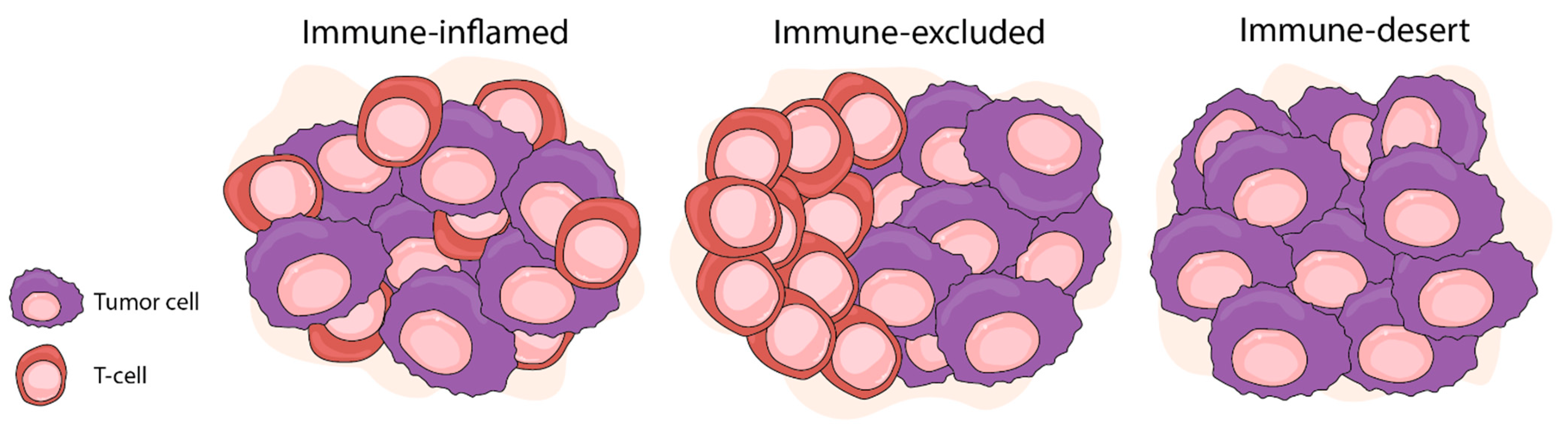
2. Interactions between the Tumor Microenvironment and Cancer

Tumor cells stimulate significant molecular, cellular, and physical changes in their surrounding microenvironment. This means that the cellular composition of the tumor microenvironment (TME) continuously evolves and can become more complex as tumors develop. In addition, there is variation in the cellular content of the TME of different tumor types, but a consistent hallmark of the TME of cancers includes immune cells, stromal cells, blood vessels, and an extracellular matrix [1]. New information about the TME indicates that it should now be considered “an active promoter of cancer progression” rather than earlier views of it being a passive supporting structure [16].
Early in tumor growth, a dynamic and reciprocal relationship develops between cancer cells and the TME. These interactions influence immune responses against cancer, local invasion, and subsequent metastatic dissemination. Tumors become infiltrated with diverse adaptive and innate immune cells that can perform both pro- and anti-tumorigenic functions. In addition, their activities are pivotal in mediating or suppressing host anti-tumor immune responses in the TME.
For cancers that are resistant to immunotherapy, recent discoveries suggest that tumors adopt several different strategies to avoid anticancer immune responses. For example, analyses of pre-treatment melanoma biopsies have demonstrated that checkpoint blockade response [17] correlated with the presence of tumor-infiltrating CD8+ lymphocytes at the invasive tumor margins. In head and neck cancer, the presence of an interferon-gamma (IFNγ) gene expression signature correlated with a better clinical response to immunotherapy [18]. Expression of IFNγ responsive genes is considered to represent an ‘inflamed’ transcriptional state in the TME, since these genes are associated with chemokine expression, antigen presentation, and cytotoxic effector molecules. These observations led to a TME classification of tumor-immune phenotypes involving three major classes of tumors: ‘immune deserts’ tumors (or ‘cold tumors’), showing no immune cell infiltration; ‘immune-excluded’ tumors (also cold) with immune cells at the tumor margins; and ‘inflamed’ tumors (or ‘hot’ tumors), showing immune infiltrates in the tumor core that can have IFNγ expression signatures [19,20,21] (Figure 1). Immune inflamed tumors, such as melanoma, are characterized by the infiltration of CD4+ and CD8+ T cells within the tumor parenchyma, suggesting the presence of a pre-existing anti-tumor response that has been dampened by an immunosuppressive microenvironment or by intrinsic T-cell tolerance. In contrast, cold tumors are characterized by the absence of pre-existing TILs and can be further subdivided into immune-excluded tumors, in which T cells have been attracted to the periphery of the tumor, but fail to infiltrate, and finally the immune-desert tumors, which have no detectable T-cell infiltrate [6].
Immunologically hot tumors, such as melanomas, are the most responsive to checkpoint blockade [17], and have a variety of infiltrating T cells in the TME and tumor [22]. In contrast, immunologically cold tumors have a low mutation load, lower and/or lowest probability of response to anti-PD1/PD-L1 treatment, are immune tolerant against self-antigens, and lack the T-cell-inflamed TME. PCa tumors are known to be sparsely infiltrated with T cells [23], suggesting that the poor immunotherapy response in PCa may be linked to their TME having an immune desert or excluded phenotype. When TILs are present in PCa, the presence of CD8+ lymphocytes have been reported to be a favorable prognostic indication [24], although an earlier study noted that the TILs present in PCa were ‘unresponsive’ or terminally differentiated [25]. A larger more recent study based on microarray gene expression profiles also reported the presence of high tumor TIL infiltrates were associated with worse distant metastasis-free survival [26].
To account for the contradictory findings in the PCa literature concerning lymphocyte density, it has been suggested that variation in the activation state of tumor specific T cells in the TME may account for the weak associations between TILs and the outcome [15]. Previous studies of PCa suggest that CD8+ lymphocytes in the TME may be inactive, suppressed, or unable to generate a functional cytotoxic response despite the presence of tumor antigen stimulation [27,28]. When looking at T-cell populations in PCa, studies have noted the presence of CD8+ CD25+ Treg cell clones that expressed FoxP3 and suppressed naive T-cell proliferation in prostate tumor-derived TILs, and there was a significant correlation between CD3+, CD8+, and FOXP3+ T-cell densities, but these were not associated with most clinical or pathologic variables. Increased T-cell density was significantly associated with ERG positivity and also with PTEN loss in the combined cohort of matched European-American and African-American ancestry patients [29,30].
Unfortunately, there is still no consensus concerning the best scoring methods amongst the large number of histologic studies of TILs. For example, some papers include only intratumoral TILs and others also score stromal TILs. For these reasons, there has been controversy about the prognostic value of scoring TILs in PCa, and there is limited appreciation of the functional status of lymphocytes in the TME [15]. It is possible that lymphocytes present in the TME are unable to perform immune activities due to mechanisms such as anergy (tolerance), exhaustion, or senescence [31]. As described below, more refined functional and transcriptomic classifications of effector cells in the microenvironment of human and murine PCa are being used to characterize the role that the TME plays in immune evasion and tumor progression.

3. Recent Lessons Learned about the Immune TME from Mouse Models of Human PCa

The development of an appropriate in vivo immunotherapy model systems for studying the cellular interactions between tumor and immune cells in the TME has been challenging because human tumors are usually grown as xenografts in immunocompromised mice. There are several recent investigations using genetically modified mouse models with functionally intact immune systems that are providing new information about underlying mechanisms of antitumor immune responses [32].
A comparison of murine model tumors to various human cancers has drawn attention to the role of immunomodulatory myeloid cells in the TME. These myeloid cells are essential for suppressing adaptive immunity and play a central role in ensuring that a developing malignancy is able to evade the host immune system [33]. Suppressive myeloid cells function by either direct cell–cell interaction with the target cells, such as T cells and NK cells, or through secreted factors. Two key classes of suppressive myeloid cells are the tumor-associated macrophages (TAMs) and myeloid-derived suppressor cells (MDSCs) [34].
The genetic background of tumors in mice promotes infiltration of specific immune-cell subsets into the TME of PCa [35]. Specifically, these authors showed that the double knockout of Pml, Zbtb7a, and Pten elevated expression of the neutrophil cytokine Cxcl5, leading to increased recruitment of these granulocytes into the TME. In contrast, Tp53 knockout mice increased expression of another cytokine, C-X-C chemokine ligand 17 (Cxcl17), which led to recruitment of MDSCs. The expression data from murine PCa was similar to human tumors in the TCGA dataset. Moreover, the TME or murine PCa also had cellular features consistent with the ‘immune desert’ phenotype with very limited intratumoral immune infiltration.
MDSCs are emerging as key players in the immunosuppression of the TME of solid tumors. The main characteristic that defines MDSCs is their ability to inhibit immune responses, including those mediated by T cells, B cells, and natural killer (NK) cells [36]. Calcinotto et al. investigated whether MDSCs might contribute directly to resistance to anti-androgen therapy, using human PCa cells and mouse models of prostate cancer. They showed that high levels of the inflammatory cytokine Il-23 mediated paracrine effects on tumor-infiltrating MDSCs by activating downstream androgen receptor (AR) target genes through the signal transducer and activator of transcription 3 (STAT3)–RORγ signaling axis in tumor cells [37].
Tumor-associated macrophages (TAMs) comprise the macrophage populations surrounding and infiltrating solid tumors that are also closely involved in mediating immune responses against cancer [33]. TAMs can be polarized by various microenvironmental stimuli to generate a heterogeneous population with different properties and functions in the immune response [38]. Human cancer cells can influence TAMs polarization by releasing cytokines, glucocorticoids, extracellular vesicles, and extracellular matrix components that give rise to a large spectrum of pro-tumoral macrophages [39]. Dimitri et al. recently investigated the interplay between different macrophage activation states and tumor cells in the TME in Pten knockout PCa [40]. The TME of these tumors were highly infiltrated by TAMs that expressed the Type2 C-X-C chemokine receptor (Cxcr2), which led to an M2 anti-inflammatory polarized macrophages when activated with its ligand Cxcl2. They showed that these PCa tumors were sensitive to inhibition of Cxcr2, which induced TAMs to re-program towards an M1 anti-tumorigenic phenotype. The authors further highlighted the therapeutic potential for modulating TAMs in the TME by combining treatment with a Cxcr2 inhibitor with infusions of Cxcr2-KO activated monocytes, which further increased the efficiency of tumor inhibition [40].
There is an increasing need to understand the specific differences that may exist in human immune responses within metastatic sites and other tumor niches. Jiao et al. [41] developed a PCa mouse model to study the mechanisms that may underlie the development of different T-cell lineages. They found that tumors growing in bones contain high levels of IL-6 and TGFβ, which provide signals to change intratumoral CD4+ T cell lineage differentiation towards polarized to a Th17 cell subset rather than the Th1 lineage. Inhibition of TGFβ plus immune checkpoint blockade led to Th1 CD4+ T cell responses and the clonal expansion of CD8+ T cells in bone tumors, which improved anti-tumor responses and increased survival in mice. These data indicate that cytokines and growth factor levels within the TME may underlie different T-cell immune activities and variation in clinical outcomes to immunotherapy. In Section 5, we discuss factors leading to bone metastasis based on single-cell analysis of TAMs, which shows how specific cytokine cues in the TME promote growth of metastatic bone PCa [42].
Distinct subsets of tumor-associated myeloid cells may play a pro-tumorigenic role either by directly mediating chemotherapy response or by suppressing recognition of the adaptive immune system and facilitating immunotherapy resistance. The diverse roles that the various myeloid cell populations play in the TME is still poorly understood cells [36], but clues coming from advanced genomic studies described in the following sections. Current areas of active investigation are to determine what combinations of the common somatic mutations of PCa and altered downstream signaling pathways influence effector cells, such as MDSCs, TAMs, and cytokines, to create conditions in the TME that favor immune evasion.

4. Role of Somatic Mutations in Shaping the Microenvironment of PCa

The TCGA group used integrative multi-omics to identify six distinct immune subtypes across 33 cancers that were associated with prognosis, genetic, and immune modulatory alterations considered to shape the specific types of immune environments in the TME [43]. The genomics of the various tumor types studied expressed vastly different immune responses suggesting that tumor-specific mutational changes may strongly influence pathways of evasion and the tumor-immune interactions. Accumulating evidence suggests that some of the common somatic mutations of PCa not only impacts classical hallmarks of cancer pathways [1], but also elicits changes in the TME, allowing cancer cells to avoid immunosurveillance [16,44,45].
Prostate cancer has a high rate of genomic instability (amplifications, deletions, and chromosomal rearrangements) [46] but a relatively low mutational burden compared with other cancers. Only four driver genes (ERG fusion, PTEN, TP53, and SPOP) are recurrently mutated in more than 10% of primary tumors [9,13]. During the early stages, PCa is predominantly an androgen-dependent disease. Eventually, cancer cells adapt to castrate androgen levels to form recurrent disease mediated through AR reactivation, acquisition of additional alterations such as PTEN loss and PI3K activation (See Table 1).

4.1. Phosphatase and Tensin Homologue (PTEN)

The PTEN tumor suppressor gene regulates the PI3K pathway. PTEN deletion is present in 20–40% of PCa and loss is strongly associated with PCa progression [64] and immune response in cancer [15,56]. Our data in human PCa [55] suggest that PCa tumors with PTEN loss have changes in the TME that could facilitate immune evasion, with a higher density of FoxP3+ Tregs and more IDO1 protein expression in the stromal and tumor compartments in PTEN-deficient tumors compared to tumors that maintained PTEN activity. Studies of Pten in murine PCa demonstrated that its loss promotes negative regulation of expression of the immunosuppressive cytokines interleukin (IL)-10, IL-6, and vascular endothelial growth factor (VEGF) by inhibiting signal transducer and activator of transcription (STAT3). The Pten-null senescent PCa tumors from mice had high infiltration of MDSCs in the TME promoted by activation of the Janus kinase (JAK)2/STAT3 pathway and secretion of chemoattractant molecules to the TME [57,58]. A recent population-based study of >6000 PCa related PTEN loss to the statin pathway and inflammation and immune activation in lethal PCa [66]. These findings are supported by a new study of Pten knockout mice, which used methylation and transcriptomic profiling to show that loss of PTEN drives global changes in DNA CpG methylation with associated gene expression changes, thus affecting several inflammatory and immune molecular pathways during PCa development [65].
Gene fusions of the ETS transcription factor family (usually involving the ERG gene) are caused by genomic rearrangements with TMPRSS2 gene and are found in over 50% of patients with clinically localized PCa [53]. As a result, the androgen-responsive promoter elements of TMPRSS2 drive the expression of the ETS family transcription factors to promote PCa progression, invasion, and tumor aggressiveness, and lead to a significantly reduced survival [13,80,81,82]. In the context of PTEN loss, ERG can restore AR target gene expression and increases AR binding to target promoters, independent of AR protein levels and circulating levels of testosterone [83]. Pten knockout mouse experiments showed that Ets overexpression led to disease progression, as well as inducing a pro-inflammatory gene signature, both of which were impacted by Ar expression levels [84]. This finding suggests that the Tmprss2-Erg fusion promotes recruitment of regulatory T cells to the tumor site [50]. Recent studies have shown an association between low levels of TILs comparing TMPRSS2-ERG fusions producing chimeric transcripts in the coding in the fusion-positive cases results in suppression of immune response by alteration of cytokine production [51,52].

4.2. TP53

TP53 mutations are found in 20–30% of PCa [61]. Loss of the TP53 protein has been shown to impact immunosuppression through increased PD1 and PDL-1 expression, and by activation of NF-kB signaling [60,85]. In primary PCa, the TP53 missense mutation was associated with higher T-cell density [35,62] and related to increase genomic instability [63]. Tp53 loss promotes the recruitment of tumor-supporting myeloid cells [35]. Pten loss in PCa models in combination with Tp53 loss increases the secretion of Cxcl17, leading to the recruitment of MDSCs [35].
Sequencing data from TCGA database revealed mutations or homozygous deletions for DNA repair genes MLH1, MSH2, BRCA2, BRCA1, ATM, CDK12, FANCD2, and RAD51B/C in primary and metastatic prostate cancer [70]. The recognition that germline or somatic mutations in DNA repair genes occur in about 25% of patients with recurrent or advanced PCa [86] allows for the use of poly(ADP-ribose) polymerase (PARP) inhibitors in this PCa molecular subtype, since these drugs can selectively kill tumors with DNA repair defects [87]. Since RNA repair defects also affects tumor immunogenicity, there is emerging interest in using checkpoint blockades combined with PARP inhibitors, as described below.

4.3. CDK12

CDK12 has been known to regulate DNA damage response genes (DDR) and modulate the expression of immune checkpoints, such as PD-1, in PCa. Mutations in CDK12 are found in 1.2 to 1.5% and 4 to 6.9% of primary-PCa and mCRPC, respectively [8,67]. Wu et al. described a biallelic loss-of-function mutation in CDK12 as a distinct molecular subtype of advanced PCa [8]. The genomic of PCa with CDK12 mutations show a widespread pattern of acquired focal tandem duplication (FTD), scattered throughout the genome and enriched in gene-dense regions. The FTD in the genome yields tumor cells with enriched gene fusion, and fusion-induced neoantigen was presented at higher levels than observed in HR or ATM-prostate cancers. This molecular subtype also demonstrates a high level of immune cell infiltration and T-cell expansion than all other PCa. Rescigno et al. founded that CD4+ infiltrate higher in CDK12 mCRPC group with a phenotype composed majority by CD4+ FOXP3-, which was associated with worse outcomes and immunosuppression [68]. Compared to CD4+ FOXP3-, the authors founded a low CD8+ infiltration load even with the high mutation burden in the CDK12 mCRPC group.

4.4. AIRE

The immune gene encoding autoimmune regulator (AIRE) protein which confers autoimmune protection was regulated by androgen/AR complex, androgen recruits AR to the AIRE promoter and thereby enhances its transcription [88]. Interesting study reports show the AIRE differential expression in the androgen-sensitive cell line compared to androgen-insensitive cell line in addition to immune cell polarization [89]. In mouse models, Aire induced Il-6 and Pge2 switches the monocyte polarization to M2 phenotype [89].
Next, generation sequencing has also been pivotal in understanding the complexity and the extent of genomic diversity that occurs during PCa progression [90]. There is accumulating evidence that specific somatic mutations in PCa trigger downstream expression changes and the abundance of non-tumor cells and immune components in the TME, allowing cancer cells to avoid immunosurveillance (see Figure 2). More detailed understanding of how the various somatic mutations can influence the dynamic interplay between tumor cells and the diverse non-cancerous cells in the TME of PCa is being provided by sensitive new single-cell analytical technologies.

5. Single-Cell Descriptions of the Immune Ecosystem of the TME of Prostate Cancer

The TME of cancer has been compared to an ecosystem that involves the delicate interplay of various cell types, including tumor cells, immune cells, stromal cells, and many others [101]. The introduction of various powerful single-cell methodologies, such as cytometry with time-of-flight (CyTOF) analysis [102,103] and single-cell genomics [104], has provided unprecedented insights into the molecular biology of the dynamic cell interactions that characterize the TME of PCa.
Single-cell methods are typically performed for many hundreds to thousands of cells in a single experiment. Recent studies have shown that analyzing gene expression using single-cell RNA-seq (scRNA-seq) at the level of individual cells provides a much greater depth of analysis than earlier bulk methods [104]. When combined with DNA sequencing, scRNA-seq allows appreciation of the in vivo impact of genomic alterations on gene expression. Importantly, scRNA-seq can assess the mutational heterogenicity and transcriptional pathways in cell populations present in the bulk tumor and the TME in an unbiased fashion at the level of individual cells. There are a number of recent examples of scRNA-seq analysis of PCa, which have investigated the heterogeneity of tumors at the single-cell level [42,105,106].
In a study by Chen et al., scRNA-seq [42] was used to study both PCa tumor intracellular heterogeneity and to examine the tumor-infiltrating immune cell content of the TME. Some intriguing observations were made regarding the TME when they compared primary PCa to matched lymph node metastases, and to adjacent non-cancerous prostate tissue. They identified a subset of TAMs with high osteoclast (OC)-activity in all samples. Since OC mediates osteodegradation and bone is the preferred site for metastatic PCa [107], this finding provides new ideas for the role of TAMs in the promotion of metastatic PCa. As discussed above, TAMs are closely involved in mediating immune responses against cancer and can produce growth factors favoring tumor outgrowth [33]. The observations of this study imply that a subset of TAMs may promote metastatic progression and immune suppression at bony sites. These findings also agree with the mouse study of Jiao et al. [41] (discussed in Section 3) in which single-cell analyses were used to demonstrate lineage programming of T cells in favor of a bone microenvironment. Chen et al. also identified PSA (KLK3) expression within the antitumor effector CD8+ T cells in lymph node metastasis sites. This second observation was puzzling, as T cells were thought to be AR negative and KLK3 is known to be an AR-dependent gene. They found that AR activity was absent in these T cells, and suggested that KLK3 expression in effector CD8+ T cells was likely transferred from exogenous sources. In keeping with this hypothesis, they found enrichment of extracellular vesicles and exosome trafficking pathways, exclusively in the PSA positive T cells. This conclusion was in keeping with the emerging role of extracellular vesicles in exogenous modulation of immune responses in the TME [108].
Song et al. (2020) used scRNA-seq to compare TMPRSS2-ERG fusion positive tumors to ERG negative PCa [109]. They found common transcriptional pathways that were upregulated in the tumor, stroma, and CD4 T cell populations of ERG negative patient tumors, including the PD-1 and IFNγ signaling pathway, suggesting that ERG tumor cells may give rise to a distinct immune cell niche in the TME.
Vickman et al. [110] used scRNA-seq analysis to define the cellular heterogeneity in the stromal carcinoma-associated fibroblasts (CAF) from primary human PCa. They identified six unique subpopulations of CAFs with altered expression of various cytokines and a large CAF subpopulation that had elevated expression of CCL2 and CXCL12. Their findings implicate a role for CAF and specific cytokine secretions in the TME for triggering inflammatory myeloid cell recruitment.
Another recent example of the power of single-cell analysis was the recent investigation of the collaborative impact of two different somatic mutations on the TME of PCa [111]. The tumor-suppressor gene CHD1 encodes a chromatin remodeler, and inactivating mutations occurs in 7–10% of human PCa. Zhao et al. used a PCa mouse model to show that Chd1 deletion resulted in less aggressive tumor growth and increased overall survival compared to prostate tumors in which Pten alone was deleted [111]. Single-cell CyTOF was used to provide high-dimensional analysis of immune cells and other TME components [102]. These data showed that PCa tumors with codeletion of Pten and Chd1 had decreased levels of immunosuppressive MDSC and TAMs and increased levels of T, B, and NK cells. These single-cell observations were clarified by functional in vitro assays that indicated that CHD1 may regulate MDSC recruitment by IL6. Their analysis showed that CHD1 binds directly to the Il6 promoter at a conserved CHD1-binding motif, so that the promoter becomes more accessible for transcriptional activation and increased expression leading to recruitment of MDSC into the TME. These observations were consistent with the regulatory role of CHD1 in establishing open chromatin. Importantly, their findings suggest that inhibiting the CHD1–IL6 signaling axis may improve PCa responses to immune checkpoint blockade by reducing the presence of immune suppressive MDSCs in tumors.
A major limitation of all single-cell multi-omics analytical methods is that they require cell suspensions as input materials. This means that spatial information of the location of the cell within the tissue context of tumor or TME is lost during dissociation. New sequence-based spatial applications of single-cell analyses are addressing these deficiencies and providing new types of maps of the TME.

6. Spatial Analysis of the Immune Architecture of the TME of Prostate Cancer

Defining previously uncharacterized subsets of immune cells by single-cell analysis is crucial to the understanding of the expression and functions of cells within the ecosystem of the TME. As mentioned previously, cell interactions and spatial relationships cannot be investigated directly by these approaches since tissue architecture and cell-to-cell contact is destroyed by pre-processing for suspension-based cell assays, such as scRNA-seq and CyTOF. Several of the studies referenced above showed that the heterogeneity and different cellular activities of the myeloid subtypes and other immune cell phenotypes in the TME were partly based on their physical orientation within the tumor or in the surrounding the periphery of the tumor [40,41,112,113]. These findings underscore the need for more accurate descriptions of the effects of somatic mutations on anti-tumoral immunity based on descriptions of the spatial organization of the various components of the immune system in the TME of PCa.
New imaging technologies with high dimensionality and greater computational resolving power have recently been combined with the detection power of single-cell sequencing to provide detailed molecular maps of the TME [114]. The research potential of these combined technologies was immediately recognized leading to Spatially resolved transcriptomics being named ‘Method of the year’ in 2020 [115]. There are several recent excellent reviews that describe different aspect of this technology [116,117], and the various platforms available [118].
The spatial architecture of immune cells in the TME can now be described in greater detail because of these advanced imaging technologies. Spatial maps of the TME contain information on the distances between cellular neighborhood within the tumor section and immune cells of interest (for example, MDSCs or specific TAM subtypes). These methods can also pinpoint and count all the TILs in relation to specific gross morphologic features or ‘cellular neighborhoods’ within the TME. Examples of PCa neighborhoods of interest for spatial mapping might be areas of focal high Gleason score, regions with perineural invasion, or margins with capsular tumor growth. For example, regions with different Gleason scores were recently shown to have distinct gene expression signatures that appear to be related to local capacity for cellular proliferation and invasion in higher Gleason score cells [119]. In addition, more detailed spatial imaging of tumor cell types of interest such as neuroendocrine, basal, or luminal cells can be directly related to the activity states of lymphocytes and other effector cells in the vicinity. Any distance-dependent metric of immune interactions such as paracrine, autocrine, or inter-cell contacts can be measured by high-resolution spatial approaches.
The first spatial transcriptomics maps of PCa used imaging methods that approached single-cell resolution [120]. They mapped gene expression in sections of primary PCa and detected activation of signaling pathways related to stress, inflammation, and angiogenesis at the tumor–TME interface periphery. A more recent spatial study focused on intratumoral gene expression and differences in cell phenotype both across and within different metastatic PCa [121]. Their analysis reports a general absence of immune cell infiltrates and a lack of PD1, PD-L1, and CTLA4 expression in the majority of metastases. There was high expression of the immune checkpoint proteins B7-H3/CD276 in areas of high AR expression.
Calagua et al. [122] studied >100 aggressive primary PCa with multiplex spatial immunofluorescence and found that 25% of tumors expressed PD-L1 and contained a high density of TILs. These lymphocytes contained subpopulations of exhausted progenitor CD8+ T cells and differentiated effector T cells. A subset of these cells was found to express TCF1, indicative of exhausted progenitor T cells that may be more likely to respond to immunotherapy. Significantly, these cells were found close to antigen-presenting cell niches within the tumor sections. Genomic analysis showed significant enrichment for somatic mutations of RB1 and BRCA2 and deep deletions in CHD1. These results suggest that a subset of localized PCa have genomic features indicating they are more likely to be immunogenic and respond to checkpoint blockade drugs.
Keam et al. [123] used multiplex spatial analysis of sections of localized PCa to map out the effects of high-dose brachytherapy. They found that many immune checkpoint molecules (e.g., B7-H3, CTLA4, PDL1, and PDL2) and TGFβ levels were increased in response to radiation. They then used a published 16-gene tumor inflammation signature [18] to differentiate tumors into their intrinsic immune activation states. Their analysis showed that most localized PCa were phenotypically cold tumors before brachytherapy. Significantly, after radiation exposure, 80% of tumors had converted into more immunologically activated ‘hot’ tumor tissue, with accompanying spatially organized immune infiltrates in the sections that were accompanied by consistent signaling changes. Their findings that ‘cold’ PCa can be converted into ‘hot’ tumors has important implications for future clinical trials involving high dose radiation and immunotherapy.

7. Recent Immunotherapy Clinical Trials in Prostate Cancer

Until recently, PCa immunotherapy has shown only modest efficacy for patients. However, now, with the increased understanding of immune mechanisms operating in the TME, immunotherapy in PCa is viewed more positively, especially for CRPC. Recent reviews addressing this area in PCa have concentrated on advances using immunotherapy combined with other approaches that facilitate the action of checkpoint blockade drugs [3,9,11].
The clinical trials phase I and II with programmed death receptor 1 (PD-1) and cytotoxic T-lymphocyte antigen-4 (CTLA-4) inhibitors have shown limited benefits [124,125,126]. The most promising strategies for mCRPC treatment combining of two different checkpoint inhibitors or combining one checkpoint inhibitor with enzalutamide, an anti-androgen approved for use in mCRPC [124,125,126]. Recent studies investigating the combination of anti-PD-1 and anti-CTLA4 had promising results in phase I/II and III [127,128,129] with the autologous cellular immunotherapy sipuleucel-T [130]. This treatment consists of autologous peripheral blood mononuclear cells, including antigen-presenting cells, which are previously activated with a recombinant fusion protein that is a prostate antigen fused to granulocyte–macrophage colony-stimulating factor, an immune-cell activator [131,132].
There are six therapies approved by the U.S. Food and Drug Administration (FDA) that have been shown to improve overall survival in men with mCRPC: docetaxel, sipuleucel-T, abiraterone, enzalutamide, cabazitaxel, and radium-223 [125] (Table 2).
There is currently considerable interest surrounding whether the combination of PD-1 and CTLA-4 inhibitors would be more efficacious in these genomically selected patients with CDK12 mutation [8,133]. Wu et al. showed that two out of four patients who received PD-L1 blockers had decreased PSA level and shrinkage of metastases [8]. In a retrospective multi-center study, Antonarakis et al. sought to clinically characterize CDK12 loss of function in PCa treated with a PD-1 inhibitor [133]. They found that CDK12-altered prostate is an aggressive subtype with poor outcomes to first-line therapy. In the same study, nine men received a PD-1 inhibitor (pembrolizumab, [n = 5], nivolumab [n = 4]); 33% of mCRPC treated patients had a PSA response and median of progression-free survival of 5.4 months [134]. In a retrospective study, Schweizer et al. described the differences between standard treatments and ICB in CDK12 mCRPC [134]. In this study, nineteen mCRPC patients received ICB (pembrolizumab [n = 15], atezolizumab [n = 1], ipilimumab + nivolumab [n = 1], tremelimumab + durvalumab [n = 1] and atezolizumab [n = 1]); 11/19 (59%) had a decline in PSA level, with 2 patients (11%) having 100% PSA decline. Therefore, tumors harboring CDK12 loss of function seem to be more aggressive and invasive than other PCa subtypes and responded to ICB. However, there were some patients from this subgroup who were non-responsive to ICB treatment despite harboring CDK12 alterations.
Another class of genetic alteration that has drawn attention for combinatorial use of immunotherapy are the mutations in the DNA repair pathway genes. Combinations of PARP inhibitors in tumors with mutations in homologous recombination genes may result in loss of compensatory DNA repair mechanisms, leading to death of cancer cell due to unrepairable DNA damage [135,136].
There is recent interest in new classes of immunotherapeutic drugs that take advantage of interactions of the innate immune system between cancer cells and macrophages through the CD47 and the signal regulatory protein alpha protein (SIRPα) [137,138]. CD47 is a molecule expressed by nearly all normal tissues and serves as a marker of self-recognition. When bound to SIRPα, located on the surface of macrophages, CD47 triggers anti-phagocytic signals. New immunotherapy drugs, such as ALX148 [139], comprise a SIRPα fusion protein that binds to CD47, which enhances phagocytosis of tumor cells by macrophages. These types of drugs are just starting to be applied in advanced solid tumors such as CRPC.

8. Conclusions

Improved understanding of the tumor biology of immune response has been greatly facilitated by recent technological advances in the analysis of the PCa microenvironment. Detailed transcriptome characterization of tumor cells and the non-cancerous cells in the TME is providing important clues about the role of somatic mutations may play in modulating the crosstalk between cancer cells and the immune system. Studies of PCa using single-cell sequencing, spatial immunobiomarkers, and functional analyses in model systems are collectively providing novel insights into the composition, function, and location of immune cells and other non-cancerous cells within the TME. These ongoing studies in mouse models and human tumors are designed to investigate the best predictive somatic mutation biomarkers for immunotherapy in advanced PCa. Molecular profiling is increasingly guiding treatment decisions and sequential treatments of advanced cancers. The application of new immunobiomarker assays in PCa promises continuing improvements in immunotherapy success in future years.

Author Contributions

C.M.M., T.V., R.B.d.R. and J.A.S. designed the review. C.M.M., T.V. and J.A.S. wrote the manuscript. T.V., W.L.-D. and L.P.C. created the figures and a table. All authors contributed to drafting, editing and reviewing. All authors have read and agreed to the published version of the manuscript.

Funding

This research was funded by FAPESP, grant numbers 2019/22912-8 (JAS) and 2020/12816-9 (LPC) and by CNPq grant PQ-1D to JAS.

Institutional Review Board Statement

Not applicable.

Informed Consent Statement

Not applicable.

Data Availability Statement

Not applicable.

Conflicts of Interest

The authors declare no conflict of interest.

References

  1. Hanahan, D.; Weinberg, R.A. Hallmarks of cancer: The next generation. Cell 2011, 144, 646–674. [Google Scholar] [CrossRef] [PubMed] [Green Version]
  2. Chen, D.S.; Mellman, I. Oncology meets immunology: The cancer-immunity cycle. Immunity 2013, 39, 1–10. [Google Scholar] [CrossRef] [PubMed] [Green Version]
  3. Waldman, A.D.; Fritz, J.M.; Lenardo, M.J. A guide to cancer immunotherapy: From T cell basic science to clinical practice. Nat. Rev. Immunol. 2020, 20, 651–668. [Google Scholar] [CrossRef] [PubMed]
  4. Topalian, S.L.; Hodi, F.S.; Brahmer, J.R.; Gettinger, S.N.; Smith, D.C.; McDermott, D.F.; Powderly, J.D.; Carvajal, R.D.; Sosman, J.A.; Atkins, M.B.; et al. Safety, Activity, and Immune Correlates of Anti–PD-1 Antibody in Cancer. N. Engl. J. Med. 2012, 366, 2443–2454. [Google Scholar] [CrossRef]
  5. Lu, S.; Stein, J.E.; Rimm, D.L.; Wang, D.W.; Bell, J.M.; Johnson, D.B.; Sosman, J.A.; Schalper, K.A.; Anders, R.A.; Wang, H.; et al. Comparison of Biomarker Modalities for Predicting Response to PD-1/PD-L1 Checkpoint Blockade: A Systematic Review and Meta-analysis. JAMA Oncol. 2019, 5, 1195–1204. [Google Scholar] [CrossRef] [PubMed]
  6. Anandappa, A.J.; Wu, C.J.; Ott, P.A. Directing traffic: How to effectively drive t cells into tumors. Cancer Discov. 2020, 10, 185–197. [Google Scholar] [CrossRef]
  7. Abida, W.; Cheng, M.L.; Armenia, J.; Middha, S.; Autio, K.A.; Vargas, H.A.; Rathkopf, D.; Morris, M.J.; Danila, D.C.; Slovin, S.F.; et al. Analysis of the Prevalence of Microsatellite Instability in Prostate Cancer and Response to Immune Checkpoint Blockade. JAMA Oncol. 2019, 5, 471–478. [Google Scholar] [CrossRef]
  8. Wu, Y.M.; Cieślik, M.; Lonigro, R.J.; Vats, P.; Reimers, M.A.; Cao, X.; Ning, Y.; Wang, L.; Kunju, L.P.; de Sarkar, N.; et al. Inactivation of CDK12 Delineates a Distinct Immunogenic Class of Advanced Prostate Cancer. Cell 2018, 173, 1770–1782. [Google Scholar] [CrossRef] [Green Version]
  9. Rebello, R.J.; Oing, C.; Knudsen, K.E.; Loeb, S.; Johnson, D.C.; Reiter, R.E.; Gillessen, S.; Van der Kwast, T.; Bristow, R.G. Prostate cancer. Nat. Rev. Dis. Prim. 2021, 7, 162–200. [Google Scholar]
  10. Wang, G.; Zhao, D.; Spring, D.J.; Depinho, R.A. Genetics and biology of prostate cancer. Genes Dev. 2018, 32, 1105–1140. [Google Scholar] [CrossRef] [Green Version]
  11. Cha, H.R.; Lee, J.H.; Ponnazhagan, S. Revisiting immunotherapy: A focus on prostate cancer. Cancer Res. 2020, 80, 1615–1623. [Google Scholar] [CrossRef] [Green Version]
  12. Petitprez, F.; Meylan, M.; de Reyniès, A.; Sautès-Fridman, C.; Fridman, W.H. The Tumor Microenvironment in the Response to Immune Checkpoint Blockade Therapies. Front. Immunol. 2020, 11, 1–11. [Google Scholar] [CrossRef]
  13. Frank, S.; Nelson, P.; Vasioukhin, V. Recent advances in prostate cancer research: Large-scale genomic analyses reveal novel driver mutations and DNA repair defects. F1000Research 2018, 7, 1173. [Google Scholar] [CrossRef] [PubMed]
  14. Jansen, C.S.; Prokhnevska, N.; Kissick, H.T. The requirement for immune infiltration and organization in the tumor microenvironment for successful immunotherapy in prostate cancer. Urol. Oncol. Semin. Orig. Investig. 2019, 37, 543–555. [Google Scholar] [CrossRef]
  15. Vitkin, N.; Nersesian, S.; Siemens, D.R.; Koti, M. The tumor immune contexture of prostate cancer. Front. Immunol. 2019, 10, 1–10. [Google Scholar] [CrossRef] [Green Version]
  16. Anderson, N.M.; Simon, M.C. The tumor microenvironment. Curr. Biol. 2020, 30, R921–R925. [Google Scholar] [CrossRef]
  17. Tumeh, P.C.; Harview, C.L.; Yearley, J.H.; Shintaku, I.P.; Taylor, E.J.M.; Robert, L.; Chmielowski, B.; Spasic, M.; Henry, G.; Ciobanu, V.; et al. PD-1 blockade induces responses by inhibiting adaptive immune resistance. Nature 2014, 515, 568–571. [Google Scholar] [CrossRef] [PubMed]
  18. Ayers, M.; Lunceford, J.; Nebozhyn, M.; Murphy, E.; Loboda, A.; Albright, A.; Kaufman, D.R.; Albright, A.; Cheng, J.D.; Kang, P.; et al. IFN-γ–related mRNA profile predicts clinical response to PD-1 blockade. J. Clin. Invest. 2017, 127. [Google Scholar] [CrossRef] [PubMed]
  19. Joyce, J.A.; Fearon, D.T. T cell exclusion, immune privilege, and the tumor microenvironment. Science 2015, 348, 74–80. [Google Scholar] [CrossRef] [PubMed] [Green Version]
  20. Chen, D.S.; Mellman, I. Elements of cancer immunity and the cancer-immune set point. Nature 2017, 541, 321–330. [Google Scholar] [CrossRef] [PubMed]
  21. Lanitis, E.; Dangaj, D.; Irving, M.; Coukos, G. Mechanisms regulating T-cell infiltration and activity in solid tumors. Ann. Oncol. 2017, 28, xii18–xii32. [Google Scholar] [CrossRef] [PubMed]
  22. Maleki Vareki, S. High and low mutational burden tumors versus immunologically hot and cold tumors and response to immune checkpoint inhibitors. J. Immunother. Cancer 2018, 6, 4–8. [Google Scholar] [CrossRef]
  23. Rådestad, E.; Egevad, L.; Jorns, C.; Mattsson, J.; Sundberg, B.; Nava, S.; Ericzon, B.G.; Henningsohn, L.; Levitsky, V.; Uhlin, M. Characterization of infiltrating lymphocytes in human benign and malignant prostate tissue. Oncotarget 2017, 8, 60257–60269. [Google Scholar] [CrossRef] [Green Version]
  24. Nardone, V.; Botta, C.; Caraglia, M.; Martino, E.C.; Ambrosio, M.R.; Carfagno, T.; Tini, P.; Semeraro, L.; Misso, G.; Grimaldi, A.; et al. Tumor infiltrating T lymphocytes expressing FoxP3, CCR7 or PD-1 predict the outcome of prostate cancer patients subjected to salvage radiotherapy after biochemical relapse. Cancer Biol. Ther. 2016, 17, 1213–1220. [Google Scholar] [CrossRef]
  25. Bronte, V.; Kasic, T.; Gri, G.; Gallana, K.; Borsellino, G.; Marigo, I.; Battistini, L.; Iafrate, M.; Prayer-Galetti, T.; Pagano, F.; et al. Boosting antitumor responses of T lymphocytes infiltrating human prostate cancers. J. Exp. Med. 2005, 201, 1257–1268. [Google Scholar] [CrossRef] [PubMed]
  26. Zhao, S.G.; Lehrer, J.; Chang, S.L.; Das, R.; Erho, N.; Liu, Y.; Sjöström, M.; Den, R.B.; Freedland, S.J.; Klein, E.A.; et al. The immune landscape of prostate cancer and nomination of PD-L2 as a potential therapeutic target. J. Natl. Cancer Inst. 2019, 111, 301–310. [Google Scholar] [CrossRef] [PubMed]
  27. Ness, N.; Andersen, S.; Valkov, A.; Nordby, Y.; Donnem, T.; Al-Saad, S.; Busund, L.T.; Bremnes, R.M.; Richardsen, E. Infiltration of CD8+ lymphocytes is an independent prognostic factor of biochemical failure-free survival in prostate cancer. Prostate 2014, 74, 1452–1461. [Google Scholar] [CrossRef]
  28. Sharma, P.; Hu-Lieskovan, S.; Wargo, J.A.; Ribas, A. Primary, Adaptive, and Acquired Resistance to Cancer Immunotherapy. Cell 2017, 168, 707–723. [Google Scholar] [CrossRef] [Green Version]
  29. Kiniwa, Y.; Miyahara, Y.; Wang, H.Y.; Peng, W.; Peng, G.; Wheeler, T.M.; Thompson, T.C.; Old, L.J.; Wang, R.F. CD8+ Foxp3+ regulatory T cells mediate immunosuppression in prostate cancer. Clin. Cancer Res. 2007, 13, 6947–6958. [Google Scholar] [CrossRef] [Green Version]
  30. Kaur, H.B.; Guedes, L.B.; Lu, J.; Maldonado, L.; Reitz, L.; Barber, J.R.; De Marzo, A.M.; Tosoian, J.J.; Tomlins, S.A.; Schaeffer, E.M.; et al. Association of tumor-infiltrating T-cell density with molecular subtype, racial ancestry and clinical outcomes in prostate cancer. Mod. Pathol. 2018, 31, 1–7. [Google Scholar] [CrossRef] [PubMed]
  31. Apetoh, L.; Smyth, M.J.; Drake, C.G.; Abastado, J.P.; Apte, R.N.; Ayyoub, M.; Blay, J.Y.; Bonneville, M.; Butterfield, L.H.; Caignard, A.; et al. Consensus nomenclature for CD8+ T cell phenotypes in cancer. Oncoimmunology 2015, 4, 37–41. [Google Scholar] [CrossRef] [Green Version]
  32. Olson, B.; Li, Y.; Lin, Y.; Liu, E.T.; Patnaik, A. Mouse models for cancer immunotherapy research. Cancer Discov. 2018, 8, 1358–1365. [Google Scholar] [CrossRef] [PubMed] [Green Version]
  33. Davidov, V.; Jensen, G.; Mai, S.; Chen, S.H.; Pan, P.Y. Analyzing One Cell at a TIME: Analysis of Myeloid Cell Contributions in the Tumor Immune Microenvironment. Front. Immunol. 2020, 11, 1–19. [Google Scholar] [CrossRef] [PubMed]
  34. Sica, A.; Porta, C.; Morlacchi, S.; Banfi, S.; Strauss, L.; Rimoldi, M.; Totaro, M.G.; Riboldi, E. Origin and functions of Tumor-Associated Myeloid Cells (TAMCs). Cancer Microenviron. 2012, 5, 133–149. [Google Scholar] [CrossRef] [PubMed] [Green Version]
  35. Bezzi, M.; Seitzer, N.; Ishikawa, T.; Reschke, M.; Chen, M.; Wang, G.; Mitchell, C.; Ng, C.; Katon, J.; Lunardi, A.; et al. Diverse genetic-driven immune landscapes dictate tumor progression through distinct mechanisms. Nat. Med. 2018, 24, 165–175. [Google Scholar] [CrossRef] [PubMed]
  36. Veglia, F.; Sanseviero, E.; Gabrilovich, D.I. Myeloid-derived suppressor cells in the era of increasing myeloid cell diversity. Nat. Rev. Immunol. 2021, 21, 1–14. [Google Scholar] [CrossRef]
  37. Calcinotto, A.; Spataro, C.; Zagato, E.; Di Mitri, D.; Gil, V.; Crespo, M.; De Bernardis, G.; Losa, M.; Mirenda, M.; Pasquini, E.; et al. IL-23 secreted by myeloid cells drives castration-resistant prostate cancer. Nature 2018, 559, 363–369. [Google Scholar] [CrossRef]
  38. Guillaume, H.; Janske, N.; Camille Corbier, C.; Nadege, T.; Stephanie, G. Tumor-associated macrophages: Shifting bad prognosis to improved efficacy in cancer therapies? Int. J. Immunother. Cancer Res. 2021, 7, 15–23. [Google Scholar] [CrossRef]
  39. Cassetta, L.; Baekkevold, E.S.; Brandau, S.; Bujko, A.; Cassatella, M.A.; Dorhoi, A.; Krieg, C.; Lin, A.; Loré, K.; Marini, O.; et al. Deciphering myeloid-derived suppressor cells: Isolation and markers in humans, mice and non-human primates. Cancer Immunol. Immunother. 2019, 68, 687–697. [Google Scholar] [CrossRef] [Green Version]
  40. Di Mitri, D.; Mirenda, M.; Vasilevska, J.; Calcinotto, A.; Delaleu, N.; Revandkar, A.; Gil, V.; Boysen, G.; Losa, M.; Mosole, S.; et al. Re-education of Tumor-Associated Macrophages by CXCR2 Blockade Drives Senescence and Tumor Inhibition in Advanced Prostate Cancer. Cell Rep. 2019, 28, 2156–2168. [Google Scholar] [CrossRef] [Green Version]
  41. Jiao, S.; Subudhi, S.K.; Aparicio, A.; Ge, Z.; Guan, B.; Miura, Y.; Sharma, P. Differences in Tumor Microenvironment Dictate T Helper Lineage Polarization and Response to Immune Checkpoint Therapy. Cell 2019, 179, 1177–1190. [Google Scholar] [CrossRef]
  42. Chen, S.; Zhu, G.; Yang, Y.; Wang, F.; Xiao, Y.-T.; Zhang, N.; Bian, X.; Zhu, Y.; Yu, Y.; Liu, F.; et al. Single-cell analysis reveals transcriptomic remodellings in distinct cell types that contribute to human prostate cancer progression. Nat. Cell Biol. 2021, 23, 87–98. [Google Scholar] [CrossRef] [PubMed]
  43. Thorsson, V.; Gibbs, D.L.; Brown, S.D.; Wolf, D.; Bortone, D.S.; Ou Yang, T.H.; Porta-Pardo, E.; Gao, G.F.; Plaisier, C.L.; Eddy, J.A.; et al. The Immune Landscape of Cancer. Immunity 2018, 48, 812–830. [Google Scholar] [CrossRef] [PubMed] [Green Version]
  44. Binnewies, M.; Roberts, E.W.; Kersten, K.; Chan, V.; Fearon, D.F.; Merad, M.; Coussens, L.M.; Gabrilovich, D.I.; Ostrand-Rosenberg, S.; Hedrick, C.C.; et al. Understanding the tumor immune microenvironment (TIME) for effective therapy. Nat. Med. 2018, 24, 541–550. [Google Scholar] [CrossRef]
  45. Havel, J.J.; Chowell, D.; Chan, T.A. The evolving landscape of biomarkers for checkpoint inhibitor immunotherapy. Nat. Rev. Cancer 2019, 19, 133–150. [Google Scholar] [CrossRef]
  46. Grasso, C.S.; Wu, Y.M.; Robinson, D.R.; Cao, X.; Dhanasekaran, S.M.; Khan, A.P.; Quist, M.J.; Jing, X.; Lonigro, R.J.; Brenner, J.C.; et al. The mutational landscape of lethal castration-resistant prostate cancer. Nature 2012, 487, 239–243. [Google Scholar] [CrossRef] [PubMed] [Green Version]
  47. Kron, K.J.; Murison, A.; Zhou, S.; Huang, V.; Yamaguchi, T.N.; Shiah, Y.J.; Fraser, M.; Van Der Kwast, T.; Boutros, P.C.; Bristow, R.G.; et al. TMPRSS2-ERG fusion co-opts master transcription factors and activates NOTCH signaling in primary prostate cancer. Nat. Genet. 2017, 49, 1336–1345. [Google Scholar] [CrossRef]
  48. Suryawanshi, A.; Tadagavadi, R.K.; Swafford, D.; Manicassamy, S. Modulation of inflammatory responses by Wnt/β-catenin signaling in dendritic cells: A novel immunotherapy target for autoimmunity and cancer. Front. Immunol. 2016, 7, 1–10. [Google Scholar] [CrossRef] [PubMed] [Green Version]
  49. Ferrandino, F.; Grazioli, P.; Bellavia, D.; Campese, A.F.; Screpanti, I.; Felli, M.P. Notch and NF-κB: Coach and players of regulatory T-Cell response in cancer. Front. Immunol. 2018, 9, 1–9. [Google Scholar] [CrossRef] [PubMed] [Green Version]
  50. Shan, L.; Ji, T.; Su, X.; Shao, Q.; Du, T.; Zhang, S. TMPRSS2-ERG Fusion Promotes Recruitment of Regulatory T cells and Tumor Growth in Prostate Cancer. Am. J. Med. Sci. 2018, 356, 72–78. [Google Scholar] [CrossRef]
  51. Kalina, J.L.; Neilson, D.S.; Lin, Y.Y.; Hamilton, P.T.; Comber, A.P.; Loy, E.M.H.; Sahinalp, S.C.; Collins, C.C.; Hach, F.; Lum, J.J. Mutational analysis of gene fusions predicts novel MHC class I-restricted T-cell epitopes and immune signatures in a subset of prostate cancer. Clin. Cancer Res. 2017, 23, 7596–7607. [Google Scholar] [CrossRef] [Green Version]
  52. Rao, S.R.; Alham, N.K.; Upton, E.; McIntyre, S.; Bryant, R.J.; Cerundolo, L.; Bowes, E.; Jones, S.; Browne, M.; Mills, I.; et al. Detailed Molecular and Immune Marker Profiling of Archival Prostate Cancer Samples Reveals an Inverse Association between TMPRSS2:ERG Fusion Status and Immune Cell Infiltration. J. Mol. Diagn. 2020, 22, 652–669. [Google Scholar] [CrossRef] [PubMed]
  53. Kumar-Sinha, C.; Tomlins, S.A.; Chinnaiyan, A.M. Recurrent gene fusions in prostate cancer. Nat. Rev. Cancer 2008, 8, 497–511. [Google Scholar] [CrossRef] [Green Version]
  54. Garcia, A.J.; Ruscetti, M.; Arenzana, T.L.; Tran, L.M.; Bianci-frias, D.; Sybert, E. Pten Null Prostate Epithelium Promotes Localized Myeloid-Derived Suppressor Cell Expansion and Immune Suppression during Tumor. Mol. Cell. Biol. 2017, 34, 2017–2028. [Google Scholar] [CrossRef] [Green Version]
  55. Vidotto, T.; Saggioro, F.P.; Jamaspishvili, T.; Chesca, D.L.; Reis, R.B.; de Albuquerque, C.G.P.; Graham, C.H.; Berman, D.M.; Siemens, D.R.; Squire, J.A.; et al. PTEN-deficient prostate cancer is associated with an immunosuppressive tumor microenvironment mediated by increased expression of IDO1 and infiltrating FoxP3+ T regulatory cells. Prostate 2019, 1, 1–11. [Google Scholar] [CrossRef] [PubMed]
  56. Vidotto, T.; Melo, C.M.; Castelli, E.; Koti, M.; dos Reis, R.B.; Squire, J.A. Emerging role of PTEN loss in evasion of the immune response to tumours. Br. J. Cancer 2020, 122, 1732–1743. [Google Scholar] [CrossRef] [PubMed]
  57. Toso, A.; Revandkar, A.; DiMitri, D.; Guccini, I.; Proietti, M.; Sarti, M.; Pinton, S.; Zhang, J.; Kalathur, M.; Civenni, G.; et al. Enhancing chemotherapy efficacy in pten-deficient prostate tumors by activating the senescence-associated antitumor immunity. Cell Rep. 2014, 9, 75–89. [Google Scholar] [CrossRef]
  58. Piro, G.; Carbone, C.; Carbognin, L.; Pilotto, S.; Ciccarese, C.; Iacovelli, R.; Milella, M.; Bria, E.; Tortora, G. Revising PTEN in the era of immunotherapy: New perspectives for an old story. Cancers 2019, 11, 1525. [Google Scholar] [CrossRef] [Green Version]
  59. Wang, X.; Grasso, C.S.; Jordahl, K.M.; Kolb, S.; Nyame, Y.A.; Wright, J.L.; Ostrander, E.A.; Troyer, D.A.; Lance, R.; Feng, Z.; et al. Copy number alterations are associated with metastatic-lethal progression in prostate cancer. Prostate Cancer Prostatic Dis. 2020, 23, 494–506. [Google Scholar] [CrossRef]
  60. Muñoz-Fontela, C.; Mandinova, A.; Aaronson, S.A.; Lee, S.W. Emerging roles of p53 and other tumour-suppressor genes in immune regulation. Nat. Rev. Immunol. 2016, 16, 741–750. [Google Scholar] [CrossRef] [Green Version]
  61. Robinson, D.; Van Allen, E.M.; Wu, Y.M.; Schultz, N.; Lonigro, R.J.; Mosquera, J.M.; Montgomery, B.; Taplin, M.E.; Pritchard, C.C.; Attard, G.; et al. Integrative clinical genomics of advanced prostate cancer. Cell 2015, 161, 1215–1228. [Google Scholar] [CrossRef] [Green Version]
  62. Kaur, H.B.; Lu, J.; Guedes, L.B.; Maldonado, L.; Reitz, L.; Barber, J.R.; De Marzo, A.M.; Tomlins, S.A.; Sfanos, K.S.; Eisenberger, M.; et al. TP53 missense mutation is associated with increased tumor-infiltrating T cells in primary prostate cancer. Hum. Pathol. 2019, 87, 95–102. [Google Scholar] [CrossRef] [PubMed] [Green Version]
  63. Hamid, A.A.; Gray, K.P.; Shaw, G.; MacConaill, L.E.; Evan, C.; Bernard, B.; Loda, M.; Corcoran, N.M.; Van Allen, E.M.; Choudhury, A.D.; et al. Compound Genomic Alterations of TP53, PTEN, and RB1 Tumor Suppressors in Localized and Metastatic Prostate Cancer. Eur. Urol. 2019, 76, 89–97. [Google Scholar] [CrossRef] [PubMed]
  64. Jamaspishvili, T.; Berman, D.M.; Ross, A.E.; Scher, H.I.; De Marzo, A.M.; Squire, J.A.; Lotan, T.L. Clinical implications of PTEN loss in prostate cancer. Nat. Rev. Urol. 2018, 15, 222–234. [Google Scholar] [CrossRef]
  65. Wang, C.; Feng, Y.; Zhang, C.; Cheng, D.; Wu, R.; Yang, Y.; Sargsyan, D.; Kumar, D.; Kong, A.N. PTEN deletion drives aberrations of DNA methylome and transcriptome in different stages of prostate cancer. FASEB J. 2020, 34, 1304–1318. [Google Scholar] [CrossRef] [Green Version]
  66. Allott, E.H.; Ebot, E.M.; Stopsack, K.H.; Gonzalez-Feliciano, A.G.; Markt, S.C.; Wilson, K.M.; Ahearn, T.U.; Gerke, T.A.; Downer, M.K.; Rider, J.R.; et al. Statin use is associated with lower risk of PTEN-null and lethal prostate cancer. Clin Cances Res. 2020, 26, 1086–1093. [Google Scholar] [CrossRef] [PubMed]
  67. Nguyen, B.; Mota, J.M.; Nandakumar, S.; Stopsack, K.H.; Weg, E.; Rathkopf, D.; Morris, M.J.; Scher, H.I.; Kantoff, P.W.; Gopalan, A.; et al. Pan-cancer Analysis of CDK12 Alterations Identifies a Subset of Prostate Cancers with Distinct Genomic and Clinical Characteristics. Eur. Urol. 2020, 78, 671–679. [Google Scholar] [CrossRef]
  68. Rescigno, P.; Gurel, B.; Pereira, R.; Crespo, M.; Rekowski, J.; Rediti, M.; Barrero, M.; Mateo, J.; Bianchini, D.; Messina, C.; et al. Characterizing CDK12-mutated prostate cancers. Clin. Cancer Res. 2021, 27, 566–574. [Google Scholar] [CrossRef]
  69. Barbieri, C.E.; Baca, S.C.; Lawrence, M.S.; Demichelis, F.; Blattner, M.; Theurillat, J.P.; White, T.A.; Stojanov, P.; Van Allen, E.; Stransky, N.; et al. Exome sequencing identifies recurrent SPOP, FOXA1 and MED12 mutations in prostate cancer. Nat. Genet. 2012, 44, 685–689. [Google Scholar] [CrossRef] [Green Version]
  70. Abeshouse, A.; Ahn, J.; Akbani, R.; Ally, A.; Amin, S.; Andry, C.D.; Annala, M.; Aprikian, A.; Armenia, J.; Arora, A.; et al. The Molecular Taxonomy of Primary Prostate Cancer. Cell 2015, 163, 1011–1025. [Google Scholar] [CrossRef] [Green Version]
  71. Gamat, M.; McNeel, D.G. Androgen deprivation and immunotherapy for the treatment of prostate cancer. Endocr. Relat. Cancer 2017, 24, T297–T310. [Google Scholar] [CrossRef]
  72. Ben-Batalla, I.; Vargas-Delgado, M.E.; von Amsberg, G.; Janning, M.; Loges, S. Influence of Androgens on Immunity to Self and Foreign: Effects on Immunity and Cancer. Front. Immunol. 2020, 11, 1–20. [Google Scholar] [CrossRef]
  73. Wu, L.; Yi, B.; Wei, S.; Rao, D.; He, Y.; Naik, G.; Bae, S.; Liu, X.M.; Yang, W.; Sonpavde, G.; et al. Loss of FOXP3 and TSC1 Accelerates Prostate Cancer Progression through Synergistic Transcriptional and Posttranslational Regulation of c-MYC. Cancer Res. 2019, 79, 1413–1426. [Google Scholar] [CrossRef] [PubMed] [Green Version]
  74. Melis, M.H.M.; Nevedomskaya, E.; Van Burgsteden, J.; Van Zeeburg, H.J.T.; Song, J.; Zevenhoven, J.; Lukas, J.A.C. The adaptive immune system promotes initiation of prostate carcinogenesis in a human c-Myc transgenic mouse model. Oncotarget 2017, 8, 93867–93877. [Google Scholar] [CrossRef] [PubMed]
  75. Rodrigues, D.N.; Casiraghi, N.; Romanel, A.; Crespo, M.; Miranda, S.; Rescigno, P.; Figueiredo, I.; Riisnaes, R.; Carreira, S.; Sumanasuriya, S.; et al. Rb1 heterogeneity in advanced metastatic castration-resistant prostate cancer. Clin. Cancer Res. 2019, 25, 687–697. [Google Scholar] [CrossRef] [Green Version]
  76. Knudsen, E.S.; Pruitt, S.C.; Hershberger, P.A.; Witkiewicz, A.K.; Goodrich, D.W. Cell cycle and beyond: Exploiting new RB1 controlled mechanisms for cancer therapy. Trends Cancer 2019, 5, 308–324. [Google Scholar] [CrossRef] [PubMed]
  77. Ali, K.; Soond, D.R.; Pineiro, R.; Hagemann, T.; Pearce, W.; Friedman, L.; Turner, M.; Okkenhaug, K. Inactivation of the PI3K p110 δ breaks regulatory T cell-mediated immune tolerance to cancer. Nature 2015, 510, 407–411. [Google Scholar] [CrossRef] [Green Version]
  78. Jenzer, M.; Keß, P.; Nientiedt, C.; Endris, V.; Kippenberger, M.; Leichsenring, J.; Stögbauer, F.; Haimes, J.; Mishkin, S.; Kudlow, B.; et al. The BRCA2 mutation status shapes the immune phenotype of prostate cancer. Cancer Immunol. Immunother. 2019, 68, 1621–1633. [Google Scholar] [CrossRef] [PubMed] [Green Version]
  79. Linch, M.; Goh, G.; Hiley, C.; Shanmugabavan, Y.; Mcgranahan, N.; Rowan, A.; Wong, Y.N.S.; King, H.; Furness, A.; Freeman, A.; et al. Intratumoural evolutionary landscape of high-risk prostate cancer: The PROGENY study of genomic and immune parameters original article. Annals Oncol. 2017, 28, 2472–2480. [Google Scholar] [CrossRef]
  80. Squire, J.A. TMPRSS2-ERG and PTEN loss in prostate cancer. Nat. Genet. 2009, 41, 509–510. [Google Scholar] [CrossRef]
  81. Hägglöf, C.; Hammarsten, P.; Strömvall, K.; Egevad, L.; Josefsson, A.; Stattin, P.; Granfors, T.; Bergh, A. TMPRSS2-ERG expression predicts prostate cancer survival and associates with stromal biomarkers. PLoS ONE 2014, 9, e86824. [Google Scholar] [CrossRef] [PubMed] [Green Version]
  82. Lucas, J.M.; Heinlein, C.; Kim, T.; Hernandez, S.A.; Malik, M.S.; True, L.D.; Morrissey, C.; Corey, E.; Montgomery, B.; Mostaghel, E.; et al. The androgen-regulated protease TMPRSS2 activates a proteolytic cascade involving components of the tumor microenvironment and promotes prostate cancer metastasis. Cancer Discov. 2014, 4, 1310–1325. [Google Scholar] [CrossRef] [Green Version]
  83. Chen, Y.; Chi, P.; Rockowitz, S.; Iaquinta, P.J.; Shamu, T.; Shukla, S.; Gao, D.; Sirota, I.; Carver, B.S.; Wongvipat, J.; et al. ETS factors reprogram the androgen receptor cistrome and prime prostate tumorigenesis in response to PTEN loss. Nat. Med. 2013, 19, 1023–1029. [Google Scholar] [CrossRef] [Green Version]
  84. Higgins, J.; Brogley, M.; Palanisamy, N.; Mehra, R.; Ittmann, M.M.; Li, J.Z.; Tomlins, S.A.; Robins, D.M. Interaction of the Androgen Receptor, ETV1, and PTEN Pathways in Mouse Prostate Varies with Pathological Stage and Predicts Cancer Progression. Horm. Cancer 2015, 6, 67–86. [Google Scholar] [CrossRef] [Green Version]
  85. Guo, G.; Cui, Y. New perspective on targeting the tumor suppressor p53 pathway in the tumor microenvironment to enhance the efficacy of immunotherapy. J. Immunother. Cancer 2015, 3, 1–11. [Google Scholar] [CrossRef] [PubMed] [Green Version]
  86. Chung, J.H.; Dewal, N.; Sokol, E.; Mathew, P.; Whitehead, R.; Millis, S.Z.; Frampton, G.M.; Bratslavsky, G.; Pal, S.K.; Lee, R.J.; et al. Prospective Comprehensive Genomic Profiling of Primary and Metastatic Prostate Tumors. JCO Precis. Oncol. 2019, 3, 1–23. [Google Scholar] [CrossRef]
  87. Lozano, R.; Castro, E.; Aragón, I.M.; Cendón, Y.; Cattrini, C.; López-Casas, P.P.; Olmos, D. Genetic aberrations in DNA repair pathways: A cornerstone of precision oncology in prostate cancer. Br. J. Cancer 2021, 124, 552–563. [Google Scholar] [CrossRef]
  88. Zhu, M.-L.; Bakhru, P.; Conley, B.; Nelson, J.S.; Free, M.; Martin, A.; Starmer, J.; Wilson, E.M.; Su, M.A. Sex bias in CNS autoimmune disease mediated by androgen control of autoimmune regulator. Nat. Commun. 2016, 7, 1–14. [Google Scholar] [CrossRef]
  89. Kalra, R.; Bhagyaraj, E.; Tiwari, D.; Nanduri, R.; Chacko, A.P.; Jain, M.; Mahajan, S.; Khatri, N.; Gupta, P. AIRE promotes androgen-independent prostate cancer by directly regulating IL-6 and modulating tumor microenvironment. Oncogenesis 2018, 7, 1–15. [Google Scholar] [CrossRef] [PubMed]
  90. Løvf, M.; Zhao, S.; Axcrona, U.; Johannessen, B.; Bakken, A.C.; Carm, K.T.; Hoff, A.M.; Myklebost, O.; Meza-Zepeda, L.A.; Lie, A.K.; et al. Multifocal Primary Prostate Cancer Exhibits High Degree of Genomic Heterogeneity. Eur. Urol. 2019, 75, 498–505. [Google Scholar] [CrossRef] [PubMed]
  91. Melaiu, O.; Lucarini, V.; Cifaldi, L.; Fruci, D. Influence of the Tumor Microenvironment on NK Cell Function in Solid Tumors. Front. Immunol. 2020, 10, 3038. [Google Scholar] [CrossRef]
  92. Pasero, C.; Gravis, G.; Guerin, M.; Granjeaud, S.; Thomassin-Piana, J.; Rocchi, P.; Paciencia-Gros, M.; Poizat, F.; Bentobji, M.; Azario-Cheillan, F.; et al. Inherent and tumor-driven immune tolerance in the prostate microenvironment impairs natural killer cell antitumor activity. Cancer Res. 2016, 76, 2153–2165. [Google Scholar] [CrossRef] [PubMed] [Green Version]
  93. Cui, Y.; Guo, G. Immunomodulatory function of the tumor suppressor p53 in host immune response and the tumor microenvironment. Int. J. Mol. Sci. 2016, 17, 1942. [Google Scholar] [CrossRef] [Green Version]
  94. Chi, N.; Tan, Z.; Ma, K.; Bao, L.; Yun, Z. Increased circulating myeloid-derived suppressor cells correlate with cancer stages, interleukin-8 and -6 in prostate cancer. Int. J. Clin. Exp. Med. 2014, 7, 3181–3192. [Google Scholar] [PubMed]
  95. Bryant, G.; Wang, L.; Mulholland, D.J. Overcoming oncogenic mediated tumor immunity in prostate cancer. Int. J. Mol. Sci. 2017, 18, 1542. [Google Scholar] [CrossRef] [PubMed]
  96. Erlandsson, A.; Carlsson, J.; Lundholm, M.; Fält, A.; Andersson, S.O.; Andrén, O.; Davidsson, S. M2 macrophages and regulatory T cells in lethal prostate cancer. Prostate 2019, 79, 363–369. [Google Scholar] [CrossRef]
  97. Antonarakis, E.S.; Shaukat, F.; Isaacsson Velho, P.; Kaur, H.; Shenderov, E.; Pardoll, D.M.; Lotan, T.L. Clinical Features and Therapeutic Outcomes in Men with Advanced Prostate Cancer and DNA Mismatch Repair Gene Mutations. Eur. Urol. 2019, 75, 378–382. [Google Scholar] [CrossRef]
  98. Armstrong, C.W.D.; Maxwell, P.J.; Ong, C.W.; Redmond, K.M.; Mccann, C.; Neisen, J.; Ward, G.A.; Chessari, G.; Johnson, C.; Crawford, N.T.; et al. PTEN deficiency promotes macrophage infiltration and hypersensitivity of prostate cancer to IAP antagonist/radiation combination therapy. Oncotarget 2016, 7, 7885–7898. [Google Scholar] [CrossRef]
  99. Bonollo, F.; Thalmann, G.N.; De Julio, M.K.; Karkampouna, S. The Role of Cancer-Associated Fibroblasts in Prostate Cancer Tumorigenesis. Cancers 2020, 12, 1887. [Google Scholar] [CrossRef]
  100. Diaz-Montero, C.M.; Finke, J.; Montero, A.J. Myeloid-derived suppressor cells in cancer: Therapeutic, predictive, and prognostic implications. Semin. Oncol. 2014, 41, 174–184. [Google Scholar] [CrossRef] [Green Version]
  101. Liu, J.; Qu, S.; Zhang, T.; Gao, Y.; Shi, H.; Song, K.; Chen, W.; Yin, W. Applications of Single-Cell Omics in Tumor Immunology. Front. Immunol. 2021, 12, 1–12. [Google Scholar] [CrossRef]
  102. Bandura, D.R.; Baranov, V.I.; Ornatsky, O.I.; Antonov, A.; Kinach, R.; Lou, X.; Pavlov, S.; Vorobiev, S.; Dick, J.E.; Tanner, S.D. Mass cytometry: Technique for real time single cell multitarget immunoassay based on inductively coupled plasma time-of-flight mass spectrometry. Anal. Chem. 2009, 81, 6813–6822. [Google Scholar] [CrossRef] [PubMed]
  103. Spitzer, M.H.; Nolan, G.P. Mass Cytometry: Single Cells, Many Features. Cell 2016, 165, 780–791. [Google Scholar] [CrossRef] [PubMed] [Green Version]
  104. Lim, B.; Lin, Y.; Navin, N. Advancing Cancer Research and Medicine with Single-Cell Genomics. Cancer Cell 2020, 37, 456–470. [Google Scholar] [CrossRef] [PubMed]
  105. Ma, X.; Guo, J.; Liu, K.; Chen, L.; Liu, D.; Dong, S.; Xia, J.; Long, Q.; Yue, Y.; Zhao, P.; et al. Identification of a distinct luminal subgroup diagnosing and stratifying early stage prostate cancer by tissue-based single-cell RNA sequencing. Mol. Cancer 2020, 19, 1–17. [Google Scholar] [CrossRef]
  106. Roditi, L.D.V.; Jacobs, A.; Rueschoff, J.H.; Bankhead, P.; Jackson, H.W.; Hermanns, T.; Fankhauser, C.D.; Poyet, C.; Chun, F.; Rupp, N.J.; et al. Single-Cell Proteomics Defines the Cellular Heterogeneity of Localized Prostate Cancer. bioRxiv 2021. [Google Scholar] [CrossRef]
  107. Cuzick, J.; Swanson, G.P.; Fisher, G.; Brothman, A.R.; Berney, D.M.; Reid, J.E.; Mesher, D.; Speights, V.O.; Stankiewicz, E.; Foster, C.S.; et al. Prognostic value of an RNA expression signature derived from cell cycle proliferation genes in patients with prostate cancer: A retrospective study. Lancet Oncol. 2011, 12, 245–255. [Google Scholar] [CrossRef] [Green Version]
  108. Xie, F.; Zhou, X.; Fang, M.; Li, H.; Su, P.; Tu, Y.; Zhang, L.; Zhou, F. Extracellular Vesicles in Cancer Immune Microenvironment and Cancer Immunotherapy. Adv. Sci. 2019, 6, 1901779. [Google Scholar] [CrossRef]
  109. Song, H.; Weinstein, H.N.W.; Allegakoen, P.; Wadsworth, M.H.; Xie, J.; Yang, H.; Feng, F.Y.; Carroll, P.R.; Wang, B.; Cooperberg, M.R.; et al. Single-cell analysis of human primary prostate cancer reveals the heterogeneity of tumor-associated epithelial cell states. bioRxiv 2020. [Google Scholar] [CrossRef]
  110. Vickman, R.E.; Broman, M.M.; Lanman, N.A.; Franco, O.E.; Sudyanti, P.A.G.; Ni, Y.; Ji, Y.; Helfand, B.T.; Petkewicz, J.; Paterakos, M.C.; et al. Heterogeneity of human prostate carcinoma-associated fibroblasts implicates a role for subpopulations in myeloid cell recruitment. Prostate 2019, 80, 1–13. [Google Scholar] [CrossRef]
  111. Zhao, D.; Cai, L.; Lu, X.; Liang, X.; Li, J.; Chen, P.; Ittmann, M.; Shang, X.; Jiang, S.; Li, H.; et al. Chromatin regulator chd1 remodels the immunosuppressive tumor microenvironment in pten-deficient prostate cancer. Cancer Discov. 2020, 10, 1374–1387. [Google Scholar] [CrossRef]
  112. Wagner, J.; Rapsomaniki, M.A.; Chevrier, S.; Anzeneder, T.; Langwieder, C.; Dykgers, A.; Rees, M.; Ramaswamy, A.; Muenst, S.; Soysal, S.D.; et al. A Single-Cell Atlas of the Tumor and Immune Ecosystem of Human Breast Cancer. Cell 2019, 177, 1330–1345. [Google Scholar] [CrossRef] [PubMed] [Green Version]
  113. Bassez, A.; Vos, H.; Van Dyck, L.; Floris, G.; Arijs, I.; Desmedt, C.; Boeckx, B.; Vanden Bempt, M.; Nevelsteen, I.; Lambein, K.; et al. A single-cell map of intratumoral changes during anti-PD1 treatment of patients with breast cancer. Nat. Med. 2021, 27, 820–832. [Google Scholar] [CrossRef]
  114. Fu, T.; Dai, L.-J.; Wu, S.-Y.; Xiao, Y.; Ma, D.; Jiang, Y.-Z.; Shao, Z.-M. Spatial architecture of the immune microenvironment orchestrates tumor immunity and therapeutic response. J. Hematol. Oncol. 2021, 14, 1–25. [Google Scholar] [CrossRef] [PubMed]
  115. Marx, V. Method of the Year: Spatially resolved transcriptomics. Nat. Methods 2021, 18, 9–14. [Google Scholar] [CrossRef] [PubMed]
  116. Zhuang, X. Spatially resolved transcriptomics adds a new dimension to genomics. Nat. Methods 2021, 18, 15–18. [Google Scholar] [CrossRef] [PubMed]
  117. Asp, M.; Bergenstråhle, J.; Lundeberg, J. Spatially Resolved Transcriptomes—Next Generation Tools for Tissue Exploration. BioEssays 2020, 42, 1–16. [Google Scholar] [CrossRef]
  118. Tan, W.C.C.; Nerurkar, S.N.; Cai, H.Y.; Ng, H.H.M.; Wu, D.; Wee, Y.T.F.; Lim, J.C.T.; Yeong, J.; Lim, T.K.H. Overview of multiplex immunohistochemistry/immunofluorescence techniques in the era of cancer immunotherapy. Cancer Commun. 2020, 40, 135–153. [Google Scholar] [CrossRef] [Green Version]
  119. Georgescu, I.; Gooding, R.J.; Doiron, R.C.; Day, A.; Selvarajah, S.; Davidson, C.; Berman, D.M.; Park, P.C. Molecular characterization of Gleason patterns 3 and 4 prostate cancer using reverse Warburg effect-associated genes. Cancer Metab. 2016, 4, 1–12. [Google Scholar] [CrossRef] [Green Version]
  120. Berglund, E.; Maaskola, J.; Schultz, N.; Friedrich, S.; Marklund, M.; Bergenstråhle, J.; Tarish, F.; Tanoglidi, A.; Vickovic, S.; Larsson, L.; et al. Spatial maps of prostate cancer transcriptomes reveal an unexplored landscape of heterogeneity. Nat. Commun. 2018, 9, 1–13. [Google Scholar] [CrossRef]
  121. Brady, L.; Kriner, M.; Coleman, I.; Morrissey, C.; Roudier, M.; True, L.D.; Gulati, R.; Plymate, S.R.; Zhou, Z.; Birditt, B.; et al. Inter- and intra-tumor heterogeneity of metastatic prostate cancer determined by digital spatial gene expression profiling. Nat. Commun. 2021, 12, 1–16. [Google Scholar] [CrossRef]
  122. Wen, J.; Huang, G.; Liu, S.; Wan, J.; Wang, X.; Zhu, Y.; Kaliney, W.; Zhang, C.; Cheng, L.; Wen, X.; et al. Polymorphonuclear MDSCs are enriched in the stroma and expanded in metastases of prostate cancer. J. Pathol. Clin. Res. 2020, 6, 171–177. [Google Scholar] [CrossRef]
  123. Keam, S.P.; Halse, H.; Nguyen, T.; Wang, M.; Van Kooten Losio, N.; Mitchell, C.; Caramia, F.; Byrne, D.J.; Haupt, S.; Ryland, G.; et al. High dose-rate brachytherapy of localized prostate cancer converts tumors from cold to hot. J. Immunother. Cancer 2020, 8, e000792. [Google Scholar] [CrossRef] [PubMed]
  124. Claps, M.; Mennitto, A.; Guadalupi, V.; Sepe, P.; Stellato, M.; Zattarin, E.; Gillessen, S.S.; Sternberg, C.N.; Berruti, A.; De Braud, F.G.M.; et al. Immune-checkpoint inhibitors and metastatic prostate cancer therapy: Learning by making mistakes. Cancer Treat. Rev. 2020, 88, 102057. [Google Scholar] [CrossRef] [PubMed]
  125. Marshall, C.H.; Antonarakis, E.S. Emerging treatments for metastatic castration-resistant prostate cancer: Immunotherapy, PARP inhibitors, and PSMA-targeted approaches. Cancer Treat. Res. Commun. 2020, 23, 100164. [Google Scholar] [CrossRef] [PubMed]
  126. Rizzo, A.; Mollica, V.; Cimadamore, A.; Santoni, M.; Scarpelli, M.; Giunchi, F.; Cheng, L.; Lopez-Beltran, A.; Fiorentino, M.; Montironi, R.; et al. Is There a Role for Immunotherapy in Prostate Cancer? Cells 2020, 9, 2051. [Google Scholar] [CrossRef]
  127. Graff, J.N.; Antonarakis, E.S.; Hoimes, C.J.; Tagawa, S.T.; Hwang, C.; Kilari, D.; Ten Tije, A.; Omlin, A.G.; McDermott, R.S.; Vaishampayan, U.N.; et al. Pembrolizumab (pembro) plus enzalutamide (enza) for enza-resistant metastatic castration-resistant prostate cancer (mCRPC): KEYNOTE-199 cohorts 4-5. J. Clin. Oncol. 2020, 38, 15. [Google Scholar] [CrossRef]
  128. Graff, J.N.; Beer, T.M.; Alumkal, J.J.; Slottke, R.E.; Redmond, W.L.; Thomas, G.V.; Thompson, R.F.; Wood, M.A.; Koguchi, Y.; Chen, Y.; et al. A phase II single-arm study of pembrolizumab with enzalutamide in men with metastatic castration-resistant prostate cancer progressing on enzalutamide alone. J. Immunother. Cancer 2020, 8, 1–11. [Google Scholar] [CrossRef]
  129. Yu, E.Y.; Piulats, J.M.; Gravis, G.; Laguerre, B.; Arija, J.A.A.; Oudard, S.; Fong, P.C.C.; Kolinsky, M.P.; Augustin, M.; Feyerabend, S.; et al. KEYNOTE-365 cohort A updated results: Pembrolizumab (pembro) plus olaparib in docetaxel-pretreated patients (pts) with metastatic castration-resistant prostate cancer (mCRPC). J. Clin. Oncol. 2020, 38, 5544. [Google Scholar] [CrossRef]
  130. Kantoff, P.W.; Higano, C.S.; Shore, N.; Berger, E.; Small, E.J. Sipuleucel T immunotherapy for CRPC. N. Engl. J. 2010, 363, 411–422. [Google Scholar] [CrossRef] [Green Version]
  131. Higano, C.S.; Schellhammer, P.F.; Small, E.J.; Burch, P.A.; Nemunaitis, J.; Yuh, L.; Provost, N.; Frohlich, M.W. Integrated data from 2 randomized, double-blind, placebo-controlled, phase 3 trials of active cellular immunotherapy with sipuleucel-T in advanced prostate cancer. Cancer 2009, 115, 3670–3679. [Google Scholar] [CrossRef]
  132. Kantoff, P.W.; Schuetz, T.J.; Blumenstein, B.A.; Michael Glode, L.; Bilhartz, D.L.; Wyand, M.; Manson, K.; Panicali, D.L.; Laus, R.; Schlom, J.; et al. Overall survival analysis of a phase II randomized controlled trial of a poxviral-based PSA-targeted immunotherapy in metastatic castration-resistant prostate cancer. J. Clin. Oncol. 2010, 28, 1099–1105. [Google Scholar] [CrossRef]
  133. Antonarakis, E.S. Cyclin-Dependent Kinase 12, Immunity, and Prostate Cancer. N. Engl. J. Med. 2018, 379, 1087–1089. [Google Scholar] [CrossRef]
  134. McGregor, B.A.; Xiao, X.W. CDK12 Loss in Prostate Cancer Imparts Poor Prognosis and Limited Susceptibility to Immune Checkpoint Inhibition. JCO Precis. Oncol. 2020, 4, 367–369. [Google Scholar] [CrossRef]
  135. Ratta, R.; Guida, A.; Scotté, F.; Neuzillet, Y.; Teillet, A.B.; Lebret, T.; Beuzeboc, P. PARP inhibitors as a new therapeutic option in metastatic prostate cancer: A systematic review. Prostate Cancer Prostatic Dis. 2020, 23, 549–560. [Google Scholar] [CrossRef]
  136. Risdon, E.N.; Chau, C.H.; Price, D.K.; Sartor, O.; Figg, W.D. PARP Inhibitors and Prostate Cancer: To Infinity and Beyond BRCA. Oncologist 2021, 26, e115. [Google Scholar] [CrossRef]
  137. Logtenberg, M.E.W.; Scheeren, F.A.; Schumacher, T.N. The CD47-SIRP α immune checkpoint. Immunity 2020, 52, 742–752. [Google Scholar] [CrossRef]
  138. Oronsky, B.; Carter, C.; Reid, T.; Brinkhaus, F.; Knox, S.J. Just eat it: A review of CD47 and SIRP-α antagonism. Semin. Oncol. 2020, 47, 117–124. [Google Scholar] [CrossRef]
  139. Chow, L.Q.M.; Gainor, J.F.; Lakhani, N.J.; Lee, K.W.; Chung, H.C.; Lee, J.; LoRusso, P.; Bang, Y.-J.; Hodi, F.S.; Santana-Davila, R.; et al. A phase I study of ALX148, a CD47 blocker, in combination with standard anticancer antibodies and chemotherapy regimens in patients with advanced malignancy. J. Clin. Oncol. 2020, 38, 3056. [Google Scholar] [CrossRef]
Figure 1. Immunological classification of tumors—inflamed (left) with high degree of cytotoxic T-cell infiltration, immune-excluded (center) with presence of T cells at invasive margins but absence in tumor tissues, and immune desert (right) absence of T cells within tumor and at margins. Prostate cancer has features of both an immune desert and an excluded phenotype (figure based on ANANDAPPA; WU; OTT, 2020) [6].
Ijms 22 09550 g001
Figure 2. Mutation and gene expression changes leading to cellular and immune evasion effects in the TME of PCa. The various somatic mutations can influence the non-cancerous cell in the tumor microenvironment of prostate cancer. CCL2 (C-C Motif Chemokine Ligand 2), CD8+ T cell (cytotoxic T lymphocyte), CSF1 (colony Stimulating Factor 1), CXCL8 (C-X-C Motif Chemokine Ligand 8), DC (dendritic cells), ERG (ETS Transcription Factor ERG), FoxP3+ (Forkhead box p3), FoxP3+ T cell (regulatory T lymphocyte), IDO1 (indoleamine 2,3-Dioxygenase 1), IL-1β (interleukin 1, beta), IL-8 (interleukin 8), MDSC (myeloid-derived suppressor cell), NK (natural killer cell), PTEN (phosphatase and tensin homologue), TGFβ (transforming growth factor beta), TME (tumor microenvironment), TMPRSS2 (Transmembrane Serine Protease 2), TP53 (Protein Coding), WNT (important pathway for immune cell maintenance and renewal). Up arrows (increase). Down arrows (decrease) [37,48,50,51,52,54,55,85,91,92,93,94,95,96,97,98,99,100].
Ijms 22 09550 g002
Table 1. Somatic mutations and immune response in PCa microenvironment.
Somatic MutationsFrequency of PCa MutatedProposed Responses of Immune Effectors in PCa MicroenvironmentReferences
Genes
ETS fusion genes20–30%Activation NOTCH, WNT and NF-kB signaling; Induction of a pro-inflammatory gene signature (Tregs and dendritic cells recruitment).[47,48,49,50,51,52,53]
PTEN loss20–40%Increased PDL1 and IDO1, FoxP3+ Tregs, resting dendritic cells, M1 macrophages, MDSCs, and CAFs; PI3K pathway regulation; Negative regulation of cytokines.[15,54,55,56,57,58,59]
TP53 loss20–30%Induction of a pro-inflammatory and immunosuppressive TME (TAM, MDSCs, T cell); Increased PD1 and PDL-1 expression; Activation of NF-kB signaling; Increased genomic instability.[35,55,60,61,62,63,64,65,66]
CDK12 inactivation1.2–6.9%Secretion of pro-tumorigenic cytokines (CCL18, /IL-8); Immune checkpoints modulation; Increased neoantigen load and immune infiltration (T cell (higher CD4+ and lower CD8+), TAM and MDSCs).[8,67,68]
SPOP/CHD16–15%Upregulation of the AR, PI3K/mTOR, ERG fusion and PD-L1; Correlated to deletion of CHD1.[13,69]
AR gene mutation1%Increased immune infiltration (Tregs and IL-10) and lower CD3+ and CD68+ cells; Decreased in IFNγ-secretion by T cells; IL-23 secreted by MDSCs can cause downstream activation of AR target genes.[70,71,72]
MYC amplification20%Pro-inflammatory and immunosuppressive TME (recruitment of TAMs and MDSCs); Induce genomic instability.[37,73,74]
RB1 loss28–30%Activation of tumor cell cycle and antigen presentation; IFN response; Reduce the immune cells mobilization and NK cells.[63,75,76]
Pathways
PI3K activation20–40%Pro-inflammatory and immunosuppressive TME (recruitment of TAMs, MDSCs and PD-L1).[37,77]
DNA repair 19%Accumulation of genetic aberrations, tumor evolution and progression; Inflamed TME phenotype; Increase IFN pathways and interactions between tumor-specific antigens and immune cells.[78]
WNT/b-catenin activation 20%Immunosuppressive TME (recruitment of FOXP3+ regulatory T cell and lower CD8+:FoxP3+ ratio).[79]
Table 2. Ongoing clinical trials (Phase III) in prostate cancer.
AgentFunction—TargetDescriptionNCT Number
Sipuleucel-TPersonalized, autologous, and cellular immunotherapy. Target the prostate specific antigen (PSA).To determine if sipuleucel-T was effective for PCa treatment. Patients with localized PCa, metastatic castration sensitive PCa or mCRPC.NCT00779402
NCT00065442
NCT03686683
NCT01133704
NCT00005947
IpilimumabHumanized IgG1 monoclonal antibody that blocks cytotoxic T lymphocyte antigen-4 (CTLA-4) and removes an inhibitory signal from reducing the activity of T lymphocytes.To evaluate whether the addition of radiotherapy or chemotherapy plus ADT improves the prognosis and survival of patients. Patients with localized PCa, metastatic castration sensitive PCa or mCRPC.NCT00861614
NCT03879122
NCT01057810
Aglatimagene
Besadenovec +
Valacyclovir
ProstAtak® kills tumor cells and stimulates a cancer vaccine effect. Killing tumor cells in an immune stimulatory environment induces the body’s immune system to detect and destroy cancer cells.To evaluate the effectiveness of ProstAtak® immunotherapy in combination with radiation therapy for patients with intermediate-high risk localized prostate cancer. NCT01436968
Bevacizumab + Cisplatin + Gemcitabine + HydroclorideHumanized monoclonal IgG antibody and inhibits angiogenesis by binding and neutralizing VEGF-A.To evaluate the effectiveness of bevacizumab to induce changes in body’s immune system and to interfere with the ability of tumor cells to grow and spread. Patients with metastatic castration sensitive PCa.NCT00942331
PROST-VAC-V
+
PROST-VAC-F
+
GM-CSF
Active immunotherapy vaccine that contains PSA as the tumor-associated antigen used to generate a T-cell response against prostate cancer.To determine whether PROSTVAC alone or in combination with GM-CSF is effective in prolonging overall survival in men with few or no symptoms from metastatic, castrate-resistant prostate cancer.NCT01322490
DCVAC
+
Docetaxel
+
Taxotere
Cell therapy platform designed to improve efficacy compared to earlier generations of dendritic cell therapies by targeting multiple antigens and applying an immune-stimulatory technique.To confirm the hypothesis that the combination of docetaxel with DCVAC/PCa followed by a maintenance therapy with DCVAC/PCa would improve overall survival in patients with metastatic castration-resistant prostate cancer.NCT02111577
Publisher’s Note: MDPI stays neutral with regard to jurisdictional claims in published maps and institutional affiliations.

