Article

Melanoma-Derived BRAFV600E Mutation in Peritumoral Stromal Cells: Implications for in Vivo Cell Fusion

1
Department of Dermatology and Allergology, University of Szeged, Szeged 6720, Hungary
2
MTA-SZTE Dermatological Research Group, Szeged 6720, Hungary
3
Institute of Immunology & Experimental Oncology, Witten/Herdecke University, Witten 58453, Germany
*
Authors to whom correspondence should be addressed.
These authors contributed equally to this work.
Int. J. Mol. Sci. 2016, 17(6), 980; https://doi.org/10.3390/ijms17060980
Submission received: 31 March 2016 / Revised: 7 June 2016 / Accepted: 13 June 2016 / Published: 21 June 2016
(This article belongs to the Special Issue Cell Fusion in Cancer)

Abstract

:
Melanoma often recurs in patients after the removal of the primary tumor, suggesting the presence of recurrent tumor-initiating cells that are undetectable using standard diagnostic methods. As cell fusion has been implicated to facilitate the alteration of a cell’s phenotype, we hypothesized that cells in the peritumoral stroma having a stromal phenotype that initiate recurrent tumors might originate from the fusion of tumor and stromal cells. Here, we show that in patients with BRAFV600E melanoma, melanoma antigen recognized by T-cells (MART1)-negative peritumoral stromal cells express BRAFV600E protein. To confirm the presence of the oncogene at the genetic level, peritumoral stromal cells were microdissected and screened for the presence of BRAFV600E with a mutation-specific polymerase chain reaction. Interestingly, cells carrying the BRAFV600E mutation were not only found among cells surrounding the primary tumor but were also present in the stroma of melanoma metastases as well as in a histologically tumor-free re-excision sample from a patient who subsequently developed a local recurrence. We did not detect any BRAFV600E mutation or protein in the peritumoral stroma of BRAFWT melanoma. Therefore, our results suggest that peritumoral stromal cells contain melanoma-derived oncogenic information, potentially as a result of cell fusion. These hybrid cells display the phenotype of stromal cells and are therefore undetectable using routine histological assessments. Our results highlight the importance of genetic analyses and the application of mutation-specific antibodies in the identification of potentially recurrent-tumor-initiating cells, which may help better predict patient survival and disease outcome.

Graphical Abstract

1. Introduction

Melanoma is a highly aggressive malignancy, which often gives rise to metastases and local recurrences following tumor-free staging [1,2]. The histopathological examination of excisional and re-excisional samples and sentinel lymph node biopsies constitutes a fundamental part of the prognostic evaluation of this disease. Thus, the ability to detect recurrent-tumor-initiating cells in the tissue sample is highly important both for prognostic purposes and for managing the disease.
As a result of tumor heterogeneity, tumor cells often differentially express phenotypic markers [3,4], rendering their detection during routine histological assessments difficult. Epithelial-mesenchymal transition, dedifferentiation, and acquisition of stem-cell characteristics have been shown to make tumor cells more malignant [3,5,6]. Although the mechanisms by which these features are acquired have not yet been fully elucidated, epigenomic and genomic alterations are thought to be the key drivers of this malignant heterogeneity [7,8].
Cell fusion has been implicated in playing a major role in tumor progression in numerous tumor types [9,10,11]. Several studies have found that hybrid cells of tumor-stromal cell fusion are more malignant than the parental tumor cells and contribute to tumor heterogeneity [12]. Moreover, it has been shown that cell fusion might alter the phenotype of the parental cells [13,14,15] and that it can be an alternative way of epithelial mesenchymal transition [16,17,18].
Therefore, we hypothesized that tumor cells could similarly adopt a stromal phenotype by fusing with peritumoral stromal cells, while maintaining their oncogenic properties. Such fusion may lead to a cell type with mixed features that can evade immune recognition and clinical detection and therefore promote tumor recurrence. Thus, the detection of such hybrid cells in the intra- and peritumoral stroma could be significant for the accurate histopathological diagnosis and subsequent therapy.

2. Results

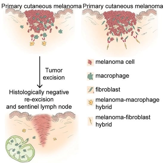
2.1. The BRAFV600E Protein Is Expressed in Subpopulations of Peritumoral Stromal Cells

To identify potential recurrent tumor-initiating cells, we screened for peritumoral cells that display a stromal phenotype but carry tumor-derived oncogenic information. For this we performed dual immunohistochemical staining on n = 11 patient-derived tissue samples of BRAFV600E melanoma with MART1 (melanoma antigen recognized by T-cells) and BRAFV600E. BRAFV600E is an oncogenic somatic mutation present in approximately 50%–60% of malignant melanomas. The staining revealed that, in addition to melanoma cells, BRAFV600E is also expressed in some subpopulations of MART1− peritumoral stromal cells with fibroblast and macrophage morphology (Figure 1a,b). To investigate whether the mutant protein originated from the tumor cells, we tested the stroma of n = 5 BRAFWT melanomas for the presence of the BRAFV600E protein. However, we could not detect the mutant protein in BRAFWT melanoma tissue samples (Supplementary Materials Figure S1), suggesting that the presence of the mutation in the stromal cells could not occur without the neighboring melanoma cells carrying the mutation. Nevertheless, though the antibody has been reported to be highly specific, and we did not see any specific signals in BRAFWT melanoma tissue samples, we cannot rule out nonspecific staining especially in case of macrophages.

2.2. Some Peritumoral Fibroblasts and Macrophages Carry the BRAFV600E Mutation in Primary Melanoma, Melanoma Metastasis and a Tumor-Free Re-Excision Sample

We wanted to confirm that the staining we observed in the BRAFV600E samples was specific, so we examined whether this oncogenic information is also present in the peritumoral cells at the genetic level. Therefore, we dual-stained BRAFV600E primary melanoma tissue samples with MART1 and either the fibroblast marker smooth muscle actin (SMA) or the monocyte-macrophage marker CD68, and isolated cell compartments consisting of 20–50, clearly MART1− but either SMA+ or CD68+ cells with fibroblast and macrophage morphology, respectively, using laser-capture microdissection (Figure 2a,b). Subsequent allele specific mutation detection PCR analyses performed on the dissected stromal cells revealed that the BRAFV600E mutant allele was present in peritumoral MART1−/SMA+ fibroblasts and MART1−/CD68+ macrophages. In addition to primary melanoma tissue samples, such peritumoral stromal cells carrying the melanoma-derived mutation were detected in lymph node and cutaneous melanoma metastases (Figure 2c). Surprisingly, BRAFV600E was also detected when analyzing MART1− macrophages dissected from a histologically tumor-free re-excision sample from a patient who subsequently developed a local recurrence. Having dissected peritumoral stromal cells from BRAFWT melanoma tissue samples, we only detected the wild-type allele, providing further evidence that BRAFV600E is only present in peritumoral cells that are adjacent to a mutant tumor. In total, we examined 86 dissected samples of 19 patients, and out of 12 patients with BRAFV600E melanoma, we found BRAFV600E-containing peritumoral cells in the tissue samples of five patients. The proportion of mutant alleles detected in these cell populations varied between 0.5% and 30% (Table 1 and Supplementary Materials Table S1).
These results indicate that some peritumoral stromal cells contain a melanoma-derived oncogene at both the DNA and protein levels. The possible recurrent tumor-initiating potential of these cells is supported by our clinical observations of the local recurrence rate in our melanoma patients diagnosed before 1998. Patients who had primary melanoma removed completely, based on a clinical diagnosis of a non-melanoma skin lesion and with histologically tumor-free excision margins developed local recurrence significantly more often (p = 0.7 × 10⁻⁵, 12 out of 228 patients) than patients with melanomas excised with 5-cm margins (8 out of 935 patients), implying that recurrent-tumor-initiating cells could indeed be present in the peritumoral stroma of malignant melanoma.

3. Discussion

After the complete resection of primary cutaneous melanoma, patients occasionally develop local recurrence, which is considered an independent prognostic factor [1]. This can be explained by hidden tumor-initiating cells containing tumor-derived genetic information as local minimal residual disease in the peritumoral area. In addition, such tumor-derived cells can also spread to distant areas such as lymph nodes.
Tumor cells are commonly identified during routine histological assessments using markers that are often insufficiently sensitive to distinguish tumor cells from stromal cells, which is supported by additional genetic analyses of sentinel lymph nodes [19]. The possible role of isolated tumor cells in the sentinel lymph node is debated, and the predictive value of additional diagnostic approaches, such as melanoma-marker detection at the mRNA level, to date, is inconclusive [20]. Nevertheless, some studies using enhanced pathological assessments have found a link between the presence of single tumor cells in the sentinel lymph node and worse disease outcome compared with completely tumor-free sentinel lymph nodes [21,22,23], and other improved tumor detection methods, such as quantitative immunocytochemistry combined with single-cell comparative genomic hybridization, have been found to better predict patient survival and disease outcome [24]. The incomplete removal of tumor cells often results in a higher recurrence rate: small excision margins lead to a more frequent tumor recurrence even if the excision margins are assessed to be tumor-free [2,25,26,27]. These data imply that recurrent-tumor-initiating cells with an altered phenotype could indeed be present in the peritumoral stroma of malignant melanoma, allowing them to remain undetected during routine diagnostic procedures.
In this study, we examined patient-derived melanoma tissue samples and detected peritumoral cells displaying stromal cell phenotype but carrying the oncogenic BRAFV600E mutation characteristic of the adjacent melanoma cells. First, we detected the mutated protein with a mutation-specific antibody. Even though nonspecific staining of the antibody in peritumoral mononuclear cells have been reported [28,29], the fact that peritumoral fibroblasts were also positive argues for a specific signal. Moreover, we only observed positivity in peritumoral cells adjacent to BRAFV600E melanoma. Nevertheless, to confirm the presence of the oncogene in these cells, we also performed genomic analyses and showed that the mutation is also present in cells with a stromal cell phenotype adjacent to BRAFV600E melanoma cells at the genetic level. We believe the mutation originates from tumor cells, as we did not detect the mutation in the stroma of BRAFWT melanoma samples. Possible mechanisms for the transfer of tumor-derived information to stromal cells include tumor-stromal cell fusion, exosomal protein transfer and the phagocytosis of tumor debris. However, subpopulations of peritumoral stromal cells carried the BRAFV600E mutation not only at protein but also at genomic level, suggesting cell fusion as the underlying mechanism. It is important to mention that, since melanoma cells can lose MART1 expression, especially on the periphery of the tumor, cell morphology and the expression patterns of CD68 or SMA were all taken into account during the process of peritumoral cell identification.
Cell fusion studies based on in vitro observations and mouse models suggest that cell fusion can confer an evolutionary benefit, such as increased metastatic potential [30] or drug resistance [31] to certain tumor–stromal cell hybrid clones. However, the majority of in vitro spontaneously formed hybrid cells undergo apoptosis [32], most likely as a result of mitotic stress. Therefore, it is possible that tumor stromal cell fusion is rather an antitumor mechanism, eliminating most of the hybrid tumor cells but occasionally giving rise to highly malignant tumor cell clones.
Studies in mice [33] imply that cell fusion may also take place in human tumors in vivo. However, it is very difficult to detect tumor–stromal cell fusion on a genetic level in humans. To address this difficulty, tumors developing in patients who have received bone marrow transplants have been investigated: genetic material originating from the donor was detected in the patients’ tumor cells [34,35], strongly suggesting a fusion event between recipient tumor cells and donor hemopoietic cells. However, inflammation resulting from treatment (e.g., whole body irradiation or chemotherapeutics) preceding bone-marrow transplantation has been reported to promote cell fusion [36,37,38]; therefore, further studies investigating fusion between tumor and stromal cells are clearly required.
Nevertheless, human in vivo studies demonstrating the relevance of tumor–stromal cell fusion in the clinical diagnosis or treatment of cancer have been lacking.
In conclusion, our results suggest that peritumoral stromal cells contain melanoma-specific oncogenic properties such as the BRAFV600E mutation derived from the neighboring tumor cells, potentially as a result of cell fusion. Based on the literature data, these cells, following a reversal to a tumorous phenotype [12], can possibly contribute to tumor recurrence. These cells are phenotypically indistinguishable from peritumoral stromal cells and are therefore not accurately detectable by routine histological assessments. Our results highlight the importance of genetic analysis and mutation-specific antibodies, especially in the case of histologically tumor-free tissue samples, for which improved methods for the detection of tumor cells have already been shown to better predict survival and outcome. Although yet to be confirmed in clinical trials, the use of genetic analyses and mutation-specific antibodies could have important prognostic and therapeutic consequences and could enable the detection of recurrent-tumor-initiating cells.

4. Experimental Section

4.1. Tissue Samples and Determination of BRAF Mutational Status

Tissue samples from n = 11 patients with BRAFV600E and n = 5 patients with BRAFWT melanoma were examined for BRAFV600E protein expression. The corresponding patient numbers for genetic analyses were n = 11 in the case of BRAFV600E melanoma and n = 8 in the case of BRAFWT melanoma. The BRAF mutational status of melanomas was determined with cobas® 4800 BRAFV600 Mutation Test (Roche Molecular Diagnostics, Pleasanton, CA, USA).

4.2. Immunohistochemistry

Formalin-fixed, paraffin-embedded tissue sections of patients with BRAFV600E malignant melanoma were stained with rabbit monoclonal MART1 (clone A19-P, DB Biotech, Kosice, Slovakia), mouse monoclonal SMA (clone 1A4, DAKO, Glostrup, Denmark), CD68 (clone PG-M1, DAKO), and BRAFV600E (Clone VE1, Springbio, Pleasanton, CA, USA) primary antibodies. The Bond Polymer Refine Detection Kit and the ChromoPlex 1 Dual Detection Kit (both from Leica, Wetzlar, Germany) were used for the visualization of single and dual histochemical staining, respectively, according to the manufacturer’s instructions. All immunohistochemical staining was scanned with a Pannoramic MIDI Slide Scanner (3DHISTECH Ltd., Budapest, Hungary) and analyzed with Pannoramic Viewer software (3DHISTECH Ltd.).

4.3. Laser-Capture Microdissection and Detection of the BRAFV600E Allele

30–100 cells were dissected from tissue sections stained either with MART1 and SMA or with MART1 and CD68 using a Palm Microbeam (Carl Zeiss Microscopy, Jena, Germany). Samples were digested with proteinase K (QIAGEN, Venlo, The Netherlands). Mutant allele (BRAF_476_mu) and gene reference (Braf_rf) TaqMan® Mutation Detection Assays (Life Technologies, Carlsbad, CA, USA) were used to detect the BRAFV600E mutant allele in the dissected samples. Samples with ΔCt (CtmutCtref) < 8 and Ctmut < 40 were considered carrying the mutant allele. MART1+ tumor cells dissected from BRAFWT and BRAFV600E melanoma were used as negative and positive controls, respectively.

4.4. Statistical Analysis

Local recurrence rate in melanoma patients were compared with the Fisher’s exact test. The level of significance was set to 0.05.

Supplementary Materials

Supplementary materials can be found at https://www.mdpi.com/1422-0067/17/6/980/s1.

Acknowledgments

The authors would like to thank their colleagues at the Dermatohistopathological Laboratory, University of Szeged, Hungary: Erika Függ and Mónika Kohajda for technical support and assistance, Márta Széll, Kornélia Szabó, Attila Bebes, Anikó Göblös, and Gábor Tax for our useful discussions and their comments, and Zoltán Tóbiás for preparing the illustrations. We are also very grateful to Ferhan Ayaydin (Biological Research Center, Hungarian Academy of Science, Szeged, Hungary) for providing the Palm Microbeam microscope and for the technical assistance. This work was supported by TÁMOP-4.2.1/B-09/1/KONV-2010-0005 and by TÁMOP-4.2.2/B grants. István B. Németh was supported by the Bolyai Scholarship 2015/17 of the Hungarian Academy of Sciences. Thomas Dittmar was supported by the Fritz-Bender-Foundation, Munich, Germany.

Author Contributions

István B. Németh conceived the study; Lajos Kemény and István B. Németh supervised the entire project; Zsuzsanna Kurgyis, Lajos V. Kemény and István B. Németh designed the experiments, analyzed the data, and generated the figures with the help of Gergely Groma, Thomas Dittmar, Lajos Kemény; István B. Németh, Zsuzsanna Kurgyis and Lajos V. Kemény performed the experiments supported by Tünde Buknicz, Ádám Jakab, Hilda Polyánka; István B. Németh, Gergely Groma, Thomas Dittmar and Kurt Zänker provided critical analysis support; Judit Oláh provided the clinical data of the melanoma patients; Zsuzsanna Kurgyis and Lajos V. Kemény wrote the manuscript.

Conflicts of Interest

The authors declare no conflict of interest.

Abbreviations

MART1melanoma antigen recognized by T-cells
SMAsmooth muscle actin

References

  1. Hocevar, M.; Dragonja, Z.; Pilko, G.; Gazic, B.; Zgajnar, J. Residual melanoma after an excisional biopsy is an independent prognostic factor for local recurrence and overall survival. Eur. J. Surg. Oncol. 2014, 40, 1271–1275. [Google Scholar] [CrossRef] [PubMed]
  2. Thomas, J.M.; Newton-Bishop, J.; A’Hern, R.; Coombes, G.; Timmons, M.; Evans, J.; Cook, M.; Theaker, J.; Fallowfield, M.; O’Neill, T.; et al. Excision margins in high-risk malignant melanoma. N. Engl. J. Med. 2004, 350, 757–766. [Google Scholar] [CrossRef] [PubMed]
  3. Polyak, K.; Weinberg, R.A. Transitions between epithelial and mesenchymal states: Acquisition of malignant and stem cell traits. Nat. Rev. Cancer 2009, 9, 265–273. [Google Scholar] [CrossRef] [PubMed]
  4. Landsberg, J.; Kohlmeyer, J.; Renn, M.; Bald, T.; Rogava, M.; Cron, M.; Fatho, M.; Lennerz, V.; Wölfel, T.; Hölzel, M.; et al. Melanomas resist T-cell therapy through inflammation-induced reversible dedifferentiation. Nature 2012, 490, 412–416. [Google Scholar] [CrossRef] [PubMed]
  5. Giancotti, F.G. Mechanisms governing metastatic dormancy and reactivation. Cell 2013, 155, 750–764. [Google Scholar] [CrossRef] [PubMed]
  6. Thiery, J.P.; Acloque, H.; Huang, R.Y.J.; Nieto, M.A. Epithelial-mesenchymal transitions in development and disease. Cell 2009, 139, 871–890. [Google Scholar] [CrossRef] [PubMed]
  7. Easwaran, H.; Tsai, H.-C.; Baylin, S.B. Cancer epigenetics: Tumor heterogeneity, plasticity of stem-like states, and drug resistance. Mol. Cell 2014, 54, 716–727. [Google Scholar] [CrossRef] [PubMed]
  8. Stoecklein, N.H.; Klein, C.A. Genetic disparity between primary tumours, disseminated tumour cells, and manifest metastasis. Int. J. Cancer 2010, 126, 589–598. [Google Scholar] [CrossRef] [PubMed]
  9. Harkness, T.; Weaver, B.A.; Alexander, C.M.; Ogle, B.M. Cell fusion in tumor development: Accelerated genetic evolution. Crit. Rev. Oncog. 2013, 18, 19–42. [Google Scholar] [CrossRef] [PubMed]
  10. Clawson, G.A. Cancer. Fusion for moving. Science 2013, 342, 699–700. [Google Scholar] [CrossRef] [PubMed]
  11. Duelli, D.; Lazebnik, Y. Cell fusion: A hidden enemy? Cancer Cell 2003, 3, 445–448. [Google Scholar] [CrossRef]
  12. Rappa, G.; Mercapide, J.; Lorico, A. Spontaneous formation of tumorigenic hybrids between breast cancer and multipotent stromal cells is a source of tumor heterogeneity. Am. J. Pathol. 2012, 180, 2504–2515. [Google Scholar] [CrossRef] [PubMed]
  13. Shabo, I.; Midtbö, K.; Andersson, H.; Åkerlund, E.; Olsson, H.; Wegman, P.; Gunnarsson, C.; Lindström, A. Macrophage traits in cancer cells are induced by macrophage-cancer cell fusion and cannot be explained by cellular interaction. BMC Cancer 2015, 15. [Google Scholar] [CrossRef] [PubMed]
  14. Terada, N.; Hamazaki, T.; Oka, M.; Hoki, M.; Mastalerz, D.M.; Nakano, Y.; Meyer, E.M.; Morel, L.; Petersen, B.E.; Scott, E.W. Bone marrow cells adopt the phenotype of other cells by spontaneous cell fusion. Nature 2002, 416, 542–545. [Google Scholar] [CrossRef] [PubMed]
  15. Kemény, L.V.; Kurgyis, Z.; Buknicz, T.; Groma, G.; Jakab, Á.; Zänker, K.; Dittmar, T.; Kemény, L.; Németh, I.B. Melanoma cells can adopt the phenotype of stromal fibroblasts and macrophages by spontaneous cell fusion in vitro. Int. J. Mol. Sci. 2016, 17. [Google Scholar] [CrossRef] [PubMed]
  16. Clawson, G.A.; Matters, G.L.; Xin, P.; Imamura-Kawasawa, Y.; Du, Z.; Thiboutot, D.M.; Helm, K.F.; Neves, R.I.; Abraham, T. Macrophage-tumor cell fusions from peripheral blood of melanoma patients. PLoS ONE 2015, 10, e0134320. [Google Scholar] [CrossRef] [PubMed]
  17. Xue, J.; Zhu, Y.; Sun, Z.; Ji, R.; Zhang, X.; Xu, W.; Yuan, X.; Zhang, B.; Yan, Y.; Yin, L.; et al. Tumorigenic hybrids between mesenchymal stem cells and gastric cancer cells enhanced cancer proliferation, migration and stemness. BMC Cancer 2015, 15. [Google Scholar] [CrossRef] [PubMed]
  18. He, X.; Li, B.; Shao, Y.; Zhao, N.; Hsu, Y.; Zhang, Z.; Zhu, L. Cell fusion between gastric epithelial cells and mesenchymal stem cells results in epithelial-to-mesenchymal transition and malignant transformation. BMC Cancer 2015, 15. [Google Scholar] [CrossRef] [PubMed]
  19. Mocellin, S.; Hoon, D.S.B.; Pilati, P.; Rossi, C.R.; Nitti, D. Sentinel lymph node molecular ultrastaging in patients with melanoma: A systematic review and meta-analysis of prognosis. J. Clin. Oncol. 2007, 25, 1588–1595. [Google Scholar] [CrossRef] [PubMed]
  20. Itakura, E.; Huang, R.-R.; Wen, D.-R.; Cochran, A.J. “Stealth” melanoma cells in histology-negative sentinel lymph nodes. Am. J. Surg. Pathol. 2011, 35, 1657–1665. [Google Scholar] [CrossRef] [PubMed]
  21. Satzger, I.; Völker, B.; Meier, A.; Schenck, F.; Kapp, A.; Gutzmer, R. Prognostic significance of isolated HMB45 or Melan A positive cells in Melanoma sentinel lymph nodes. Am. J. Surg. Pathol. 2007, 31, 1175–1180. [Google Scholar] [CrossRef] [PubMed]
  22. Murali, R.; DeSilva, C.; McCarthy, S.W.; Thompson, J.F.; Scolyer, R.A. Sentinel lymph nodes containing very small (<0.1 mm) deposits of metastatic melanoma cannot be safely regarded as tumor-negative. Ann. Surg. Oncol. 2012, 19, 1089–1099. [Google Scholar] [PubMed]
  23. Murali, R.; Thompson, J.F.; Shaw, H.M.; Scolyer, R.A. The prognostic significance of isolated immunohistochemically positive cells in sentinel lymph nodes of melanoma patients. Am. J. Surg. Pathol. 2008, 32, 1106–1108. [Google Scholar] [CrossRef] [PubMed]
  24. Ulmer, A.; Dietz, K.; Hodak, I.; Polzer, B.; Scheitler, S.; Yildiz, M.; Czyz, Z.; Lehnert, P.; Fehm, T.; Hafner, C.; et al. Quantitative measurement of melanoma spread in sentinel lymph nodes and survival. PLoS Med. 2014, 11, e1001604. [Google Scholar] [CrossRef] [PubMed]
  25. Ng, A.K.; Jones, W.O.; Shaw, J.H. Analysis of local recurrence and optimizing excision margins for cutaneous melanoma. Br. J. Surg. 2001, 88, 137–142. [Google Scholar] [CrossRef] [PubMed]
  26. Hudson, L.E.; Maithel, S.K.; Carlson, G.W.; Rizzo, M.; Murray, D.R.; Hestley, A.C.; Delman, K.A. 1 or 2 cm margins of excision for T2 melanomas: Do they impact recurrence or survival? Ann. Surg. Oncol. 2013, 20, 346–351. [Google Scholar] [CrossRef] [PubMed]
  27. Pasquali, S.; Haydu, L.E.; Scolyer, R.A.; Winstanley, J.B.; Spillane, A.J.; Quinn, M.J.; Saw, R.P.M.; Shannon, K.F.; Stretch, J.R.; Thompson, J.F. The importance of adequate primary tumor excision margins and sentinel node biopsy in achieving optimal locoregional control for patients with thick primary melanomas. Ann. Surg. 2013, 258, 152–157. [Google Scholar] [CrossRef] [PubMed]
  28. Fisher, K.E.; Cohen, C.; Siddiqui, M.T.; Palma, J.F.; Lipford, E.H.; Longshore, J.W. Accurate detection of BRAF p.V600E mutations in challenging melanoma specimens requires stringent immunohistochemistry scoring criteria or sensitive molecular assays. Hum. Pathol. 2014, 45, 2281–2293. [Google Scholar] [CrossRef] [PubMed]
  29. Long, G.V.; Wilmott, J.S.; Capper, D.; Preusser, M.; Zhang, Y.E.; Thompson, J.F.; Kefford, R.F.; von Deimling, A.; Scolyer, R.A. Immunohistochemistry is highly sensitive and specific for the detection of V600E BRAF mutation in melanoma. Am. J. Surg. Pathol. 2013, 37, 61–65. [Google Scholar] [CrossRef] [PubMed]
  30. Rachkovsky, M.; Sodi, S.; Chakraborty, A.; Avissar, Y.; Bolognia, J.; McNiff, J.M.; Platt, J.; Bermudes, D.; Pawelek, J. Melanoma × macrophage hybrids with enhanced metastatic potential. Clin. Exp. Metastasis 1998, 16, 299–312. [Google Scholar] [CrossRef] [PubMed]
  31. Nagler, C.; Hardt, C.; Zänker, K.S.; Dittmar, T. Co-cultivation of murine BMDCs with 67NR mouse mammary carcinoma cells give rise to highly drug resistant cells. Cancer Cell Int. 2011, 11. [Google Scholar] [CrossRef] [PubMed]
  32. Wang, R.; Sun, X.; Wang, C.Y.; Hu, P.; Chu, C.-Y.; Liu, S.; Zhau, H.E.; Chung, L.W.K. Spontaneous cancer-stromal cell fusion as a mechanism of prostate cancer androgen-independent progression. PLoS ONE 2012, 7, e42653. [Google Scholar] [CrossRef] [PubMed]
  33. Chakraborty, A.K.; Sodi, S.; Rachkovsky, M.; Kolesnikova, N.; Platt, J.T.; Bolognia, J.L.; Pawelek, J.M. A Spontaneous murine melanoma lung metastasis comprised of host × tumor hybrids. Cancer Res. 2000, 60, 2512–2519. [Google Scholar] [PubMed]
  34. Lazova, R.; LaBerge, G.S.; Duvall, E.; Spoelstra, N.; Klump, V.; Sznol, M.; Cooper, D.; Spritz, R.A.; Chang, J.T.; Pawelek, J.M. A Melanoma brain metastasis with a donor-patient hybrid genome following bone marrow transplantation: First evidence for fusion in human cancer. PLoS ONE 2013, 8, e66731. [Google Scholar] [CrossRef] [PubMed]
  35. Yilmaz, Y.; Lazova, R.; Qumsiyeh, M.; Cooper, D.; Pawelek, J. Donor Y chromosome in renal carcinoma cells of a female BMT recipient: visualization of putative BMT-tumor hybrids by FISH. Bone Marrow Transplant. 2005, 35, 1021–1024. [Google Scholar] [CrossRef] [PubMed]
  36. Nygren, J.M.; Liuba, K.; Breitbach, M.; Stott, S.; Thorén, L.; Roell, W.; Geisen, C.; Sasse, P.; Kirik, D.; Björklund, A.; et al. Myeloid and lymphoid contribution to non-haematopoietic lineages through irradiation-induced heterotypic cell fusion. Nat. Cell Biol. 2008, 10, 584–592. [Google Scholar] [CrossRef] [PubMed]
  37. Espejel, S.; Romero, R.; Alvarez-Buylla, A. Radiation damage increases Purkinje neuron heterokaryons in neonatal cerebellum. Ann. Neurol. 2009, 66, 100–109. [Google Scholar] [CrossRef] [PubMed]
  38. Johansson, C.B.; Youssef, S.; Koleckar, K.; Holbrook, C.; Doyonnas, R.; Corbel, S.Y.; Steinman, L.; Rossi, F.M.V.; Blau, H.M. Extensive fusion of haematopoietic cells with Purkinje neurons in response to chronic inflammation. Nat. Cell Biol. 2008, 10, 575–583. [Google Scholar] [CrossRef] [PubMed]
Figure 1. Peritumoral stromal cells express the melanoma-derived oncogenic BRAFV600E protein. (a,b) Tissue samples of a patient with BRAFV600E melanoma stained for the melanoma marker melanoma antigen recognized by T-cells (MART1) (red), the BRAFV600E mutant protein (brown) and hematoxylin (blue). Brown arrows indicate peritumoral MART1−/BRAFV600E+ cells displaying fibroblast (a) or macrophage (b) morphology. Blue arrows indicate MART1−/BRAFV600E− stromal cells (light blue from hematoxylin). Red lines indicate MART1+ melanoma cells. Black bars indicate 50 µm.
Ijms 17 00980 g001
Figure 2. Peritumoral stromal cells contain the melanoma-derived oncogenic BRAFV600E at the genomic level. (a) BRAFV600E melanoma tissue sample stained for MART1 (red), smooth muscle actin (SMA) (brown), and hematoxylin (blue) before (left panel) and after (right panel) laser-capture microdissection of MART1−/SMA+ fibroblasts (white line); (b,c) Tissue samples of BRAFV600E (b) primary melanoma and (c) melanoma metastasis stained for MART1 (red), CD68 (brown), and hematoxylin (blue) before (left panels) and after (right panels) laser-capture microdissection of MART1−/CD68+ macrophages (black line). Black bars indicate 150 µm.
Ijms 17 00980 g002
Table 1. Peritumoral fibroblasts and macrophages in BRAFV600E melanoma carry the melanoma-derived BRAFV600E mutation at the genetic level.
Tissue 1Npos/Nex 2,3
Fibroblasts 4 DissectedMacrophages 5 Dissected
BRAFV600E primary melanoma2/21/1
BRAFV600E melanoma metastasis2/42/4
Histologically tumor-free tissue (from patients with BRAFV600E melanoma)0/41/3
BRAFWT primary melanoma0/40/1
1 Tissue samples were dual-stained either with melanoma antigen recognized by T-cells (MART1) & smooth muscle actin (SMA) or with MART1 & CD68 and examined by a certified pathologist (IBN); 2 Npos: number of patients where BRAFV600E-positive peritumoral stromal cells were found; Nex: the number of patients examined; 3 Dissected cell samples were considered positive for BRAFV600E if the prevalence of the mutant allele was higher than 0.39% (ΔCt < 8); 4 MART1−/SMA+ cells were considered fibroblasts; 5 MART1−/CD68+ cells were considered macrophages.

