Article

Architectural Polychromy on the Athenian Acropolis: An In Situ Non-Invasive Analytical Investigation of the Colour Remains

by
Eleni Aggelakopoulou
1,*,
Sophia Sotiropoulou
2,* and
Georgios Karagiannis
3
1
Acropolis Restoration Service (YSMA), Hellenic Ministry of Culture and Sports, 10 Polygnotou Str., 10555 Athens, Greece
2
Foundation for Research and Technology—Hellas, Institute of Electronic Structure and Laser (FORTH-IESL), N.Plastira 100, Vassilika Vouton, 70013 Crete, Greece
3
Ormylia Foundation, Art Diagnosis Centre, 63071 Ormylia, Greece
*
Authors to whom correspondence should be addressed.
Heritage 2022, 5(2), 756-787; https://doi.org/10.3390/heritage5020042
Submission received: 13 February 2022 / Revised: 11 March 2022 / Accepted: 23 March 2022 / Published: 1 April 2022
(This article belongs to the Special Issue Polychromy in Ancient Sculpture and Architecture)

Abstract

:
The preservation of the Athenian Acropolis monuments constitutes an ongoing top-priority national project of global significance and impact. The project concerning the analytical investigation of the polychromy of the Acropolis monuments presented in this paper was part of the Acropolis Restoration Service (YSMA) program (2011–2015), regarding the restoration of the two corners of the west entablature of the Parthenon, which exhibited severe static damage, and a parallel restoration program of the Propylaea. The scope of this research was to investigate the materials in the paint decoration remains on the monuments by applying, entirely in situ, numerous non-invasive techniques on selected architectural members of the Parthenon and the Propylaea. The research focused, mainly, on surfaces where traces of colour or decoration patterns were visible to the naked eye. Furthermore, surfaces that are referred to in the literature as decorated but that are currently covered with weathering crusts (of white or black colour) and/or layers of patina (of yellowish and orange-brown hue), were also examined. The techniques applied in situ on the Acropolis monuments were X-ray fluorescence, micro-Raman, and Fourier Transform InfraRed (FTIR) spectroscopic techniques, conducted with the use of handheld or portable instruments. The scientific data gathered in situ are discussed in this paper to enhance our knowledge of the architectural polychromy of the classical period. Further investigation by applying analytical techniques on a few selected micro-samples would be highly complementary to this present work.

1. Introduction

The earliest information about polychromy on the classical monuments of the Athenian Acropolis is provided by European travelers, architects, and painters of the 19th century, who refer to elements of colour on the architectural members and sculptures of the temples, with pigments that were visible at that time to the naked eye. The earliest chemical investigations on “coatings” and traces of “paint” from the Parthenon and other monuments of the Classical period in Athens extend back at least to the early nineteenth century. Following these first studies on the “Polychromy”, as the term was established in the early 19th century, a long period elapsed without any substantial unambiguous data on the extent and composition of the colour that adorned the temples of classical antiquity [1,2]. All the observations recorded in the literature, relating to the polychromy of the metopes of the Parthenon—especially the west metopes—noted from the nineteenth century up to 1960, as well as certain isolated analyses of paint samples, were collated by Frank Brommer [1].
As stated by Ian Jenkins and Andrew Middleton [2], Michael Faraday (1837) was the first to study samples of colour taken from the Propylaea and the Hephaesteum. He concluded that the samples of blue colour consisted of either copper carbonate (referring to azurite) or copper-containing glass/frit (certainly relating to Egyptian blue) which, according to his investigation, had been admixed with “a wax”. Five years later, X. Landerer (1842) accomplished analyses of several paint samples from the Parthenon and other monuments. Landerer came to the following conclusions: the reds consisted of iron oxide or cinnabar; green was identified as verdigris; the blue pigment examined was titaniferous copper oxide; the yellow colour contained iron, clay minerals, and calcium carbonate. Regarding the black colours, two samples were examined, one was mentioned to contain “fer brûlé” while the other sample was some type of carbon black. He also investigated two white colours, one of which consisted of lead white and the other one of a mixture of lime and clay. Like Faraday, Landerer also found evidence of the presence of wax in the examined samples of paint.
In the past few decades, considerable work has been accomplished by the Greek authorities regarding an investigation of the polychromy of the Acropolis monuments, within the framework of the respective restoration and conservation programs. In 1989, a study was carried out regarding polychromy, focusing on the Parthenon’s east front. Blue colour was found on the mutules of the east cornice blocks and red colour on the viae. The same blue colour was also observed on the taenia of the east metopes and on the face and the meroi of the east triglyphs. By means of X-ray diffraction analysis and electron beam microanalysis, Egyptian blue and hematite were identified in the blue and red colours, respectively [3].
After the dismantling and the transportation of the Parthenon’s east metopes to the new Acropolis Museum in 2007, the remaining colours were observed in various areas of eight metopes (east metopes 3, 4, 9–14), among the so-called “patinas”. More specifically, traces of blue and red colour were detected on the taenia and the background of the metopes, while the blue colour was also observed on the astragal. Moreover, on the lower part of east metope 9 (which depicts Apollo fighting with a giant), the contour lines of a snake painted red became clearer after laser cleaning. Furthermore, in the case of the Parthenon’s west pediment, on the back of the himation’s folds on the Kecrops figure, where the orange-brown layer of patina is preserved, significant remains of the blue colour were observed on protected parts of the folds, on top of the patina layer [4,5].
At the same time, the Parthenon’s west frieze was dismantled for laser cleaning and conservation before being transferred to the new Acropolis Museum (2007). In the course of laser cleaning, completed in 2005, considerable areas of orange-brown patina layers (“epidermis”) were revealed. Moreover, clear remnants of a light green colour were found on the himation of rider 17 (slab IX), on the left arm and on the folds hanging down behind the figure’s back, as well as on the folds of the chitoniskos of rider 21 on slab XI [6]. During 2012–2013, visible induced infrared luminescence (VIL) imaging was carried out on the Parthenon’s west frieze, while it was stored in the new Acropolis Museum. Egyptian blue pigment was detected mainly on the horses’ eyes (the horses of figures 15, 17, and 19 on the slabs VIII, IX, and X, respectively) and on parts of the figures’ clothing (figures 15, 17, 19, and 20 on slabs VIII, ΙΧ, X, and XI, respectively). Especially in the case of slab ΙX, Egyptian blue apparently covered the entire surface of the mantle of figure 17 and was also used to represent the horse’s eye contour [7].
Investigation of the polychromy of the coffered ceiling of the Maidens’ Porch in the Erechtheion began in 2008, within the framework of laser-assisted cleaning and conservation projects. Thanks to the application of VIL imaging, Egyptian blue was revealed as the main colour for the background of the ceiling; this finding has been confirmed via the analysis of representative micro-samples. Lead white was found as a priming layer, as well as in the highlights of specific decorative patterns. Significant data were obtained from the study of cross-sections throughout the entire stratigraphy of the original and historic layer. In two samples, the presence of egg was identified using chromatographic analysis and was attributed to a binding medium, a result that needs further investigation with the analysis of additional samples [8,9].
Research continued during the last restoration project (2011–2015) regarding the restoration of the two corners of the west entablature of the Parthenon, which faced severe damage, in regard to the structural stability of the building. In parallel, a restoration program also began on the Propylaea. For that purpose, scaffolds were erected, and, for the first time in a hundred years, the upper part of both monuments were accessible. Observation of the marble surface at close range revealed several areas in both monuments that preserved evidence of polychromy. In some cases, the design is visible through the decay layer, presenting a contrast due to its different texture or thickness, as a result of the differential material degradation of the initially painted and unpainted areas. In other cases, layers or traces of paint still survive on the marble surface, most often below οr interlaced with the decay crusts. Finally, there are cases where the decorative pattern is discernible due to the incised outlines of the design. The specific architectural members of the Parthenon’s west entablature and of the Propylaea, where traces of the decorative patterns and pigments still survived, were selected for analytical investigation.
The methodology applied for the investigation of the polychromy took into account appropriate state-of-the-art methodology [10,11,12,13,14,15,16,17] and consisted of four phases: (a) Phase A—documentation of the conserved paint layers; (b) Phase Β—imaging-based diagnostic techniques applied in situ; (c) Phase C—non-invasive techniques applied in situ; and (d) Phase D—micro-sampling and the application of analytical techniques to representative samples.
In recent decades, research has been extended and intensified, bringing new evidence to the fore regarding the architectural polychromy of the Classical world, in which colour is finely integrated into or interlaced with the sculptural elements of the architecture. The new findings have uncovered fertile interdisciplinary scientific ground to be explored, interpreted, and understood, forming a new perception of Classical aesthetics as a basis for reconstructing the original appearance of architectural monuments. In this context, technical studies on the surviving traces of polychromy are nowadays carried out systematically, following an integrated protocol, extending and exhausting the possibilities of non-invasive investigations, thus minimizing and reserving the need for sampling or micro-destructive measurements until the very last, conclusive stages of the analytical methodology.
In the present paper, the results of Phase C are presented, with part of the results for phases A and B presented in previous works [18,19]. The non-invasive investigation of the polychromy of the Acropolis monuments constituted an integral part of the entire analytical methodological approach, which was adopted to address the investigation of colour remains or surviving decorative patterns on the Parthenon’s west entablature and specific architectural members of the Propylaea. It consisted of optical macro- and micro-examination, elemental (X-ray fluorescence), and molecular (micro-Raman and Fourier Transform InfraRed—FTIR) analysis on selected architectural members of the Parthenon and the Propylaea, carried out on site. The application of a multi-technique approach combining XRF, micro-Raman, and FTIR spectroscopies, constitutes a well-established analytical protocol for the in situ investigation of paintings or paint decoration on various substrates. A wide spectrum of valuable works can be found in the recent literature, demonstrating the complementarity of these techniques and proving the worth of applying them jointly for a complete characterization of both original materials and degradation products found in the paint layers [20,21,22,23,24,25,26]. In the present paper, the results are presented, interpreted, and discussed in a synthetic manner, based also on the interdisciplinary synergy and collaborative work that was carried out on site.
Further investigation by applying analytical techniques on a minimal number of micro-samples, as selected after the evaluation of the in situ analytical data, is ongoing, in order to validate and extend the interpretation of the data obtained in situ, and for a further understanding of the techniques employed in the paint decoration of these ancient monuments.

2. Materials and Methods

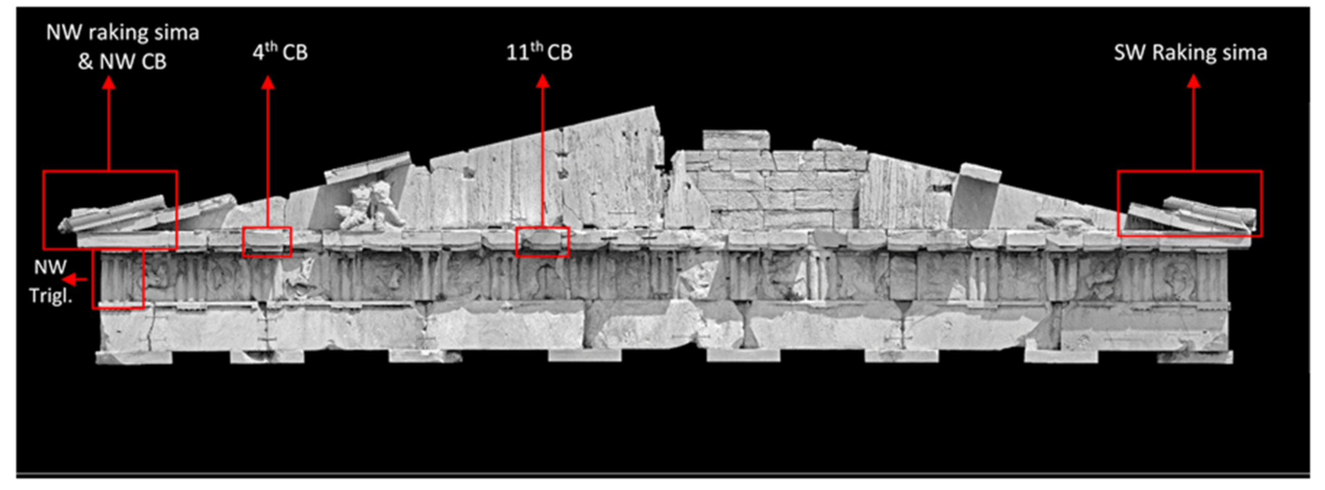
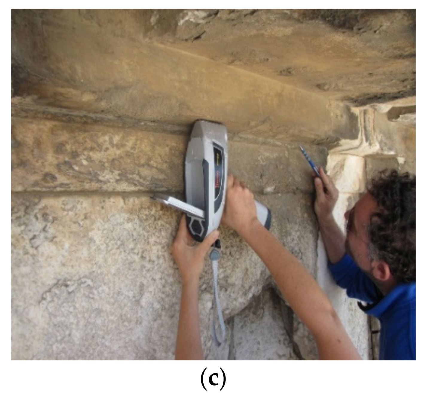
2.1. Selection of Architectural Members for In Situ Analytical Investigation

Based on the preliminary studies (phases A and B), including visual observation using video-microscopy and, in certain cases, technical imaging (VIL), a number of architectural members of the Parthenon and the Propylaea that bear vestiges of polychromy was selected for analysis. Τhe specific sites of interest that have been investigated with in situ measurements are listed here and, in detail, in Table 1. Access to the sites was accomplished by scaffolds that were erected around the Parthenon and Propylaea during the last restoration program (2011–2015) of the Acropolis monuments. A few members were investigated on the ground after their dismantling for restoration and before their repositioning in the monuments.
The research focused on surfaces where traces of colour and decoration were visible to the naked eye. However, the study also extended to surfaces that have been referred to in the literature as decorated but that are currently covered with encrustations (of white or black colour) and/or layers of patina (of yellowish and orange-brown hue). In the latter cases, no decorative pattern is, thus, visually perceptible.
More specifically, in this paper, the results of our investigation on the following architectural members of the Parthenon are presented (Figure 1 and Figure 2):
  • The northwest (NW) triglyph;
  • The 4th and the 11th horizontal cornice blocks (CBs) of the west side (the numbering starts from north to south);
  • The northwest (NW) raking sima with the lion-head false water spout;
  • The southwest (SW) raking sima;
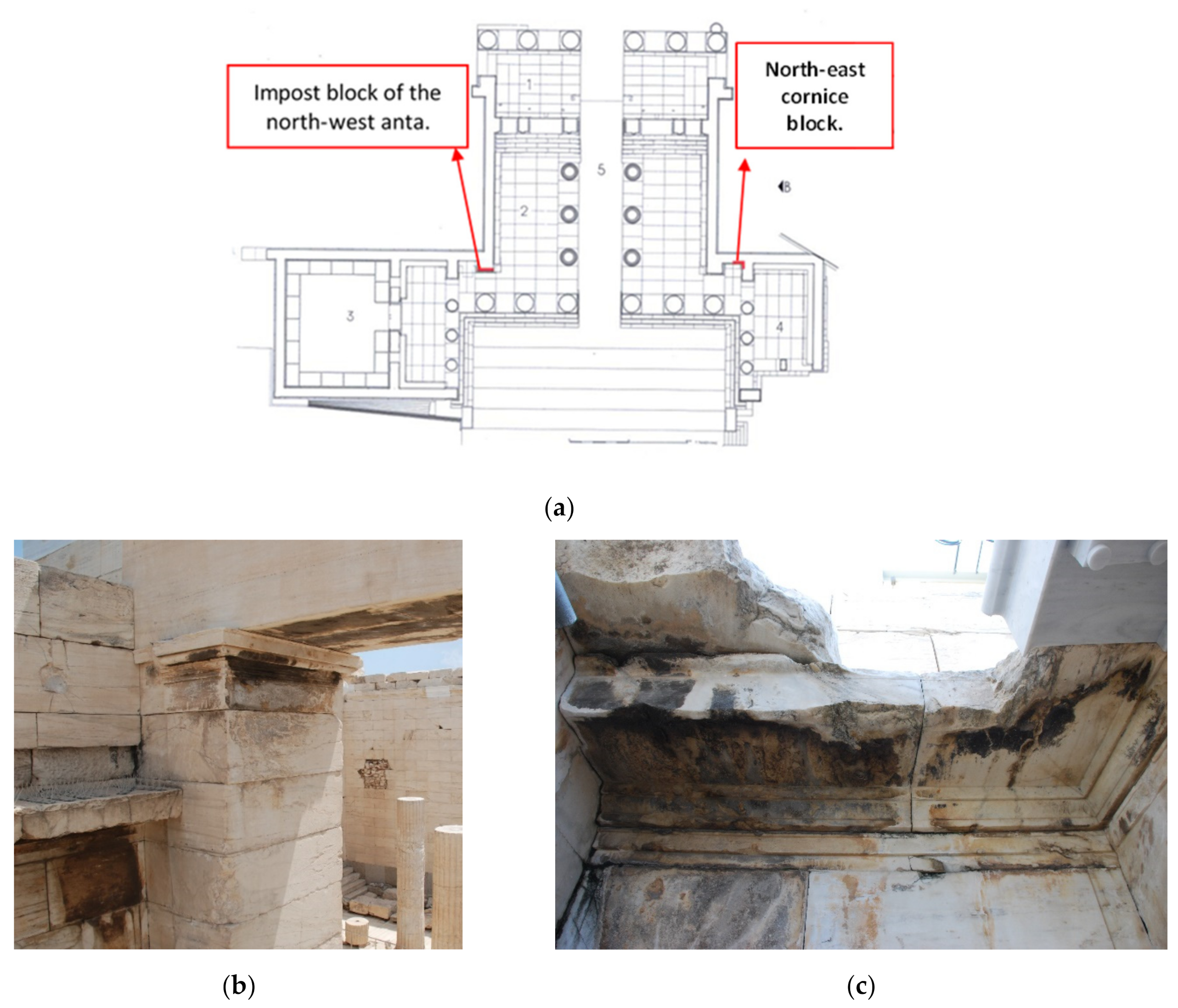
  • The impost block of the southwest (SW) anta.
In the case of the Propylaea, the results on the following investigated architectural members are presented (Figure 3):
  • The impost block of the northwest (NW) anta in the central building;
  • The northeast (NE) cornice block (CB) in the niche between the south wing and the southwest anta of the west portico.

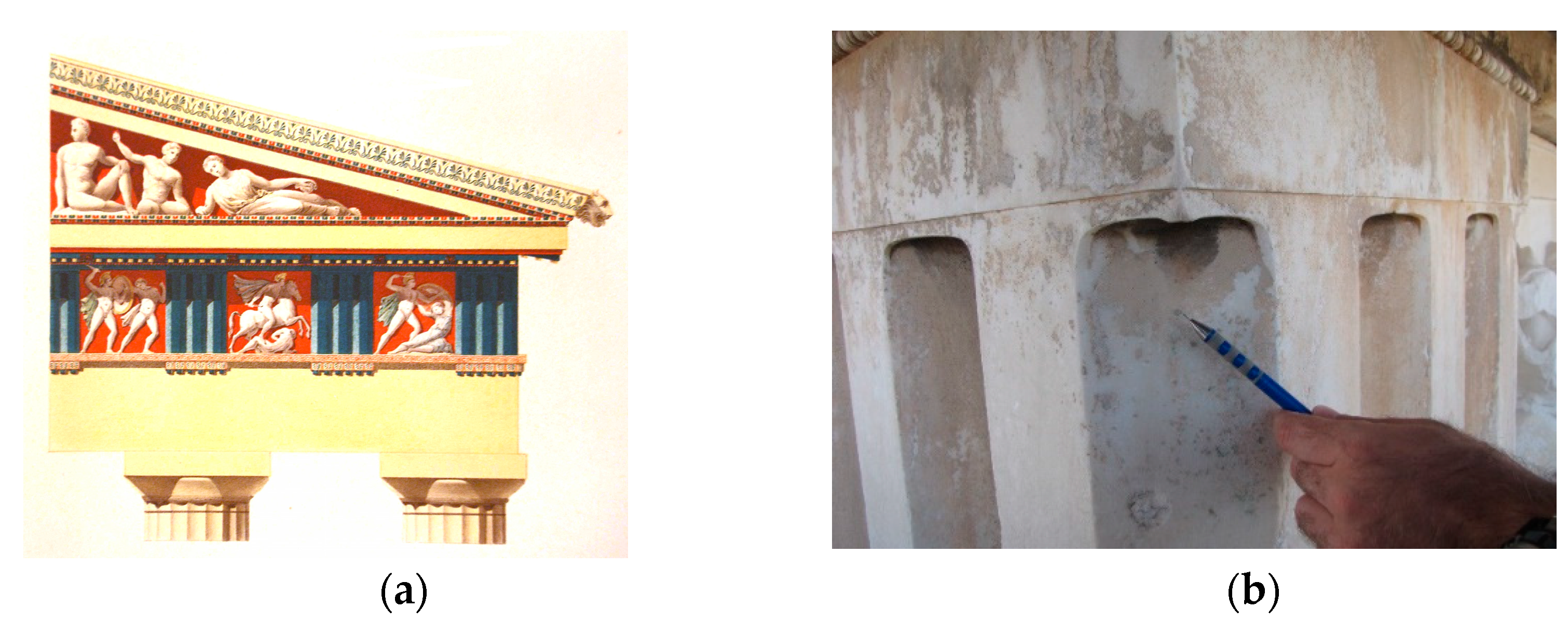
2.2. Historical colour Representations Available in the Literature

For each investigated architectural member, a reference is made to historical colour representations, dating from the 19th century. Although these hand-coloured drawings cannot be considered to be accurate reproductions, neither can they be dismissed as arbitrary reconstructions. They were made after thorough observation of the monuments, in centuries past, when that polychromy was preserved to, presumably, a significant extent. They can, therefore, serve as a valuable source of information that should, however, be interpreted with caution and investigated and validated or disproved on a case-by-case basis, based on the evidence provided by this investigation of the colour remains still existing on the monuments.

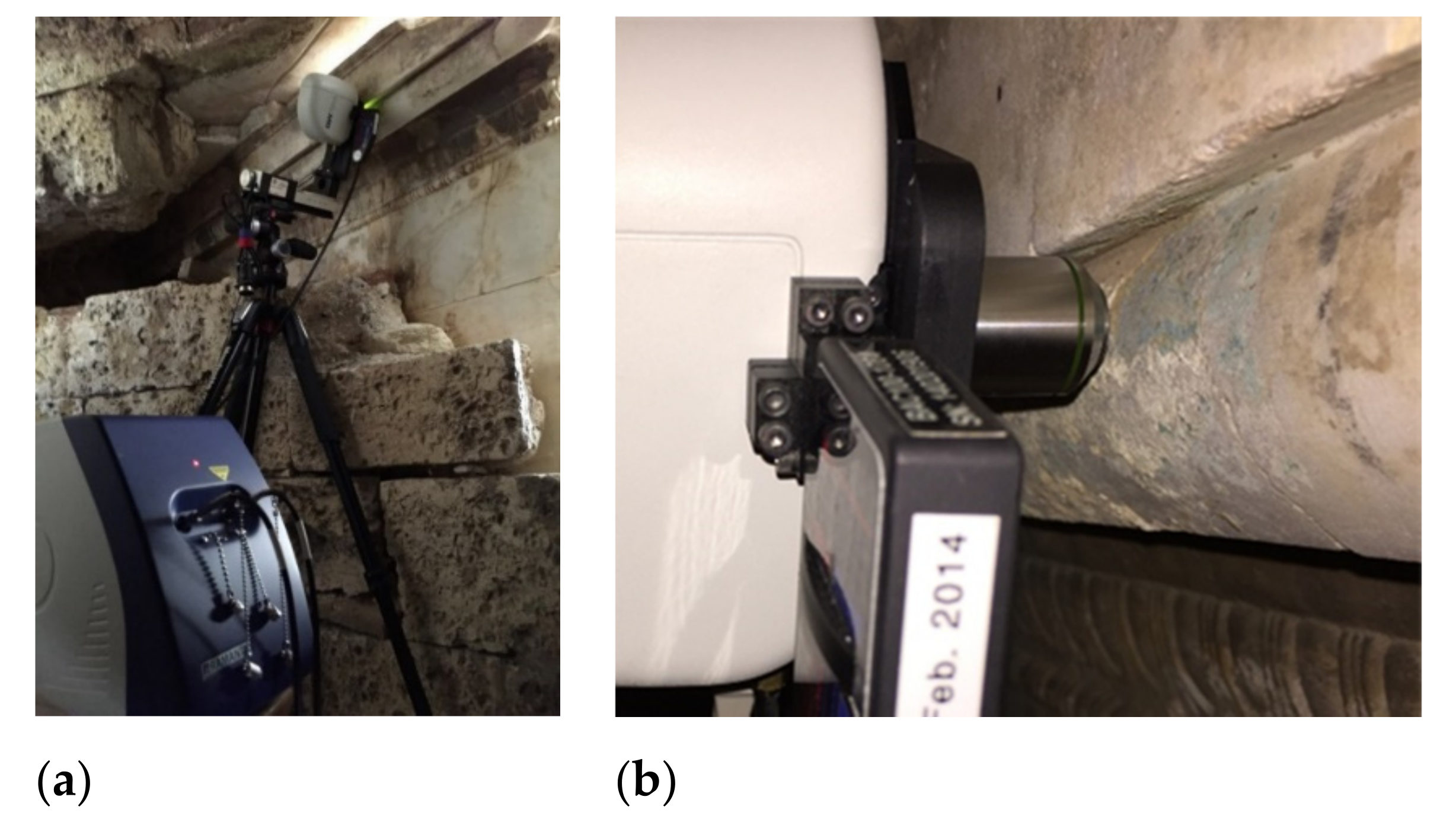
2.3. Analytical Investigation Methods and Experimental Details

Two consecutive and entirely in situ analytical campaigns were organized in June and July 2015.
In the first campaign, X-ray fluorescence measurements were carried out with a handheld XRF device, NITON XL3t GOLDD+, with an analytical range extending from Z = 13 (aluminum) to Z = 92 (uranium) (Figure 1). The instrumental specifications of the device include: tube type—Ag anode, voltage—50 kV, current—200 µA, acquisition time 60–90 s; an integrated CCD camera for locating the area of interest and storing images, with a typical spot size (Φ: ~10 mm) and optional (Φ: 3 mm) marked on the digital image stored and attached to the measurement. The use of a handheld device was advantageous as it gave the flexibility to perform a great number of measurements within a short time period. It was possible to visit all the architectural members bearing polychromy remains that were accessible with the scaffolding and to obtain a first screening for the proper subsequent selection of regions of interest (ROI)s for the molecular analyses performed during the second campaign.
In the second campaign, micro-Raman measurements were carried out with a portable B&W Tek i-Raman EX device equipped with a laser excitation source operating at 1064 nm; spectra were recorded in the spectral range of (175–2500) cm−1 with a spectral resolution of 9.5 cm−1 at 1296 nm. The probe was attached to a camera with different objectives for previewing the ROI and recording images of them. The spot size of the laser beam was 100 μm or 40 μm, when focused with the objective lens at 20× or 50×, respectively. The probe-camera-objective setup has been adapted to be mounted on a photographic tripod to assure flexibility on the scaffolding. To enable precision in the adjustment of optical focusing, a fine-tuning Aerotech stage was combined with a Manfrotto 454 micrometric positioning sliding plate (Figure 4). Such flexibility in the setup was mandatory to meet the challenging measuring conditions on the scaffolding, in particular on the upper part of the Parthenon and the Propylaea, where the weather conditions (due to the wind and the light intensity) were particularly adverse. Due to these particular working conditions, Raman measurements were also carried out with adapted measurement parameters, minimizing the integration time, as it was impossible to guarantee complete immobilization and constant lighting conditions for longer time intervals.
FTIR measurements were performed selectively on a few samples with the portable ALPHA device by Bruker in the midIR spectral range of 4000–400 cm−1 using the ATR module, averaging 64 scans with a spectral resolution of 4 cm−1. All spectra are presented after baseline correction and normalization.
Microphotographs, taken with a portable digital microscope (I-SCOPE-USB2, Moritex Corporation, magnification ×30, ×50, and ×120) are also presented in this work for a more comprehensive presentation of the results.

3. Visual Examination and Analytical Results Obtained on the Traces of Polychromy Preserved on the Monuments

Thorough visual examination, combining the use of the naked eye and a handheld digital microscope, allowed areas preserving traces of the original polychromy to be pinpointed and the chemical composition of the pigments to be investigated with analytical instruments.
The results of the analytical investigation combining XRF elemental analysis and molecular analysis with vibrational spectroscopies, micro-Raman analysis, and FTIR is presented separately for each examined architectural member. The XRF spectroscopy results for all the investigated architectural members are summarized in Table 2.

3.1. Parthenon North-West Angular Triglyph

3.1.1. Visual Evidence vs. Assumptions Based on Historical colour Reproductions

According to the colour representations available in the literature, the triglyphs were painted blue (Figure 5a) [29,30]. After thorough visual examination with the naked eye and with digital microscopy, traces of light green-blue and blue colours were observed on the meroi and the glyphs of the triglyphs, as can be observed in the northwest triglyph (Figure 5b).

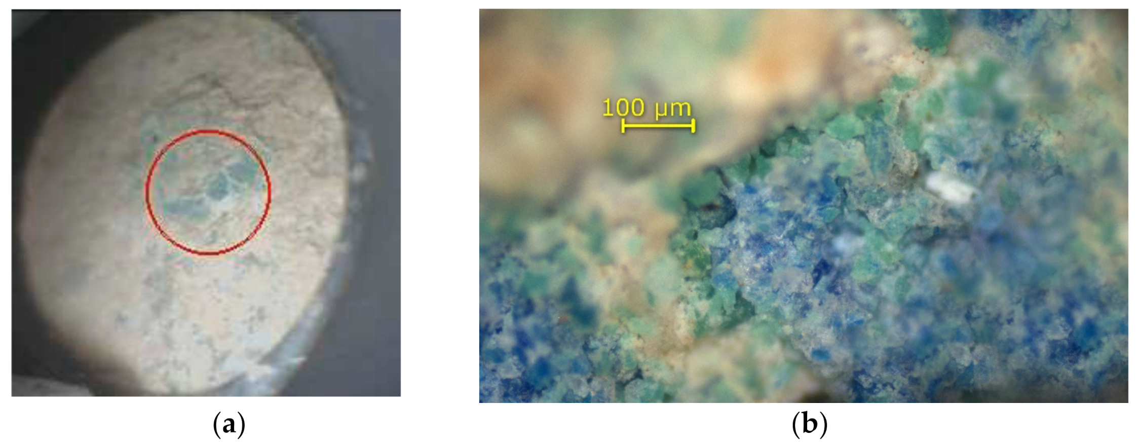
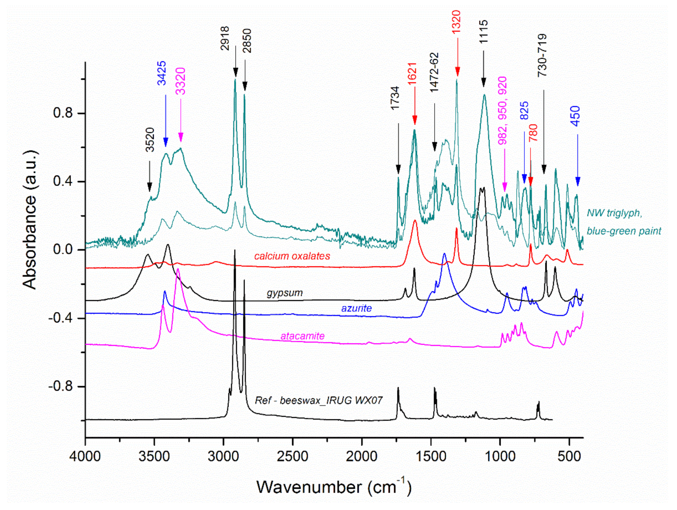
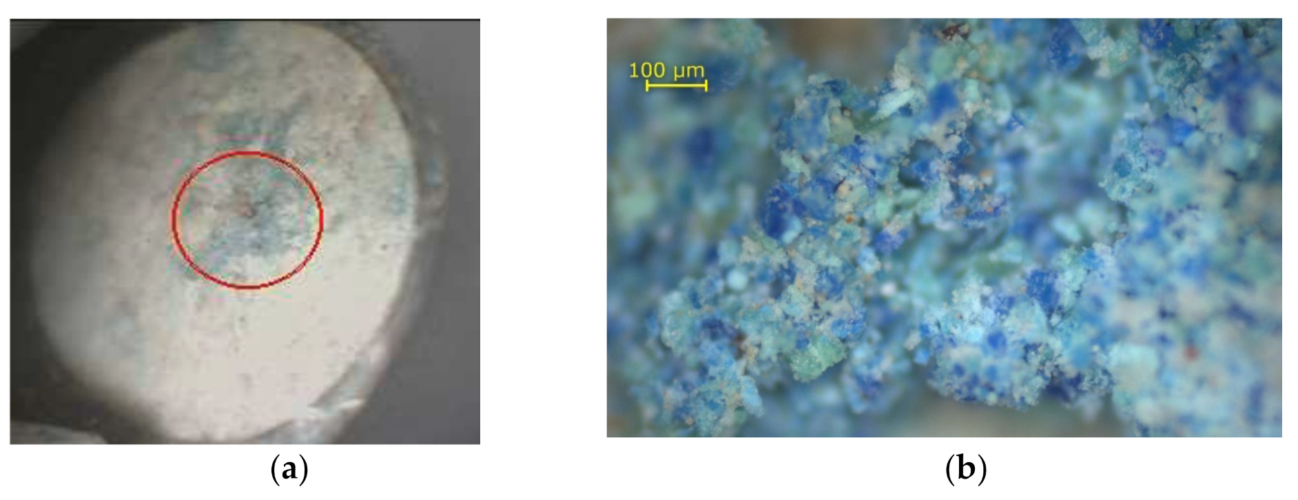
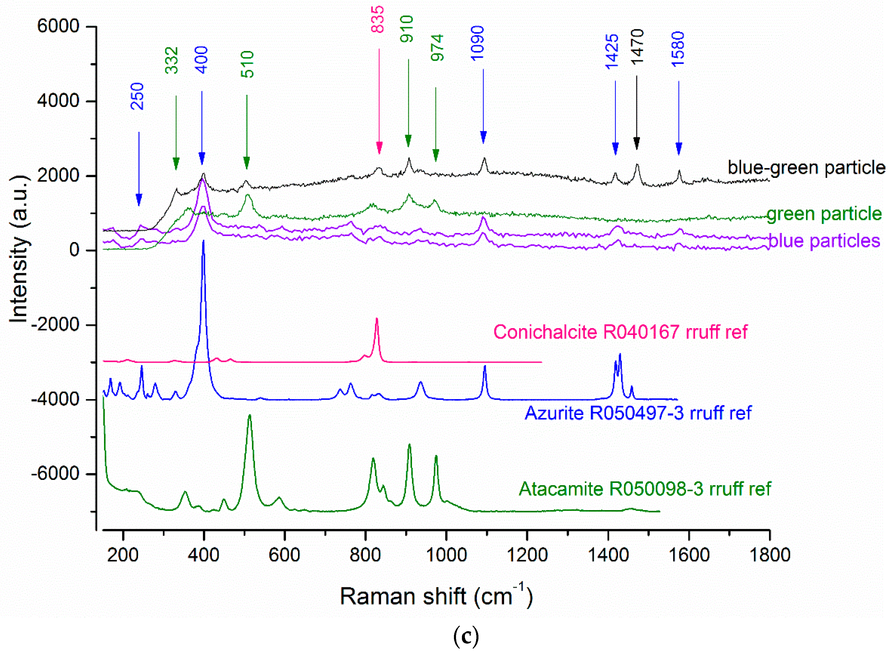
3.1.2. In Situ Analytical Investigation Results

In situ spectroscopic measurements have been carried out, focusing with the handheld XRF analyzer on the few traces still visible to the naked eye of light green-blue colour on the angular glyph of the NW triglyph (Figure 6a). Under the microscope, the colour is inhomogeneous, containing blue and green particles (Figure 6b). Point XRF measurements evidenced a copper-based pigment, as well as a high chlorine content (see Table 2).
Micro-Raman spectra that were collected on the few blue particles discernible among the predominantly light green grains present the characteristic bands of azurite (250 w; 400 s; 760 w; 1090 m; 1425 w; 1580 w) cm−1 [31]. The spectra on the light green particles evidenced the characteristic bands of atacamite at (130–140 m; 515 m; 820 w; 910 w; 975 w) cm−1 (Figure 7). This corresponds to the light green colour that was observed macroscopically and justifies the presence of chlorine in the XRF spectra. More precisely, the band at 515 cm−1 can be attributed to the O–Cu–O symmetric stretching vibrations, while the bands at 910, 975 cm−1 are assigned to the hydroxyl deformation modes of OH bonded to Cu (Cu–OH) [32,33].
The FTIR spectra acquired in ATR mode for a few particles sampled from the green-blue paint showed additional valuable information (Figure 8). The absorption bands at 3444, 3325, 982, 950, and 920 cm−1 confirmed the identification of atacamite, while those at 3425, 1430, 825 cm−1 confirmed azurite, already evidenced in the micro-Raman spectra; however, they also showed additional bands indicating the presence of malachite (1390, 1047, 875 cm−1) [34].
Moreover, in the same FTIR spectra (Figure 8), the doublet at 2916 and 2850 cm−1 corresponding to the symmetric and antisymmetric stretching vibrations of the CH2 groups, respectively, provided clear evidence for an organic compound in the paint. The organic content was further identified as pure wax, on the basis of all its characteristic absorption bands being present in the spectrum: the carbonyl band of esters at about 1734 cm−1 (stretching vibrations of C=O groups), the doublet at 1472 and 1462 cm−1, due to the planar deformation vibrations (or scissoring) of CH2 groups, and the doublet at 730 and 719 cm−1 assigned to an ordered orthorhombic packing of the long aliphatic chains [35,36,37].
Additionally, calcium oxalates affirmed by the sharp bands at 1621 and 1320 cm−1, corresponding to the antisymmetric and symmetric stretching of CO, respectively (Figure 8), were identified as constituents of the orange-brown patina layer present in the investigated area, emerging on the marble surface and also spread across the light green-blue paint remains [38,39]. In this same patina layer in the area surrounding the colour, gypsum and calcite were identified using in situ micro-Raman measurements (Figure 9).
As part of the Conservation projects coordinated by the Acropolis Restoration Service to protect the Acropolis monuments and slow down their further deterioration, several detailed multi-technique analytical studies were carried out in situ and ex situ, allowing the full characterization of the external surface that forms a cohesive layered structure, firmly attached to the marble. These layers, including thin whitish or orange patinas or thicker black and white decay crusts, were fully identified and are associated with the deterioration of the marble, attributed to a combination of mechanical, physical, and chemical factors along with atmospheric pollution, the microclimate, and the microstructure of the Pentelic marble [6,38,40,41,42,43].

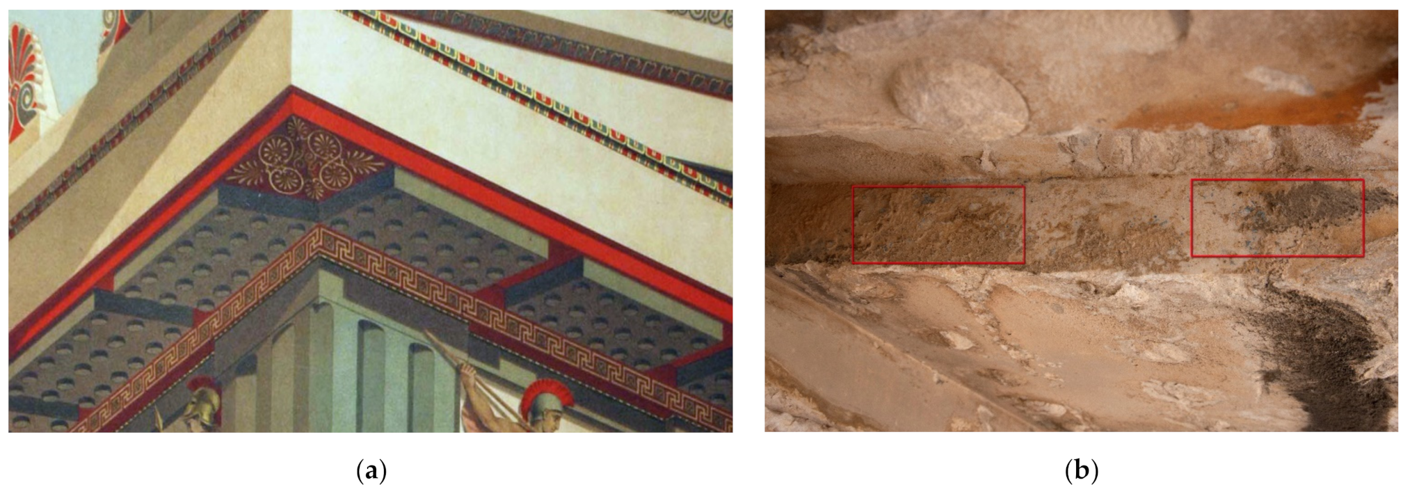
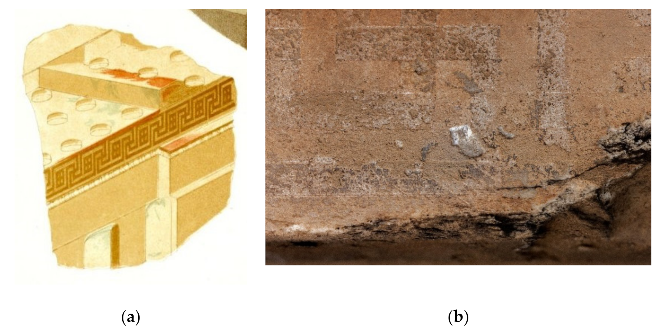
3.2. Parthenon Horizontal Cornice Blocks of the West Entablature

The next area of interest for investigating visible traces of polychromy on the Parthenon’s west entablature was the compact layer of horizontal cornice blocks placed on the top of the triglyphs and metopes, below the pediment and the roof. The front face of the cornices ends upward in a Doric cyma and downward in a taenia. The mutules, which alternate with viae, are decorated with eighteen (18) guttae (drop-like projections) each. The cornice blocks end in a taenia at the base of the cornice block.

3.2.1. Visual Evidence vs. Assumptions Based on Historical colour Reproductions

Based on assumptions and 19th-century colour renderings, the decoration of the Doric cyma consisted of tetrangular leaves with vertical separating areas among them (Figure 10a). At present, what still remains to suggest this pattern is a grid that is merely discernible through a layer of patina. This is present in most of the architectural members investigated; however, no clear evidence of any colour is present on the cyma. Presumably, the blue colour was used for the mutules’ background and the red colour for painting the viae. The taenia at the base of the cornice blocks was decorated with a meander pattern that is still discernible today, although it is partly overlaid with orange-brown layers of patina. In the area of the viae and the mutules of the west cornice blocks, a black crust layer was extensively present, a phenomenon that occurs mainly on surfaces that are sheltered from water and is attributed to the weathering effect of a sulfur dioxide (SO2)-polluted atmosphere on the stone surface (Figure 10b) [44].
Besides the black encrustation, orange-brown patina layers were also observed. These interlaced layers of crusts and patina have lost their cohesion, especially on the borders of their formation, causing exfoliation. In certain areas, where the superficial weathering crust or patina layers are loose, flaking paint layers appear underneath, preserved under the crust, as observed in the case of the fourth west cornice block in Figure 10b. In this architectural member, bright blue paint was clearly observed, both on the background of the mutule and on the vertical side. On the same block, the red colour is also preserved on the via area.
On the other hand, on the taenia at the base of the cornice along most of the west cornice blocks, the imprint of the meander pattern is preserved as a result of the differential decay of the paint and the background (Figure 11b). The preservation of the polychromy remains can be explained by the fact that this area is protected from direct exposure to environmental factors.
Merely perceptible traces of red colour were observed in various places in the background of the meander pattern, presumably painted in red and forming a continuum with the bottom side of the taenia; the pattern is well-preserved on the eleventh cornice block (Figure 11).

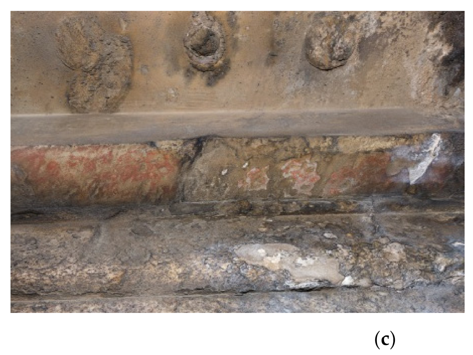
3.2.2. Investigation of Visible Traces of Blue colour on the 4th Cornice Block

In situ XRF measurements of the vivid light blue colour on the north face of the fourth cornice block evidenced the use of a copper-based pigment with a non-negligible amount of Si. Under the microscope, the colour is homogeneously blue (Figure 12b).
In situ micro-Raman spectra on the blue paint present the main characteristic bands that are attributed to Si-O in cuprorivaite, at around 430 cm−1 and at 1085 cm−1 [46,47]. Additionally, weak but clearly detectable bands in the spectra, at 1062, 1129, 1293, and 1440 cm−1 (Figure 12c), were assigned to a wax, corresponding to C-C (carbon–carbon) stretching and CH2 and CH3 deformation [48,49].
The identification of Egyptian blue has been confirmed in the FTIR spectra (Figure 12d), with the characteristic bands associated with the Cu, Ca silicates in the cuprorivate. The bands at 1230, 1162, 1050, and 1008 cm−1 are attributed to the antisymmetric stretching, while those at 800, 755, 664, and 595 cm−1 are attributed to the symmetric stretching. The absorptions at 521 and 484 cm−1 have been assigned to the deformation mode of the Si-O-Si group [46]. Equally, the identification of wax has been confirmed, based on the carbonyl band of esters at about 1734 cm−1, along with the bands at (2916 and 2850, 1472 and 1462, 730 and 719) cm−1 corresponding to the different vibration modes of the CH2 groups. The presence of Egyptian blue and wax was further confirmed with spectroscopic micro-analysis of a micro-sample taken from the blue paint on the 4th cornice block [50].

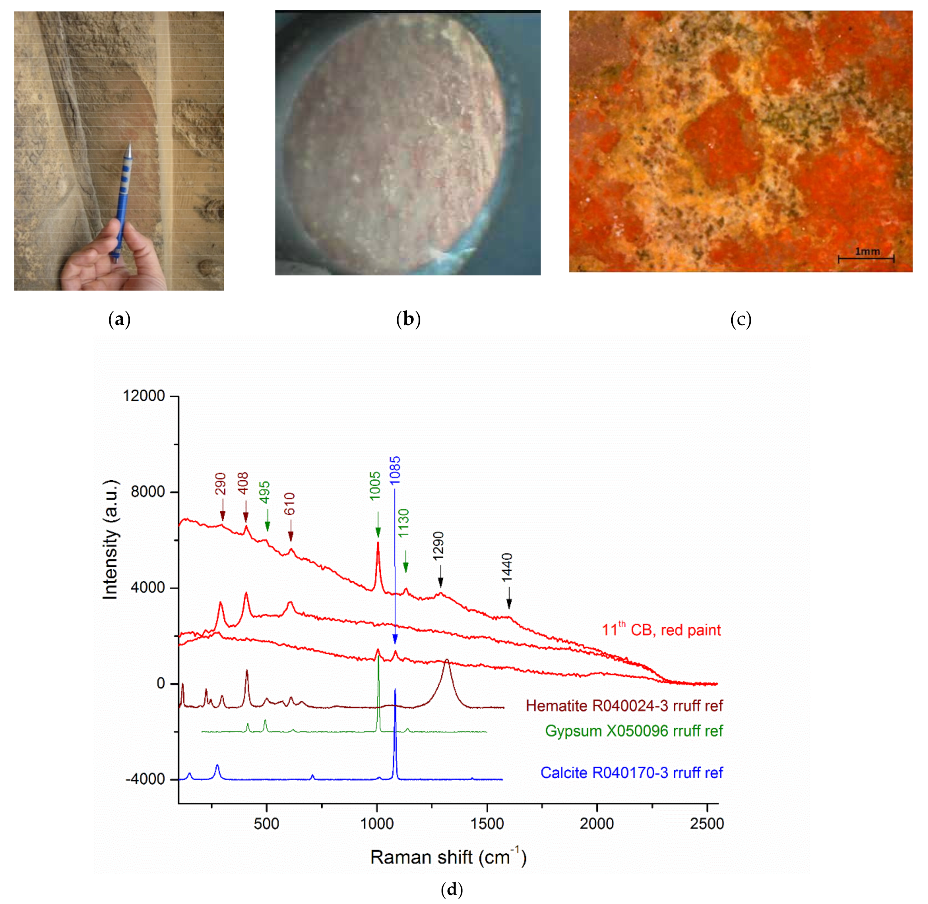
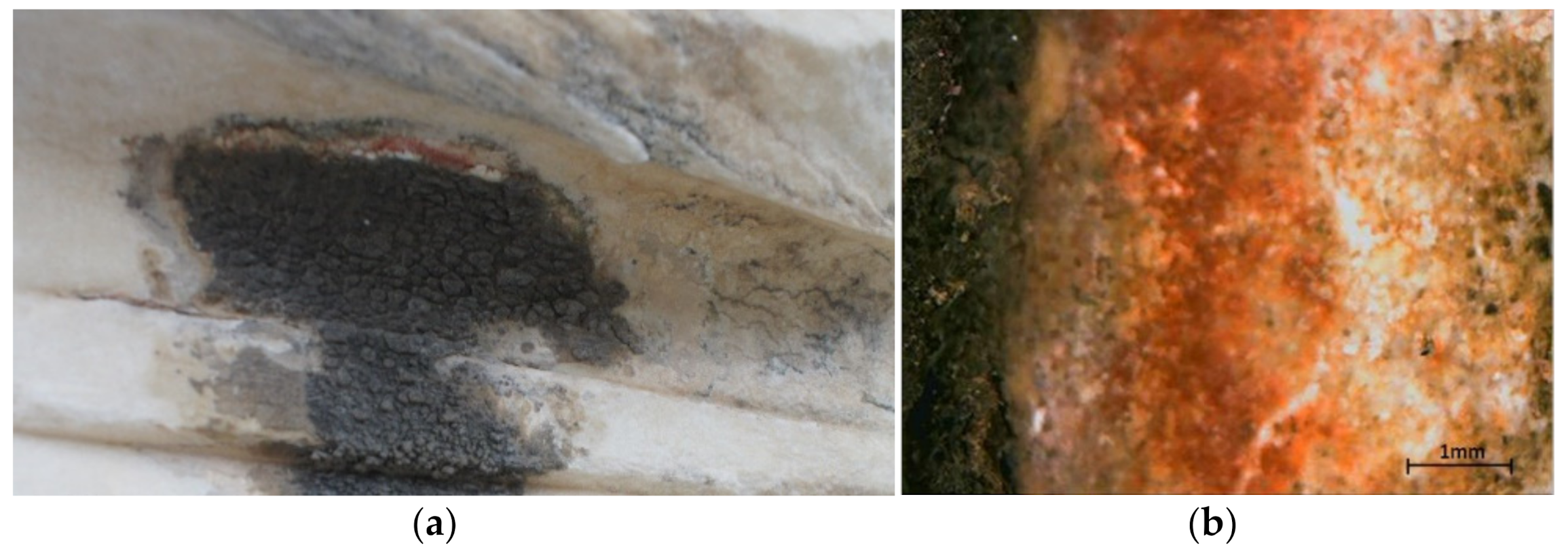
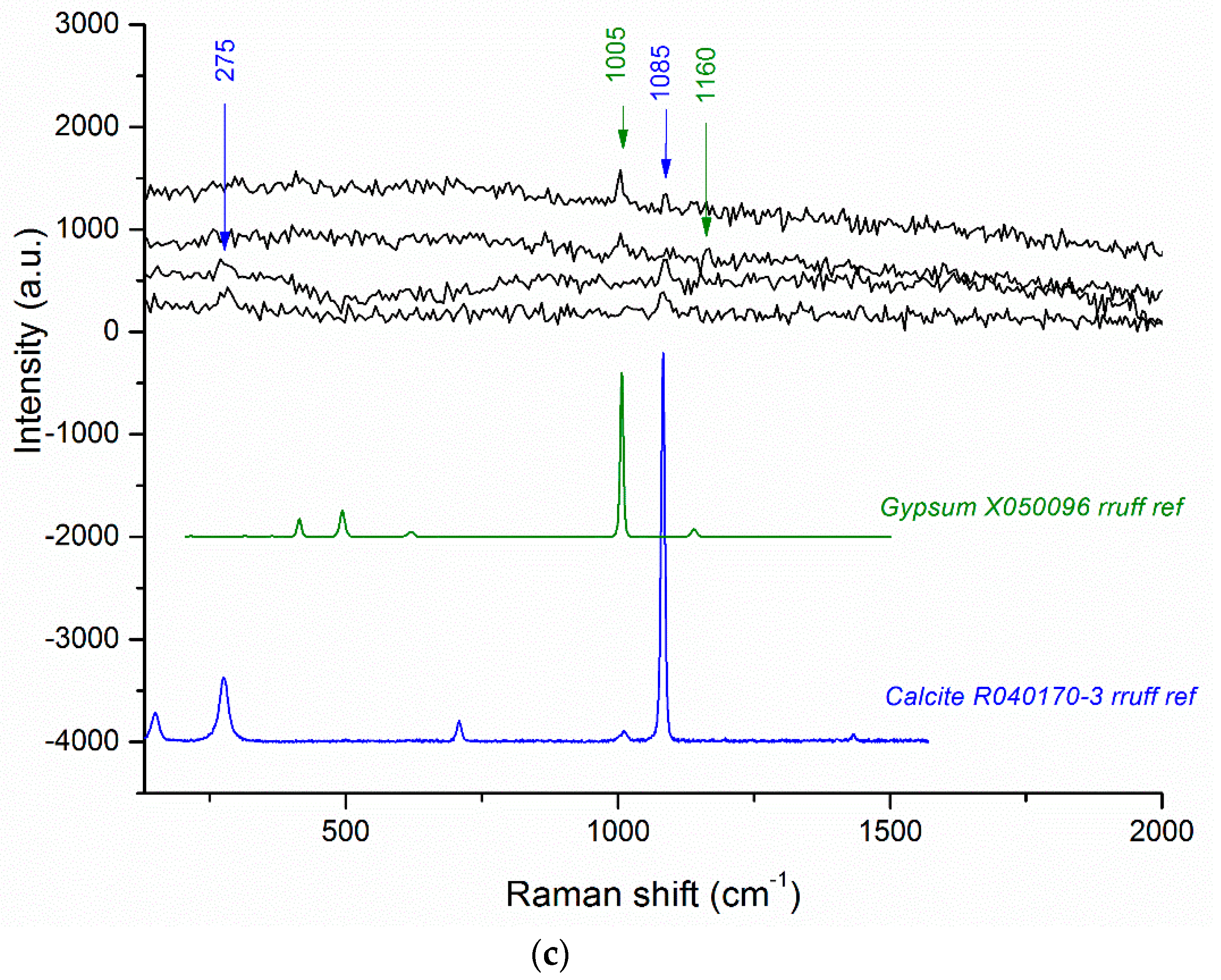
3.2.3. Investigation of Visible Traces of Red Colour on the 11th Cornice Block

In the taenia at the base of the cornice blocks (11th cornice block—Figure 13), XRF measurements showed an iron-based pigment that was identified as red ochre thanks to the characteristic bands of hematite Fe2O3 (220; 289; 408 m; 610 m; 1300 cm−1) in the micro-Raman spectra (Figure 13d). Additional analytical data on a micro-sample taken from the red paint in the same area converged to make an identification of red ochre [50].
Additionally, on some whitish particles focused under the Raman microscope, calcite CaCO3 (280, 1085 cm−1) and gypsum CaSO4·(2H2O) (490 w; 1004 vs; 1128 m) were identified, justifying the high amount of sulfur in the XRF measurements.

3.3. Parthenon NW and SW Raking Simae

Of particular interest and artistic value are the lion-head false waterspouts that decorated the four corners of the Parthenon entablature. The west face of the raking sima consisted of two parts, (a) the corner of the raking sima with a taenia above, an ovolo and a taenia at the bottom, and (b) the lowest corner of the raking cornice block, with a Lesbian cyma above and a flat triangular surface at the bottom (Figure 14).

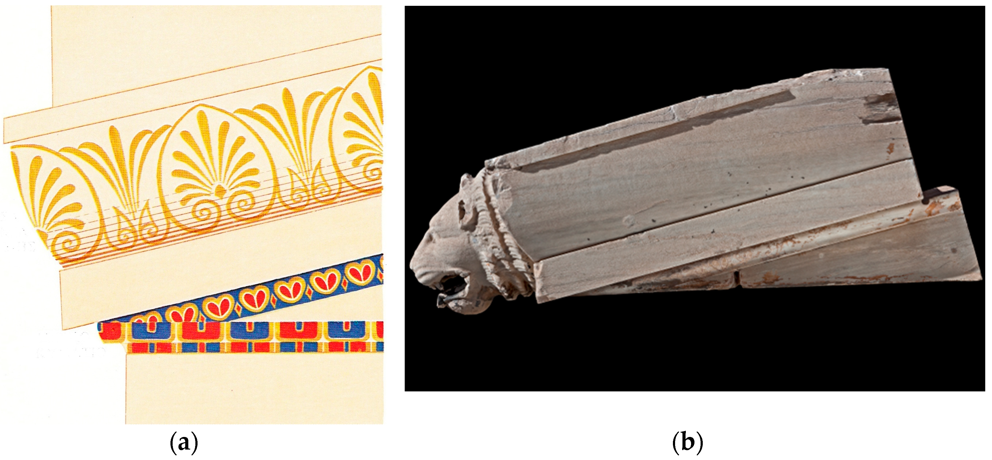
3.3.1. Visual Observation vs. Assumptions Based on Historical Colour Reproductions

colour representations of the Parthenon raking sima that are available in the literature referred to the existing decoration in the ovolo and in the Lesbian cyma (Figure 14a). The ovolo on the sima was decorated with gold-painted anthemia and lotus blossoms, while the Lesbian cyma was decorated with heart-shaped leaves and with smaller lozenges alternating among them. Currently, no traces of the painted decoration at the ovolo have survived on the raking simas, at least that are visible to the naked eye, and only lightly carved lines from the anthemion leaves, surrounded with a closed border, can be discerned.
The Lesbian cyma is in a relatively good state of preservation, obviously due to the fact that this area is partly protected from direct exposure to environmental factors. The decorative pattern still exists, along with a layer of patina of yellowish and in some areas of orange-brown hue. Fragmented parts of the double-bordered heart-shaped leaves and the outline of the lozenges, painted with red colour, have been preserved until the present day on both the northwest and southwest members (Figure 15a).

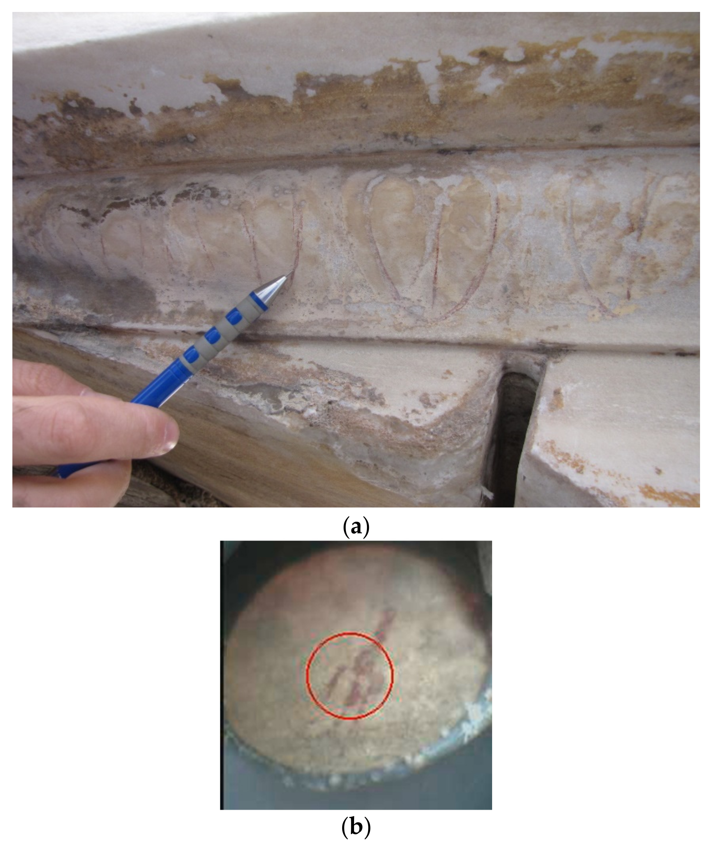
3.3.2. Investigation of the Heart-Shaped Leaves on the Lesbian Cyma of the NW and SW Raking Sima

An iron-based red pigment has been confirmed in the double outlines of the heart-shaped incised decoration, only identified in the XRF measurements (Figure 15b).

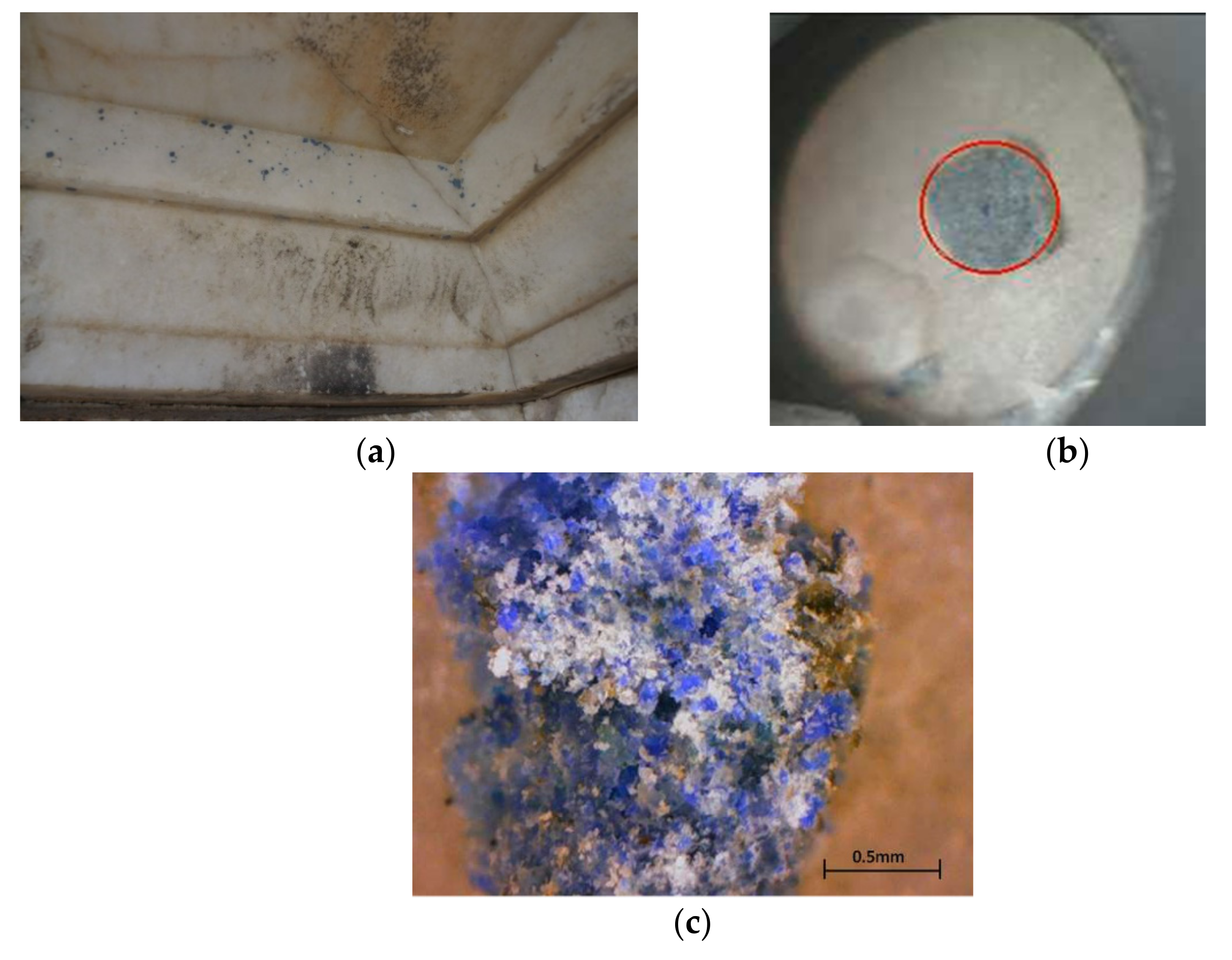
3.4. Parthenon Impost Block of the South-West Anta

The impost blocks of the antae of the Parthenon’s porches are composed of the following parts: (a) the abacus, which is crowned with a very fine Lesbian cyma; (b) the Doric cyma, which was decorated with tetrangular leaves that were painted alternately in red and blue; (c) the hypotrachelium below, decorated with a carved Ionic molding and a carved astragal; and (d) a lower facade. The impost block of the southwest anta had been embodied in the masonry of the tower that was constructed during the medieval era in the southwest corner of the opisthonaos. Due to the partial demolition of the masonry in this area, the decoration of this architectural member has been revealed. The masonry was constructed in contact with the impost block, providing a certain degree of protection against the environmental factors; it has, therefore, preserved some polychromy remains in the area corresponding to the Doric cyma.

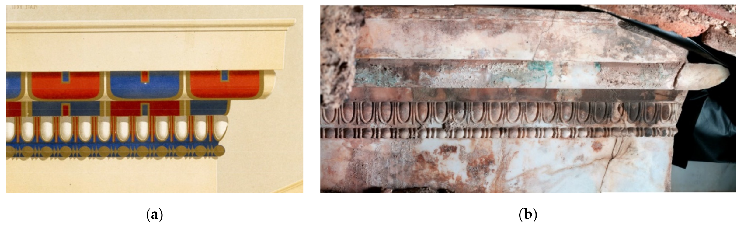
3.4.1. Visual Observation vs. Assumptions Based on Historical Colour Reproductions

Presently, all the exposed faces of this architectural member are covered with various types of crusts (white, black, or an orange-brown colour) resulting from the interaction of the building materials of the masonry and environmental factors with the ancient painted marble surface at micro and macro scales. However, the grid of the tetrangular leaves with the vertical separating areas between them, as well as a blue-green colour, is still visible in some areas (Figure 16). Furthermore, traces of a green-blue pigment can be observed in the background of the Ionic molding.

3.4.2. Analytical Results of the Investigation of the Impost Block of the South-West Anta

In the area corresponding to the blue section of the decoration (Figure 16), XRF measurements carried out on the still-preserved light blue-green colour (Figure 17a) indicated not only the use of a copper-based pigment but also a high chlorine content. Under the microscope, the colour is inhomogeneous, containing blue and light green particles in similar proportions. Focusing on the blue grains, the Raman spectra presented the characteristic bands of azurite (250 m; 400 vs; 535 w; 740 w(sh); 760 m; 835 m; 930 w; 1090 m; 1425 m; 1580 m) (Figure 17b,c). Focusing on particle aggregates of the lighter green-blue colour, additional Raman bands were obtained that were attributed to atacamite (332, 510, 910, 974 cm−1), which is consistent with the presence of chlorine evidenced in the XRF spectra. The particles responsive to the Raman spectra, showing mainly the bands assigned to atacamite, have a lighter green hue under the microscope. Further investigation into the scarce particles of the deeper olive-green colour helped to assign the strong band at 830 cm−1 to the arsenate (AsO43−) symmetric stretching vibrations in conichalcite, CaCu(AsO4)(OH) [52], which explains the low content of arsenic detected in the XRF measurements in this area.
XRF measurements were also carried out on the region that corresponds to the red section of the decoration pattern according to the colour drawings of the 19th century, which today is covered by a superficial white weathering crust (Figure 16b). A clearly resolved detection of the Hg lines in the XRF spectra was certainly attributable to cinnabar. Close observation with the naked eye and under the microscope did not allow any red particles to be located on the surface; however, on the basis of the XRF findings, likely enabled thanks to the penetrative ability of X-rays, it can be assumed that particles of red cinnabar pigment are preserved under the white crust, consisting mainly of calcite, as evidenced by the micro-Raman spectra.

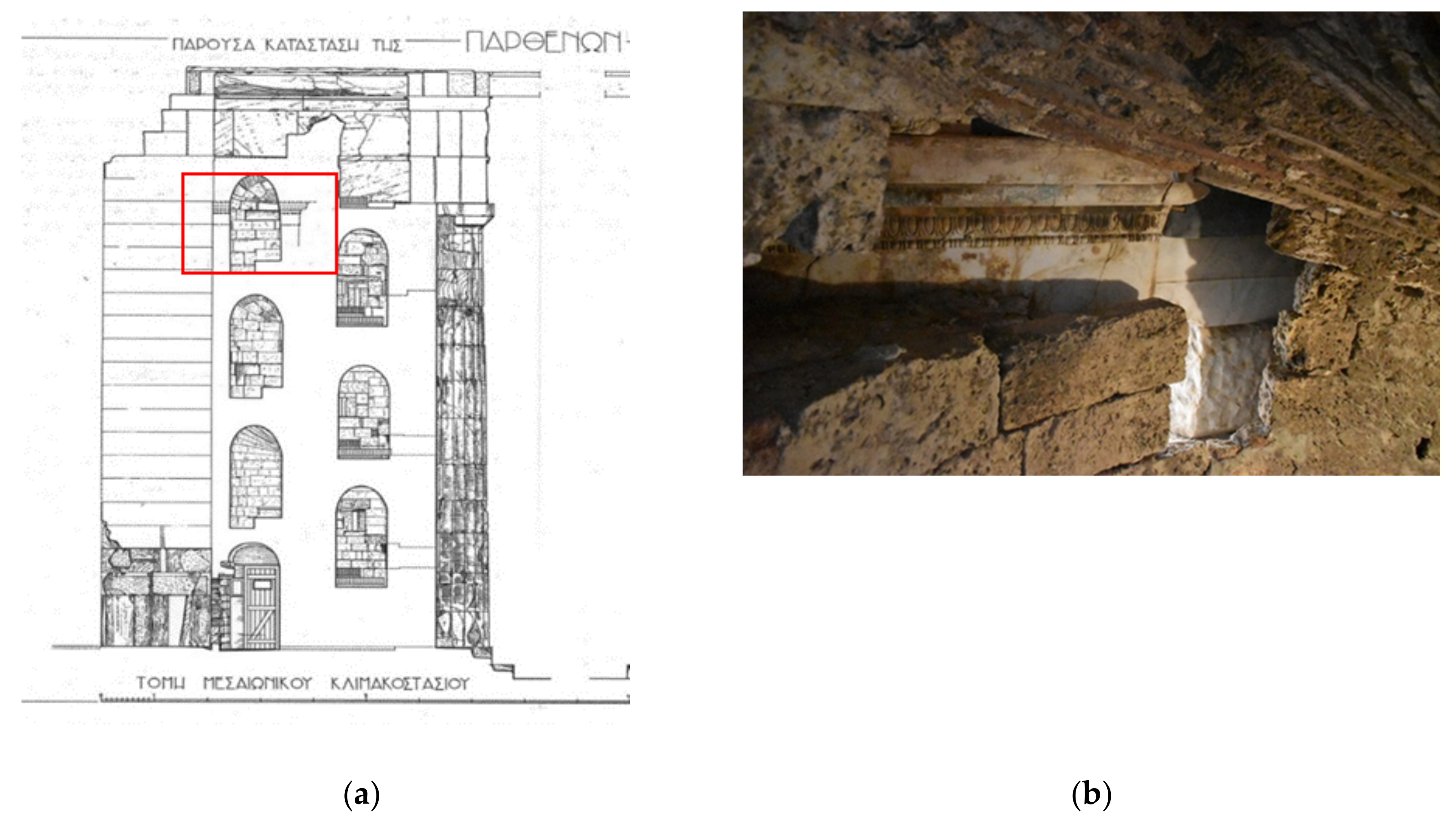
3.5. Propylaea—Impost Block of the North-West Anta in the Central Building

3.5.1. Visual Observation vs. Assumptions Based on Historical Colour Reproductions

The impost blocks of the antae in the central building of the Propylaea consisted of the following parts: (a) an abacus with a Lesbian cyma as a crowning molding; (b) a Doric cyma that was adorned with a carved pattern of Doric leaves; (c) a group of three annulets with intervals in between; and, finally, (d) a lower flat fascia. Currently, on the impost block of the northwest anta, traces of colour are preserved in the carved parts of the Doric cyma. More specifically, the red colour is preserved on the west and south faces of the Doric cyma on the marble surface, along with a widespread white crust under a thick overlying black crust. Additionally, the green colour has survived under the black crust on the south face of the block. This is apparent in the intervals between the annulets in areas where the crust has exfoliated (Figure 18).

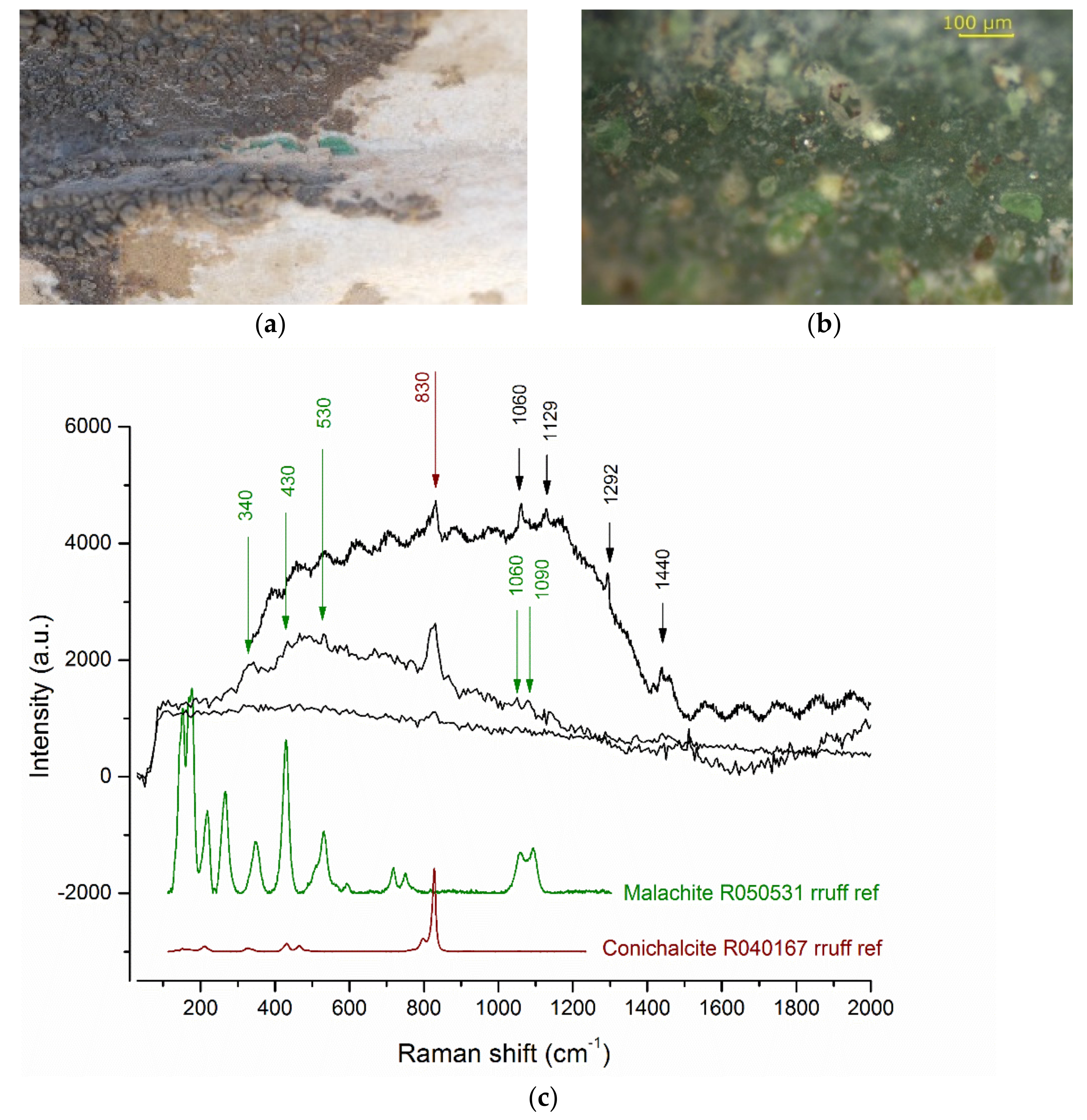
3.5.2. Investigation of Colour Remains at the South Side of the Impost Block of the North-West Anta in the Propylaea Central Building (Green and Red Colour)

The first areas of focus for investigating the nature of the pigments used in the Propylaea polychromy were the vivid red-coloured areas, visually discernible under a thick overlying black crust in the east and south faces of the Doric cyma (Figure 19a,b). An iron-based red colour is identified on the basis of XRF measurements.
For this architectural member, the micro-Raman spectra were collected under particularly difficult weather conditions. Strong winds made it difficult to maintain stability and we had to cut off the ambient daylight with the black cloths we had set up, to achieve effectively dark and stable conditions. It should also be mentioned that the selected areas of interest were not easily accessible, due to both the limited free working space left by the scaffolding setup and the relief shape of the architectural member itself as the colour was preserved only in the cavities. To overcome these operational constraints, the spectra were collected over very short integration times and, therefore, show a great deal of noise. Only the bands at 1000 and 1085 cm−1 were resolved, which are assigned to the more intense peaks of gypsum and calcite, respectively (Figure 19c). Both calcium sulfate and carbonate are attributed to the composition of the white crust, which is visibly intercalated between the red colour and the thick black crust (Figure 19a,b). Although the micro-Raman spectra collected in this area did not provide any information relating to the red colour, it is assumed that an iron-based ochre pigment is used, based on the XRF elemental data, in which iron is the only detectable element that can be associated with a red pigment.
In the area of the interval between the second and the third annulet (from bottom to the top), on the south side of the impost block, the green colour was attested to survive under the black crust and it was visible in areas where the crust has exfoliated (Figure 20a). XRF measurements carried out on the remains of green colour indicated the presence of a copper-based pigment with a considerable amount of arsenic (As), see Table 2. Under the microscope, the colour is of a relatively homogeneous green shade, with some distinct brighter green particles dispersed in a matrix of darker olive-green hue (Figure 20b).
The micro-Raman spectra collected from the green colour showed the main characteristic band of the arsenate group in conichalcite, CaCu(AsO4)(OH), at 830 cm−1 (Figure 20c). The identification of conichalcite is in agreement with the indications provided by the XRF spectra, in which arsenic was detected at a high content comparable to that of copper. However, additional weak bands in the Raman spectrum at 430 and 530 nm, as well as the doublet at 1055–85 cm−1, could be attributed to malachite, Cu2CO3(OH)2. The possible admixture of malachite in the green pigment would explain the tone of the green colour as perceived by the naked eye, which is more saturated and colder in tone than the typical olive-green colour of conichalcite. Further investigation is, however, required to confirm or contest the uncertain identification of malachite in the Raman spectra. Finally, the weak Raman bands at 1062, 1129, 1293, and 1440 cm−1 were attributed to organic material and correspond to a spectroscopic fingerprint similar to that recognized in the architectural members of the Parthenon (the ΝW triglyph and the 4th CB).

3.6. Propylaea—North-East Cornice Block in the Niche between the South Wing and the Southwest Anta of the West Portico

The second area of the Propylaea that was investigated is the area of Ionic cornice blocks in the niche between the south wing and the southwest anta of the west portico. The Ionic cornice blocks consisted of (a) the mutule area without guttae, (b) a taenia at the base of the mutule, (c) a Lesbian cyma, and (d) a taenia at the base of the block.
Traces of ancient decoration are observed οn the northeast cornice block. More specifically, several dispersed traces of an intense blue colour are present in the area of the taenia at the base of the mutules (Figure 21). For this architectural member, a pattern of heart-shaped leaves was noticed in the area of the Lesbian cyma, of which only the outline in black colour survives. Moreover, on the area of the mutules of the cornice blocks, black crusts were extensively present.
Based exclusively on the XRF elemental analysis data on the blue traces, a copper pigment has been identified. Additionally, the detection of a relatively high percentage of silicon, along with copper, supports the identification of Egyptian blue, which was suggested by the characteristic response of the blue pigment when applying the VIL imaging technique. The homogeneous colour, with bright blue particles, that was observed under the microscope also supports the identification of Egyptian blue in the blue details.

4. Discussion

In the following paragraphs, the materials identified through the in situ analytical campaign at the Acropolis monuments are summarized and discussed synthetically, allowing additional interpretations and comparative considerations with the 18–19th-century colour renderings found in the literature.

4.1. Materials Identified in the Blue and Green Colour of Polychromy Remains

Azurite was identified on two of the investigated architectural members on the Parthenon’s west entablature, namely, on the NW angular triglyph and the impost block of the southwest anta (see Table 3).
Azurite is a basic copper carbonate (2CuCO3·Cu(OH)2), a natural blue mineral, and its use as a pigment is attested back to the Old Kingdom’s Fourth Dynasty in Egypt. However, the use of azurite in Egyptian paintings seems extremely rare [53,54]; in a parallel chronological context, in prehistoric Aegean paintings, to date, azurite has not been identified.
In the broader context of the prehistoric Aegean, azurite has been known since the Early Cycladic period II [55,56,57]; it has been found as a content of marble vessels, while its use in the decoration of meso-Cycladic figurines, although suggested, has not been confirmed with certainty [58]. Azurite was found as an offering in Early Cycladic rich tombs in the form of ground pigment, as a content of stone vessels, in marine shells serving as pallets, or in colour containers in the shape of bone pipes. These finds of azurite in a funerary context may be associated with specific rituals, but they also refer to a possible use of the pigment as a cosmetic, intended for makeup or the colouration of the body [57,59,60].
The use of azurite reappears during the archaic period, in the painted decoration of limestone or marble architectural members, as evidenced in cases of the archaic monumental temples; most prominently, those of Aphaia on Aegina Island [61] and the Ekatombedos in the Acropolis of Athens [62]. During the Classical and Hellenistic periods, azurite was gradually displaced by Egyptian blue.
Azurite is again in extensive use by the Middle Ages, on both Byzantine and Western mural paintings in particular, and it is mainly on monuments from this period that it has been extensively investigated using analytical techniques. Azurite presents frequent chromatic alterations that correspond to diverse chemical modifications. The scenario of visually perceptible darkening corresponds to the formation of black copper oxides (CuO); however, when azurite modifies to greenish tones, the secondary minerals of the atacamite group (nantokite, CuCl, and paratacamite/atacamite, Cu2Cl(OH)3) are among the most frequently identified end-degradation products [63,64]. The degradation of azurite in murals, executed with the fresco technique, in particular, seems to be related to an alkaline pH, while its use with an organic binder significantly improves its stability in bright light and atmospheric conditions. When azurite degrades in green compounds, malachite is formed (CuCO3 Cu(OH)2) in the first stages, whereas green copper chlorides such as paratacamite and atacamite are subsequently formed, as identified in several studies.
Accordingly, in the examined areas of the polychromy of the Parthenon, atacamite was also detected in the same spots that azurite was identified, and was assigned to the light green particles of the paint as observed under the microscope (see Figure 6b and Figure 17b).
Atacamite is a copper oxychloride, Cu2Cl(OH)3, and is known to have been used as a green pigment of natural provenance [65] or as a synthetic product [66] in other parts of the world and in later historical periods. This is not surprising because atacamite occurs as a natural mineral in the oxidation zones of copper deposits [67,68,69]. It is also known that atacamite occurs as a chloride corrosion product on copper and bronze objects [70,71]. When identified in paintings, the detection of secondary elements, such as arsenic or tin, in the elemental analysis of atacamite constitutes an indication of its synthetic nature, where bronze is used as the copper source.
In the present case, the inhomogeneous appearance of the paint investigated on the Parthenon NW angular triglyph and the impost block of the SW anta, observed under the microscope as being made up of blue, dark, and lighter green particles, together with the identification of both azurite and malachite in the blue and dark green particles, respectively, leave no doubt as to the presence of atacamite in the light green particles. In this context, the latter is the final degradation product of azurite, with malachite being an intermediate product in the sequence of reactions involved in the weathering path. In the first stage, the chemical transformation of azurite to malachite is possible in the open air and involves the replacement of some of the carbon dioxide (CO2) units with water (H2O), changing the carbonate:hydroxide ratio from 1:1 (azurite) to a 1:2 ratio (malachite), following the chemical reaction, 2 Cu3(CO3)2(OH)2 + H2O → 3 Cu2(CO3)(OH)2 + CO2. In the second stage, the occurrence of chloride content (from salt water) in the air humidity may catalyze the formation of paratacamite and atacamite (Cu2Cl(OH)3) [32,72,73]. The partial, relatively extensive but incomplete chemical alteration of azurite to atacamite (with malachite as an intermediate alteration product) explains the macroscopically observed colour of the areas where azurite was used. The colour of these remaining traces is inhomogeneous, ranging from bluish-green to light green depending on the degree of alteration.
Conichalcite, another copper-based mineral of deeper olive-green tone, has been identified in two architectural members, the impost block of the Parthenon’s SW anta and the impost block of the Propylaea’s NW anta (see Table 3).
In nature, conichalcite is found associated with other minerals, often with malachite, in the oxidation zones of copper ores, and is also characterized as a weathering product of enargite, Cu3AsS4. In the first instance of its identification on the Parthenon (the impost block of the SW anta), the few identified particles of conichalcite are scant in the paint mixture, are most probably incidental, and do not have an impact on the macroscopic colour. In the case of the impost block of the anta in the central building of the Propylaea, the significant amount of As in the XRF spectra suggests that conichalcite constitutes a major compound affecting the macroscopic colour of the green paint, which is also composed of malachite. However, as conichalcite occurs in nature and is associated with malachite, its presence in the green colour could be attributed with the same probability to a natural admixture of malachite or to a deliberate mixture in the painter’s palette. Therefore, in the two cases that conichalcite was identified, what emerged from the comparative microscopic observation of the colour mixture is that in the first case, the original colour was blue, with azurite being the main ingredient; however, in the second case, the colour was green, with the main ingredients being malachite and conichalcite. The identification of malachite and conichalcite, without any other compound in the paint, is unique among the analyzed areas of colour remains in this study, in both the monuments of the Propylaea and the Parthenon. It is considered to be the only case for which, based on analytical evidence, we can suggest the identification of an original green colour on investigated parts of the architectural polychromy of the Acropolis temples. This analytically confirmed instance of green colour used in the lower part of the Doric cyma, in the intervals between the annulets, and on the impost block of the northwest anta, is also affirmed in the colour rendering by Penrose [29] (Plate 26; Figure 18a).
In the broader context of ancient Greek paintings, conichalcite has been identified on Macedonian paintings of the 4th century and on some gravestones of the Hellenistic period from Demetrias, often associated with malachite [57]. Conichalcite, as a deliberately chosen pigment yielding its characteristic warm green-olive colour, was first identified on the chryselephantine (gold and ivory) couch of the tomb of Philip II at Aegae [73,74,75]. Its use was further attested in the later fourth century in the wall decoration of a cubic-form Macedonian tomb of Pella [76]. The availability of conichalcite, as suggested by its use in Macedonian paintings and artifacts, must have been connected to the occurrence of local copper resources in the area of Chalcidice [73,74,75].
However, in the Athenian context of the Classical period, when endeavoring to assign the provenance of the identified natural copper-based pigments, azurite, malachite, and conichalcite, their origin should presumably be investigated in the mines along the Attic-Cycladic crystalline complex. More specifically, the western Cycladic islands, Kea, Kythnos, Seriphos, and Siphnos, on which there existed referenced sources of iron, lead, silver, and copper since the early Bronze Age, should probably be explored as candidates for their provenance [77]. According to recent systematic field campaigns, followed by analytical studies, the presence of early copper smelting installations was observed in surface surveys and was associated with existing ore sources on the island of Kythnos [78,79].
Egyptian blue has been identified on two architectural members, on the 4th cornice block of the Parthenon’s west entablature and on the NE cornice block in the niche between the south wing and the SW anta of the west portico of the Propylaea. In both cases, the Egyptian blue that was detected preserved its original physicochemical integrity, without any observable indication of chromatic or chemical change. Based on the XRF elemental analysis, only the major elements of calcium, copper, and silicium (Ca, Cu, Si) assigned to cuprorivaite, the main crystalline phase of the pigment, were detected without any secondary or minor elements, such as tin, zinc, or arsenic (Sn, Zn, As), that would suggest any alloy to be the possible source of copper in the manufacture of the pigment.
The analytical confirmation for the alternate use of the two pigments, Egyptian blue and azurite, in the blue colour details of the Parthenon entablature polychromy is of particular interest. Furthermore, it is worth mentioning that the laboratory investigation of paint micro-samples from the cornice blocks revealed that azurite and Egyptian blue were used in the meander and chess decorative patterns of the taenia, respectively, located at the cornice blocks’ base [50].
The analytically proven, relatively extensive chemical alteration of azurite to atacamite (with malachite as an intermediate alteration product) offers a clear explanation for the colour alteration of blue, now visible macroscopically as bluish-green to light green, depending on the degree of alteration. However, the original appearance of pure azurite would have been of an intense deep blue, forming bichromy in the areas where it was used in juxtaposition with Egyptian blue, the latter being of a vivid but lighter tone, as retained in the unaltered remaining traces. The colour contrast between the two blue tones, appositely inserted in the design of the flat or relief decorative patterns on the architectural forms, should certainly be understood in relation to the lighting contrasts and shadows lent by the changing direct and diffuse sunlight, interacting and enhancing the three-dimensionality of the entablature, as suggested in the historical colour reproductions (see Figure 5a).
The use of both Egyptian blue and azurite in the same monument, in different but adjacent architectural members, and even in different details of the same architectural member, undoubtedly raises multi-dimensional questions. It testifies to the simultaneous availability of the two pigments but raises questions about the criteria, whether aesthetic or technical, for the selective use or combination of the two pigments in different architectural members or decorative details. Finally, this is of historical interest because it may signify the gradual replacement of azurite, known to be widely used in the archaic period, in the painted decoration of stone in architectural monuments (such as the archaic Parthenon, the Temple of Aphaia) with Egyptian blue in the architectural decoration of the Classical period. Although atacamite and malachite have been identified on certain decorative details of the Parthenon’s west entablature, where green traces of colour had been observed, those materials have been attributed, without any doubt, to alteration products of the blue azurite that was originally used.
The original blue colour of these details is additionally confirmed when taking under consideration the rendering of blue colour details in colour reproductions of the 19–20th century. However, from this current study, there is one instance of analytical evidence for an original green colour applied to the architectural polychromy of the Acropolis temples, that is, on the impost block of the NW (S) anta of the Propylaea’s central building. Such an assumption is supported by the fact that the colour is entirely green, also under the microscope and malachite is, in this case, identified in mixture with conichalcite, another green pigment, as evidenced by the micro-Raman spectroscopy. The green paint layer in this case does not present any signs of weathering but, unfortunately, neither does it present any clue for suggesting whether the mixture of the two copper-based pigments in the green paint is intentional or natural. The coexistence of conichalcite in copper mines containing malachite is reported in Macedonia and is identified in monuments of the classical period, but conichalcite is known also as a mineral in the Classical mines of the Lavrion district in East Attica [80].

4.2. Materials Identified in the Red Colour of the Polychromy Remains

In the architectural members examined for red traces, where they were preserved, iron oxides were identified in all cases, first by detecting a significant amount of iron in the XRF spectra and at the same time by the absence of any other element relating to a red chromophore. In addition, in some cases, the molecular structure of hematite was identified in the micro-Raman spectra collected from the red paint traces, as seen in Figure 13 (Parthenon, 11th cornice block (W)).
The case of the Parthenon impost block of the SW anta is an exception, where, in the XRF measurements (see Figure 16b and Table 2), mercury was detected in an area corresponding to the red section of the decoration pattern. Mercury was detected in measurements on a white surface-calcite crust and was interpreted as evidence of the use of cinnabar, traces of which are probably preserved under the white crust. Measurements made in other spots of the same architectural member, where red would also be expected based on the reconstructed decoration pattern corresponding to this area, revealed only iron, suggesting the use of red ochre. However, the use of cinnabar does not exclude the use of red ochre, since the two pigments could have been combined, either as a mixture or in superimposed layers. In the latter case, it is possible that the cinnabar, if used on a superficial surface layer, may have been lost today.

4.3. Preliminary Data for the Binding Medium

Although the focus of this study and the methodology applied was set to shed light on the identification of the pigments used in the polychromy of the architectural sculptures of the Athenian Classical temples, some additional information was obtained in parallel with both micro-Raman and FTIR spectroscopies, converging to the screening of organic material in the paint layer in several instances. The sites of measurement that gave such information (Parthenon: NW triglyph; 4th cornice block (W), 11th cornice block (W), impost block of the SW anta, and Propylaea: impost block of the NW anta in the central building), as seen in Table 4, were located in protected areas or areas that were substantially covered by a superficial crust, in any case, presumably forming favorable conditions for the better preservation of the identified organic compound in the paint. There was indisputable evidence for the identification of a pure wax in the paint, based on the assignment of both FTIR and Raman bands in the instances mentioned above. In all these cases, pure wax was ascertained without any bands referring to other additional compounds, such as lipids or carboxylates, that would suggest any treatment or degradation of the wax. The wax is presumably inherent to the paint layer and was not identified in any adjacent areas in which the paint is not preserved, which weakens its attribution to a non-original layer that was possibly added to play a consolidation role. Additionally, in the FTIR spectra evidencing the wax compound, the characteristic bands of calcium oxalates are well-resolved. However, the oxalate bands and those assigned to the wax compound do not show any correlation. The oxalate bands are present in all FTIR spectra, including those acquired on the surrounding areas of the localized paint remains; for their attribution, the several possible sources of formation extensively discussed in the literature should be taken into account. However, for the wax compound that is undoubtedly present in the paint in some of the investigated sites where traces of paint have survived, further investigation should be carried out to consolidate this preliminary attribution of wax to the binding medium before drawing conclusions as to the characterization of the painting technique.

5. Conclusions

This paper summarizes the results obtained from the in situ investigation of selected architectural members, where traces of polychromy were still visible on the architectural sculptures of the Parthenon and the Propylaea temples. Two blue pigments, azurite and Egyptian blue, and two red pigments, cinnabar and hematite, seem to be the main pigments in the polychromy of the Athenian Acropolis monuments. Although the survived colour traces are sparsely distributed on the monuments and are too fragmentary to reconstruct a complete picture, this methodical study of the colour patterns as integrated into the design of the architectural sculptures, assisted by some indications stemming from the critical consideration and interpretation of historical colour reproductions found in the literature, is meant to contribute in a small way to our understanding and efforts to approach the original appearance of classical beauty.

Author Contributions

Conceptualization, S.S.; analytical methodology and investigation, S.S. and G.K.; monuments’ historical and conservation context, E.A.; writing—original draft preparation, review and editing, S.S. and E.A. All authors have read and agreed to the published version of the manuscript.

Funding

This research was co-funded by the National Resources of Greece and the European Regional Development Fund (ERDF), through the Regional Operational Program (ROP) of Attica 2007–2013, grant no: SAΕ 2010SE01480147.

Acknowledgments

Acknowledgments are due to the director of YSMA, V. Eleftheriou, and the members of ESMA for their support of the present research. The authors would also like to thank I. P. Kotsifakos, A. Sotiropoulos and A. Panou (conservators of the Parthenon), and K. Frantzikinaki and E. Fragkiadaki (conservators of the Propylaea) for their contributions in terms of documentation and in the application of the non-invasive techniques on site. In situ analytical measurements on the Acropolis monuments were carried out with portable instruments provided by the Ormylia Foundation, Art Diagnosis Center, during the previous affiliation of the second author.

Conflicts of Interest

The authors declare no conflict of interest. The funders had no role in the design of the study; in the collection, analyses, or interpretation of data; in the writing of the manuscript, or in the decision to publish the results.

References

  1. Brommer, F. Die Metopen des Parthenon. Katalog und Untersuchung; von Zabern: Mainz am Rhein, Germany, 1967; Volume 1–2. [Google Scholar]
  2. Jenkins, I.D.; Middleton, A.P. Paint on the Parthenon Sculptures. Annu. Br. Sch. Athens 1988, 83, 183–207. [Google Scholar] [CrossRef]
  3. Kouzeli, K.; Belogiannis, N.; Dogani, G.; Tolias, A. Μελέτη των έγχρωμων στρωμάτων που διακρίνονται στις επιφάνειες των μνημείων. In Μελέτη αποκαταστάσεως του Παρθενώνος ΙΙa; Korres, M., Ed.; Hellenic Ministry of Culture: Athens, Greece, 1989; pp. 198–202. [Google Scholar]
  4. Vlassopoulou, C. New investigations into the polychromy of the Parthenon. In Circumlitio. The Polychromy of Antique and Mediaeval Sculpture; Brinkmann, V., Primavesi, O., Hollein, M., Eds.; Hirmer Verlag: München, Germany, 2010; pp. 219–223. [Google Scholar]
  5. Vlassopoulou, C. The polychromy of the Parthenon Metopes. In An Archaeologist’s Eye. The Parthenon Drawings of Katherine A. Schwab, Exhibition Catalogue; Bucher, G.S., Deupi, J., Eds.; Fairfield University: Fairfield, CT, USA, 2013; pp. 14–17. [Google Scholar]
  6. Papakonstantinou, E.; Panou, A.; Franzikinaki, K.; Tsimereki, A.; Frantzi, G. The surface conservation project of the Acropolis monuments: Studies and Interventions. In Proceedings of the XXI International CIPA Symposium, Athens, Greece, 1–6 October 2007; Available online: https://www.isprs.org/proceedings/xxxvi/5-c53/papers/FP111.pdf (accessed on 30 January 2022).
  7. Panou, A.; Frantzikinaki, Κ.; Papakonstantinou, Ε. Conservation and cleaning of the west frieze of the Parthenon. In The Acropolis Restoration Works, Vol. A, Interventions on the Acropolis Monuments 2000–2012: Completed Projects (Digital ed.); Bouras, C.H., Eleftheriou, V., Eds.; Ministry of Culture: Athens, Greece, 2013. [Google Scholar]
  8. Frantzi, G.; Maridaki, A.; Papakonstantinou, E.; Verri, G.; Sotiropoulou, S.; Brekoulaki, H. The revelation of the decorative pattern of the coffered ceiling of the Maidens’ Porch in the Erechtheion. In Proceedings of the 7th Round Table on Greek and Roman Sculptural and Architectural Polychromy, Athens, Greece, 7–8 November 2013; Brekoulaki, H., Ed.; in preparation. [Google Scholar]
  9. Frantzi, G.; Frantzikinaki, K.; Panou, A.; Papakonstantinou, E.; Maridaki, A.; Pouli, P.; Fotakis, C. Analytical studies to investigate the safeguarding of the original surfaces upon laser cleaning interventions at the Athens Acropolis Monuments. In Proceedings of the 11th International Conference on Lasers in the Conservation of Artwork (LACONA XI), Cracow, Poland, 20–23 September 2016. [Google Scholar]
  10. Brinkmann, V. Κόρη ή Θεά; Το αίνιγμα της «πεπλοφόρου» από την Aθηναϊκή Aκρόπολη. In Πολύχρωμοι θεοί: χρώματα στα αρχαία γλυπτά; Brinkmann, V., Kaltsas, Ν., Wünsche, R., Eds.; Εθνικό Aρχαιολογικό Μουσείο: Athens, Greece, 2007; pp. 97–110. [Google Scholar]
  11. Verri, G.; Saunders, D.; Ambers, J.; Sweek, T. Digital mapping of Egyptian blue: Conservation implications. Stud. Conserv. 2010, 55 (Suppl. S2), 220–224. [Google Scholar] [CrossRef]
  12. Bourgeois, B.; Jockey, P.H. Approches nouvelles de la polychromie des sculptures hellénistiques de Délos (résumé). In Comptes Rendus des Séances de L’academie des Inscriptions et Belles-Lettres; 145ᵉ année, N. 1; Nabu Press: New York, NY, USA, 2001; pp. 629–665. [Google Scholar] [CrossRef]
  13. Sargent, M.L. Recent Investigation of the Polychromy of a Metropolitan Roman Garland Sarcophagus. In Ttracking Colour—The Polychromy of Greek and Roman Sculpture in the Ny Carlsberg Glyptotek, Preliminary Report 3; The Copenhagen Polychromy Network; Glyptoteket: København, Denmark, 2011; pp. 14–34. [Google Scholar]
  14. Sotiropoulou, S.; Perdikatsis, V.; Birtacha, K.; Apostolaki, C.; Devetzi, A. Physicochemical characterization and provenance of colouring materials from Akrotiri-Thera in relation to their archaeological context and application. Archaeol. Anthr. Sci. 2012, 4, 263–275. [Google Scholar] [CrossRef]
  15. Brekoulaki, C.; Perdikatsis, V. Ancient Painting on Macedonian Funerary Monuments, IV-III c.B.C.: A Comparative Study on the Use of colour. In Proceedings of the Colour in Ancient Greece: The Role of Colour in Ancient Greece Art and Architecture (700-31 B.C.), Thessaloniki, Greece, 12–16 April 2000; Τiverios, Μ.A., Tsiafake, D.S., Eds.; Aristotelean University Press: Thessaloniki, Greece, 2002; pp. 147–155. [Google Scholar]
  16. Verri, G. The use and distribution of Egyptian blue: A study by visible-induced luminescence imaging. In The Nebamun Wall Paintings; Middleton, A., Uprichard, K., Eds.; Archetype Publications: London, UK, 2008; pp. 41–50. [Google Scholar]
  17. Østergaard, J.S. Colour shifts: On methodologies in research on the polychromy of Greek and Roman sculpture. Proc. Dan. Inst. Athens 2017, 8, 149. [Google Scholar]
  18. Aggelakopoulou, E.; Panou, A.; Kotsifakos, I.P.; Moutsatsou A., P.; Bakolas, A.; Karoglou, M.; Sioumpara, E.P. Technical investigation of the polychromy of the NW raking sima of the Parthenon. In Proceedings of the 7th International Round Table on Greek and Roman Sculptural and Architectural Polychromy, The Acropolis Museum, Athens, Greece, 7–8 November 2013. in preparation. [Google Scholar]
  19. Aggelakopoulou, E.; Sioumpara, E.; Eleftheriou, V. Investigation of the polychromy of the Parthenon’s west front. In The Parthenon. Colour, Materiality and Aesthetics; Abbe, M., Norman, N., Eds.; Cambridge University Press: Cambridge, CA, USA, 2014; in preparation. [Google Scholar]
  20. Miliani, C.; Rosi, F.; Brunetti, B.G.; Sgamellotti, A. In situ Non-invasive Study of Artworks: The MOLAB Multi-technique Approach. Acc. Chem. 2010, 43, 728–738. [Google Scholar] [CrossRef]
  21. Vandenabeele, P.; Donais, M.K. Mobile spectroscopic instrumentation in archaeometry research. Appl. Spectrosc. 2016, 70, 27–41. [Google Scholar] [CrossRef]
  22. Colomban, P. The on-site/remote Raman analysis with mobile instruments: A review of drawbacks and success in cultural heritage studies and other associated fields. J. Raman Spectrosc. 2012, 43, 1529–1535. [Google Scholar] [CrossRef]
  23. Bersani, D.; Lottici, P.P. Raman spectroscopy of minerals and mineral pigments in archaeometry. J. Raman Spectrosc. 2016, 47, 499–530. [Google Scholar] [CrossRef]
  24. Brunetti, B.G.; Miliani, C.; Rosi, F.; Doherty, B.; Monico, L.; Romani, A.; Sgamellotti, A. Non-invasive Investigations of Paintings by Portable Instrumentation: The MOLAB Experience. Top Curr. Chem. (Z) 2016, 47, 499–530. [Google Scholar] [CrossRef] [Green Version]
  25. Rousaki, A.; Moens, L.; Vandenabeele, P. Archaeological investigations (archaeometry). Phys. Sci. Rev. 2018, 3, 20170048. [Google Scholar] [CrossRef]
  26. Castro, K.; Sarmiento, A.; Maguregui, M.; Martínez-Arkarazo, I.; Etxebarria, N.; Angulo, M.; Urrutikoetxea Barrutia, M.; González-Cembellín, J.M.; Madariaga, J.M. Multianalytical approach to the analysis of English polychromed alabaster sculptures: μRaman, μEDXRF and FTIR spectroscopies. Anal. Bioanal. Chem. 2008, 392, 755–763. [Google Scholar] [CrossRef] [PubMed]
  27. Korres, M. The Parthenon. In The Parthenon—Architecture and Conservation, Catalogue for the Exhibition; Korres, M., Panetsos, G.A., Seki, T., Eds.; Osaka City Museum, Foundation of Hellenic Culture: Athens, Greece, 1996; pp. 12–73. [Google Scholar]
  28. Tanoulas, A. The propylaea of the Athenian Acropolis during the Middle Ages. Ph.D. Thesis, The Archaeological Society at Athens, Athens, Greece, 1997. [Google Scholar]
  29. Penrose, F.C. An Investigation of the Principles of Athenian Architecture; or the Results of a Recent Survey, Conducted Chiefly with Reference to the Optical Refinements Exhibited in the Construction of the Ancient Buildings of Athens; Society of Dilettanti: London, UK, 1852; pp. 58–59. [Google Scholar] [CrossRef]
  30. Hittorff, J.I. Restitution du Temple d’Empédocle à Sélinonte, ou, L’architecture Polychrome Chez les Grecs; Librairie De Firmin Didot Frères; Imprimeurs De L’Institut: Paris, France, 1851. [Google Scholar] [CrossRef]
  31. Frost, R.L.; Martens, W.N.; Rintoul, L.; Mahmutagic, E.; Kloprogge, J.T. Raman spectroscopic study of azurite and malachite at 298 and 77 K. J. Raman Spectrosc. 2002, 33, 252–259. [Google Scholar] [CrossRef] [Green Version]
  32. Frost, R.L.; Martens, W.; Kloprogge, J.T.; Williams, P.A. Raman spectroscopy of the basic copper chloride minerals atacamite and paratacamite: Implications for the study of copper, brass and bronze objects of archaeological significance. J. Raman Spectrosc. 2002, 33, 801–806. [Google Scholar] [CrossRef] [Green Version]
  33. Coccato, A.; Bersani, D.; Coudray, A.; Sanyova, J.; Moens, L.; Vandenabeele, P. Raman spectroscopy of green minerals and reaction products with an application in Cultural Heritage research. J. Raman Spectrosc. 2016, 47, 1429–1443. [Google Scholar] [CrossRef]
  34. Goldsmith, J.A.; Ross, S.D. The infra-red spectra of azurite and malachite. Spectrochim. Acta Part A Mol. Spectrosc. 1968, 24, 2131–2137. [Google Scholar] [CrossRef]
  35. Snyder, G. Vibrational correlation splitting and chain packing for the crystalline n-alkanes. J. Chem. Phys. 1979, 71, 3229. [Google Scholar] [CrossRef]
  36. Cuní, J.; Cuní, P.; Eisen, B.; Savizky, R.; Bove, J. Characterization of the binding medium used in Roman encaustic paintings on wall and wood. Anal. Methods 2012, 4, 659–669. [Google Scholar] [CrossRef]
  37. Stacey, R.J.; Dyer, J.; Mussell, C.; Lluveras-Tenorio, A.; Colombini, M.P.; Duce, C.; La Nasa, J.; Cantisani, E.; Prati, S.; Sciutto, G.; et al. Ancient encaustic: An experimental exploration of technology, ageing behaviour and approaches to analytical investigation. Microchem. J. 2018, 138, 472–487. [Google Scholar] [CrossRef]
  38. Maravelaki-Kalaitzaki, P. Black crusts and patinas on Pentelic marble from the Parthenon and Erechtheum (Acropolis, Athens): Characterization and origin. Anal. Chim. Acta 2005, 532, 187–198. [Google Scholar] [CrossRef]
  39. Conti, C.; Casati, M.; Colombo, C.; Realini, M.; Brambilla, L.; Zerbi, G. Phase transformation of calcium oxalate dihydrate–monohydrate: Effects of relative humidity and new spectroscopic data. Spectrochim. Acta Part A Mol. Biomol. Spectrosc. 2014, 128, 413–419. [Google Scholar] [CrossRef]
  40. Skoulikidis, T.H. Papakonstantinou-Ziotis, E. Mechanism of Sulphation by Atmospheric SO2 of the Limestones and Marbles of the Ancient Monuments and Statues: I. Observations in situ (Acropolis) and laboratory measurements. Br. Corros. J. 1981, 16, 63–69. [Google Scholar] [CrossRef]
  41. Kouzeli, K.; Beloyiannis, N.; Tolias, C.; Dogani, Y. Monochromatic layers with and without oxalates on the Parthenon. In Proceedings of the International Symposium on the Oxalate Films: Origin and Significance in the Conservation of Works of Art, Milan; Centro CNR Gino Bozza: Milan, Italy, 1989; p. 327. [Google Scholar]
  42. Polikreti, K.; Maniatis, Y. Micromorphology, composition and origin of the orange patina on the marble surfaces of Propylaea (Acropolis, Athens). Sci. Total Environ. 2003, 308, 111–119. [Google Scholar] [CrossRef]
  43. Palagia, O.; Pike, S.S. Art historical and scientific perspectives on the nature of the orange-red patina of the Parthenon. In Interdisciplinary Studies on Ancient Stone; Pensabene, P., Gasparini, E., Eds.; ASMOSIA X: Rome, Italy, 2015; pp. 881–888. [Google Scholar]
  44. Sabbioni, C.; Zappia, G.; Ghedini, N.; Gobbi, G.; Favoni, O. Contribution of atmospheric deposition to the formation of damage layers. Sci. Total Environ. 1995, 167, 49–56. [Google Scholar] [CrossRef]
  45. Fenger, L.P. Doric Polychrome: Studies in the Application of Colour on the Doric Temple; Asher: Berlin, Germany, 1886. [Google Scholar] [CrossRef]
  46. Baraldi, P.; Bondioli, F.; Fagnano, C.; Ferrari, A.M.; Tinti, A.; Vinella, M. Study of the vibrational spectrum of cuprorivaite. Ann. Chim. 2001, 91, 679–692. [Google Scholar] [PubMed]
  47. Guineau, B. L’ etude des pigments par les moyens de la microspectrometrie Raman. In Datation-Caractérisation des Peintures Pariétales et Murales; Hackens, T., Delamare, F., Helly, B., Eds.; Centre Universitaire Européen pour les Biens Culturels: Strasbourg, France, 1987; Volume 17, pp. 259–294. [Google Scholar]
  48. Špaldoňová, A.; Havelcová, M.; Lapčák, L.; Machovič, V.; Titěra, D. Analysis of beeswax adulteration with paraffin using GC/MS, FTIR-ATR and Raman spectroscopy. J. Apic. Res. 2021, 60, 73–83. [Google Scholar] [CrossRef]
  49. Vandenabeele, P.; Wehling, B.; Moens, L.; Edwards, H.; De Reu, M.; Van Hooydonk, G. Analysis with micro-Raman spectroscopy of natural organic binding media and varnishes used in art. Anal. Chim. Acta 2000, 407, 261–274. [Google Scholar] [CrossRef]
  50. Aggelakopoulou, E.; Bakolas, A. What were the colours of the Parthenon? Investigation of the entablature’s cornice blocks. J. Archaeol. Sci. 2022, 140, 105553. [Google Scholar] [CrossRef]
  51. Orlandos, A. H αρχιτεκτονική του Παρθενώνος, vols 1-3; Archaeological Society at Athens: Athens, Greece, 1977. [Google Scholar]
  52. Reddy, B.; Frost, R.; Martens, W. Characterization of conichalcite by SEM, FTIR, Raman and electronic reflectance spectroscopy. Mineral. Mag. 2005, 69, 155–167. [Google Scholar] [CrossRef] [Green Version]
  53. Hedegaard, S.B.; Delbey, T.; Brøns, C.; Rasmussen, K.L. Painting the Palace of Apries II: Ancient pigments of the reliefs from the Palace of Apries, Lower Egypt. Herit. Sci. 2019, 7, 54. [Google Scholar] [CrossRef] [Green Version]
  54. Scott, D.A. A review of ancient Egyptian pigments and cosmetics. Stud. Conserv. 2016, 61, 185–202. [Google Scholar] [CrossRef]
  55. Birtacha, K. Κυκλάδες. In Aργοναύτης. Τιμητικός τόμος για τον Καθηγητή Χρ. Γ. Ντούμα από τους μαθητές του; Vlachopoulos, A., Birtacha, K., Eds.; H Καθημερινή AΕ: Athens, Greece, 2003; pp. 263–276. [Google Scholar]
  56. Birtacha, K. Examining the paint on Cycladic figurines. In Early Cycladic Sculpture in Context; Marthari, M., Renfrew, C., Boyd, M., Eds.; Oxbow: Cambridge, UK; Oxford, UK; Philadelphia, PA, USA, 2017; pp. 491–501. [Google Scholar]
  57. Brecoulaki, H. Precious colours in Ancient Greek polychromy and painting: Material aspects and symbolic values. Rev. Archéologique 2014, 1, 3–35. [Google Scholar] [CrossRef]
  58. Hendrix, Ε. Some Methods for Revealing Paint on Early Cycladic Figures. In Metron: Measuring the Aegean Bronze Age (Aegaeum, 24); Foster, K.P., Laffineur, R.L., Eds.; Liege University: Liege, Belgium, 2003; pp. 139–145. [Google Scholar]
  59. Tsountas, C. ‘Κυκλαδικά’, AΕ. 1899; pp. 104–113. [Google Scholar]
  60. Sotiropoulou, S.; Brecoulaki, H.; Papazoglou, E. Colouring Matters Dated to the 3rd and 2nd Millennium BC, from the Prehistoric Collection at the Archaeological Museum of Athens, Analytical Data Obtained in the Framework of this Project Granted by the Institute of Aegean Prehistory (INSTAP). 2012; Unpublished Data.
  61. Riederer, J. Die Bemalung des Aphaia-Tempels auf Ägina. BBA 1977, 2, 67–72. [Google Scholar]
  62. Sioumpara, E.; Sotiropoulou, S.; Karagiannis, G. Recent analytical results about the Polychromy of the archaic Parthenon and the Polychromy of the first Doric Temples from the 6th c.B.C. In Proceedings of the 8th International Round Table on Polychromy in Ancient Sculpture and Architecture, Centre de Recherche et de Restauration des Musées de France (C2RMF), Paris, France, 15–16 November 2016. [Google Scholar]
  63. Coccato, A.; Moens, L.; Vandenabeele, P. On the stability of mediaeval inorganic pigments: A literature review of the effect of climate, material selection, biological activity, analysis and conservation treatments. Herit. Sci. 2017, 5, 12. [Google Scholar] [CrossRef] [Green Version]
  64. Lluveras, A.; Boularand, S.; Andreotti, A.; Vendrell-Saz, M. Degradation of azurite in mural paintings: Distribution of copper carbonate, chlorides and oxalates by SRFTIR. Appl. Phys. A 2010, 99, 363–375. [Google Scholar] [CrossRef]
  65. Tomasini, E.P.; Rúa Landa, C.; Siracusano, G.; Maier, M.S. Atacamite as a natural pigment in a South American colonial polychrome sculpture from the late XVI century. J. Raman Spectrosc. 2013, 44, 637–642. [Google Scholar] [CrossRef]
  66. Liu, L.; Gong, D.; Yao, Z.; Xu, L.; Zhu, Z.; Eckfeld, T. Characterization of a Mahamayuri Vidyarajni Sutra excavated in Lu’an, China. Herit. Sci. 2019, 7, 77. [Google Scholar] [CrossRef] [Green Version]
  67. Wiggins, M.B.; Heath, E.L.; Booksh, K.; Alcántara-García, J. Multi-Analytical Study of Copper-Based Historic Pigments and their Alteration Products. Appl. Spectrosc. 2019, 73, 1255–1264. [Google Scholar] [CrossRef] [PubMed]
  68. Wiggins, M.B.; Liu, M.; Liu, C.; Booksh, K.S. Polymorph identification in green Chinese architectural paints using Raman imaging and multivariate curve resolution. J. Chemom. 2021, 35, e3308. [Google Scholar] [CrossRef]
  69. Yong, L. Copper trihydroxychlorides as pigments in China. Stud. Conserv. 2012, 57, 106–111. [Google Scholar] [CrossRef]
  70. Chang, T.; Maltseva, A.; Volovitch, P.; Wallinder, I.O.; Leygraf, C. A mechanistic study of stratified patina evolution on Sn-bronze in chloride-rich atmospheres. Corros. Sci. 2020, 166, 108477. [Google Scholar] [CrossRef]
  71. Scott, D. A Review of Copper Chlorides and Related Salts in Bronze Corrosion and as Painting Pigments. Stud. Conserv. 2000, 45, 39–53. [Google Scholar] [CrossRef]
  72. Dei, L.; Ahle, A.; Baglioni, P.; Dini, D.; Ferroni, E. Green degradation products of azurite in wall paintings: Identification and conservation treatment. Stud. Conserv. 1998, 43, 80–88. [Google Scholar] [CrossRef]
  73. Brecoulaki, H.; Perdikatsis, V. The green colour in ancient Greek painting [in Greek]. In Proceedings of the 2nd International Conference on Ancient Greek Technology, Athens, Greece, 17–21 October 2005; pp. 179–185. [Google Scholar]
  74. Brécoulaki, H. Sur la technè de la peinture grecque ancienne d’après les monuments funéraires de Macédoine. Bull. De Corresp. Hellénique 2000, 124, 189–216. [Google Scholar] [CrossRef]
  75. Brecoulaki, H. La Peinture Funéraire de Macedoine, Emplois et Fonctions de la Couleur, IVe-IIe s. av. J.-C. (Meletemata, 48); The National Research Foundation: Athens, Greece, 2006; pp. 425–429. [Google Scholar]
  76. Maniatis, Y.; Sakellari, H.; Kavousanaki, D.; Minos, N. Φυσικοχημικός χαρακτηρισμός χρωστικών από του κιβωτιόσχημο τάφο της Πέλλας. In Κιβωτιόσχημος τάφος με ζωγραφική διακόσμηση από την Πέλλα; Lilimbaki-Akamati, M., Ed.; Ministry of Culture, 17th Ephorate of Prehistoric and Classical Antiquities: Thessaloniki, Greece, 2007; pp. 138–175. [Google Scholar]
  77. Renfrew, C. Cycladic Metallurgy and the Aegean Early Bronze Age. Am. J. Archaeol. 1967, 71, 1–20. [Google Scholar] [CrossRef]
  78. Bassiakos, Y.; Philaniotou, O. Early Copper Production on Kythnos. Archaeological Evidence and Analytical Approaches to the Reconstruction of the Metallurgical Process. In Metallurgy in the Early Bronze Age Aegean (Sheffield Studies in Aegean Archaeology, 7; Day, P.M., Doonan, R.C.P., Eds.; Oxbow: Oxford, UK, 2007; pp. 19–56. [Google Scholar]
  79. Catapotis, M. On the Spatial Organisation of Copper Smelting Activities in the Southern Aegean during the Early Bronze Age, In Metallurgy in the Early Bronze Age Aegean (Shefield Studies in Aegean Archaeology; Day, P.M., Doonan, R.C.P., Eds.; Oxbow: Oxford, UK, 2007; pp. 207–223. [Google Scholar]
  80. Katerinopoulos, A.; Zissimopoulou, E. Minerals of the Lavrion Mines; Association of Greek Collectors of Minerals and Fossils: Athens, Greece, 1994. [Google Scholar]
Figure 1. The west side of the Parthenon entablature. Orthophotomosaic—D. Mavromati (YSMA archive). Red frames: Architectural members investigated in situ, with the findings presented in this paper.
Heritage 05 00042 g001
Figure 2. The Parthenon. (a) Section of the medieval tower in the opisthonaos (M. Korres, pl. 8 [27]). The position of the impost block of the southwest anta inside the medieval tower (red frame), (b) Macroscopic view of the impost block of the southwest anta.
Heritage 05 00042 g002
Figure 3. The Propylaea. (a) Top view (ground plan) (A. Tanoulas, pl. 1 [28]) where the investigated areas are marked in red. (b) Central building. North wall, south face: impost block of the north-west anta, macroscopic view. (c) South wing, west face, northeast cornice block: macroscopic view.
Heritage 05 00042 g003
Figure 4. (a) The mobile micro-Raman setup used for the in situ measurements, positioned at the impost block of the southwest anta of the Parthenon. (b) Detailed view of the micro-Raman head, brought close to the paint surface for taking measurements. (c) The handheld X-ray fluorescence spectrometer in action on the 11th west cornice block of the Parthenon.
Heritage 05 00042 g004aHeritage 05 00042 g004b
Figure 5. (a) Colour representation of Parthenon architrave’s polychromy (Hittorff, 1851, pl. 8 [30]), (b) NW angular triglyph, northwest glyph, macroscopic photo. Spots of light green-blue colour, from which pXRF and micro-Raman measurements have been taken.
Heritage 05 00042 g005
Figure 6. NW triglyph. (a) The area of the XRF-measured spot is outlined in red on the area indicated in Figure 5b; the results of the XRF are given in Table 2 (b) Microphotograph of the light green-blue colour under optical microscopy, of the area in which XRF and micro-Raman spectra have been acquired.
Heritage 05 00042 g006
Figure 7. Micro-Raman spectra, taken through the 50× objective lens, for several blue and light green grains of the light green-blue colour on the angular glyph of the NW triglyph. Reference spectra of azurite and atacamite from the rruff.info database are presented on the same graph for comparison.
Heritage 05 00042 g007
Figure 8. Two FTIR spectra, acquired in ATR mode from the green-blue traces of the angular glyph of the NW triglyph. Reference spectra of beeswax, atacamite, azurite, gypsum, and calcium oxalates from the IRUG and rruff.info databases are presented for comparison purposes.
Heritage 05 00042 g008
Figure 9. Micro-Raman spectra taken through the 50× objective lens on the patina layer of the marble surface surrounding the traces of light green-blue colour still visible with the naked eye on the angular glyph of the NW triglyph. Reference spectra of gypsum and calcite from the rruff.info database are presented in comparison.
Heritage 05 00042 g009
Figure 10. (a) Colour drawing of the cornice blocks dating from the 19th century (Fenger, pl.2, 1886) [45]. (b) The current state of the fourth west cornice block (north face). Black crusts and patina layers cover the surface of the mutule and viae. The guttae show severe damage and decay. Blue paint is visible in the areas where the crust has exfoliated (red frame).
Heritage 05 00042 g010
Figure 11. (a) Colour drawing of the Parthenon’s entablature, according to F.C. Penrose (pl. 1, detail) [29]. (b) Twenty-first west cornice block. The pattern of the meander on the taenia, at the base of the block (c) Eleventh west cornice block. View of the bottom side of the taenia, painted in red.
Heritage 05 00042 g011aHeritage 05 00042 g011b
Figure 12. The fourth cornice block of the west entablature. (a) The area on which pXRF measurements have been taken; the XRF results are given in Table 2. (b) Image of the vivid light blue colour under digital optical microscopy at original magnification (50×). (c) Micro-Raman spectra taken through the 50× objective lens on the light blue paint layer preserved under the exfoliating layers of black crust and patina, on the north face of the 4th CB. A reference spectrum of Egyptian blue from the UCL database is presented in the same graph for comparison. (d) FTIR spectrum acquired in ATR mode on a few particles of the light blue paint from the paint layer, preserved under the exfoliating layers of black crust and patina, on the north face of the 4th CB. The reference spectra of beeswax from the IRUG database and Egyptian blue are presented for comparison purposes.
Heritage 05 00042 g012aHeritage 05 00042 g012b
Figure 13. The 11th cornice block of the west entablature. (a) Area of the taenia, on which pXRF measurements have been taken. (b) Macrophotograph of the spot measured with the XRF analyzer. The corresponding XRF results are given in Table 2. (c) Image of red colour under optical microscopy, original magnification (50×). (d) Representative micro-Raman spectra, collected from the red paint on the 11th CB, on which XRF measurements have been taken as well. Measurements have been acquired with the 20× objective lens fitted to the camera of the instrument, with a measuring area corresponding to the diameter of the laser spot: Φ ~100 µm.
Heritage 05 00042 g013
Figure 14. (a) Colour representation of the raking sima polychromy (Orlandos, 1977 [51] p. 644). (b) The NW raking sima, west face (macroscopic photo).
Heritage 05 00042 g014
Figure 15. NW raking sima, west face—Lesbian cyma. (a) Area on which pXRF measurements have been taken. (b) Macrophotograph with the XRF measured spot, outlined in red (~3 mm). The results of the XRF are given in Table 2.
Heritage 05 00042 g015
Figure 16. (a) Colour representation of the Doric cyma polychromy (Penrose, plate 23, detail). (b) Impost block of the SW anta; macroscopic photo of the north side.
Heritage 05 00042 g016
Figure 17. Impost block of the SW anta, north side. (a) Macrophotograph with the XRF-measured spot, outlined in red, on the area corresponding to the blue section of the decoration. The results of the XRF are given in Table 2. (b) Microphotograph of the light green-blue colour under optical microscopy, in the area from which XRF and micro-Raman spectra have been acquired. (c) Micro-Raman spectra acquired in different blue, light green, and dark olive-green particles, discernible under high magnification (50×), identifying azurite, atacamite, and conichalcite, respectively.
Heritage 05 00042 g017aHeritage 05 00042 g017b
Figure 18. (a) Impost block of the central building of the Propylaea (coloured drawing, Penrose. pl. 26 [29]). (b) Impost block of the north-west anta, south side, macroscopic view. A red paint layer is visible underneath the black crust on the Doric cyma (red frame). A green paint layer is preserved underneath the black crust on the interval between the second and the third annulet (from the bottom to the top).
Heritage 05 00042 g018
Figure 19. Impost block of the central building of Propylaea, south side. (a) Area on which pXRF measurements have been taken. (b) Microphotograph of the red colour under optical microscopy, original magnification (30×). (c) Micro-Raman spectra, collected on the area of the south side of the impost block, in which the red colour is visible to the naked eye under layers of white and black crust. The spectra were taken with very short integration times of 10 s, due to the hostile weather conditions. The main bands at 1005 and 1085 cm−1 are assigned, respectively, to gypsum and calcite, which are recognized as the main compounds of the white crust intercalated between the red colour and the thick black crust.
Heritage 05 00042 g019aHeritage 05 00042 g019b
Figure 20. Impost block of the central building of the Propylaea, south side. (a) Detail from the interval between the annulets. The green colour was discernible under the black crust. (b) Microphotograph of the green colour under optical microscopy, in the area from which the pXRF and micro-Raman measurements have been taken. The results of the XRF are given in Table 2. (c) The micro-Raman spectra allowed us to identify conichalcite and malachite in the green paint.
Heritage 05 00042 g020
Figure 21. Cornice blocks, south wing, NE CB, west side. (a) Detail of the taenia at the bottom of the mutules where traces of vivid light blue colour are visible to the naked eye, in the area from which pXRF measurements have been taken. (b) Macrophotograph of the area where pXRF was applied. The results of the XRF are given in Table 2. (c) Image of the blue colour under digital optical microscopy (×50).
Heritage 05 00042 g021
Table 1. The architectural members of the Parthenon and the Propylaea, investigated in situ with XRF, micro-Raman, and FTIR spectroscopies, are indicated with “+”.
Architectural MemberDescriptionColour to the Naked EyeXRFμ-RamanFTIR
PARTHENON
NW TriglyphNW angular glyphLight green-blue+++
The 4th CB (west side)Vertical (north) face of the mutuleVivid light blue+++
The 11th CB (west side)Via areaRed++
NW raking sima (west face)Lesbian Cyma
Heart-shaped leaves
Red+
SW raking sima (west face)Lesbian Cyma
Heart-shaped leaves and lozenge
Red+
Impost block of the SW anta, north faceDoric cymaLight green-blue++
White crust 1+
PROPYLAEA
Impost block of the anta of the central buildingImpost block of the NW anta (south face)Bright green 2++
Red 3++
Cornice blocks, south wingNorth-east cornice block. Taenia at the base of the mutule (west face)Vivid light blue with black crust+
East cornice block. Lesbian cyma (west face)Red with black crust+
1 White crust on the area corresponding to the red colour, according to representations. 2 Visible under the black crust, οn the interval between the annulets. 3 Visible under the black crust on the Doric cyma.
Table 2. XRF data results.
Architectural Member, Area or PatternColour under the MicroscopeXRF Data
Major ElementsMinor Elements
PARTHENON entablature
NW triglyph; NW angular glyph.Inhomogeneous: blue and light green particlesCa, Cu, ClFe, Si, S, Pb
4th west cornice block; vertical (north) face of the mutules.Homogeneous vivid light blueCa, Fe, Cu, Si, SCl, Pb
11th west cornice block; via areaRedCa, S, Fe, Si
NW raking sima; west face—Lesbian cyma, heart-shaped leaves Homogeneous redCa, S, Fe, SiSr, Pb
SW raking sima; west face—Lesbian cyma, heart-shaped leavesHomogeneous redCa, FeSi, P, S, Pb
Impost block of the SW anta; north face, Doric cyma Inhomogeneous: blue and light green particlesCa, Cu, ClFe, Si, P, S, As
Impost block of the SW anta; north face, Doric cymaWhite crust on the area corresponding to red colourCa, S, Hg, Fe, Si K, P, Cu, Pb
PROPYLAEA
Impost block of the NW anta; west face, Doric cymaHomogeneous redCa, S, FeSi, Pb, Sr
Impost block of the NW anta; south face, interval between the annuletsHomogeneous bright greenCa, Cu, AsSi, S, Fe, Pb, Sr
South wing, NE CB; west face, taenia at the base of the mutulesHomogeneous vivid light blueCa, Cu, SiS, Fe
Table 3. Identified materials in the investigated architectural members of the Parthenon’s west entablature and the Propylaea.
As Preserved at PresentArchitectural Member; AreaOriginal
(Proposed Based on Analytical Results)
Alteration
Identified MaterialsActual Colour Original Colour Original Pigments
Azurite, malachite, atacamitelight green-blueParthenon,
NW angular triglyph
bright blueazuriteyes
Impost block of the SW anta
Egyptian bluelight blueParthenon,
4th CB (W)
light blueEgyptian blueno
Propylaea,
NE CB (S wing)
Malachite, conichalcitebright greenPropylaea,
Impost block of the NW (S) anta
bright greenmalachite,
conichalcite
no
Hematite/Red ochrered redHematite or red ochreno
Cinnabar, Red ochre under crustwhiteParthenon, Impost block of the SW antaredCinnabar, red ochreyes
Table 4. The paint remains in the architectural members of the Parthenon and the Propylaea, in which an organic material (possibly a binding medium) has been identified in situ with FTIR or micro-Raman spectroscopies.
Architectural MemberOriginal Pigments Colour to the Naked EyeFTIRμ-RamanIdentified Compounds
PARTHENON
NW angular triglyphAzuriteLight green-blueFigure 7Figure 8Wax, Ca-oxalates
4th cornice block (W)Egyptian blueVivid light blueFigure 12cFigure 12dWax, Ca-oxalates
11th cornice block (W)HematiteRed Figure 13Wax
Impost block of the SW antaAzuriteLight green-blue Figure 17Wax
PROPYLAEA
Impost block of the NW (S) anta (central building)Malachite and conichalciteBright green Figure 20Wax
Publisher’s Note: MDPI stays neutral with regard to jurisdictional claims in published maps and institutional affiliations.

