Case Report

Greig Cephalopolysyndactyly Contiguous Gene Syndrome: Case Report and Literature Review

1
Faculty of Medicine and Pharmacy, University of Oradea, 410073 Oradea, Romania
2
Regional Center of Medical Genetics Bihor, 410445 Oradea, Romania
3
Municipal Clinical Hospital “Dr. Gavril Curteanu”, 410469 Oradea, Romania
4
Regional Center of Medical Genetics Dolj, 200349 Craiova, Romania
5
Human Genomics Laboratory, Faculty of Medicine, University of Medicine and Pharmacy Craiova, 200642 Craiova, Romania
6
Division of Clinical Genetics, Faculty of Medicine, Departament of Laboratory Medicine, University of Debrecen, 4032 Debrecen, Hungary
7
Faculty of Medicine, Departament of Pediatrics, University of Debrecen, 4032 Debrecen, Hungary
*
Authors to whom correspondence should be addressed.
Genes 2021, 12(11), 1674; https://doi.org/10.3390/genes12111674
Submission received: 18 September 2021 / Revised: 18 October 2021 / Accepted: 21 October 2021 / Published: 23 October 2021
(This article belongs to the Section Genetic Diagnosis)

Abstract

:
Greig cephalopolysyndactyly syndrome (GCPS) is a rare genetic disorder (about 200 cases reported), characterized by macrocephaly, hypertelorism, and polysyndactyly. Most of the reported GCPS cases are the results of heterozygous loss of function mutations affecting the GLI3 gene (OMIM# 175700), while a small proportion of cases arise from large deletions on chromosome 7p14 encompassing the GLI3 gene. To our knowledge, only 6 patients have been reported to have a deletion with an exact size (given by genomic coordinates) and a gene content larger than 1 Mb involving the GLI3 gene. This report presents a patient with Greig cephalopolysyndactyly contiguous gene syndrome (GCP-CGS) diagnosed with a large, 18 Mb deletion on chromosome 7p14.2-p11.2. Similar cases are reviewed in the literature for a more accurate comparison between genotype and phenotype.

1. Introduction

Greig cephalopolysyndactyly syndrome (GCPS) is a rare genetic disorder (~200 cases reported), characterized by macrocephaly, hypertelorism, polysyndactyly and most commonly normal intelligence [1,2,3,4].
Both small-scale (missense, nonsense, splicing variants, small deletions, small insertions, and indels) and large-scale alterations (gross deletions, gross insertions, and translocations) affecting the GLI3 gene (~300 kb), located on chromosome 7p14.1 can cause GCPS (MIM# 175700) [5] in an autosomal dominant mode of inheritance [1,2,6,7,8].
Cases with a chromosomal microdeletion encompassing GLI3 (deletions larger than 1 Mb) are referred to as GPS-CGS, and they typically present with a more complex neurobehavioral phenotype including intellectual disability, severe motor retardation and neurological symptoms [2,6].
In this study, we describe a child of non-consanguineous parents with severe developmental disability diagnosed with a large interstitial deletion of chromosome 7p, using array comparative genomic hybridization. The molecular karyotype revealed a deletion of 18 Mb on chromosome 7p14.2-p11.2, which is the largest chromosomal deletion described in the related literature so far [1,2,3,4,5,6,7,8,9,10,11,12,13,14,15,16,17,18,19].

2. Materials and Methods

2.1. Patient

A 3-year-old boy was referred to our Genetics Department of Municipal Clinical Hospital “Dr. Gavril Curteanu” Oradea, Romania, due to psychomotor developmental delay and multiple congenital anomalies. He is the second child of healthy and unrelated parents, whose first child is a healthy girl.
The patient was born from an uneventful pregnancy as the second child of the parents, in the 34th week, via vaginal delivery. Macrosomia (the birth weight was 3030 g, 97th percentile and length 48 cm, 90th percentile), macrocephaly (OFC was 36 cm, 1 cm > 97th percentile), and congenital malformations of the face and limbs were noted at birth. Perinatal hypoxia was suspected based on the Apgar scores (5/1, 7/5, and 8/10).

2.2. Neuroimaging

Cranial ultrasound at birth and magnetic resonance imaging (MRI) was performed at 1 year and at 3 years.

2.3. Classical and Molecular Cytogenetic Investigations

An informed consent was obtained from the patient’s parents as legal representatives, to collect blood, take pictures, and access medical data.
Karyotyping was performed at the Clinical Genetics Center of the University of Debrecen, Hungary, from peripheral blood using the G-banding technique, with 500 bands, 15 metaphases were analyzed. Image analysis was done using CytoVision Software.
Molecular cytogenetic analyses were performed at the Regional Center of Medical Genetics Dolj (Romania). Genomic DNA was extracted from peripheral venous blood using Wizard® Genomic DNA Purification Kit (Promega, Madison, WI, USA), following the manufacturer’s protocol. DNA concentration and quality were measured using a spectrophotometer (Eppendorf Biophotometer). A total of 0.5 µg of genomic DNA was used for array comparative genomic hybridization (aCGH) analysis following the manufacturer’s protocol. It was performed using a microarray slide with 120,000 oligonucleotide and 60,000 SNP probes (180K) covering the entire human genome with an average spatial resolution of 25 kb DNA (G4890A, ID design: 029830_20100921, Genom hg38 build; Agilent). The post-hybridization data were obtained using the Feature Extraction programme, and their subsequent analysis was performed with the CytoGenomics programme, Agilent.

3. Results

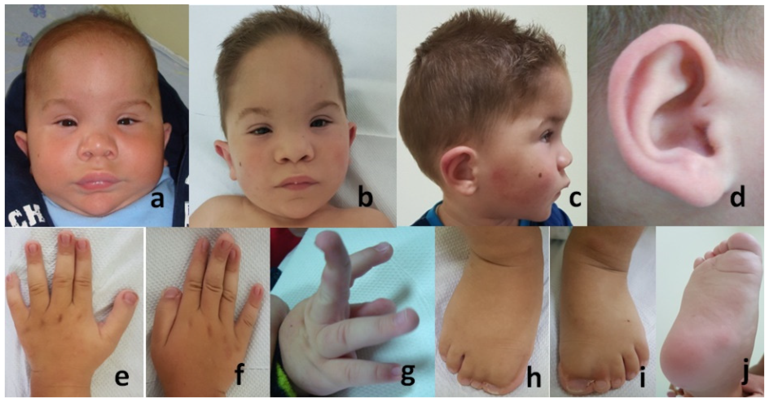
3.1. Morphological Evaluation of the Patient

When he was one year old, macrocephaly (OFC: 48.5 cm, 95th percentile) was observed (Figure 1a) and at the age of three, macro-acrocephaly (OFC: 53 cm, 1 cm > 95th percentile) was seen (Figure 1b,c). Observations included prominent metopic suture and high anterior hairline with right frontal upsweep; sparse eyebrows, hypertelorism, upslanted telecanthus, and palpebral fissures; convergent strabism; depressed and wide nasal bridge, anteverted nares, broad nasal base, and broad and high inserted columella; deep naso-labial creases; long and broad philtrum, and thick upper lip; microdontia; pointed chin with a horizontal deep crease; pigmented nevus on the right cheek (Figure 1a–c); low inserted ears with out-sticking upper one-third, and a pit on the crus of the helix (Figure 1c,d).
Digital anomalies of the hands and feet (Figure 1e–j) included squared, broad thumbs (e,f); joint laxity with dorsiflexion of the distal phalanges (f,g); post-operative scar after surgical correction of preaxial polydactyly and broad hallux with cutaneous complete syndactyly of interdigital spaces I–II and II–III on both feet; bilateral pes planus (h,i); and deep horizontal skin cutaneous crease at the base of the hallux (j).
Additional anomalies were: congenital stridor (almost disappeared at the age of 3 years), small umbilical hernia, postoperative scar after surgical intervention for right cryptorchidism (at age 16 month), and a retractile left testicle.
Severe cognitive and motor delays were observed: head control was achieved at 6–7 months, sitting at 2 years, independent walking has not yet been achieved, and speech is limited to pronouncing a few syllables. The Mental Scale (Bayley II) score at 3 years of age was equivalent to 8 months.
Growth was assessed based on the Fenton Growth chart for preterm infants [20] and the Longitudinal Child Growth Standards of the Central Statistics Office of Hungary, by Joubert, Darvay and Ágfalvi, 2006 [21].
Morphological features are described according to the Human Malformation Termi-nology [22,23,24,25].

3.2. Neuroimaging

The cranial ultrasound at birth was not informative and the cranial MRI at 1 year and at 3 years showed hypoplasia of the corpus callosum, stenosis of the cerebral aqueduct, and mild supratentorial ventriculomegaly.

3.3. Conventional and Molecular Cytogenetic Investigations

The result of the karyotype and the aCGH analysis are described in Table 1.
The clinical diagnosis of GCPS was made immediately after birth based on the characteristic phenotype suggestive of the syndrome.
Therapeutic interventions were focusing on correcting the digital anomalies to improve the patient’s grasping manipulation and walking skills, as well as to improve cognitive functions by introducing continuous cognitive and kinetic stimulation.
The clinical course during the 6-year follow-up suggested a stationary condition regarding somatic development, which at the age of 6 years converged to normal, with weight being 22 kg (50–75th percentile), height 116 cm (50th percentile), and head circumference 53 cm (90–95th percentile). Regarding the psychomotor development, there was a mild improvement: at the age of 6 years, he is more active, his ability to cooperate gradually improves and his speech comprehension develops in terms of simple, often repetitive stimulances; vocalization is more monosyllabic; he enjoys listening to rhymes and music; the Mental Scale (Bayley II) score equal to that of a 15 month-old toddler; his right hand is more skillful, he inserts a few geometric elements, shows some of his body parts, climbs, kneels, clings, sometimes gets up, and steps with support by one hand.

4. Discussion

The diagnosis of GCPS was suggested immediately after birth based on the characteristic clinical signs of the syndrome and was confirmed by classical and molecular karyotyping.
The G-banded karyotype was 46,XY,del(7)(p13p15).
Molecular karyotype analysis (array-CGH) revealed an 18.37 Mb interstitial deletion on the short arm of chromosome 7. Deletion breakpoints are located between p14.2 and p11.2 bands (35830920-54201451). The OMIM genes located in the deleted region are described in Table 1. The analysis of haploinsufficiency described a difference of 330 kb, which is due to the different localization of the CNV and SNP probes.
Of the 205 genes included in the deleted segment, 22 were phenotype associated OMIM genes. Nine genes: GLI3 (Gli-Kruppel Family Member 3; OMIM# 16540), ANLN (Actin-Binding Protein Anillin; OMIM# 616027), POU6F2 (Pou Domain, Class 6, Transcription2; OMIM# 609062), GCK (Glucokinase; OMIM#138079), CCM2 (Scaffold Protein; OMIM# 607929), IKZF1 (Ikaros Family Zinc Finger 1; OMIM# 603023), CDK13 (Cyclin-Dependent Kinase 13; OMIM# 603309), CAMK2B (Calcium/Calmodulin-Dependent Protein Kinase Ii-β; OMIM# 607707), and RALA (Ras-Like Protooncogene A; OMIM# 179550) are known to be responsible for autosomal dominant mendelian disorders, of which GLI3 is the gene causing GCPS. Loss of function mutations of GLI3 determine the classic phenotype with craniofacial dysmorphism (macrocephaly, hypertelorism) and polysyndactyly; while gain of function mutations determine the Pallister-Hall syndrome (MIM# 146510) [3,8,10,13].
Mutations of the ANLN gene have been linked to functional or structural renal anomalies; those of the POU6F2 gene are associated with susceptibility to Wilms tumor (MIM# 601583); mutations of the GCK gene cause MODY II diabetes; pathogenic variants of the CCM2 gene cause cavernous cerebral malformation; and those of the IKZF1 gene result in immunodefficiency, providing a reason for close follow-up and screening for comorbidities in our patient.
Five years after the diagnosis, we have reevaluated our case; we updated the OMIM genes involved in the deleted segment and we found three recently phenotype-associated, autosomal dominant OMIM genes: CDK13, CAMK2B, and RALA. Only the RALA gene has been mentioned in four cases before (Jennifer J Johnston et al.) [6,7], but at that time (2003 and 2007) it had no known associated phenotype.
The CDK13 and CAMK2B genes have been recently annotated OMIM genes, related to syndromic neurodevelopmental disorders (congenital heart defects, dysmorphic facial features, intellectual developmental disorder associated to MIM# 617360, mental retardation, and autosomal dominant 54 associated to MIM# 617799) [26,27,28,29,30,31,32,33].
CDK13 codes for a member of the cyclin-dependent serine/threonine protein kinase (STK) family, which phosphorylate targets, such as RNA polymerase II-involved in extracellular and growth signaling [33]. The majority of mutations in CDK13 are missense mutations, occurring within the protein kinase domain leading to significant loss of catalytic activity. Hamilton et al. (2018) postulated their dominant-negative effect [26]. Genotype–phenotype studies suggested a trend toward milder phenotypes in patients with mutations predicted to cause haploinsufficiency of CDK13 [30].
The CAMK2B gene encodes a subunit of calcium/calmodulin-dependent protein kinase II (CaM kinase II, CAMK2), a multifunctional serine/threonine kinase that has critical roles in the induction of hippocampal long-term potentiation and, as such, in synaptic plasticity, learning, and memory. Both loss-of-function and splice variants have been extensively studied and functionally characterized by Küry et al. in 2017 [31].
The RALA gene encodes a low molecular mass ras-like GTP-binding protein that shares about 50% similarity with other ras proteins. In neuronal culture systems, RALA has been implicated in the development, plasticity, polarization, migration, branching, and spine growth of neurons, as well as the renewal of synaptic vesicles and trafficking of NMDA, AMPA, and dopamine receptors to the postsynaptic membrane. Loss of function for RALA causes a severe neural tube defect; de novo missense variation disrupting the GTP/GDP-binding functions of RALA lead to developmental delay, intellectual disability, and related phenotypes (Hiatt-Neu-Cooper neurodevelopmental syndrome, MIM# 619311) [34].
The deletion of these genes could have influenced the phenotype of our patient, especially the intellectual status, but their impact is indistinguishable from the GCPS-CGS phenotype. The macrosomia, hypertelorism, prominent metopic suture without confirmed craniosynostosis, anomaly of the corpus callosum, and polysyndactyly can be attributed to Greig cephalopolysyndactyly syndrome and haploinsufficiency of GLI3. Intellectual deficiency is rare in loss-of-function mutations of GLI3 associated with GCPS syndrome, but is frequently described and correlates with the size of deletion of GCPS-CGS [31].
Since the identification of CDK13, CAMK2B and RALA as syndromic, intellectual disability genes, we assume that the involvement of CDK13, CAMK2B and RALA genes in the deletion of our patient explains the presence and the severity of the intellectual disability. Callosal anomalies have also been reported in the CAMK2B-related intellectual disability [31].
To our knowledge, chromosomal deletion encompassing the genes CDK13, CAMK2B or RALA as a mechanism for intellectual disability related neurodevelopment delay disorders, have not been described so far.
Twelve further genes in the deleted segment: NME8 (NME/NM23 Family; OMIM# 607421), MPLKIP (M-Phase-Specific Plk1-Interacting Protein; OMIM# 609188), BLVRA (Biliverdin Reductase A; OMIM# 109750), PGAM2 (Phosphoglycerate Mutase 2; OMIM# 612931), OGDH (Oxoglutarate Dehydrogenase; OMIM# 613022), ADCY1 (Adenylate Cyclase 1; OMIM# 103072), DDC (Dopa Decarboxylase; OMIM# 107930), AEBP1 (Ae-Binding Protein 1; OMIM# 602981), SFRP4 (Secreted Frizzled-Related Protein 4; OMIM# 606570), PKD1L1 (Polycystin 1-Like; OMIM# 609721), VPS41 (Subunit Of Hops Complex; OMIM# 605485), and SUGCT (Succinyl-Coa: Glutarate-Coa Transferase; OMIM# 609187) are related to recessive conditions, and the deletion of a single allele will not manifest as a disease unless the second allele has a mutation. The clinical signs of our patient cannot be correlated with the effects of these genes. Regarding the DDC gene, this would require further investigation (sequencing) given the risk of acute lymphoblastic leukemia (MIM# 613067) [14,35].
The NPC1L1 (NPC-Like Intracellular Cholesterol Transporter 1; OMIM# 608010) gene is responsible for the intestinal absorption of cholesterol, it is the molecular target for the drug ezetimibe, and variants in this gene affect response to this drug. The inheritance of the trait is not known, but monitoring cholesterol profile is relatively easy.
We only assume that the patient’s deletion is de novo because, although the parents do not show clinical signs of the disease, it would have been necessary to perform q-PCR or microarray for them to exclude the inheritance of the deleted segment and high-resolution karyotype or FISH test to detect balanced chromosomal rearrangements. Genetic testing was declined by both parents, they are not willing to have further children.
A review of the literature of the patients with Greig cephalopolysyndactyly contiguous gene syndrome caused by a structural chromosomal anomaly (deletion) with a size greater than 1 Mb is summarized in Table 2 [3,4,6,7,11,13,16,17,18,19].
The genotype–phenotype correlation regarding the neurodevelopment in Greig cephalopolysyndactyly contiguous gene syndrome in our case and a further six cases (with available genomic coordinates) from the literature is presented in Table 3.
The haploinsufficiency of the CDK13, CAMK2B and RALA genes could influence the patients’ neurodevelopment in five cases and the CAMK2B gene in two cases.

5. Conclusions

The case presented here, with an 18 Mb deleted chromosomal segment, is the largest deletion described in the related literature so far.
By describing the multitude of symptoms in our patient related to this large spanning deletion, encompassing several other genes than GLI3 responsible for intellectual disability, and by comparing our patient’s features with literature data, we aimed to broaden the phenotypic and genotypic spectrum of GPS-CGS and find out about the possible comorbidities and prognosis. Sequence analysis would be useful to determine whether the variants on the other allele could contribute to the phenotype, therefore a whole exome sequencing has yet to be performed.

Author Contributions

Conceptualization, K.K., M.B.; investigation, K.K., A.U., I.S., M.I., S.Ş.Ş. and A.P.; resources, C.D.P., A.U., Z.S., I.S., M.I., S.Ş.Ş., A.P. and A.J.; writing—original draft preparation, K.K., M.B., C.M.J. and A.J.; writing—review and editing, K.K., M.B., I.S., K.S., C.N.S. and A.S.; visualization, M.B., A.S. and C.N.S.; supervision, M.B., K.S. and A.U.; validation, K.S. All authors have read and agreed to the published version of the manuscript.

Funding

This paper received financial support through the project “SmartDoct—High quality programs for doctoral students and postdoctoral researchers of the University of Oradea to increase the relevance of research and innovation in the context of regional economy”, ID/Project code: 123008, co-financed by the European Social Fund through the Human Capital Operational Program 2014–2020.

Institutional Review Board Statement

The study was conducted according to the guidelines of the Declaration of Helsinki, and approved by the Ethics Committee of Municipal Clinical Hospital “Dr. Gavril Curteanu” Oradea, Romania (protocol code 32811/15.09.2021).

Informed Consent Statement

Written informed consent has been obtained from the parents of the patient to publish this paper.

Data Availability Statement

Not applicable.

Acknowledgments

The authors would like to thank the family who participated in the study.

Conflicts of Interest

The authors declare no conflict of interest.

References

  1. Biesecker, L.G.; Johnston, J.J. Greig Cephalopolysyndactyly Syndrome; Adam, M.P., Ardinger, H.H., Pagon, R.A., Eds.; University of Washington: Seattle, WA, USA, 1993; pp. 1993–2020. [Google Scholar]
  2. Biesecker, L.G. The Greig cephalopolysyndactyly syndrome. Orphanet. J. Rare Dis. 2008, 3, 10. [Google Scholar] [CrossRef] [Green Version]
  3. Niida Yo Ozaki, M.; Takase, E.; Yokoyama, T.; Yamada, S. A Girl with Greig Cephalopolysyndactyly Contiguous Gene Deletion Syndrome: The Importance and Usefulness of DNA Microarray Analysis. Genetics 2015, S7, 4. [Google Scholar] [CrossRef]
  4. Schulz, S.; Volleth, M.; Muschke, P.; Wieland, I.; Wieacker, P. Greig cephalopolysyndactyly (GCPS) contiguous gene syndrome in a boy with a 14 Mb deletion in region 7p13-14 caused by a paternal balanced insertion (5; 7). Appl. Clin. Genet. 2008, 1, 19–22. [Google Scholar] [CrossRef] [PubMed] [Green Version]
  5. McKusick, V.A. Mendelian Inheritance in Man. Available online: http://www.ncbi.nlm.nih.gov/omim (accessed on 25 May 2021).
  6. Johnston, J.J.; Olivos-Glander, I.; Turner, J.; Aleck, K.; Bird, L.M.; Mehta, L.; Schimke, R.N.; Heilstedt, H.; Spence, J.E.; Blancato, J.; et al. Clinical and molecular delineation of the Greig cephalopolysyndactyly contiguous gene deletion syndrome and its distinction from acrocallosal syndrome. Am. J. Med. Genet. A 2003, 123A, 236–242. [Google Scholar] [CrossRef]
  7. Johnston, J.J.; Walker, R.L.; Davis, S.; Facio, F.; Turner, J.T.; Bick, D.P.; Daentl, D.L.; Ellison, J.W.; Meltzer, P.S.; Biesecker, L.G. Zoom-in comparative genomic hybridisation arrays for the characterisation of variable breakpoint contiguous gene syndromes. J. Med. Genet. 2007, 44, e59. [Google Scholar] [CrossRef]
  8. Johnston, J.J.; Olivos-Glander, I.; Killoran, C.; Elson, E.; Turner, J.T.; Peters, K.F.; Abbott, M.H.; Aughton, D.J.; Aylsworth, A.S.; Bamshad, M.J.; et al. Molecular and Clinical Analyses of Greig Cephalopolysyndactyly and Pallister-Hall Syndromes: Robust Phenotype Prediction from the Type and Position of GLI3 Mutations. Am. J. Hum. Genet. 2005, 76, 609–622. [Google Scholar] [CrossRef] [PubMed] [Green Version]
  9. Pettigrew, A.L.; Greenberg, F.; Caskey, C.T.; Ledbetter, D.H. Greig syndrome associated with an interstitial deletion of 7p: Confirmation of the localization of Greig syndrome to 7p13. Hum. Genet. 1991, 87, 452–456. [Google Scholar] [CrossRef] [PubMed]
  10. Siracusano, M.; Riccioni, A.; Baratta, A.; Baldi, M.; Curatolo, P.; Mazzone, L. Autistic symptoms in Greig cephalopolysyndactyly syndrome: A family case report. J. Med. Case Rep. 2019, 13, 100. [Google Scholar] [CrossRef] [PubMed] [Green Version]
  11. Hurst, J.A.; Jenkins, D.; Vasudevan, P.C.; Kirchhoff, M.; Skovby, F.; Rieubland, C.; Gallati, S.; Rittinger, O.; Kroisel, P.M.; Johnson, D.; et al. Metopic and sagittal synostosis in Greig cephalopolysyndactyly syndrome: Five cases with intragenic mutations or complete deletions of GLI3. Eur. J. Hum. Genet. 2011, 19, 757–762. [Google Scholar] [CrossRef]
  12. Kroisel, P.M.; Petek, E.; Wagner, K. Phenotype of five patients with Greig syndrome and microdeletion of 7p13. Am. J. Med. Genet. 2001, 102, 243–249. [Google Scholar] [CrossRef]
  13. Démurger, F.; Ichkou, A.; Mougou-Zerelli, S.; Le Merrer, M.; Goudefroye, G.; Delezoide, A.-L.; Quélin, C.; Manouvrier, S.; Baujat, G.; Fradin, M.; et al. New insights into genotype–phenotype correlation for GLI3 mutations. Eur. J. Hum. Genet. 2014, 23, 92–102. [Google Scholar] [CrossRef] [PubMed] [Green Version]
  14. Mendoza-Londono, R.; Kashork, C.D.; Shaffer, L.G.; Krance, R.; Plon, S.E. Acute lymphoblastic leukemia in a patient with Greig cephalopolysyndactyly and interstitial deletion of chromosome 7 del(7)(p11.2 p14) involving the GLI3 and ZNFN1A1 genes. Genes Chromosomes Cancer 2005, 42, 82–86. [Google Scholar] [CrossRef] [PubMed]
  15. Chotai, K.A.; Brueton, L.A.; van Herwerden, L.; Garrett, C.; Hinkel, G.K.; Schinzel, A.; Mueller, R.F.; Speleman, F.; Winter, R.M. Six cases of 7p deletion: Clinical, cytogenetic, and molecular studies. Am. J. Med. Genet. 1994, 51, 270–276. [Google Scholar] [CrossRef]
  16. Debeer, P.; Devriendt, K.; De Smet, L.; Deravel, T.; Gonzalez-Meneses, A.; Grzeschik, K.-H.; Fryns, J.-P. The spectrum of hand and foot malformations in patients with Greig cephalopolysyndactyly. J. Child. Orthop. 2007, 1, 143–150. [Google Scholar] [CrossRef] [PubMed] [Green Version]
  17. Williams, P.G.; Hersh, J.H.; Yen, F.F.; Barch, M.J.; Kleinert, H.E.; Kunz, J.; Kalff-Suske, M. Greig cephalopolysyndactyly syndrome: Altered phenotype of a microdeletion syndrome due to the presence of a cytogenetic abnormality. Clin. Genet. 1997, 52, 436–441. [Google Scholar] [CrossRef] [PubMed]
  18. Schwarzbraun, T.; Windpassinger, C.; Ofner, L.; Vincent, J.B.; Cheung, J.; Scherer, S.W.; Wagner, K.; Kroisel, P.M.; Petek, E. Genomic analysis of five chromosome 7p deletion patients with Greig cephalopolysyndactyly syndrome (GCPS). Eur. J. Med. Genet. 2006, 49, 338–345. [Google Scholar] [CrossRef] [PubMed]
  19. Zneimer, S.M.; Cotter, P.D.; Stewart, S.D. Telomere-telomere (end to end) fusion of chromosomes 7 and 22 with an interstitial deletion of chromosome 7p11.2→p15.1: Phenotypic consequences and possible mechanisms. Clin Genet. 2000, 58, 129–133. [Google Scholar] [CrossRef] [PubMed]
  20. Fenton, T.R.; Kim, J.H. A systematic review and meta-analysis to revise the Fenton Growth chart for preterm infants. BMC Pediatr. 2013, 13, 59. [Google Scholar] [CrossRef] [Green Version]
  21. Joubert, K.; Gy, G.; Darvay, S.; Ágfalvi, R. Results of the Hungarian Longitudinal Child Growth Study: From Birth to the Age of 18 Years (I). January 2010. Available online: https://edit.elte.hu/xmlui/bitstream/handle/10831/10549/Kutjel83_honlapra.pdf?sequence=1 (accessed on 17 October 2021).
  22. Allanson, J.E.; Cunniff, C.; Hoyme, H.E.; McGaughran, J.; Muenke, M.; Neri, G. Elements of morphology: Standard terminology for the head and face. Am. J. Med. Genet. A 2009, 149A, 6–28. [Google Scholar] [CrossRef] [Green Version]
  23. Biesecker, L.G.; Aase, J.M.; Clericuzio, C.; Gurrieri, F.; Temple, I.K.; Toriello, H. Elements of morphology: Standard terminology for the hands and feet. Am. J. Med. Genet. A 2009, 149A, 93–127. [Google Scholar] [CrossRef] [Green Version]
  24. Carey, J.C.; Cohen MMJr Curry, C.J.; Devriendt, K.; Holmes, L.B.; Verloes, A. Elements of morphology: Standard terminology for the lips, mouth, and oral region. Am. J. Med. Genet. A 2009, 149A, 77–92. [Google Scholar] [CrossRef]
  25. Hennekam, R.C.; Biesecker, L.G.; Allanson, J.E.; Hall, J.G.; Opitz, J.M.; Temple, I.K.; Carey, J.C.; Elements of Morphology Consortium. Elements of morphology: General terms for congenital anomalies. Am. J. Med. Genet. A 2013, 161A, 2726–2733. [Google Scholar] [CrossRef]
  26. Hamilton, M.J.; Suri, M. CDK13-related disorder. Adv. Genet. 2019, 3, 163–182. [Google Scholar] [CrossRef]
  27. Bostwick, B. CDK13-Related Disorder; Adam, M.P., Ardinger, H.H., Pagon, R.A., Eds.; University of Washington: Seattle, WA, USA, 1993; Available online: https://www-ncbi-nlm-nih-gov.ezproxy.u-pec.fr/books/NBK536784/ (accessed on 31 January 2019).
  28. Sifrim, A.; Hitz, M.-P.; Wilsdon, A.; Breckpot, J.; Al Turki, S.H.; Thienpont, B.; McRae, J.; Fitzgerald, T.W.; Singh, T.; Swaminathan, G.J.; et al. Distinct genetic architectures for syndromic and nonsyndromic congenital heart defects identified by exome sequencing. Nat. Genet. 2016, 48, 1060–1065. [Google Scholar] [CrossRef]
  29. Greifenberg, A.K.; Hönig, D.; Pilarova, K.; Düster, R.; Bartholomeeusen, K.; Bösken, C.A.; Anand, K.; Blazek, D.; Geyer, M. Structural and Functional Analysis of the Cdk13/Cyclin K Complex. Cell Rep. 2016, 14, 320–331. [Google Scholar] [CrossRef] [PubMed] [Green Version]
  30. Hamilton, M.J.; Caswell, R.; Canham, N.; Cole, T.; Firth, H.V.; Foulds, N.; Heimdal, K.; Hobson, E.; Houge, G.; Joss, S.; et al. Heterozygous mutations affecting the protein kinase domain of CDK13 cause a syndromic form of developmental delay and intellectual disability. J. Med Genet. 2017, 55, 28–38. [Google Scholar] [CrossRef] [PubMed] [Green Version]
  31. Küry, S.; Van Woerden, G.M.; Besnard, T.; Onori, M.P.; Latypova, X.; Towne, M.C.; Cho, M.T.; Prescott, T.E.; Ploeg, M.A.; Sanders, S.; et al. De Novo Mutations in Protein Kinase Genes CAMK2A and CAMK2B Cause Intellectual Disability. Am. J. Hum. Genet. 2017, 101, 768–788. [Google Scholar] [CrossRef] [PubMed]
  32. Rizzi, S.; Spagnoli, C.; Salerno, G.G.; Frattini, D.; Caraffi, S.G.; Trimarchi, G.; Moratti, C.; Pascarella, R.; Garavelli, L.; Fusco, C. Severe intellectual disability, absence of language, epilepsy, microcephaly and progressive cerebellar atrophy related to the recurrent de novo variant p (P139L) of the CAMK2B gene: A case report and brief review. Am. J. Med. Genet. Part A 2020, 182, 2675–2679. [Google Scholar] [CrossRef]
  33. Heiman, P.; Drewes, S.; Ghaloul-Gonzalez, L. A familial case of CAMK2B mutation with variable expressivity. SAGE Open Med. Case Rep. 2021, 9. [Google Scholar] [CrossRef]
  34. Hiatt, S.M.; Neu, M.B.; Ramaker, R.C.; Hardigan, A.A.; Prokop, J.W.; Hancarova, M.; Prchalova, D.; Havlovicova, M.; Prchal, J.; Stranecky, V.; et al. De novo mutations in the GTP/GDP-binding region of RALA, a RAS-like small GTPase, cause intellectual disability and developmental delay. PLoS Genet. 2018, 14, e1007671. [Google Scholar] [CrossRef] [Green Version]
  35. Sava, C.N.; Ritli, L.; Balmoş, A.B.; Iuhas, A.R.; Marian, P.; Motorca, M.A.; Lele, L.A.; Straciuc, O.; Zaha, D.C.; Jurcă, M.C.; et al. Unusual extramedullary relapses in a case of common B-cell acute lymphoblastic leukemia. Case Rep. Rev. Lit. 2019, 60, 249–254. [Google Scholar]
Figure 1. (ad) shows cranio-facial features of the patient: macrocephaly at the age of 1 year (a), and macro-acrocephaly at 3 years (b,c); prominent high anterior hairline with right frontal upsweep; sparse eyebrows, hypertelorism, telecanthus, and upslanted palpebral fissures; convergent strabism; depressed and wide nasal bridge, anteverted nares, and broad nasal base with broad and high inserted columella; long and broad philtrum, and deep naso-labial fold; thick upper lip; pointed chin with a horizontal deep crease; pigmented nevus on the right cheek (ac); low inserted ears with out-sticking upper one-third of the pinnae, and a pit on the crus of the helix (c,d). (ej) Digital anomalies of the hands and feet: squared broad thumbs (e,f); joint laxity with dorsiflexion of the distal phalanges (f,g); broad hallux with cutaneous complete syndactyly of interdigital spaces I–II and II–III on both feet; bilateral ped planus (h,i) and deep horizontal cutaneous skin crease at the base of the hallux (j).
Genes 12 01674 g001
Table 1. Results of the cytogenetic- and the molecular cytogenetic investigations.
ResultsSize of the Deleted Segment (Mb)OMIM Genes
Karyotype46,XY, del (7)(p13-p15)
aCGHarr[hg38]7p14.2-p11.2 (35830920-54201451)x118.04ANLN, NME8, POU6F2, MPLKIP, GLI3, BLVRA, GCK, NPC1L1, OGDH, CCM2, ADCY1, IKZF1, DDC, PGAM2, CDK13, AEBP1, CAMK2B, SFRP4, PKD1L1, VPS41, SUGCT, RALA
LOH-18.37ANLN, NME8, POU6F2, MPLKIP, GLI3, BLVRA, PGAM2, GCK, NPC1L1, OGDH, CCM2, ADCY1, IKZF1, DDC, CDK13, AEBP1, CAMK2B, SFRP4, PKD1L1, VPS41, SUGCT, RALA, SEPTIN7
aCGH: comparative genomic hibridisation; a: array; arr: array; hg38: human genome version 38; LOH: loss of heterozygosity; Mb: megabase.
Table 2. Review of literature of the patients with GCP-CGS due to a chromosomal deletion encompassing GLI3 (>1 Mb).
ReferencesChromosomal
Localisation
Size of the Deleted Segment
(Mb)
Array Coordinates
Present case7p14.2-p11.218.37arr[hg38](35830920_54201451)del
Niida Y.
et al [3] (2015)
7p14.1-p12.36.2arr[hg19](41076615_47282889)del
Demurger F. et al. [13] (2015)7p13-p157arr[hg19](38521704_45810267)del
9arr[hg19](35674000_37280000)
_(46116000_46598000)del
Jane A Hurst
et al [11] (2011)
7p13-p14.16.0 arr[hg18](39013006_39213707)del
6.8arr[hg18](39130081_45492392)del
7p12.3-p14.18.3arr[hg18](40845981_40855164)
_(49136714_49160830)del
Solveig Schulz et a [4] (2008)7p13-7p1414NA
Debeer Philippe et al. [16] (2007)7p14.3NANA
7p14.3
Jennifer J Johnston
et al. [6,7]
(2003, 2007)
7p14.11.8NA
7p14.1-7p133.2
7p14.14.1
7p14.1-7p135.2
7p14.1-7p135.9
7p14.2-7p14.16.3
7p14.1-7p12.38.4
7p14.2-7p139.8
7p14.2-7p1310.3
Kroisel PM
et al [12]
Schwarbraun T. et al. [18]
(2001, resp. 2005)
7p13

4.5-15


NA
7p12.3-p13
7p12.3-p13
7p12.3-p14.2
7p11.2-p13
Williams PG et al. [17] (1997)7p13-p15.1NANA
Zneimer SM et al. [19] (2000)45,XY,der(22;7)
(p13;p22.3)del
(7)-(p11.2-p15.1)
NANA
NA: not available.
Table 3. Genotype–phenotype correlation.
ReferencesPatients’s
Age
Size of the Deleted Segment
(Mb)
Phenotype Associated OMIM Genes
Neurodevelopment
Present case *3 years
followed to age 6
18.37ANLN, NME8, POU6F2, MPLKIP, GLI3, BLVRA, GCK, NPC1L1, OGDH, CCM2, ADCY1, IKZF1, DDC, PGAM2, CDK13, AEBP1, CAMK2B, SFRP4, PKD1L1, VPS41, SUGCT, RALASevere intellectual disability,
speech
and
developmental delay
Niida Y. et al. * [3]
(2015)
2 years6.2GLI3, GCK, CCM2, AEBP1, CAMK2B, PGAM2, BLVRA, ADCY1, NPC1L1, OGDHDevelopmental delay
Demurger F. et al.* [13] (2015)NA7GLI3, AEBP1, MPLKIP, GCK, CCM2, CDK13, CAMK2B, VPS41, BLVRA, PGAM2, ADCY1, SUGCT, POU6F2, NPC1L1, RALA, OGDHDevelopmental delay
NA9GLI3, GCK, CDK13, MPLKIP AEBP1, CCM2, CAMK2B, SFRP4, VPS41, PGAM2, BLVRA, NME8, SUGCT, ADCY1, NPC1L1, POU6F2, ANLN, RALA, OGDHDevelopmental
delay
Jane A Hurst et al.* [11]
(2011)
2 years6.0 GLI3, GCK, CCM2, CDK13, MPLKIP, AEBP1, CAMK2B, BLVRA, PGAM2, SUGCT, RALA, POU6F2, NPC1L1, OGDHDevelopmental delay
5 years6.8GLI3, GCK, CCM2, CDK13, MPLKIP, AEBP1, CAMK2B, BLVRA, PGAM2, SUGCT, RALA, NPC1L1, POU6F2,
OGDH
Severe intellectual disability,
speech and developmental delay
15 years8.3GLI3, GCK, CCM2, AEBP1, CAMK2B, PKD1L1, BLVRA, PGAM2, SUGCT, ADCY1, NPC1L1, OGDHSevere intellectual disability, speech and
delayed
development
*: updated cases based on the available array coordinates; Neurodevelopment disorders related genes: CDK13, CAMK2B, RALA.
Publisher’s Note: MDPI stays neutral with regard to jurisdictional claims in published maps and institutional affiliations.

