

Progress in Surface Modification of Titanium Implants by Hydrogel Coatings
by
Huangqin Chen
1,†,
Rui Feng
1,†,
Tian Xia
1,
Zhehan Wen
1,
Qing Li
1,
Xin Qiu
1,
Bin Huang
1,* and
Yuesheng Li
2,* 1
Department of Stomatology, School of Stomatology and Ophthalmology, Xianning Medical College, Hubei University of Science and Technology, Xianning 437100, China
2
Hubei Key Laboratory of Radiation Chemistry and Functional Materials, Non-Power Nuclear Technology Collaborative Innovation Center, Hubei University of Science and Technology, Xianning 437100, China
*
Authors to whom correspondence should be addressed.
†
These authors contributed equally to this work.
Gels 2023, 9(5), 423; https://doi.org/10.3390/gels9050423
Submission received: 5 May 2023
/
Revised: 16 May 2023
/
Accepted: 17 May 2023
/
Published: 18 May 2023
(This article belongs to the Special Issue Hydrogel for Sustained Delivery of Therapeutic Agents)
Abstract
:Although titanium and titanium alloys have become the preferred materials for various medical implants, surface modification technology still needs to be strengthened in order to adapt to the complex physiological environment of the human body. Compared with physical or chemical modification methods, biochemical modification, such as the introduction of functional hydrogel coating on implants, can fix biomolecules such as proteins, peptides, growth factors, polysaccharides, or nucleotides on the surface of the implants, so that they can directly participate in biological processes; regulate cell adhesion, proliferation, migration, and differentiation; and improve the biological activity on the surface of the implants. This review begins with a look at common substrate materials for hydrogel coatings on implant surfaces, including natural polymers such as collagen, gelatin, chitosan, and alginate, and synthetic materials such as polyvinyl alcohol, polyacrylamide, polyethylene glycol, and polyacrylic acid. Then, the common construction methods of hydrogel coating (electrochemical method, sol–gel method and layer-by-layer self-assembly method) are introduced. Finally, five aspects of the enhancement effect of hydrogel coating on the surface bioactivity of titanium and titanium alloy implants are described: osseointegration, angiogenesis, macrophage polarization, antibacterial effects, and drug delivery. In this paper, we also summarize the latest research progress and point out the future research direction. After searching, no previous relevant literature reporting this information was found.
1. Introduction
In the 1940s, some scholars implanted pure titanium (Ti) into the femur of mice without causing adverse reactions, which proved that Ti had good biocompatibility [1]. Later, more and more scholars began to apply pure Ti in dental implants, joint prostheses, and other clinical fields. However, in the process of application, it was found that the low hardness and poor wear resistance of Ti did not meet the requirements of the force parts of knee joint and hip joint, promoting research into and development of titanium alloys. Ti6Al4V is an α + β alloy with higher hardness, better wear resistance, and better workability compared with Ti (Table 1). However, aluminum (Al) and vanadium (V) in Ti6Al4V alloy are harmful metal elements, which have the risk of releasing after implantation into the human body. There is an urgent need to develop new medical titanium alloys with better biocompatibility. Representative materials are Ti–Nb–Zr and Ti–Nb–Zr–ME (Me (metal)) systems, especially Ti–Nb–Zr–Si (TNZS) alloy, which not only has better biocompatibility, but also has improved corrosion resistance, and can better match with human bone tissue [2].
At present, titanium and titanium alloys have become the preferred materials for medical metal products because of their lower density, higher specific strength, and better biocompatibility.
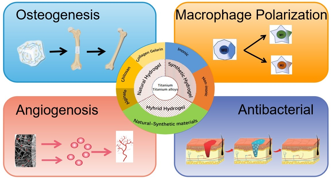
Although medical titanium and titanium alloys have outstanding properties, it is still necessary to strengthen the research on surface modification technology to match the complex physiological environment in human body (Figure 1) [3]. Now, construction of functional coatings on titanium and titanium alloys has attracted more and more attention. For example, titanium nitride [4], titanium aluminum nitride [5], and titanium dioxide [6] coatings significantly improve wear resistance and corrosion resistance of titanium and titanium alloys. The modification of Si–TiO2 nanotubes on the Ti substrate generates a nanostructured and hydrophilic surface, which can promote cell growth. Moreover, the existence of the TiO2 nanotubes and Si element improves the in vitro osteogenic differentiation of MC3T3-E1 cells and early bone-formation around the implanted screws [7]. Coating of silver (Ag), copper (Cu), zinc (Zn), and other antibacterial metal elements show excellent antibacterial properties [8,9]. Adding Cu with Ti-15Mo reduces the possibility of bacterial infection during biomedical implant surgeries [10].
Differently from physical or chemical modification methods such as micro-arc oxidation and sandblasting to prepare oxide film or rough surface on the surface of implants, biochemical modification of fixing specific proteins, peptides, growth factors, polysaccharides, nucleotides, and other biomolecules on the surface of implants can directly participate in biological processes and regulate cell adhesion, proliferation, migration, and differentiation [11,12]. Hydrogel is a type of hydrophilic three-dimensional network structure formed by natural or artificial synthetic polymer materials through the gelation process of sol, which is widely used in many fields such as tissue engineering, drug delivery, and biosensors. The functional hydrogel coatings on titanium implants can effectively coordinate the advantages of hydrogel (lubricity, biocompatibility, and controlled release) with those of implants (stiffness, strength, and toughness) [13], and change the electrochemical behavior of titanium implants and enhance corrosion resistance [14].
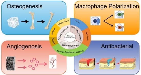
In this review, we will first introduce the classification of hydrogel coatings on the surface of titanium implants, including natural hydrogels and synthetic hydrogels, according to the composition of the hydrogel matrix. We will then introduce the common binding methods of hydrogel coatings and titanium implants, such as the electrochemical precipitation method, the sol–gel method, and the layer-by-layer self-assembly method. Subsequently, the improvement of titanium implants by hydrogel coating on osseointegration, angiogenesis, macrophage polarization, antibacterial, and drug delivery are summarized in detail. Finally, the possible problems and future development direction of hydrogel coatings are presented in order to provide reference for scientific research workers in related fields. After searching, no previous research reporting of this information was found.
2. Classification of Hydrogel Coatings
According to the main components of the hydrogel matrix, hydrogel coatings can be divided into natural hydrogel coatings and synthetic hydrogel coatings (Table 2).
2.1. Natural Hydrogel Coating
Natural hydrogel is composed of natural biological materials which are highly similar to the extracellular matrix. It is considered as good biomimetic material in tissue engineering because of its complete bioactivity in promoting cell adhesion, proliferation, differentiation, and biodegradation. Common natural biomaterials are collagen and gelatin from animal protein, hyaluronic acid from animal epithelium and connective tissue, chitosan from shells of crustaceans, and alginate from the cytoplasm and cell wall.
2.1.1. Collagen-Based Hydrogel Coating
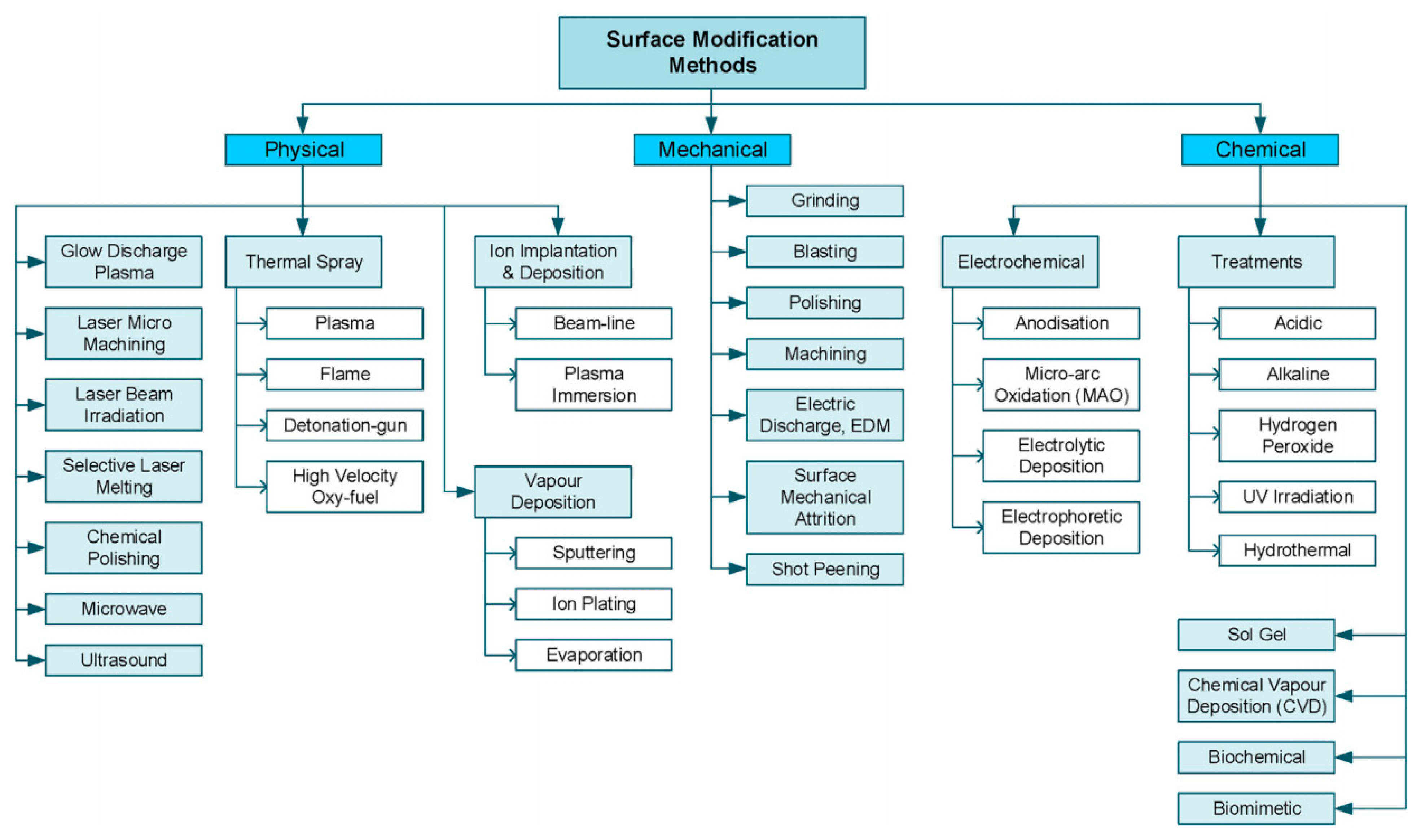
Collagen is the main component of the extracellular matrix in mammals, and is mainly distributed in the cornea, cartilage, bone, blood vessels, viscera, intervertebral discs, and dentin, and plays an important role in supporting and protecting the body and organs. Collagen has the advantages of non-cytotoxicity, good biocompatibility, easy absorption, small immune response, low antigenicity, etc. Coating titanium alloys with collagen promotes adhesion, proliferation, and differentiation of born-forming cells [30,31,32,33] as well as fibroblasts [34,35]. In comparison with uncoated commercially pure titanium, collagen coating significantly improves bone mineralization and maturation [36]. More rapid osteointegration will be achieved if the coating is combined with vitamins [37], phospholipid [38], or hydroxyapatite (HA) [39,40,41]. Besides osteogenesis, collagen coating can support the timely conversion of macrophages from the pro-inflammatory M1 to the pro-healing M2 phenotype, inhibiting inflammatory reaction and generating a beneficial osteoimmune microenvironment [42,43] (Figure 2). Simultaneously, collagen coating prominently facilitates angiogenesis of endothelial cells [42] and strengthens local blood supply restoration through sustained release of vascular endothelial growth factor (VEGF) [44].
2.1.2. Gelatin-Based Hydrogel Coating
Gelatin is a product of collagen hydrolysis but retains arginine–glycine–aspartic (RGD) cell adhesion peptide and protease degradation sites of collagen with lower immunogenicity. Gelatin-based hydrogel coatings enhance the integration between implant and tissue [45], and promote biological activity by loading various growth factors. For example, gelatin coating loaded with VEGF/bone morphogenetic protein 2 (BMP-2) shell-core microspheres promoted osteogenic differentiation and osseointegration effectively in 3D-printed porous titanium alloy [46]. In 2000, Van den Bulcke et al. introduced the methacryl group into modified gelatin for the first time to prepare methacryl amide gelatin, which gave the gelatin the property of photo-cross-linking under the photoinitiator and light [47]. When loaded with a short cationic antimicrobial peptide and synthetic silicate nanoparticles, the photo-cross-linked gelatin-based hydrogel coating demonstrated excellent antimicrobial activity and enhanced osteogenesis [48]. The addition of ginger inhibited the growth of S. mutans and P. gingivalis [49]. Methylacrylamide gelatin combined with photosensitizer and photocatalyst offers direct fibroblast activation [50] (Figure 3) and multi-mode photothermal and photodynamic antibacterial effects [51]. Furthermore, the allylated gelatin co-encapsulated human umbilical vein endothelial cells (HUVECs) and human mesenchymal stromal cells (hMSCs) support and achieve concurrent vasculogenic and osteogenic performance [52].
2.1.3. Chitosan-Based Hydrogel Coating
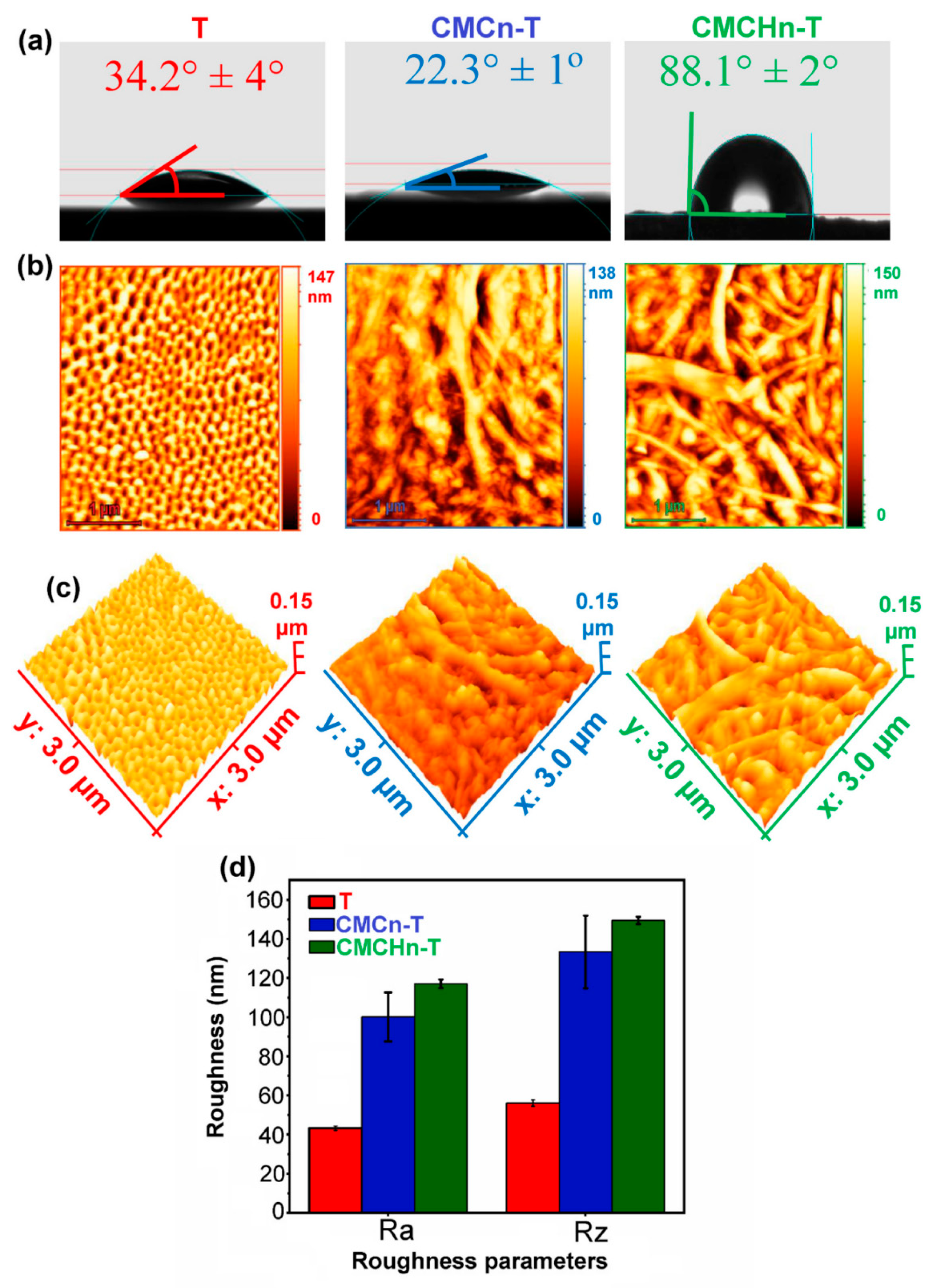
Chitosan, the deacetylated chitin, is the only natural alkaline polysaccharide with charge. Due to their pH, ionic strength, and temperature sensitivity, chitosan-based hydrogels have good application prospects in the fields of targeting, sustained drug release, tissue engineering, and medical dressings [53]. Chitosan-based hydrogel coatings increase the antibacterial ability of the implant by loading antibacterial agents [54,55,56], or metal ions (Ag, Cu) [57,58,59,60]. Coatings give the implant the photocatalytic antibacterial effect by modifying or loading novel semiconductor materials, such as graphene [61], molybdenum disulfide [62], black phosphorus [63], and molybdenum diselenide [64]. They also promote osteogenesis through loading drugs, (for example, pitavastatin [65] and quercetin [66]), active substances (insulin growth factor binding protein-3 [67] and BMP-2 [68]) and inorganic matter (HA [69,70] and bioactive glass [71]). In addition, chitosan combines with polyanions such as gelatin [72,73], hyaluronic acid [74], and sodium alginate [75,76] to form a polyelectrolyte complex, promoting the surface functionalization of titanium. Modified carboxymethyl chitosan nanofibers, as a novel implant coating on titania nanotube arrays, inhibit bacterial colony formation and increase osteoblast cell survival [77] (Figure 4). Similarly, carboxymethyl chitosan loaded with silver nanoparticles enhances the antibacterial properties of the titanium alloy [78].
2.1.4. Alginate-Based Hydrogel Coating
Alginate is a type of linear hydrophilic polysaccharide existing in brown algae. It forms hydrogels by ionic cross-linking with Ca2+ and other polyvalent inorganic cations. There are a large number of –OH and –COOH groups on the alginate skeleton, which can be modified by chemical or physical methods to achieve controlled release of cells or bioactive molecules in response to temperature, pH, and light [79]. Composite coating formed by alginate crosslinking with collagen enhances the cell adhesion of titanium implants [80,81]. Alginate and chitosan coating improves the biomineralization, the antibacterial activity, and corrosion resistance [75,82]. The addition of Ag further promotes the antibacterial ability and reduces the bacterial adhesion [83,84]. Alginate-based hydrogel coatings also provide sustained antibacterial activity by loading various antibacterial agents such as gentamicin [68], vancomycin [85], and chlorchloridine [86], and improving in vitro osteogenic differentiation as well as bone integration by loading BMP2 [87] and RGD [88].
2.2. Synthetic Hydrogel Coatings
The synthetic hydrogels have great application potential due to the wide source of raw materials, simple synthesis method, and controllable composition and structure. Synthetic hydrogels can be divided into the following functional groups: non-ionic hydrogels including polyvinyl alcohol (PVA), polyacrylamide (PAM), poly N-isopropylacrylamide (PNIPAm), polyethylene glycol (PEG), poly (lacto-glycolic acid) (PLGA), etc., and ionic hydrogels such as polyacrylic acid (PAA).
PVA hydrogels with porous titanium bases are being developed to repair or replace articular cartilage due to their high mechanical strength [89]. The Ti–hydrogel artificial cartilage material constructed with polydopamine (PDA), PVA, HA, or PAA as raw materials is an ideal high-strength and low-friction biomimetic cartilage material [90]. The novel “soft (PVA hydrogel layer)–hard (porous Ti6Al4V alloy substrate)” structure improves the surface wettability and tribological properties of Ti6Al4V alloy [91].
PAM hydrogel is an injectable soft-tissue-filling material. PAM-based hydrogels in combination with titanium-oxide nanotubes are also widely used as potential candidates for cartilage replacement [92]. PAM/PVA hydrogel on Ti6Al4V alloy configuration combines the good load-bearing capacity of the rigid substrate and the excellent lubrication of the hydrogel layer [93]. The cross-linked network porous structure of hydrogel is the main factor accounting for the low dynamic friction [94].
PNIPAm is a thermo-responsive polymer with lower critical solution temperature (LCST) around 32 °C. When the temperature is above the LCST, the polymer chains become hydrophobic and collapse, resulting in dense crosslinking networks in which the loaded molecules are more likely to be trapped, thus leading to slow release [95].
PEG is obtained by glycol dehydration polycondensation. The functional-group-hydroxyl at the end of the molecular chain is prone to chemical reactions and chemical modifications [96]. PLGA–PEG–PLGA hydrogels, polymerization of PLGA with PEG, are suitable for drug loading in vitro and sustained drug release in vivo, owing to the thermo-sensitive properties [97].
PAA hydrogels are three-dimensional macromolecules containing a large number of carboxyl groups that cannot move freely. When the pH value of the solution is different, it presents different degrees of shrinkage or swelling state. It can be used to prepare a simple and low-cost hydrogel-based bone adhesive to improve the osseointegration and anti-infection ability of the bone-implant interface [98].
3. Binding Method of Hydrogel Coating and Titanium Implant (Preparation Method of Hydrogel Coating)
Titanium implants have some disadvantages and one of the effective strategies is to prepare multifunctional hydrogel coatings on the surface. The most commonly used preparation method is sol–gel method.
3.1. Electrochemical Methods
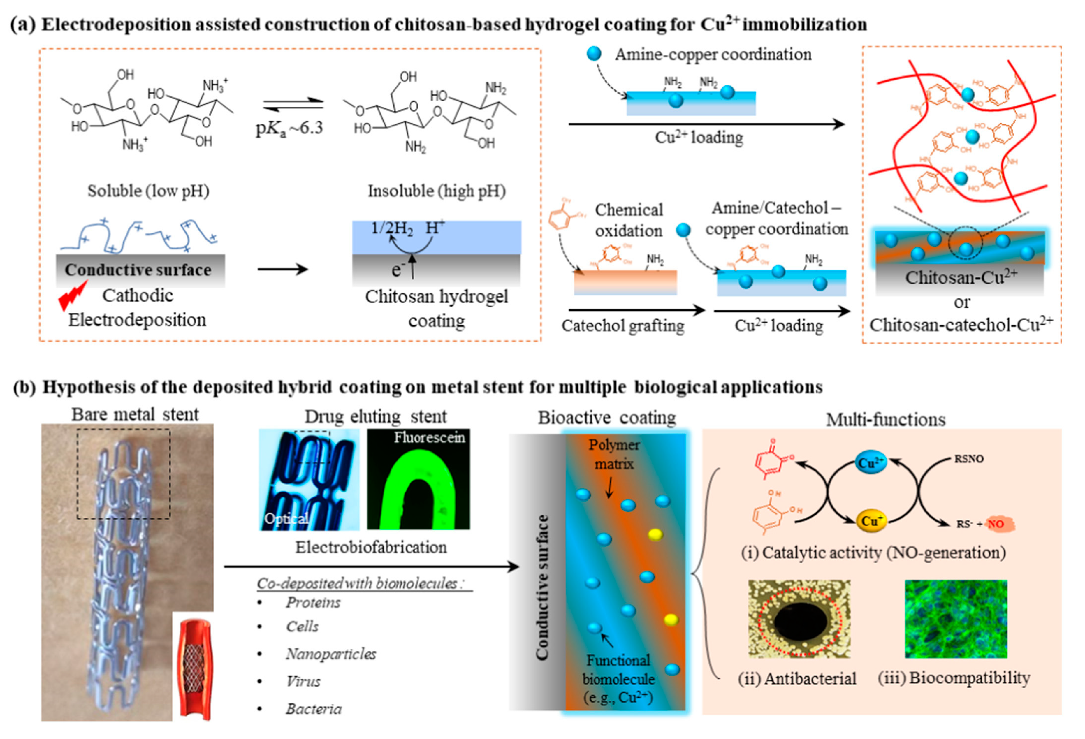
The electrochemical methods for preparing hydrogel coatings mainly include electrochemical deposition and electrophoretic deposition. Electrochemical deposition refers to the process of forming coatings on the surface of metals or alloys in aqueous or non-aqueous solutions of inorganic salts and bio-active factors, which is a promising technique for surface modification of implants with various shapes, especially deformed structures [99,100]. The chitosan hydrogel coating, which is a versatile platform for Cu immobilization and precisely controlled synthesis via electrochemical deposition, has in vitro cell biocompatibility and catalyzed nitric-oxide-generation activity [101] (Figure 5). Electrophoretic deposition refers to the phenomenon of powder particles deposited from the suspension on the electrodes with opposite charges and certain shapes, relying on the action of the direct current [102]. The lanthanum- and silicate-substituted composite coating on a titanium implant achieved by the electrophoretic deposition method exhibited strong osteogenic ability [103]. Moreover, UV irradiation can be used as a crosslinking activator to deposit gentamicin-loaded agarose hydrogels, controlling the release of the loaded antibacterial agents while improving cell integration [104].
3.2. Sol–Gel Method
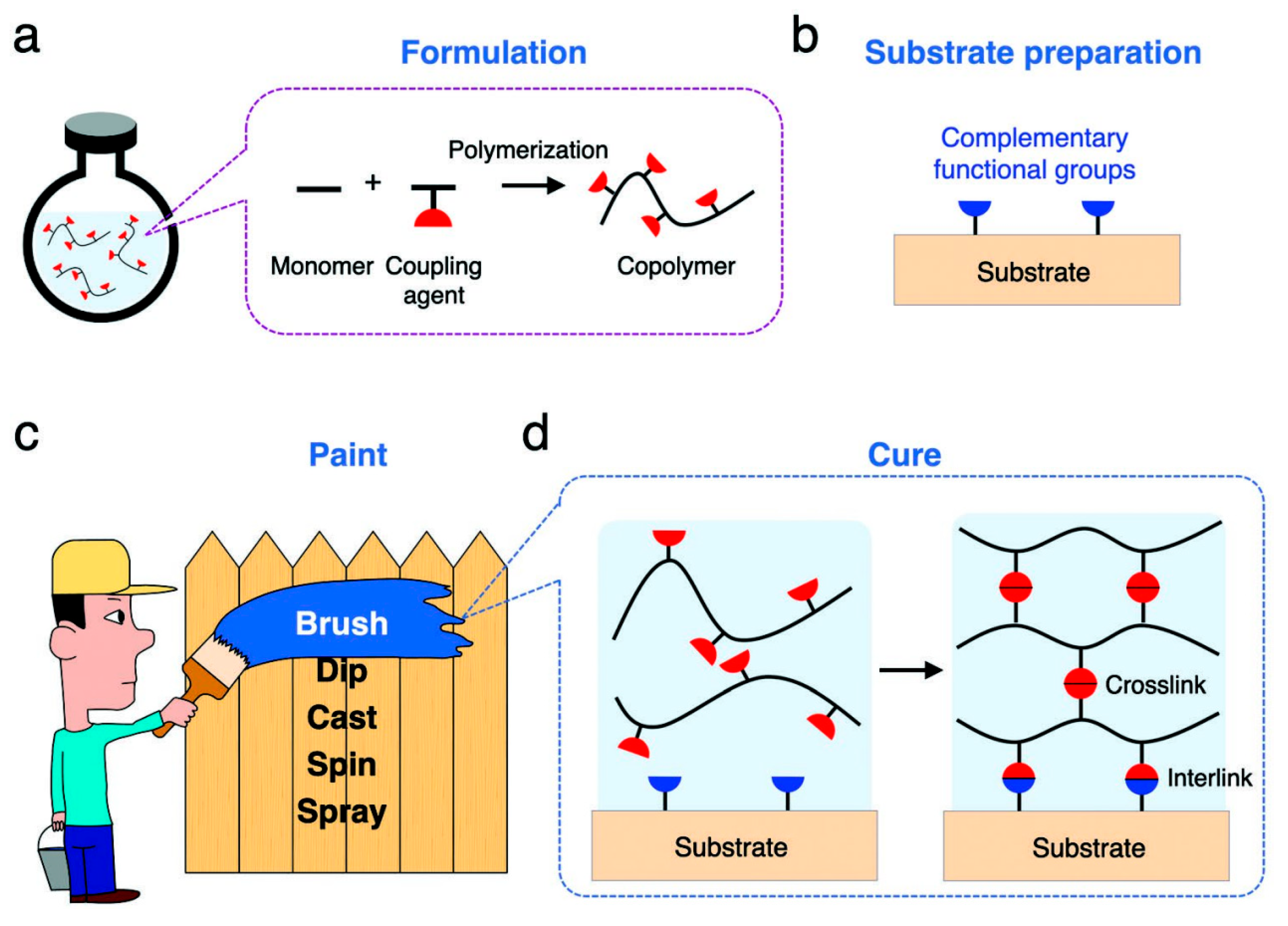
Hydrogel coatings are mostly prepared by the sol–gel method. That is, monomers and coupling agents, as well as substances with different functions (initiators, loaded drugs, etc.), are dissolved in water to generate a free-radical polymerization reaction to form uncross-linked polymer chains. Then, the formulated aqueous solution is coated on the prepared matrix, and the polymer chain is cross-linked into a polymer network by the coupling agent. Finally, the polymer network is connected to the matrix by reacting with complementary functional groups on the matrix surface (Figure 6) [105].
Common methods of applying aqueous solution to the substrate are spraying [106], spin [26,107], and impregnated lift [52,108]. In order to enhance integration between implant material and hydrogel, a PDA layer was introduced onto the surface of the titanium alloy. Through chemical crosslinking between PDA and gelatin [45,51] or HRP/H2O2 catalysis [78], the hydrogel precursor could simply form a firm gel layer on the titanium alloy plate. Methylacryylated gelatin (GelMA) is a photo-cross-linked gelatin derivative. The photoinitiator [109] or catechol motifs [50,110] stabilize the GelMA hydrogel system and make the coating tightly adhere to titanium substrates after 365 nm UV exposure. The sol–gel method caused by ionizing radiation is a safe, simple operation with no polluting effects. Unfortunately, this method is rarely used in the preparation of hydrogel coatings on titanium and titanium alloys.
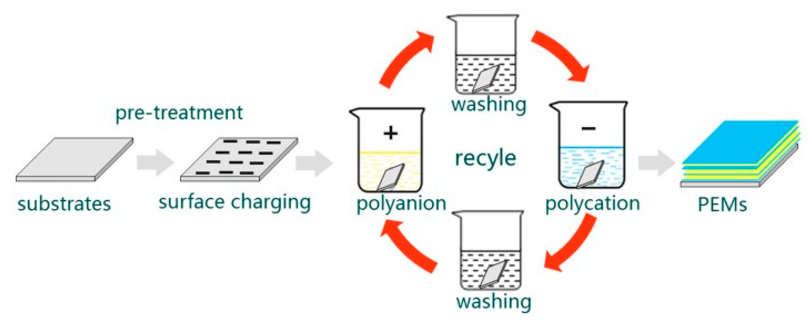
3.3. Layer-by-Layer Self-Assembly
Layer-by-layer self-assembly (LBL) is a popular surface modification method that uses electrostatic adsorption to self-assemble layers of materials with opposite charges into multilayer structures [111] (Figure 7). The assembly process is simple and gentle, and can maintain the biological activity of cytokines and achieve sustained release drug delivery [112]. However, it is necessary to pretreat the Ti surface with microarc oxidation, electrochemical deposition technology [113], etc., to firmly immobilize the multilayers. As one of the silyl reagents, 3-aminopropyl triethoxysilane is often used to aminofunctionalize titanium substrates, promoting covalent coupling to form precursor layers and facilitating the construction of future multilayer coatings [114]. The titanium alloy surfaces can also conjugate with dopamine as the base layer, which enables the deposition of gelatin molecules of hydrogel precursor [115]. PDA is a common mussel-inspired anchoring polymer and exhibits powerful reactivity to various bioactive molecules containing carboxyl groups, amino groups, and thiol groups. A multilayer type-I-collagen decorated nanoporous network was successfully developed on alkali-treated titanium surfaces via PDA coating and LBL [42]. The phase-transited lysozyme provides a new approach to achieving a high binding force that is superior to dopamine, and which forms an amyloid-like microfiber net that tightly adheres to Ti surfaces according to the transition process of lysozyme based on the β-sheet of lysozyme microfibers [116,117]. In addition, tannic acid is a low-cost plant polyphenol, which can bind materials tightly via hydrogen bonds, Michael addition reactions, Schiff base reactions, etc., due to the composition of a glucose core and a hydroxyl-rich phenolic shell, show great potential in LBL [118].
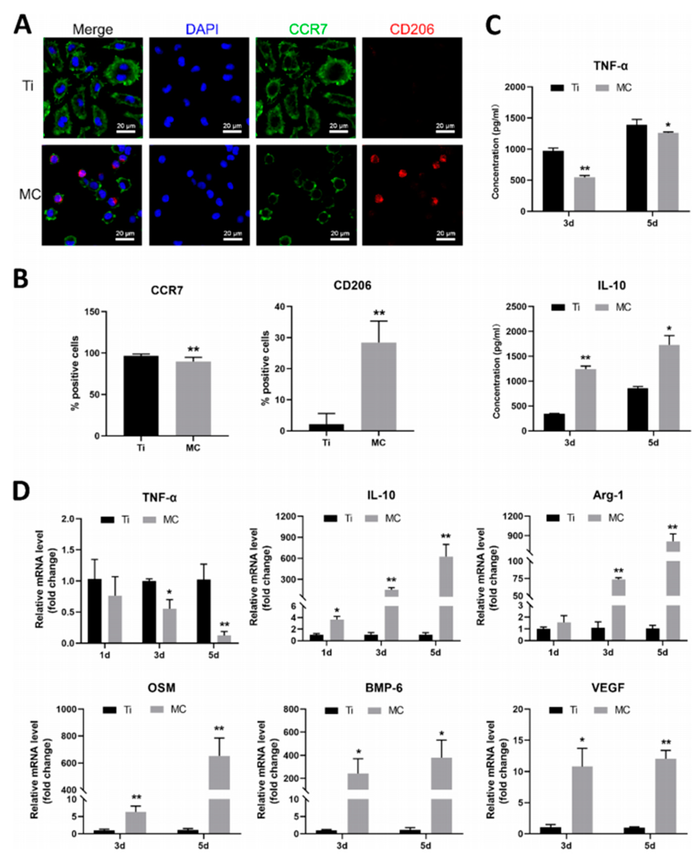
4. Characterization Methods of Surface Modification
Characterization methods for hydrogel coatings include nuclear magnetic resonance (NMR), Fourier transform infrared spectroscopy (FTIR), X-ray diffraction (XRD), and scanning electron microscopy (SEM). The position of the resonance signal on the NMR spectrum reflects the local structure of the sample molecules, such as functional groups. FTIR is the absorption spectrum generated by the absorption of specific wavelengths of infrared light during the vibrational energy level transition of bond-forming atoms in compound molecules, and is mainly used for structural analysis, qualitative identification, and quantitative analysis. XRD is the crystal structure analysis of hydrogel precursor polymer, such as fibroin protein, collagen, and other natural macromolecules containing crystal structure, or loaded nanoparticles such as biological glass and HA. Moreover, SEM and 3D optical profilometer are used to detect the morphological changes and thickness of hydrogel coating, respectively.
5. Application of the Hydrogel Coating
5.1. Osseointegration
Osseointegration is the direct contact between the implant and the bone tissue under the optical microscope, without fibrous connective tissue. Good osseointegration is a key factor in the long-term success of implants. Physical and chemical modification methods, such as changing surface properties [119] and loading inorganic substances, mainly indirectly affect cell behavior, with limitations in improving osteogenic activity [120,121]. Biochemical modifications caused by biomolecules [122,123,124,125,126,127] immobilized on the surface of titanium implants directly participate in biological processes and are more effective in inducing bone-formation, especially in poor bone conditions [128].
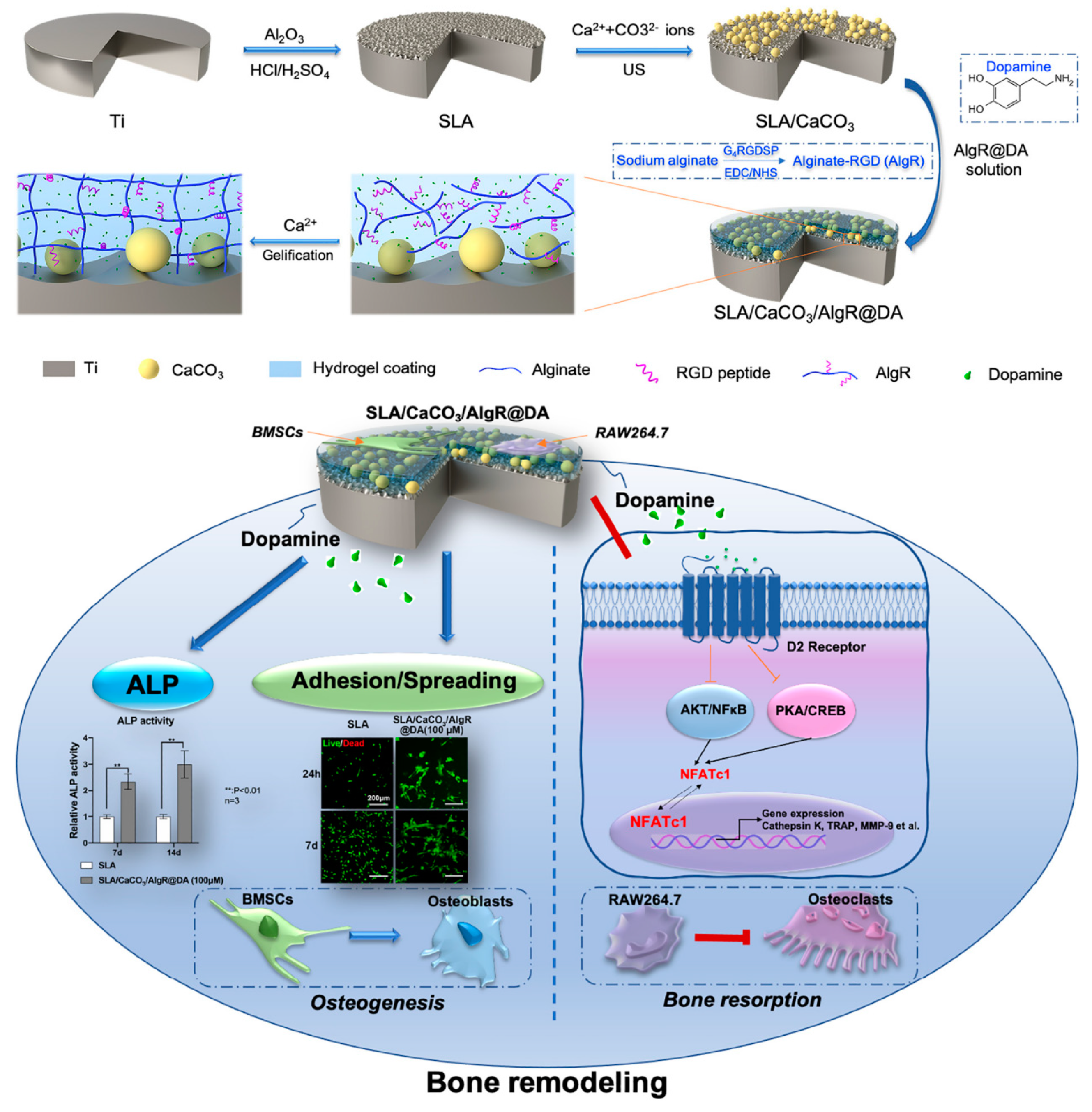
Hydrogel is a three-dimensional network cross-linked structure, which can not only simulate the extracellular matrix environment and develop bio-mimetic implants to design and repair bone defects [129], but also serve as drug carrier to carry and slowly release various active substances to promote bone-formation. Poloxamer-407 hydrogel loaded with simvastatin induces endogenous osteogenic growth factors and promotes bone ingrowth [130]. Pluronic F-127 hydrogel controls the release of 1α,25-Dihydroxyvitamin D3 as a bio-cap [131]. A non-toxic click hydrogel that rapidly polymerizes in situ provides localized controlled delivery of osteoprotective factor Semaphorin 3A [132]. Hydrogel containing BMP-2 facilitates dimensionally stable bone regeneration [133]. Dopamine-loaded RGD coatings on a vaterite-modified titanium surface successfully provided a solution to bone remodeling imbalance in osteoporosis by promoting osteoblasts and inhibiting osteoclasts at different concentrations [88] (Figure 8). Titanium implants loaded with human bone marrow mesenchymal stem cells (hBMSCs) show superior tissue ingrowth, and the synergic action of the bioactive hydrogel and hBMSCs increases both the bone deposition and integration [134]. In addition to these active ingredients, some inorganic substances such as a tri-calcium phosphate- [135], HA- [136], and silica-nanoparticle-loaded [137] hybrid hydrogels also improve the osteogenic ability of titanium implants, especially in combination with BMP-2 [138] or osteoblasts [139].
5.2. Angiogenesis
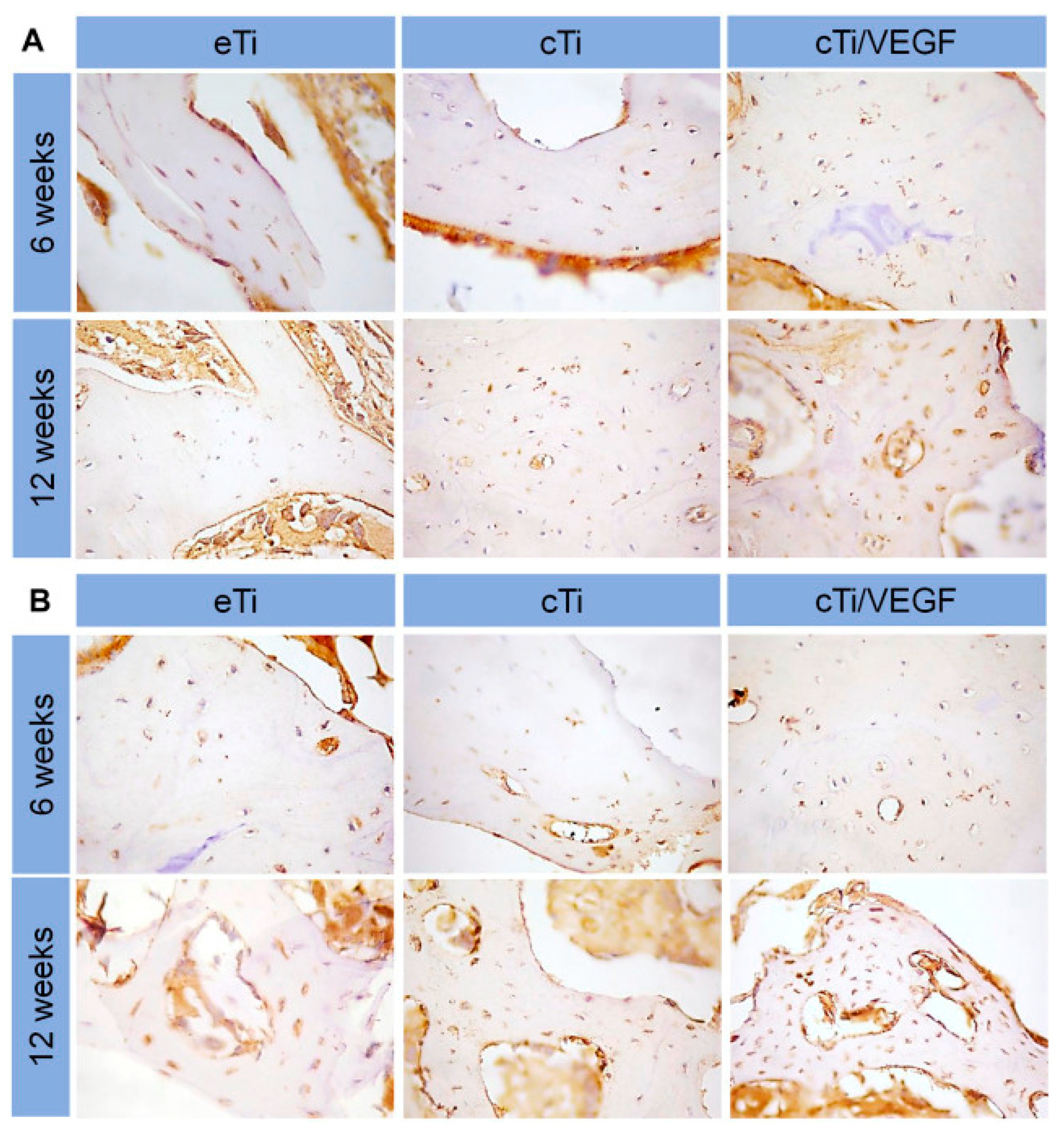
Adequate blood supply plays an indispensable role in promoting bone regeneration, and angiogenesis promotion has become one of the key factors for the success of titanium implants. Hydrogels can act as carriers for drugs, growth factors, and cells, to promote angiogenesis around titanium implants. The combination of simvastatin-loaded hydrogel coating with porous titanium alloy significantly improved the formation of new blood vessels around rabbit tibial implants, providing an effective strategy for bone integration and bone growth [140]. The heat-sensitive collagen hydrogel/porous titanium alloy scaffold system equipped with VEGF, increased vascular permeability, promoted proliferation and induction of HUVECs, and aided in angiogenic-mediated bone regeneration [44] (Figure 9). The composite scaffold loaded with VEGF and BMP continuously provided angiogenic and osteogenic growth factors at the site of osseous defect, thus exhibiting higher bone integration capacity and new bone amount [46,141]. Combining cell-laden hydrogels with porous titanium alloys develops a vascularized bone implant. Co-encapsulating hMSCs with HUVECs [52] or endothelial progenitor cells (EPCs) [142], support HUVEC- and EPC-spreading and vascular-like network formation, along with osteogenesis of hMSCs.
5.3. Macrophage Polarization
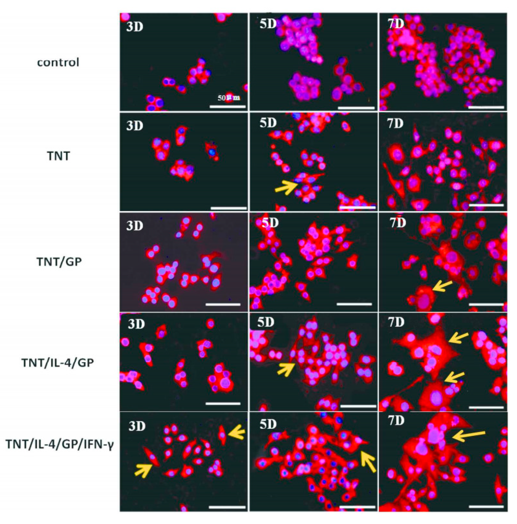
Macrophage polarization is a reversible and modified dynamic process involving in the occurrence, development and outcome of many immune inflammatory diseases, including peri-implantitis. The introduction of hydrogel for “reprogramming” of the macrophage state is a novel strategy to induce resolution of inflammation [143]. Interleukin-4 (IL-4) is a common inflammatory factor, which can regulate the antigen-presenting ability of macrophages, inhibit the secretion of inflammatory factors such as IL-1 β and TNF- α, and promote the differentiation of macrophages into profibrotic macrophages to secrete TGF-β. IL-4-loading of a hydrogel system on titanium modulated pro-inflammatory reactions [110]. Hydrogels containing interferon-γ and IL-4 were able to modulate the transformation with a stronger effect than those containing only IL-4 [144] (Figure 10). Combination of IL-4 and cell adhesive motif (RGD) onto the Ti substrate synergistically generated a more favorable early-stage osteo-immune environment with superior osteogenic properties [145]. Dexamethasone, as a glucocorticoid, can also regulate macrophage polarization and plays an important role in the regression of inflammation. The novel DNA hydrogel on the titanium surface, as the platform for dexamethasone delivery, extends the half- life of the release profile [146]. Reactive oxygen species (ROS) produced by macrophages regulate a variety of physiological functions including endothelial cells growth, migration, and mesenchymal stem cells activation. Removing excessive ROS by a two-component hydrogel coating containing borate ester bond and thymosin β4 favors M1 to M2 phenotype switch of macrophages and inflammatory response regulation [147].
5.4. Antibacterial
Bacterial biofilm formation can cause implant infection and osseointegration loss, resulting in loosening and dropping. Hydrogels with good biocompatibility and drug loading capability can slowly release various antibacterial components to prevent initial bacterial adhesion [13]. Designing and constructing a hydrogel drug-controlled release system by loading with antibacterial drugs such as gentamicin [104,108,148] or vancomycin [98,149,150] on a titanium surface is a frequently used strategy. Antibacterial peptides have garnered more attention as alternative antibacterial agents of implant coating due to their unique antibacterial mechanism [151,152]. Bacteriophage-loaded hydrogels also showed excellent antimicrobial activity in inhibiting attachment and colonization of multidrug-resistant E. faecalis surrounding and within femoral tissues [153]. Metal antibacterial agents are introduced into the implant hydrogel coating because of their broad-spectrum antibacterial properties and no drug resistance. Among them, silver ion is most commonly used [26,78,154,155]. Metal oxide antimicrobial agents such as zinc oxide [109] and calcium oxide [110] also show significant antibacterial ability in the coating, although Zn ion has renal absorption toxicity. Photodynamic therapy is a promising modality in antibacterial material design. The introduction of photosensitizer Chlorin e6 with laser-triggered ROS generation property exhibited a remarkable and rapid antibacterial activity when the laser power was 1 W cm−2 [50]. Coatings with semiconductor photocatalytic materials, such as bismuth [51] and red phosphorus [156], can produce ROS, kill bacteria and eradicate biofilm under light, which might provide a novel multimodal antibacterial and anti-biofilm treatment for infection.
5.5. Drug Delivery
Hydrogels have been widely used in various fields of medicine as vehicles to control the continuous release of drugs [157]. Loading cefuroxime, tetracycline, amoxicillin, or acetylsalicylic acid through hydrogel coating can improve the anti-infection effect of the implant [158]. Loading bone-metabolism-related drugs, proteins, peptides, and growth factors has demonstrated better osseointegration, especially in challenged degenerative conditions, such as osteoporosis, osteoarthritis, and osteogenesis imperfecta [159]. Similarly, hydrogels can load with cytokines to promote macrophage polarization [160] and angiogenesis [141], which have great potential for application in bone-tissue regeneration and repair.
6. Conclusions and Future Protects
In recent decades, metallic materials have been widely used in the field of biomaterials for their good mechanical properties and biocompatibility. Among them, the application prospects of biomedical titanium alloy are particularly remarkable. However, some disadvantages of titanium alloys limit their further application. Therefore, scientists have been working to explore improvements in the properties of titanium alloys. Hydrogel coatings can serve as ideal carriers to introduce drugs, peptides, metal ions, growth factors, and cells to effectively bio-modify titanium alloys. In this study, we have reviewed the popular matrix of hydrogel coatings, especially the natural materials such as collagen, gelatin, chitosan, and alginate. The usual modification methods are the electrochemical method, the sol–gel method, and layer-by-layer self-assembly. Hydrogel coatings significantly improve the properties of the titanium implant in osseointegration, angiogenesis, macrophage polarization, antibacterial effects, and drug delivery.
Although the improvement of titanium alloy caused by hydrogel coating is obvious, there are still some problems worth noting: (1) The mechanical properties of hydrogel coating are poor, and whether some inorganic fillers can be added to promote bone integration and mechanical properties needs further research. (2) The strong bond between the hydrogel coating and titanium alloy needs to be further strengthened. (3) Some semiconductor materials have excellent photocatalytic properties, and the introduction of semiconductor materials in hydrogel coatings is promising for photodynamic therapy. Therefore, future research should focus on these aspects to further improve the properties of hydrogel coating on titanium alloy.
Author Contributions
Conceptualization, B.H., Y.L., R.F. and H.C.; formal analysis, X.Q.; data curation, T.X.; writing—original draft preparation, R.F., H.C., Q.L. and Z.W.; writing—review and editing, B.H.; visualization. All authors have read and agreed to the published version of the manuscript.
Funding
This work was supported by the Key R&D Plan of Hubei Provincial Department of Science and Technology (Major Health Project, No. 2022BCE026), the Hubei Provincial Colleges and Universities Outstanding Young and Middle-aged Technological Innovation Team Project (No. T2020022), the Xianning City Key Program of Science & Technology (No. 2021GXYF021), the Special Fund for medical research of Hubei University of Science and Technology (2022YKY14), the Special fund of the School of Stomatology and Ophthalmology of Hubei University of Science and Technology (2020XZ32, 2020WG01), the Science Development Foundation of Hubei University of Science & Technology (No. 2020TD01, 2021ZX01, 2022FH09), and the Provincial College Students Innovation Training Program (S202210927030, S202210927031).
Institutional Review Board Statement
Not applicable.
Informed Consent Statement
Not applicable.
Data Availability Statement
Not applicable.
Conflicts of Interest
The authors declare no conflict of interest.
References
- Bothe, R.T.; Beaton, L.E.; Davenport, H.A. Reaction of bone to multiple metallic implants. Surg. Gynecol. Obstet. 1940, 71, 598–602. [Google Scholar]
- Bordbar-Khiabani, A.; Gasik, M. Electrochemical and biological characterization of Ti-Nb-Zr-Si alloy for orthopedic applications. Sci. Rep. 2023, 13, 2312. [Google Scholar] [CrossRef] [PubMed]
- Alipal, J.; Mohd Pu’ad, N.A.S.; Nayan, N.H.M. An updated review on surface functionalisation of titanium and its alloys for implants applications. Mater. Today Proc. 2021, 42, 270–282. [Google Scholar] [CrossRef]
- Del Castillo, R.; Chochlidakis, K.; Galindo-Moreno, P.; Ercoli, C. Titanium Nitride Coated Implant Abutments: From Technical Aspects and Soft tissue Biocompatibility to Clinical Applications. A Literature Review. J. Prosthodont. 2022, 31, 571–578. [Google Scholar] [CrossRef]
- Prabhakar, V.; Chidambaranathan, A.S.; Balasubramanium, M. Effect of Cathodic Arc Plasma Deposition on Shear Bond Strength between Palladium Cobalt Chromium Coated with Titanium Nitride and Titanium Aluminium Nitride with Ceramic. Contemp. Clin. Dent. 2021, 12, 49–54. [Google Scholar] [PubMed]
- Zhang, R.; Wan, Y.; Ai, X.; Zhang, D. Corrosion resistance and biological activity of TiO2 implant coatings produced in oxygen-rich environments. Proc. Inst. Mech. Eng. Part H J. Eng. Med. 2017, 231, 20–27. [Google Scholar] [CrossRef] [PubMed]
- Zhao, X.; You, L.; Wang, T.; Li, B. Enhanced Osseointegration of Titanium Implants by Surface Modification with Silicon-doped Titania Nanotubes. Int. J. Nanomed. 2020, 15, 8583–8594. [Google Scholar] [CrossRef]
- Shimabukuro, M. Antibacterial Property and Biocompatibility of Silver, Copper, and Zinc in Titanium Dioxide Layers Incorporated by One-Step Micro-Arc Oxidation: A Review. Antibiotics 2020, 9, 716. [Google Scholar] [CrossRef] [PubMed]
- Alshimaysawee, S.; Fadhel Obaid, R.; Al-Gazally, M.E.; Bathaei, M.S. Recent Advancements in Metallic Drug-Eluting Implants. Pharmaceutics 2023, 15, 223. [Google Scholar] [CrossRef]
- Yuan, Y.X.; Luo, R.D.; Ren, J.K.; He, Z.Y. Design of a new Ti-Mo-Cu alloy with excellent mechanical and antibacterial properties as implant materials. Mater. Lett. 2022, 306, 130875. [Google Scholar] [CrossRef]
- Lupi, S.M.; Torchia, M.; Rizzo, S. Biochemical Modification of Titanium Oral Implants: Evidence from in Vivo Studies. Materials 2021, 14, 2798. [Google Scholar] [CrossRef] [PubMed]
- Zheng, Q.C.; Mao, L.L.; Shi, Y.T.; Hu, Y.H. Biocompatibility of Ti-6Al-4V titanium alloy implants with laser microgrooved surfaces. Mater. Technol. 2022, 37, 2039–2048. [Google Scholar] [CrossRef]
- Bohara, S.; Suthakorn, J. Surface coating of orthopedic implant to enhance the osseointegration and reduction of bacterial colonization: A review. Biomater. Res. 2022, 26, 26. [Google Scholar] [CrossRef] [PubMed]
- De Giglio, E.; Cometa, S.; Cioffi, N.; Sabbatini, L. Analytical investigations of poly (acrylic acid) coatings electrodeposited on titanium-based implants: A versatile approach to biocompatibility enhancement. Anal. Bioanal. Chem. 2007, 389, 2055–2063. [Google Scholar] [CrossRef]
- Nagai, M.; Hayakawa, T.; Fukatsu, A.; Kato, T. In vitro study of collagen coating of titanium implants for initial cell attachment. Dent. Mater. J. 2002, 21, 250–260. [Google Scholar] [CrossRef]
- Hauser, J.; Ring, A.; Schaffran, A.; Langer, S. In vivo analysis of tissue response to plasma-treated collagen-I-coated titanium alloys. Eur. Surg. Res. 2009, 43, 262–268. [Google Scholar] [CrossRef]
- Raita, Y.; Komatsu, K.; Hayakawa, T. Pilot study of gingival connective tissue responses to 3-dimensional collagen nanofiber-coated dental implants. Dent. Mater. J. 2015, 34, 847–854. [Google Scholar] [CrossRef] [PubMed]
- Vanderleyden, E.; Van Bael, S.; Chai, Y.C.; Dubruel, P. Gelatin functionalised porous titanium alloy implants for orthopaedic applications. Mat. Sci. Eng. C Mater. 2014, 42, 396–404. [Google Scholar] [CrossRef]
- Mahin, T.; Torab, A.; Negahdari, R.; Sharifi, S. The Antibacterial Effects of Healing Abutments Coated with Gelatin-curcumin Nanocomposite. Pharm. Nanotechnol. 2023. [Google Scholar] [CrossRef]
- López-Valverde, N.; Aragoneses, J.; López-Valverde, A.; Aragoneses, J.M. Role of chitosan in titanium coatings. trends and new generations of coatings. Front. Bioeng. Biotechnol. 2022, 10, 907589. [Google Scholar] [CrossRef]
- Bumgardner, J.D.; Chesnutt, B.M.; Yuan, Y.; Ong, J.L. The integration of chitosan-coated titanium in bone: An in vivo study in rabbits. Implant 2007, 16, 66–79. [Google Scholar] [CrossRef] [PubMed]
- Peng, Z.X.; Ao, H.Y.; Wang, L.; Tang, T.T. Quaternised chitosan coating on titanium provides a self-protective surface that prevents bacterial colonisation and implant-associated infections. RSC Adv. 2015, 5, 54304–54311. [Google Scholar] [CrossRef]
- Jian, Y.H.; Yang, C.; Zhang, J.X.; Du, Y.M. One-step electrodeposition of Janus chitosan coating for metallic implants with anti-corrosion properties. Colloid Surf. 2022, 641, 128498. [Google Scholar] [CrossRef]
- Guo, C.C.; Cui, W.D.; Wang, X.W.; Chen, J.L. Poly-L-lysine/Sodium Alginate Coating Loading Nanosilver for Improving the Antibacterial Effect and Inducing Mineralization of Dental Implants. ACS Omega 2020, 5, 10562–10571. [Google Scholar] [CrossRef] [PubMed]
- Yuan, N.; Jia, L.; Geng, Z.; Liu, Y. The Incorporation of Strontium in a Sodium Alginate Coating on Titanium Surfaces for Improved Biological Properties. Biomed. Res. Int. 2017, 2017, 9867819. [Google Scholar] [CrossRef] [PubMed]
- Abdul Azam, F.A.; Ismail, H.; Abdul Hamid, M.A. Characterizations on Morphology and Adhesion of Calcium Silicate Coating on Ti6Al4V Substrate. Key Eng. Mater. 2016, 694, 83–87. [Google Scholar] [CrossRef]
- Sille, I.E.; Pissinis, D.E.; Fagali, N.S.; Schilardi, P.L. Antimicrobial-Loaded Polyacrylamide Hydrogels Supported on Titanium as Reservoir for Local Drug Delivery. Pathogens 2023, 12, 202. [Google Scholar] [CrossRef]
- Buxadera-Palomero, J.; Calvo, C.; Torrent-Camarero, S.; Rodríguez, D. Biofunctional polyethylene glycol coatings on titanium: An in vitro-based comparison of functionalization methods. Colloid Surf. 2017, 152, 367–375. [Google Scholar] [CrossRef]
- Xiao, D.Q.; Liu, Q.; Weng, J. Room-temperature attachment of PLGA microspheres to titanium surfaces for implant-based drug release. Appl. Surf. Sci. 2014, 30, 112–118. [Google Scholar] [CrossRef]
- Iafiscol, M.; Quirici, N.; Foltran, I.; Rimondini, L. Electrospun collagen mimicking the reconstituted extracellular matrix improves osteoblastic differentiation onto titanium surfaces. J. Nanosci. Nanotechnol. 2013, 13, 4720–4726. [Google Scholar] [CrossRef]
- Becker, D.; Geissler, U.; Hempel, U.; Bierbaum, S.; Scharnweber, D.; Worch, H.; Wenzel, K.W. Proliferation and differentiation of rat calvarial osteoblasts on type I collagen-coated titanium alloy. J. Biomed. Mater. Res. 2002, 59, 516–527. [Google Scholar] [CrossRef] [PubMed]
- Bierbaum, S.; Hempel, U.; Geissler UHanke, T.; Scharnweber, D.; Wenzel, K.W.; Worch, H. Modification of Ti6Al4V surfaces using collagen i, iii, and fibronectin. Ii. Influence on osteoblast responses. J. Biomed. Mater. Res. A 2003, 67, 431–438. [Google Scholar] [CrossRef] [PubMed]
- Geissler, U.; Hempel, U.; Wolf, C.; Scharnweber, D.; Worch, H.; Wenzel, K. Collagen type I-coating of Ti6Al4V promotes adhesion of osteoblasts. J. Biomed. Mater. Res. 2000, 51, 752–760. [Google Scholar] [CrossRef]
- Ritz, U.; Nusselt, T.; Sewing, A.; Hofmann, A. The effect of different collagen modifications for titanium and titanium nitrite surfaces on functions of gingival fibroblasts. Clin. Oral. Investig. 2017, 21, 255–265. [Google Scholar] [CrossRef] [PubMed]
- Sharan, J.; Koul, V.; Dinda, A.K.; Singh, M.P. Bio-functionalization of grade V titanium alloy with type I human collagen for enhancing and promoting human periodontal fibroblast cell adhesion—An in-vitro study. Colloid Surf. 2018, 161, 1–9. [Google Scholar] [CrossRef]
- Morra, M.; Cassinelli, C.; Meda, L.; Giardino, R. Surface analysis and effects on interfacial bone microhardness of collagen-coated titanium implants: A rabbit model. Int. J. Oral Maxillofac. Implant. 2005, 20, 23–30. [Google Scholar]
- Ciobanu, G.; Ciobanu, O. Investigation on the effect of collagen and vitamins on biomimetic hydroxyapatite coating formation on titanium surfaces. Mat. Sci. Eng. C Mater. 2013, 33, 1683–1688. [Google Scholar] [CrossRef]
- Ruiz, G.C.M.; Cruz, M.A.E.; Faria, A.N.; Ramos, A.P. Biomimetic collagen/phospholipid coatings improve formation of hydroxyapatite nanoparticles on titanium. Mat. Sci. Eng. C Mater. 2017, 77, 102–110. [Google Scholar] [CrossRef]
- Patty, D.J.; Nugraheni, A.D.; Dewi Ana, I.; Yusuf, Y. Mechanical Characteristics and Bioactivity of Nanocomposite Hydroxyapatite/Collagen Coated Titanium for Bone Tissue Engineering. Bioengineering 2022, 9, 784. [Google Scholar] [CrossRef]
- Pokorný, M.; Suchý, T.; Kotzianová, A.; Čejka, Z. Surface Treatment of Acetabular Cups with a Direct Deposition of a Composite Nanostructured Layer Using a High Electrostatic Field. Molecules 2020, 25, 1173. [Google Scholar] [CrossRef]
- Iwanami-Kadowaki, K.; Uchikoshi, T.; Uezono, M.; Moriyama, K. Development of novel bone-like nanocomposite coating of hydroxyapatite/collagen on titanium by modified electrophoretic deposition. J. Biomed. Mater. Res. A 2021, 109, 1905–1911. [Google Scholar] [CrossRef] [PubMed]
- Zhao, Y.; Bai, L.; Zhang, Y.; Hang, R. Type I collagen decorated nanoporous network on titanium implant surface promotes osseointegration through mediating immunomodulation, angiogenesis, and osteogenesis. Biomaterials 2022, 288, 121684. [Google Scholar] [CrossRef] [PubMed]
- Shao, J.; Weng, L.; Li, J.; Lin, J. Regulation of Macrophage Polarization by Mineralized Collagen Coating to Accelerate the Osteogenic Differentiation of Mesenchymal Stem Cells. ACS Biomater. Sci. Eng. 2022, 8, 610–619. [Google Scholar] [CrossRef] [PubMed]
- Li, Y.; Liu, Y.; Bai, H.; Huang, L. Sustained Release of VEGF to Promote Angiogenesis and Osteointegration of Three-Dimensional Printed Biomimetic Titanium Alloy Implants. Front. Bioeng. Biotechnol. 2021, 9, 757767. [Google Scholar] [CrossRef] [PubMed]
- Dinh, T.N.; Hou, S.; Park, S.; Jeong, K.J. Gelatin Hydrogel Combined with Polydopamine Coating to Enhance Tissue Integration of Medical Implants. ACS Biomater. Sci. Eng. 2018, 4, 3471–3477. [Google Scholar] [CrossRef] [PubMed]
- Liu, Z.; Xu, Z.; Wang, X.; Jia, R. Construction and osteogenic effects of 3D-printed porous titanium alloy loaded with VEGF/BMP-2 shell-core microspheres in a sustained-release system. Front. Bioeng. Biotechnol. 2022, 10, 1028278. [Google Scholar] [CrossRef] [PubMed]
- Van den Bulcke, A.I.; Bogdanov, B.; de Rooze, N.; Schacht, E.H.; Cornelissen, M.; Berghmans, H. Structural and rheological properties of methaerylamide modified gelatin hydrogels. Biomacromolecules 2000, 1, 31–38. [Google Scholar] [CrossRef]
- Cheng, H.; Yue, K.; Kazemzadeh-Narbat, M.; Khademhosseini, A. Mussel-Inspired Multifunctional Hydrogel Coating for Prevention of Infections and Enhanced Osteogenesis. ACS Appl. Mater. Interfaces 2017, 9, 11428–11439. [Google Scholar] [CrossRef] [PubMed]
- Kim, S.Y.; Choi, A.J.; Park, J.E.; Lee, M.H. Antibacterial Activity and Biocompatibility with the Concentration of Ginger Fraction in Biodegradable Gelatin Methacryloyl (GelMA) Hydrogel Coating for Medical Implants. Polymers 2022, 14, 5317. [Google Scholar] [CrossRef] [PubMed]
- He, Y.; Leng, J.; Li, K.; Cai, K. A multifunctional hydrogel coating to direct fibroblast activation and infected wound healing via simultaneously controllable photobiomodulation and photodynamic therapies. Biomaterials 2021, 278, 121164. [Google Scholar] [CrossRef]
- Ding, Y.; Ma, R.; Liu, G.; Cai, K. Fabrication of a New Hyaluronic Acid/Gelatin Nanocomposite Hydrogel Coating on Titanium-Based Implants for Treating Biofilm Infection and Excessive Inflammatory Response. ACS Appl. Mater. Interfaces 2023, 15, 13783–13801. [Google Scholar] [CrossRef] [PubMed]
- Li, J.; Cui, X.; Lindberg, G.C.J.; Woodfield, T.B.F. Hybrid fabrication of photo-clickable vascular hydrogels with additive manufactured titanium implants for enhanced osseointegration and vascularized bone formation. Biofabrication 2022, 14, 034103. [Google Scholar] [CrossRef]
- Taokaew, S.; Kaewkong, W.; Kriangkrai, W. Recent Development of Functional Chitosan-Based Hydrogels for Pharmaceutical and Biomedical Applications. Gels 2023, 9, 277. [Google Scholar] [CrossRef]
- Stevanović, M.; Đošić, M.; Janković, A.; Mišković-Stanković, V. Gentamicin-Loaded Bioactive Hydroxyapatite/Chitosan Composite Coating Electrodeposited on Titanium. ACS Biomater. Sci. Eng. 2018, 4, 3994–4007. [Google Scholar] [CrossRef] [PubMed]
- Zarghami, V.; Ghorbani, M.; Bagheri, K.P.; Shokrgozar, M.A. Improving bactericidal performance of implant composite coatings by synergism between Melittin and tetracycline. J. Mater. Sci. Mater. Med. 2022, 33, 46–58. [Google Scholar] [CrossRef] [PubMed]
- Asadi, S.; Mortezagholi, B.; Hadizadeh, A.; Chaiyasut, C. Ciprofloxacin-Loaded Titanium Nanotubes Coated with Chitosan: A Promising Formulation with Sustained Release and Enhanced Antibacterial Properties. Pharmaceutics 2022, 14, 1359. [Google Scholar] [CrossRef] [PubMed]
- Divakar, D.D.; Jastaniyah, N.T.; Altamimi, H.G.; Haleem, S. Enhanced antimicrobial activity of naturally derived bioactive molecule chitosan conjugated silver nanoparticle against dental implant pathogens. Int. J. Biol. Macromol. 2018, 108, 790–797. [Google Scholar] [CrossRef] [PubMed]
- Pawłowski, Ł.; Wawrzyniak, J.; Banach-Kopeć, A.; Zieliński, A. Antibacterial properties of laser-encapsulated titanium oxide nanotubes decorated with nanosilver and covered with chitosan/Eudragit polymers. Biomater. Adv. 2022, 138, 212950. [Google Scholar] [CrossRef]
- Zhang, T.; Qin, X.; Gao, Y.; Yin, P. Functional chitosan gel coating enhances antimicrobial properties and osteogenesis of titanium alloy under persistent chronic inflammation. Front. Bioeng. Biotechnol. 2023, 11, 1118487. [Google Scholar] [CrossRef] [PubMed]
- Han, J.; Hassani Besheli, N.; Deng, D.; Yang, F. Tailoring Copper-Doped Bioactive Glass/Chitosan Coatings with Angiogenic and Antibacterial Properties. Tissue Eng. Part C Methods 2022, 28, 314–324. [Google Scholar] [CrossRef]
- Stevanović, M.; Djošić, M.; Janković, A.; Mišković-Stanković, V. Antibacterial graphene-based hydroxyapatite/chitosan coating with gentamicin for potential applications in bone tissue engineering. J. Biomed. Mater. Res. A 2020, 108, 2175–2189. [Google Scholar] [CrossRef]
- Zhu, M.; Liu, X.; Tan, L.; Wu, S. Photo-responsive chitosan/Ag/MoS2 for rapid bacteria-killing. J. Hazard. Mater. 2020, 383, 121122. [Google Scholar] [CrossRef]
- Bose, S.; Surendhiran, D.; Chun, B.S.; Kang, H.W. Facile synthesis of black phosphorus-zinc oxide nanohybrids for antibacterial coating of titanium surface. Colloid Surf. 2022, 219, 112807. [Google Scholar] [CrossRef]
- Chai, M.; An, M.; Zhang, X. Construction of a TiO2/MoSe2/CHI coating on dental implants for combating Streptococcus mutans infection. Mat. Sci. Eng. C Mater. 2021, 129, 112416. [Google Scholar] [CrossRef]
- Chen, W.; Xie, G.; Lu, Y.; Bao, J. An improved osseointegration of metal implants by pitavastatin loaded multilayer films with osteogenic and angiogenic properties. Biomaterials 2022, 280, 121260. [Google Scholar] [CrossRef]
- Wang, B.; Chen, L.; Xie, J.; Yang, L. Coating Polyelectrolyte Multilayers Loaded with Quercetin on Titanium Surfaces by Layer-By-Layer Assembly Technique to Improve Surface Osteogenesis Under Osteoporotic Condition. J. Biomed. Nanotechnol. 2021, 17, 1392–1403. [Google Scholar] [CrossRef] [PubMed]
- Takanche, J.S.; Kim, J.E.; Jang, S.; Yi, H.K. Insulin growth factor binding protein-3 enhances dental implant osseointegration against methylglyoxal-induced bone deterioration in a rat model. J. Periodontal. Implant Sci. 2022, 52, 155–169. [Google Scholar] [CrossRef]
- Tao, B.; Deng, Y.; Song, L.; Cai, K. BMP2-loaded titania nanotubes coating with pH-responsive multilayers for bacterial infections inhibition and osteogenic activity improvement. Colloid Surf. 2019, 177, 242–252. [Google Scholar] [CrossRef] [PubMed]
- Zhang, T.; Zhang, X.; Mao, M.; Sun, H. Chitosan/hydroxyapatite composite coatings on porous Ti6Al4V titanium implants: In vitro and in vivo studies. J. Periodontal. Implant Sci. 2020, 50, 392–405. [Google Scholar] [CrossRef] [PubMed]
- Gaafar, M.S.; Yakout, S.M.; Barakat, Y.F.; Sharmoukh, W. Electrophoretic deposition of hydroxyapatite/chitosan nanocomposites: The effect of dispersing agents on the coating properties. RSC Adv. 2022, 12, 27564–27581. [Google Scholar] [CrossRef]
- Rastegari, S.; Salahinejad, E. Surface modification of Ti-6Al-4V alloy for osseointegration by alkaline treatment and chitosan-matrix glass-reinforced nanocomposite coating. Carbohyd. Polym. 2019, 205, 302–311. [Google Scholar] [CrossRef] [PubMed]
- Ma, K.; Cai, X.; Zhou, Y.; Jiang, T. In Vitro and In Vivo Evaluation of Tetracycline Loaded Chitosan-Gelatin Nanosphere Coatings for Titanium Surface Functionalization. Macromol. Biosci. 2017, 17, 201600130. [Google Scholar] [CrossRef] [PubMed]
- Zhao, D.; Dong, H.; Niu, Y.; Zhang, Z. Electrophoretic deposition of novel semi-permeable coatings on 3D-printed Ti-Nb alloy meshes for guided alveolar bone regeneration. Dent. Mater. 2022, 38, 431–443. [Google Scholar] [CrossRef] [PubMed]
- Tang, J.; Chen, L.; Yan, D.; Shen, L. Surface Functionalization with Proanthocyanidins Provides an Anti-Oxidant Defense Mechanism That Improves the Long-Term Stability and Osteogenesis of Titanium Implants. Int. J. Nanomed. 2020, 15, 1643–1659. [Google Scholar] [CrossRef]
- Vakili, N.; Asefnejad, A. Titanium coating: Introducing an antibacterial and bioactive chitosan-alginate film on titanium by spin coating. Biomed. Eng. Biomed. Tech. 2020, 65, 621–630. [Google Scholar] [CrossRef] [PubMed]
- Jabłoński, P.; Kyzioł, A.; Pawcenis, D.; Kyzioł, K. Electrostatic self-assembly approach in the deposition of bio-functional chitosan-based layers enriched with caffeic acid on Ti-6Al-7Nb alloys by alternate immersion. Biomater. Adv. 2022, 136, 212791. [Google Scholar] [CrossRef] [PubMed]
- Rahnamaee, S.Y.; Dehnavi, S.M.; Bagheri, R.; Karimi, A. Boosting bone cell growth using nanofibrous carboxymethylated cellulose and chitosan on titanium dioxide nanotube array with dual surface charges as a novel multifunctional bioimplant surface. Int. J. Biol. Macromol. 2023, 228, 570–581. [Google Scholar] [CrossRef]
- Ren, Y.; Qin, X.; Barbeck, M.; Liu, C. Mussel-Inspired Carboxymethyl Chitosan Hydrogel Coating of Titanium Alloy with Antibacterial and Bioactive Properties. Materials 2021, 14, 6901. [Google Scholar] [CrossRef]
- Abasalizadeh, F.; Moghaddam, S.V.; Alizadeh, E.; Akbarzadeh, A. Alginate-based hydrogels as drug delivery vehicles in cancer treatment and their applications in wound dressing and 3D bioprinting. J. Biol. Eng. 2020, 14, 8. [Google Scholar] [CrossRef]
- Vrana, N.E.; Dupret-Bories, A.; Bach, C.; Lavalle, P. Modification of macroporous titanium tracheal implants with biodegradable structures: Tracking in vivo integration for determination of optimal in situ epithelialization conditions. Biotechnol. Bioeng. 2012, 109, 2134–2146. [Google Scholar] [CrossRef]
- Gregurec, D.; Wang, G.; Pires, R.H.; Moya, S.E. Bioinspired titanium coatings: Self-assembly of collagen-alginate films for enhanced osseointegration. J. Mater. Chem. 2016, 4, 1978–1986. [Google Scholar] [CrossRef] [PubMed]
- Shaygani, H.; Seifi, S.; Shamloo, A.; Ebrahimi, S. Novel bilayer coating on gentamicin-loaded titanium nanotube for orthopedic implants applications. Int. J. Pharm. 2023, 636, 122764. [Google Scholar] [CrossRef] [PubMed]
- Duan, Y.; Wu, Y.; Yan, R.; Ma, H. Chitosan-sodium alginate-based coatings for self-strengthening anticorrosion and antibacterial protection of titanium substrate in artificial saliva. Int. J. Biol. Macromol. 2021, 184, 109–117. [Google Scholar] [CrossRef] [PubMed]
- Yuan, Z.; Liu, P.; Hao, Y.; Cai, K. Construction of Ag-incorporated coating on Ti substrates for inhibited bacterial growth and enhanced osteoblast response. Colloid Surf. 2018, 171, 597–605. [Google Scholar] [CrossRef] [PubMed]
- Lian, Q.; Zheng, S.; Shi, Z.; Cheng, H. Using a degradable three-layer sandwich-type coating to prevent titanium implant infection with the combined efficient bactericidal ability and fast immune remodeling property. Acta Biomater. 2022, 154, 650–666. [Google Scholar] [CrossRef]
- Perni, S.; Alotaibi, H.F.; Yergeshov, A.A.; Prokopovich, P. Long acting anti-infection constructs on titanium. J. Control. Release 2020, 326, 91–105. [Google Scholar] [CrossRef] [PubMed]
- Chen, T.; Wang, S.; He, F.; Zou, S. Promotion of Osseointegration Using Protamine/Alginate/Bone Morphogenic Protein 2 Biofunctionalized Composite Coating on Nanopolymorphic Titanium Surfaces. J. Biomed. Nanotechnol. 2018, 14, 933–945. [Google Scholar] [CrossRef]
- Wang, M.; Wang, C.; Zhang, Y.; Lin, Y. Controlled release of dopamine coatings on titanium bidirectionally regulate osteoclastic and osteogenic response behaviors. Mat. Sci. Eng. C Mater. 2021, 129, 112376. [Google Scholar] [CrossRef]
- Chen, T.; Brial, C.; McCarthy, M.; Maher, S.A. Synthetic PVA Osteochondral Implants for the Knee Joint: Mechanical Characteristics during Simulated Gait. Am. J. Sport. Med. 2021, 49, 2933–2941. [Google Scholar] [CrossRef]
- Chen, K.; Liu, S.Y.; Wu, X.F.; Zhang, D.K. Mussel-inspired construction of Ti6Al4V-hydrogel artificial cartilage material with high strength and low friction. Mater. Lett. 2020, 265, 127421. [Google Scholar] [CrossRef]
- Cui, L.L.; Chen, J.Y.; Yan, C.Q.; Xiong, D.S. Articular Cartilage Inspired the Construction of LTi-DA-PVA Composite Structure with Excellent Surface Wettability and Low Friction Performance. Tribol. Lett. 2021, 69, 41. [Google Scholar] [CrossRef]
- Awasthi, S.; Gaur, J.K.; Pandey, S.K.; Srivastava, C. High-Strength, Strongly Bonded Nanocomposite Hydrogels for Cartilage Repair. ACS Appl. Mater. Interfaces 2021, 13, 24505–24523. [Google Scholar] [CrossRef] [PubMed]
- Shi, Y.; Liu, J.; Li, J.L.; Xiong, D.S.; Dini, D. Improved mechanical and tribological properties of PAAm/PVA hydrogel-Ti6Al4V alloy configuration for cartilage repair. J. Polym. Res. 2022, 29, 515. [Google Scholar] [CrossRef]
- Deng, Y.L.; Sun, J.J.; Ni, X.Y.; Yu, B. Tribological properties of hierarchical structure artificial joints with poly acrylic acid (AA)-poly acrylamide (AAm) hydrogel and Ti6Al4V substrate. J. Polym. Res. 2020, 27, 157. [Google Scholar] [CrossRef]
- Chen, Y.H.; Cheng, W.H.; Teng, L.J.; Wang, Y.J. Graphene Oxide Hybrid Supramolecular Hydrogels with Self-Healable, Bioadhesive and Stimuli-Responsive Properties and Drug Delivery Application. Macromol. Mater. Eng. 2018, 303, 1700660. [Google Scholar] [CrossRef]
- Xupeng, G.; Fangyuan, Y.; Ming, C. Drug release kinetics of rifampicin from composite gel coating on surface of titanium alloy. Orthop. J. China 2018, 26, 649–654. [Google Scholar]
- Jing, Z.; Ni, R.; Wang, J.; Liu, Z. Practical strategy to construct anti-osteosarcoma bone substitutes by loading cisplatin into 3D-printed titanium alloy implants using a thermosensitive hydrogel. Bioact. Mater. 2021, 6, 4542–4557. [Google Scholar] [CrossRef] [PubMed]
- Wang, X.Y.; Fang, X.; Gao, X.; Qin, Y.G. Strong adhesive and drug-loaded hydrogels for enhancing bone-implant interface fixation and anti-infection properties. Colloid Surf. 2022, 219, 112817. [Google Scholar] [CrossRef] [PubMed]
- Huang, T.; Yu, Z.; Yu, Q.; Yang, G. Electrochemical deposition of lithium coating on titanium implant with enhanced early stage osseointegration. J. Biomed. Mater. Res. 2022, 110, 2399–2410. [Google Scholar] [CrossRef] [PubMed]
- Lu, M.; Chen, H.; Yuan, B.; Zhang, X. Electrochemical Deposition of Nanostructured Hydroxyapatite Coating on Titanium with Enhanced Early Stage Osteogenic Activity and Osseointegration. Int. J. Nanomed. 2020, 15, 6605–6618. [Google Scholar] [CrossRef] [PubMed]
- Wang, B.; Hua, J.; You, R.; Ma, L. Electrochemically deposition of catechol-chitosan hydrogel coating on coronary stent with robust copper ions immobilization capability and improved interfacial biological activity. Int. J. Biol. Macromol. 2021, 181, 435–443. [Google Scholar] [CrossRef] [PubMed]
- Croes, M.; Bakhshandeh, S.; van Hengel, I.A.J.; Amin Yavari, S. Antibacterial and immunogenic behavior of silver coatings on additively manufactured porous titanium. Acta Biomater. 2018, 81, 315–327. [Google Scholar] [CrossRef]
- Yin, X.; Yan, L.; Jun Hao, D.; Liu, Z. Calcium alginate template-mineral substituted hydroxyapatite hydrogel coated titanium implant for tibia bone regeneration. Int. J. Pharm. 2020, 582, 119303. [Google Scholar] [CrossRef]
- Soylu, H.M.; Chevallier, P.; Copes, F.; Mantovani, D. A Novel Strategy to Coat Dopamine-Functionalized Titanium Surfaces with Agarose-Based Hydrogels for the Controlled Release of Gentamicin. Front. Cell. Infect. Microbiol. 2021, 11, 678081. [Google Scholar] [CrossRef]
- Yao, X.; Liu, J.; Yang, C.; Suo, Z. Hydrogel Paint. Adv. Mater. 2019, 31, e1903062. [Google Scholar] [CrossRef] [PubMed]
- Tolle, C.; Riedel, J.; Mikolai, C.; Menzel, H. Biocompatible Coatings from Smart Biopolymer Nanoparticles for Enzymatically Induced Drug Release. Biomolecules 2018, 8, 103. [Google Scholar] [CrossRef] [PubMed]
- Tsikopoulos, K.; Bidossi, A.; Drago, L.; Papaioannidou, P. Is Implant Coating with Tyrosol- and Antibiotic-loaded Hydrogel Effective in Reducing Cutibacterium (Propionibacterium) acnes Biofilm Formation? A Preliminary in Vitro Study. Clin. Orthop. Relat. Res. 2019, 477, 1736–1746. [Google Scholar] [CrossRef]
- Wu, Y.; Hu, F.; Yang, X.; Zhang, X. Titanium surface polyethylene glycol hydrogel and gentamicin-loaded cross-linked starch microspheres release system for anti-infective drugs. J. Drug Target. 2023, 31, 217–224. [Google Scholar] [CrossRef] [PubMed]
- Leng, J.; He, Y.; Yuan, Z.; Cai, K. Enzymatically-degradable hydrogel coatings on titanium for bacterial infection inhibition and enhanced soft tissue compatibility via a self-adaptive strategy. Bioact. Mater. 2021, 6, 4670–4685. [Google Scholar] [CrossRef]
- He, Y.; Li, K.; Yang, X.; Cai, K. Calcium Peroxide Nanoparticles-Embedded Coatings on Anti-Inflammatory TiO2 Nanotubes for Bacteria Elimination and Inflammatory Environment Amelioration. Small 2021, 17, e2102907. [Google Scholar] [CrossRef] [PubMed]
- Stoetzel, S.; Malhan, D.; Wild, U.; El Khassawna, T. Osteocytes Influence on Bone Matrix Integrity Affects Biomechanical Competence at Bone-Implant Interface of Bioactive-Coated Titanium Implants in Rat Tibiae. Int. J. Mol. Sci. 2021, 23, 374. [Google Scholar] [CrossRef]
- Shi, Q.; Qian, Z.; Liu, D.; Liu, H. Surface Modification of Dental Titanium Implant by Layer-by-Layer Electrostatic Self-Assembly. Front. Physiol. 2017, 8, 574. [Google Scholar] [CrossRef]
- Wang, D.; Chen, M.W.; Wei, Y.J.; Cai, K.Y. Construction of Wogonin Nanoparticle-Containing Strontium-Doped Nanoporous Structure on Titanium Surface to Promote Osteoporosis Fracture Repair. Adv. Healthc. Mater. 2022, 11, e2201405. [Google Scholar] [CrossRef]
- Lv, H.; Chen, Z.; Yang, X.; Gao, P. Layer-by-layer self-assembly of minocycline-loaded chitosan/alginate multilayer on titanium substrates to inhibit biofilm formation. J. Dent. 2014, 42, 1464–1472. [Google Scholar] [CrossRef]
- Chen, M.; Huang, L.; Shen, X.; Hu, Y. Construction of multilayered molecular reservoirs on a titanium alloy implant for combinational drug delivery to promote osseointegration in osteoporotic conditions. Acta Biomater. 2020, 105, 304–318. [Google Scholar] [CrossRef]
- Zhong, X.; Song, Y.; Yang, P.; Li, C. Titanium Surface Priming with Phase-Transited Lysozyme to Establish a Silver Nanoparticle-Loaded Chitosan/Hyaluronic Acid Antibacterial Multilayer via Layer-by-Layer Self-Assembly. PLoS ONE 2016, 11, e0146957. [Google Scholar] [CrossRef] [PubMed]
- Song, Y.; Ma, A.; Ning, J.; Li, C. Loading icariin on titanium surfaces by phase-transited lysozyme priming and layer-by-layer self-assembly of hyaluronic acid/chitosan to improve surface osteogenesis ability. Int. J. Nanomed. 2018, 13, 6751–6767. [Google Scholar] [CrossRef]
- Han, M.; Dong, Z.; Li, J.; Li, J. Mussel-inspired self-assembly engineered implant coatings for synergistic anti-infection and osteogenesis acceleration. J. Mater. Chem. 2021, 9, 8501–8511. [Google Scholar] [CrossRef] [PubMed]
- Iwata, N.; Nozaki, K.; Horiuchi, N.; Nagai, A. Effects of controlled micro-/nanosurfaces on osteoblast proliferation. J. Biomed. Mater. Res. 2017, 105, 2589–2596. [Google Scholar] [CrossRef] [PubMed]
- Yu, Y.; Jin, G.; Xue, Y.; Sun, J. Multifunctions of dual Zn/Mg ion co-implanted titanium on osteogenesis, angiogenesis and bacteria inhibition for dental implants. Acta Biomater. 2017, 49, 590–603. [Google Scholar] [CrossRef] [PubMed]
- Lee, S.; Chang, Y.Y.; Lee, J.; Shin, H. Surface engineering of titanium alloy using metal-polyphenol network coating with magnesium ions for improved osseointegration. Biomater. Sci. 2020, 8, 3404–3417. [Google Scholar] [CrossRef] [PubMed]
- Wu, Y.; Tang, H.; Liu, L.; Wang, A. Biomimetic titanium implant coated with extracellular matrix enhances and accelerates osteogenesis. Nanomedicine 2020, 15, 1779–1793. [Google Scholar] [CrossRef] [PubMed]
- Lu, X.; Xiong, S.; Chen, Y.; Yang, B. Effects of statherin on the biological properties of titanium metals subjected to different surface modification. Colloid Surf. 2020, 188, 110783. [Google Scholar] [CrossRef] [PubMed]
- Scarano, A.; Lorusso, F.; Orsini, T.; Valbonetti, L. Biomimetic Surfaces Coated with Covalently Immobilized Collagen Type I: An X-ray Photoelectron Spectroscopy, Atomic Force Microscopy, Micro-CT and Histomorphometrical Study in Rabbits. Int. J. Mol. Sci. 2019, 20, 724. [Google Scholar] [CrossRef] [PubMed]
- Liu, J.; Tang, Y.; Yang, W.; Cai, K. Functionalization of titanium substrate with multifunctional peptide OGP-NAC for the regulation of osteoimmunology. Biomater. Sci. 2019, 7, 1463–1476. [Google Scholar] [CrossRef] [PubMed]
- Zhang, G.; Zhang, X.; Yang, Y.; Zhang, X. Dual light-induced in situ antibacterial activities of biocompatibleTiO2/MoS2/PDA/RGD nanorod arrays on titanium. Biomater. Sci. 2020, 8, 391–404. [Google Scholar] [CrossRef] [PubMed]
- Teng, F.Y.; Tai, I.C.; Ho, M.L.; Tseng, C.C. Controlled release of BMP-2 from titanium with electrodeposition modification enhancing critical size bone formation. Mat. Sci. Eng. C Mater. 2019, 105, 109879. [Google Scholar] [CrossRef] [PubMed]
- Zhao, H.; Huang, Y.; Zhang, W.; Shi, Q. Mussel-Inspired Peptide Coatings on Titanium Implant to Improve Osseointegration in Osteoporotic Condition. ACS Biomater. Sci. Eng. 2018, 4, 2505–2515. [Google Scholar] [CrossRef]
- Ma, L.; Wang, X.; Zhou, Y.; Zhang, Y. Biomimetic Ti-6Al-4V alloy/gelatin methacrylate hybrid scaffold with enhanced osteogenic and angiogenic capabilities for large bone defect restoration. Bioact. Mater. 2021, 6, 3437–3448. [Google Scholar] [CrossRef]
- Zhang, W.; Sun, C.G.; Song, C.L. 3D printed porous titanium cages filled with simvastatin hydrogel promotes bone ingrowth and spinal fusion in rhesus macaques. Biomater. Sci. 2020, 8, 4147–4156. [Google Scholar] [CrossRef]
- He, P.; Zhang, H.; Li, Y.; Yang, S. 1α,25-Dihydroxyvitamin D3-loaded hierarchical titanium scaffold enhanced early osseointegration. Mat. Sci. Eng. C Mater. 2020, 109, 110551. [Google Scholar] [CrossRef] [PubMed]
- Deng, J.; Cohen, D.J.; Sabalewski, E.L.; Boyan, B.D. Semaphorin 3A delivered by a rapidly polymerizing click hydrogel overcomes impaired implant osseointegration in a rat type 2 diabetes model. Acta Biomater. 2023, 157, 236–251. [Google Scholar] [CrossRef] [PubMed]
- Vaquette, C.; Mitchell, J.; Fernandez-Medina, T.; Ivanovski, S. Resorbable additively manufactured scaffold imparts dimensional stability to extraskeletally regenerated bone. Biomaterials 2021, 269, 120671. [Google Scholar] [CrossRef]
- Lovati, A.B.; Lopa, S.; Talò, G.; Moretti, M. In vivo evaluation of bone deposition in macroporous titanium implants loaded with mesenchymal stem cells and strontium-enriched hydrogel. J. Biomed. Mater. Res. 2015, 103, 448–456. [Google Scholar] [CrossRef] [PubMed]
- Kang, H.J.; Hossain, M.; Park, S.S.; Im, S.B.; Lee, B.T. Microstructures and biological properties of 3D-printed titanium intervertebral spacer with the tri-calcium phosphate loaded demineralized bone matrix hydrogel. Mater. Lett. 2021, 303, 130519. [Google Scholar] [CrossRef]
- Kwon, K.A.; Juhasz, J.A.; Brooks, R.A.; Best, S.M. Bioactive conformable hydrogel-carbonated hydroxyapatite nanocomposite coatings on Ti-6Al-4V substrates. Mater. Technol. 2020, 35, 727–733. [Google Scholar] [CrossRef]
- Alavi, S.E.; Panah, N.; Page, F.; Gholami, M. Hydrogel-based therapeutic coatings for dental implants. Eur. Polym. J. 2022, 181, 111652. [Google Scholar] [CrossRef]
- Lee, J.H.; Jin, Y.Z. Recombinant human bone morphogenetic protein-2 loaded porous beta-tricalcium phosphate microsphere-hyaluronic acid composites promoted osseointegration around titanium implants. Int. J. Polym. Mater. Polym. Biomater. 2019, 68, 368–374. [Google Scholar] [CrossRef]
- Kumar, A.; Nune, K.C.; Misra, R.D.K. Design and biological functionality of a novel hybrid Ti-6Al-4V/hydrogel system for reconstruction of bone defects. J. Tissue Eng. Regen. 2018, 12, 1133–1144. [Google Scholar] [CrossRef] [PubMed]
- Liu, H.; Li, W.; Liu, C.; Song, C.L. Incorporating simvastatin/poloxamer 407 hydrogel into 3D-printed porous Ti6Al4V scaffolds for the promotion of angiogenesis, osseointegration and bone ingrowth. Biofabrication 2016, 8, 045012. [Google Scholar] [CrossRef]
- Che, Z.J.; Sun, Y.F.; Luo, W.B.; Huang, L.F. Bifunctionalized hydrogels promote angiogenesis and osseointegration at the interface of three-dimensionally printed porous titanium scaffolds. Mater. Des. 2022, 223, 111118. [Google Scholar] [CrossRef]
- Zhao, H.; Shen, S.; Zhao, L.; Zhuo, N. 3D printing of dual-cell delivery titanium alloy scaffolds for improving osseointegration through enhancing angiogenesis and osteogenesis. BMC Musculoskelet. Disord. 2021, 22, 734. [Google Scholar] [CrossRef]
- Li, B.E.; Zhang, L.; Wang, D.H.; Zhao, X.Y. Thermo-sensitive hydrogel on anodized titanium surface to regulate immune response. Surf. Coat. Technol. 2021, 405, 126624. [Google Scholar] [CrossRef]
- Gao, L.; Li, M.; Yin, L.; Feng, B. Dual-inflammatory cytokines on TiO2 nanotube-coated surfaces used for regulating macrophage polarization in bone implants. J. Biomed. Mater. Res. 2018, 106, 1878–1886. [Google Scholar] [CrossRef] [PubMed]
- Li, M.; Wei, F.; Yin, X.; Zhou, Y. Synergistic regulation of osteoimmune microenvironment by IL-4 and RGD to accelerate osteogenesis. Mat. Sci. Eng. C Mater. 2020, 109, 110508. [Google Scholar] [CrossRef]
- Chen, F.; He, Y.; Li, Z.; Zhang, Y. A novel tunable, highly biocompatible and injectable DNA-chitosan hybrid hydrogel fabricated by electrostatic interaction between chitosan and DNA backbone. Int. J. Pharm. 2021, 606, 120938. [Google Scholar] [CrossRef] [PubMed]
- Li, X.; Xu, K.; He, Y.; Cai, K. ROS-responsive hydrogel coating modified titanium promotes vascularization and osteointegration of bone defects by orchestrating immunomodulation. Biomaterials 2022, 287, 121683. [Google Scholar] [CrossRef]
- Sun, C.K.; Ke, C.J.; Lin, Y.W.; Sun, J.S. Transglutaminase Cross-Linked Gelatin-Alginate-Antibacterial Hydrogel as the Drug Delivery-Coatings for Implant-Related Infections. Polymers 2021, 13, 414. [Google Scholar] [CrossRef]
- Huang, H.; Wu, Z.; Yang, Z.; Xie, X. In vitro application of drug-loaded hydrogel combined with 3D-printed porous scaffolds. Biomed. Mater. 2022, 17, 065019. [Google Scholar] [CrossRef] [PubMed]
- Boot, W.; Vogely, H.C.; Jiao, C.; Gawlitta, D. Prophylaxis of implant-related infections by local release of vancomycin from a hydrogel in rabbits. Eur. Cells Mater. 2020, 39, 108–120. [Google Scholar] [CrossRef]
- Zhang, X.; Wang, W.; Chen, J.; Lai, M. Peptide GL13K releasing hydrogel functionalized micro/nanostructured titanium enhances its osteogenic and antibacterial activity. J. Biomat. Sci. Polym. 2022, 18, 1–17. [Google Scholar] [CrossRef]
- Lin, H.Y.; Chiou, W.S.; Shiue, S.J.; Cheng, J.K. Non-RGD peptide H-ckrwwkwirw-NH2 grafting accentuates antibacterial and osteoinductive properties of biopolymer coating. Soft Mater. 2020, 18, 487–498. [Google Scholar] [CrossRef]
- Barros, J.A.R.; Melo, L.D.R.; Silva, R.A.R.D.; Monteiro, F.J. Encapsulated bacteriophages in alginate-nanohydroxyapatite hydrogel as a novel delivery system to prevent orthopedic implant-associated infections. Nanomed. Nanotechnol. Biol. Med. 2020, 24, 102145. [Google Scholar] [CrossRef] [PubMed]
- Qiao, S.; Wu, D.; Li, Z.; Gu, Y. The combination of multi-functional ingredients-loaded hydrogels and three-dimensional printed porous titanium alloys for infective bone defect treatment. J. Tissue Eng. 2020, 11, 2041731420965797. [Google Scholar] [CrossRef] [PubMed]
- Li, Z.; Zhao, Y.; Wang, Z.; Wang, J. Engineering Multifunctional Hydrogel-Integrated 3D Printed Bioactive Prosthetic Interfaces for Osteoporotic Osseointegration. Adv. Healthc. Mater. 2022, 11, e2102535. [Google Scholar] [CrossRef] [PubMed]
- Li, Y.; Liu, X.; Li, B.; Wu, S. Near-Infrared Light Triggered Phototherapy and Immunotherapy for Elimination of Methicillin-Resistant Staphylococcus aureus Biofilm Infection on Bone Implant. ACS Nano 2020, 14, 8157–8170. [Google Scholar] [CrossRef] [PubMed]
- Chen, H.; Qiu, X.; Xia, T.; Li, Y. Mesoporous Materials Make Hydrogels More Powerful in Biomedicine. Gels 2023, 9, 207. [Google Scholar] [CrossRef] [PubMed]
- Andrade Del Olmo, J.; Pérez-Álvarez, L.; Sáez Martínez, V.; Alonso, J.M. Multifunctional antibacterial chitosan-based hydrogel coatings on Ti6Al4V biomaterial for biomedical implant applications. Int. J. Biol. Macromol. 2023, 231, 123328. [Google Scholar] [CrossRef] [PubMed]
- Barik, A.; Chakravorty, N. Targeted Drug Delivery from Titanium Implants: A Review of Challenges and Approaches. Adv. Exp. Med. Biol. 2020, 1251, 1–17. [Google Scholar]
- Ding, Y.; Liu, G.; Liu, S.; Cai, K. A multi-function hydrogel-coating engineered implant for rescuing biofilm infection and boosting osseointegration by macrophage-related immunomodulation. Adv. Healthc. Mater. 2023, 4, e2300722. [Google Scholar] [CrossRef]
Figure 1.
Surface modification methods of titanium and its alloys [3].
Figure 1.
Surface modification methods of titanium and its alloys [3].

Figure 2.
Functional evaluation for BMDM polarization through surface biomarkers (A,B), inflammatory factor secreting pattern (C), and related gene expression levels (D). CCR7 and TNF-α served as the M1-polarized markers; CD206, IL-10, and Arg-1 served as the M2-polarized markers; OSM, BMP-6, and VEGF served as the pro-regeneration biomarker. * p < 0.05; ** p < 0.01 [43].

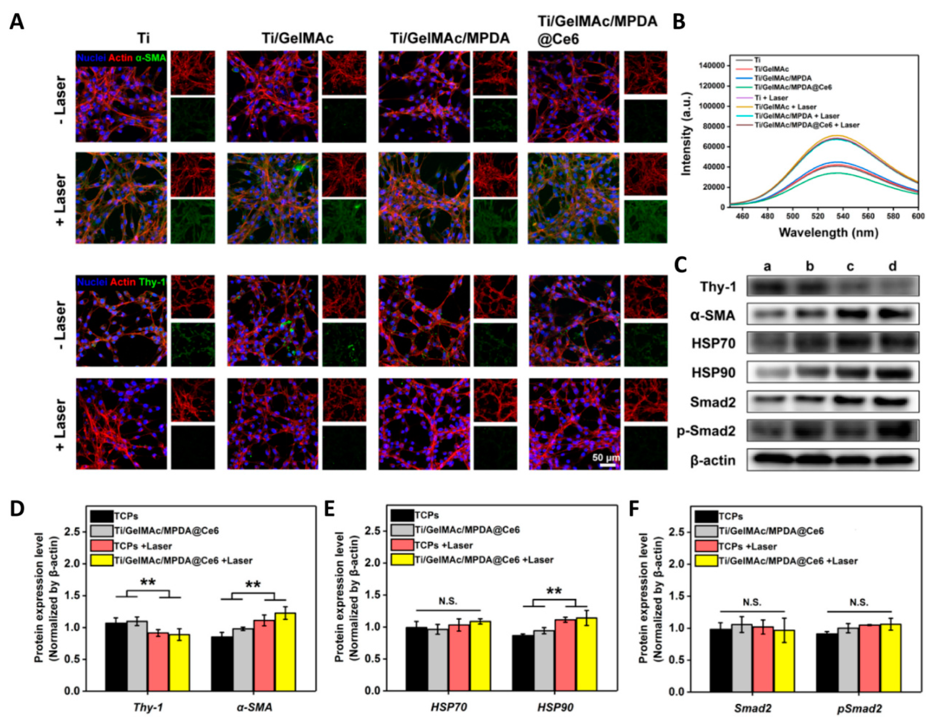
Figure 3.
Fibroblast activation, or fibroblast-myofibroblast transition, of NIH/3T3 cells after irradiation [50]. (A) Representative fluorescence images of NIH/3T3 cells cultured with different samples for 2 days. The expression of α-SMA and Thy-1 was stained as green. (B) Cellular ATP level reflected by luminescence intensity after different treatments. (C) Western blotting detecting the expression of Thy-1, α-SMA, HSP70, HSP90, Smad-2, and p-Smad-2 after different treatments. a: TCPs; b: Ti/GelMAc/MPDA@Ce6; c: TCPs + Laser; d: Ti/GelMAc/MPDA@Ce6 + Laser. (D–F) Quantitative analysis according to Western blotting results. (n = 6, ** p < 0.01, N.S.: no significance). (For interpretation of the references to color in this figure legend, the reader is referred to the Web version of this article.)

Figure 4.
Wettability and topography of the implant’s surface [77]. (a) Contact angle values and water droplet images, (b,c) 2D and 3D AFM images, and (d) average maximum height of the profile (Rz) and the arithmetic mean roughness (Ra) of the synthesized samples (p < 0.05).
Figure 4.
Wettability and topography of the implant’s surface [77]. (a) Contact angle values and water droplet images, (b,c) 2D and 3D AFM images, and (d) average maximum height of the profile (Rz) and the arithmetic mean roughness (Ra) of the synthesized samples (p < 0.05).

Figure 5.
Electrochemical construction of a catechol-grafted chitosan film for Cu2+ incorporation [101].
Figure 5.
Electrochemical construction of a catechol-grafted chitosan film for Cu2+ incorporation [101].

Figure 6.
Principle of hydrogel coating [105]. (a) Formulation: monomer units and coupling agents copolymerize into polymer chains, but do not crosslink into a network, resulting in an aqueous solution. The solution may also contain other compounds for various functions but are not drawn here. (b) Substrate preparation: functional groups complementary to the coupling agents are imparted onto the surface of a substrate. (c) Paint: The aqueous solution is painted on the substrate by various operations. (d) Cure: The coupling agents react with each other to crosslink the polymer chains into network and react with the complementary functional groups to interlink the polymer network to the substrate.

Figure 7.
Layer-by-layer electrostatic self-assembly [112].
Figure 7.
Layer-by-layer electrostatic self-assembly [112].

Figure 8.
Hydrogel coatings on titanium bidirectionally regulate osteoclastic and osteogenic response behaviors [88].
Figure 8.
Hydrogel coatings on titanium bidirectionally regulate osteoclastic and osteogenic response behaviors [88].

Figure 9.
Sustained release of VEGF promotes COL I (A) and CD 31 (B) expression in bone-surrounding scaffolds 6 and 12 weeks after implantation (n = 3) [44].
Figure 9.
Sustained release of VEGF promotes COL I (A) and CD 31 (B) expression in bone-surrounding scaffolds 6 and 12 weeks after implantation (n = 3) [44].

Figure 10.
Fluorescence images of macrophage morphologies on dual-inflammatory cytokine (interferon-γ and IL-4)-coated TiO2 nanotube surfaces [144]. Activated macrophages are indicated by yellow arrows.
Figure 10.
Fluorescence images of macrophage morphologies on dual-inflammatory cytokine (interferon-γ and IL-4)-coated TiO2 nanotube surfaces [144]. Activated macrophages are indicated by yellow arrows.


Table 1.
Development of medical titanium and titanium alloys.
| Classification | Time | Representative Material | Advantage | Disadvantage |
|---|---|---|---|---|
| α | 1960s | Ti | Good biocompatibility | Low strength, poor wear resistance |
| α + β | 1970s | Ti6Al4V | Higher hardness, better wear resistance, lower elastic modulus, better mechanical compatibility | Biological toxicity of metal ions Al and V |
| 1980s | Ti6Al7Nb Ti5Al2.5Fe | Better biocompatibility | Easy corrosion, biological toxicity of Al metal ions | |
| β | 1990s | Ti13Nb13Zr Ti12Mo6Zr2Fe Ti15Mo | The low modulus of elasticity is close to that of human bones, non-biological toxicity of metal ions | Biological activity, abrasion resistance, and corrosion resistance still need to be improved |
Table 2.
Classification of hydrogel coatings.
| Classification | Representative Material | Advantage | Reference |
|---|---|---|---|
| Natural hydrogel coatings | Collagen-based | Improve the attachment of the peri-implant soft tissue to titanium at early stages | [15] |
| Enhance tissue vascularization and reduce inflammatory response | [16] | ||
| Improve gingival connective tissue response to titanium implants | [17] | ||
| Gelatin-based | Improve surface bio-activity | [18] | |
| Load with antibacterial agent curcumin | [19] | ||
| Chitosan-based | Enhance the antibacterial activity and osteoinductive properties | [20] | |
| Develop a close bony apposition or the osseointegration of dental/craniofacial and orthopedic implants | [21] | ||
| Provide a self-protective surface that prevents bacterial colonisation and implant-associated infections | [22] | ||
| Great potential in implant anticorrosion | [23] | ||
| Alginate-based | Improve the antibacterial effect and induce mineralization of dental implants | [24] | |
| Successively functionalize titanium surface | [25] | ||
| Synthetic hydrogels coatings | Polyvinyl alcohol | Improve the calcium silicate coating-to-substrate adhesion. | [26] |
| Polyacrylamide | Antimicrobial-loaded hydrogel coatings | [27] | |
| Polyethylene glycol | Lower albumin adsorption and presented a decreased fibroblast, Streptococcus sanguinis and Lactobacillus salivarius adhesion. | [28] | |
| Poly (lacto-glycolic acid) | Drug release | [29] | |
| Polyacrylic acid | Acts as both an effective bioactive surface and an effective anti-corrosion barrier | [14] |
Disclaimer/Publisher’s Note: The statements, opinions and data contained in all publications are solely those of the individual author(s) and contributor(s) and not of MDPI and/or the editor(s). MDPI and/or the editor(s) disclaim responsibility for any injury to people or property resulting from any ideas, methods, instructions or products referred to in the content. |
© 2023 by the authors. Licensee MDPI, Basel, Switzerland. This article is an open access article distributed under the terms and conditions of the Creative Commons Attribution (CC BY) license (https://creativecommons.org/licenses/by/4.0/).
Share and Cite
MDPI and ACS Style
Chen, H.; Feng, R.; Xia, T.; Wen, Z.; Li, Q.; Qiu, X.; Huang, B.; Li, Y. Progress in Surface Modification of Titanium Implants by Hydrogel Coatings. Gels 2023, 9, 423. https://doi.org/10.3390/gels9050423
AMA Style
Chen H, Feng R, Xia T, Wen Z, Li Q, Qiu X, Huang B, Li Y. Progress in Surface Modification of Titanium Implants by Hydrogel Coatings. Gels. 2023; 9(5):423. https://doi.org/10.3390/gels9050423
Chicago/Turabian StyleChen, Huangqin, Rui Feng, Tian Xia, Zhehan Wen, Qing Li, Xin Qiu, Bin Huang, and Yuesheng Li. 2023. "Progress in Surface Modification of Titanium Implants by Hydrogel Coatings" Gels 9, no. 5: 423. https://doi.org/10.3390/gels9050423
Note that from the first issue of 2016, this journal uses article numbers instead of page numbers. See further details here.