
Sol–Gel Co-Precipitation Synthesis, Anticoagulant and Anti-Platelet Activities of Copper-Doped Nickel Manganite Nanoparticles

 , , ,
, , ,  and
and 
Abstract
:1. Introduction
2. Results and Discussion
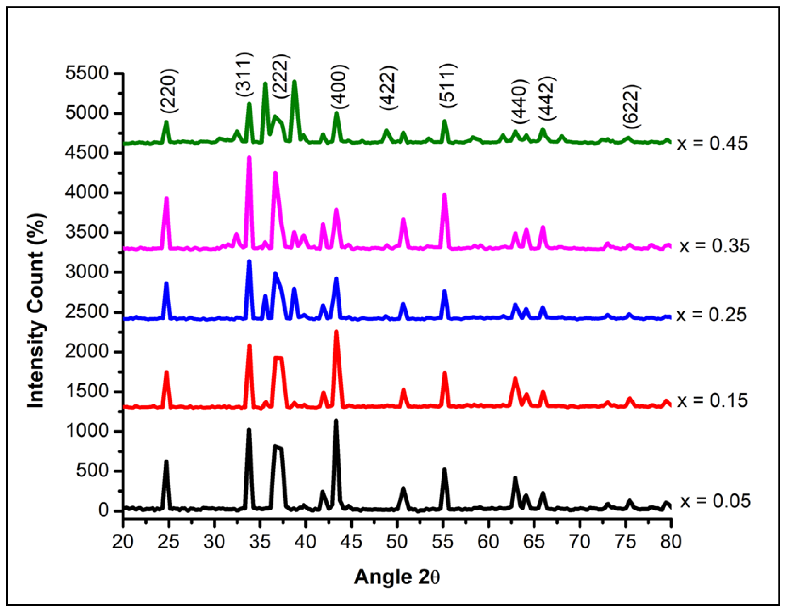
2.1. Structural Studies
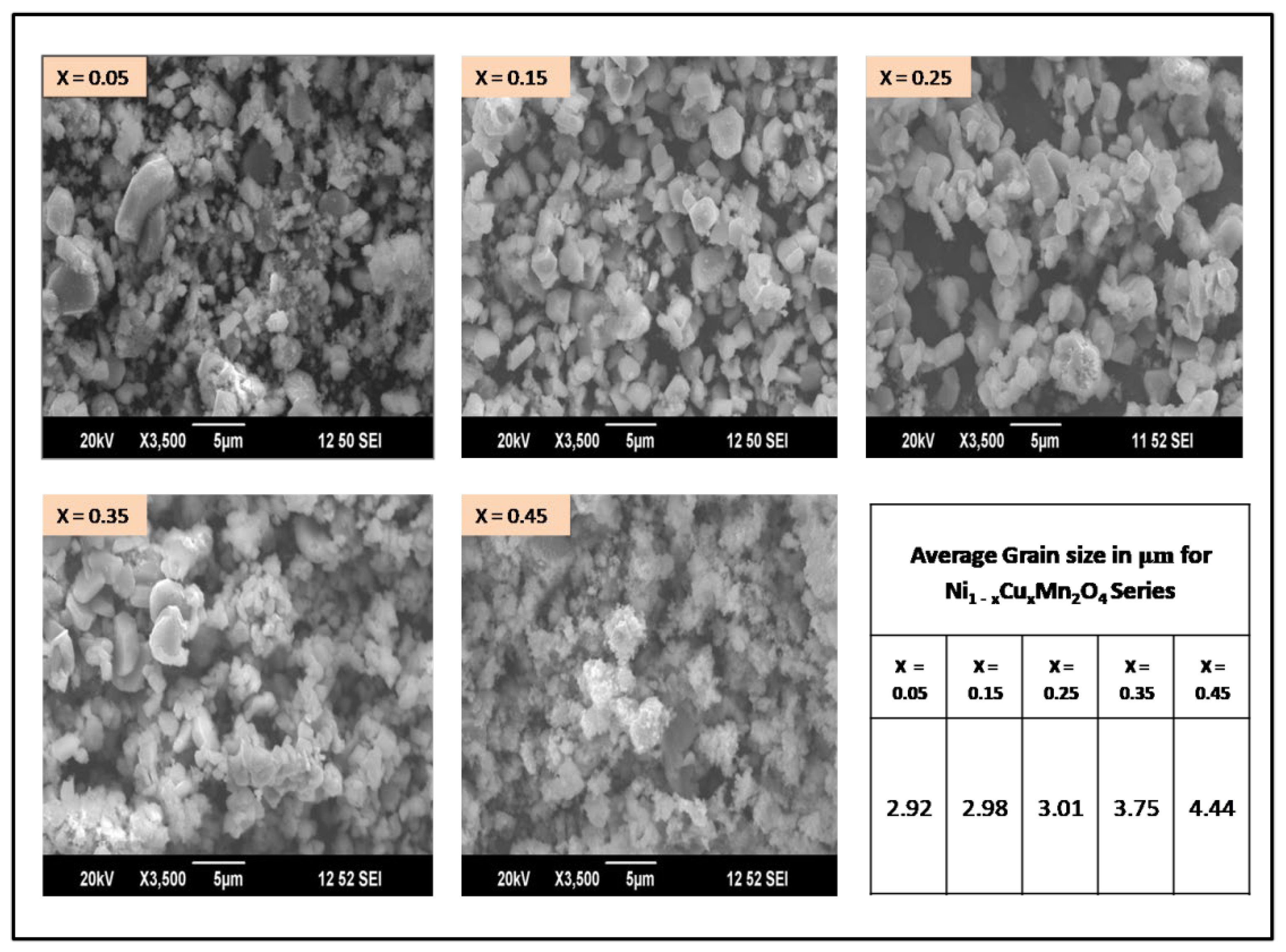
2.2. Morphological Studies
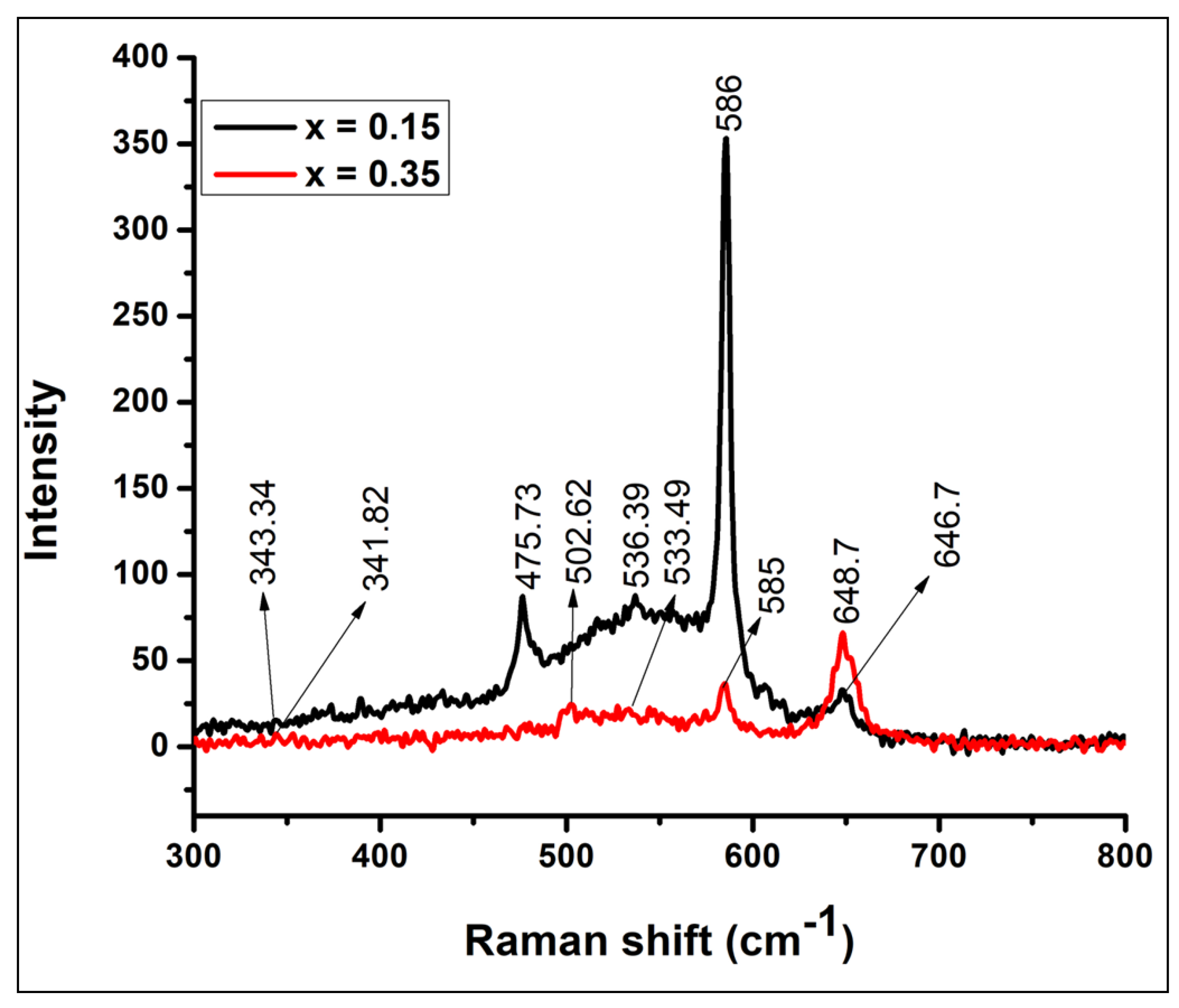
2.3. Raman Studies
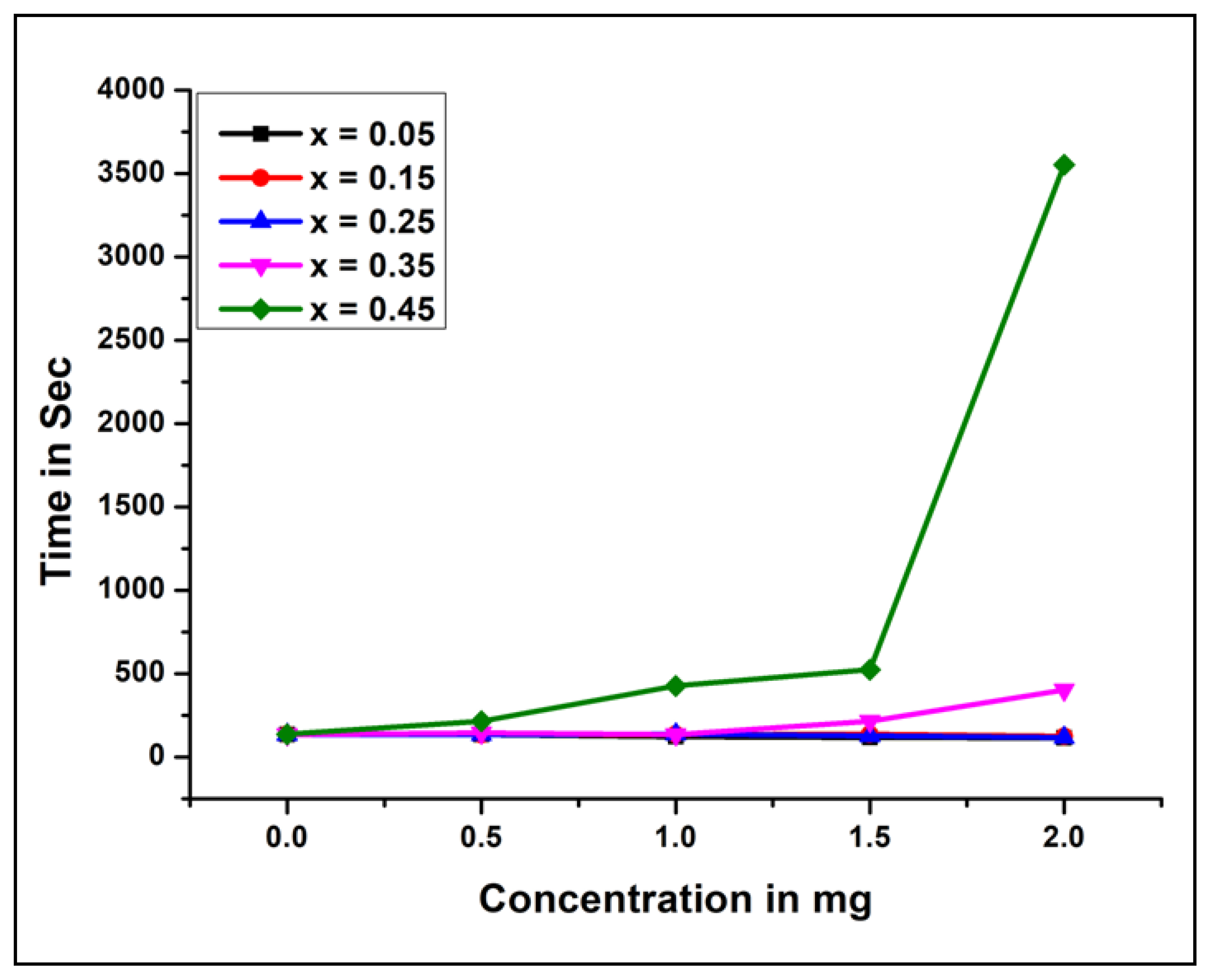
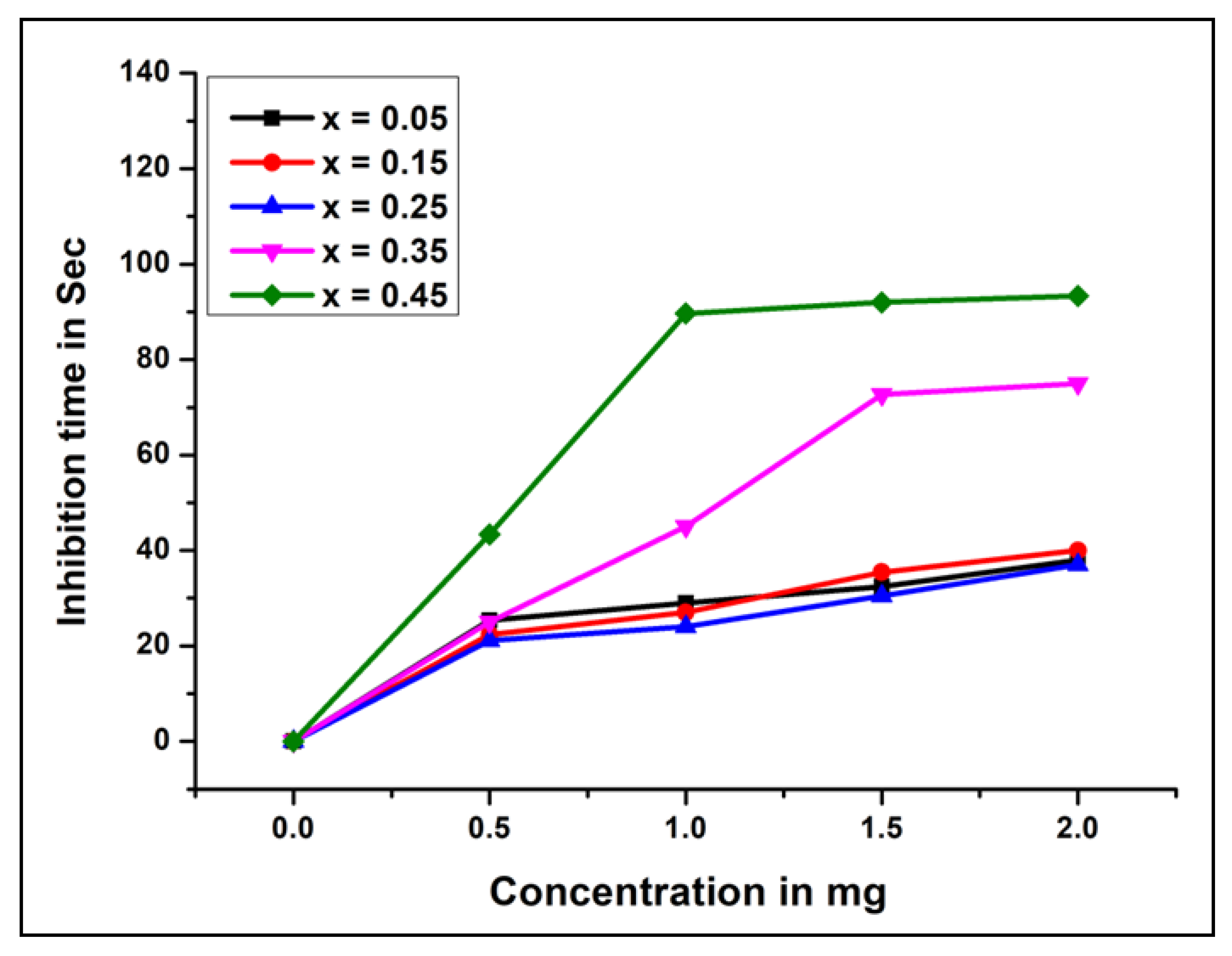
2.4. Anticoagulant and Antiplatelet Analysis
2.4.1. Human Plasma Clotting Time
2.4.2. Citrated Human Plasma Coagulation Time
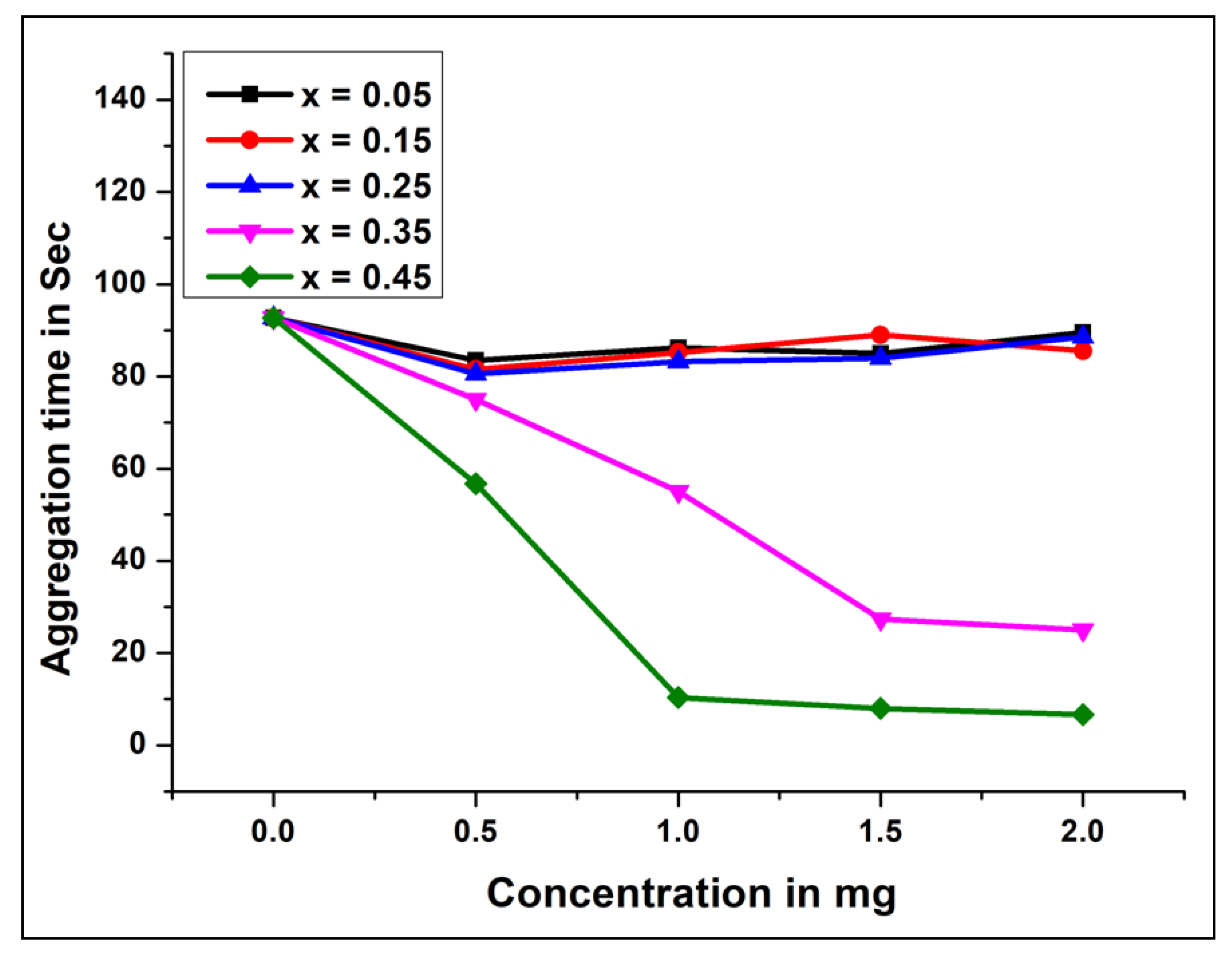
2.4.3. Antiplatelet Aggregation Property
2.4.4. Antiplatelet Aggregation Effect of NCB-NPs with the Usage of ADP as an Agonist
2.4.5. Hemolytic Activity
3. Conclusions
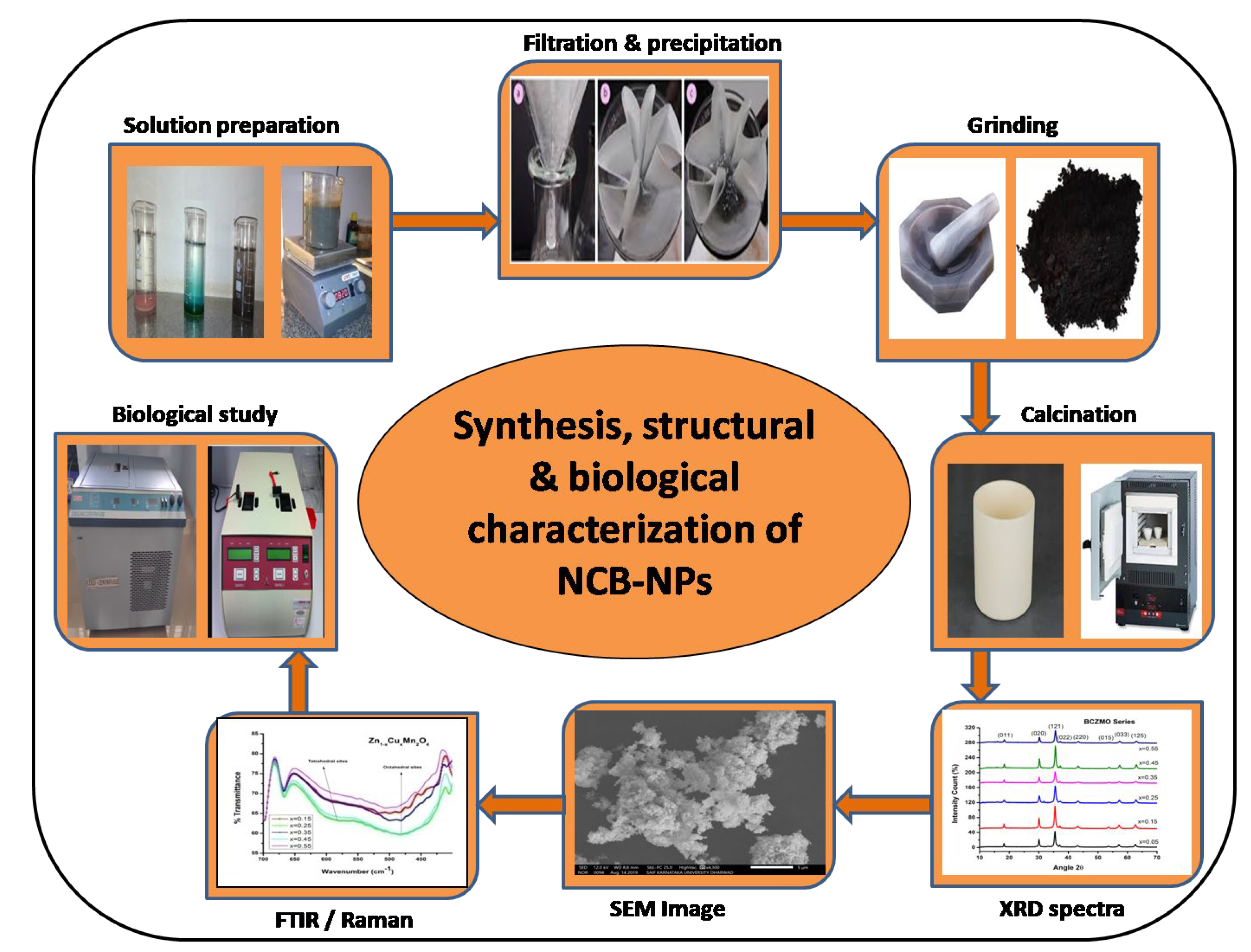
4. Experimental
4.1. Process of Synthesis
4.2. Characterization Techniques
4.3. Anticoagulant and Antiplatelet Activity
4.3.1. Plasma Recalcification Time
4.3.2. Preparation of Platelet-Rich Plasma (PRP) and Platelet-Poor Plasma (PPP)
4.3.3. Platelet Aggregation
4.3.4. Hemolytic Assay
4.3.5. Hemolytic Activity
Author Contributions
Funding
Institutional Review Board Statement
Informed Consent Statement
Acknowledgments
Conflicts of Interest
References
- Kanagasubbulakshmi, S.; Kadirvelu, K. Green synthesis of Iron oxide nanoparticles using Lagenaria siceraria and evaluation of its Antimicrobial activity. Def. Life Sci. J. 2017, 2, 422–427. [Google Scholar] [CrossRef]
- Gottimukkala, K.S.V.; Harika, R.P.; Zamare, D. Green synthesis of iron nanoparticles using green tea leaves extract. J. Nanomed. Biother. Discov. 2017, 7, 4172. [Google Scholar]
- Sun, C.; Ma, M.; Yang, J.; Zhang, Y.; Chen, P.; Huang, W.; Dong, X. Phase-controlled synthesis of α-NiS nanoparticles confined in carbon nanorods for High Performance Supercapacitors. Sci. Rep. 2014, 4, 7054. [Google Scholar] [CrossRef] [Green Version]
- Leng, Y.; Fu, L.; Ye, L.; Li, B.; Xu, X.; Xing, X.; He, J.; Song, Y.; Leng, C.; Guo, Y.; et al. Protein-directed synthesis of highly monodispersed, spherical gold nanoparticles and their applications in multidimensional sensing. Sci. Rep. 2016, 6, 28900. [Google Scholar] [CrossRef] [PubMed]
- Rehman, F.U.; Zhao, C.; Jiang, H.; Wang, X. Biomedical applications of nano-titania in theranostics and photodynamic therapy. Biomater. Sci. 2016, 4, 40–54. [Google Scholar] [CrossRef] [Green Version]
- Simeonidis, K.; LiébanaViñas, S.; Wiedwald, U.; Ma, Z.; Li, Z.-A.; Spasova, M.; Patsia, O.; Myrovali, E.; Makridis, A.; Sakellari, D.; et al. A versatile large-scale and green process for synthesizing magnetic Nanoparticles with tunable magnetic hyperthermia features. RSC Adv. 2016, 6, 53107–53117. [Google Scholar] [CrossRef]
- Strayer, A.; Ocsoy, I.; Tan, W.; Jones, J.B.; Paret, M.L. Low Concentrations of a Silver Based Nanocomposite to Manage Bacterial Spot of Tomato in the Green house. Plant. Dis. 2016, 100, 1460–1465. [Google Scholar] [CrossRef] [Green Version]
- Duman, F.; Ocsoy, I.; Kup, F.O. Chamomile flower extract-directed CuO nanoparticle formation for its antioxidant and DNA cleavage properties. Mater. Sci. Eng. C 2016, 60, 333–338. [Google Scholar] [CrossRef]
- Karatoprak, G.Ş.; Aydin, G.; Altinsoy, B.; Altinkaynak, C.; Koşar, M.; Ocsoy, I. The Effect of Pelargonium endlicherianum Fenzl. root extracts on formation of nanoparticles and their antimicrobial activities. Enzyme Microb. Technol. 2017, 97, 21–26. [Google Scholar] [CrossRef]
- Jadhav, R.N.; Mathad, S.N.; Puri, V. Studies on the properties of Ni0.6Cu0.4Mn2O4 NTC ceramic due to Fe doping. Ceram. Int. 2012, 38, 5181–5188. [Google Scholar] [CrossRef]
- Bobo, D.; Robinson, K.J.; Islam, J.; Thurecht, K.J.; Corrie, S.R. Nanoparticle-based medicines: A review of FDA-approved materials and clinical trials to date. Pharm. Res. 2016, 33, 2373–2387. [Google Scholar] [CrossRef] [PubMed]
- Mathad, S.N.; Jadhav, R.N.; Pawar, R.P.; Puri, V. Studies on rod shaped bismuth strontium manganite ceramics. Sci. Adv. Mater. 2012, 6, 1276–1281. [Google Scholar] [CrossRef]
- Shashidharagowda, H.; Mathad, S.N. Effect of incorporation of copper on structural properties of spinel nickel manganites by co-precipitation method. Mater. Sci. Energy Technol. 2020, 3, 201–208. [Google Scholar] [CrossRef]
- Sanpo, N.; Berndt, C.C.; Wang, J. Microstructural and antibacterial properties of zinc-substituted cobalt ferrite nanopowders synthesized by sol-gel method. J. Appl. Phys. 2012, 112, 084333. [Google Scholar] [CrossRef]
- Larbi, T.; Doll, K.; Amlouk, M. Temperature dependence of Raman spectra and first principles study of NiMn2O4 magnetic spinel oxide thin films. Application in efficient photocatalytic removal of RhB and MB dyes. Spectrochim. Acta Part A Mol. Biomol. Spectrosc. 2019, 216, 117–124. [Google Scholar] [CrossRef]
- Malavasi, L.; Galinetto, P.; Mozzati, M.C.; Azzoni, C.B.; Flor, G. Raman spectroscopy of AMn2O4 (A = Mn, Mg and Zn) spinels. Phys. Chem. Chem. Phys. 2002, 4, 3876–3880. [Google Scholar] [CrossRef]
- Gubbiveeranna, V.; Kusuma, C.G.; Bhavana, S.; Sumachirayu, C.K.; Nagaraju, S. Anti-hemostatic protease from Jatropha curcas latex with fibrinogen lytic activity. J. Pharmacogn. Phytochem. 2019, 8, 1303–1310. [Google Scholar]
- Gubbiveeranna, V.; Nagaraju, S. Ethnomedicinal, Phytochemical constituents and Pharmacological activities of tridaxprocumbens: A review. Int. J. Pharm. Pharm. Sci. 2016, 8, 1–7. [Google Scholar]
- Shivaiah, N.; Kempaiah, K. ‘Partitagin’, a unique β, γ-fibrinogenase that inhibits platelet aggregation from Hippasa partita spider venom. Blood Coagul. Fibrinolysis 2011, 22, 24–28. [Google Scholar] [CrossRef]
- Kitchen, S.; Gray, E.; Mackie, I.; Baglin, T.; Makris, M.; The BCSH Committee. Measurement of Non-Coumarin Anticoagulants and their Effects on Tests of Haemostasis: Guidance from the British Committee for Standards in Haematology. Br. J. Haematol. 2014, 166, 830–841. [Google Scholar] [CrossRef]
- Lee, J.; Jeong, L.; Jung, E.; Ko, C.; Seon, S.; Noh, J.; Lee, D. Thrombus targeting aspirin particles for near infrared imaging and on-demand therapy of thrombotic vascular diseases. J. Control Release 2019, 304, 164–172. [Google Scholar] [CrossRef] [PubMed]
- Kang, C.; Gwon, S.; Song, C.; Kang, P.M.; Park, S.-C.; Jeon, J.; Hwang, D.W.; Lee, D. Fibrin-Targeted and H2O2-Responsive Nanoparticles as a Theranostics for Thrombosed Vessels. ACS Nano 2017, 11, 6194–6203. [Google Scholar] [CrossRef]
- Jung, E.; Kang, C.; Lee, J.; Yoo, D.; Hwang, D.W.; Kim, D.; Park, S.C.; Lim, S.K.; Song, C.; Lee, D. Molecularly Engineered Theranostic Nanoparticles for Thrombosed Vessels: H2O2-Activatable Contrast-Enhanced Photoacoustic Imaging and Antithrombotic Therapy. ACS Nano 2018, 12, 392–401. [Google Scholar] [CrossRef] [PubMed]
- Nishida, N.; Yano, H.; Nishida, T.; Kamura, T.; Kojiro, M. Angiogenesis in Cancer. Vasc. Health Risk Manag. 2006, 2, 213–219. [Google Scholar] [CrossRef]
- Li, Z.; Delaney, M.K.; O’Brien, K.A.; Du, X. Signaling during platelet adhesion and activation. Arterioscler. Thromb. Vasc. Biol. 2010, 30, 2341–2349. [Google Scholar] [CrossRef] [PubMed] [Green Version]
- Anselmo, A.C.; Modery-Pawlowski, C.L.; Menegatti, S.; Kumar, S.; Vogus, D.R.; Tian, L.L.; Chen, M.; Squires, T.M.; Sen Gupta, A.; Mitragotri, S. Platelet-like nanoparticles: Mimicking shape, flexibility and surface biology of platelets to target vascular injuries. ACS Nano 2014, 8, 11243–11253. [Google Scholar] [CrossRef]
- Fröhlich, E. Action of Nanoparticles on Platelet Activation and Plasmatic Coagulation. Curr. Med. Chem. 2016, 23, 408–430. [Google Scholar] [CrossRef]
- Lateef, A.; Akande, M.A.; Ojo, S.A.; Folarin, B.I.; Gueguim-Kana, E.B.; Beukes, L.S. Paper wasp nest-mediated biosynthesis of silver nanoparticles for antimicrobial, catalytic, anticoagulant, and thrombolytic applications. 3 Biotech. 2016, 6, 140. [Google Scholar] [CrossRef] [Green Version]
- Kim, H.K.; Choi, M.J.; Cha, S.H.; Koo, Y.K.; Jun, S.H.; Cho, S.; Park, Y. Earthworm extracts utilized in the green synthesis of gold nanoparticles capable of reinforcing the anticoagulant activities of heparin. Nanoscale Res. Lett. 2013, 8, 542. [Google Scholar] [CrossRef] [Green Version]
- Kim, H.S.; Jun, S.H.; Koo, Y.K.; Cho, S.; Park, Y. Green synthesis and nanotopography of heparin-reduced gold nanoparticles with enhanced anticoagulant activity. J. Nanosci. Nanotechnol. 2013, 13, 2068–2076. [Google Scholar] [CrossRef]
- Marulasiddeshwara, M.B.; Dakshayani, S.S.; Sharath Kumar, M.N.; Chethana, R.; Raghavendra Kumar, P.; Devaraja, S. Facile-one pot-green synthesis, antibacterial, antifungal, antioxidant and antiplatelet activities of lignin capped silver nanoparticles: A promising therapeutic agent. Mater. Sci. Eng. C 2017, 81, 182–190. [Google Scholar] [CrossRef] [PubMed]
- Hany, I.K.; Ismet, B.; Alan, B. Complement-coagulation cross-talk: A potential mediator of the physiological activation of complement by low pH. Front. Immunol. 2015, 6, 215. [Google Scholar]
- Bharadwaj, S.S.; Poojary, B.; Sharath Kumar, M.N.; Jayanna, K.; Mugaranja, P.K.; Madan Kumar, S.; Das, A.J.; Kulal, A.; San-naningaiah, D. Efficient synthesis and in silico studies of the benzimidazole hybrid scaffold with the quinolinyloxadiazole skeleton with potential α-glucosidase inhibitory, anticoagulant, and antiplatelet activities for type-II diabetes mellitus management and treating thrombotic disorders. ACS Omega 2018, 3, 12562–12574. [Google Scholar] [PubMed]
- Quick, A.J. The prothrombin in hemophilia and in obstructive jaundice. J. Boil. Chem. 1935, 109, 73–74. [Google Scholar]
- Ardlie, N.G.; Han, P. Enzymatic Basis for Platelet Aggregation and Release: The Significance of the ‘Platelet Atmosphere’ and the Relationship between Platelet Function and Blood Coagulation. Br. J. Haematol. 1974, 26, 331–356. [Google Scholar] [CrossRef]
- Shin, S.Y.; Lee, M.K.; Kim, K.L.; Hahm, K.S. Structure-antitumor and hemolytic activity relationships of synthetic peptides derived from cecropin A-magainin 2 and cecropin A-melittin hybrid peptides. J. Pept. Res. 1997, 50, 279–285. [Google Scholar] [CrossRef]


| x | Crystallite Size D nm | Dislocation Density fD × 1015/m2 | Grain Size in μm | ν1 cm−1 (Th Site) | ν2 cm−1 (Oh Site) |
|---|---|---|---|---|---|
| 0.05 | 25.03 | 1.83 | 2.72 | 582.42 | 412.71 |
| 0.15 | 24.15 | 1.87 | 2.87 | 586.28 | 410.78 |
| 0.25 | 26.38 | 1.46 | 3.05 | 588.21 | 408.85 |
| 0.35 | 27.15 | 1.36 | 3.75 | 590.14 | 408.68 |
| 0.45 | 26.18 | 1.69 | 4.42 | 592.06 | 407.92 |
Publisher’s Note: MDPI stays neutral with regard to jurisdictional claims in published maps and institutional affiliations. |
© 2021 by the authors. Licensee MDPI, Basel, Switzerland. This article is an open access article distributed under the terms and conditions of the Creative Commons Attribution (CC BY) license (https://creativecommons.org/licenses/by/4.0/).
Share and Cite
H., S.; Mathad, S.; Malladi, S.; Gubbiveeranna, V.; G., K.C.; S., N.; Patil, A.Y.; Khan, A.; Rub, M.A.; Asiri, A.M.; et al. Sol–Gel Co-Precipitation Synthesis, Anticoagulant and Anti-Platelet Activities of Copper-Doped Nickel Manganite Nanoparticles. Gels 2021, 7, 269. https://doi.org/10.3390/gels7040269
H. S, Mathad S, Malladi S, Gubbiveeranna V, G. KC, S. N, Patil AY, Khan A, Rub MA, Asiri AM, et al. Sol–Gel Co-Precipitation Synthesis, Anticoagulant and Anti-Platelet Activities of Copper-Doped Nickel Manganite Nanoparticles. Gels. 2021; 7(4):269. https://doi.org/10.3390/gels7040269
Chicago/Turabian StyleH., Shashidharagowda, Shridhar Mathad, Shridhar Malladi, Vinod Gubbiveeranna, Kusuma C. G., Nagaraju S., Arun Y. Patil, Anish Khan, Malik Abdul Rub, Abdullah M. Asiri, and et al. 2021. "Sol–Gel Co-Precipitation Synthesis, Anticoagulant and Anti-Platelet Activities of Copper-Doped Nickel Manganite Nanoparticles" Gels 7, no. 4: 269. https://doi.org/10.3390/gels7040269