Article

Enhanced Light-Induced Biosynthesis of Fatty Acids Suitable for Biodiesel Production by the Yellow-Green Alga Eustigmatos magnus

by
Izabela Krzemińska
1,*,
Artur Nosalewicz
1,
Emilia Reszczyńska
2 and
Barbara Pawlik-Skowrońska
3
1
Institute of Agrophysics, Polish Academy of Sciences, Doświadczalna 4, 20-290 Lublin, Poland
2
Department of Plant Physiology and Biophysics, Institute of Biological Sciences, Maria Curie-Skłodowska University, Akademicka Street 19, 20-033 Lublin, Poland
3
Department of Hydrobiology and Protection of Ecosystems, University of Life Sciences in Lublin, Dobrzańskiego 37, 20-262 Lublin, Poland
*
Author to whom correspondence should be addressed.
Energies 2020, 13(22), 6098; https://doi.org/10.3390/en13226098
Submission received: 26 October 2020 / Revised: 13 November 2020 / Accepted: 19 November 2020 / Published: 21 November 2020
(This article belongs to the Special Issue Algal Biotechnology and Biofuels)

Abstract

:
Optimization of the fatty acid profile in microalgae is one of the key strategies for obtaining valuable products and sustainable biofuels. Light intensity and light regimes exert an impact on the growth and metabolic process in microalgae. The objective of the present investigations was to assess the effect of light intensity and continuous light vs. photoperiod conditions on the growth and changes in the biomass composition in Eustigmatos magnus, with a focus on bioactive molecules such as lipids and fatty acids. The highest daily productivity of Eustigmatos magnus biomass and lipid yields were detected at continuous illumination and at the highest intensity of light. The results show that the content and composition of fatty acids was influenced by the culture conditions. The biomass of Eustigmatos magnus contained the highest concentrations of polyunsaturated fatty acids in the pphotoperiod conditions with the highest light intensity. This study shows that Eustigmatos magnus has a capacity for the accumulation of palmitoleic acid. A high intensity of continuous light improves the profile of fatty acids in Eustigmatos magnus, which can be suitable for biodiesel applications. At the high intensity of continuous light, Eustigmatos magnus lipids are characterized by high content of oleic acids and low content of saturated and monounsaturated acids.

1. Introduction

Eukaryotic microalgae have been being recognized as a new promising feedstock production of biodiesel and such bioproducts as lipids, pigments, carbohydrates, and vitamins [1]. Photoautotrophic microalgae are the primary producer of medium-chain (C10–C14) and long-chain (C16–C18, C20–C22) polyunsaturated fatty acids. Microalgal lipids containing C14–C20 fatty acids can be used for the production of biodiesel, which is non-toxic and eco-friendly [2]. Long-chain polyunsaturated fatty acids are commercially important as well. They comprise two groups of essential acids: omega ω-3 and ω-6. The omega 3 group is represented by an essential fatty acid, i.e., eicosapentaenoic acid (EPA, 20:5 n-3), which is an important target in microalgal biotechnology due to its special role in human nutrition [3].
Only certain algal classes are capable of the production of EPA. High concentrations of EPA have been detected in the lipids of several species of the genus Nannochloropsis, e.g., N. gaditana and N. oculata. Trachydiscus minatus and diatoms, e.g., Phaeodactylum tricornutum, are other species capable of EPA biosynthesis [2].
The modern research on algal biotechnology is focused on oleaginous microalgae of the genus Nannochloropsis representing the class Eustigmatophyceae, as they are regarded as the primary candidates for algal biofuel production and producers of EPA [4]. The use of these species suggests that other representatives of Eustigmatophyceae may be applied for production of biofuels or EPA as well. Eustigmatophyceae is a class of yellow-green unicellular coccoid algae living mainly in soil and freshwater. The physiology and biochemistry of Eustigmatos magnus, i.e., a unicellular alga from the class Eustigmatophyceae, is poorly recognized. Previous studies indicate that Eustigmatos magnus is a promising producer of β-carotene [5]. Recent investigations have demonstrated that Eustigmatos magnus biomass can be a feedstock for biogas production [6]. The metabolic profile of Eustigmatos magnus has not yet been fully characterized, particularly in terms of the ability of the alga to synthesize lipids and fatty acids.
Light intensity and the light regime play a significant role, as they determine the rate and course of metabolic and reproductive processes [7]. Determination of the conditions that ensure the production of biomass with the desired biochemical composition is an important issue due to the diverse requirements of algal species for light and growth conditions.
The present study is the first to demonstrate the impact of light intensity and continuous light vs. photoperiod conditions on biomass production, content of pigments, photosynthetic apparatus, and metabolic regulation of fatty acids in Eustigmatos magnus. The aim of the study was to analyze the relationships between physiological and photochemical parameters and changes induced by light in the lipid metabolism of Eustigmatos magnus. The study also assessed the suitability of fatty acids from Eustigmatos magnus as a source of palmitic and eicosapentaenoic acids and material for biodiesel production.

2. Results

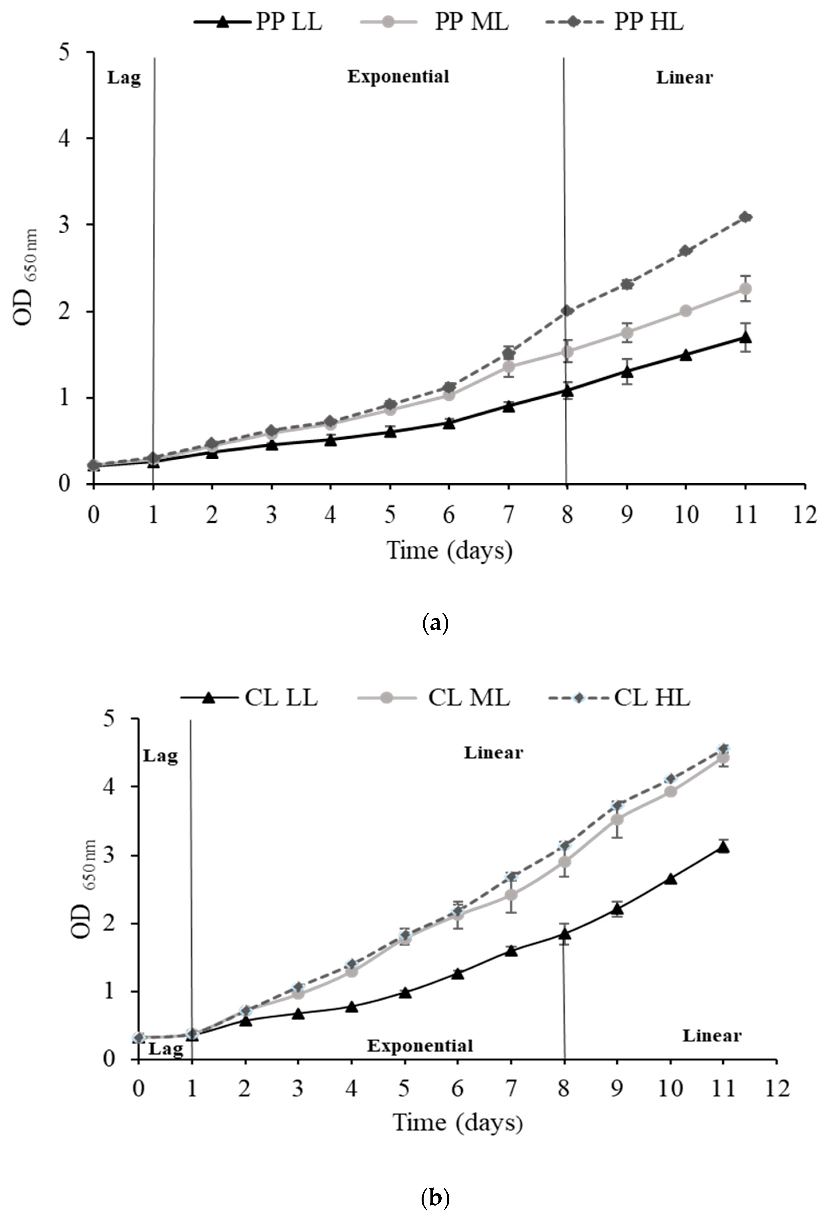
2.1. Analysis of Eustigmatos magnus Growth

The parameters of Eustigmatos magnus growth are presented in Table 1. The light conditions applied in the experiment exerted a significant impact on the specific growth rate, doubling time, and biomass productivity. In the photoperiod conditions (PP), the specific growth rate increased with the increasing light intensity from 0.207 µ (30 µmol m−2 s−1, LL—low light intensities) to 0.275 µ (400 µmol m−2 s−1, HL—high light intensities). In the continuous lighting conditions (CL), the specific Eustigmatos magnus growth rate increased with the light intensity and was higher than that recorded in the photoperiod variant. The highest specific growth rate of 0.344 µ was exhibited by Eustigmatos magnus in the conditions of continuous illumination and at light intensity of 400 µmol m−2 s−1. The increase in the light intensity was accompanied by reduction of the doubling time in both the continuous illumination and photoperiod variants. Additionally, the culture conditions had an impact on biomass productivity. The highest daily productivity of biomass was found in Eustigmatos magnus cultures grown under continuous illumination and at the highest light intensity.
The Eustigmatos magnus growth curves are shown in Figure 1a,b. The course of the algal growth curves differed depending on the lighting conditions applied. In the case of the photoperiod, regardless of the light intensity, the curves were similar: a short lag phase (0–1 day) was followed by the exponential phase (1–7 day) and the linear phase (8–11 day). In contrast, the ML and HL growth curves under continuous illumination differed from those recorded in the LL conditions. The growth curves in cultures growing at the three analyzed light intensities were characterized by a short log phase (0–1 day). The growth curve in the HL and ML conditions was similar: the log phase was immediately followed by the linear growth phase (1–11 day). In the LL conditions, the cells entered the exponential growth phase after the log phase, and then the linear phase started, likewise in the photoperiod conditions. There was no stationary growth phase in all the continuous light intensity and photoperiod variants during the 11 days of culture.

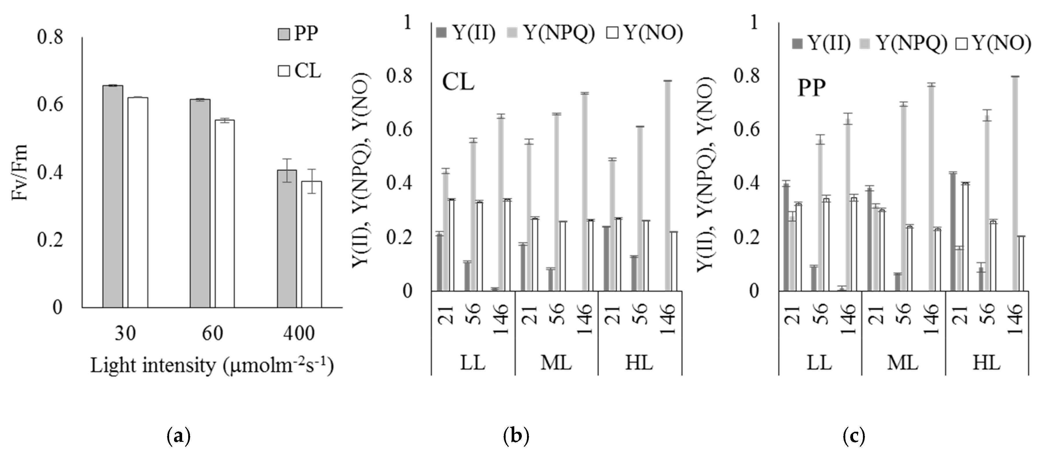
2.2. Content of Pigments and Analysis of Chlorophyll Fluorescence

The analysis of Eustigmatos magnus pigments was carried out based on chromatograms and absorption spectra. The Eustigmatos magnus cells contained carotenoid pigments: violaxanthin, voucheriaxanthin-like structure, zeaxanthin, and carotene as well as chlorophyll a and β-carotene (Table 2).
The amount of the pigments depended on the illumination conditions. As shown in the table, the amount of violaxanthin increased in the continuous light conditions, with its highest amount recorded in the CL HL variant (Table 2). Changes in the amount of β-carotene and chlorophyll a were detected as well. The amount of β-carotene increased in the CL HL and PP HL variants. The highest amount of zeaxanthin was observed in the HL conditions in both the continuous illumination and photoperiod conditions. The lowest content was detected in LL in the photoperiod and in the ML continuous illumination variants.
The Eustigmatos magnus algae grown at the different light regimes were characterized by various degrees of photoinhibition. There were similar downward trends in the maximum quantum yield (Fv/Fm) for both PP and CL with the increase in the light intensity (Figure 2a).
A slight (significant) decrease in the maximum quantum yield of PSII (Fv/Fm) was observed with an increase at the lowest light intensity and a strong decrease with the increase from 60 to 400 µmol m−2 s−1, which indicated a clear inhibitory effect of high light. At all values of light intensity, the values of Fv/Fm were significantly lower for CL. However, the differentiation at 400 µmol m−2 s−1 was not significant. The analysis of the chlorophyll fluorescence induction curves (Figure 2b,c) revealed that the non-regulated dissipation of excess energy (Y(NO)) at the level of 0.2–0.4. Y(NO) reached the highest level at HL for algae grown in LL in the photoperiod variant. Non-photochemical quenching (Y(NPQ)) displayed a clear upward trend with an increase in the actinic light intensity. Such response was noted for both the duration and all light intensities. Although the maximum values of Y(NPQ) were similar between both CL and PP, indicating similar functioning of protective mechanisms against high light, different responses were noted at the lowest light (21 µmol m−2 s−1), for which Y(NPQ) was significantly lower by 41–60% in the PP treatment (conditions in which the lowest PUFA content was noted, Table 3). The lower Y(NPQ) was associated with the highest Y(II) in these conditions (PP—21 µmol m−2 s−1). Importantly, the data presented in Figure 2 are steady-state values obtained after ca. 5 min of illumination during the measurements of chlorophyll fluorescence with the specified intensity light. Such a low value of Y(NPQ) for PP HL 21 µmolm−2 s−1 was a result of adaptation to such light.
As shown above, these conditions stimulated lipid productivity (Figure 3), yet at the cost of the lowest specific growth rate (Table 1). Based on these analyses, it can be concluded that under CL irradiance with HL, Eustigmatos magnus displaying a reduction of photochemical quenching (Y(II)) was able to maintain its high growth rate (Table 1) in the conditions of longer illumination time than under PP.

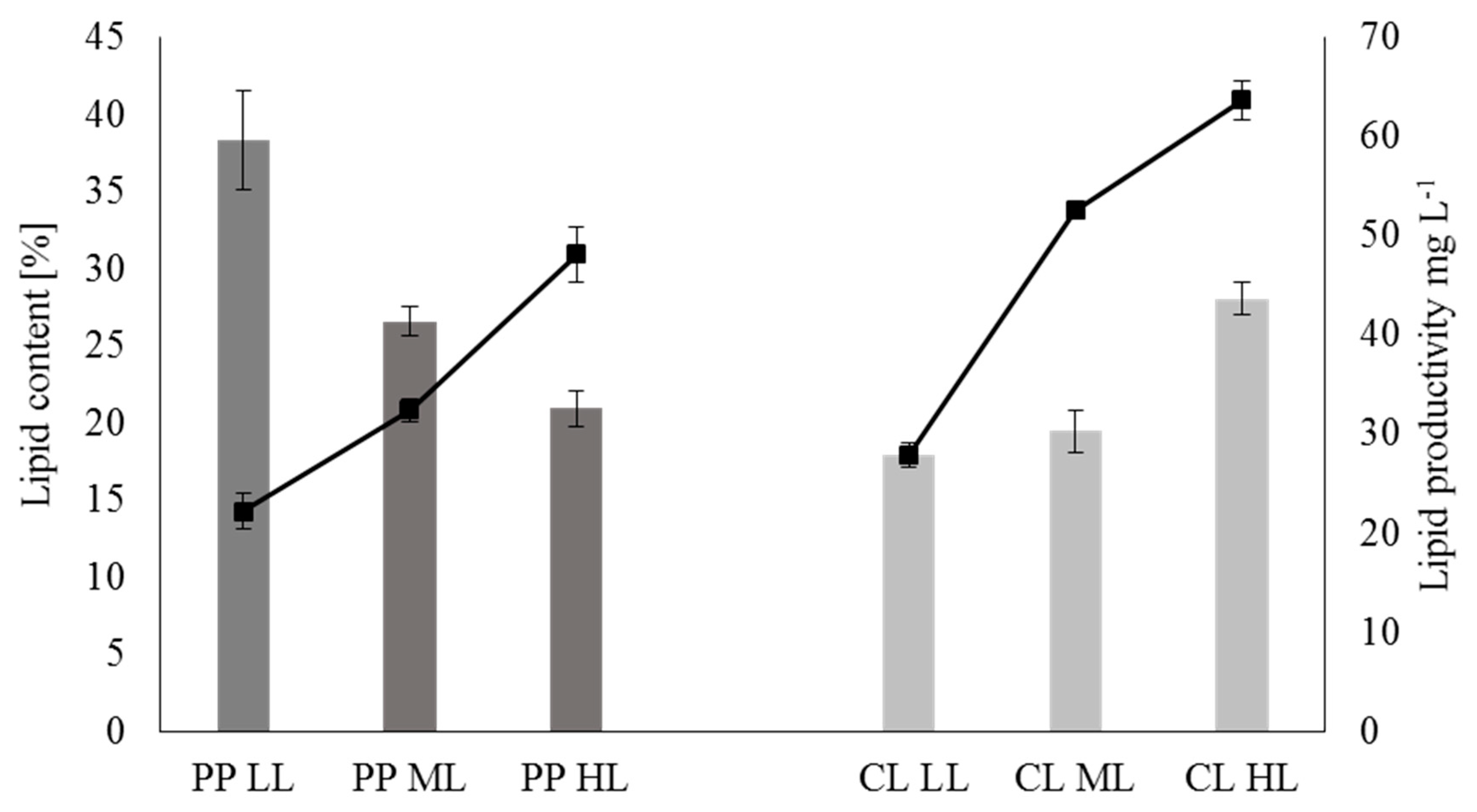
2.3. Analysis of Lipid Metabolism

The impact of the light intensity in the photoperiod (PP) and continuous illumination (CL) variants on the lipid content in Eustigmatos magnus cells is shown in Figure 3.
The lipid content had the maximum values at the low light intensity in the photoperiod variant and at the high light intensity at continuous illumination. The light intensity in the photoperiod conditions had a significant impact on the lipid content in the Eustigmatos magnus cells (Figure 3). There were significant differences between the Eustigmatos magnus lipid content at the high light intensities and at the low and moderate light intensities in the continuous illumination variant. The maximum lipid content in the Eustigmatos magnus cells (28.06%) obtained at HL in the continuous light conditions was 1.5-fold higher than in the LL growth variant. In the photoperiod variant, the content of lipids decreased as the light intensity increased.
The influence of the algal culture conditions on lipid productivity is shown in Figure 3. Higher lipid productivity was recorded in the continuous light conditions at each of the three light intensities tested. The lipid productivity was in the range from 22.20 to 63.64 mg L−1 for the LL photoperiod and HL continuous light, respectively.

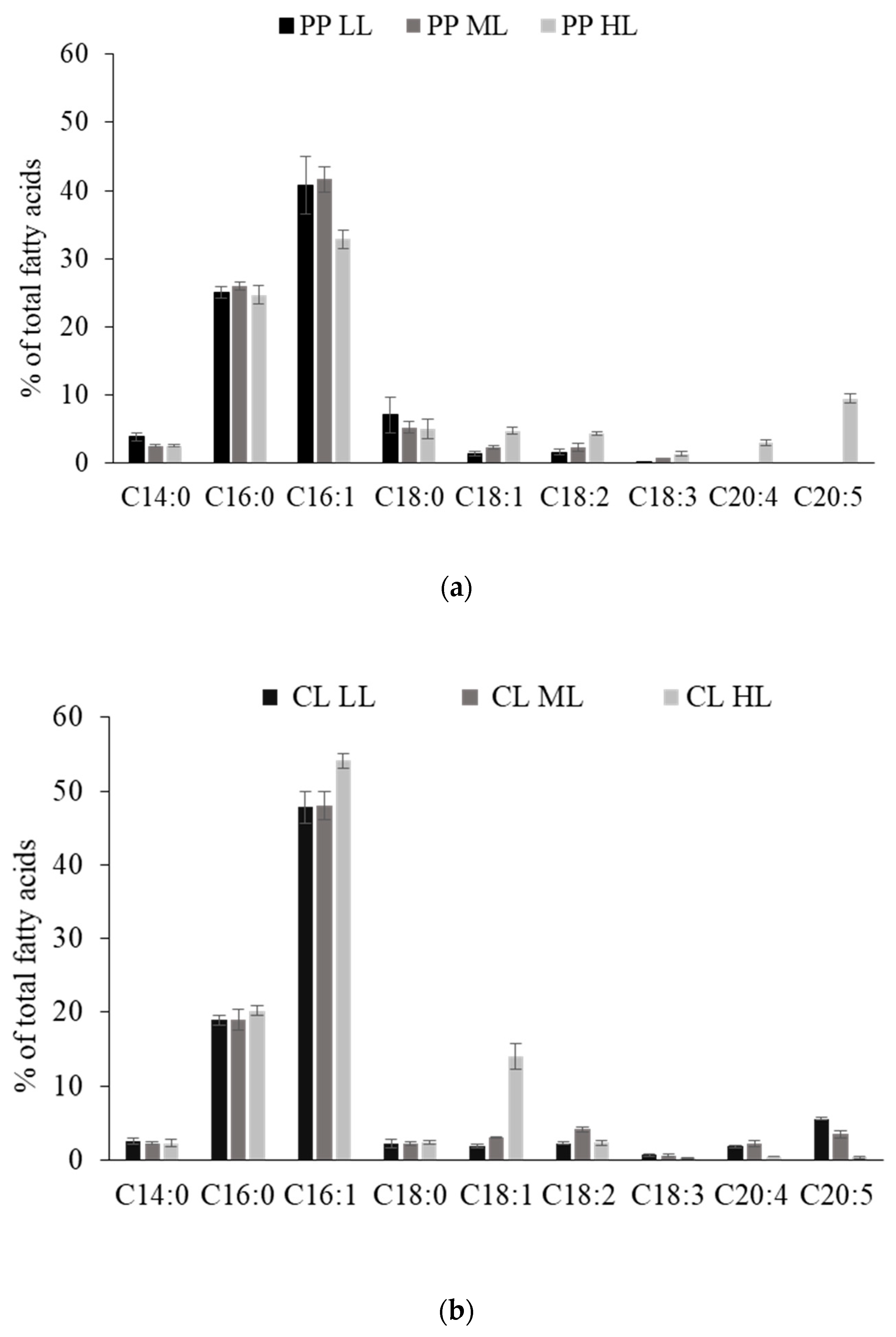
2.4. Analysis of the Fatty Acid Profile

The distribution of fatty acids in Eustigmatos magnus cultured at the three light intensities (LL, ML, HL) in the photoperiod and continuous illumination variants is shown in Figure 4a,b.
The main fatty acids identified in Eustigmatos magnus were represented by myristic acid (C14:0), palmitic acid (C16:0), palmitoleic acid (C16:1), stearic acid (C18:0), oleic acid (C18:1), linoleic acid (C18:2), arachidonic acid (C20:4), and eicosapentaenoic acid (C20:5). Palmitoleic and palmitic acids were most abundant. The results show that the culture conditions exerted an impact on the fatty acid profile. Eustigmatos magnus cells growing in the photoperiod conditions had higher content of C16:0 and C18:0 than that recorded in the continuous light conditions. The highest C16:1 content was found in the continuous light variant.
The results also showed that the higher light intensity in the photoperiod conditions induced an accumulation of eicosapentaenoic acid in the algal cells. The highest percentage of eicosapentaenoic acid (9.495%) was detected in cells cultured in the HL PP conditions. It was also found that the content of eicosapentaenoic acid in the Eustigmatos magnus cells declined from 5.59% to 0.33% in response to the increasing light intensity in the continuous light variant.
To provide detailed information about the effect of light on the distribution of fatty acids in the lipid pool in Eustigmatos magnus, the sum of saturated (SFA), monounsaturated (MUFA), and polyunsaturated (PUFA) fatty acids was compared with the sum of C16–C18 fatty acids (Table 3).
The content of SFAs, MUFAs, and PUFAs varied depending on the light conditions. The results showed that in comparison with the photoperiod, continuous illumination reduced the amounts of SFA in the cells at all the three light intensities. It was also found that continuous light stimulated the accumulation of MUFAs. The highest MUFA percentage (68.305%) was recorded in Eustigmatos magnus cells from the HL CL culture. The highest PUFA content (14.97%) was detected in the HL PP variant. The results showed that the C16–C18 acids represented from 73.1% to 94% of all fatty acids.

3. Discussion

The investigation results suggest that Eustigmatos magnus has an ability to grow and produce biomass efficiently in a broad range of light intensity from 30 to 400 µmol m−2 s−1 with no photoinhibition. In this study, the high light intensities (400 µmol m−2 s−1) and continuous light promoted the specific growth rate as well as biomass productivity of Eustigmatos magnus. Sirisuk et al. has also found maximum biomass production in Nannochloropsis salina under 24:0 h continuous LED illumination [8]. As reported by Meseck et al. a longer day length (24:0, 16:8; 12:12; 8:16 light:dark cycles) and higher light intensities resulted in higher yields of Tetraselmis chui biomass [9]. The results obtained by Mitra et al. [10] demonstrated that the productivity of Nannochloropsis gaditana biomass increased at two light intensities of 60 and 150 µmol m−2 s−1 [10]. In these studies, higher biomass productivity accompanied the increasing number of light hours from 12:12 to 18:6 light:dark. A further increase in the number of hours of light to 24 h did not increase the biomass productivity of Nannochloropsis gaditana. Matos et al. studied Nannochloropsis gaditana growing in four different photoperiod regimes and showed the maximum biomass productivity under 12L:12D and the maximum specific growth rate under the 16L:08D cycle [11]. In turn, the highest lipid content was noted under the 16L:08D cycle.
Eustigmatophyceae are characterized by the presence of chlorophyll a only, but no content of chlorophylls b and c. Violaxanthin, vaucheriaxanthin, zeaxanthin, and β-carotene are their main pigments [12,13]. The decrease in the chlorophyll a content in the Eustigmatos magnus cells observed in this study in the continuous lighting variant was caused by the appearance of its oxidized forms in the CL HL experiment. The increase in the β-carotene content in the photoperiod and continuous lighting HL variants may have been associated with stress caused by nitrate deficiency, which affected its intracellular accumulation [5]. As demonstrated by other authors [14], violaxanthin and vaucheriaxanthin are the dominant pigments in Eustigmatophycae algae. Vaucheriaxanthin is most often present in a free form and as an ester [15,16,17,18,19,20]. Literature data indicate the presence of the photosynthetic antenna complex of violaxanthin/vaucheriaxanthin with chlorophyll a, which represents the light-harvesting complexes (LHC) family, in Eustigmatophyta. The presence of the antenna complex ensures the involvement of xanthophylls in excitation energy transfer to chlorophyll a. Interestingly, the percentage content of pigments varies depending on the light intensity and the CL and PP lighting cycle. Both violoxanthin and vaucheriaxanthin in ML CL may be responsible for the effective quenching of chlorophyll a triplet states, and it is not known which of these pigments is more preferred in this mechanism [14]. It is possible that an adequate content of xanthophyll pigments may have a protective effect on chlorophyll a; therefore, no significant decrease in its content is observed (PP LL, ML, HL). Additionally, it seems that the photosynthetic apparatus in the case of CL HL does not regenerate, and the level of pigments obtained may represent the maximum value [17]. The functioning of the “photosynthetic apparatus” is important and depends on the lighting conditions. In the case of white light, its efficiency is approximately 60% [14]. The increase in the zeaxanthin content in the HL conditions with CL and PP (Table 2) may indicate the action of the violaxanthine cycle, likewise in higher plants [18,21]. In the case of the PP, a higher regeneration of pigments in the dark phase of growth is possible.
As indicated by the decrease in Fv/Fm, photoinhibition causes damage to the PSII protein complex [22]. In a study on green microalga Dunaliella salina, Xu et al. reported a photoinhibition-induced decrease in Fv/Fm at light intensities above 200 µmol m−2 s−1 [23]. Simionato et al. showed that Fv/Fm could help to evaluate the suitability of algae for outdoor production at variable irradiance [22]. The higher Fv/Fm values observed at all light intensities in the PP vs. CL treatment (Figure 2a), indicating lower photoinhibition, may be related to the higher SFA content in Eustigmatos magnus (Table 3). SFA synthesis requiring substantial levels of ATP and NADPH may enhance dissipation of excess light energy and prevent photochemical damage to algal cells [24]. Low values of non-regulated energy dissipation Y(NO) indicate the ability of an organism to be protected against damage caused by excess light [25]. The values recorded in our experiment demonstrate that algae are able to maintain their growth and functioning under HL, irrespective of the photoperiod. Regulated non-photochemical quenching involves xanthophyll cycle-mediated processes, with high light-induced accumulation of zeaxanthin [26]. As in the case of Nannochloropsis sp. [27], the non-photochemical quenching in our experiment was stimulated by the exposure to HL when it was mediated by the xanthophyll cycle, which was reflected by the increased zeaxanthin content (Table 2).
Under continuous illumination, the highest total lipid content in the Eustigmatos magnus cells was detected in the highest light intensity variant. Light intensity is regarded as one of the determinants of the lipid accumulation process, which is enhanced by strong illumination [28]. We have demonstrated that the increased lipid accumulation in the Eustigmatos magnus cells exposed to the highest light intensity is the result of light but not the depletion of nutrients, which is also supported by the highest biomass productivity in the HL CL conditions. Mitra et al. reported that light intensities increased from 60 to 150 μmol m−2 s−1 (under 12:12 h light:dark) promoted the accumulation of total lipids and lipid productivity in N. gaditana [10]. The total lipid content increased from 22.43% to 38.63% and lipid productivity increased from 8.38 to 13.86 mg L−1 day−1, respectively [10].
Matos et al. demonstrated that autotrophic N. gaditana cells cultured under continuous illumination accumulated lower amounts of lipids (10.75%) than algae exposed to 16:8 light/dark cycles (16.7%) [11]. In this study, the light intensity and the light/dark cycle were found to have an impact on lipid productivity in Eustigmatos magnus. The highest lipid productivity (63.64 mg L−1) was noted in the variant with continuous lighting and at the highest light intensity applied. The lipid productivity increased with the increasing light intensity in both the continuous light and photoperiod variants. Enhanced lipid productivity recorded not in stressful conditions but in conditions of maximum growth under CL, similarly to our findings (Figure 3 CL HL), which was observed in a study on Chlamydomonas reinhardtii [29]. The authors suggest that enhanced oil accumulation may occur to balance enhanced photosynthetic productivity at a limited capacity for the direct consumption thereof [29]. The results indicate that Eustigmatos magnus cells require a longer lighting cycle and higher light intensity to achieve increased lipid productivity. Lipid productivity is regarded as one of the key parameters in assessment of the suitability of strains to be used in algal biotechnology, e.g., biodiesel production [30].
In recent years, issues related to the fatty acid profile in microalgae have been widely investigated. However, there are still no studies of the fatty acid profile in Eustigmatos magnus. This is the first description of alterations in the fatty acid profile in Eustigmatos magnus in response to various light conditions. This study clearly shows that Eustigmatos magnus algae have a capacity for the accumulation of palmitoleic acid (C16:1). Palmitoleic acid was the dominant acid in both the photoperiod and continuous lighting conditions. Wang et al. studied the fatty acid profile of eustigmatophycean microalgae at different initial nitrogen concentrations. The C16:1 fatty acid was dominant in four of the six investigated species [13]. With its properties, palmitoleic acid is used in biodiesel production and as an ingredient of cosmetics, functional foods, and pharmaceuticals [13]. As shown in this study, extension of the lighting time from 16 to 24 h promoted the accumulation of the C16:1 acid in Eustigmatos magnus cells. The continuous illumination induced an increase in the palmitoleic acid content in the Eustigmatos magnus cells in comparison with those cultured in the photoperiod conditions. In the continuous illumination variant, the higher light intensity contributed to an increase in the C16:1 content from 47.8% to 54.12%.
Eicosapentaenoic acid is one of the high-value omega-3 PUFAs, which are necessary for e.g., normal function of the human nervous system [31]. The present study has shown that Eustigmatos magnus is a producer of the essential eicosapentaenoic acid. PUFAs are components of structural lipids and scavenge reactive oxygen species, which cause lipid peroxidation via the removal of hydrogen from the unsaturated chain of fatty acids [32]. EPA-rich biomass of Eustigmatos magnus was obtained at the highest light intensity in the photoperiod light regime. The EPA percentage was negatively correlated with light intensity in the continuous light conditions, which is in agreement with the results obtained in investigations of Nannnochloropsis spp. by Pal et al. [33]. Based on the present results, it can be concluded that the use of appropriate light intensity and duration is an effective way to increase the EPA content in Eustigmatos magnus biomass.
The content of SFA, MUFA, and PUFA and the amount of C16-C18 are important indicators in assessment of the suitability of algal biomass for the production of biodiesel [13]. In the variant with continuous lighting and the highest light intensity, the Eustigmatos magnus cells were characterized by the highest content of C16-C18 fatty acids, representing 93.9% of their total amount. High biodiesel quality is associated with a high content of C16-C18 [28]. Noteworthy, in the CL HL conditions, there was an increase not only in the content of palmitoleic acid (C16:1) but also in the level of oleic acids, i.e., from 1.81% to 14.01% of the total fatty acids in Eustigmatos magnus (Figure 4). Biodiesel performance can be optimized by a high content of C16:1 and C18:1 acids due to their properties with respect to oxidative stability and cold flow [34].
Another aspect in biodiesel quality is the higher content of MUFAs and the lower content of SFAs and PUFAs [13]. The decrease in the SFA content and the increase in the MUFA content in the Eustigmatos magnus biomass together with the extension of the lighting time (from photoperiod to continuous lighting conditions) are in agreement with the study conducted by Matos et al. [11]. These authors observed an increase in the MUFA content (from 5.0 to 24.1%) and a decrease in the SFA content (from 84.7 to 46.5%) in the autotrophic cultivation of N. gaditana upon extension of the illumination time from 16 to 24 h of light (at illumination of 100 µmol m−2 s−1). In turn, the content of PUFA in the N. gaditana biomass increased with the extension of the illumination time in the culture [11]. In the present study, the content of polyunsaturated fatty acids increased with the increasing light intensity in the photoperiod conditions and declined with the increasing light intensity in the continuous light variant. The decline in the PUFA content in the continuous light conditions may be associated with oxidative damage of PUFA induced by the high light intensity [35]. The results show that the content of saturated fatty acids in Eustigmatos magnus was similar for the three tested light intensities for both the photoperiod and continuous lighting conditions. It was also found that the Eustigmatos magnus cells increased the accumulation of monounsaturated fatty acids from 49.8% to 68.3% with the growing light intensity in the continuous illumination variant.

4. Conclusions

The fatty acid profile in Eustigmatos magnus cells growing in the high light conditions meets the requirements for biodiesel quality in terms of the higher content of MUFAs, the low content of SFAs and PUFAs, and the higher content of C16-C18. This indicates that Eustigmatos magnus cells are suitable as a feedstock for biodiesel production. The findings also demonstrate that Eustigmatos magnus biomass is a natural source of a high-value bioproduct, i.e., eicosapentaenoic acid.

5. Materials and Methods

5.1. Microalgal Strain and Culture Conditions

The initial Eustigmatos magnus strain was provided by the SAG Culture Collection of Algae (Göttingen, Germany). The axenic cultures of Eustigmatos magnus were grown in BG 11 medium (Sigma-Aldrich, Saint-Louis, MI, USA). The experiments were conducted in 500-mL sterile Erlenmeyer flasks containing 200 mL of BG 11 medium (Sigma-Aldrich) in the conditions of aeration with sterile air and orbital shaking at 100 rpm in a KK1200 growth chamber (POL EKO, Wodzisław Śląski Poland). The growth temperature was 22 ± 1 °C. Eustigmatos magnus cultures were cultivated under continuous illumination (CL) and with a cycle (PP) of light (16 h) and dark (8 h) at three light intensities: 30 (LL), 60 (ML), and 400 μmol m−2 s−1 (HL). The cells of Eustigmatos magnus were grown for 11 days.

5.2. Growth Density Measurement

Spectrophotometric measurements of Eustigmatos magnus growth were performed at 650 nm with the use of a spectrophotometer. Based on spectrophotometric measurements, the basic growth parameters, i.e., specific growth rate and biomass doubling time, were calculated. The specific growth rate was determined for Eustigmatos magnus cells in the exponential growth phase (0–7 days) in accordance with the following formula:
µ (day−1) = ln (N2/N1)/(T2 − T1)
where µ = specific growth rate, N1 and N2 = initial dry weight of biomass (T1) and the end of the exponential growth phase (T2), respectively.
The biomass doubling time (Td) was determined from the specific growth rate:
Td (h) = (ln2/µ) × 24.

5.3. Biomass Determination

For determination of dry weight, the culture was filtered through filters (Whatman GF/C, Uppsala, Sweden). Next, the filters were dried in a laboratory drier at 90 °C to constant weight and weighed.
Based on the dry weight, the biomass productivity Bp (g L−1day−1) was determined with the formula:
Bp = DCW/t
where DCW—dry cell weight (g L−1), and t (day)—culture time in days [36].

5.4. Pigment Analysis

Pigments were extracted from Eustigmatos magnus cells using a solvent mix at the following proportions and composition of the solvents: hexane/methanol/chloroform in a 1:1:1. The extraction mixture was added to the cells, which were then vortexed and centrifuged. The supernatant was filtered using a 0.22 µm polytetrafluoroethylene (PTFE) filter. The composition of the pigment pool was analyzed by high-performance liquid chromatography (HPLC) technique using the LC-20AD system (Shimadzu, Japan) and a SPD-M20A detector. The analyses were performed with the use of an RP C18 column (length 250 mm, internal diameter 4.6 mm, grain 5 µm) with a pre-column. The pigments were separated in the following gradient: Solvent A acetonitrile/methanol/water (72:8:3, v:v) and Solvent B acetonitrile/dichloromethane/methanol (54:28:18; v:v). The pigments were detected at a wavelength of 441 nm and a flow of 1.5 mL/min.

5.5. Lipid Extraction

After the culture, the Eustigmatos magnus biomass was centrifuged and washed with distilled water. Next, lipids were extracted with a modified gravimetric Bligh and Dyer method as in Krzemińska et al. [28]. Briefly, lipids were extracted from the ultrasound-treated Eustigmatos magnus biomass with a mixture of chloroform/methanol (1:2, v/v). Next, the solvent was evaporated on a vacuum evaporator, and the lipid content was determined gravimetrically.
Lipid productivity (Lp) was calculated as in Nascimento et al. [37]:
Lp = Bp × Lc
where Bp = biomass productivity and Lc = lipid content. Lp was expressed in milligrams per liter per day.
Extracted lipids were suspended in 99% n-hexane.

5.6. Determination of the Fatty Acid Composition

The fatty acid composition was determined after the cultivation process. The process of transesterification of lipids was carried out as in Krzemińska et al. [28]. Briefly, the analyses consisted of the following steps: after extraction, crude oil was mixed with 0.5 M KOH in HPLC grade methanol and hydrolyzed at 80 °C for 1 h. Next, 10% BF3 in 100% methanol was added. Esterification was performed at 100 °C for 20 min. Next, 99% n-hexane and a saturated NaCl solution were added. The samples were analyzed with the use of a Trace GC Ultra chromatograph coupled with an ion trap mass spectrometer ITQ 1100 (Thermo Scientific, Madison, WI, USA) equipped with a 105 m Rtx-2330 column with I.D. of 0.25 mm and 0.25 µm film thickness (Restek, Bellefonte, PA, USA). Helium was used as the carrier gas (2.4 mL/min flow rate). The components were identified based on a previous analysis of the mixture of standard solutions provided by Supelco®37 Component FAME Mix solutions (Supelco, Sigma-Aldrich, Saint-Louis, USA).

5.7. Determination of the Effective Quantum Yield of PSII Photochemistry

The maximum quantum yield Fv/Fm of photosystem PSII, quantum yield of regulated energy dissipation in PS II Y(NPQ), and quantum yield of non-regulated energy dissipation in PS II Y(NO) were measured using a Pulse Amplitude Modulated fluorometer IMAGING-PAM Maxi with a CCD Camera IMAG-K7 (Walz, Effeltrich, Germany). The 30-min dark adaptation (DA) and fluorescence measurements were conducted at an air temperature of 22 °C. Eustigmatos magnus grown at various light intensities were analyzed using 24-well culture plates (Nunc, Thermo Scientific) and placed on a shaker during DA to prevent sedimentation. The measurements of chlorophyll fluorescence parameters were conducted at three actinic light intensities: 21, 56, and 146 μmol m−2 s−1. The values of Fv/Fm, Y(NPQ), and Y(NO) were calculated as in Kramer et al. [38].
The presented data are the mean and standard error values (unless indicated otherwise) of the measurements (exact n is specified under the figures or in the description of the table). The results were analyzed statistically using confidence tests with the ANOVA analysis of variance (STATISTICA 13). The HSD Tukey test was used for comparison of the means at p < 0.05.

Author Contributions

I.K. conceived and performed the whole set of the experiments, analyzed and interpreted the results obtained, wrote and revised the manuscript and obtained the funds required. A.N. performed the measurements and analysis of the chlorophyll fluorescence and wrote a part of the manuscript. E.R. performed the measurements of the content of pigments and analysed and described the data obtained. B.P.-S. took part in conceptualization, reviewing and editing of the manuscript. All authors have read and approved the final version of manuscript.

Funding

This research was funded by the National Science Centre, Poland, grant number 2016/23/D/NZ9/02670 [2017–2020] to I.K.

Conflicts of Interest

The authors declare no conflict of interest.

References

  1. Magierek, E.; Krzemińska, I. Effect of stress conditions on improvement of lipid and carbohydrate accumulation under photoautotrophic cultivation of Chlorophyta. Phycologia 2018, 57, 601–618. [Google Scholar] [CrossRef]
  2. Li-Beisson, Y.; Thelen, J.J.; Fedosejevs, E.; Harwood, J.L. The lipid biochemistry of eukaryotic algae. Prog. Lipid Res. 2019, 74, 31–68. [Google Scholar] [CrossRef] [PubMed]
  3. Khozin-Goldberg, I.; Solovchenko, A.; Pal, D.; Cohen, Z.; Boussiba, S. Omega-3 and omega-6 LC-PUFA from photosynthetic microalgae: Studies on Parietochlorisincisa and Nannochloropsis sp. In Polyunsaturated Fatty Acids: Sources, Antioxidant Properties and Health Benefits; Catala, A., Ed.; Nova Science Publishers: New York, NY, USA, 2006. [Google Scholar]
  4. Tamburic, B.; Guruprasad, S.; Radford, D.T.; Szabó, M.; McC Lilley, R.; Larkum, A.W.D.; Franklin, J.B.; Kramer, D.M.; Blackburn, S.I.; Raven, J.A.; et al. The effect of diel temperature and light cycles on the growth of Nannochloropsis oculata in a photobioreactor matrix. PLoS ONE 2014, 9, e86047. [Google Scholar] [CrossRef] [Green Version]
  5. Li, Z.; Ma, X.; Li, A.; Zhang, C. A novel potential source of β-carotene: Eustigmatos cf. polyphem (Eustigmatophyceae) and pilot β-carotene production in bubble column and flat panel photobioreactors. Bioresour. Technol. 2012, 117, 257–263. [Google Scholar] [CrossRef]
  6. Gruber-Brunhumer, M.R.; Schöberl, A.; Zohar, E.; Koenigsberger, S.; Bochmann, G.; Uher, B.; Lang, I.; Schagerl, M.; Fuchs, W.; Drosg, B. Cultivation of the microalga Eustigmatos magnus in different photobioreactor geometries and subsequent anaerobic digestion of pre-treated biomass. Biomass Bioenergy 2019, 128, 105303. [Google Scholar] [CrossRef]
  7. Krzemińska, I.; Pawlik-Skowrońska, B.; Trzcińska, M.; Tys, J. Influence of photoperiods on the growth rate and biomass productivity of green microalgae. Bioprocess Biosyst. Eng. 2014, 37, 735–741. [Google Scholar] [CrossRef] [Green Version]
  8. Sirisuk, P.; Ra, C.-H.; Jeong, G.-T.; Kim, S.-K. Effects of wavelength mixing ratio and photoperiod on microalgal biomass and lipid production in a two-phase culture system using LED illumination. Bioresour. Technol. 2018, 253, 1715–1781. [Google Scholar] [CrossRef]
  9. Meseck, S.L.; Alix, J.H.; Wikfors, G.H. Photoperiod and light intensity effects on growth and utilization of nutrients by the aquaculture feed microalga, Tetraselmischui (PLY429). Aquaculture 2005, 246, 393–404. [Google Scholar] [CrossRef]
  10. Mitra, M.; Patidar, S.K.; George, B.; Shah, F.; Mishra, S. A euryhaline Nannochloropsis gaditana with potential for nutraceutical (EPA) and biodiesel production. Algal Res. 2015, 8, 161–167. [Google Scholar] [CrossRef]
  11. Matos, Â.P.; Cavanholi, M.G.; Moecke, E.H.S.; Sant’Anna, E.S. Effects of different photoperiod and trophic conditions on biomass, protein and lipid production by the marine alga Nannochloropsis gaditana at optimal concentration of desalination concentrate. Bioresour. Technol. 2017, 224, 490–497. [Google Scholar] [CrossRef]
  12. Li, Z.; Sun, M.; Li, Q.; Li, A.; Zhang, C. Profiling of carotenoids in six microalgae (Eustigmatophyceae) and assessment of their β-carotene productions in bubble column photobioreactor. Biotechnol. Lett. 2012, 34, 2049–2053. [Google Scholar] [CrossRef] [PubMed]
  13. Wang, F.; Huang, L.; Gao, B.; Zhang, C. Optimum production conditions, purification, identification, and antioxidant activity of violaxanthin from microalga Eustigmatos cf. polyphem (Eustigmatophyceae). Mar. Drugs 2018, 16, 190. [Google Scholar] [CrossRef] [PubMed] [Green Version]
  14. Niedzwiedzki, D.M.; Wolf, B.M.; Blankenship, R.E. Excitation energy transfer in the far-red absorbing violaxanthin/vaucheriaxanthin chlorophyll a complex from the eustigmatophyte alga FP5. Photosynth. Res. 2019, 140, 337–354. [Google Scholar] [CrossRef] [PubMed]
  15. Keşan, G.; Litvín, R.; Bína, D.; Durchan, M.; Šlouf, V.; Polívka, T. Efficient light-harvesting using non-carbonyl carotenoids: Energy transfer dynamics in the VCP complex from Nannochloropsis oceanica. Biochim. Et Biophys. Acta Bioenerg. 2016, 1857, 370–379. [Google Scholar] [CrossRef]
  16. Stoyneva-Gärtner, M.; Stoykova, P.; Uzunov, B.; Dincheva, I.; Atanassov, I.; Draganova, P.; Borisova, C.; Gärtner, G. Carotenoids in five aeroterrestrial strains from Vischeria/Eustigmatos group: Updating the pigment pattern of Eustigmatophyceae. Biotechnol. Biotechnol. Equip. 2019, 33, 250–267. [Google Scholar] [CrossRef] [Green Version]
  17. Lubián, L.M.; Montero, O. Excess light-induced violaxanthin cycle activity in Nannochloropsis gaditana (Eustigmatophyceae): Effects of exposure time and temperature. Phycologia 1998, 37, 16–23. [Google Scholar] [CrossRef]
  18. Larkum, A.W.D.; Grossmann, A.R.; Raven, J.A. Photosynthesis in Algae: Biochemical and Physiological Mechanisms. In Advances in Photosynthesis and Respiration Including Bioenergy and Related Processes; Larkum, A.W.D., Grossmann, A.R., Raven, J.A., Eds.; Springer: Berlin/Heidelberg, Germany, 2020. [Google Scholar]
  19. Wolf, B.M.; Niedzwiedzki, D.M.; Magdaong, N.C.M.; Roth, R.; Goodenough, U.; Blankenship, R.E. Characterization of a newly isolated freshwater Eustigmatophyte alga capable of utilizing far-red light as its sole light source. Photosynth. Res. 2018, 135, 177–189. [Google Scholar] [CrossRef]
  20. Litvín, R.; Bína, D.; Herbstová, M.; Gardian, Z. Architecture of the light-harvesting apparatus of the eustigmatophyte alga Nannochloropsis oceanica. Photosynth. Res. 2016, 130, 137–150. [Google Scholar] [CrossRef]
  21. Lubián, L.M.; Montero, O.; Moreno-Garrido, I.; Huertas, I.E.; Sobrino, C.; González-del Valle, M.; Parés, G. Nannochloropsis (Eustigmatophyceae) as source of commercially valuable pigments. J. Appl. Phycol. 2000, 12, 249–255. [Google Scholar] [CrossRef]
  22. Simionato, D.; Sforza, E.; Corteggiani Carpinelli, E.; Bertucco, A.; Giacometti, G.M.; Morosinotto, T. Acclimation of Nannochloropsis gaditana to different illumination regimes: Effects on lipids accumulation. Bioresour. Technol. 2011, 102, 6026–6032. [Google Scholar] [CrossRef]
  23. Xu, Y.; Ibrahim, I.M.; Harvey, P.J. The influence of photoperiod and light intensity on the growth and photosynthesis of Dunaliella salina (chlorophyta) CCAP 19/30. Plant Physiol. Biochem. 2016, 106, 305–315. [Google Scholar] [CrossRef] [Green Version]
  24. Richmond, A. Handbook of Microalgal Culture: Biotechnology and Applied Phycology; Blackwell Publishing Company: Oxford, UK, 2004; pp. 125–177. [Google Scholar]
  25. Huang, W.; Zhang, S.-B.; Cao, K.-F. Evidence for leaf fold to remedy the deficiency of physiological photoprotection for photosystem II. Photosynth. Res. 2012, 110, 185–191. [Google Scholar] [CrossRef]
  26. Perkins, R.; Williamson, C.; Lavaud, J.; Mouget, J.-L.; Campbell, D.A. Time-dependent upregulation of electron transport with concomitant induction of regulated excitation dissipation in Haslea diatoms. Photosynth. Res. 2018, 137, 377–388. [Google Scholar] [CrossRef] [Green Version]
  27. Cao, S.; Zhang, X.; Xu, D.; Fan, X.; Mou, S.; Wang, Y.; Ye, N.; Wang, W. A transthylakoid proton gradient and inhibitors induce a non-photochemical fluorescence quenching in unicellular algae Nannochloropsis sp. Febs Lett. 2013, 587, 1310–1315. [Google Scholar] [CrossRef] [Green Version]
  28. Krzemińska, I.; Piasecka, A.; Nosalewicz, A.; Simionato, D.; Wawrzykowski, J. Alterations of the lipid content and fatty acid profile of Chlorella protothecoides under different light intensities. Bioresour. Technol. 2015, 196, 72–77. [Google Scholar] [CrossRef]
  29. Goold, H.D.; Cuiné, S.; Legeret, B.; Liang, Y.; Brugière, S.; Auroy, P.; Javot, H.; Tardif, M.; Jones, B.; Beisson, F.; et al. Saturating light induces sustained accumulation of oil in plastidal lipid droplets in Chlamydomonas reinhardtii. Plant Physiol. 2016, 171, 2406–2417. [Google Scholar] [CrossRef] [Green Version]
  30. Griffiths, M.J.; Harrison, S.T.L. Lipid productivity as a key characteristic for choosing algal species for biodiesel production. J. Appl. Phycol. 2009, 21, 493–507. [Google Scholar] [CrossRef]
  31. Meng, Y.; Jiang, J.; Wang, H.; Cao, X.; Xue, S.; Yang, Q.; Wang, W. The characteristics of TAG and EPA accumulation in Nannochloropsis oceanica IMET1 under different nitrogen supply regimes. Bioresour. Technol. 2015, 179, 483–489. [Google Scholar] [CrossRef]
  32. Rezayian, M.; Niknam, V.; Ebrahimzadeh, H. Oxidative damage and antioxidative system in algae. Toxicol. Rep. 2019, 6, 1309–1313. [Google Scholar] [CrossRef]
  33. Pal, D.; Khozin-Goldberg, I.; Cohen, Z.; Boussiba, S. The effect of light, salinity, and nitrogen availability on lipid production by Nannochloropsis sp. Appl. Microbiol. Biotechnol. 2011, 90, 1429–1441. [Google Scholar] [CrossRef]
  34. Pinzi, S.; Garcia, I.L.; Lopez-Gimenez, F.J.; Luque de Castro, M.D.; Dorado, G.; Dorado, M.P. The ideal vegetable oil-based biodiesel composition: A review of social, economical and technical implications. Energy Fuels 2009, 23, 2325–2341. [Google Scholar] [CrossRef]
  35. Sharma, K.K.; Schuhmann, H.; Schenk, P.M. High lipid induction in microalgae for biodiesel production. Energies 2012, 5, 1532–1553. [Google Scholar] [CrossRef] [Green Version]
  36. Go, S.; Lee, S.-J.; Jeong, G.-T.; Kim, S.-K. Factors affecting the growth and the oil accumulation of marine microalgae, Tetraselmis suecica. Bioprocess Biosyst. Eng. 2011, 35, 145–150. [Google Scholar] [CrossRef]
  37. Nascimento, I.A.; Marques, S.S.I.; Cabanelas, I.T.D.; Pereira, S.A.; Druzian, J.I.; de Souza, C.O.; Vich, D.V.; de Carvalho, G.C.; Nascimento, M.A. Screening microalgae strains for biodiesel production: Lipid productivity and estimation of fuel quality based on fatty acids profiles as selective criteria. Bioenerg. Res. 2012, 6, 1–13. [Google Scholar] [CrossRef]
  38. Kramer, D.M.; Johnson, G.; Kiirats, O.; Edwards, G.E. New fluorescence parameters for the determination of QA redox state and excitation energy fluxes. Photosynth. Res. 2004, 79, 209–218. [Google Scholar] [CrossRef]
Figure 1. Growth curves of Eustigmatos magnus cultivated at low (LL, black triangles), medium (ML, light gray circles), and high light intensity (HL, dark gray circles, dotted line) in the conditions of (a) photoperiod (PP) (b) continuous light (CL). The lines indicate the lag, exponential, and linear growth phases. For continuous light ML and HL, the phases are marked above the growth curves; for LL, the phases are marked below the growth curve. Data are expressed as means (±SD), n = 9.
Energies 13 06098 g001
Figure 2. (a) Maximum Fv/Fm and (b,c) effective PS II quantum yield Y(II), quantum yield of regulated Y(NPQ), and non-regulated energy dissipation Y(NO) measured at 21, 56, and 156 µmol m2 s−1 for algal grown at low, moderate, and high light; photoperiod (PP)—gray bars—continuous lighting (CL)—white bars. Data are expressed as means +/− standard error (n = 18 (a), n = 6 (b,c)).
Energies 13 06098 g002
Figure 3. Impact of culture conditions: LL—low light, ML—medium light, and HL—high light intensity on the lipid content (dark gray bars—photoperiod, light gray bars—continuous light) and lipid productivity (black squares) in Eustigmatos magnus. Data are expressed as means (±SD), n = 9.
Energies 13 06098 g003
Figure 4. Fatty acid composition in Eustigmatos magnus cultivated in photoperiod conditions—PP (a) and continuous light conditions—CL (b). Data are expressed as means (±SD), n = 9.
Energies 13 06098 g004
Table 1. Impact of culture conditions on the specific growth rate, doubling time, and biomass productivity of Eustigmatos magnus (data are means (±SD), n = 9).
Illumination Intensity
(µmol m−2 s−1)
Specific Growth Rate
0–7 Days µ(d−1)
Doubling Time
0–7 Days (hr)
Biomass Productivity
(g L−1 day−1)
16/8-h light and dark cycle (PP)
30 LL0.207 ± 0.01 80.26 ± 2.67 0.083 ± 0.007
60 ML0.256 ± 0.0164.64 ± 1.37 0.143 ± 0.01
400 HL0.275 ± 0.0160.32 ± 1.810.251 ± 0.012
Continuous 24-h light (CL)
30 LL0.251 ± 0.00666.10 ± 1.500.161 ± 0.007
60 ML0.310 ± 0.0254.32 ± 2.740.283 ± 0.016
400 HL0.344 ± 0.00748.32 ± 1.110.343 ± 0.028
Table 2. Content of pigments in Eustigmatos magnus cultivated in variants: CL—continuous illumination and PP—photoperiod: LL—low light, ML—medium light, HL—high light. The sum of carotenoids and chlorophyll a is regarded as 100%, and each component is a proportional part of the total percentage.
Individual Carotenoids and Chlorophyll Content [%] of the Total Pigment
PigmentContinuous 24-h Light
CL
16/8-h Light and Dark Cycle
PP
LLMLHLLLMLHL
Violaxanthin33.00 ± 3.0021.50 ± 1.0235.91 ± 2.3820.68 ± 3.1517.29 ± 0.6922.60 ± 1.14
Xantophyll-like voucheriaxanthin structure20.01 ± 2.0113.78 ± 0.3817.82 ± 0.6016.39 ± 0.8711.75 ± 0.6111.90 ± 0.79
Zeaxanthin0.82 ± 0.330.69 ± 0.143.58 ± 0.210.21 ± 0.060.23 ± 0.022.55 ± 0.10
Chlorophyll a40.68 ± 1.6559.25 ± 3.3127.61 ± 0.8557.97 ± 0.0669.47 ± 2.7053.52 ± 1.44
β-carotene5.71 ± 0.884.13 ± 0.7215.72 ± 1.144.96 ± 0.332.29 ± 0.0411.12 ± 1.39
Table 3. Impact of culture conditions on the fatty acid profile in Eustigmatos magnus (data are means (±SD), n = 9).
Fatty Acid Profile
16/8-h Light and Dark Cycle (PP)
illumination intensity
(µmol m−2 s−1)
30
LL
60
ML
400
HL
saturated fatty acids35.953 ± 2.90734.468 ± 1.40530.202 ± 3.083
monounsaturated fatty acids43.320 ± 4.68144.68 ± 2.03638.238 ± 1.495
polyunsaturated fatty acids2.395 ± 0.4293.625 ± 0.45514.973 ± 2.066
total C16–18 (%,w/w)75.957 ± 1.11078.993 ± 2.09473.070 ± 1.570
Continuous 24-h light (CL)
illumination intensity
(µmol m−2 s−1)
30
LL
60
ML
400
HL
saturated fatty acids 23.703 ± 0.42722.470 ± 1.48324.685 ± 1.011
monounsaturated fatty acids49.816 ± 2.39851.597 ± 2.51768.305 ± 2.537
polyunsaturated fatty acids9.849 ± 2.7777.750 ± 1.5924.512 ± 0.238
total C16–18 (%,w/w)78.355 ± 3.48778.948 ± 5.42493.960 ± 1.599
Publisher’s Note: MDPI stays neutral with regard to jurisdictional claims in published maps and institutional affiliations.

