
68Ga-DOTATATE and 68Ga-Pentixafor PET/CT in a Patient with Castleman Disease of the Retroperitoneum

Department of Nuclear Medicine, The First Affiliated Hospital of Chongqing Medical University, No. 1 Youyi Road, Chongqing 400016, China
*
Author to whom correspondence should be addressed.
Diagnostics 2024, 14(4), 372; https://doi.org/10.3390/diagnostics14040372
Submission received: 7 December 2023
/
Revised: 4 February 2024
/
Accepted: 6 February 2024
/
Published: 8 February 2024
(This article belongs to the Special Issue The Use of PSMA in Nuclear Medicine beyond Prostate Cancer)
Abstract
:This is a case of a 42-year-old man with recurrent symptoms of dizziness and a newly found retroperitoneal mass with no 131I-MIBG uptake who was referred for restaging with 68Ga-DOTATATE PET/CT and local 68Ga-pentixafor PET/CT. The examinations both showed intense radioactivity uptake in the retroperitoneal mass and no abnormal uptake in the right adrenal nodule. Two lesions showed distinct properties of radioactivity uptake, which suggested the possibility of different sources. A postoperative pathological test revealed that the morphology and immunohistochemistry of the retroperitoneal mass was found to be consistent with Castleman disease, and the right adrenal gland was normal tissue.
Figure 1.
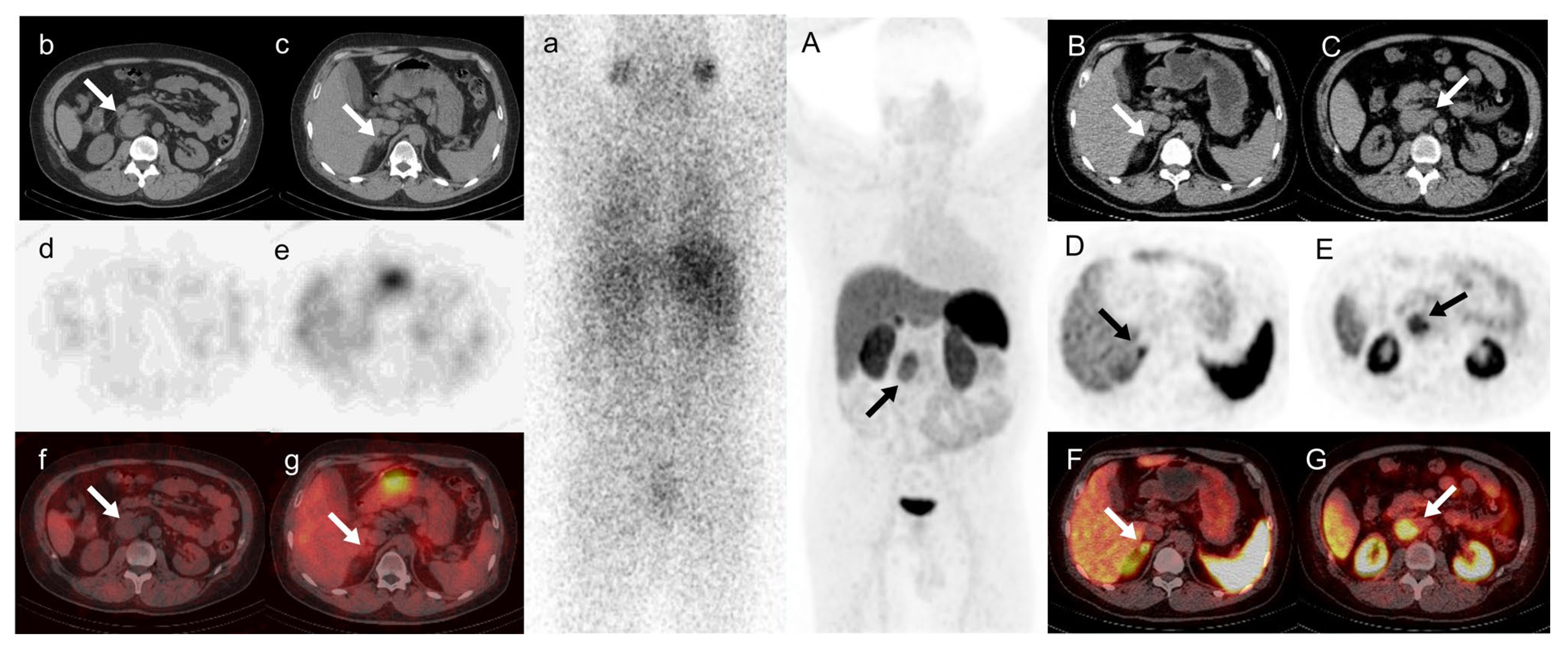
A 42-year-old man presented who had experienced repeated dizzy spells for 4 years. His blood pressure was 220/140 mmHg at the time of the attack. He complained of recent edema in both ankles and foamy urine. An upper abdominal CT scan revealed a suspicious retroperitoneal mass and a nodule on the lateral branch of the right adrenal gland. His catecholamine levels were slightly elevated, his aldosterone-to-renin ratio was 51.4 [pg/mL]/[uIU/mL], and captopril and saline inhibition tests were positive. Clinicians made a comprehensive diagnosis of primary aldosteronism, which may be associated with paraganglioma. Therefore, it is suggested to improve the relevant imaging examination, especially radionuclide functional imaging. 131I-MIBG SPECT images (a–g) indicated no radioactive uptake by the retroperitoneal mass or adrenal nodule. 68Ga-DOTATATE PET/CT images ((A) MIP; (B,D,F) arrows, adrenal nodule; (C,E,G) arrows, retroperitoneal mass) showed no significant increase in radioactive uptake in the right adrenal nodule. The SUVmax was 7.6 (the SUVmax of the left normal adrenal tissue was 7.2), while there was somatostatin receptor expression on the retroperitoneal mass with an SUVmax of 8.1. The SUVmax in the retroperitoneal mass is higher than in the right adrenal nodule, implying the mass had a higher level of CXCR4 expression. Castleman disease (CD) is a heterogeneous lymphoproliferative disease [1]. The key to its treatment lies in the evaluation of the systemic condition, but because of its varying locations and clinical manifestations, diagnosis can be quite challenging [2,3,4,5]. Thus, effective diagnostic methods are needed [6]. In this case, we found high uptake for both 68Ga-DOTATATE PET/CT and local 68Ga-pentixafor PET/CT. As we observed in this case, CD lesions exhibited notable 68Ga-DOTATATE absorption, which was also recently reported in two additional CD patients [4,7]. A CT scan of the former patient revealed a mediastinal mass after a neuroendocrine tumorectomy [4], and a scan of the latter patient revealed an inadvertent pancreatic mass [7]. To rule out a neuroendocrine condition, they both received 68Ga-DOTATATE PET/CT in a clinical setting. A surgical pathology test verified that they both had CD. Although the exact role of 68Ga-DOTATATE PET/CT in CD remains unclear, it interestingly presents a unique idea for the diagnosis of the disease: 68Ga-DOTATATE PET could be a helpful diagnostic tool. In this case, the high 68Ga-pentixafor uptake of CD was accidentally found in the process of evaluating primary aldosteronism with 68Ga-pentixafor PET/CT. This indicated that there is a certain degree of CXCR4 expression in CD, due to which CXCR4 may be highly expressed in a variety of solid tumors, and this receptor may be a suitable target for molecular imaging [8,9]. Of course, this also can assist in the clinical diagnosis of CD and reflect the expression of CXCR4. The focus of CD in this patient was in the retroperitoneum rather than the mediastinum, which is the most common location for unicentric CD [10]. Both 68Ga-DOTATATE and 68Ga-pentixafor PET/CT exhibited positive uptake, which has not previously been reported in a case of CD. We provide new imaging data for diagnosing CD. The positive uptake of both 68Ga-DOTATATE and 68Ga-pentixafor in lesions may further confirm this suspicion when the possibility of CD is considered clinically.
Figure 1.
A 42-year-old man presented who had experienced repeated dizzy spells for 4 years. His blood pressure was 220/140 mmHg at the time of the attack. He complained of recent edema in both ankles and foamy urine. An upper abdominal CT scan revealed a suspicious retroperitoneal mass and a nodule on the lateral branch of the right adrenal gland. His catecholamine levels were slightly elevated, his aldosterone-to-renin ratio was 51.4 [pg/mL]/[uIU/mL], and captopril and saline inhibition tests were positive. Clinicians made a comprehensive diagnosis of primary aldosteronism, which may be associated with paraganglioma. Therefore, it is suggested to improve the relevant imaging examination, especially radionuclide functional imaging. 131I-MIBG SPECT images (a–g) indicated no radioactive uptake by the retroperitoneal mass or adrenal nodule. 68Ga-DOTATATE PET/CT images ((A) MIP; (B,D,F) arrows, adrenal nodule; (C,E,G) arrows, retroperitoneal mass) showed no significant increase in radioactive uptake in the right adrenal nodule. The SUVmax was 7.6 (the SUVmax of the left normal adrenal tissue was 7.2), while there was somatostatin receptor expression on the retroperitoneal mass with an SUVmax of 8.1. The SUVmax in the retroperitoneal mass is higher than in the right adrenal nodule, implying the mass had a higher level of CXCR4 expression. Castleman disease (CD) is a heterogeneous lymphoproliferative disease [1]. The key to its treatment lies in the evaluation of the systemic condition, but because of its varying locations and clinical manifestations, diagnosis can be quite challenging [2,3,4,5]. Thus, effective diagnostic methods are needed [6]. In this case, we found high uptake for both 68Ga-DOTATATE PET/CT and local 68Ga-pentixafor PET/CT. As we observed in this case, CD lesions exhibited notable 68Ga-DOTATATE absorption, which was also recently reported in two additional CD patients [4,7]. A CT scan of the former patient revealed a mediastinal mass after a neuroendocrine tumorectomy [4], and a scan of the latter patient revealed an inadvertent pancreatic mass [7]. To rule out a neuroendocrine condition, they both received 68Ga-DOTATATE PET/CT in a clinical setting. A surgical pathology test verified that they both had CD. Although the exact role of 68Ga-DOTATATE PET/CT in CD remains unclear, it interestingly presents a unique idea for the diagnosis of the disease: 68Ga-DOTATATE PET could be a helpful diagnostic tool. In this case, the high 68Ga-pentixafor uptake of CD was accidentally found in the process of evaluating primary aldosteronism with 68Ga-pentixafor PET/CT. This indicated that there is a certain degree of CXCR4 expression in CD, due to which CXCR4 may be highly expressed in a variety of solid tumors, and this receptor may be a suitable target for molecular imaging [8,9]. Of course, this also can assist in the clinical diagnosis of CD and reflect the expression of CXCR4. The focus of CD in this patient was in the retroperitoneum rather than the mediastinum, which is the most common location for unicentric CD [10]. Both 68Ga-DOTATATE and 68Ga-pentixafor PET/CT exhibited positive uptake, which has not previously been reported in a case of CD. We provide new imaging data for diagnosing CD. The positive uptake of both 68Ga-DOTATATE and 68Ga-pentixafor in lesions may further confirm this suspicion when the possibility of CD is considered clinically.

Figure 2.
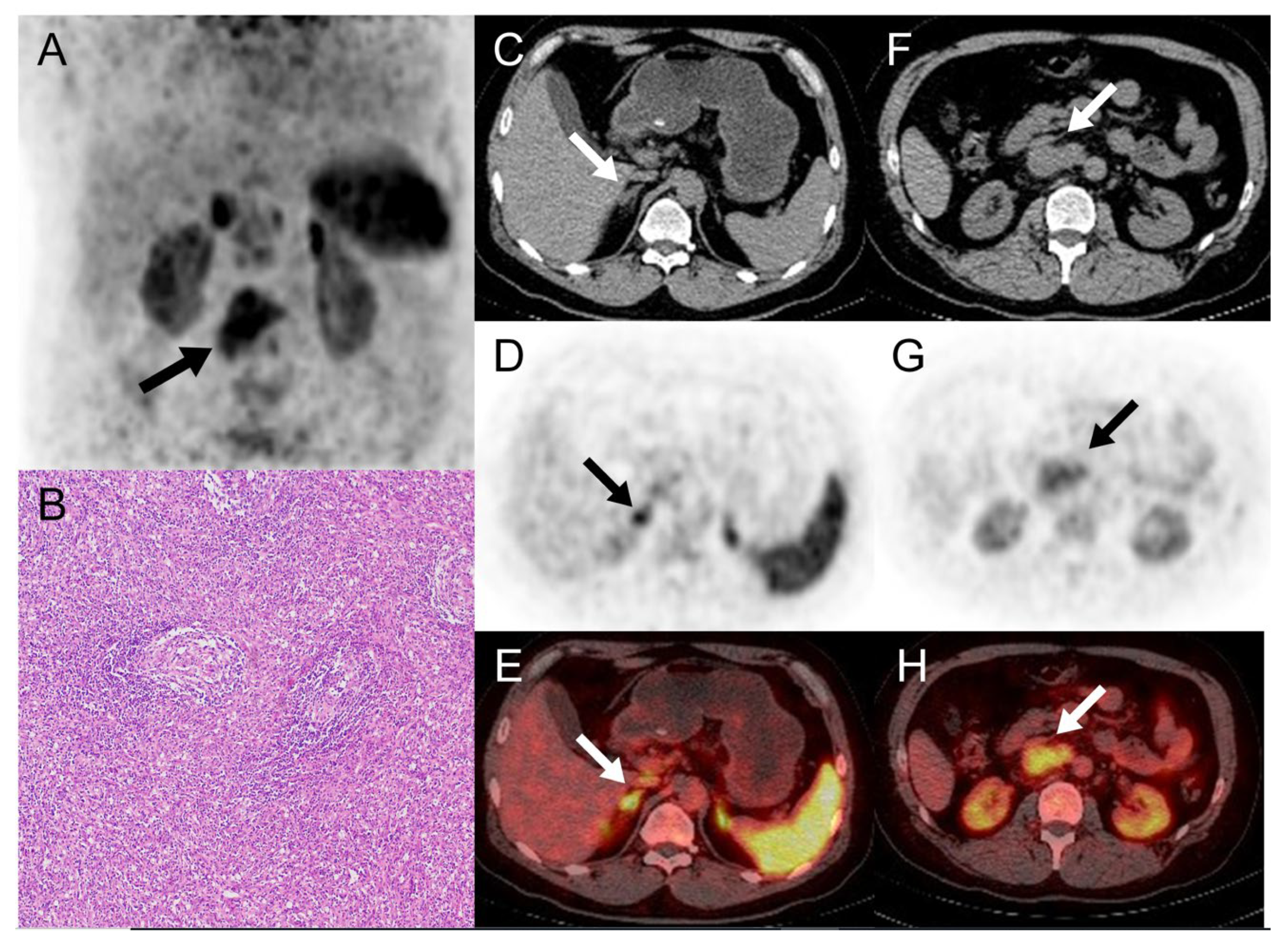
The MIP, axial CT, PET, and fusion PET/CT images of local 68Ga-pentixafor PET/CT showed no abnormal uptake in the right adrenal nodule ((C–E), arrows, SUVmax, 7.3; the contralateral normal adrenal gland, SUVmax, 7.1) and the presence of CXCR4 expression in the retroperitoneal mass (A,F–H), arrows, SUVmax, 6.2). Robot-assisted laparoscopic resection of the retrovena cava tumor and right adrenal gland was performed. A postoperative pathological test of the retroperitoneal mass revealed atypical large-cell lymphoid proliferation in a microscopic section of hematoxylin–eosin stain (B) (original magnification × 10), the immunohistochemistry analyses was consistent with Castleman’s disease ((C,D) hyaline vascular type), and the right adrenal nodule was normal tissue.
Figure 2.
The MIP, axial CT, PET, and fusion PET/CT images of local 68Ga-pentixafor PET/CT showed no abnormal uptake in the right adrenal nodule ((C–E), arrows, SUVmax, 7.3; the contralateral normal adrenal gland, SUVmax, 7.1) and the presence of CXCR4 expression in the retroperitoneal mass (A,F–H), arrows, SUVmax, 6.2). Robot-assisted laparoscopic resection of the retrovena cava tumor and right adrenal gland was performed. A postoperative pathological test of the retroperitoneal mass revealed atypical large-cell lymphoid proliferation in a microscopic section of hematoxylin–eosin stain (B) (original magnification × 10), the immunohistochemistry analyses was consistent with Castleman’s disease ((C,D) hyaline vascular type), and the right adrenal nodule was normal tissue.

Author Contributions
Conceptualization and investigation: R.Z.; writing—original draft preparation: L.X.; writing—review and editing: H.P. All authors have read and agreed to the published version of the manuscript.
Funding
This research received no external funding.
Institutional Review Board Statement
The Institutional Review Board of Chongqing Medical University approved this study (approval number: K2023-039, approval date: 19 March 2023).
Informed Consent Statement
Informed consent was obtained from all subjects involved in the study. Written informed consent has been obtained from the patient to publish this paper.
Data Availability Statement
Most data generated during this study are included in this published article. The data are available from the corresponding author on reasonable request, if you are confused about some of the data.
Conflicts of Interest
The authors declare no conflicts of interest.
References
- Carbone, A.; Borok, M.; Damania, B.; Gloghini, A.; Polizzotto, M.N.; Jayanthan, R.K.; Fajgenbaum, D.C.; Bower, M. Castleman disease. Nat. Rev. Dis. Primers 2021, 7, 84. [Google Scholar] [CrossRef] [PubMed]
- Li, F.; Xiao, L.; Cai, H.; Li, L. Colonic Castleman Disease on FDG PET/CT. Clin. Nucl. Med. 2023, 48, 71–72. [Google Scholar] [CrossRef] [PubMed]
- Ding, J.; Cheng, X.; Hou, G.; Jing, H.; Huo, L. Adrenal Castleman Disease on 99mTc-HYNIC-TOC Scan and FDG PET/CT. Clin. Nucl. Med. 2021, 46, 71–73. [Google Scholar] [CrossRef] [PubMed]
- Verçosa, A.F.A.; Flamini, M.; Loureiro, L.V.M.; Flamini, R.C. 68Ga-DOTATATE and 18F-FDG in Castleman Disease. Clin. Nucl. Med. 2020, 45, 868–870. [Google Scholar] [CrossRef] [PubMed]
- Wang, P.; Hou, G.; Li, F.; Cheng, X. Hypermetabolic Unicentric Castleman Disease of Kidney on FDG PET/CT. Clin. Nucl. Med. 2021, 46, 510–511. [Google Scholar] [CrossRef] [PubMed]
- Abramson, J.S. Diagnosis and Management of Castleman Disease. J. Natl. Compr. Cancer Netw. JNCCN 2019, 17, 1417–1419. [Google Scholar] [CrossRef] [PubMed]
- Liu, S.L.; Luo, M.; Gou, H.X.; Yang, X.L.; He, K. Castleman disease of the pancreas mimicking pancreatic malignancy on 68Ga-DOTATATE and 18F-fluorodeoxyglucose positron emission tomography/computed tomography: A case report. World J. Gastrointest. Surg. 2022, 14, 514–520. [Google Scholar] [CrossRef] [PubMed]
- Buck, A.K.; Serfling, S.E.; Lindner, T.; Hänscheid, H.; Schirbel, A.; Hahner, S.; Fassnacht, M.; Einsele, H.; Werner, R.A. CXCR4-targeted theranostics in oncology. Eur. J. Nucl. Med. Mol. Imaging 2022, 49, 4133–4144. [Google Scholar] [CrossRef] [PubMed]
- Werner, R.A.; Kircher, S.; Higuchi, T.; Kircher, M.; Schirbel, A.; Wester, H.J.; Buck, A.K.; Pomper, M.G.; Rowe, S.P.; Lapa, C. CXCR4-Directed Imaging in Solid Tumors. Front. Oncol. 2019, 9, 770. [Google Scholar] [CrossRef] [PubMed]
- Dispenzieri, A.; Fajgenbaum, D.C. Overview of Castleman disease. Blood 2020, 135, 1353–1364. [Google Scholar] [CrossRef] [PubMed]
Disclaimer/Publisher’s Note: The statements, opinions and data contained in all publications are solely those of the individual author(s) and contributor(s) and not of MDPI and/or the editor(s). MDPI and/or the editor(s) disclaim responsibility for any injury to people or property resulting from any ideas, methods, instructions or products referred to in the content. |
© 2024 by the authors. Licensee MDPI, Basel, Switzerland. This article is an open access article distributed under the terms and conditions of the Creative Commons Attribution (CC BY) license (https://creativecommons.org/licenses/by/4.0/).
Share and Cite
MDPI and ACS Style
Zuo, R.; Xu, L.; Pang, H. 68Ga-DOTATATE and 68Ga-Pentixafor PET/CT in a Patient with Castleman Disease of the Retroperitoneum. Diagnostics 2024, 14, 372. https://doi.org/10.3390/diagnostics14040372
AMA Style
Zuo R, Xu L, Pang H. 68Ga-DOTATATE and 68Ga-Pentixafor PET/CT in a Patient with Castleman Disease of the Retroperitoneum. Diagnostics. 2024; 14(4):372. https://doi.org/10.3390/diagnostics14040372
Chicago/Turabian StyleZuo, Rui, Lu Xu, and Hua Pang. 2024. "68Ga-DOTATATE and 68Ga-Pentixafor PET/CT in a Patient with Castleman Disease of the Retroperitoneum" Diagnostics 14, no. 4: 372. https://doi.org/10.3390/diagnostics14040372
Note that from the first issue of 2016, this journal uses article numbers instead of page numbers. See further details here.