Article

Protection and Rehabilitation Effects of Cordyceps militaris Fruit Body Extract and Possible Roles of Cordycepin and Adenosine

1
Department of General and Inorganic Chemistry, Hanoi University of Pharmacy, Hanoi 111000, Vietnam
2
Center for Research and Technology Transfer, Vietnam Academy of Science and Technology (VAST), Hanoi 122100, Vietnam
3
Institute of Biotechnology, Vietnam Academy of Science and Technology (VAST), Hanoi 122100, Vietnam
4
Transdisciplinary Science and Engineering Program, Graduate School of Advanced Science and Engineering, Hiroshima University, Hiroshima 739-8529, Japan
5
Center for The Planetary Health and Innovation Science (PHIS), the IDEC Institute, Hiroshima University, Higashi-Hiroshima 739-8529, Japan
*
Author to whom correspondence should be addressed.
Compounds 2022, 2(4), 388-403; https://doi.org/10.3390/compounds2040032
Submission received: 26 November 2022 / Revised: 14 December 2022 / Accepted: 16 December 2022 / Published: 19 December 2022
(This article belongs to the Special Issue Feature Papers in Compounds (2022–2023))

Abstract

:
Cordyceps militaris is a valued medicinal fungus in folk medicine in East Asia. It contains two major nucleosides, cordycepin and adenosine, which have been reported to have potential antineoplastic, antioxidant, and anti-inflammatory activities. This paper aimed to study the effect of C. militaris extract on the reproductive function of a mouse model, evaluating possible toxicity, androgenic activity, and protective and rehabilitative effects against damages caused by sodium valproate (VPA). There was no death and abnormalities observed in mice. Androgen activity was also shown in young male rats by an improvement in several sexual organs. The protective effect of C. militaris extract was explained by the gain of sexual organs’ weight, testosterone concentration, and seminiferous tubule size as well as the enhancement of sperm density, alive sperm percentage, and the progressive forward movement of sperm. The pregnancy rate of female rats paired with VPA-administered male rats (500 mg/kg/day) increased proportionally with the higher dose of C. militaris extract. In the rehabilitation study, an incline in the weight of the Cowper’s gland and glans (0.112 g/kg/day) and testicle and prostate (0.336 g/kg/day) as well as an improvement of the sperm forward progressive movement was observed. The percentage of unprogressive sperm and immotile sperm has reduced. These results suggest that C. militaris is a potential supplement to reduce the negative effects of VPA and improve reproductive function, in which the two major constituents cordycepin and adenosine may play an active role.

1. Introduction

Sodium valproate (VPA), due to its wide therapeutic properties, is amongst the preferred remedies for epileptic symptoms such as the Lennox–Gastaut syndrome, tonic–clonic seizures, absence seizures, and myoclonic seizures [1,2]. Furthermore, it was suggested as an advantageous drug for other diseases such as infantile spasms, bipolar psychiatric disorders, and migraine. Although it is beneficial to medical applications to a certain extent, on the other hand, VPA has been observed to have negative impacts on reproductive endocrine function as well as the semen quality of epileptic patients [3,4]. Long-term exposure to VPA in animals can lead to testicular atrophy and semen abnormality [5,6]. Additionally, an increase in free radical levels is considered a consequence of VPA treatment, and the VPA-associated complications might be explained by the biotransformation and/or alterations in natural antioxidants within VPA [7,8,9]. To negate such toxic effects, antioxidants found in plant extracts are promising factors due to their protective attributes against chemical-induced toxicity. However, their effects on VPA-induced reproductive function have remained an unconfirmed topic for medication purposes.
Fungi create a variety of secondary metabolites, some of which are beneficial compounds [10,11]. Fungi phytotoxicity is determined by the number, intensity, and variety of phytochemicals present [12]. Many phytochemicals derived from fungi are used to mitigate harmful effects, and throughout the history of medicinal use in Eastern and Chinese medicine, cordyceps has emerged as a miracle cure for many common ailments [13]. The main source from nature collected in Tibet is a parasitic fungus on worms [14]. It is very difficult and expensive to obtain such original natural products. Instead, the discovery of the similarity of chemical constituents between C. militaris and Cordyceps sinensis has become the alternative solution to derived products of Cordyceps sinensis [15]. There have been many publications showing that the content of two main active ingredients, adenosine and cordycepin, in C. militaris is much higher and superior to that of the Cordyceps sinesis strain [16].
C. militaris (L.), a fungus specie of the Ascomycetes class, is widely used for therapeutic purposes due to its diverse physiological and pharmacological properties, such as anti-fatigue, anti-stress, antioxidant, anti-fungal, and anti-cancer [17,18,19,20,21]. Numerous active components have been found in this living organism such as adenine, cordyheptapeptide, amino acids, polysaccharides, fatty acids, cordycepin, and adenosine [22]. Cordycepin and anucleoside analog (3′-deoxyadenosine) in particular are among the most active compounds that can contribute to tumor cell apoptosis, a decrease in tumor cell proliferation, and several therapeutic activities (antineoplastic, antioxidant, and anti-inflammatory activities) [23,24,25]. There have been many publications on the toxicity, acute toxicity, and reproductive effects of C. militaris extract in experimental animals, but most of the previous publications only targeted one gender or lacked uniform toxicity assessments [26,27,28,29]. Within the medicinal field in China, C. militaris extract is widely applied to ease the lung and strengthen the kidney in order to cure hyperglycemia, renal dysfunction, hyperlipidemia, and liver diseases [30]. Moreover, the beneficial effects of C. militaris have not only been shown on such types of diseases, but more importantly, they have been believed to play a certain role in the treatment of impotence, seminal emission, and infertility [31]. Indeed, C. militaris supplementation can improve sperm quality in several animals, including boars and rats. Within two months of supplementation with C. militaris, growth in the proportion of motile sperm cells and the sperm morphology of boars has been observed, while the serum testosterone and estradiol-17 of rats after 6 weeks of C. militaris treatment increased significantly [32,33]. The enhancement of reproductive function and the repair of reproductive dysfunction of cyclophosphamide-induced mice were also described [34].
Thus, in order to affirm the benefits of C. militaris and conduct further research on its effects on the fertility system, an evaluation of the protective and rehabilitation potential, the androgenic activity, as well as the safety of the fungus were performed on mouse models.

2. Materials and Methods

2.1. Materials and Reagent

2.1.1. Animals

Healthy Wistar rats were obtained from the National Institute of Hygiene and Epidemiology and Vietnam Military Medical Academy (Hanoi, Vietnam), respectively. Wistar rats, both male and female, were obtained in two age groups; the premature rats were 42–50 days old, and the mature weight was approximately 180 g. After transportation to the laboratory and before the onset of the experiments, the animals were allowed to adapt to the laboratory environment and conditions for 5–7 days. Before and during experimentation, the animals were fed standard food, which consisted of diet cubes containing carbohydrate (66.67%), protein (19.31%), minerals (5.71%), cellulose (4.87%), and fat (3.44%), and were supplied with water freely.

2.1.2. Material and Chemicals

C. militaris extracts (CE) were collected and commercialized by Hoa Binh Biopharm Joint Stock Company, Hoa Binh City, Vietnam. Sodium valproate (Dépakine) tablet was produced by Sanofi Synthelabo Inc. Testosterone propionate (Tesmon—injection tubes of 25 mg/mL) produced by Tai Yu Chemical & Pharma Co., Ltd. (Zhudong Township, Taiwan). Test kits for hematology analysis was purchased from Hospitex Diagnostics (Sesto Fiorentino, Toscana, Italy) and DIALAB® GmbH (Vienna, Austria).

2.2. Experimental Instrument

A Semi-Auto Biochemistry Analyzer (Erba, India) was employed to quantify the parameters. Sperm analyzer CASA (IVOS II TM, MICROPTIC, Hamilton Thorne, Barcelona, Sapin) was employed to determine the sperm quality of the male animals.

2.3. Toxicity Evaluation of C. militaris Extract on Experimental Animals

2.3.1. Acute Toxicity on Swiss Webster White Mice

The experiment was conducted following the Litchfield–Wilcoxon method [35]. In detail, Swiss Webster white mice weighing 20 ± 2 g were divided into groups of 10 each. Each group was treated orally with C. militaris extract from the highest non-lethal dose to the lowest lethal dose causing 100% death. Mice were fasted for 12 h and adequately hydrated before the oral intake. The number of dead mice in the first 72 h and their general condition in 7 days after the oral intake was observed and recorded (eating habits, neural activity, walking, excretion, etc.). If the rat died, anatomical dissection was performed in order to assess the gross organ damage. The lethal dose of 50% (LD50) was determined by the percentage of dead mice within the first 72 h.

2.3.2. Semi-Chronic Toxicity on Wistar Rats

Wistar rats were divided randomly into 3 groups and were exposed to the following 90-day regimens of (1) distilled water (S1—Control Group) with a volume of 1.0 mL/100 g/day; (2) C. militaris extract (S2—Group 2) with the dose of 0.336 g/kg/day and the same volume; (3) S3—Group 3 were exposed to another dose of C. militaris extract (1.008 g/kg/day) with the same volume. The regimens were given once a day in the morning. Several parameters were evaluated during and after the intake period included overall condition, weight, hematopoietic function (red blood cell, white blood cell count, mean corpuscular volume, hemoglobin, hematocrit, white blood cell formula, and platelet count), liver function (total bilirubin, albumin, total cholesterol, AST, and ALT), and kidney function (plasma creatinine level). The evaluation of all parameters was performed before the regimens and within 30 days, 60 days, and 90 days and after the regimens. Finally, the animals were sacrificed for the overall anatomical dissection after the 90-day period.

2.4. Androgenic Activity of C. militaris Extract on Castrated Premature Male Wistar Rats

Hershberger’s approach was applied to conduct the examination [36]. The castrated premature male Wistar rats were reared during examination for 5–7 days in the laboratory environment and then were randomly divided into 6 groups. Each group was treated with different regimens: (1) A1—Control Group (uncastrated rats) was administered reference solution (10 mL/kg/day); (2) A2—Group 2 was exposed to reference solution (10 mL/kg/day); (3) A3—Group 3 was injected with testosterone propionate at 0.4 g/kg/day; (4) A4—Group 4 was given CE (0.112 g/kg/day); (5) A5—Group 5 was treated with a 3-times-higher dose of CE (0.336 g/kg/day); A6—Group 6 was given a 5-times-higher dose of CE (0.560 g/kg/day). The testicles of the animals were removed in groups A2-A6, except for group A1. After resting for the next 7 days, animals in each group were given the corresponding regimens for the following 10 days. Twenty-four hours later, the animals were weighed and then sacrificed for anatomical dissection. The testosterone levels in the animals’ blood were determined, and genital organs (seminal vesicle, Cowper’s gland, supralevator abscess, prostate, and glans) were isolated and weighed.

2.5. Effects of C. militaris Extract on Reproductive Function of Wistar Rats Induced by VPA

2.5.1. Protective Effect

In this study, forty male Wistar rats were randomly divided into 4 groups, 3 of which were treated with VPA (500 mg/kg/day): (1) P1—Control Group was given distilled water (10 mL/kg/day) without the treatment of VPA; (2) P2—Group 2 was administered VPA then given distilled water (10 mL/kg/day); (3) P3—Group 3 was first exposed to VPA then given CE (0.112 g/kg/day); (4) P4—Group 4 was exposed to VPA then given CE with a 3-times-higher dose (0.336 g/kg/day). The regimens for each group were given twice a day for 7 weeks. On the 5th week after the first regimens, the mating procedure was performed by moving 1 male rat and 2 female rats into one cage for 2 weeks (14 days). After 7 weeks of study, with male rats, evaluation of sperm density, living sperm rate, sperm mobility, sperm morphology, and seminiferous tubule size were examined; with female rats, mean of corpus luteum, normal pregnancy, normally developed fetuses, egg loss rate, early fetal death rate, and late fetal death rate were examined. Notably, the parameters of corpus luteum, normal pregnancy, and normally developed fetuses were calculated by the mean of counted cells/pregnancy/fetuses over one female rat individual. The egg loss rate was determined by the subtraction of corpus luteum to normally developed fetuses, while early and late fetal death rate were calculated by the number of fetal deaths over the total normal pregnancies.

2.5.2. Rehabilitation Effect

In this study, healthy male Wistar rats were randomly divided into 4 groups with different stages of regimens: (1) R1—Control Group was treated only with distilled water; (2) R2—Group 2 was exposed to VPA (500 mg/kg/day) in the first 7 weeks (stage 1), then given distilled water in the next 10 days (stage 2); (3) R3—Group 3 was administered VPA (500 mg/kg/day) in stage 1 then given CE (0.112 g/kg/day) in stage 2; (4) R4—Group 4 was administered VPA (500 mg/kg/day) in stage 1 then given a 3-times-higher dose of CE (0.336 g/kg/day) in stage 2. After the study period, the mating procedure was performed within 10 days. Similar to the protective assay, the same parameters for both male and female rats were evaluated.

2.6. Statistical Analysis

Excel 2016 was employed to process raw data after the initial measurement. The data were presented as mean ± standard deviation (SD) or percentage (%). The means and SD of all parameters related to the weight of sexual organs and reproductive function were calculated for all the experimental group by two-way ANOVA employing SPSS 15.0 (SPSS, Chicago, IL). The means of individual groups were compared using post hoc two-tailed t tests for independent variables with equal variance not assumed (SPSS). The difference was significant when p < 0.05.

3. Results

3.1. Impact of Possible Toxicity of C. militaris on Experimental Animals

3.1.1. Acute Toxicity on Swiss Webster White Mice

The dose of 89.0 g/kg (weight of the animal) of C. militaris extract (CE) was the highest intake applied to the mice in this experiment. The results showed that there were no deaths or abnormalities observed in the mice, including in weight, general condition, skin, fur, eyes, and mucous membranes; no behavior disorders were observed during the first 72 h after the regimens. In the following 7 days, the treated mice were still in normal condition. The parameter LD50 could not be determined due to the limitation of the volume and concentration of the CE used for the intake in the mice. The highest dose of CE that could be ingested orally in the mice was 89.0 g/kg (463.54 times higher than the equivalent suggested dose for human).

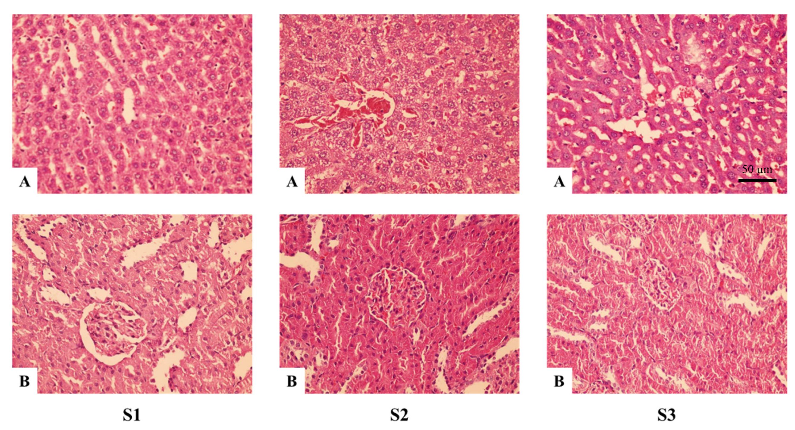
3.1.2. Semi-Chronic Toxicity in Wistar Rats

Within the experimental time of 90 days, the rats from all three groups were in normal condition, healthy, having bright eyes and dry stool. There were no behavioral abnormalities observed in any of the rats during the experimentation. Table 1 depicts the long-term effect of the CE intake on the weight, hematopoiesis, hepatic panel, and renal function of the Wistar rats and also the changes of the parameters recorded before the regimen and on the 30th, 60th, and 90th days of the regimen. The results showed that changes were only recorded in the weight of the rats, which increased steadily throughout the experimental period in all the groups of S1, S2, and S3. The weight of rats given CE (both dosages of 0.336 and 1.008 g/kg/day) was higher than that of the control group (given only distilled water). There were no significant alterations in the haematopoiesis, hepatic panel, or renal function of the rats over the course of the experimental period or between the three different groups.
Figure 1 illustrates the observations of the hepatic (Figure 1A) and renal (Figure 1B) microbody of Wistar rats in the 400× magnification after the 90-day regimen. All of the obtained observations revealed no damage or degenerations of the hepatic and renal cells. No significant difference was observed between the cells of the biological control group (S1) and the groups given CE (S2, S3). The microbody images confirmed the nontoxicity of CE to the function of both the liver and kidney of the rats.
The dissection of the rats was performed in order to evaluate the alteration in the weight of sexual organs and the level of testosterone in line with the different regimens. Table 2 shows that the androgenic activity of CE was not observed evenly in all the examined parameters. As compared to group A2 which was given a reference solution, the testosterone content in rats given CE witnessed a proportional trend along the increasing dosage of 0.112–0.560 g/kg/day but was not significantly different (p > 0.05). The significant increases in all three dosages of CE given to rats were detected in the weight of several organs, such as the vesicles and prostate. At the highest content of CE given to rats (0.560 g/kg/day), the weights of 4 out of 5 sexual organs (vesicles, prostate, glans, and Cowper’s gland) were significantly changed (p < 0.05), typically 1.4 to 3.5 times higher than that of group A2. In the group of castrated rats treated with testosterone propionate (A3), all of the parameters observed a significant increase as compared to the biological control group (A1).

3.2. Effect of C. militaris Extract on the Reproduction Function of Wistar Rats Induced by VPA

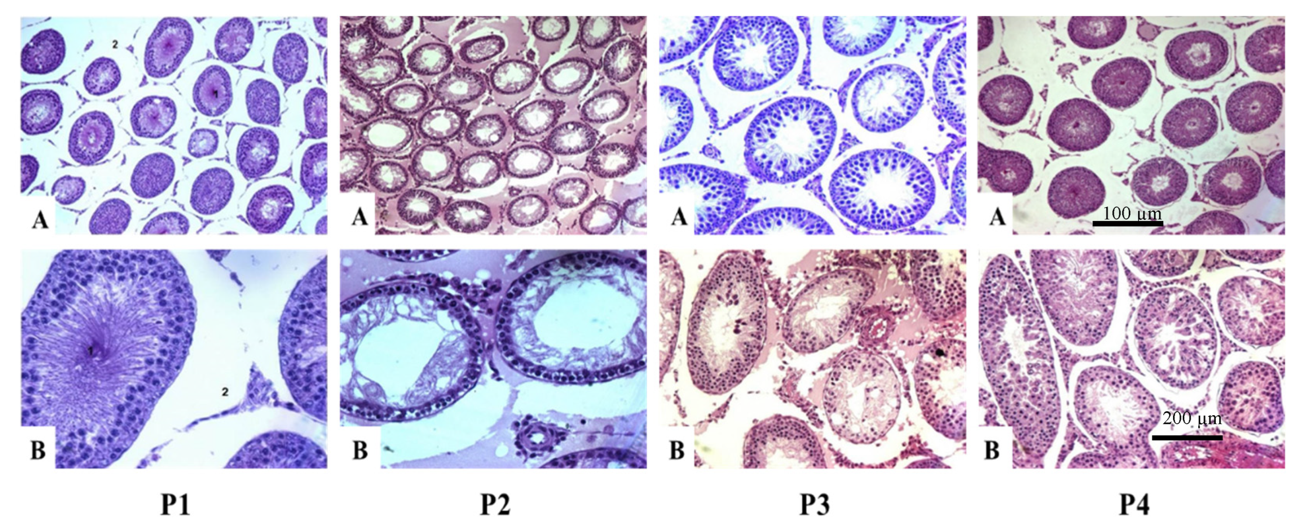
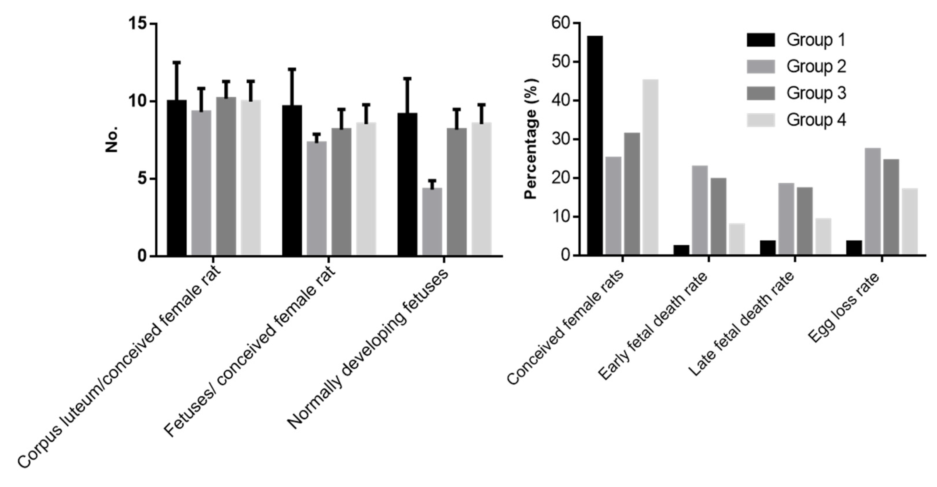
3.2.1. Protective Effect

The reproduction protective activity of CE was studied using mature male Wistar rats exposed to both VPA and CE at the same time. VPA has shown its toxicity against rat reproduction and played the role of a toxic reproductive agent in this study. CE was fed simultaneously with VPA in order to observe the Cordyceps’ protective activity via the difference between treatments with CE and biological control and VPA controls. The fertility-related parameters were measured and evaluated in both genders, male and female rats. In male rats, the measured parameters included sexual organs’ weight, properties of semen from the epididymis, testicular morphology, and seminiferous tubule sizes (Table 3) (Figure 2). Meanwhile, within the female rat group, the indicators of interest were conception rate, number of corpus luteus, fetuses, and fetal death during pregnancy development (Figure 3).
Sodium valproate control (Group P2) was treated with 500 mg/kg/day of VPA for seven consecutive weeks. The VPA treatment showed a significant impact on the rat reproductive system, which is illustrated by the decrease in sexual organs’ weights, semen and sperm quality, as well as testosterone production and the size of seminiferous tubules (Table 3). There were no more than 2 parameters out of the 13 evaluated, such as the weight of the Cowper’s gland and glans, which did not show significant differences between VPA control and biological control (Group P1). Additionally, Group P3 consisted of rats who took an equal dose of VPA as in Group P2 but were treated with 0.112 g/kg CE in the meantime and showed a similar effect on fertility-related parameters to Group 2. However, compared to the biological control, Group P3 had five parameters statistically close to Group P1.
By contrast, there was a significant improvement in both the quality and quantity of fertility-related factors within the group of rats treated with a higher amount of CE (0.336 g/kg) (Group P4). The positive impact of CE was seen in the increase of sexual organs’ weight, testosterone concentration, and the size of seminiferous tubules. The quality of semen and sperm was enhanced and demonstrated via the higher sperm density, percentage of live sperms, and the progressive forward movement of sperms (Table 3). The testicular morphology of male rats with reproductive failure induced by VPA was shown in Figure 3. The seminiferous tubules were round and taut in the biological control group P1, with a thin basement membrane. Most tubules were narrow and contained many spermatozoa. The epithelium was thick and had all types of sperm cells: spermatocytes, spermatocytes, pre-sperm, and spermatozoa. The proportions of spermatogenic cells varied in the seminiferous tubules. The interstitial spaces were sparse with small blood vessels (Figure 2(P1)). By contrast, the reproductive failure induced by VPA groups exhibited different morphologies. The differences were distinguished between groups treated with and without CE. There were no signs of infection in the testicles of Groups P2,3, or 4. One-third of the mice in Group 2 had congestion and fluid retention in the interstitial spaces, but Groups P3 and P4 did not. Group P2’s seminiferous tubules had a small size, wide lumen, and thin epithelium, while Group P3 also displayed a wide lumen and thin epithelium. Still, there are only Sertoli cells, spermatocytes, and spermatozoa in the epithelium, no pre-sperm or sperm (Figure 2(P2,P3)). With a high concentration of CE, most of the mice in Group P4 had a healthy testicular structure similar to the biological control group P1 (Figure 2(P4)).
The number female rats which conceived and their pregnancy developments were observed by dissection at day 14 to 17 of their pregnancy. The percentage of pregnant female mice paired with male mice in Group P2 was lowest in the tested treatments at 25%, and the numbers increased in correlation with additional CE. Even though the group was treated with 0.336 g/kg CE, the females in Group P4 that conceived were less than the biological control. However, the ratio of corpus luteum to a conceived female rat was kept the same among all four groups. Additionally, the number of fetuses per conceiving female rat and normal developing fetuses decreased significantly compared with the control group (p < 0.05). At the same time, the rate of fetal death and egg loss increased significantly (p < 0.05) in all three treated groups (Figure 2).
However, trending data can be seen in Figure 3, in which CE showed an effective impact on rat reproduction. By increasing the concentration of CE, the number of conceived female rats and normally developing pregnancies was increased while the ratio of fetal death was decreased.

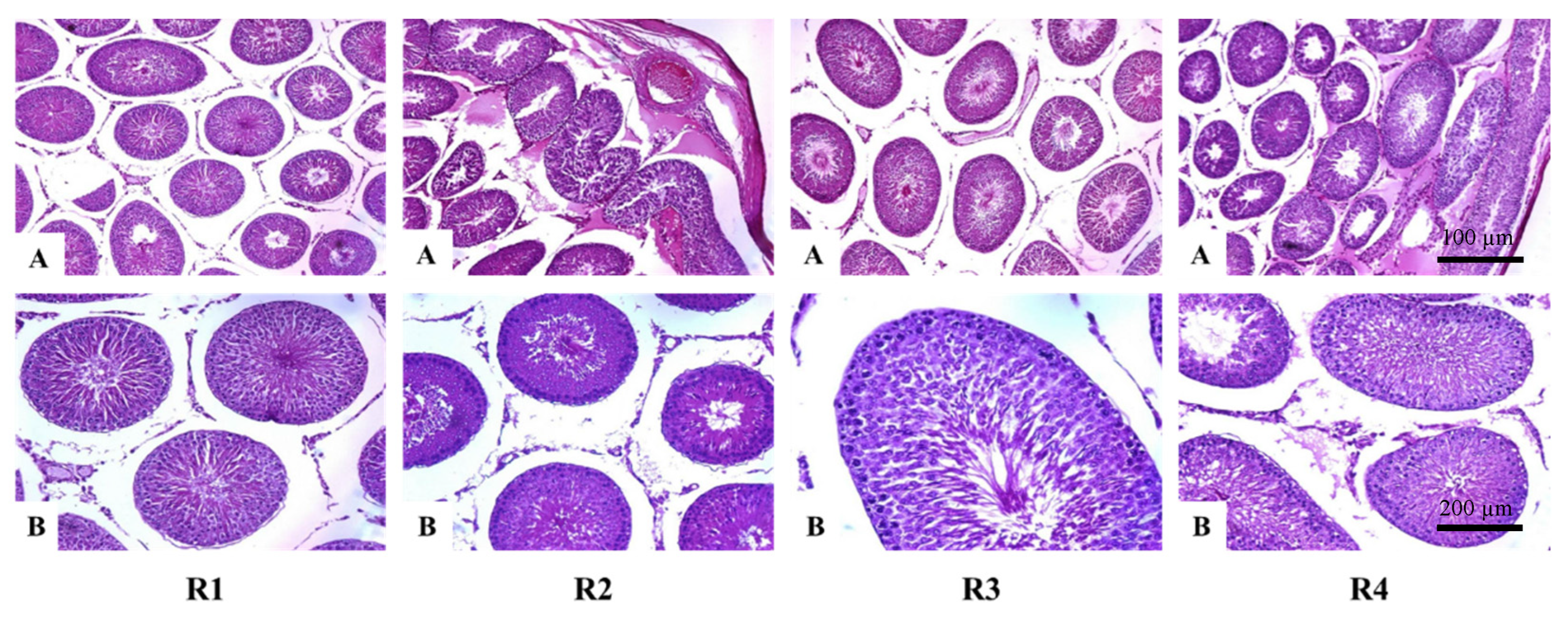
3.2.2. Rehabilitation Effect

The rehabilitation activity of CE was studied by using different doses of CE after induced reproductive failure in male Wistar rats by VPA. After seven weeks of taking VPA 500 mg/kg/day, the tested groups were treated with 0.112 g/kg/day and 0.336 g/kg/day of CE for 10 days and named Group R3 and Group R4, respectively. Group R2 included Wistar male rats that took VPA 500 mg/kg/day for seven days and functioned as a control. Group R1 was the biological control, including male rats using distilled water instead of VPA. After 10 days treated with CE, male rats were mated with healthy female rats, and the pregnancy of the female was used to evaluate the recovery activity of CE along with the male rat’s fertility-related properties.
With the treatment using CE for male rats with reproductive failure caused by VPA, the effects observed included the increase of the weight of the Cowper’s gland and glans (0.112 g/kg/day) and testicle and prostate (0.336 g/kg/day); the increase of sperm density and alive sperm as well as the testosterone in rat blood; and the tendency of widening the diameter of the seminiferous tubules (Table 4). As a result of treatment with CE, the improvement of sperm forward progressive movement was detected, simultaneously reducing the percentage of sperm that do not progress and the percentage of sperm that were not motile (Figure 4). The number of female rats that conceived increased, but the opposite was true for fetal deaths (Figure 5).
Two-thirds of the mice in Groups 3 and 4 showed normal testicular tubule morphology, and they were not congested and had fluid retention. The testicular tubule was round and taut with thick seminiferous epithelium and consisted of all types of spermatocytes. This proportion was higher than that in Group 2, in which half of the mice had slight fluid retention in the interstitial spaces. One-third of the mice had little fluid retention in the basement membrane, and the epithelium had limited sperm.

4. Discussion

The beneficial uses of C. militaris as a medicinal material have been widely demonstrated and documented in many studies. Significantly, it has been found that the biological activities of C. militaris varied more than those of Cordyceps sinesis, a similar species, containing a comparable content of phytochemicals but costing much more than C. militaris due to its scarcity in nature [37,38]. C. militaris showed therapeutic attributes such as being ergogenic, immune-stimulating, antitumor, antioxidant, anti-inflammatory, antiviral, neuroprotective, and hypolipemic [38,39]. Several pharmaceutical effects of C. militaris originated from phytochemicals such as polysaccharides and phenolic compounds. These two groups of substances are associated with anti-inflammatory, hypoglycemic, and antioxidant activity [37].
Cordycepin and adenosine are the major and noticeable active compounds of Cordyceps militaris and represent chemical markers for the quality control of Cordyceps [40,41], in which the most bioactive substance is cordycepin, which was first isolated from C. militaris and then found to be present in Cordyceps sinensis [42] and Cordyceps kyushuensis [43]. Since cordycepin is the natural derivative of adenosine [44], their chemical structures are closely similar (Figure 6). Notably, the lack of oxygen in the 3′ position of its ribose moiety is the reason for the indistinguishability of the two substances for some enzymes [45]. Thus, it could be involved in several reactions. For example, cordycepin could interrupt the premature synthesis of an RNA molecule by binding with the RNA itself and exhibit various biological effects in the regulation of inflammation and platelet aggregation [46,47,48]. Moreover, cordycepin can even act as an alternative indirect precursor of ATP, which was found to enhance physical performance in lower body exercise [49]. Cordycepin isolated from C. militaris exhibited antitumor activities due to its inhibitory effect against the proliferation and migration of human bladder cancer cells [50]. Additionally, with adenosine, a purine nucleoside, after its extracellular formation, it can diffuse to the cell membrane and be recognized and incorporated by specific cell-surface structures, so-called adenosine receptors [51,52]. This kind of role makes adenosine both a potent negative inotropic agent and a coronary vasodilator [53]. In addition, several reports postulated that adenosine has many biological and pharmacological effects [40,54], including potential for the treatment of chronic heart failure [55], and it has been reported as a potential solid inhibitor of HIV-1 [56]. Taken together, cordycepin and adenosine represent excellent immunomodulators regulating immune cell activation via the suppression of over-expressed inflammation.
In this study, the oral administration of CE to tested Wistar rats had positive effects on the reproduction of male rats when they were exposed to a fertility-reducing substance, VPA, and it improved their reproductive fertility system, which was reduced by VPA. VPA in this study showed its toxic activity on Wistar rats, as demonstrated in Group 2 of both experiments (Table 3 and Table 4). This result is consistent with previous studies on VPA in experimental rats, where VPA induces the shedding of testicular epithelial cells and changes in the sperm motility, morphology, sperm density, and cellular structure of the testis [57,58]. However, these effects are reversible [58]. Many studies have shown the ability to reverse the reduced-fertility effect caused by VPA using active ingredients such as TD0014 (a herbal formula) or cinnamon [59,60]. CE from this study has been shown to protect and reverse VPA-induced damage in male rats over an exposure period of seven weeks. Several previous studies showed similar results when investigating the effect of cordyceps products on rats. For example, C. militaris improved sperm concentration, motility, and viability by reducing the oxidative stress on testicular functions [61]. In addition, in a study of a different breed of rat, Sprague–Dawley rat, the mycelia of C. militaris showed a significant increase in sperm concentration, and the fungal mycelia were treated as a dietary supplement [34].
The protective activity of CE against VPA in this study was also observed in others caused by different toxic ingredients such as bisphenol A or streptozotocin [27,62]. Through enhancing the production of testosterone and another hormone, such as luteinizing hormone, the fertilities of reproduction-reduced rats were significantly improved by the addition of CE or cordycepin or mycelium in both in vivo and in vitro conditions [27,62,63,64,65]. Remarkably, several scientific papers confirmed the key contribution of cordycepin in the enhancement of the fertility function of animals. Kopalli et al. (2019) indicated that the administration of cordycepin at a high dose of 10 and 20 mg/kg in Sprague–Dawley rats improved their sperm quality significantly. Moreover, another result of this study also revealed the possible involvement of cordycepin in the recovery of the spermatogenesis-related gene expression of old rats in which sexual dysfunction was found [66]. The stimulatory mechanism of cordycepin’s impact on the enhancement of testosterone production in the Leydig cells of mice was also explained by a previous study. The authors observed that the activation of cordycepin was through the cyclic adenosine 3′,5′-monophosphate (cAMP)—Protein Kinase A (PKA) signal transduction pathway, inducing StAR protein expression for the stimulation steroidogenesis in the mice testes [67]. As compared to cordycepin, although the act of adenosine to the fertility function is less straight forward, its supportive role in the reproductive process should not be neglected. Adenosine receptors (ARs), the first class of purine receptors, bind with adenosine to from endogenous ligand. Through these receptors, adenosine is activated and exerts its biological effects [68]. It was found that the influence of adenosine displaying through A1ARs, the first adenosine receptors, can protect the embryo of mice from hypoxia. Notably, with the shortage of oxygen in blood, embryos without A1ARs had higher death rates and developed more stiffly than those with A1ARs [69]. Additionally, Bellezza and Minelli (2017) also reviewed the functional role and potential activities of ARs in testes and spermatogenesis [70]. The paper stated that the activation of ARs can control the levels of cAMP, whose activity in sperm cells is deeply involved in the regulation of sperm motility.
Additionally, it can be seen that this study evaluated five variables of C. militaris extract, including acute toxicity, semi-chronic toxicity, androgenic, and protective and rehabilitative effects that possibly affect to the condition of the experimental animals. An integration between a numerous dataset with multiple variables and an appropriate multivariate statistical mean is another promising perspective that needs comprehensive research and studies to look for the development of modeling procedures in the future. In fact, several studies have been conducted to search for the inter-correlation between multivariate data analysis and the toxicity of different factors to experimental animals in order to garner a better understanding of those toxic effects on their body. Mansouri et al. (2014) have collected a huge dataset of over 300 toxicity tests from Toxicity Reference Database to uncover the correlation between chemicals and biological activity [71]. Pattern recognition was employed collectively with the comprehensive mixtures of chemical fingerprinting in the study of Ingvar et al. (2004) in order to recognize the main contributors to toxicity as well as to predict the toxicity of additional mixtures [72]. Thus, the boundary of this study also suggests a different perspective of the potential application of multivariate statistics in the given dataset for the search for an appropriate modeling approach.
In this paper, a study of the major bioactive compounds that contributed to the protection and rehabilitation effects has not been conducted. However, based on the results of the aforementioned research, it is suggested that the combination of the two main compounds—cordycepin and adenosine—may have simultaneous impact on the property of CE to enhance the reproduction function of rats. The findings of this research showed a promising application of this material in the development of the medicinal sector.

5. Conclusions

This study documented the impact of possible toxicity, androgenic activity, and the protective effect of C. militaris extract against reproductive damages caused by sodium valproate in mouse models. The results of this study can serve as evidence that C. militaris could significantly protect testicles and recover fertility damaged by sodium valproate. Specifically, treatment of rats with C. militaris extract (0.336 g/kg) right after VPA administration (500 mg/kg/day) decelerated the reduction in the weight of sexual organs as well as the sperm quality. Meanwhile, rats treated with C. militaris extract (0.336 g/kg) after 7 weeks of daily VPA administration (500 mg/kg/day) tended to reclaim normal-sized sexual organs and normal sperm quality. It was also shown that treatment with a higher dose of C. militaris extract also increased the pregnancy rates of female rats, and more importantly, C. militaris extract caused no harm to tested rats on both the acute and semi-chronic term. Further clinical tests should be performed in order to clarify the possible contributive role of cordycepin and adenosine in the effects of C. militaris on the reproductive function of rats as well as the pharmaceutical potential of C. militaris for medical research.

Author Contributions

Conceptualization, T.N.M., M.X.B. and H.N.A.; methodology, M.X.B., D.T.N.A. and H.N.A.; formal analysis, T.N.M. and L.V.A.; data curation, D.T.N.A. and H.N.A.; writing—original draft preparation, B.Q.M., T.N.M. and H.N.A.; writing—review and editing, T.N.M. and T.D.X.; supervision, T.D.X. and N.Q.T.; funding acquisition, T.N.M. All authors have read and agreed to the published version of the manuscript.

Funding

This research was funded by Vietnam Academy of Science and Technology under grant number THTEXS.02/21-24.

Institutional Review Board Statement

Not applicable.

Informed Consent Statement

Not applicable.

Data Availability Statement

Not applicable.

Acknowledgments

The extraction method was provided by the Vietnam Academy of Science and Technology. Greenvital Health Science Institute is appreciated for partly supporting this research.

Conflicts of Interest

The authors declare no conflict of interest.

References

  1. Semah, F.; Picot, M.-C.; Derambure, P.; Dupont, S.; Vercueil, L.; Chassagnon, S.; Marchal, C.; Thomas, P.; Ryvlin, P. The Choice of Antiepileptic Drugs in Newly Diagnosed Epilepsy: A National French Survey. Epileptic Disord. 2004, 6, 255–265. [Google Scholar] [PubMed]
  2. Mattson, R.H.; Cramer, J.A.; Collins, J.F.; Smith, D.B.; Delgado-Escueta, A.V.; Browne, T.R.; Williamson, P.D.; Treiman, D.M.; McNamara, J.O.; McCutchen, C.B. Comparison of Carbamazepine, Phenobarbital, Phenytoin, and Primidone in Partial and Secondarily Generalized Tonic-Clonic Seizures. N. Engl. J. Med. 1985, 313, 145–151. [Google Scholar] [CrossRef] [PubMed]
  3. Curtis, V.L.; Oelberg, D.G.; Willmore, L.J. Infertility Secondary to Valproate. J. Epilepsy 1994, 7, 259–261. [Google Scholar] [CrossRef]
  4. Isojärvi, J.I.T.; Taubøll, E.; Herzog, A.G. Effect of Antiepileptic Drugs on Reproductive Endocrine Function in Individuals with Epilepsy. CNS Drugs 2005, 19, 207–223. [Google Scholar] [CrossRef]
  5. Nishimura, T.; Sakai, M.; Yonezawa, H. Effects of Valproic Acid on Fertility and Reproductive Organs in Male Rats. J. Toxicol. Sci. 2000, 25, 85–93. [Google Scholar] [CrossRef]
  6. Sveberg Røste, L.; Taubøll, E.; Berner, A.; Berg, K.A.; Aleksandersen, M.; Gjerstad, L. Morphological Changes in the Testis after Long-Term Valproate Treatment in Male Wistar Rats. Seizure 2001, 10, 559–565. [Google Scholar] [CrossRef] [Green Version]
  7. Graf, W.; Oleinik, O.; Glauser, T.; Maertens, P.; Eder, D.; Pippenger, C. Altered Antioxidant Enzyme Activities in Children with a Serious Adverse Experience Related to Valproic Acid Therapy. Neuropediatrics 1998, 29, 195–201. [Google Scholar] [CrossRef]
  8. Klee, S.; Johanssen, S.; Ungemach, F.R. Evidence for a Trigger Function of Valproic Acid in Xenobiotic-Induced Hepatotoxicity. Pharmacol. Toxicol. 2000, 87, 89–95. [Google Scholar] [CrossRef]
  9. Bykov, I.L.; Mal’tsev, A.N.; Gurinovich, V.A.; Nefedov, L.I. Biochemical Basis of Valproic Acid Toxicity: Role of Oxidative Stress and Effects of L-carnitine. Biomed. Khim. 2004, 50, 384–389. [Google Scholar]
  10. Osivand, A.; Araya, H.; Appiah, K.; Mardani, H.; Ishizaki, T.; Fujii, Y. Allelopathy of Wild Mushrooms—An Important Factor for Assessing Forest Ecosystems in Japan. Forests 2018, 9, 773. [Google Scholar] [CrossRef] [Green Version]
  11. Idrees, H.; Javaid, A. Screening of Some Pathogenic Fungi for Their Herbicidal Potential Against Parthenium Weed. Pak. J. Phytopathol. 2008, 20, 150–155. [Google Scholar]
  12. Xuan, T.D.; Shinkichi, T.; Khanh, T.D.; Chung, I.M. Biological Control of Weeds and Plant Pathogens in Paddy Rice by Exploiting Plant Allelopathy: An Overview. Crop. Prot. 2005, 24, 197–206. [Google Scholar] [CrossRef]
  13. Ziment, I.; Tashkin, D.P. Alternative Medicine for Allergy and Asthma. J. Allergy Clin. Immunol. 2000, 106, 603–614. [Google Scholar] [CrossRef] [PubMed]
  14. Winkler, D. Cordyceps sinensis. Field Mycol. 2010, 11, 60–67. [Google Scholar] [CrossRef]
  15. Chou, T.-Y.; Kuo, H.-P.; Tsai, S.-F.; Huang, S.-T.; Yang, M.-J.; Lee, S.-S.; Chang, C.-C. Doubled Production of Cordycepin Analogs in Cultured Cordyceps militaris by Addition of Andrea Droppings. Nat. Prod. Res. 2021, 35, 5459–5464. [Google Scholar] [CrossRef]
  16. Huang, L.-F.; Liang, Y.-Z.; Guo, F.-Q.; Zhou, Z.-F.; Cheng, B.-M. Simultaneous Separation and Determination of Active Components in Cordyceps sinensis and Cordyceps militarris by LC/ESI-MS. J. Pharm. Biomed. Anal. 2003, 33, 1155–1162. [Google Scholar] [CrossRef]
  17. Dong, J.Z.; Wang, S.H.; Ai, X.R.; Yao, L.; Sun, Z.W.; Lei, C.; Wang, Y.; Wang, Q. Composition and Characterization of Cordyxanthins from Cordyceps militaris Fruit Bodies. J. Funct. Foods 2013, 5, 1450–1455. [Google Scholar] [CrossRef]
  18. Das, S.K.; Masuda, M.; Sakurai, A.; Sakakibara, M. Medicinal Uses of the Mushroom Cordyceps militaris: Current State and Prospects. Fitoterapia 2010, 81, 961–968. [Google Scholar] [CrossRef]
  19. Koh, J.-H.; Kim, K.-M.; Kim, J.-M.; Song, J.-C.; Suh, H.-J. Antifatigue and Antistress Effect of the Hot-Water Fraction from Mycelia of Cordyceps sinensis. Biol. Pharm. Bull. 2003, 26, 691–694. [Google Scholar] [CrossRef] [Green Version]
  20. Liu, J.-Y.; Feng, C.-P.; Li, X.; Chang, M.-C.; Meng, J.-L.; Xu, L.-J. Immunomodulatory and Antioxidative Activity of Cordyceps militaris Polysaccharides in Mice. Int. J. Biol. Macromol. 2016, 86, 594–598. [Google Scholar] [CrossRef]
  21. Cho, S.H.; Kang, I.-C. The Inhibitory Effect of Cordycepin on the Proliferation of Cisplatin-Resistant A549 Lung Cancer Cells. Biochem. Biophys. Res. Commun. 2018, 498, 431–436. [Google Scholar] [CrossRef] [PubMed]
  22. Quy, T.; Xuan, T. Xanthine Oxidase Inhibitory Potential, Antioxidant and Antibacterial Activities of Cordyceps militaris (L.) Link Fruiting Body. Medicines 2019, 6, 20. [Google Scholar] [CrossRef] [PubMed]
  23. Wang, H.-J.; Pan, M.-C.; Chang, C.-K.; Chang, S.-W.; Hsieh, C.-W. Optimization of Ultrasonic-Assisted Extraction of Cordycepin from Cordyceps militaris Using Orthogonal Experimental Design. Molecules 2014, 19, 20808–20820. [Google Scholar] [CrossRef] [PubMed] [Green Version]
  24. Lee, H.J.; Burger, P.; Vogel, M.; Friese, K.; Brüning, A. The Nucleoside Antagonist Cordycepin Causes DNA Double Strand Breaks in Breast Cancer Cells. Investig. New Drugs 2012, 30, 1917–1925. [Google Scholar] [CrossRef]
  25. Wu, W.-C.; Hsiao, J.-R.; Lian, Y.-Y.; Lin, C.-Y.; Huang, B.-M. The Apoptotic Effect of Cordycepin on Human OEC-M1 Oral Cancer Cell Line. Cancer Chemother. Pharmacol. 2007, 60, 103–111. [Google Scholar] [CrossRef]
  26. Zhou, X.; Yao, Y. Unexpected Nephrotoxicity in Male Ablactated Rats Induced by Cordyceps militaris: The Involvement of Oxidative Changes. Evid. Based Complement. Altern. Med. 2013, 2013, 786528. [Google Scholar] [CrossRef] [Green Version]
  27. Wang, J.; Chen, C.; Jiang, Z.; Wang, M.; Jiang, H.; Zhang, X. Protective Effect of Cordyceps militaris Extract against Bisphenol A Induced Reproductive Damage. Syst. Biol. Reprod. Med. 2016, 62, 249–257. [Google Scholar] [CrossRef] [Green Version]
  28. Xu, Y.-F. Effect of Polysaccharide from Cordyceps militaris (Ascomycetes) on Physical Fatigue Induced by Forced Swimming. Int. J. Med. Mushrooms 2016, 18, 1083–1092. [Google Scholar] [CrossRef]
  29. Deshmukh, L.; Sharma, A.K.; Sandhu, S.S. Contrive Himalayan Soft Gold Cordyceps Species: A Lineage of Eumycota Bestowing Tremendous Pharmacological and Therapeutic Potential. Curr. Pharmacol. Rep. 2020, 6, 155–166. [Google Scholar] [CrossRef]
  30. Yu, R.; Yin, Y.; Yang, W.; Ma, W.; Yang, L.; Chen, X.; Zhang, Z.; Ye, B.; Song, L. Structural Elucidation and Biological Activity of a Novel Polysaccharide by Alkaline Extraction from Cultured Cordyceps militaris. Carbohydr. Polym. 2009, 75, 166–171. [Google Scholar] [CrossRef]
  31. Zhu, J.S.; Halpern, G.M.; Jones, K. The Scientific Rediscovery of an Ancient Chinese Herbal Medicine: Cordyceps sinensis: Part I. J. Altern. Complement. Med. 1998, 4, 289–303. [Google Scholar] [CrossRef] [PubMed]
  32. Lin, W.-H.; Tsai, M.-T.; Chen, Y.-S.; Hou, R.C.-W.; Hung, H.-F.; Li, C.-H.; Wang, H.-K.; Lai, M.-N.; Jeng, K.-C.G. Improvement of Sperm Production in Subfertile Boars by Cordyceps militaris Supplement. Am. J. Chin. Med. 2007, 35, 631–641. [Google Scholar] [CrossRef] [PubMed]
  33. Chang, Y.; Jeng, K.-C.; Huang, K.-F.; Lee, Y.-C.; Hou, C.-W.; Chen, K.-H.; Cheng, F.-Y.; Liao, J.-W.; Chen, Y.-S. Effect of Cordyceps militaris Supplementation on Sperm Production, Sperm Motility and Hormones in Sprague-Dawley Rats. Am. J. Chin. Med. 2008, 36, 849–859. [Google Scholar] [CrossRef] [PubMed] [Green Version]
  34. Jin, L.; Chen, S.Z. The Effect of Chinese Caterpillar Fungus Ontestis Oxidative Damage Induced by Cyclopho-Sphamide in the Mice. Matern. Child Health Care China 2008, 23, 1858–1860. [Google Scholar]
  35. Litchfield, J.T., Jr.; Wilcoxon, F. A Simplified Method of Evaluating Dose-Effect Experiments. J. Pharmacol. Exp. Ther. 1949, 96, 99–113. [Google Scholar] [PubMed]
  36. Hershberger, L.G.; Shipley, E.G.; Meyer, R.K. Myotrophic Activity of 19-Nortestosterone and Other Steroids Determined by Modified Levator Ani Muscle Method. Proc. Soc. Exp. Biol. Med. 1953, 83, 175–180. [Google Scholar] [CrossRef]
  37. Jędrejko, K.J.; Lazur, J.; Muszyńska, B. Cordyceps militaris: An Overview of Its Chemical Constituents in Relation to Biological Activity. Foods 2021, 10, 2634. [Google Scholar] [CrossRef]
  38. Yu, H.M.; Wang, B.-S.; Huang, S.C.; Duh, P.-D. Comparison of Protective Effects between Cultured Cordyceps militaris and Natural Cordyceps sinensis against Oxidative Damage. J. Agric. Food Chem. 2006, 54, 3132–3138. [Google Scholar] [CrossRef] [PubMed]
  39. Tuli, H.S.; Sandhu, S.S.; Sharma, A.K. Pharmacological and Therapeutic Potential of Cordyceps with Special Reference to Cordycepin. 3 Biotech 2014, 4, 153. [Google Scholar] [CrossRef] [Green Version]
  40. Chiang, S.-S.; Liang, Z.-C.; Wang, Y.-C.; Liang, C.-H. Effect of Light-Emitting Diodes on the Production of Cordycepin, Mannitol and Adenosine in Solid-State Fermented Rice by Cordyceps militaris. J. Food Compost. Anal. 2017, 60, 51–56. [Google Scholar] [CrossRef]
  41. Iamtham, S.; Kaewkam, A.; Chanprame, S.; Pan-utai, W. Effect of Spirulina Biomass Residue on Yield and Cordycepin and Adenosine Production of Cordyceps militaris Culture. Bioresour. Technol. Rep. 2022, 17, 100893. [Google Scholar] [CrossRef]
  42. Cunningham, K.G.; Hutchinson, S.A.; Manson, W.; Spring, F.S. 508. Cordycepin, a Metabolic Product from Cultures of Cordyceps militaris (Linn.) Link. Part I. Isolation and Characterisation. J. Chem. Soc. 1951, 508, 2299. [Google Scholar] [CrossRef]
  43. Ling, J.-Y.; Sun, Y.-J.; Zhang, H.; Lv, P.; Zhang, C.-K. Measurement of Cordycepin and Adenosine in Stroma of Cordyceps Sp. by Capillary Zone Electrophoresis (CZE). J. Biosci. Bioeng. 2002, 94, 371–374. [Google Scholar] [CrossRef]
  44. Kim, J.; Shin, J.Y.; Choi, Y.-H.; Lee, S.Y.; Jin, M.H.; Kim, C.D.; Kang, N.-G.; Lee, S. Adenosine and Cordycepin Accelerate Tissue Remodeling Process through Adenosine Receptor Mediated Wnt/β-Catenin Pathway Stimulation by Regulating GSK3b Activity. Int. J. Mol. Sci. 2021, 22, 5571. [Google Scholar] [CrossRef] [PubMed]
  45. Chen, Y.-C.; Chen, Y.-H.; Pan, B.-S.; Chang, M.-M.; Huang, B.-M. Functional Study of Cordyceps sinensis and Cordycepin in Male Reproduction: A Review. J. Food Drug Anal. 2017, 25, 197–205. [Google Scholar] [CrossRef] [PubMed] [Green Version]
  46. Liu, Y.; Wang, J.; Wang, W.; Zhang, H.; Zhang, X.; Han, C. The Chemical Constituents and Pharmacological Actions of Cordyceps sinensis. Evid. Based Complement. Altern. Med. 2015, 2015, 575063. [Google Scholar] [CrossRef] [Green Version]
  47. Cho, H.-J.; Cho, J.Y.; Rhee, M.H.; Park, H.-J. Cordycepin (3′-Deoxyadenosine) Inhibits Human Platelet Aggregation in a Cyclic AMP- and Cyclic GMP-Dependent Manner. Eur. J. Pharmacol. 2007, 558, 43–51. [Google Scholar] [CrossRef]
  48. Jeong, J.-W.; Jin, C.-Y.; Kim, G.-Y.; Lee, J.-D.; Park, C.; Kim, G.-D.; Kim, W.-J.; Jung, W.-K.; Seo, S.K.; Choi, I.-W. Anti-Inflammatory Effects of Cordycepin via Suppression of Inflammatory Mediators in BV2 Microglial Cells. Int. Immunopharmacol. 2010, 10, 1580–1586. [Google Scholar] [CrossRef]
  49. Freitas, M.C.; Cholewa, J.M.; Gerosa-Neto, J.; Gonçalves, D.C.; Caperuto, E.C.; Lira, F.S.; Rossi, F.E. A Single Dose of Oral ATP Supplementation Improves Performance and Physiological Response during Lower Body Resistance Exercise in Recreational Resistance-Trained Males. J. Strength Cond. Res. 2019, 33, 3345–3352. [Google Scholar] [CrossRef]
  50. Lee, E.-J.; Kim, W.-J.; Moon, S.-K. Cordycepin Suppresses TNF-Alpha-Induced Invasion, Migration and Matrix Metalloproteinase-9 Expression in Human Bladder Cancer Cells: Cordycepin Inhibits Invasion, Migration and Mmp-9 Expression. Phytother. Res. 2010, 24, 1755–1761. [Google Scholar] [CrossRef]
  51. Ralevic, V.; Burnstock, G. Receptors for Purines and Pyrimidines. Pharmacol. Rev. 1998, 50, 413–492. [Google Scholar] [PubMed]
  52. Fredholm, B.B.; IJzerman, A.P.; Jacobson, K.A.; Klotz, K.N.; Linden, J. International Union of Pharmacology. XXV. Nomenclature and Classification of Adenosine Receptors. Pharmacol. Rev. 2001, 53, 527–552. [Google Scholar] [PubMed]
  53. Drury, A.N.; Szent-Györgyi, A. The Physiological Activity of Adenine Compounds with Especial Reference to Their Action upon the Mammalian Heart1. J. Physiol. 1929, 68, 213–237. [Google Scholar] [CrossRef] [PubMed]
  54. Gu, Y.-X.; Wang, Z.-S.; Li, S.-X.; Yuan, Q.-S. Effect of Multiple Factors on Accumulation of Nucleosides and Bases in Cordyceps militaris. Food Chem. 2007, 102, 1304–1309. [Google Scholar] [CrossRef]
  55. Kitakaze, M.; Hori, M. Adenosine Therapy: A New Approach to Chronic Heart Failure. Expert Opin. Investig. Drugs 2000, 9, 2519–2535. [Google Scholar] [CrossRef]
  56. Jiang, Y.; Wong, J.H.; Fu, M.; Ng, T.B.; Liu, Z.K.; Wang, C.R.; Li, N.; Qiao, W.T.; Wen, T.Y.; Liu, F. Isolation of Adenosine, Iso-Sinensetin and Dimethylguanosine with Antioxidant and HIV-1 Protease Inhibiting Activities from Fruiting Bodies of Cordyceps militaris. Phytomedicine 2011, 18, 189–193. [Google Scholar] [CrossRef]
  57. Bairy, L.; Paul, V.; Rao, Y. Reproductive Toxicity of Sodium Valproate in Male Rats. Indian J. Pharmacol. 2010, 42, 90–94. [Google Scholar] [CrossRef] [Green Version]
  58. Tallon, E.; O’Donovan, L.; Delanty, N. Reversible Male Infertility with Valproate Use: A Review of the Literature. Epilepsy Behav. Rep. 2021, 16, 100446. [Google Scholar] [CrossRef]
  59. Ha, T.T.; Anh, P.; Tuyen, P.B.; Thanh, M.P.; Lien, N.; Binh, P.Q.; Phuong, P.T.; Huy, D.Q.; Tam, T.; Tuyet, N. Protective Role of TD0014 against Sodium Valproate-Induced Reproductive Toxicity in Male Wistar Rats. Med. Sci. 2021, 25, 1241–1247. [Google Scholar]
  60. HMA Hamouda, M.; S Abdel Aal, F.; HY El-Mashad, F. Effect of Sodium Valproate on the Structure of the Renal Cortex of Adult Male Albino Rat and the Role of Cinnamon. Al Azhar Med. J. 2019, 48, 1–28. [Google Scholar] [CrossRef]
  61. Van Nguyen, T.; Chumnanpuen, P.; Parunyakul, K.; Srisuksai, K.; Fungfuang, W. A Study of the Aphrodisiac Properties of Cordyceps militaris in Streptozotocin-Induced Diabetic Male Rats. Vet. World 2021, 14, 537–544. [Google Scholar] [CrossRef] [PubMed]
  62. Huang, Y.-L.; Leu, S.-F.; Liu, B.-C.; Sheu, C.-C.; Huang, B.-M. In Vivo Stimulatory Effect of Cordyceps sinensis Mycelium and Its Fractions on Reproductive Functions in Male Mouse. Life Sci. 2004, 75, 1051–1062. [Google Scholar] [CrossRef] [PubMed]
  63. Hsu, C.C.; Huang, Y.L.; Tsai, S.J.; Sheu, C.C.; Huang, B.M. In Vivo and in Vitro Stimulatory Effects of Cordyceps sinensis on Testosterone Production in Mouse Leydig Cells. Life Sci. 2003, 73, 2127–2136. [Google Scholar] [CrossRef] [PubMed]
  64. Dong, C.-H.; Yao, Y.-J. In Vitro Evaluation of Antioxidant Activities of Aqueous Extracts from Natural and Cultured Mycelia of Cordyceps sinensis. Lebenson. Wiss. Technol. 2008, 41, 669–677. [Google Scholar] [CrossRef] [PubMed]
  65. Pao, H.-Y.; Pan, B.-S.; Leu, S.-F.; Huang, B.-M. Cordycepin Stimulated Steroidogenesis in MA-10 Mouse Leydig Tumor Cells through the Protein Kinase C Pathway. J. Agric. Food Chem. 2012, 60, 4905–4913. [Google Scholar] [CrossRef]
  66. Kopalli, S.R.; Cha, K.-M.; Lee, S.-H.; Hwang, S.-Y.; Lee, Y.-J.; Koppula, S.; Kim, S.-K. Cordycepin, an Active Constituent of Nutrient Powerhouse and Potential Medicinal Mushroom Cordyceps militaris Linn., Ameliorates Age-Related Testicular Dysfunction in Rats. Nutrients 2019, 11, 906. [Google Scholar] [CrossRef] [Green Version]
  67. Leu, S.-F.; Poon, S.L.; Pao, H.-Y.; Huang, B.-M. The in Vivo and in Vitro Stimulatory Effects of Cordycepin on Mouse Leydig Cell Steroidogenesis. Biosci. Biotechnol. Biochem. 2011, 75, 723–731. [Google Scholar] [CrossRef] [Green Version]
  68. Sheth, S.; Brito, R.; Mukherjea, D.; Rybak, L.P.; Ramkumar, V. Adenosine Receptors: Expression, Function and Regulation. Int. J. Mol. Sci. 2014, 15, 2024–2052. [Google Scholar] [CrossRef] [Green Version]
  69. Rivkees, S.A.; Wendler, C.C. Adverse and Protective Influences of Adenosine on the Newborn and Embryo: Implications for Preterm White Matter Injury and Embryo Protection. Pediatr. Res. 2011, 69, 271–278. [Google Scholar] [CrossRef]
  70. Bellezza, I.; Minelli, A. Adenosine in Sperm Physiology. Mol. Asp. Med. 2017, 55, 102–109. [Google Scholar] [CrossRef]
  71. Mansouri, K.; Martin, M.; Judson, R. Multivariate Analysis of Toxicity Experimental Results of Environmental Endpoints; FutureToxII: Chapel Hill, NC, USA, 2014. [Google Scholar]
  72. Eide, I.; Neverdal, G.; Thorvaldsen, B.; Arneberg, R.; Grung, B.; Kvalheim, O.M. Toxicological Evaluation of Complex Mixtures: Fingerprinting and Multivariate Analysis. Environ. Toxicol. Pharmacol. 2004, 18, 127–133. [Google Scholar] [CrossRef] [PubMed]
Figure 1. Morphology of hepatic (A) and renal (B) microbody of Wistar rats after the 90-day regimen of distilled water and C. militaris extract.
Compounds 02 00032 g001
Figure 2. Testicular morphology of Wistar male rats administered VPA then given CE within 7 weeks. A: 250× (except P3-A: 500×); B: 500×. (P1)—Control group (administered only distilled water); (P2)—administered VPA then distilled water (10 mL/kg/day); (P3)—administered VPA then CE (0.112 g/kg/day); (P4)—administered VPA then CE (0.336 g/kg/day).
Compounds 02 00032 g002
Figure 3. The effect of C. militaris extract on the number of female rats that conceived and pregnancy development.
Compounds 02 00032 g003
Figure 4. Testicular morphology of male Wistar rats administered VPA for 7 weeks followed by intaking CE and DW for the next 10 days. A: 250×; B: 500× (except R3-B: 1000×). (R1)—Control group (administered only distilled water); (R2)—administered VPA in the first 7 weeks then distilled water (10 mL/kg/day) in the next 10 days; (R3)—administered VPA in the first 7 weeks then CE (0.112 g/kg/day) in the next 10 days; (R4)—administered VPA in the first 7 weeks then CE (0.336 g/kg/day) in the next 10 days.
Compounds 02 00032 g004
Figure 5. The effect of C. militaris extract on the number of female rats that conceived by reproductive-rehabilitated male Wistar rats and their pregnancy development.
Compounds 02 00032 g005
Figure 6. Chemical structures of cordycepin and adenosine.
Compounds 02 00032 g006
Table 1. Semi-chronic evaluation of C. militaris extract on Wistar rats via weight, haematopoiesis, hepatic panel, and henal function.
ParameterS1 a (Control Group)—DW S2 a—CE
(0.336 g/kg/day)
S3 a—CE
(1.008 g/kg/day)
Day 0Day 30Day 60Day 90Day 0Day 30Day 60Day 90Day 0Day 30Day 60Day 90
Weight (g)179.5 ± 11.9197.0 ± 25.0 *215.5 ± 34.5 *229.5 ± 24.7 *180.5 ± 13.8 a210.0 ± 22.1 *230.5 ± 41.4 *241.5 ± 43.9 *177.0 ± 15.1199.0 ± 22.8 *226.5 ± 38.0 *236.0 ± 39.4 *
Haematopoiesis
  Red blood cell (T/L)8.4 0.38.3 ± 0.4 **8.6 ± 0.4 **8.6 ± 0.5 **8.3 ± 0.58.4 ± 0.5 **8.6 ± 0.3 **8.2 ± 0.5 **8.3 ± 0.58.5 ± 0.5 **8.3 ± 0.5 **8.2 ± 0.4 **
  Hemoglobin level (g/dl)13.4 ± 0.513.4 ± 0.4 **13.5 ± 0.4 **13.5 ± 0.5 **13.4 ± 0.713.2 ± 0.4 **13.2 ± 0.5 **13.2 ± 0.4 **13.2 ± 0.313.2 ± 0.5 **13.2 ± 0.5 **13.1 ± 0.5 **
  Hematocrit (%)39.8 ± 1.740.0 ± 1.5 **39.8 ± 1.8 **39.4 ± 1.9 **41.0 ± 1.941.4 ± 1.5 **41.90 ± 1.3 **40.7 ± 1.9 **39.9 ± 1.840.5 ± 1.6 **39.3 ± 1.9 **39.8 ± 1.9 **
  Mean corpuscular volume (fl)48.0 ± 1.848.7 ± 1.6 **48.5 ± 1.3 **48.3 ± 1.8 **49.1 ± 1.049.5 ± 1.7 **49.4 ± 1.2 **48.7 ± 1.9 **49.1 ± 1.648.6 ± 1.7 **47.7 ± 1.5 **47.5 ± 1.1 **
  White blood cell (G/l)8.7 ± 1.68.9 ± 1.7 **9.2 ± 1.2 **9.3 ± 1.0 **9.1 ± 1.39.4 ± 1.2 **9.8 ± 1.2 **9.6 ± 1.2 **8.6 ± 1.68.4 ± 1.1 **9.6 ± 1.3 **9.3 ± 1.5 **
  Platelet count (G/l)536.3 ± 75.9 501.8 ± 45.3 **506.7 ± 61.7 **513.1 ± 76.2 **562.8 ± 74.8519.8 ± 43.1 **534.1 ± 59.7 **502.5 ± 63.9 **529.7 ± 62.4515.6 ± 48.2 **502.6 ± 64.6 **497.9 ± 59.5 **
Hepatic panel
  AST (UI/l)75.5 ± 9.574.4 ± 9.6 **72.3 ± 10.9 **79.9 ± 10.5 **74.0 ± 9.281.3 ± 9.4 **77.6 ± 10.3 **80.4 ± 8.1 **72.5 ± 10.878.7 ± 10.8 **72.1 ± 8.8 **74.8 ± 8.7 **
  ALT (UI/l)52.8 ± 6.355.9 ± 8.2 **55.2 ± 9.9 **54.9 ± 6.2 **57.9 ± 9.157.4 ± 7.5 **56.8 ± 10.2 **59.6 ± 9.1 **53.6 ± 7.457.2 ± 9.0 **56.2 ± 9.4 **55.1 ± 7.7 **
  Bilirubin13.5 ± 0.413.6 ± 0.6 **13.5 ± 0.5 **13.4 ± 0.5 **13.4 ± 0.513.5 ± 0.4 **13.6 ± 0.6 **13.5 ± 0.4 **13.6 ± 0.513.6 ± 0.3 **13.4 ± 0.5 **13.4 ± 0.4 **
  Albumin (g/dl)2.8 ± 0.22.7 ± 0.3 **2.7 ± 0.2 **2.7 ± 0.3 **2.8 ± 0.22.7 ± 0.2 **2.8 ± 0.3 **2.7 ± 0.2 **2.7 ± 0.12.8 ± 0.3 **2.8 ± 0.3 **2.8 ± 0.2 **
Renal function
  Total cholesterol (mmol/l)1.5 ± 0.21.5 ± 0.2 **1.5 ± 0.2 **1.6 ± 0.2 **1.5 ± 0.21.9 ± 0.2 **1.7 ± 0.2 **1.6 ± 0.3 **1.5 ± 0.11.8 ± 0.3 **1.7 ± 0.2 **1.6 ± 0.2 **
  Creatinin (mg/dl)1.07 ± 0.071.06 ± 0.11 **1.05 ± 0.05 **1.06 ± 0.12 **1.06 ± 0.101.04 ± 0.07 **1.08 ± 0.09 **1.05 ± 0.08 **1.07 ± 0.091.06 ± 0.05 **1.05 ± 0.10 **1.08 ± 0.08 **
p < 0.05: *; p > 0.05: **; a: remark of the significant difference between the examination groups. DW: distilled water; CE: C. militaris extract 3.2. Study of androgenic activity of C. militaris on rats
Table 2. Androgenic activity of C. militaris extract on the reproductive function of premature male Wistar rats.
A1A2A3A4A5A6
Biological Control (Uncastrated + RS)RS
(10 mL/kg)
p2-1TP
(0.4 g/kg)
p3-2CE
(0.112 g/kg)
p4-2CE
(0.336 g/kg)
p5-2CE
(0.560 g/kg)
p6-2
Weight of sexual organs (mg/100 g body weight)
  Vesicles15.81 ± 7.01.92 ± 0.92****75.11 ± 9.49****4.33 ± 1.83***4.51 ± 1.32***3.97 ± 2.20**
  Prostate12.51 ± 4.31.42 ± 0.66****25.38 ± 4.42****3.10 ± 1.46**4.26 ± 2.64**3.16 ± 1.58**
  Glans39.04 ± 13.1619.62 ± 6.19****36.46 ± 12.13***17.87 ± 5.48**18.10 ± 5.56**26.79 ± 12.73**
  Cowper1.16 ± 0.330.37 ± 0.23****6.08 ± 2.30****0.62 ± 0.52*1.01 ± 0.70***1.28 ± 1.01**
  Levator Ani Muscle38.85 ± 18.1822.63 ± 6.22**85.78 ± 28.01****32.57 ± 10.27*24.53 ± 8.01*21.56 ± 7.53*
Testosterone (mmol/l)0.371 ± 0.2780.119 ± 0.069**8.278 ± 5.755***0.097 ± 0.021*0.125 ± 0.032*0.216 ± 0.154*
p > 0.05: *; p < 0.05: **; p < 0.01: ***; p < 0.001: ****; RS: Reference solution; TP: Testosterone propionate; CE: C. militaris extract
Table 3. Protective effect of C. militaris extract on male Wistar rats against the influence of sodium valproate.
P1P2P3P4
Biological ControlVPA + DWp2-1VPA + CE
(0.112 g/kg)
p3-1p3-2 VPA + CE
(0.336 g/kg)
p4-1p4-2
Weight of sexual organs (mg/100 g body weight)
  Testicular1268.2 ± 113.9604.6 ± 202.9***533.6 ± 67.5*** 807.4 ± 243.9***
  Vesicles110.1 ± 44.957.4 ± 25.4*79.8 ± 18.7 104.4 ± 23.1 **
  Prostate69.3 ± 26.734.8 ± 22.3*42.6 ± 9.2* 48.3 ± 18.4
  Cowper17.2 ± 5.815.5 ± 7.6 17.1 ± 5.7 16.5 ± 4.2
  Glans48.2 ± 7.539.4 ± 7.1 37.8 ± 3.1 48.5 ± 10.7
  Levator Ani Muscle250.1 ± 67.3162.6 ± 53.3*168.3 ± 43.4 209.0 ± 50.3
  Epididymis371.1 ± 43.1175.5 ± 38.5***191.3 ± 41.4*** 221.6 ± 59.2***
Semen and Sperm properties
  Sperm density144.7 ± 18.732.8 ± 8.9***36.3 ± 3.4*** 132.5 ± 29.5 ***
  Alive sperm (%)93.6 ± 2.777.5 ± 10.9**80.0 ± 11.3** 89.5 ± 4.8 **
  Forward progression30.3 ± 11.04.7 ± 0.8***5.5 ± 1.5*** 30.3 ± 12.2 ***
  Slow-progressive motility12.3 ± 4.52.2 ± 0.8***2.0 ± 0.9*** 8.5 ± 3.0****
  Non-progressive motility8.4 ± 4.012.5 ± 1.9*11.3 ± 2.2 8.6 ± 2.3 **
  No-mobility49.0 ± 10.580.7 ± 2.9***79.5 ± 7.0*** 52.6 ± 14.8
Testosterone (mmol/l)6.67 ± 2.42 0.96 ± 0.39***1.41 ± 0.63*** 24.69 ± 9.39******
Seminiferous tubules (picxel)452.74 ± 55.12326.09 ± 62.71**316.45 ± 9.44*** 422.81 ± 52.94 *
p > 0.05: *; p < 0.05: **; p < 0.01: ***; RS: reference solution; TP: testosterone propionate; CE: C. militaris extract
Table 4. Rehabilitation effect of C. militaris extract on the reproductive functions of male Wistar rats.
R1R2R3R4
Biological ControlVPA + DWp2-1VPA + CE
(0.112 g/kg)
p3-1p3-2 VPA + CE
(0.336 g/kg)
p4-1p4-2
Weight of sexual organs (mg/100 g body weight)
  Testicular1294.5 ± 128.5703.0 ± 80.2***759 ± 137.1*** 801.4 ± 82.2****
  Vesicles109.7 ± 30.7100.7 ± 18.1 96.3 ± 26.4 108.1 ± 43.9
  Prostate76.6 ± 16.647.0 ± 17.5**65.1 ± 22.0 78 ± 21.1 **
  Cowper’s Gland20.8 ± 8.115.9 ± 3.5 18.8 ± 5.0 *19.9 ± 4.7
  Glans42.6 ± 5.037.7 ± 8.4 36.8 ± 5.0* 39 ± 4.0
  Levator Ani Muscle213.4 ± 68.6194.4 ± 83.9 227.9 ± 54.3 210.9 ± 69.3
  Epididymis341.4 ± 85.5197.8 ± 51.9***244.3 ± 43.4** 236.8 ± 58.3**
Semen and Sperm properties
  Sperm density161.8 ± 36.379.2 ± 9.3***135.7 ± 58.0 *147.7 ± 45.5 ***
  Alive sperm (%)68.6 ± 8.953.2 ± 19.0*71.6 ± 9.6 *67.6 ± 5.6 *
  Forward progression28.9 ± 8.79.3 ± 2.9***13.8 ± 3.8****13.8 ± 4.6****
  Slow-progressive motility15.7 ± 5.66.4 ± 2.6***8.9 ± 2.9*** 7.4 ± 2.0***
  Non-progressive motility6.4 ± 3.67.0 ± 2.2*4.1 ± 1.5***4.2 ± 1.2***
  No-mobility54.5 ± 4.977.7 ± 4.9***73.2 ± 5.5*****74.6 ± 4.8****
Testosterone (mmol/l)7.0 ± 1.65.9 ± 0.9 8.4 ± 2.1 **9.2 ± 2.2****
Seminiferous tubules (picxel)430.2 ± 20.1380.9 ± 35.3*416.1 ± 51.9 413.2 ± 34.9
p < 0.05: *; p < 0.01: **; p < 0.001: ***; n = 10; VPA: Sodium valproate; DW: distilled water; CE: C. militaris extract
Publisher’s Note: MDPI stays neutral with regard to jurisdictional claims in published maps and institutional affiliations.

