
Influence of Material Composition on Structural and Optical Properties of HfO2-TiO2 Mixed Oxide Coatings


Abstract
:1. Introduction
2. Experimental Section
3. Results and Discussion
4. Conclusions
Acknowledgments
Author Contributions
Conflicts of Interest
References
- Mazur, M.; Wojcieszak, D.; Domaradzki, J.; Kaczmarek, D.; Poniedziałek, A.; Domanowski, P. Investigation of microstructure, micro-mechanical and optical properties of HfTiO4 thin films prepared by magnetron co-sputtering. Mater. Res. Bull. 2015, 72, 116–122. [Google Scholar] [CrossRef]
- Jain, R.K.; Gautam, Y.K.; Dave, V.; Chawla, A.K.; Chandra, R. A study on structural, optical and hydrophobic properties of oblique angle sputter deposited HfO2 films. Appl. Surf. Sci. 2013, 283, 332–338. [Google Scholar] [CrossRef]
- Lin, S.S.; Liao, C.S.; Fan, S.Y. Effects of substrate temperature on properties of HfO2, HfO2:Al and HfO2:W films. Surf. Coat. Technol. 2015, 271, 269–275. [Google Scholar] [CrossRef]
- Lin, S.S. Optical properties of HfO2 nanoceramic films asa function of N-Bi co-doping. Ceram. Int. 2014, 40, 5707–5713. [Google Scholar] [CrossRef]
- Vyas, S.; Tiwary, R.; Shubham, K.; Chakrabarti, P. Study the target effect on the structural, surface and optical properties of TiO2 thin film fabricated by RF sputtering method. Superlattices Microst. 2015, 80, 215–221. [Google Scholar] [CrossRef]
- Vargas, M.; Murphy, N.R.; Ramana, C.V. Structure and optical properties of nanocrystalline hafnium oxide thin films. Opt. Mater. 2014, 37, 621–628. [Google Scholar] [CrossRef]
- Franta, D.; Ohlídal, I.; Nečas, D.; Vižd'a, F.; Caha, O.; Hasoň, M.; Pokorný, P. Optical characterization of HfO2 thin films. Thin Solid Films 2011, 519, 6085–6091. [Google Scholar] [CrossRef]
- Vlček, J.; Belosludtsev, A.; Rezek, J.; Houška, J.; Čapek, J.; Čerstvý, R.; Haviar, S. High-rate reactive high-power impulse magnetron sputtering of hard and optically transparent HfO2 films. Surf. Coat. Technol. 2015. [Google Scholar] [CrossRef]
- Linn, S.S.; Li, H.R. The optical properties of hydrophilic Hf-doped HfO2 nanoceramic films. Ceram. Int. 2013, 39, 7677–7683. [Google Scholar] [CrossRef]
- Jena, S.; Tokas, R.B.; Misal, J.S.; Rao, K.D.; Udupa, D.V.; Thakur, S.; Sahoo, N.K. Effect of O2/Ar gas flow ratio on the optical properties and mechanical stress of sputtered HfO2 thin films. Thin Solid Films 2015, 592, 135–142. [Google Scholar] [CrossRef]
- Khoshman, J.M.; Khan, A.; Kordesch, M.E. Amorphous hafnium oxide thin films for antireflection optical coatings. Surf. Coat. Technol. 2008, 202, 2500–2502. [Google Scholar] [CrossRef]
- Khoshman, J.M.; Kordesch, M.E. Optical properties of a-HfO2 thin films. Surf. Coat. Technol. 2006, 201, 3530–3535. [Google Scholar] [CrossRef]
- Garcia, J.C.; Lino, A.T.; Scolfaro, L.M.R.; Leite, J.R.; Freire, V.N.; Farias, G.A.; da Silva, E.F. Band structure derived properties of HfO2 from first principles calculations. AIP Conf. Proc. 2005, 772, 189–190. [Google Scholar]
- Vargas, M.; Murphy, N.R.; Ramana, C.V. Tailoring the index of refraction of nanocrystalline hafnium oxide thin films. Appl. Phys. Lett. 2014, 104, 101907. [Google Scholar] [CrossRef]
- Al-Kuhaili, M.F. Optical properties of hafnium oxide thin films and their application in energy-efficient windows. Opt. Mater. 2004, 27, 383–387. [Google Scholar] [CrossRef]
- Mazur, M.; Morgiel, J.; Wojcieszak, D.; Kaczmarek, D.; Kalisz, M. Effect of Nd doping on structure and improvement of the properties of TiO2 thin films. Surf. Coat. Technol. 2015, 270, 57–65. [Google Scholar] [CrossRef]
- Domaradzki, J.; Kaczmarek, D.; Prociow, E.L.; Borkowska, A.; Kudrawiec, R.; Misiewicz, J.; Schmeisser, D.; Beuckert, G. Characterization of nanocrystalline TiO2-HfO2 thin films prepared by low pressure hot target reactive magnetron sputtering. Surf. Coat. Technol. 2006, 200, 6283–6287. [Google Scholar] [CrossRef]
- Twu, M.J.; Chiou, A.H.; Hu, C.C.; Hsu, C.Y.; Kuo, C.G. Properties of TiO2 films deposited on flexible substrates using direct current magnetron sputtering and using high power impulse magnetron sputtering. Polym. Degrad. Stabil. 2015, 117, 1–7. [Google Scholar] [CrossRef]
- Choi, K.H.; Duraisamy, N.; Muhammad, N.M.; Kim, I.; Choi, H.; Jo, J. Structural and optical properties of electrohydrodynamically atomized TiO2 nanostructured thin films. Appl. Phys. A 2012, 107, 715–722. [Google Scholar] [CrossRef]
- Bedikyan, L.; Zakhariev, S.; Zakharieva, M. Titanium dioxide thin films: preparation and optical properties. J. Chem. Tech. Metall. 2013, 48, 555–558. [Google Scholar]
- Lin, J.; Wang, B.; Sproul, W.D.; Ou1, Y.; Dahan, I. Anatase and rutile TiO2 films deposited by arc-free deep oscillation magnetron sputtering. J. Phys. D Appl. Phys. 2013, 46, 084008. [Google Scholar] [CrossRef]
- Heo, C.H.; Lee, S.B.; Boo, J.H. Deposition of TiO2 thin films using RF magnetron sputtering method and study of their surface characteristics. Thin Solid Films 2005, 475, 183–188. [Google Scholar] [CrossRef]
- Cisneros-Morales, M.C.; Aita, C.R. Crystallization, metastable phases, and demixing in a hafnia-titania nanolaminate annealed at high temperature. J. Vac. Sci. Technol. A 2010, 28, 1161–1168. [Google Scholar] [CrossRef]
- Chen, F.; Bin, X.; Hella, C.; Shi, X.; Gladfelter, W.L.; Campbell, S.A. A study of mixtures of HfO2 and TiO2 as high-k gate dielectrics. Microelectron. Eng. 2004, 72, 263–266. [Google Scholar] [CrossRef]
- Zhang, J.W.; Hea, G.; Zhou, L.; Chen, H.S.; Chen, X.S.; Chen, X.F.; Deng, B.; Lv, J.G.; Sun, Z.Q. Microstructure optimization and optical and interfacial properties modulation of sputtering-derived HfO2 thin films by TiO2 incorporation. J. Alloy. Compd. 2014, 611, 253–259. [Google Scholar] [CrossRef]
- Li, H.J.; Price, J.; Gardner, M.; Lu, D.; Kwong, D.L. High permittivity quaternary metal (HfTaTiOx) oxide layer as an alternative high-k gate dielectric. Appl. Phys. Lett. 2006, 89, 103523. [Google Scholar] [CrossRef]
- Deen, D.A.; Champlain, J.G.; Koester, S.J. Multilayer HfO2/TiO2 gate dielectric engineering of graphene field effect transistors. Appl. Phys. Lett. 2013, 103, 073504. [Google Scholar] [CrossRef]
- Triyoso, D.H.; Hegde, R.I.; Wang, X.D.; Stoker, M.W.; Rai, R.; Ramon, M.E.; White, B.E., Jr.; Tobin, P.J. Characteristics of mixed oxides and nanolaminates of atomic layer deposited HfO2-TiO2 gate dielectrics. J. Electrochem. Soc. 2006, 153, 834–839. [Google Scholar] [CrossRef]
- Kaczmarek, D.; Domaradzki, J.; Adamiak, B.; Dora, J.; Maguda, S. A Method of Depositing Films in the Multitarget System for Magnetron Sputtering. Pol. Pat. Appl. PL396389, 2011. [Google Scholar]
- Mazur, M.; Kalisz, M.; Wojcieszak, D.; Grobelny, M.; Mazur, P.; Kaczmarek, D.; Domaradzki, J. Determination of structural, mechanical and corrosion properties of Nb2O5 and (NbyCuy−1)Ox thin films deposited on Ti6Al4V alloy substrates for dental implant applications. Mater. Sci. Eng. 2015, 47, 211–221. [Google Scholar] [CrossRef] [PubMed]
- Kalisz, M.; Grobelny, M.; Mazur, M.; Wojcieszak, D.; Świniarski, M.; Zdrojek, M.; Domaradzki, J.; Kaczmarek, D. Mechanical and electrochemical properties of Nb2O5, Nb2O5:Cu and graphene layers deposited on titanium alloy (Ti6Al4V). Surf. Coat. Technol. 2015, 271, 92–99. [Google Scholar] [CrossRef]
- Klug, H.P.; Alexander, L.E. X-ray Diffraction Procedures for Polycrystalline and Amorphous Materials, 2nd ed.; John Wiley and Sons: New York, NY, USA, 1974. [Google Scholar]
- Mazur, M.; Howind, T.; Gibson, D.; Kaczmarek, D.; Song, S.; Wojcieszak, D.; Wenzhong, Z.; Mazur, P.; Domaradzki, J.; Placido, F. Investigation of structural, optical and micro-mechanical properties of (NdyTi1−y)Ox thin films deposited by magnetron sputtering. Mater. Design 2015, 85, 377–388. [Google Scholar] [CrossRef]
- Domaradzki, J.; Kaczmarek, D.; Prociow, E.; Wojcieszak, D.; Sieradzka, K.; Mazur, M.; Lapinski, M. Study of structural and optical properties of TiO2:Tb thin films prepared by high energy reactive magnetron sputtering method. Opt. Appl. 2009, 39, 815–823. [Google Scholar]
- Powder Diffraction File, Card 34–0104, Joint Committee on Powder Diffraction Standards: Philadelphia, PA, USA, 1984.
- Powder Diffraction File, Card 21–1276, Joint Committee on Powder Diffraction Standards: Philadelphia, PA, USA, 1967.
- W.Theiss Hard- and Software. Available online: http://www.mtheiss.com (accessed on 18 March 2016).
- Wood, D.L.; Nassau, K.; Kometani, T.Y.; Nash, D.L. Optical properties of cubic hafnium stabilized with yttria. Appl. Opt. 1990, 29, 604–607. [Google Scholar] [CrossRef] [PubMed]
- Devore, J.R. Refractive Indices of Rutile and Sphalerite. J. Opt. Soc. Am. 1951, 41, 416–419. [Google Scholar] [CrossRef]
- Kermadi, S.; Agoudjil, N.; Sali, S.; Zougar, L.; Boumaour, M.; Broch, L.; Naciri, A.En.; Placido, F. Microstructure and optical dispersion characterization of nanocomposite sol-gel TiO2-SiO2 thin films with different compositions. Spectrochim. Acta A 2015, 145, 145–154. [Google Scholar] [CrossRef] [PubMed]
- Subramanian, M.; Vijayalakshmi, S.; Venkataraj, S.; Jayavel, R. Effect of cobalt doping on the structural and optical properties of TiO2 films prepared by sol-gel process. Thin Solid Films 2008, 516, 3776–3782. [Google Scholar] [CrossRef]
- Dave, V.; Dubey, P.; Gupta, H.O.; Chandra, R. Influence of sputtering pressure on the structural, optical and hydrophobic properties of sputtered deposited HfO2 coatings. Thin Solid Films 2013, 549, 2–7. [Google Scholar] [CrossRef]
- Bauer, G. Absolutwerte der optischen Absorptionskonstanten von Alkalihalogenidkristallen im Gebiet ihrer ultravioletten Eigenfrequenzen. Ann. Phys. 1934, 411, 434–464. [Google Scholar] [CrossRef]
- Humard, M.; Riassetto, D.; Roussel, F.; Bourgeois, A.; Berthome, G.; Joud, J.C.; Langlet, M. Enhanced persistence of natural super-hydrophilicity in TiO2-SiO2 composite thin films deposited via a sol-gel route. Surf. Sci. 2008, 602, 3364–3374. [Google Scholar] [CrossRef]


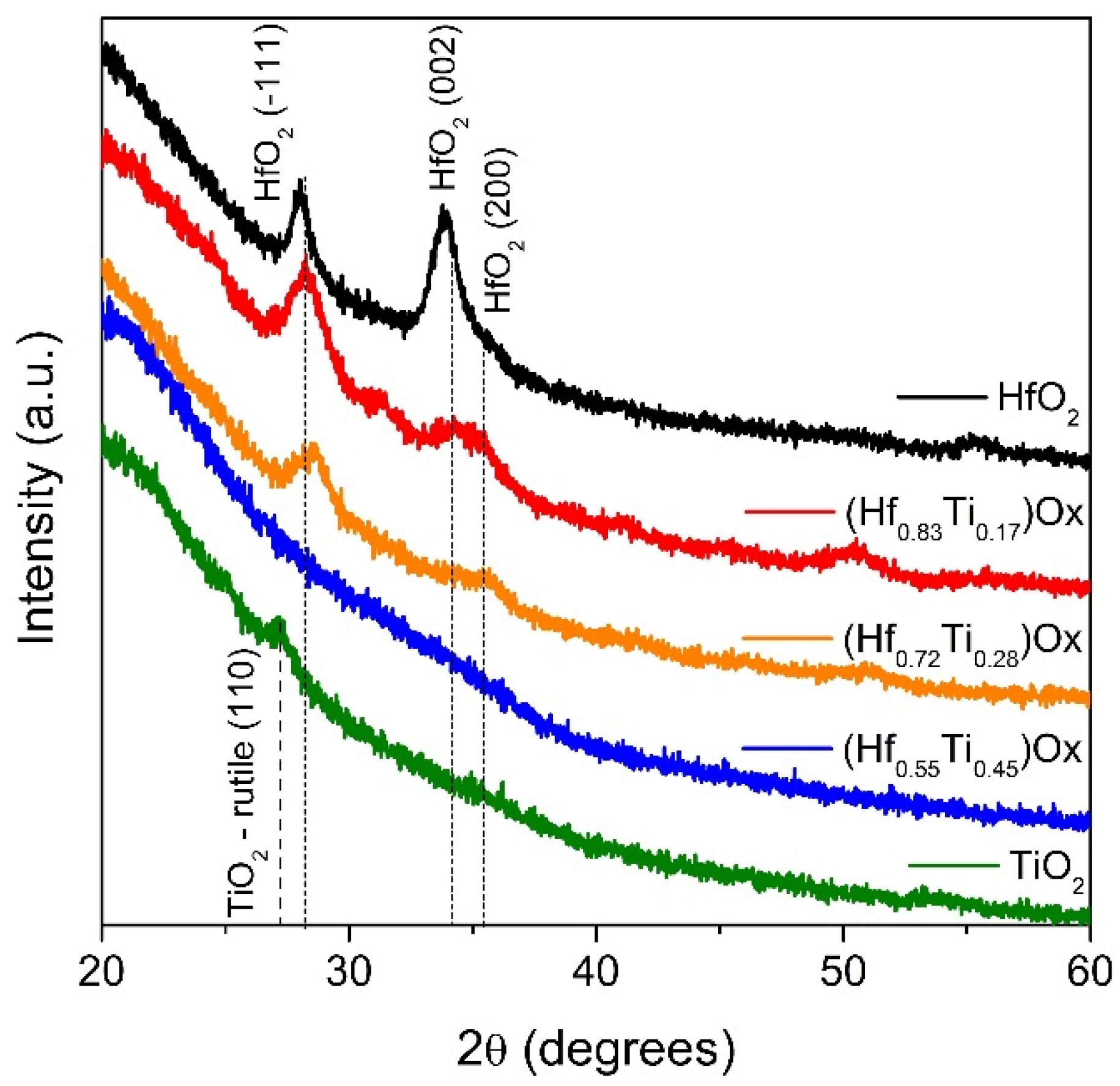
| Thin Film | Crystal Plane | D (nm) | d (Å) | Δd (%) | Type of Stress |
|---|---|---|---|---|---|
| PDF No. 65-1142 HfO2–monoclinic [35] | (−111) | – | 3.145 | – | – |
| PDF No. 21-1276 TiO2–rutile [36] | (110) | – | 3.247 | – | – |
| HfO2 | (−111) | 10.7 | 3.171 | +0.81 | tension |
| (Hf0.83Ti0.17)Ox | 6.7 | 3.155 | +0.32 | tension | |
| (Hf0.72Ti0.28)Ox | 7.4 | 3.129 | −0.53 | compression | |
| (Hf0.55Ti0.45)Ox | amorphous | – | – | – | – |
| TiO2 | (110) | 10.8 | 3.261 | +0.43 | tension |
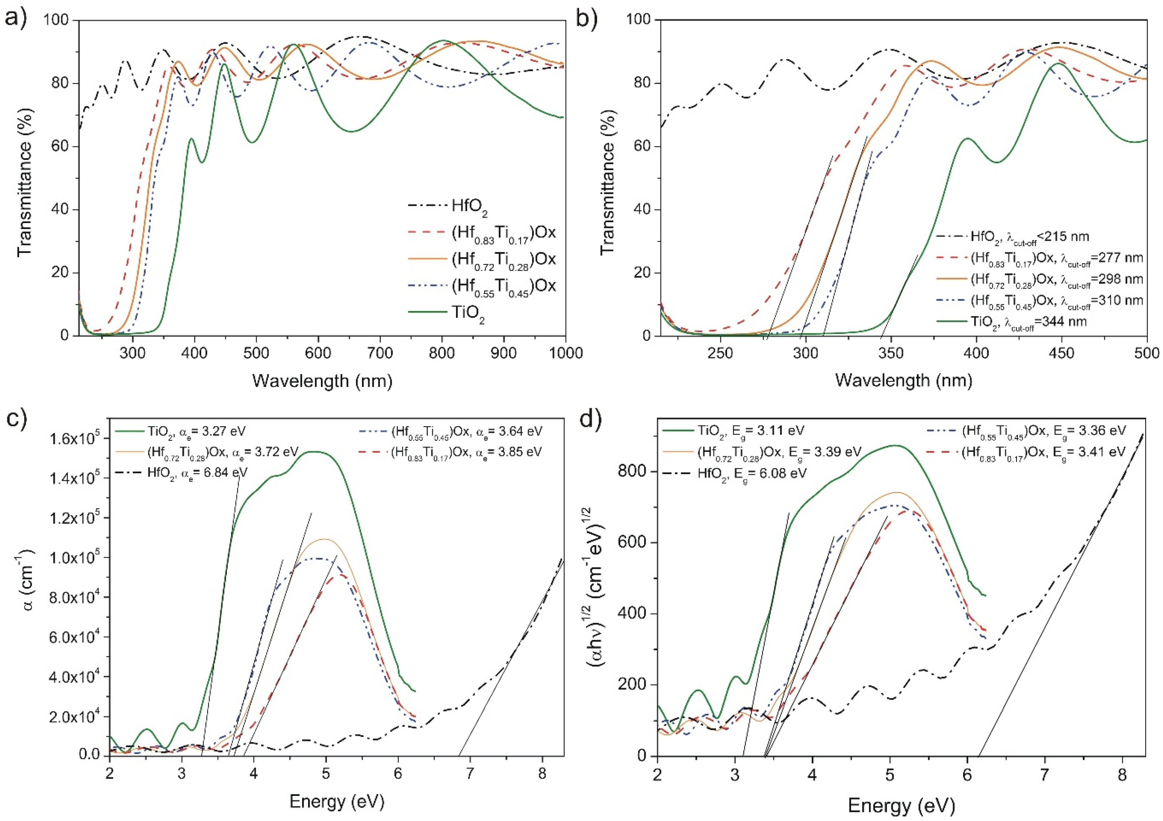
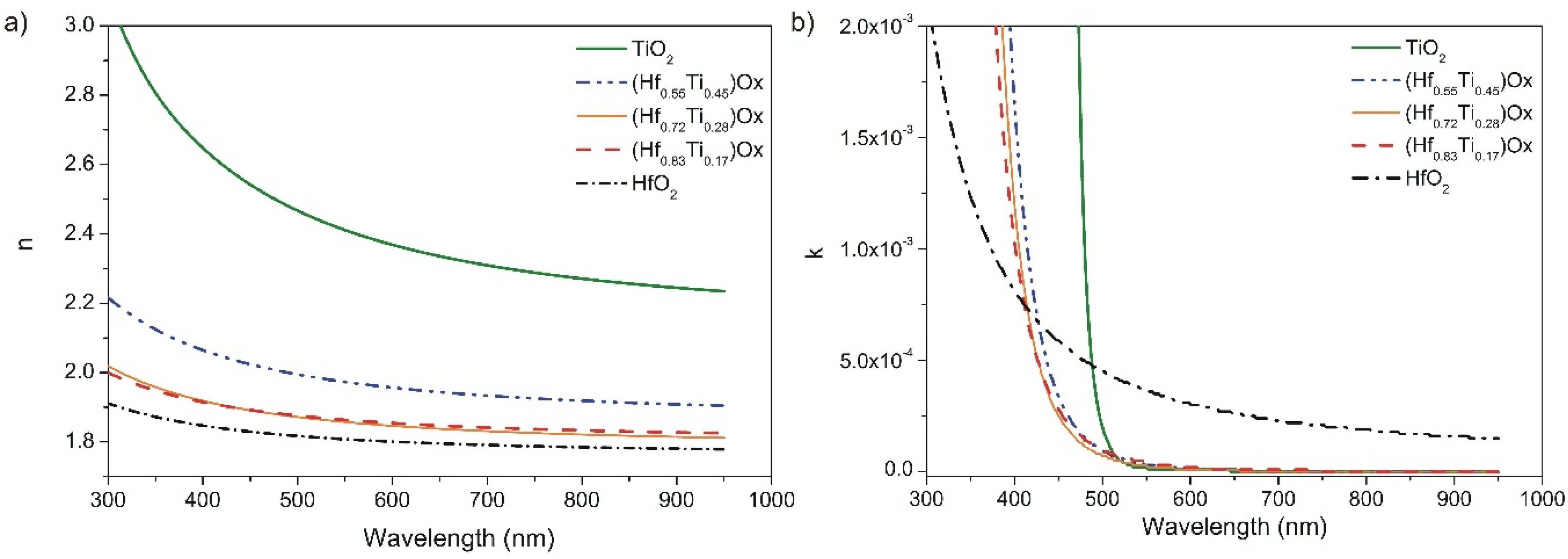
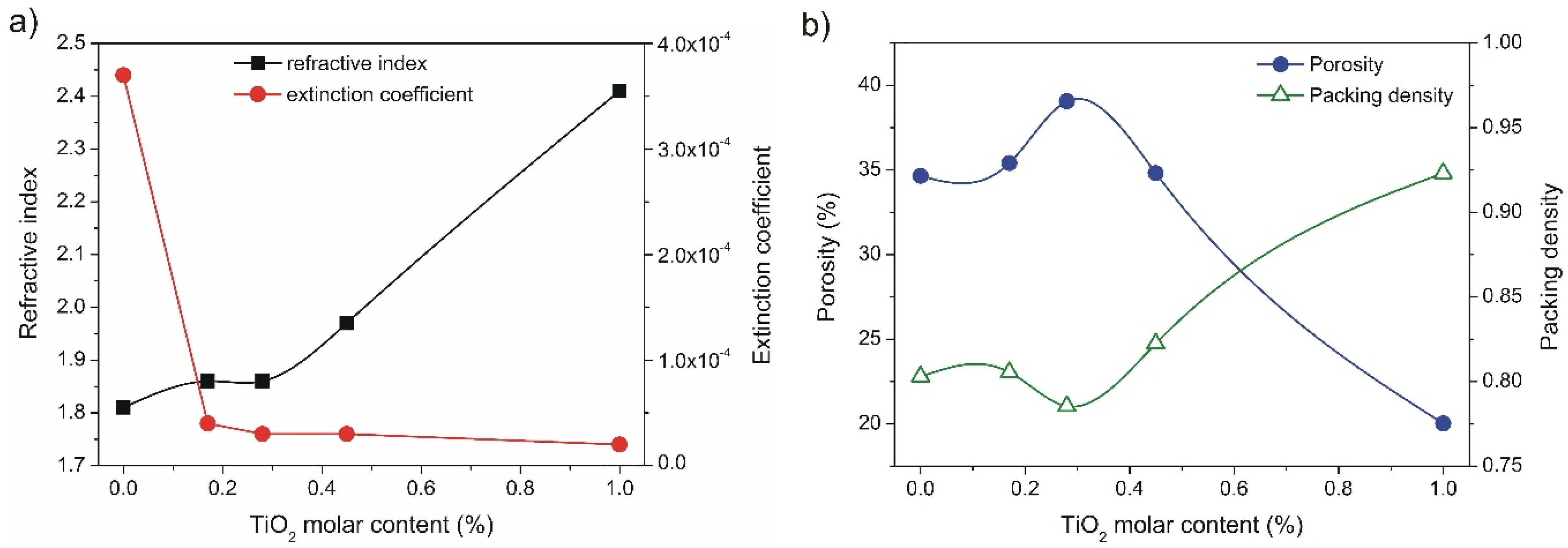
| Thin Film | λcut-off (nm) | Eg (eV) | n (at 550 nm) | k (at 550 nm) | P (%) | PD |
|---|---|---|---|---|---|---|
| HfO2 | <215 ± 2 | 6.08 ± 0.07 | 1.81 | 3.7× 10⁻⁴ | 34.7 | 0.80 |
| (Hf0.83Ti0.17)Ox | 277 ± 2 | 3.41 ± 0.03 | 1.86 | 4 × 10⁻⁵ | 35.4 | 0.81 |
| (Hf0.72Ti0.28)Ox | 298 ± 2 | 3.39 ± 0.03 | 1.86 | 3 × 10⁻⁵ | 39.1 | 0.79 |
| (Hf0.55Ti0.45)Ox | 310 ± 2 | 3.36 ± 0.02 | 1.97 | 3 × 10⁻⁵ | 34.8 | 0.82 |
| TiO2 | 344 ± 2 | 3.11 ± 0.02 | 2.41 | 2 × 10⁻⁵ | 20.0 | 0.92 |
© 2016 by the authors; licensee MDPI, Basel, Switzerland. This article is an open access article distributed under the terms and conditions of the Creative Commons by Attribution (CC-BY) license (http://creativecommons.org/licenses/by/4.0/).
Share and Cite
Mazur, M.; Kaczmarek, D.; Domaradzki, J.; Wojcieszak, D.; Poniedzialek, A. Influence of Material Composition on Structural and Optical Properties of HfO2-TiO2 Mixed Oxide Coatings. Coatings 2016, 6, 13. https://doi.org/10.3390/coatings6010013
Mazur M, Kaczmarek D, Domaradzki J, Wojcieszak D, Poniedzialek A. Influence of Material Composition on Structural and Optical Properties of HfO2-TiO2 Mixed Oxide Coatings. Coatings. 2016; 6(1):13. https://doi.org/10.3390/coatings6010013
Chicago/Turabian StyleMazur, Michal, Danuta Kaczmarek, Jaroslaw Domaradzki, Damian Wojcieszak, and Agata Poniedzialek. 2016. "Influence of Material Composition on Structural and Optical Properties of HfO2-TiO2 Mixed Oxide Coatings" Coatings 6, no. 1: 13. https://doi.org/10.3390/coatings6010013