Article

Study on Corrosion Resistance and Biological Properties of the Double Glow Plasma Nb-Zr Biological Implantation Alloying Layers

1
Institute of Advanced Materials and Flexible Electronics (IAMFE), School of Chemistry and Materials Science, Nanjing University of Information Science and Technology, Nanjing 210044, China
2
Department of Orthopaedic Surgery, Ningbo Medical Treatment Center Lihuili Hospital, Ningbo 315040, China
*
Authors to whom correspondence should be addressed.
Coatings 2022, 12(7), 942; https://doi.org/10.3390/coatings12070942
Submission received: 4 May 2022 / Revised: 24 June 2022 / Accepted: 26 June 2022 / Published: 2 July 2022
(This article belongs to the Special Issue Plasma Thin Films)

Abstract

:
In order to improve the corrosion resistance of implant materials and understand the corrosion mechanisms, we prepared a biomedical Nb-Zr alloying layer on 316L stainless steel using double-layer glow plasma surface-alloying technology and investigated the effects of gas pressures on its surface structure, mechanical properties, and corrosion behavior. In particular, the surface states of the substrate and alloying layers were investigated using 3D confocal micrographs, the water contact angle, and UV reflectance, which aims to study the effect of the surface quality on corrosion resistance and discuss the corrosion mechanisms. The results show that the working pressure has an effect on the current density, the sputtering amount of the alloying elements, and the diffusion process of the alloying elements during glow discharge. The Nb-Zr alloying layer prepared under a pressure of 40 Pa had a uniform and dense surface structure, and the distribution was island-like. A Nb-Zr alloying layer with a thickness of 15 μm was successfully obtained, including the diffusion layer and the deposition layer. Simultaneously, the elements Nb and Zr were gradually distributed along the depth, and a high Nb concentration formed in the Nb-Zr alloying layer. The solid solution formed by Zr in the Nb layer significantly improved the microhardness and corrosion resistance of the substrate. The Nb-Zr alloying layer prepared under a pressure of 40 Pa had the lowest corrosion current density and excellent corrosion resistance, which originated from the passive film formed by the Nb-Zr alloying layer that could inhibit the invasion of corrosive ions and improve the corrosion resistance. In addition, the Nb-Zr alloying layer could promote cell proliferation during long-term use and had good biocompatibility. Our study provides an efficient, high-quality processing method for the surface modification of biomedical metallic materials to form thicker Nb-Zr alloying layers as a cost-effective alternative to bulk Nb-based alloys.

1. Introduction

Biomaterials are special functional materials that can replace, repair, induce regeneration, and enhance the function of body cells, tissues, and organs and have great application potential [1]. As a common biological material, medical 316L stainless steel has a low price and strong processability, and it is often used in bone tissue repair and cardiovascular scaffolds. However, in the clinical medical process, the complex physiological environment in the human body will reduce its corrosion performance, resulting in implant corrosion failure [2]. Therefore, understanding the corrosion failure and improving the corrosion resistance of implant materials have become two important aspects of implant design.
Selecting the appropriate surface modification technology to improve the corrosion resistance and biocompatibility of medical 316L stainless steel in the human environment has important application value. Deposition of Nb coatings on substrates is a known method that combines high-strength alloying elements with the excellent corrosion resistance and excellent biocompatibility of Nb coatings to achieve the originally designed functions [3,4]. It has been reported that metal alloys of Ta, Nb, and Zr have good biocompatibility and bone tissue inductivity, which can promote the adhesion, proliferation, and differentiation of fibroblasts [5]. In addition, the chemical properties of Nb and Zr metals exhibit their strong stability in various inorganic salts, inorganic acids, and organic acids, which is attributed to the formation of dense passive films due to the oxidation of Nb and Zr [6,7,8]. Moreover, the combination of a Zr alloy implant and bone interface has better osseointegration than a titanium implant [9,10], which is a promising orthopedic implant material.
Various deposition methods have been proposed to prepare Nb-Zr alloy coatings, including electrodeposition, physical vapor deposition (PVD), ion beam-assisted deposition (IBAD), and high-energy micro-arc alloying (HEMA). However, the preparation of a Nb-Zr alloy coating is still a difficult problem in clinical application, and different processing techniques have a great influence on its properties and microstructure [11,12,13]. The chemical composition and surface structure of the implant surface affect the ability of the cells to attach, proliferate, and form bone and are important factors affecting the bonding strength and long-term stability of the implant [14,15]. Skliarova et al. [16] deposited Nb-Zr alloy coatings with different alloy compositions by magnetron co-sputtering, and the coatings crystallized well. The microstructure and diffusion barrier efficiency of the coating were studied, which decreased with the increase in Nb content. Zhou et al. [17] prepared Nb-Zr alloys with different Nb contents through vacuum arc melting and studied the microstructure, electrochemical corrosion, and cytotoxicity of the Nb-Zr alloys. An electrochemical polarization test showed that the corrosion resistance increased with the increase in Nb content. XPS detected that the main components on the surface of the pure Zr and Nb-Zr alloy samples after corrosion were ZrO2 and Nb2O5, respectively. The results showed that the passivation film formed by oxidation of Nb and Zr had excellent corrosion resistance. Frutos et al. [18] prepared Ti-Nb-Zr ternary alloy coatings with various complex microstructures by carefully controlling the Nb and Zr concentrations by the DC magnetron sputtering deposition technique. Compared with Ti6Al4V, the Ti-Nb-Zr ternary alloy coating enabled better adhesion of osteoblasts to the surface and improved bioactivity. The beneficial effect of the surface structure on the biocompatibility of materials may be attributed to the improvement of the surface morphology and surface roughness [19,20]. The above experimental results show that these newly developed Nb-Zr alloys have great potential to be candidates for orthopedics.
Different from other surface technology for preparing the biological coating [21], double glow plasma surface alloying (DGPSA) technology is a novel surface modification method [22]. It uses the principle of double glow plasma discharge to directly form an alloying layer on the surface of the substrate. DGPSA alloying layers can obtain corrosion-resistant layers with different thicknesses from a few microns to hundreds, which is very vital to controlling the corrosion life of a material surface. Furthermore, it is important to obtain strong adhesion between the layer and the substrate containing a diffusion layer with a certain thickness. For example, Liu et al. [23] prepared a β-Ta layer using a dual-glow plasma surface alloying technique, which exhibited an extremely dense and uniform microstructure with a thickness of 40 μm. The β-Ta coating not only provided good corrosion protection for Ti-6Al-4V but also endowed it with a higher corrosion resistance in Ringer’s physiological solution.
In this paper, we designed an optimized and cost-effective biocompatible Nb-Zr alloying layer to replace the bulk Nb-based alloy with a thicker Nb-Zr alloying layer. The Nb-Zr alloying layers were prepared on medical 316L stainless steel using the method of DGPSA technology, and the thicknesses of the alloying layers were regulated by changing the working pressure. The effects of the microstructure and surface state of the alloying layer on the corrosion resistance and biocompatibility were systematically studied under different pressures. Nb-Zr alloying layers with optimized biocompatibility are of great value for their potential applications in the implantation field. Our technology can also provide efficient, high-quality processing for the surface modification of implant materials to replace the block alloys with surface alloying.

2. Materials and Methods

2.1. Material Preparation

As shown in Figure 1a, the double-layer glow plasma alloying technique was used to alloy the surface of 316L stainless steel. In the vacuum chamber, a source electrode and two cathode electrodes are provided. A target is placed on the source to provide alloying elements, and a sample is placed on the cathode. During operation, the source target material and the cathode sample can generate a glow, forming a “double glow” discharge phenomenon. Under stable double glow conditions, the argon plasma bombards the surface of the source target, and the alloying elements are sputtered out. Under the action of the electric field, it flies to the surface of the sample at high speed, and after adsorption and deposition, it diffuses into the surface of the sample to form an alloying layer composed of alloying elements to achieve special physical and chemical properties. The element content and thickness of the alloying layer can generally be adjusted by process parameters such as the gas pressure, source voltage, cathode voltage, and working time [24].
Commercially pure 316L stainless steel (20 mm × 20 mm × 3 mm) was used as the base, which was polished with 800 #, 1000 #, 1500 #, and 2000 # silicon carbide sandpaper in turn and ultrasonically cleaned in 95% alcohol and deionized water. The cathode structure during double glow plasma sputtering is shown in Figure 1b. Using Nb-Zr alloy as the target material, four Nb-Zr strips (100 mm × 20 mm × 3 mm) were used to form a fence-like target structure to provide alloying elements for the matrix. First, the vacuum degree of the furnace was pumped to below 6 × 10⁻⁴ Pa, and then pure argon gas was introduced into the furnace cavity until the pressure reached 30–50 Pa. We increase the cathode voltage to 400–500 V and held it at this level for 10 min to warm up the substrate. The source voltage was increased to 800–850 V, the cathode voltage was maintained at 400–500 V, and the working time was 2 h.
The sample test was divided into three groups, and each group of experiments needed to complete the corresponding surface organization, corrosion performance, and biocompatibility experiments. A total of 10 samples were prepared for each group, and some tests were performed by statistical analysis. The detailed experimental parameters are shown in Table 1.

2.2. Characterization of Surface State of Alloying Layer

The phase composition was detected by X-ray diffraction (XRD, Smart Lab/3 kW, Rigaku, Tokyo, Japan). The XRD spectrum was recorded from 10° to 90° using 40 kV Cu Ka radiation at a scan rate of 5°/min. We observed the surface morphology of the sample surface with a scanning electron microscope (SEM, Gemini 300, Carl Zeiss Meditec AG, Oberkochen, Germany) under an acceleration voltage of 10 kV and an energy dispersive X-ray spectrometer (EDS, Bruker, Billerica, MA, USA) combined with SEM elemental composition analysis. The surface roughness was obtained in the selected area by a confocal three-dimensional profilometer (SM-1000, Think Focus, Shanghai, China), and the surface roughness (sq) was calculated. Hydrophilicity was evaluated by measuring the static water contact angle of the sample surface at room temperature with a contact angle measuring instrument (JC2000D, Powereach, Shanghai, China). Drop volumes of 2 uL were chosen to avoid gravitation-induced shape alteration and to diminish the evaporation effects. Each sample measured 5 data at different positions and took the average value. The difference between the measured value of each point and the average value was less than 5° as a valid value. The diffuse reflectance of the sample surface was obtained by ultraviolet spectroscopy (UV-3600 Plus, Shimadzu, Kyoto−fu, Japan). The nanohardness was measured by loading-unloading experiments using a nanoindenter (Tribolndenter TI-900, Hysitron, Billerica, MA, USA). In order to avoid the influence of the thickness of the alloy layer during the nanoindentation hardness test, the indentation depth was about 1/10 of the thickness of the alloy layer. We measured 5–10 points on a sample for calculating the average value, based on an average error of less than 5% as the effective value.

2.3. Electrochemical Corrosion Measurement

The electrochemical experiment was carried out on the CHI660 electrochemical workstation (CH Instruments, Shanghai, China) using a three-electrode system consisting of platinum foil and a calomel electrode as the counter electrode and reference electrode, respectively. The working electrolyte was a 3.5% NaCl solution, and all experiments were carried out in a 37 °C water bath. The specimen was covered with AB adhesive, leaving a working surface of 10 mm × 10 mm for electrochemical measurements.
The open circuit potential (OCP) measurement was performed on the packaged sample and maintained up to 3600 s. Electrochemical impedance spectroscopy (S) measurements were performed on an open circuit potential (OCP) basis by applying a sinusoidal potential perturbation of 10 mV and a frequency sweep from 10 kHz to 0.01 Hz. The potentiodynamic polarization measurement was performed from −0.7 V to 0.4 V and at a scan rate of 1 mV/s.

2.4. Biocompatibility Test

In vitro cell viability assays were performed according to ISO 10993-5:1999. The cells selected for assessing the cell viability of the Nb-Zr alloying layer were rabbit mesenchymal stem cells (The MSCs cell line was obtained from Ningbo University Animal Laboratory.). The samples were sterilized before the experiment, and the front and back sides of the samples were irradiated under a UV lamp for 1 h to sterilize them. We immersed the samples in an RPMI-1640 (Roswell Park Memorial Institute-1640) cell culture medium. The cell culture medium volume was calculated according to the sample area, and 1 mL of the medium was used for every 0.25 cm2. The cells were seeded in 96-well plates at a density of 1 × 10⁴ cells/mL and with 200 μL per well, and they cultured for 24 h at 37 °C and 5% CO2 in a humidified atmosphere. At the same time, a blank control group and an experimental group were set up with 6 parallel samples in each group. After culturing for 1, 3, and 7 days, the medium was aspirated and washed three times with phosphate buffer saline (PBS), and then 200 μL of RPMI-1640 medium containing 10% Cell Counting Kit-8 (CCK-8) was added and incubated in the incubator for 2 h in the dark. To assess cell viability, absorbance (OD value) was measured at 450 nm with a microplate reader (Bio-RAD680), and cell viability was detected according to each group of absorbance values. We calculated the relative cell growth rate (RGR) according to Equation (1):
RGR ( % ) = ( OD T / OD N )   ×   100 %
where ODT represents the OD value of the test group and ODN represents the OD value of the blank control group.
Samples were placed in well plates seeded with green fluorescent protein (GFP) fluorescent protein-transfected rabbit bone marrow mesenchymal stem cells and incubated at 37 °C for 7 days. The well plate was taken out and washed twice with PBS solution, and then 1 mL of 2.5% glutaraldehyde was added to each well and allowed to stand at room temperature for 10 min. After washing twice with PBS solution, 1 mL of 4′,6-diamidino-2-phenylindole (DAPI) was added, left to stand for 5 min at room temperature, and after being washed twice with PBS solution, the growth of cells on the surface of the sample was observed under a laser confocal microscope.

3. Results

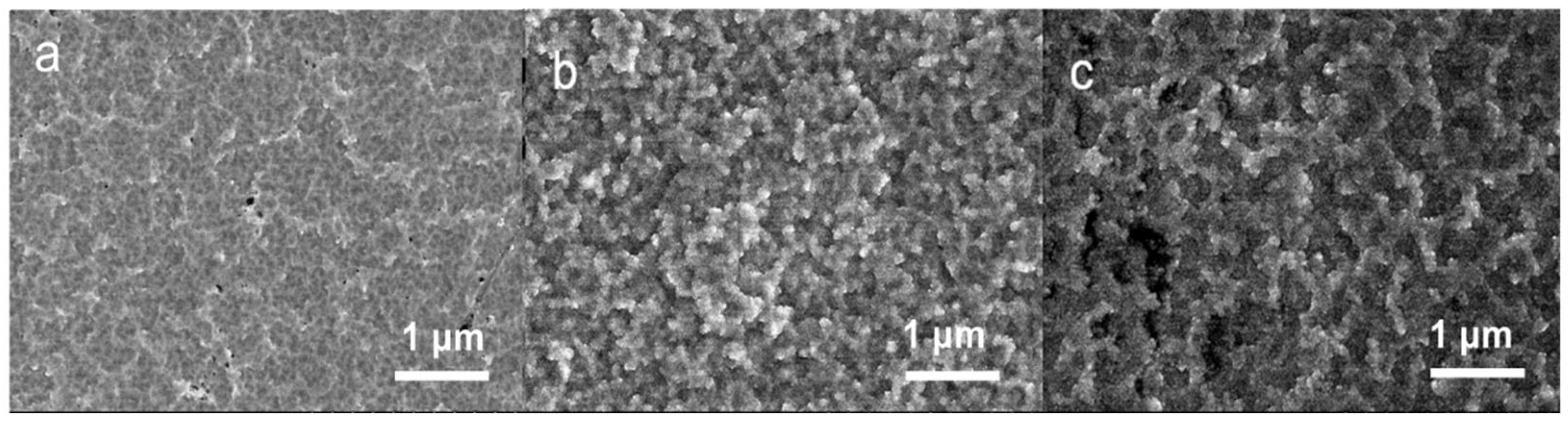
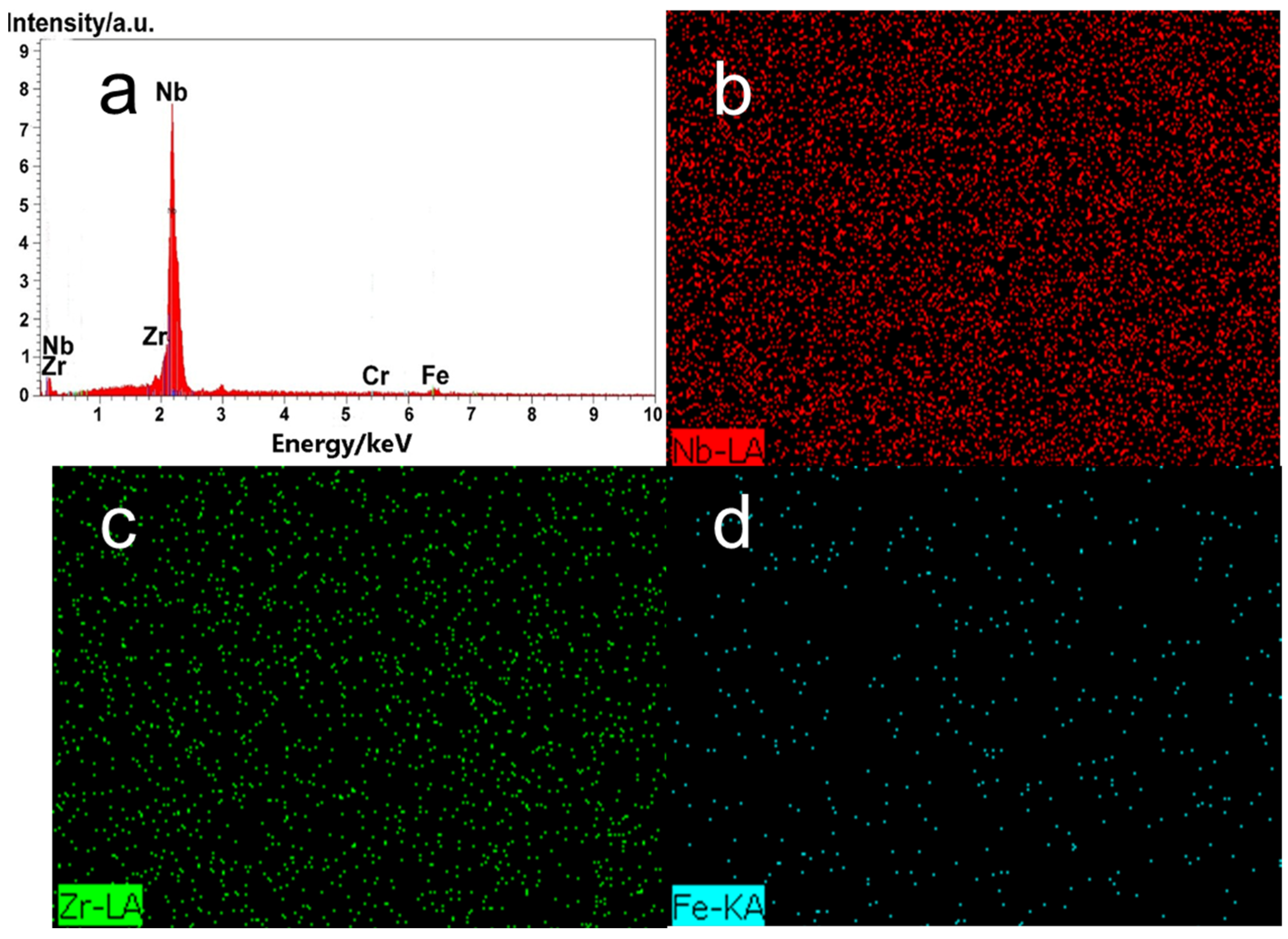
3.1. Composition and Microstructure Analysis

Figure 2 shows the XRD spectrum of the Nb-Zr alloying layer. Nb-Zr alloying layers with different crystallinities were prepared on the surface of the 316L stainless steel. The alloying layer had Nb characteristic peaks at 38.415° and 69.476° along the (110) and (211) crystal planes, respectively, and the Zr characteristic peak coincided with Nb at 69.476°. The diffraction peaks were sharp and intense, and the intensity varied with the working air pressure. When the gas pressure was 30 Pa, the characteristic peak of Fe appeared in the alloying layer, indicating that the thickness of the alloying layer was low and the content of the matrix elements was high. When the gas pressure was 40 Pa, the crystallinity of the surface alloying layer was the best. The second characteristic peak of Zr was also presented at 36.518°, which was oriented along the (101) crystal plane. There were no characteristic peaks in the form of alloys in the Nb-Zr alloying layer, indicating that both Fe and Zr exist in the Nb layer in the form of solid solutions.
Figure 3 shows the SEM micrographs of the Nb-Zr alloying layers prepared at (a) 30 Pa, (b) 40 Pa, and (c) 50 Pa pressures. From the figures, it can be seen that the microstructure of the studied Nb-Zr alloying layers varied with the pressure. The Nb-Zr alloying layer prepared by 40 Pa of air pressure had a good surface morphology, uniform and dense alloying layer with an island-like structure, and no cracks or pores on the surface. When the air pressure was 30 Pa, the supply of alloying elements was insufficient, so the surface of the substrate could not be completely filled with alloying elements, and the growth of the surface alloying layer was inhibited. Thus, the surface morphology of the alloying layer was incomplete. When the gas pressure was 50 Pa, the reverse sputtering effect on the surface of the substrate was enhanced, and the bombardment of high-energy particles aroused the active atoms that had been adsorbed on the surface of the substrate, which affected the surface integrity of the alloying layer.
Figure 4 shows the element distribution diagram on the surface of the Nb-Zr alloying layer sample prepared at 40 Pa of pressure. The image shows that the surface elements of the sample were mainly Nb, Zr, Fe, and Cr, and there were no peaks for the other elements in the energy spectrum, which indicates that the alloying layer did not appear to be oxidized during the preparation process. According to Table 2, the contents of Nb and Zr were 85.18% and 8.11%, respectively, and the contents of Fe and Cr were 5.10% and 1.61%, respectively.
The cross-sectional morphologies and element content of the Nb-Zr alloying layers varied with the depth of the alloying layer, as shown in Figure 5. The results show that the cross-sectional morphology of the alloying layers of all samples was complete, being without micropores and cracks, and the thickness of the alloying layer changed with the deposition pressure. The cross-sectional thicknesses of the alloying layer prepared by 30 Pa, 40 Pa, and 50 Pa of pressure were about 6 μm, 10 μm, and 14 μm, respectively, and the alloying layer was divided into a deposition layer and a diffusion layer. The deposition layer was mainly composed of the alloying elements Nb and Zr. The content of the Nb and Zr elements was far more than that of the matrix element. The alloying elements Nb and Zr and the matrix element Fe coexisted in the diffusion layer. In the diffusion layer, Nb and Zr atoms diffused into the substrate, Fe atoms diffused outward, and the content of the diffused alloying elements decreased with the depth. The thickness of the Nb-Zr alloying layer prepared at 30 Pa of pressure was only about 6 μm. When the deposition pressure was too low, the supply of the target elements was insufficient, resulting in a low thickness of the alloying layer on the surface of the substrate. The thickness of the alloying layer at a pressure of 40 Pa was approximately 10 μm, while the thickness of the alloy layer at a pressure of 50 Pa was 15 μm.

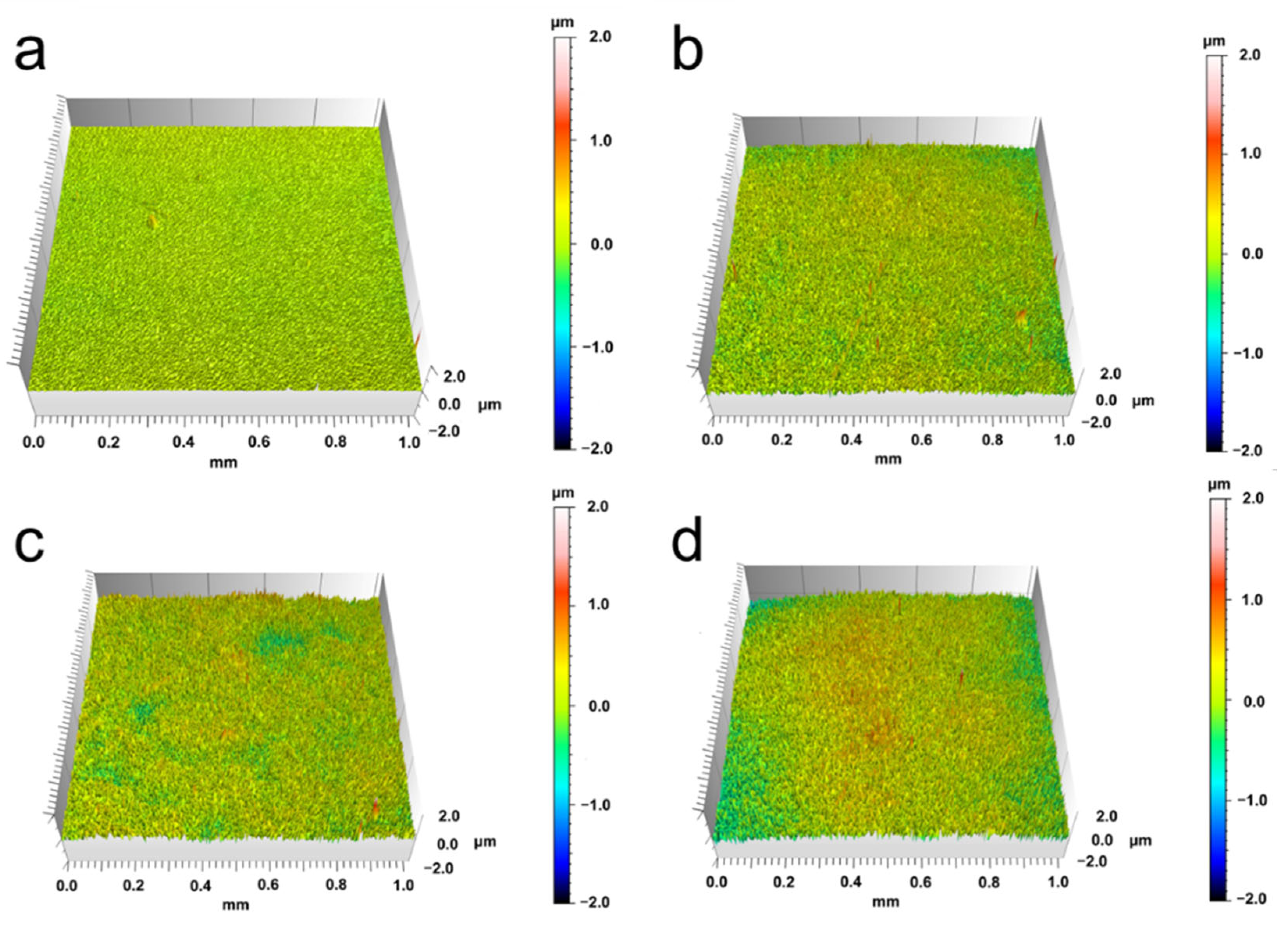
3.2. Surface State Analysis

Figure 6 shows the surface roughness of the samples prepared at different air pressures. After the alloying layers were prepared on the surface, the surface roughness increased significantly, and the air pressure directly affected the value of the surface roughness. The Nb-Zr alloying layer surface height parameter Sq (root mean square height) was calculated as follows:
Sq = 1 A   A   Z 2   ( x , y )   dx   dy
The surface roughness of the substrate was 0.053 μm. The 30 Pa alloying layer had a small surface undulation, and the surface roughness was 0.168 μm. The surface of the alloying layer prepared by 50 Pa of air pressure was composed of a large number of fluctuations. Its surface roughness was the largest at 0.298 μm, and the surface was the roughest. In contrast, the surface of the alloying layer prepared under 40 Pa of pressure was smoother, the surface geometric texture was more uniform and regular, and the surface roughness was 0.224 μm.
Figure 7a shows the water contact angles of the corresponding samples. It can be observed that the water contact angle of the substrate surface was about 65°. After the alloying layer was deposited, the water contact angle significantly increased. The water contact angle on the surface of the 30 Pa alloying layer sample was about 83°, and with the increase in air pressure, the water contact angle rose to 92° and 98° for 40 Pa and 50 Pa, respectively. The increased water contact angle confirms that the Nb-Zr alloying layer obtained during the plasma deposition process enhanced the hydrophobicity of the substrate surface. The superhydrophobic surface showed better potential in terms of blood compatibility, and the ability of the surface to adsorb cells was affected by the surface roughness and the amount of crystal grains. The reflectivity test was performed on the substrate and the Nb-Zr alloying layers prepared at different pressures as shown in Figure 7b. In the 780–2500-nm wavelength range, the average reflectivity of the substrate was only 2%, the average reflectivities of the alloying layer samples prepared under 30 Pa and 40 Pa of pressure were close to 10% and 20%, respectively, and the samples prepared under 50 Pa of pressure showed an average reflectivity of about 30%.
It can be seen from Table 3 that the experimentally measured surface hardness levels of the Nb-Zr alloying layer were greater than 3.52 GPa for the 316L substrate, being 5.53 GPa, 8.02 GPa, and 8.12 GPa at 30, 40, and 50 Pa of pressure, respectively. The data show that after the surface of the 316L substrate was modified by DGPSA technology, the plastic deformation resistance improved, and the mechanical properties were enhanced. Surface hardness reflects the strength of the material’s ability to resist plastic deformation and destructive force, and it can also measure the interface bonding strength of the alloying layer. Figure 8 shows the variation curve of the surface load with displacement for the 316L matrix and Nb-Zr alloying sample. The experimental curves obtained by the nano-indentation test were smooth and continuous, having no bending and breakpoints, indicating that the tested samples did not have cracks or fractures during the nano-indentation test.

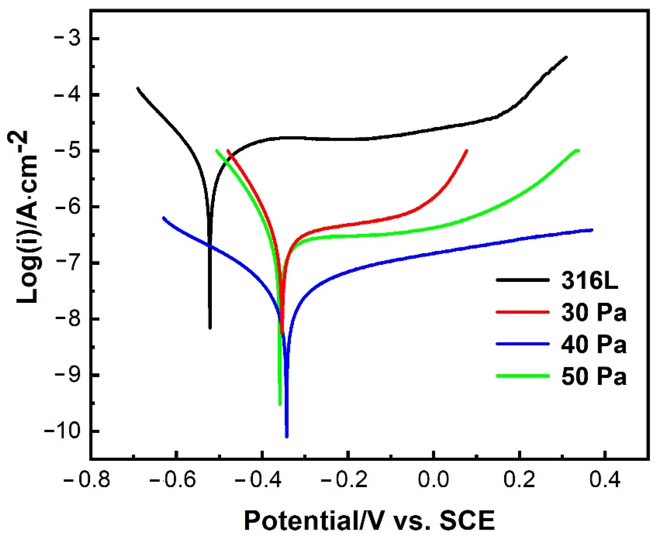
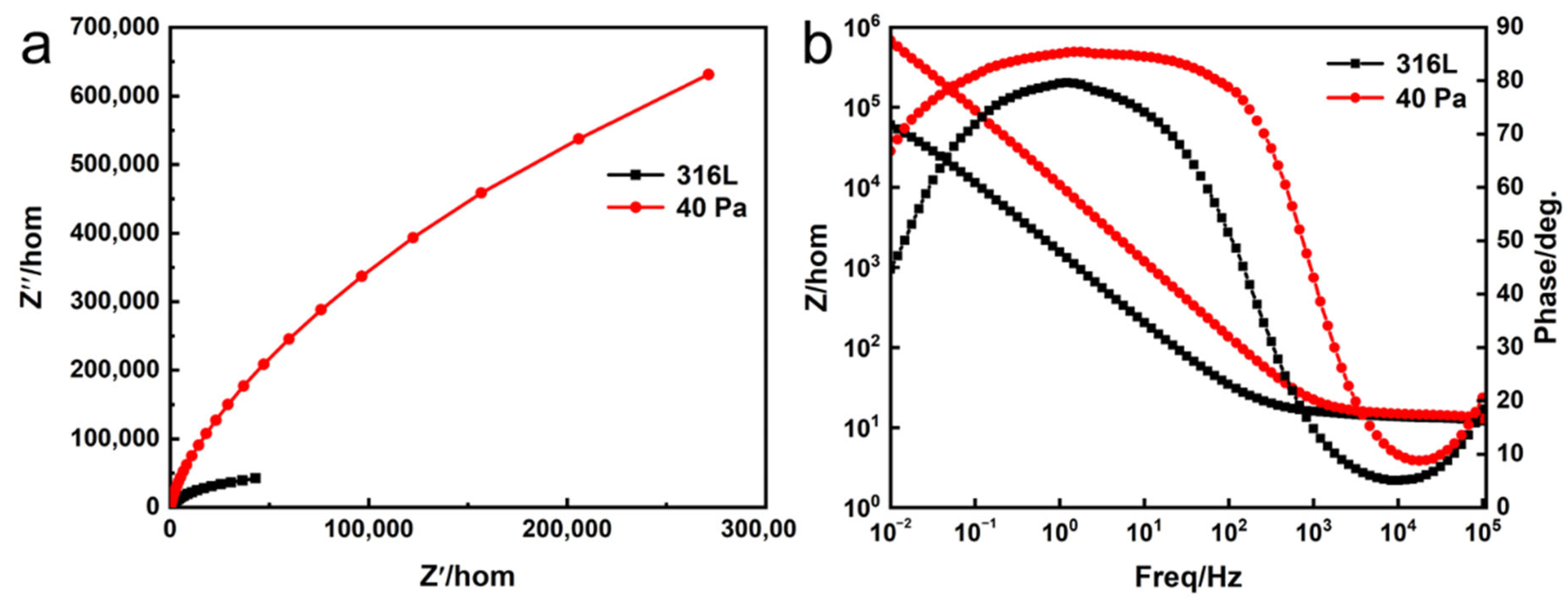
3.3. Corrosion Resistance

Figure 9 shows the potentiodynamic polarization curves of the 316L substrate and the Nb-Zr alloying layers prepared under pressures of 30 Pa, 40 Pa, and 50 Pa. The test samples exhibited very similar polarization behaviors as the potential increased. They were passivated and showed the passive regions before the protective films were broken. These data are similar to the experimental results for Nb-Zr alloying layers obtained by Zhou et al. The average values Eccor and Icorr from the polarization curves calculated by CorrView software (CorrView 3.10, Solartron Analytical, Oak Ridge, TN, USA) are presented in Table 4. For the substrate and Nb-Zr alloying layers prepared at different gas pressures, the Iccor values of all test samples were 5.05 × 10⁻⁶ (A·cm2), 1.35 × 10⁻⁷ (A·cm2), 1.26 × 10⁻⁸ (A·cm2), and 1.16 × 10⁻⁸ (A·cm2). It can be seen that the corrosion resistance of the Nb-Zr alloying layer was improved regardless of the pressure. This indicates that the Nb-Zr alloying layer on the surface of the 316L substrate inhibited the intrusion of the Cl ions of the corrosive solution and increased the corrosion resistance. For the Nb-Zr alloying layers prepared at different working pressures, the corrosion potential Ecorr and corrosion rate first increased and then decreased with the working pressure, while the natural corrosion current Icorr first decreased and then increased. The corrosion current and rate of the alloying layer at 40 Pa were the lowest, and the corrosion resistance was better. The polarization potentials of the substrate and the Nb-Zr alloying layers are shown in Table 4. Compared with the substrate, the polarization potential of the Nb-Zr alloying layers increased to −0.35 V, −0.34 V, and −0.36 V at 30, 40, and 50 Pa of pressure, respectively. It can be seen from the material corrosion evaluation method that the higher the self-corrosion potential of the material, the smaller the self-corrosion current, and the better the corrosion resistance. The passivation zone is observed on the polarization curve, which indicates that a stable passivation film is formed on the surface of the Nb-Zr alloying layers [25,26]. Therefore, the prepared Nb-Zr alloying layer is expected to improve the corrosion resistance of these biomedical alloys in body fluids.
Figure 10 shows the EIS results after immersion in a 3.5% NaCl solution to reach a steady state in the form of a Nyquist diagram and Bode diagram. Figure 10a shows a Nyquist plot with similar behavior, indicating a typical capacitive response. It can be observed in the Nyquist diagram that the capacitance loop diameter of the Nb-Zr alloying layer was much larger than that of the 316L substrate, indicating that the alloying layer had a higher electrochemical performance [27,28]. An electrochemical reaction occurs in the electrolyte to form an oxide film with high impedance, which increases the resistance when the charge is transferred. The greater the capacitive reactance arc amplitude, the greater the electrochemical reaction impedance, and the better the corrosion resistance [29]. The Bode plot of the 40-Pa alloying layer sample in Figure 10b shows that the curve consists of a flat part at high frequencies, which was caused by the resistance of the electrolyte, and varies linearly over a wide frequency range, indicating the capacitive behavioral response of the passivation film [30,31]. In addition, the maximum phase angle of the Nb-Zr alloying layer was about −85° in the frequency range from 0.1 to 100 Hz, which was significantly higher than that of the 316L substrate. It is worth noting that the phase angle plateau of the Nb-Zr alloying layer was wider, but the substrate was significantly reduced. In the low frequency region, a high impedance value of about 106 (Ω·cm2) was obtained for the Nb-Zr alloying layer, while significantly lower impedance values were observed for the 316L substrate. These results reveal the excellent protective properties of the passivation film on the surface of the Nb-Zr alloying layer [32].

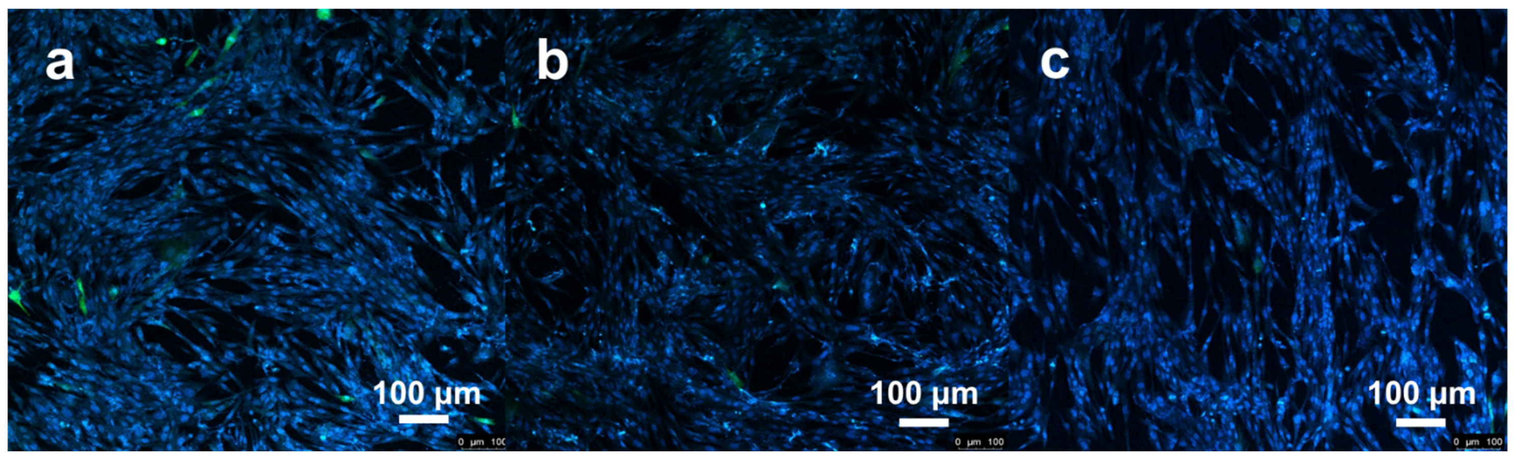
3.4. Biocompatibility Analysis

By studying the microstructure, surface state, and corrosion resistance of an Nb-Zr alloy layer, the optimum working pressure of an Nb-Zr alloy layer prepared by DGPSA technology was found to be 40 Pa. In order to test whether the Nb-Zr alloying layer prepared in this experiment had ideal biocompatibility, the biological activity of the Nb-Zr alloying layer was preliminarily evaluated by in vitro cell proliferation experiments. CCK-8 is a commonly used method for detecting the cell activity of implanted materials which can objectively and truly reflect the adhesion and proliferation of cells [33]. Statistical analysis of the cellular absorbance data was performed using GraphPad Prism (GraphPad Prism 9, GraphPad, CA, USA). Two-way ANOVA was selected to determine the significance of any statistical differences between groups, and significance was determined by a value of p < 0.05.
The cell absorbance data of the blank control group, 316L matrix group, and Nb-Zr alloying layer group measured by CCK-8 are shown in Figure 11. Compared with the blank control group, the Nb-Zr alloying layer exhibited good biocompatibility. The cell viability in the sample extracts increased with the culture time. After culturing for 1 day, the cell viability on the surface of the Nb-Zr alloying layer prepared under a working pressure of 40 Pa was greater than that of the 316L substrate. After 7 days of culturing, the number of cells on the surface of the Nb-Zr alloying layer was much higher than that on the surface of the substrate. The cell activity value on the surface of the sample is related to the surface toxicity of the material and the adsorption and proliferation of cells [34]. Biomedical metal materials will inevitably cause the problem of metal ion release. Compared with the blank control group, the cell viability of the alloying layer samples decreased, but the cell viability was greater than 90%. According to the requirements of the national standard GB/T16886.5, when the surface cell viability is greater than 75%, the material is considered to be non-toxic.
Figure 12 shows the fluorescence confocal images of the surface cells of the blank control group, the 316L substrate group, and the Nb-Zr alloying layer group after 7 days of culturing. It can be seen that the cells grew well on the surface of the Nb-Zr alloying layer, and the cells were fibrous and densely grown, covering the entire surface, similar to the growth morphology of the cells in the blank control group. As shown in Figure 12, the cell morphologies indicate that the surface roughness increase in the Nb-Zr alloying layer promoted cell adhesion and proliferation. Therefore, the investigated Nb-Zr alloying layers were considered to be biocompatible in vitro.

4. Discussion

Based on the above results, the prepared Nb-Zr alloy layer exhibited excellent corrosion resistance under a pressure of 40 Pa. The pressure of the DGPSA affects the microstructure, crystal orientation, and surface state of the Nb-Zr alloy layer, which in turn induces changes in the mechanical, corrosion, and biological properties. According to the characteristics of the glow discharge, the working air pressure has an important influence on the current density, sputtering amount of alloying elements, space transport, and adsorption and diffusion on the substrate surface during the glow discharge process [35]. The sputtering rate of the source target is inversely proportional to the power of the working gas pressure:
S 1 P n
where S is the sputtering throughput, P is the working pressure, and n is an empirical value between 2 and 5. Low pressure seems to be beneficial to increasing the amount of sputtering, but the surface of the cathode substrate is subject to strong self-sputtering under low pressure, which is not conducive to the deposition and diffusion of high concentrations of alloying elements on the surface of the substrate to form an alloying layer. Moreover, the low-pressure plasma density is low, the bombardment ability to the surface of the substrate is weak, and it is not conducive to the high-temperature diffusion of alloying elements. Although higher gas pressure is beneficial to increase the sputtering amount of alloying elements to be infiltrated, it is easy to increase the reverse sputtering effect on the surface of the substrate, desorb the deposited alloying elements, and cause the surface of the alloying layer to be incomplete [36]. Therefore, if the air pressure is too low or too high, the quality of the alloying layer will be affected. Only by selecting the appropriate working air pressure and maintaining the balance of supply and adsorption of the alloying elements during the sputtering process can the alloying elements be optimally utilized. Under a pressure of 40 Pa, the surface morphology of the alloying layer showed a uniform and dense structure with an island-like distribution of grains. Different air pressures led to different surface morphologies of the alloying layers. The changes in surface morphology were related to the growth properties of the atomic clusters during DGPSA sputtering. Higher pressure caused the occurrence of the reverse sputtering effect and the bombardment of high-energy particles, which affected the surface integrity of the alloying layers, while the lower pressure could not supply enough alloying elements, so the surface of the substrate could not be completely filled with alloying elements, and the surface morphology of the alloying layer was incomplete. The cross-sectional SEM morphology and EDS results show that the elements Nb and Zr were gradually distributed along the depth, and a high Nb concentration formed in the Nb-Zr alloying layer, confirming the process of element out diffusion into the alloying layer region. With the increase in the working pressure, the plasma density of Ar increased, and the sputtering intensity of the source and cathode also increased. This provided more alloying elements and induced more vacancies on the substrate surface to increase the thickness of the alloying layer. A large amount of alloying elements was deposited, which led to the alloying layer becoming thicker. However, the higher gas pressure led to a change of the surface structure of the Nb-Zr alloying layer, and then the excessive thickness of the alloying layer would increase the internal stress. Therefore, the thickness of the alloying layer was affected by the gas pressure.
In addition, differences in the chemical composition and surface structure of the surface of the Nb-Zr alloying layer were also found, which were caused by the differences in the sputtering rates of the elements. XRD studies show that all Nb-Zr alloying layers have Nb and Zr phases, and the elements Zr and Fe are a solid solution when distributed in the Nb layer. The composition and grain orientation of the Nb-Zr alloying layer affect the growth and corrosion rate of the passivation film. Studies have shown that the addition of zirconium can improve the corrosion resistance of the alloy, but the increase in Zr will reduce the surface reactivity. In order to better improve the surface properties of medical 316L stainless steel, a specific proportion of the Nb-Zr alloying layer is selected to enhance the biological activity of the material [37]. Wang et al. [38] investigated the effect of grain orientation on electrochemical behavior in single niobium crystals with different orientations and found that the passive film formed on the (100) plane had the lowest donor density, but it decreased with increasing pH levels. Therefore, the addition of Zr will not only affect the growth orientation of the alloying layer with the change in pressure, but due to the additional Zr orientation, better corrosion resistance can also be obtained, and it is easier to form a passivation film on the orientation.
The surface state reflects the surface qualities and properties of alloying layers, which affect the corrosion resistance and biocompatibility. The change in the surface roughness of the Nb-Zr alloying layer will affect the surface properties, corrosion resistance, and biocompatibility of the material [39], which will be related to the surface contact angle. The deposited alloying layers greatly increase the surface roughness and the exposed grain area and theoretically should promote cell adhesion, proliferation, and differentiation. The reflectivities of the base sample and the alloying layer samples increase with the increase in air pressure. The results show that the size of the grains and surface roughness of the sample surface affected the single reentry of the light incident on this area, so the reflectivities of the alloying layer samples were relatively high. Based on the data in Figure 3 and Figure 6, it can be seen that the surface morphology of the alloying layer sample prepared under 40 Pa of pressure had a more regular arrangement, which was more conducive to the control of the chemical corrosion process.
The nanoindentation results show that the surface hardness of the Nb-Zr alloying layer increased from 3.52 GPa to 8.12 GPa compared with the matrix, which is beneficial to the interfacial bonding strength of the alloying layer. The surface state results show that the Nb-Zr alloying layer enhanced the substrate surface roughness and exposed the grain area, which can promote cell adhesion, proliferation, and differentiation. During the nanoindentation loading process, when the indentation depth was the same, the corresponding load of the Nb-Zr alloying layer was greater than that of the 316L matrix, indicating that under the same test load, the surface of the Nb-Zr alloying layer was more resistant to plastic deformation. At the same indentation depth, the load values on the surface of the 40-Pa and 50-Pa alloying layers were close, and both were better than those of the 30-Pa sample, indicating better resistance to external loads.
The corrosion resistance shows that the Nb-Zr alloying layer improved the corrosion performance of the 316L substrate. The ElS data show that during the electrochemical corrosion process, a passivation film could be formed on the surface of the Nb-Zr alloying layer to inhibit the intrusion of corrosion ions. The analysis of polarization curve data shows that the alloying layer prepared at 40 Pa of pressure had the best corrosion resistance. The oxidation of Nb and Zr can form a dense passivation film, which exhibits excellent corrosion resistance. Studies have shown that surface morphology plays a very important role in cell–material interactions and is a factor that affects the biocompatibility of materials. Cell adhesion prefers ridges and horns and less so the textured surfaces of pits. Ponsonnet et al. [40] studied the relationship between the surface roughness of titanium alloys and the proliferation of fibroblasts and found that when the peak-to-valley difference was less than 1 μm, the surface had higher cell proliferation ability. This shows that the surface roughness of biomaterials can improve cell activity, and the more uniform the distribution of rough particles on the surface, the higher the biological activity. Wang et al. [41] showed that cells prefer to proliferate on rough surfaces, the prepared petal-like structures are more conducive to cell adhesion, spreading, proliferation, and differentiation, and cells tend to grow on rough surfaces. The surface showed better biocompatibility with an increasing petal density. The results of in vitro cell proliferation experiments showed that compared with the polished matrix structure, the surface with the rough structure was more favorable for cell adhesion, spreading, and proliferation. The improved biocompatibility was due to the increased specific surface area of the material, which provided a larger cell contact area and adhesion sites.
In a word, the Nb-Zr alloying layers can not only improve the surface structure and corrosion resistance of medical 316L stainless steel but also have good biocompatibility. Therefore, the Nb-Zr alloying layer is a promising orthopedic implant material as a cost-effective alternative to bulk Nb-based alloys.

5. Conclusions

In our current study, a uniform and dense Nb-Zr alloying layer was successfully formed on medical 316L stainless steel by the DGPSA process. The surface microstructures and compositions of the alloyed layers were conducted by SEM, EDS, and XRD. The surface water contact angle and UV reflectance from the surface show information about the roughness of the surface, which affects the surface electrochemical properties. The nano-mechanical properties of the Nb-Zr layers and the substrate were analyzed by the depth-sensing nanoindentation measurement to characterize the surface mechanical properties. All the results showed that the Nb-Zr alloying layer effectively improved the mechanical properties, corrosion resistance, and bioactivity of the medical 316L stainless steel. The important findings of the study are summarized as follows:
(1)
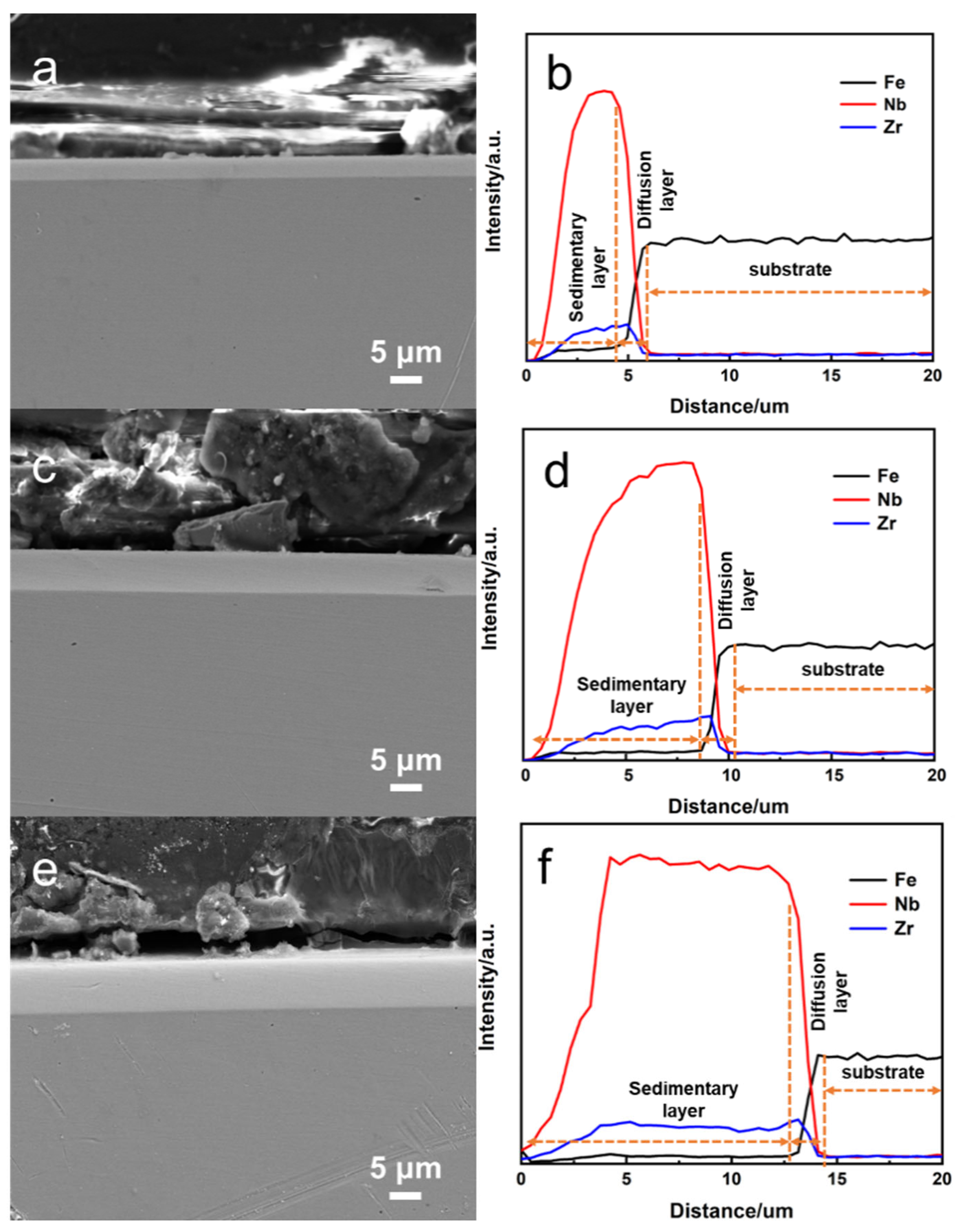
By comparing the surface quality and corrosion performance, the optimal pressure parameter was found to be about 40 Pa. Simultaneously, we prepared a thicker Nb-Zr alloying layer, and the total thickness of it was about 15 μm, including the deposition layer and interdiffusion diffusion layer (2 μm).
(2)
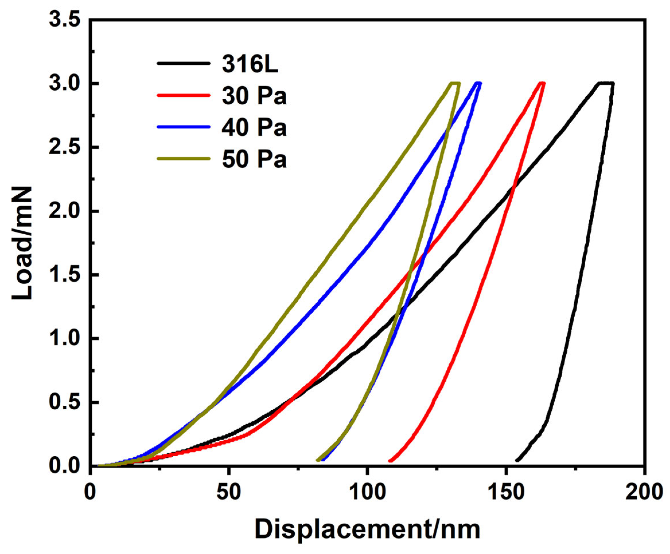
The maximum microhardness of the Nb-Zr alloying layer was 8 GPa, which was 2.3 times that of the matrix.
(3)
The corrosion test shows that the alloy layer inhibited the intrusion of corrosion ions and improved the corrosion resistance.
(4)
The rough surface of the Nb-Zr alloying layer was more conducive to the adhesion, diffusion, and proliferation of cells, and its biological activity was higher than that of the medical 316L stainless steel.

Author Contributions

Conceptualization, funding acquisition, writing—review and editing, and supervision, H.W.; writing—original draft preparation, and validation, K.Z.; resources, C.X.; methodology, J.D.; investigation, J.R.; funding acquisition, data curation, and supervision, Z.P. All authors have read and agreed to the published version of the manuscript.

Funding

This work was supported by the “Science and Technology Innovation 2025” Major Special Project of Ningbo (No. 2019B10064), Zhejiang Provincial Natural Science Foundation of China under grant no. LGF21H060003, Programs Supported by the Ningbo Natural Science Foundation (202002N3195), and the Foundation Project for Medical Science and Technology of Zhejiang Province (2022KY1083).

Institutional Review Board Statement

Not applicable.

Informed Consent Statement

Not applicable.

Data Availability Statement

The data presented in this study are also available upon request from the corresponding author.

Conflicts of Interest

The authors declare no conflict of interest.

References

  1. Qu, H.W.; Fu, H.Y.; Han, Z.Y.; Sun, Y. Biomaterials for bone tissue engineering scaffolds: A review. RSC Adv. 2019, 9, 26252–26262. [Google Scholar] [CrossRef] [PubMed] [Green Version]
  2. Li, Y.H.; Yang, C.; Zhao, H.D.; Qu, S.G.; Li, X.Q.; Li, Y.Y. New developments of Ti-based alloys for biomedical applications. Materials 2014, 7, 1709–1800. [Google Scholar] [CrossRef] [PubMed] [Green Version]
  3. Navarrete, R.O.; Olaya, J.J.; Ramírez, C.; Rodil, S.E. Biocompatibility of niobium coatings. Coatings 2011, 1, 72–87. [Google Scholar] [CrossRef]
  4. Wang, X.J.; Li, Y.C.; Lin, J.G.; Yamada, Y.; Hodgson, P.D.; Wen, C.E. In vitro bioactivity evaluation of titanium and niobium metals with different surface morphologies. Acta Biomater. 2008, 4, 1530–1535. [Google Scholar] [CrossRef]
  5. Yang, Y.L.; Song, Z.Q.; Xu, J. Corrosion fatigue behavior of MRI-compatible Nb-60Ta-2Zr alloy in exposure to simulated physiological solution. Int. J. Fatigue 2022, 155, 106583. [Google Scholar] [CrossRef]
  6. Li, B.Q.; Xie, R.Z.; Lu, X. Microstructure, mechanical property and corrosion behavior of porous Ti-Ta-Nb-Zr. Bioact. Mater. 2020, 5, 564–568. [Google Scholar] [CrossRef]
  7. Ji, P.F.; Li, B.; Chen, B.H.; Wang, F.; Ma, W.; Zhang, X.Y.; Ma, M.Z.; Liu, R.P. Effect of Nb addition on the stability and biological corrosion resistance of Ti-Zr alloy passivation films. Corros. Sci. 2020, 170, 108696. [Google Scholar] [CrossRef]
  8. Chui, P.F.; Jing, R.; Zhang, F.G.; Li, J.G.; Feng, T. Mechanical properties and corrosion behavior of β-type Ti-Zr-Nb-Mo alloys for biomedical application. J. Alloys Compd. 2020, 842, 155693. [Google Scholar] [CrossRef]
  9. Rubitschek, F.; Niendorf, T.; Karaman, I.; Maier, H.J. Corrosion fatigue behavior of a biocompatible ultrafine-grained niobium alloy in simulated body fluid. J. Mech. Behav. Biomed. 2012, 5, 181–192. [Google Scholar] [CrossRef]
  10. Wang, Y.L.; Zhang, Y.F.; Sculean, A.; Bosshardt, D.D.; Miron, R.J. Macrophage behavior and interplay with gingival fibroblasts cultured on six commercially available titanium, zirconium, and titanium-zirconium dental implants. Clin. Oral Investig. 2019, 23, 3219–3227. [Google Scholar] [CrossRef]
  11. Li, B.Q.; Li, C.L.; Wang, Z.X.; Lu, X. Preparation of Ti-Nb-Ta-Zr alloys for load-bearing biomedical applications. Rare Metals 2019, 38, 571–576. [Google Scholar] [CrossRef]
  12. Rokosz, K.; Hryniewicz, T.; Raaen, S.; Chapon, P.; Prima, F. Development of copper-enriched porous coatings on ternary Ti-Nb-Zr alloy by plasma electrolytic oxidation. Int. J. Adv. Manuf. Technol. 2017, 89, 2953–2965. [Google Scholar] [CrossRef] [Green Version]
  13. Kumar, A.M.; Adesina, A.Y.; Hussein, M.A.; Ramakrishna, S.; Al-Aqeeli, N.; Akhtar, S.; Saravanan, S. PEDOT/FHA nanocomposite coatings on newly developed Ti-Nb-Zr implants: Biocompatibility and surface protection against corrosion and bacterial infections. Mater. Sci. Eng. C 2019, 98, 482–495. [Google Scholar] [CrossRef] [PubMed]
  14. Rahman, M.; Dutta, N.K.; Choudhury, N.R. Magnesium alloys with tunable interfaces as bone implant materials. Front. Bioeng. Biotech. 2020, 8, 564. [Google Scholar] [CrossRef] [PubMed]
  15. Liu, W.; Liu, S.F.; Wang, L.Q. Surface modification of biomedical titanium alloy: Micromorphology, microstructure evolution and biomedical applications. Coatings 2019, 9, 249. [Google Scholar] [CrossRef] [Green Version]
  16. Skliarova, H.; Azzolini, O.; Johnson, R.R.; Palmieri, V. Co-sputtered amorphous Nb-Ta, Nb-Zr and Ta-Zr coatings for corrosion protection of cyclotron targets for [18F] production. J. Alloys Compd. 2015, 639, 488–495. [Google Scholar] [CrossRef] [Green Version]
  17. Zhou, F.Y.; Wang, B.L.; Qiu, K.J.; Lin, W.J.; Li, L.; Wang, Y.B.; Nie, F.L.; Zheng, Y.F. Microstructure, corrosion behavior and cytotoxicity of Zr-Nb alloys for biomedical application. Mater. Sci. Eng. C 2012, 32, 851–857. [Google Scholar] [CrossRef]
  18. Frutos, E.; Karlík, M.; Jiménez, J.A.; Langhansová, H.; Lieskovská, J.; Polcar, T. Development of new β/α″-Ti-Nb-Zr biocompatible coating with low Young’s modulus and high toughness for medical applications. Mater. Design 2018, 142, 44–55. [Google Scholar] [CrossRef] [Green Version]
  19. Yuan, B.; Cheng, Q.W.; Zhao, R.; Zhu, X.D.; Yang, X.; Yang, X.; Zhang, K.; Song, Y.M.; Zhang, X.D. Comparison of osteointegration property between PEKK and PEEK: Effects of surface structure and chemistry. Biomaterials 2018, 170, 116–126. [Google Scholar] [CrossRef]
  20. Singh, P.; Srivastava, S.; Singh, S.K. Nanosilica: Recent progress in synthesis, functionalization, biocompatibility, and biomedical applications. ACS Biomater. Sci. Eng. 2019, 5, 4882–4898. [Google Scholar] [CrossRef]
  21. Prosolov, K.A.; Mitrichenko, D.V.; Prosolov, A.B.; Nikolaeva, O.O.; Lastovka, V.V.; Belyavskaya, O.A.; Chebodaeva, V.A.; Glukhov, I.A.; Litvinova, L.S.; Shupletsova, V.V.; et al. Zn-doped CaP-based coatings on Ti–6Al–4V and Ti–6Al–7Nb alloys prepared by magnetron sputtering: Controllable biodegradation, bacteriostatic, and osteogenic activities. Coatings 2021, 11, 809. [Google Scholar] [CrossRef]
  22. Wang, Q.; Zhang, P.Z.; Wei, D.B.; Chen, X.H.; Wang, R.N.; Wang, H.Y.; Feng, K.T. Microstructure and sliding wear behavior of pure titanium surface modified by double-glow plasma surface alloying with Nb. Mater. Design 2013, 52, 265–273. [Google Scholar] [CrossRef]
  23. Liu, L.L.; Xu, J.; Lu, X.L.; Munroe, P.; Xie, Z.H. Electrochemical corrosion behavior of nanocrystalline β-Ta coating for biomedical applications. ACS Biomater. Sci. Eng. 2016, 2, 579–594. [Google Scholar] [CrossRef]
  24. Yi, J.W.; Miao, Q.; Liang, W.P.; Ding, Z.; Qi, Y.; Lin, H.; Huang, C.J.; Li, Y. A study for pre-processing of Nb diffusion in Nb-N layer by double-glow plasma alloying. J. Alloys Compd. 2020, 820, 153121. [Google Scholar] [CrossRef]
  25. Zareidoost, A.; Yousefpour, M. A study on the mechanical properties and corrosion behavior of the new as-cast TZNT alloys for biomedical applications. Mater. Sci. Eng. C 2020, 110, 110725. [Google Scholar] [CrossRef]
  26. Dong, J.J.; Fan, L.; Zhang, H.B.; Xu, L.K.; Xue, L.L. Electrochemical performance of passive film formed on Ti-Al-Nb-Zr alloy in simulated deepsea environments. Acta Metall. Sin. 2020, 33, 595–604. [Google Scholar] [CrossRef]
  27. Pillis, M.F.; Oliveira, M.C.L.; Antunes, R.A. Surface chemistry and the corrosion behavior of magnetron sputtered niobium oxide films in sulfuric acid solution. Appl. Surf. Sci. 2018, 462, 344–352. [Google Scholar] [CrossRef]
  28. Shi, K.Y.; Zhang, Y.; Zhang, J.Z.; Xie, Z.H. Electrochemical properties of niobium coating for biomedical application. Coatings 2019, 9, 546. [Google Scholar] [CrossRef] [Green Version]
  29. Qiao, Y.X.; Wang, X.Y.; Yang, L.L.; Wang, X.J.; Chen, J.; Wang, Z.B.; Zhou, H.L.; Zou, J.S.; Wang, F.H. Effect of aging treatment on microstructure and corrosion behavior of a Fe-18Cr-15Mn-0.66N stainless steel. J. Mater. Sci. Technol. 2022, 107, 197–206. [Google Scholar] [CrossRef]
  30. Han, M.K.; Kim, J.Y.; Hwang, M.J.; Song, H.J.; Park, Y.J. Effect of Nb on the microstructure, mechanical properties, corrosion behavior, and cytotoxicity of Ti-Nb alloys. Materials 2015, 8, 5986–6003. [Google Scholar] [CrossRef] [Green Version]
  31. Yang, J.; Wang, Z.B.; Qiao, Y.X.; Zheng, Y.G. Synergistic effects of deposits and sulfate reducing bacteria on the corrosion of carbon steel. Corros. Sci. 2022, 199, 110210. [Google Scholar] [CrossRef]
  32. Hussein, M.A.; Kumar, M.; Drew, R.; Al-Aqeeli, N. Electrochemical corrosion and in vitro bioactivity of nano-grained biomedical Ti-20Nb-13Zr alloy in a simulated body fluid. Materials 2018, 11, 26. [Google Scholar] [CrossRef] [PubMed] [Green Version]
  33. Zhang, J.; Ren, L.; Yang, K. Cytotoxicity of Ti-6Al-4V-5Cu alloy to MC3T3-E1 cells. Acta Metall. Sin. 2021, 34, 694–700. [Google Scholar] [CrossRef]
  34. Zhou, X.; Xu, D.K.; Geng, S.J.; Fan, Y.Q.; Liu, M.X.; Wang, Q.; Wang, F.H. Mechanical properties, corrosion behavior and cytotoxicity of Ti-6Al-4V alloy fabricated by laser metal deposition. Mater. Charact. 2021, 179, 111302. [Google Scholar] [CrossRef]
  35. Wu, H.Y.; Zhang, P.Z.; Wang, L.; Zhao, H.F.; Xu, Z. The role of process parameters in plasma surface chromising of Ti2AlNb-based alloys. Appl. Surf. Sci. 2009, 256, 1333–1340. [Google Scholar] [CrossRef]
  36. Wu, H.Y.; Zhao, X.M.; Li, J.L.; Jiang, S.; Chen, Z. Effect of processing factors on the microstructure and gradual diffusion of tungstenized layers. Appl. Surf. Sci. 2019, 477, 232–240. [Google Scholar] [CrossRef]
  37. Martins, D.Q.; Osório, W.R.; Souza, M.E.P.; Caram, R.; Garcia, A. Effects of Zr content on microstructure and corrosion resistance of Ti-30Nb-Zr casting alloys for biomedical applications. Electrochim. Acta 2008, 53, 2809–2817. [Google Scholar] [CrossRef]
  38. Wang, W.; Alfantaz, A. An electrochemical impedance spectroscopy and polarization study of the role of crystallographic orientation on electrochemical behavior of niobium. Electrochim. Acta 2014, 131, 79–88. [Google Scholar] [CrossRef]
  39. Kazemi, M.; Ahangarani, S.; Esmailian, M.; Shanaghi, A. Investigation on the corrosion behavior and biocompatibility of Ti-6Al-4V implant coated with HA/TiN dual layer for medical applications. Surf. Coat. Tech. 2020, 397, 126044. [Google Scholar] [CrossRef]
  40. Ponsonnet, L.; Reybier, K.; Jaffrezic, N.; Comte, V.; Lagneau, C.; Lissac, M.; Martelet, C. Relationship between surface properties (roughness, wettability) of titanium and titanium alloys and cell behaviour. Mater. Sci. Eng. C 2003, 23, 551–560. [Google Scholar] [CrossRef]
  41. Wang, X.L.; Li, B.E.; Zhou, L.X.; Ma, J.W.; Zhang, X.L.; Li, H.P.; Liang, C.Y.; Liu, S.M.; Wang, H.S. Influence of surface structures on biocompatibility of TiO2/HA coatings prepared by MAO. Materi. Chem. Phys. 2018, 215, 339–345. [Google Scholar] [CrossRef]
Figure 1. (a) The schematic diagram of the DGPSA device. (b) Tooling structure of cathode and source in double glow plasma sputtering process.
Coatings 12 00942 g001
Figure 2. XRD patterns of Nb-Zr prepared at pressures of 30 Pa, 40 Pa, and 50 Pa.
Coatings 12 00942 g002
Figure 3. SEM micrographs.of Nb-Zr alloying layers at pressures of (a) 30 Pa, (b) 40 Pa, and (c) 50 Pa.
Coatings 12 00942 g003
Figure 4. EDS element distribution: (a) energy spectrum, (b) Nb, (c) Zr, and (d) Fe of Nb-Zr alloying layer prepared at a pressure of 40 Pa.
Coatings 12 00942 g004
Figure 5. SEM images of cross-sections and changes in element content of Nb-Zr alloying layers at (a,b) 30 Pa, (c,d) 40 Pa, and (e,f) 50 Pa.
Coatings 12 00942 g005
Figure 6. Three−dimensional confocal micrographs of (a) 316L substrate and Nb-Zr alloying layer and at pressures of (b) 30 Pa, (c) 40 Pa, and (d) 50 Pa.
Coatings 12 00942 g006
Figure 7. (a) Surface water contact angle and (b) UV reflectance of Nb-Zr alloying layers prepared at pressures of 30 Pa, 40 Pa, and 50 Pa.
Coatings 12 00942 g007
Figure 8. Variation curves of load versus displacement on the surface of alloying layers on 316L substrate and at 30 Pa, 40 Pa and 50 Pa of pressure.
Coatings 12 00942 g008
Figure 9. Potentiodynamic polarization curves of Nb-Zr alloying layers prepared with 316L substrate and at pressures of 30 Pa, 40 Pa, and 50 Pa.
Coatings 12 00942 g009
Figure 10. Electrochemical impedance spectra of Nb-Zr alloying layers prepared at 40 Pa of pressure: (a) Nyquist and (b) Bode.
Coatings 12 00942 g010
Figure 11. Absorbance maps of cells in the blank control group, matrix group, and alloying layer group after 1, 3, and 7 days of proliferation. (*** p < 0.0005, **** p < 0.0001).
Coatings 12 00942 g011
Figure 12. Fluorescence confocal images of surface cells in (a) blank control group, (b) substrate group, and (c) alloying layer group after 1, 3, and 7 days of proliferation.
Coatings 12 00942 g012
Table 1. Preparation parameters for samples of Nb-Zr alloying layers.
SamplePressureSourceCathodeTimeTemperature
130 Pa800–850 V400–500 V2 h800 °C
240 Pa
350 Pa
Table 2. Element contents of Nb-Zr alloying layer prepared at a pressure of 40 Pa.
Elementwt.%at.%
Nb85.1881.28
Zr8.117.88
Fe5.108.09
Cr1.612.74
Table 3. Surface hardness of 316L substrate and alloying layers at 30 Pa, 40 Pa, and 50 Pa of pressure.
SampleHardness/GPa
316L3.52
30 Pa5.53
40 Pa8.02
50 Pa8.12
Table 4. Corrosion parameters of Nb-Zr alloying layer prepared by 316L substrate and at 30 Pa, 40 Pa, and 50 Pa of gas pressure.
SampleIcorr (A·cm2)Ecorr (V)Corrosion Rate
Substrate5.05 × 10−6−0.520.059
30 Pa1.35 × 10−7−0.350.0016
40 Pa1.26 × 10−8−0.340.00015
50 Pa1.16 × 10−7−0.360.0014
Publisher’s Note: MDPI stays neutral with regard to jurisdictional claims in published maps and institutional affiliations.

