
The Inflammatory Lung Microenvironment; a Key Mediator in MSC Licensing
by
 and
and
Hazel Dunbar
1,2, 
Daniel J Weiss
3,
Sara Rolandsson Enes
4,
John G Laffey
5,6 and
Karen English
1,2,* 1
Department of Biology, Maynooth University, W23 F2H6 Maynooth, Ireland
2
Kathleen Lonsdale Institute for Human Health Research, Maynooth University, W23 F2H6 Maynooth, Ireland
3
Department of Medicine, 226 Health Science Research Facility, Larner College of Medicine, University of Vermont, Burlington, VT 05405, USA
4
Department of Experimental Medical Science, Faculty of Medicine, Lund University, 22100 Lund, Sweden
5
Regenerative Medicine Institute (REMEDI) at CÚRAM Centre for Research in Medical Devices, Biomedical Sciences Building, National University of Ireland Galway, H91 W2TY Galway, Ireland
6
Department of Anaesthesia, Galway University Hospitals, SAOLTA University Health Group, H91 YR71 Galway, Ireland
*
Author to whom correspondence should be addressed.
Cells 2021, 10(11), 2982; https://doi.org/10.3390/cells10112982
Submission received: 7 October 2021
/
Revised: 28 October 2021
/
Accepted: 29 October 2021
/
Published: 2 November 2021
(This article belongs to the Special Issue Cell Therapy for Lung Disease)
Abstract
:Recent clinical trials of mesenchymal stromal cell (MSC) therapy for various inflammatory conditions have highlighted the significant benefit to patients who respond to MSC administration. Thus, there is strong interest in investigating MSC therapy in acute inflammatory lung conditions, such as acute respiratory distress syndrome (ARDS). Unfortunately, not all patients respond, and evidence now suggests that the differential disease microenvironment present across patients and sub-phenotypes of disease or across disease severities influences MSC licensing, function and therapeutic efficacy. Here, we discuss the importance of licensing MSCs and the need to better understand how the disease microenvironment influences MSC activation and therapeutic actions, in addition to the need for a patient-stratification approach.
1. Introduction
The morbidity and mortality associated with acute respiratory disease have never been more prominent than during the COVID-19 pandemic. In particular, the lack of therapeutics for treating lung inflammatory conditions including acute respiratory distress syndrome (ARDS) have highlighted the urgent unmet need for new therapeutic approaches. Mesenchymal stromal cells (MSCs) derived from both mouse and human bone marrow (BM), human umbilical cord (UC), adipose tissue (AT) and amniotic (A) tissue have shown positive outcomes in a broad spectrum of lung diseases [1] in preclinical studies, including asthma [2,3], idiopathic pulmonary fibrosis (IPF) [4,5,6], chronic obstructive pulmonary disease (COPD) [7,8], acute lung injury (ALI) [9,10,11], and acute respiratory distress syndrome (ARDS) [12,13,14]. Clinical trials have investigated MSC therapy (BM and UC) in IPF [15], COPD [16,17,18], ARDS [19,20] and in COVID-19 associated ARDS [21] showing safety but have not yet shown efficacy. While MSCs (BM or AT) have been approved in some countries for use in treating acute graft versus host disease (aGvHD) [22,23,24] and for Crohn’s fistula [25,26], MSC therapy has not yet been approved for lung inflammatory disorders [27]. However, MSCs have a proven safety profile in clinical trials [27] for inflammatory lung disorders and there are currently 151 clinical trials investigating MSCs as a lung intervention (https://ClinicalTrials.gov/, accessed on 5 October 2021), many of these for COVID-19-associated ARDS. While significant progress has been made in understanding the mechanisms by which MSC mediate their anti-inflammatory and pro-reparative effects in vitro, there are gaps in our understanding of how MSCs mediate their therapeutic effects in vivo [28,29]. A significant body of work has clearly identified the requirement for MSCs to be activated or licensed by signals such as pro-inflammatory cytokines to mediate MSC therapeutic effects [28,30]. It is becoming increasingly apparent that the disease inflammatory environment into which MSCs are administered is of critical importance for MSC capacity to suppress inflammation and promote repair [27,28]. The success of clinical application of MSC administration is limited by unclear understanding of the role of the microenvironment on MSC capacity to suppress inflammation and modulate immune responses. In the context of inflammatory lung disorders such as ARDS, there is now a general consensus that hyper- and hypo- inflammatory traits can be identified in ARDS patients [31]. Similarly, there are significant differences between the lung microenvironments of mild, moderate and severe asthmatics [32]. Thus, a better understanding of how the disease microenvironment may influence MSC therapeutic efficacy would help to identify the patients in which MSC therapy may be of most benefit. Herein, we provide an overview of our current understanding of how the disease microenvironment impacts MSC therapeutic efficacy and potential strategies to license or activate the anti-inflammatory and pro-reparative functions of MSCs. Where possible, we will focus on studies in inflammatory lung disease as well as evidence from other inflammatory conditions that might help us to better understand the role of the disease microenvironment and the potential for licensing to impact on MSC protective effects and therapeutic efficacy.
2. The Progress in the Clinical Translation of MSCs for Inflammatory Lung Disease
While earlier studies have not provided clear evidence of efficacy in phase 1/2 randomised controlled trials of MSCs in COPD [17] and ARDS [19,20] there are some reports of positive effects. A phase 2 trial enrolling 62 randomised patients with COPD, deemed the systemic administration of BM-MSC safe, although there were no differences in pulmonary function testing or with the 6-min walking test. However, a decrease in C-reactive protein was observed in comparison to elevated C-reactive protein (CRP) levels upon study entry (NCT00683722) [17]. In the context of the growing body of research suggesting the importance of the inflammatory lung profile of patients and its role in activating or licensing MSCs, a post-hoc analysis of the trial data was performed with stratification of COPD patients based on baseline levels of circulating inflammatory marker CRP. Interestingly, the data demonstrated that Remestemcel-L (BM-MSC) provided significant improvements in forced expiratory volume in one second, forced vital capacity and six-minute walk distance at 120 days post-infusion in patients with a higher baseline CRP [18]. In the context of ARDS, the findings from MSC clinical trials have not been clear-cut with respect to efficacy but studies have had favourable outcomes. Many of the current clinical trials investigating MSCs in lung inflammatory conditions are in COVID-19 ARDS and there is now a growing body of literature supportive of MSC efficacy in both non COVID-19 ARDS and in COVID-19 ARDS. A double-blinded randomised phase 2a safety trial investigating the use of allogeneic bone marrow derived-MSCs (BM-MSC) in severe ARDS patients showed there was no infusion-related haemodynamic or respiratory adverse events, proving their safety, although, the data could not support a claim for MSC efficacy in this trial and that may have been associated with reduced viability of the cell therapy product (NCT02097641). However, patients infused with BM-MSCs (with higher viability post-thaw) had lower concentrations of angiopoietin 2 (Ang-2) in their plasma after 6 h [19]. Interestingly, a nested cohort study within a phase 2a trial investigating BM-MSCs for moderate-to-severe ARDS, demonstrated that MSC treatment significantly reduced airspace total protein, Ang-2, IL-6 and soluble tumour necrosis factor (TNF) receptor-1 concentrations within a 48 h window following administration (NCT02097641) [33]. In addition, a phase 1 study of UC-MSCs in moderate-to-severe ARDS showed safety and reduction of circulating inflammatory biomarkers (ISRCTN52319075) [34].
Positive findings from the completed Athersys MUST-ARDS phase 1/2 randomised, double blind, placebo-controlled exploratory clinical study of MultiStem® (BM derived human MSC-like cells) therapy in ARDS have been reported in a published conference abstract [35]. Interestingly, MultiStem® therapy enhanced ventilator-free days and ICU-free days and reduced mortality [35]. Ricordi and colleagues have also published the findings from their double-blind randomised control phase 1/2a trial of UC-MSCs in COVID-19 ARDS reporting significantly improved patient survival and significant decreases in pro-inflammatory cytokines in UC-MSC treated subjects at day 6 (NCT04355728) [21]. Similarly, a randomised controlled clinical trial investigating UC-MSCs reported improved survival rate, reduced length of stay and ventilator use as well as a decrease in IL-6 in patients who received UC-MSCs in COVID-19 ARDS (NCT04457609) [36]. Moreover, Mesoblast have reported positive initial findings from their phase 3 randomised, double-blind, placebo-controlled trial investigating Remestemcel-L in COVID-19 ARDS at international conferences (NCT04371393) [37]. Importantly, the work from Calfee and colleagues and others supports the idea of identifying phenotypes of ARDS [38,39] or treatable traits [40] and using that information to facilitate a personalised medicine approach. Together these studies suggest that patient stratification to identify disease phenotypes that might best respond to MSC therapy may increase the chance for MSC therapeutic efficacy.
3. The Importance of the Inflammatory Disease Microenvironment on MSC Therapeutic Efficacy
Pre-clinical studies have shown that the microenvironment present at the time of MSC administration influences MSCs’ actions and thus potential therapeutic efficacy (Figure 1). For example, if MSCs are administered too early before disease onset (such as in GvHD) [41,42,43], or if signals required for their immunomodulation (such as NFκB, TNF-α receptor, or IFN-γ) [41,44] are blocked, MSCs lose their protective effects. Activation or licensing of MSC immunomodulation often involves activation of NF-κB and NF-κB regulated genes. In situations, where pro-inflammatory cytokines are limited or NF-κB is inhibited, MSC efficacy is impaired [42,43,45]. Moreover, in microenvironments which prevent MSC immunomodulation or promote MSC death, the presence of allogeneic MSCs may even promote harm [29,44,46]. For example, MSCs promoted fibrotic changes and inhibited re-epithelialization in an acid-induced ALI model with high levels of oxidative stress, whereby the conditions present in this model either negatively impacted the MSCs or prevented their ability to immunomodulate and promote repair [47].
Notably, expression of PPARδ in mouse MSCs has been shown to impair MSC efficacy in a mouse model of arthritis, while knockdown or antagonism of PPARδ-enhanced mouse MSC efficacy via increased nitric oxide (NO) production [48]. In a humanised mouse model of acute GvHD, agonism of PPARδ in human BM-derived MSCs significantly impaired MSC therapeutic efficacy [49]. With respect to pre-clinical lung injury and sepsis models, upregulation of PPARδ signalling has been shown to play an important role in LPS-induced ALI and in caecal ligation puncture-induced sepsis [50,51]. Notably, angiopoietin-like protein 4 (Angptl4), a known target gene of PPARδ is upregulated in LPS-induced ALI [52] and has recently been reported as a clinical biomarker for ARDS [53]. Together, this evidence suggests that PPARδ ligands (agonists) are present in LPS-induced ALI which may contribute to microenvironmental impact on MSC therapeutic efficacy.
4. Licensing of MSCs Enhances Their Therapeutic Efficacy
IFN-γ, TNF-α and IL-1β have been identified as key mediators that facilitate the activation of MSC immunomodulation, in vitro [54,55,56,57]. A number of pre-clinical studies have demonstrated the capacity to enhance MSC therapeutic effects using a licensing approach in lung diseases including ALI, asthma and IPF (Table 1, Figure 1). Moreover, there is much we can learn about the mechanistic effects of licensed MSCs in other disease models, particularly where licensed MSCs have been administered systemically via the tail vein (Table 2). The licensing of mouse MSCs [41] or human BM-MSCs with IFN-γ before administration, enhanced MSC therapeutic efficacy in a mouse model [41] and a humanised mouse model of GvHD [43,49,54]. IFN-γ licensing of human BM-MSCs also enhanced therapeutic efficacy in pre-clinical models of TNBS-induced colitis and DSS-induced colitis [58]. Importantly, MSCs require stimulation with pro-inflammatory cytokines to produce immunomodulatory secreted factors like prostaglandin-E2 (PGE-2) [55,59], indolamine-2,3-dioxygenase (IDO) [60] and TNFα-stimulated gene 6 (TSG-6) [61], responsible for MSC therapeutic efficacy. TNF-α can act as an adjuvant for IFN-γ, where the two pro-inflammatory cytokines can work synergistically [62]. Licensing MSCs with TNF-α alone, enhances the production or secretion of factors such as PGE2, IDO, HGF and TSG-6 [55,63,64,65] and has been shown to enhance MSC efficacy in a number of disease models (reviewed in [66]).
Trophic factors, including those contained within extracellular vesicles (EVs) produced by MSCs, are thought to play a key role in mediating MSC therapeutics effects. EVs can transfer therapeutic cargo, such as mRNA, miRNA and even organelles like mitochondria, to ameliorate lung injury [70,80,81,82]. EVs are of particular interest, as they have been identified as a main effector of MSC paracrine function, playing a pivotal role for intracellular communication and boasting a therapeutic effect equivalent to that of their parent cells, MSCs [81]. Along with boosting MSC therapeutic potential, licensing with inflammatory cytokines can also boost EV efficacy [70,83,84]. The use of human and mouse BM- and human UC-MSC-derived EVs has been documented in pre-clinical models of lung disease, such as neonatal chronic lung disease [85], pneumonia [70,86,87], allergic asthma [3,88] and ALI [74,89]. EVs secreted from UC-MSCs and licensed with IFN-γ were more effective in attenuating E. coli-induced injury compared with EV from unlicensed UC-MSC in a rat model of E. coli pneumonia [70]. Furthermore, IFN-γ licensed UC-MSC EVs, but not naïve UC-MSC EVs, had the ability to reduce lung protein permeability, alveolar inflammation and alveolar-arterial oxygen gradient in injured lungs compared with controls [70].
Other cytokine licensing approaches include the use of oncostatin M and TGF-β. Licensing of BM-MSCs with oncostatin M enhanced MSC therapeutic efficacy in a bleomycin-induced fibrosis model [90], while TGF-β licensing of MSC enhanced MSC survival in a rat ALI model [69]. TGF-β-licensed mouse BM-MSCs have also been utilised in a corneal allograft mouse model, where they modulated the immune response by suppressing the effector T cell population and induced Tregs within the lung following intravenous administration [77].
‘Multi-cytokine licensing’, or ‘composite priming’ involving a cocktail of cytokines, including combinations of IFN-γ, TNF-α and IL-1β for MSC licensing, has also been investigated [43,55]. In this context, TNF-α and IL-1β increased the MSC expression of the IFN-γ receptor, enhancing the MSC immunoregulatory effects; thus, IL-1β can optimise the therapeutic effects initiated by MSC licensing with IFN-γ/TNF-α [91]. In line with this, monocyte-derived IL-1β activation of multipotent adult progenitor cells (MAPCs) was required for MAPC suppression of IL-7-induced CD4 T-cell proliferation [92]. In vivo, rat BM-MSCs, pre-licensed with TNF-α and IL-1β, promoted corneal allograft survival via myeloid cell-mediated induction of regulatory T cells in the lung [78].
In addition to cytokines, pre-conditioning, or licensing MSCs with toll-like receptor (TLR) ligands can enhance therapeutic efficacy in a wide array of inflammatory diseases. MSCs express a number of TLRs [93]. MSCs licensed with the TLR3 ligand Poly(I:C) provided enhanced therapeutic effects in pre-clinical models of TNBS-induced colitis [94], in cardiomyopathic hamsters [95] and in atopic dermatitis [96]. Licensed MSC-derived EVs via the TLR3 ligand Poly I:C, also exhibited beneficial effects with enhanced antimicrobial activity in pre-clinical mouse and ex-vivo-perfused human lung injured with severe E. coli pneumonia [87,97]. TLR4 priming of MSCs via LPS enhanced MSC efficacy in an experimental autoimmune encephalitis (EAE) model [98], while LPS-primed MSC-derived exosomes provided accelerated wound healing in a diabetic cutaneous wound model [99]. Using an alternative approach, Yu and Chiang utilised the TLR2 agonist Pam3CSK4 to license mouse BM-MSCs and showed that TLR2 activated mouse MSCs further decreased eosinophil infiltration in the lung and IL-4/IL-5 secretion in the bronchoalveolar lavage fluid (BALF) and had a greater impact on lung function compared to MSCs in an OVA-induced allergic airway model [100]. Early work from Waterman et al. highlighted the role of differential TLR ligation in driving pro- or anti-inflammatory activation of human MSCs [93]. Pre-conditioning with differential TLR agonists can modulate the MSC secretome, where TLR4 activation with LPS enhances secretion of pro-inflammatory mediators and TLR3 activation with Poly(I:C) increases secretion of immunosuppressive factors such as IDO and PGE2 [101,102]. Ligation of TLR2, but not TLR4, inhibited the chemotaxis of murine BM-MSC and reduced their ability to expand Treg populations in vitro [103]. Moreover, murine BM-MSCs licensed with TLR4 and IFN-γ alleviated liver fibrosis in mice infected with S. japonicum cercariae, compared with BM-MSCs licensed with TLR2 and IFN-γ, which exacerbated the immunopathology in vivo [104]. In a mouse model of experimental autoimmune encephalomyelitis (EAE), BM-MSCs licensed with Poly(I:C) reduced the proliferation of CD3+ T cells, compared with BM-MSCs licensed with LPS, which increased CD3+ T-cell proliferation. Following i.p. administration, Poly(I:C)-licensed BM-MSCs alleviated EAE severity in contrast to LPS-licensed BM-MSCs, where their immunosuppressive effects were reversed [105]. The differential effects of various TLR ligands on MSCs may be associated with the downstream activation of pro- or anti-inflammatory mediators by MSCs. It is also likely that differential disease microenvironments will alter TLR activation of MSCs in vivo.
The oxygen concentration in ex-vivo culture can have a significant impact on MSC function, particularly when there is a large difference between the oxygen concentration used during ex-vivo culture (usually normoxic) and the concentration available at the site of in vivo administration in inflammatory disease (usually hypoxic). Pre-conditioning in a hypoxic environment can enhance MSC survival [106,107,108,109,110] and MSCs’ secretion of trophic factors associated with their therapeutic efficacy [110,111,112,113]. Hypoxic licensing is of relevance, as a disruption in oxygen homeostasis results in a hypoxic environment in many inflammatory lung diseases [114], such as ARDS, wherein gas exchange is impaired [115,116]. Hypoxic pre-conditioning of MSCs enhanced MSC survival and therapeutic efficacy in bleomycin-induced lung fibrosis [67]. This enhanced survival was found to be partially linked to an upregulation in hepatocyte growth factor (HGF) [4]. The importance of HGF’s role in MSC cytoprotection has also been demonstrated using shRNA knockdown of HGF in human MSCs in a bleomycin-induced IPF model [4] and in an elastase-induced COPD model [8]. Differentially, MSCs exposed to hyperoxia (95% oxygen) can have an enhanced paracrine effect when administered to rats with oxygen-induced neonatal lung injury, due to an increase in stannocalcin-1 (STC-1) [117]. STC-1 has been described as having an important role in anti-apoptotic effects when secreted by MSCs [118]. UC-MSC derived microvesicles had enhanced angiogenesis potential following licensing with hypoxia, both in vitro and in vivo [119]. Alternatively, MSCs can also be pre-conditioned by culturing in anoxia, where ischemic MSC derived-exosomes have been shown to have enhanced protection in endotoxin-induced ALI in mice [120].
5. Exogenous Licensing of MSCs
There is growing evidence that different lung inflammatory environments, illustrated by utilising serum and BALF collected from various different inflammatory lung conditions as a surrogate, leads to altered MSC behaviours [2,47,121,122]. For example, ex-vivo exposure of murine MSCs to BALF or serum from mice with house dust mite (HDM)-induced allergic airway inflammation promoted increased expression of anti-inflammatory mediators (IDO, IL1RN, TSG-6, IL-10, TGF-β) and enhanced MSC therapeutic efficacy in HDM-mediated allergic airway inflammation [2]. Pre-conditioning of human MSCs with ARDS patient (moderately severe ARDS secondary to bacterial pneumonia) serum led to enhanced production of IL-10 and IL-1RN and decreased production of IL-6, IL-1 and IL-8 [73]. In contrast, BALF from patients with cystic fibrosis had toxic effects on human BM-MSCs, mediated by Aspergillus species-induced mitochondrial dysfunction and MSC death [123]. In the context of BALF from ARDS patients, a range of pro and anti-inflammatory mediators are induced in human MSCs following exposure to ARDS BALF (taken from ARDS patients without sepsis). Of particular interest, IL-1β present in these ARDS BALF samples was predictive of the induction of IL-6, IL-8 and FAS by human BM-MSCs following ex-vivo exposure to the BALF [124]. MSCs exposed to ARDS patient BALF samples were less effective at driving an anti-inflammatory macrophage phenotype compared to MSCs exposed to BALF from other lung conditions (including acute exacerbations of CF) [121]. A different study used pooled ARDS BALF and following exposure to these samples, human BM-MSCs promoted an anti-inflammatory and phagocytic macrophage phenotype, in vitro [74]. The limitations of these studies are that the ARDS patient etiologic profiles were not well described, and the use of different experimental conditions make it difficult to compare the findings from these studies. However, these studies highlight that the disease microenvironment present in the lung has the potential to have a significant positive or indeed negative effect on MSC therapeutic effects. As such, this underscores the need to better understand the disease microenvironment, how it influences MSC efficacy, and the potential benefits associated with a patient stratification approach to identify the patients who are most likely to respond to MSC therapy.
6. Endogenous Licensing of MSCs
Thus far, we have discussed the evidence supporting the fact that MSCs can influence their microenvironment once administered in vivo and the positive impact that exogenous licensing can have on MSC therapeutic effects. The previous paragraph alluded to the fact that MSCs can also be influenced by their microenvironment, for example, ex vivo, following exposure to BALF or serum from inflammatory conditions. Based on the status of the microenvironment, MSCs can either have a beneficial or detrimental effect in the case of acute lung injury [47]. One of the earliest studies focusing on the microenvironmental effects on MSC efficacy showed that MSCs failed to modulate the immune response in GvHD driven by IFN-γ knock out T cells, demonstrating the importance of IFN-γ for activation of MSC immunomodulatory function in vivo [41]. In alignment with this, protective effects of BM-MSC administration in a mouse model of allergic airway inflammation were lost in IFN-γ receptor knockout mice [125]. While systemic administration of mouse BM-MSCs protected against ventilator-induced lung injury in mice, the same MSCs exacerbated injury in an acid-induced ALI mouse model [47]. Exacerbated injury was associated with higher BALF concentrations of IL-6, fibronectin, and lower levels of total antioxidant capacity (TAC) [47], highlighting conditions which negatively impact MSC functions. The proteomic profile of more than half of a cohort of ARDS (severe pneumonia without sepsis) patients serum samples (n = 33) shared this profile of high plasma fibronectin and low levels of TAC, suggesting that the microenvironment present in these patients may not be optimal for MSC administration [47]. This study from Islam et al. was an important study in highlighting that there may be lung inflammatory conditions that may not be compatible with MSC therapeutic efficacy. Notably, a recent clinical trial of BM-MSC in ARDS (inclusion criteria: sepsis with/out pneumonia, pneumonia without sepsis, aspiration only) showed a non-significant trend of higher 28-day mortality in patients after treatment with MSCs compared to that of placebo (NCT02097641) [19]. This may be associated with baseline imbalances in the severity of illness and low viability of the MSCs utilized in this trial, however, it is also possible that the microenvironment present in some of these patients was sub-optimal for MSC therapeutic efficacy. These investigators are now conducting another trial in a select population of patients with ARDS resulting from trauma as opposed to sepsis or pneumonia to better evaluate this issue. Importantly, there are also many pre-clinical studies that have identified ways in which the lung inflammatory environment facilitates the licensing or activation of MSCs. In addition to the positive effects associated with endogenous production or presence of NF-κB, TNF-α receptor, or IFN-γ [41,44,45], endogenous TNF-α has also been shown to play a key role in licensing MSCs, in vivo, through the induction of TSG-6. Human MSCs expressing high levels of TSG-6 improved survival and preserved body weight in a murine bleomycin model, compared with the control. Similarly, human MSCs attenuated LPS-induced inflammation in the lung via secretion of TSG-6, as knock down of TSG-6 expression abrogated the human BM-MSCs’ anti-inflammatory effects in this murine model of ALI [126]. Moreover, entrapment of BM-MSCs in the lung leads to their activation and production of TSG-6, which has been shown to play a role in protection against myocardial infarction in mice [64]. Endogenous TNF-α or TNF receptors present in the disease microenvironment have been shown to play a key role in MSC efficacy in pre-clinical models of sepsis [127] and cardiomyopathy [128]. BM-MSCs from TNF-α or TNF-R1 knockout mice did not protect against caecal ligation and puncture-induced sepsis following intravenous administration [79]. An elegant study identified an important role for induced pluripotent stem cells (iPSC)-MSCs sensitivity to endogenous TNF-α in protection mediated via mitochondrial transfer in anthracycline-induced cardiomyopathy [128]. Specifically, transplantation of iPSC-MSCs but not TNFαIP2 (TNF-α induced protein that regulates tunnelling nanotube (TNT) formation) knockdown iPSC-MSCs protected against cardiomyopathy [128].
In addition to pro-inflammatory cytokines, toll-like receptors (TLR) and their ligands also influence MSC efficacy and function in vivo. TLR4 knockout mouse BM-MSCs failed to protect against E. coli-induced pneumonia [10] and were not efficacious in pre-clinical models of EAE [98] in comparison with wildtype MSCs. On the other hand, the microenvironment in the failing heart or myocardial infarct promoted a pro-inflammatory phenotype in both resident and transplanted mouse MSCs via TLR4 activation [129]. Interestingly, TLR4 knockout MSCs maintained their expression of CD47 (a “don’t eat me” signal), increasing their survival and facilitating their protective effects in a pre-clinical model of myocardial infarction [129].
Some of the most interesting studies focused on understanding how MSCs mediate their effect, have identified an important role for host macrophages present at the site of administration. A growing body of recent literature suggests that MSC–macrophage crosstalk plays a key role [130,131] in shaping MSC anti-inflammatory effects. An increase in anti-inflammatory or non-classical monocytes or macrophages has been reported in a range of disease models following MSC administration [79,82,92,132,133,134,135,136]. With respect to mechanisms, the transfer of mitochondria from MSCs to macrophages has been shown to enhance their bioenergetics [84] and promote an anti-inflammatory phenotype [11,82]. Use of clodronate liposomes to delete macrophages in vivo has demonstrated that the presence of macrophages are essential for MSC-mediated anti-inflammatory effects in preclinical models of ARDS, corneal allo-transplantation, liver injury and DSS-induced colitis [11,133,137,138]. Phagocytosis of MSCs [139,140], MSC-EVs [141], MSC cytoplasm [142] or MSC mitochondrial transfer via tunnelling nanotubes [11] can drive anti-inflammatory macrophage phenotypes. Building complexity upon those findings, one study showed that macrophage phagocytosis of MSCs that have been killed by cytotoxic T cells plays a key role in human BM-MSC protection against GvHD following i.v. administration [76]. Importantly, i.v. administration of apoptotic MSCs did not have the same level of protection as live MSCs [76]. Moreover, while live MSCs were effective in combination with immunosuppressive drugs, heat-killed MSCs were not efficacious in pre-clinical allogeneic heart transplantation [143]. Many of these findings highlighting the importance of the MSC-macrophage crosstalk in driving MSC activation and therapeutic effects are in studies using systemic administration of MSCs, whereby MSCs become trapped in the lung and mediate their effects even in inflammatory conditions distal to the lung. Thus, if we are to better understand exactly how the disease microenvironment influences MSC licensing and therapeutic efficacy and to identify the mechanism used by MSCs in mediating their effects, then we need to include in-depth studies of the lung environments present before and after MSC administration when MSCs are administered intravenously.
7. Patient Stratification to Identify Responders to MSC-Based Therapy
MSCs are usually detected for only a short time (72 h) in the lung or any other organ following systemic administration [29,140,144], however, their longevity can be enhanced in an injured lung or in licensed MSCs. Interestingly, exposure to healthy control BALF promotes human BM-MSC expression of HLA-DR, arguably increasing recognition and clearance of the MSCs [124]. This doesn’t occur with ARDS BALF exposure, suggesting that the ARDS inflammatory environment may be protective of MSC survival [124]. We have also demonstrated that IFN-γ licensing of human BM-MSCs enhances their longevity in the short-term, in vivo, while pre-exposure to a PPARδ agonist significantly reduces MSC longevity in vivo in a humanised mouse model of aGvHD [49]. Despite the short time-frame, MSCs can mediate significant protective effects when administered to conditions where there is an acute inflammatory insult such as in ARDS. Data from chronic lung patients where MSCs have been investigated within clinical trials but have not demonstrated efficacy in COPD [17,145,146,147], or IPF [15,148]. Furthermore, preclinical evidence suggests that MSCs cannot promote the regeneration of fibrotic tissue when administered during established bleomycin-induced IPF [4,149,150] and delayed administration of MSC in an elastase-induced COPD model reduced MSC efficacy. Given that patients with IPF and COPD are likely to receive MSCs at a time when the disease is fully established, this data suggests that MSCs may not be efficacious. In the context of the acute nature of ARDS [151], and the growing body of literature supporting the potential for MSC efficacy in ARDS [19,21,35,36], it seems sensible that MSCs may be most suitable in acute inflammatory conditions. Moreover, the data discussed in this review also supports the idea that differential disease microenvironments present in some diseases or sub-phenotypes of disease may be better suited to facilitate MSC activation and lead to optimal MSC therapeutic effects. For example, stratification of ARDS patients based on the hyper and hypo-inflammatory phenotypes may lead to the identification of responders to MSC therapy. Similarly, COPD patient stratification based on CRP baseline levels may enhance MSC efficacy, suggesting that even in some chronic diseases, the inflammatory environment may dictate potential MSC efficacy. Moreover, the potential to enhance MSC efficacy via pre-conditioning or licensing before administration to patients may provide a solution to try to enhance MSC therapy in heterogeneous patients where MSCs may not receive activation signals, negatively influencing their efficacy. Interestingly, Horwitz and colleagues have registered a phase I clinical trial investigating IFN-γ-licensed MSCs as a prophylaxis against aGvHD (NCT04328714). The findings from this study are eagerly awaited as the first study evaluating licensed MSCs in clinical trials.
8. Conclusions
In patients who respond to MSC therapy, these cells can have significant effects on the morbidity and in some cases mortality of patients who are very unwell and who have limited options. Although it may seem that limited progress has been made in the translation of MSC therapy to patients with inflammatory conditions (particularly in the lung), the field has learned much about how these cells respond to the inflammatory/disease microenvironment in which they find themselves following administration. Considering this, there is also a significant volume of research to be done in order for us to fully appreciate and understand how best to utilise these cells, so that we can identify the mechanisms of action and critical quality attributes required by the regulatory agencies. In our opinion, significant efforts should be made at the pre-clinical model stage and in patients following MSC administration to better identify how the disease microenvironment influences MSC licensing, function and efficacy.
Author Contributions
H.D. and K.E. conceived the design, concept, and wrote the manuscript. D.J.W., S.R.E. and J.G.L. provided references and reviewed the manuscript. All authors have read and agreed to the published version of the manuscript.
Funding
This research was funded by an Irish Research Council Laureate Award to KE (IRCLA/2017/288). This publication has emanated from research supported in part by a Grant from Science Foundation Ireland under Grant number 20/FFP-A/8948. DJW is supported by grants from the US National Institutes of Health, Department of Defense, Cystic Fibrosis Foundation, and by United Therapeutics Inc. SRE is supported by grants from The Swedish Heart-Lung Foundation, Alfred Österlund Foundation, Magnus Bergvalls Foundation, and The Royal Physiographic Society of Lund.
Conflicts of Interest
S.R.E. and D.J.W. are editors of this special issue but were not involved in the peer review of this manuscript. H.D., K.E. and J.G.L. declare no conflict of interest.
References
- Cruz, F.F.; Rocco, P.R.M. The potential of mesenchymal stem cell therapy for chronic lung disease. Expert Rev. Respir. Med. 2019, 14, 31–39. [Google Scholar] [CrossRef] [PubMed]
- Abreu, S.C.; Xisto, D.G.; De Oliveira, T.B.; Blanco, N.G.; De Castro, L.L.; Kitoko, J.; Olsen, P.; Lopes-Pacheco, M.; Morales, M.M.; Weiss, D.J.; et al. Serum from Asthmatic Mice Potentiates the Therapeutic Effects of Mesenchymal Stromal Cells in Experimental Allergic Asthma. Stem Cells Transl. Med. 2018, 8, 301–312. [Google Scholar] [CrossRef] [PubMed] [Green Version]
- Cruz, F.F.; Borg, Z.D.; Goodwin, M.; Sokocevic, D.; Wagner, D.E.; Coffey, A.; Antunes, M.; Robinson, K.L.; Mitsialis, S.A.; Kourembanas, S.; et al. Systemic Administration of Human Bone Marrow-Derived Mesenchymal Stromal Cell Extracellular Vesicles AmelioratesAspergillusHyphal Extract-Induced Allergic Airway Inflammation in Immunocompetent Mice. Stem Cells Transl. Med. 2015, 4, 1302–1316. [Google Scholar] [CrossRef] [PubMed] [Green Version]
- Cahill, E.F.; Kennelly, H.; Carty, F.; Mahon, B.P.; English, K. Hepatocyte Growth Factor Is Required for Mesenchymal Stromal Cell Protection Against Bleomycin-Induced Pulmonary Fibrosis. Stem Cells Transl. Med. 2016, 5, 1307–1318. [Google Scholar] [CrossRef] [PubMed]
- Cargnoni, A.; Romele, P.; Signoroni, P.B.; Farigu, S.; Magatti, M.; Vertua, E.; Toschi, I.; Cesari, V.; Silini, A.R.; Stefani, F.R.; et al. Amniotic MSCs reduce pulmonary fibrosis by hampering lung B-cell recruitment, retention, and maturation. Stem Cells Transl. Med. 2020, 9, 1023–1035. [Google Scholar] [CrossRef] [PubMed]
- Moroncini, G.; Paolini, C.; Orlando, F.; Capelli, C.; Grieco, A.; Tonnini, C.; Agarbati, S.; Mondini, E.; Saccomanno, S.; Goteri, G.; et al. Mesenchymal stromal cells from human umbilical cord prevent the development of lung fibrosis in immunocompetent mice. PLoS ONE 2018, 13, e0196048. [Google Scholar] [CrossRef]
- Antunes, M.A.; Abreu, S.C.; Cruz, F.F.; Teixeira, A.C.; Lopes-Pacheco, M.; Bandeira, E.; Olsen, P.; Diaz, B.L.; Takyia, C.M.; Freitas, I.P.R.G.; et al. Effects of different mesenchymal stromal cell sources and delivery routes in experimental emphysema. Respir. Res. 2014, 15, 1–14. [Google Scholar] [CrossRef] [PubMed] [Green Version]
- Kennelly, H.; Mahon, B.; English, K. Human mesenchymal stromal cells exert HGF dependent cytoprotective effects in a human relevant pre-clinical model of COPD. Sci. Rep. 2016, 6, 38207. [Google Scholar] [CrossRef]
- Devaney, J.; Horie, S.; Masterson, C.; Elliman, S.; Barry, F.; O’Brien, T.; Curley, G.; O’Toole, D.; Laffey, J. Human mesenchymal stromal cells decrease the severity of acute lung injury induced by E. coli in the rat. Thorax 2015, 70, 625–635. [Google Scholar] [CrossRef] [PubMed] [Green Version]
- Gupta, N.; Sinha, R.; Krasnodembskaya, A.; Xu, X.; Nizet, V.; Matthay, M.A.; Griffin, J.H. The TLR4-PAR1 Axis Regulates Bone Marrow Mesenchymal Stromal Cell Survival and Therapeutic Capacity in Experimental Bacterial Pneumonia. Stem Cells 2018, 36, 796–806. [Google Scholar] [CrossRef] [PubMed] [Green Version]
- Jackson, M.V.; Morrison, T.J.; Doherty, D.F.; McAuley, D.; Matthay, M.A.; Kissenpfennig, A.; O’Kane, C.; Krasnodembskaya, A.D. Mitochondrial Transfer via Tunneling Nanotubes is an Important Mechanism by Which Mesenchymal Stem Cells Enhance Macrophage Phagocytosis in the In Vitro and In Vivo Models of ARDS. Stem Cells 2016, 34, 2210–2223. [Google Scholar] [CrossRef] [PubMed] [Green Version]
- Curley, G.; Jerkic, M.; Dixon, S.; Hogan, G.; Masterson, C.; O’Toole, D.; Devaney, J.; Laffey, J. Cryopreserved, Xeno-Free Human Umbilical Cord Mesenchymal Stromal Cells Reduce Lung Injury Severity and Bacterial Burden in Rodent Escherichia coli–Induced Acute Respiratory Distress Syndrome. Crit. Care Med. 2017, 45, e202–e212. [Google Scholar] [CrossRef] [PubMed]
- Horie, S.; Gonzalez, H.E.; Laffey, J.G.; Masterson, C.H. Cell therapy in acute respiratory distress syndrome. J. Thorac. Dis. 2018, 10, 5607–5620. [Google Scholar] [CrossRef] [PubMed]
- Masterson, C.; Devaney, J.; Horie, S.; O’Flynn, L.; Deedigan, L.; Elliman, S.; Barry, F.; O’Brien, T.; O’Toole, D.; Laffey, J.G. Syndecan-2–positive, Bone Marrow–derived Human Mesenchymal Stromal Cells Attenuate Bacterial-induced Acute Lung Injury and Enhance Resolution of Ventilator-induced Lung Injury in Rats. Anesthesiology 2018, 129, 502–516. [Google Scholar] [CrossRef] [PubMed]
- Glassberg, M.K.; Minkiewicz, J.; Toonkel, R.L.; Simonet, E.S.; Rubio, G.A.; DiFede, D.; Shafazand, S.; Khan, A.; Pujol, M.V.; LaRussa, V.F.; et al. Allogeneic Human Mesenchymal Stem Cells in Patients With Idiopathic Pulmonary Fibrosis via Intravenous Delivery (AETHER). Chest 2017, 151, 971–981. [Google Scholar] [CrossRef] [PubMed]
- Armitage, J.D.; Tan, D.B.; Sturm, M.; Moodley, Y.P. Transcriptional profiling of circulating mononuclear cells from patients with chronic obstructive pulmonary disease receiving mesenchymal stromal cell infusions. Stem Cells Transl. Med. 2021, 10, 1470–1481. [Google Scholar] [CrossRef]
- Weiss, D.J.; Casaburi, R.; Flannery, R.; LeRoux-Williams, M.; Tashkin, D.P. A Placebo-Controlled, Randomized Trial of Mesenchymal Stem Cells in COPD. Chest 2013, 143, 1590–1598. [Google Scholar] [CrossRef] [Green Version]
- Weiss, D.J.; Segal, K.; Casaburi, R.; Hayes, J.; Tashkin, D. Effect of mesenchymal stromal cell infusions on lung function in COPD patients with high CRP levels. Respir. Res. 2021, 22, 1–11. [Google Scholar] [CrossRef]
- Matthay, M.A.; Calfee, C.S.; Zhuo, H.; Thompson, B.T.; Wilson, J.G.; Levitt, J.E.; Rogers, A.J.; Gotts, J.E.; Wiener-Kronish, J.P.; Bajwa, E.K.; et al. Treatment with allogeneic mesenchymal stromal cells for moderate to severe acute respiratory distress syndrome (START study): A randomised phase 2a safety trial. Lancet Respir. Med. 2018, 7, 154–162. [Google Scholar] [CrossRef]
- Wilson, J.G.; Liu, K.D.; Zhuo, H.; Caballero, L.; McMillan, M.; Fang, X.; Cosgrove, K.; Vojnik, R.; Calfee, C.S.; Lee, J.-W.; et al. Mesenchymal stem (stromal) cells for treatment of ARDS: A phase 1 clinical trial. Lancet Respir. Med. 2014, 3, 24–32. [Google Scholar] [CrossRef] [Green Version]
- Lanzoni, G.; Linetsky, E.; Correa, D.; Cayetano, S.M.; Alvarez, R.A.; Kouroupis, D.; Gil, A.A.; Poggioli, R.; Ruiz, P.; Marttos, A.C.; et al. Umbilical cord mesenchymal stem cells for COVID-19 acute respiratory distress syndrome: A double-blind, phase 1/2a, randomized controlled trial. Stem Cells Transl. Med. 2021, 10, 660–673. [Google Scholar] [CrossRef] [PubMed]
- Galipeau, J. Mesenchymal Stromal Cells for Graft-versus-Host Disease: A Trilogy. Biol. Blood Marrow Transplant. 2020, 26, e89–e91. [Google Scholar] [CrossRef] [PubMed]
- Galipeau, J.; Sensébé, L. Mesenchymal Stromal Cells: Clinical Challenges and Therapeutic Opportunities. Cell Stem Cell 2018, 22, 824–833. [Google Scholar] [CrossRef] [PubMed] [Green Version]
- Pittenger, M.F.; Discher, D.E.; Péault, B.M.; Phinney, D.G.; Hare, J.M.; Caplan, A.I. Mesenchymal stem cell perspective: Cell biology to clinical progress. NPJ Regen. Med. 2019, 4, 1–15. [Google Scholar] [CrossRef] [PubMed] [Green Version]
- Meng, Z.W.; Baumgart, D.C. Darvadstrocel for the treatment of perianal fistulas in Crohn’s disease. Expert Rev. Gastroenterol. Hepatol. 2020, 14, 405–410. [Google Scholar] [CrossRef] [PubMed]
- Panés, J.; García-Olmo, D.; Van Assche, G.; Colombel, J.F.; Reinisch, W.; Baumgart, D.C.; Dignass, A.; Nachury, M.; Ferrante, M.; Kazemi-Shirazi, L.; et al. Expanded allogeneic adipose-derived mesenchymal stem cells (Cx601) for complex perianal fistulas in Crohn’s disease: A phase 3 randomised, double-blind controlled trial. Lancet 2016, 388, 1281–1290. [Google Scholar] [CrossRef]
- Enes, S.R.; Krasnodembskaya, A.D.; English, K.; Dos Santos, C.C.; Weiss, D.J. Research Progress on Strategies that can Enhance the Therapeutic Benefits of Mesenchymal Stromal Cells in Respiratory Diseases With a Specific Focus on Acute Respiratory Distress Syndrome and Other Inflammatory Lung Diseases. Front. Pharmacol. 2021, 12, 811. [Google Scholar] [CrossRef]
- English, K. Mechanisms of mesenchymal stromal cell immunomodulation. Immunol. Cell Biol. 2012, 91, 19–26. [Google Scholar] [CrossRef] [PubMed] [Green Version]
- Weiss, D.J.; English, K.; Krasnodembskaya, A.; Isaza-Correa, J.M.; Hawthorne, I.J.; Mahon, B.P. The Necrobiology of Mesenchymal Stromal Cells Affects Therapeutic Efficacy. Front. Immunol. 2019, 10, 1228. [Google Scholar] [CrossRef]
- English, K.; French, A.; Wood, K.J. Mesenchymal Stromal Cells: Facilitators of Successful Transplantation? Cell Stem Cell 2010, 7, 431–442. [Google Scholar] [CrossRef] [PubMed] [Green Version]
- Sinha, P.; Calfee, C.S. Phenotypes in acute respiratory distress syndrome. Curr. Opin. Crit. Care 2019, 25, 12–20. [Google Scholar] [CrossRef] [PubMed]
- Desai, D.; Brightling, C. Cytokine and anti-cytokine therapy in asthma: Ready for the clinic? Clin. Exp. Immunol. 2009, 158, 10–19. [Google Scholar] [CrossRef]
- Wick, K.D.; Leligdowicz, A.; Zhuo, H.; Ware, L.B.; Matthay, M.A. Mesenchymal stromal cells reduce evidence of lung injury in patients with ARDS. JCI Insight 2021, 6. [Google Scholar] [CrossRef]
- Yip, H.-K.; Fang, W.-F.; Li, Y.-C.; Lee, F.-Y.; Lee, C.-H.; Pei, S.-N.; Ma, M.-C.; Chen, K.-H.; Sung, P.-H.; Lee, M.S. Human Umbilical Cord-Derived Mesenchymal Stem Cells for Acute Respiratory Distress Syndrome. Crit. Care Med. 2020, 48, e391–e399. [Google Scholar] [CrossRef]
- Bellingan, G.; Jacono, F.; Bannard-Smith, J.; Brealey, D.; Meyer, N.; Thickett, D.; Young, D.; Bentley, A.; McVerry, B.; Wunderink, R.; et al. Primary Analysis of a Phase 1/2 Study to Assess MultiStem® Cell Therapy, a Regenerative Advanced Therapy Medicinal Product (ATMP), in Acute Respiratory Distress Syndrome (MUST-ARDS). Am. J. Respir. Crit. Care Med. 2019, 199, A7353. [Google Scholar] [CrossRef]
- Dilogo, I.H.; Aditianingsih, D.; Sugiarto, A.; Burhan, E.; Damayanti, T.; Sitompul, P.A.; Mariana, N.; Antarianto, R.D.; Liem, I.K.; Kispa, T.; et al. Umbilical cord mesenchymal stromal cells as critical COVID-19 adjuvant therapy: A randomized controlled trial. Stem Cells Transl. Med. 2021, 10, 1279–1287. [Google Scholar] [CrossRef] [PubMed]
- Ilic, D.; Liovic, M. Industry updates from the field of stem cell research and regenerative medicine in November 2020. Regen. Med. 2021, 16, 323–329. [Google Scholar] [CrossRef] [PubMed]
- Matthay, M.A.; Arabi, Y.M.; Siegel, E.R.; Ware, L.B.; Bos, L.D.J.; Sinha, P.; Beitler, J.R.; Wick, K.D.; Curley, M.A.Q.; Constantin, J.-M.; et al. Phenotypes and personalized medicine in the acute respiratory distress syndrome. Intensiv. Care Med. 2020, 46, 2136–2152. [Google Scholar] [CrossRef]
- Vasquez, C.R.; Gupta, S.; Miano, T.A.; Roche, M.; Hsu, J.; Yang, W.; Holena, D.N.; Reilly, J.P.; Schrauben, S.J.; Leaf, D.E.; et al. Identification of Distinct Clinical Subphenotypes in Critically Ill Patients With COVID-19. Chest 2021, 160, 929–943. [Google Scholar] [CrossRef] [PubMed]
- Reddy, K.; Calfee, C.S.; McAuley, D.F. Acute Respiratory Distress Syndrome Subphenotypes beyond the Syndrome: A Step toward Treatable Traits? Am. J. Respir. Crit. Care Med. 2021, 203, 1449–1451. [Google Scholar] [CrossRef]
- Polchert, D.; Sobinsky, J.; Douglas, G.W.; Kidd, M.; Moadsiri, A.; Reina, E.; Genrich, K.; Mehrotra, S.; Setty, S.; Smith, B.J.; et al. IFN-γ activation of mesenchymal stem cells for treatment and prevention of graft versus host disease. Eur. J. Immunol. 2008, 38, 1745–1755. [Google Scholar] [CrossRef] [PubMed] [Green Version]
- Sudres, M.; Norol, F.; Trenado, A.; Grégoire, S.; Charlotte, F.; Levacher, B.; Lataillade, J.-J.; Bourin, P.; Holy, X.; Vernant, J.-P.; et al. Bone Marrow Mesenchymal Stem Cells Suppress Lymphocyte Proliferation In Vitro but Fail to Prevent Graft-versus-Host Disease in Mice. J. Immunol. 2006, 176, 7761–7767. [Google Scholar] [CrossRef] [PubMed] [Green Version]
- Tobin, L.M.; Healy, M.E.; English, K.; Mahon, B.P. Human mesenchymal stem cells suppress donor CD4+T cell proliferation and reduce pathology in a humanized mouse model of acute graft-versus-host disease. Clin. Exp. Immunol. 2012, 172, 333–348. [Google Scholar] [CrossRef] [PubMed]
- Ankrum, J.; Ong, J.F.; Karp, J.M. Mesenchymal stem cells: Immune evasive, not immune privileged. Nat. Biotechnol. 2014, 32, 252–260. [Google Scholar] [CrossRef] [PubMed] [Green Version]
- Dorronsoro, A.; Ferrin, I.; Salcedo, J.M.; Jakobsson, E.; Fernández-Rueda, J.; Lang, V.; Sepulveda, P.; Fechter, K.; Pennington, D.; Trigueros, C. Human mesenchymal stromal cells modulate T-cell responses through TNF-α-mediated activation of NF-κB. Eur. J. Immunol. 2013, 44, 480–488. [Google Scholar] [CrossRef]
- Zhang, H.; Li, Y.; Slutsky, A.S. Precision medicine for cell therapy in acute respiratory distress syndrome. Lancet Respir. Med. 2019, 7, e13. [Google Scholar] [CrossRef] [Green Version]
- Islam, D.; Huang, Y.; Fanelli, V.; Delsedime, L.; Wu, S.; Khang, J.; Han, B.; Grassi, A.; Li, M.; Xu, Y.; et al. Identification and Modulation of Microenvironment Is Crucial for Effective Mesenchymal Stromal Cell Therapy in Acute Lung Injury. Am. J. Respir. Crit. Care Med. 2019, 199, 1214–1224. [Google Scholar] [CrossRef] [PubMed]
- Luz-Crawford, P.; Ipseiz, N.; Espinosa-Carrasco, G.; Caicedo, A.; Tejedor, G.; Toupet, K.; Loriau, J.; Scholtysek, C.; Stoll, C.; Khoury, M.; et al. PPARβ/δ directs the therapeutic potential of mesenchymal stem cells in arthritis. Ann. Rheum. Dis. 2016, 75, 2166–2174. [Google Scholar] [CrossRef] [PubMed] [Green Version]
- Carty, F.; Dunbar, H.; Hawthorne, I.J.; Ting, A.E.; Stubblefield, S.R.; Hof, W.V.; English, K. IFN-γ and PPARδ influence the efficacy and retention of multipotent adult progenitor cells in graft vs host disease. Stem Cells Transl. Med. 2021, 10, 1561–1574. [Google Scholar] [CrossRef]
- Haskova, Z.; Hoang, B.; Luo, G.; Morgan, L.A.; Billin, A.; Barone, F.C.; Shearer, B.G.; Barton, M.E.; Kilgore, K.S. Modulation of LPS-induced pulmonary neutrophil infiltration and cytokine production by the selective PPARβ/δ ligand GW0742. Inflamm. Res. 2008, 57, 314–321. [Google Scholar] [CrossRef]
- Kapoor, A.; Shintani, Y.; Collino, M.; Osuchowski, M.F.; Busch, D.; Patel, N.S.A.; Sepodes, B.; Castiglia, S.; Fantozzi, R.; Bishop-Bailey, D.; et al. Protective Role of Peroxisome Proliferator–activated Receptor-β/δ in Septic Shock. Am. J. Respir. Crit. Care Med. 2010, 182, 1506–1515. [Google Scholar] [CrossRef] [PubMed]
- Guo, L.; Li, S.; Zhao, Y.; Qian, P.; Ji, F.; Qian, L.; Wu, X.; Qian, G. Silencing Angiopoietin-Like Protein 4 (ANGPTL4) Protects Against Lipopolysaccharide-Induced Acute Lung Injury Via Regulating SIRT1 /NF-kB Pathway. J. Cell. Physiol. 2015, 230, 2390–2402. [Google Scholar] [CrossRef] [PubMed]
- Wang, T.; Lan, Y.; Wang, H.; Zeng, N.; Wen, F. Angiopoietin like 4 as a clinical biomarker for acute respiratory distress syndrome. Eur. Respir. J. 2019, 54. [Google Scholar] [CrossRef]
- Corbett, J.M.; Hawthorne, I.; Dunbar, H.; Coulter, I.; Ni Chonghaile, M.; Flynn, C.M.; English, K. Cyclosporine A and IFNγ licencing enhances human mesenchymal stromal cell potency in a humanised mouse model of acute graft versus host disease. Stem Cell Res. Ther. 2021, 12, 1–10. [Google Scholar] [CrossRef] [PubMed]
- English, K.; Barry, F.P.; Field-Corbett, C.P.; Mahon, B. IFN-γ and TNF-α differentially regulate immunomodulation by murine mesenchymal stem cells. Immunol. Lett. 2007, 110, 91–100. [Google Scholar] [CrossRef] [PubMed]
- Krampera, M.; Cosmi, L.; Angeli, R.; Pasini, A.; Liotta, F.; Andreini, A.; Santarlasci, V.; Mazzinghi, B.; Pizzolo, G.; Vinante, F.; et al. Role for Interferon-γ in the Immunomodulatory Activity of Human Bone Marrow Mesenchymal Stem Cells. Stem Cells 2006, 24, 386–398. [Google Scholar] [CrossRef] [PubMed]
- Ren, G.; Su, J.; Zhang, L.; Zhao, X.; Ling, W.; L’Huillie, A.; Zhang, J.; Lu, Y.; Roberts, A.I.; Ji, W.; et al. Species Variation in the Mechanisms of Mesenchymal Stem Cell-Mediated Immunosuppression. Stem Cells 2009, 27, 1954–1962. [Google Scholar] [CrossRef] [PubMed]
- Duijvestein, M.; Wildenberg, M.E.; Welling, M.M.; Hennink, S.; Molendijk, I.; van Zuylen, V.L.; Bosse, T.; Vos, A.C.W.; de Jonge-Muller, E.S.M.; Roelofs, H.; et al. Pretreatment with Interferon-γ Enhances the Therapeutic Activity of Mesenchymal Stromal Cells in Animal Models of Colitis. Stem Cells 2011, 29, 1549–1558. [Google Scholar] [CrossRef]
- Rabani, R.; Volchuk, A.; Jerkic, M.; Ormesher, L.; Garces-Ramirez, L.; Canton, J.; Masterson, C.; Gagnon, S.; Tatham, K.C.; Marshall, J.; et al. Mesenchymal stem cells enhance NOX2-dependent reactive oxygen species production and bacterial killing in macrophages during sepsis. Eur. Respir. J. 2018, 51, 1702021. [Google Scholar] [CrossRef]
- Boyt, D.T.; Boland, L.K.; Burand, A.J.; Brown, A.J.; Ankrum, J.A. Dose and duration of interferon γ pre-licensing interact with donor characteristics to influence the expression and function of indoleamine-2,3-dioxygenase in mesenchymal stromal cells. J. R. Soc. Interface. 2020, 17, 20190815. [Google Scholar] [CrossRef]
- Wang, G.; Cao, K.; Liu, K.; Xue, Y.; Roberts, A.I.; Li, F.; Han, Y.; Rabson, A.B.; Wang, Y.; Shi, Y. Kynurenic acid, an IDO metabolite, controls TSG-6-mediated immunosuppression of human mesenchymal stem cells. Cell Death Differ. 2017, 25, 1209–1223. [Google Scholar] [CrossRef] [PubMed] [Green Version]
- Carvalho, A.S.; Sousa, M.R.R.; Alencar-Silva, T.; Carvalho, J.L.; Saldanha-Araujo, F. Mesenchymal stem cells immunomodulation: The road to IFN-γ licensing and the path ahead. Cytokine Growth Factor Rev. 2019, 47, 32–42. [Google Scholar] [CrossRef]
- De Witte, S.F.H.; Franquesa, M.; Baan, C.; Hoogduijn, M.J. Toward Development of iMesenchymal Stem Cells for Immunomodulatory Therapy. Front. Immunol. 2016, 6, 648. [Google Scholar] [CrossRef] [PubMed] [Green Version]
- Lee, R.H.; Pulin, A.A.; Seo, M.J.; Kota, D.J.; Ylostalo, J.; Larson, B.L.; Semprun-Prieto, L.; Delafontaine, P.; Prockop, D.J. Intravenous hMSCs Improve Myocardial Infarction in Mice because Cells Embolized in Lung Are Activated to Secrete the Anti-inflammatory Protein TSG-6. Cell Stem Cell 2009, 5, 54–63. [Google Scholar] [CrossRef] [Green Version]
- Prasanna, S.J.; Gopalakrishnan, D.; Shankar, S.R.; Vasandan, A.B. Pro-Inflammatory Cytokines, IFNγ and TNFα, Influence Immune Properties of Human Bone Marrow and Wharton Jelly Mesenchymal Stem Cells Differentially. PLoS ONE 2010, 5, e9016. [Google Scholar] [CrossRef] [PubMed]
- Yan, L.; Zheng, D.; Xu, R.-H. Critical Role of Tumor Necrosis Factor Signaling in Mesenchymal Stem Cell-Based Therapy for Autoimmune and Inflammatory Diseases. Front. Immunol. 2018, 9, 1658. [Google Scholar] [CrossRef] [PubMed] [Green Version]
- Lan, Y.-W.; Choo, K.-B.; Chen, C.-M.; Chung-Hsing, H.; Chen, Y.-B.; Hsieh, C.-H.; Kuo, H.-P.; Chong, K.-Y. Hypoxia-preconditioned mesenchymal stem cells attenuate bleomycin-induced pulmonary fibrosis. Stem Cell Res. Ther. 2015, 6, 1–17. [Google Scholar] [CrossRef] [PubMed] [Green Version]
- Liu, Y.-Y.; Chiang, C.-H.; Hung, S.-C.; Chian, C.-F.; Tsai, C.-L.; Chen, W.-C.; Zhang, H. Hypoxia-preconditioned mesenchymal stem cells ameliorate ischemia/reperfusion-induced lung injury. PLoS ONE 2017, 12, e0187637. [Google Scholar] [CrossRef] [PubMed]
- Li, D.; Liu, Q.; Qi, L.; Dai, X.; Liu, H.; Wang, Y. Low levels of TGF-β1 enhance human umbilical cord-derived mesenchymal stem cell fibronectin production and extend survival time in a rat model of lipopolysaccharide-induced acute lung injury. Mol. Med. Rep. 2016, 14, 1681–1692. [Google Scholar] [CrossRef]
- Varkouhi, A.K.; Jerkic, M.; Ormesher, L.; Gagnon, S.; Goyal, S.; Rabani, R.; Masterson, C.; Spring, C.; Chen, P.Z.; Gu, F.X.; et al. Extracellular Vesicles from Interferon-γ–primed Human Umbilical Cord Mesenchymal Stromal Cells Reduce Escherichia coli-induced Acute Lung Injury in Rats. Anesthesiology 2019, 130, 778–790. [Google Scholar] [CrossRef] [PubMed]
- Nonaka, P.N.; Falcones, B.; Farre, R.; Artigas, A.; Almendros, I.; Navajas, D. Biophysically Preconditioning Mesenchymal Stem Cells Improves Treatment of Ventilator-Induced Lung Injury. Arch. Bronconeumol. 2019, 56, 179–181. [Google Scholar] [CrossRef] [PubMed]
- Lv, H.; Yuan, X.; Zhang, J.; Lu, T.; Yao, J.; Zheng, J.; Cai, J.; Xiao, J.; Chen, H.; Xie, S.; et al. Heat shock preconditioning mesenchymal stem cells attenuate acute lung injury via reducing NLRP3 inflammasome activation in macrophages. Stem Cell Res. Ther. 2021, 12, 1–16. [Google Scholar] [CrossRef]
- Bustos, M.L.; Huleihel, L.; Meyer, E.M.; Donnenberg, A.D.; Donnenberg, V.S.; Sciurba, J.D.; Mroz, L.; McVerry, B.J.; Ellis, B.M.; Kaminski, N.; et al. Activation of Human Mesenchymal Stem Cells Impacts Their Therapeutic Abilities in Lung Injury by Increasing Interleukin (IL)-10 and IL-1RN Levels. Stem Cells Transl. Med. 2013, 2, 884–895. [Google Scholar] [CrossRef]
- Morrison, T.J.; Jackson, M.V.; Cunningham, E.K.; Kissenpfennig, A.; McAuley, D.; O’Kane, C.; Krasnodembskaya, A.D. Mesenchymal Stromal Cells Modulate Macrophages in Clinically Relevant Lung Injury Models by Extracellular Vesicle Mitochondrial Transfer. Am. J. Respir. Crit. Care Med. 2017, 196, 1275–1286. [Google Scholar] [CrossRef] [PubMed]
- Silva, J.D.; De Castro, L.L.; Braga, C.L.; Oliveira, G.P.; Trivelin, S.A.; Barbosa-Junior, C.M.; Morales, M.M.; Dos Santos, C.C.; Weiss, D.J.; Lopes-Pacheco, M.; et al. Mesenchymal Stromal Cells Are More Effective Than Their Extracellular Vesicles at Reducing Lung Injury Regardless of Acute Respiratory Distress Syndrome Etiology. Stem Cells Int. 2019, 2019, 1–15. [Google Scholar] [CrossRef] [PubMed] [Green Version]
- Galleu, A.; Riffo-Vasquez, Y.; Trento, C.; Lomas, C.; Dolcetti, L.; Cheung, T.S.; von Bonin, M.; Barbieri, L.; Halai, K.; Ward, S.; et al. Apoptosis in mesenchymal stromal cells induces in vivo recipient-mediated immunomodulation. Sci. Transl. Med. 2017, 9, 158–175. [Google Scholar] [CrossRef] [PubMed] [Green Version]
- Lynch, K.; Treacy, O.; Chen, X.; Murphy, N.; Lohan, P.; Islam, N.; Donohoe, E.; Griffin, M.D.; Watson, L.; McLoughlin, S.; et al. TGF-β1-Licensed Murine MSCs Show Superior Therapeutic Efficacy in Modulating Corneal Allograft Immune Rejection In Vivo. Mol. Ther. 2020, 28, 2023–2043. [Google Scholar] [CrossRef] [PubMed]
- Murphy, N.; Treacy, O.; Lynch, K.; Morcos, M.; Lohan, P.; Howard, L.; Fahy, G.; Griffin, M.D.; Ryan, A.E.; Ritter, T. TNF-α/IL-1β—Licensed mesenchymal stromal cells promote corneal allograft survival via myeloid cell-mediated induction of Foxp3 + regulatory T cells in the lung. FASEB J. 2019, 33, 9404–9421. [Google Scholar] [CrossRef] [PubMed] [Green Version]
- Németh, K.; Leelahavanichkul, A.; Yuen, P.; Mayer, B.; Parmelee, A.; Doi, K.; Robey, P.; Leelahavanichkul, K.; Koller, B.H.; Brown, J.M.; et al. Bone marrow stromal cells attenuate sepsis via prostaglandin E2–dependent reprogramming of host macrophages to increase their interleukin-10 production. Nat. Med. 2008, 15, 42–49. [Google Scholar] [CrossRef] [PubMed] [Green Version]
- Abreu, S.C.; Weiss, D.J.; Rocco, P.R.M. Extracellular vesicles derived from mesenchymal stromal cells: A therapeutic option in respiratory diseases? Stem Cell Res. Ther. 2016, 7, 1–10. [Google Scholar] [CrossRef] [PubMed] [Green Version]
- Lee, M.J.W.; Matthay, M.M.A. Is a Part Better than the Whole for Cell-based Therapy for Acute Respiratory Distress Syndrome? Anesthesiology 2019, 130, 683–685. [Google Scholar] [CrossRef] [Green Version]
- Phinney, D.; Di Giuseppe, M.; Njah, J.; Sala-Llinas, E.; Shiva, S.; Croix, C.M.S.; Stolz, D.B.; Watkins, S.; Di, Y.P.; Leikauf, G.; et al. Mesenchymal stem cells use extracellular vesicles to outsource mitophagy and shuttle microRNAs. Nat. Commun. 2015, 6, 8472. [Google Scholar] [CrossRef] [PubMed]
- Harting, M.T.; Srivastava, A.; Zhaorigetu, S.; Bair, H.; Prabhakara, K.S.; Furman, N.E.T.; Vykoukal, J.V.; Ruppert, K.A.; Cox, C.S.; Olson, S.D. Inflammation-Stimulated Mesenchymal Stromal Cell-Derived Extracellular Vesicles Attenuate Inflammation. Stem Cells 2017, 36, 79–90. [Google Scholar] [CrossRef] [PubMed] [Green Version]
- Zhang, Q.; Fu, L.; Liang, Y.; Guo, Z.; Wang, L.; Ma, C.; Wang, H. Exosomes originating from MSCs stimulated with TGF-β and IFN-γ promote Treg differentiation. J. Cell. Physiol. 2018, 233, 6832–6840. [Google Scholar] [CrossRef] [PubMed]
- Aslam, M.; Baveja, R.; Liang, O.D.; Fernandez-Gonzalez, A.; Lee, C.; Mitsialis, S.A.; Kourembanas, S. Bone Marrow Stromal Cells Attenuate Lung Injury in a Murine Model of Neonatal Chronic Lung Disease. Am. J. Respir. Crit. Care Med. 2009, 180, 1122–1130. [Google Scholar] [CrossRef] [PubMed] [Green Version]
- Hao, Q.; Gudapati, V.; Monsel, A.; Park, J.H.; Hu, S.; Kato, H.; Lee, J.H.; Zhou, L.; He, H. Mesenchymal Stem Cell–Derived Extracellular Vesicles Decrease Lung Injury in Mice. J. Immunol. 2019, 203, 1961–1972. [Google Scholar] [CrossRef] [PubMed]
- Monsel, A.; Zhu, Y.-G.; Gennai, S.; Hao, Q.; Hu, S.; Rouby, J.-J.; Rosenzwajg, M.; Matthay, M.A.; Lee, J.W. Therapeutic Effects of Human Mesenchymal Stem Cell–derived Microvesicles in Severe Pneumonia in Mice. Am. J. Respir. Crit. Care Med. 2015, 192, 324–336. [Google Scholar] [CrossRef] [Green Version]
- De Castro, L.L.; Xisto, D.G.; Kitoko, J.Z.; Cruz, F.F.; Olsen, P.C.; Redondo, P.A.G.; Ferreira, T.P.T.; Weiss, D.J.; Martins, M.A.; Morales, M.M.; et al. Human adipose tissue mesenchymal stromal cells and their extracellular vesicles act differentially on lung mechanics and inflammation in experimental allergic asthma. Stem Cell Res. Ther. 2017, 8, 151. [Google Scholar] [CrossRef] [PubMed] [Green Version]
- Silva, J.D.; Su, Y.; Calfee, C.S.; Delucchi, K.L.; Weiss, D.; McAuley, D.F.; O’Kane, C.; Krasnodembskaya, A.D. Mesenchymal stromal cell extracellular vesicles rescue mitochondrial dysfunction and improve barrier integrity in clinically relevant models of ARDS. Eur. Respir. J. 2020, 58, 2002978. [Google Scholar] [CrossRef] [PubMed]
- Lan, Y.-W.; Theng, S.-M.; Huang, T.-T.; Choo, K.-B.; Chen, C.-M.; Kuo, H.-P.; Chong, K.Y. Oncostatin M-Preconditioned Mesenchymal Stem Cells Alleviate Bleomycin-Induced Pulmonary Fibrosis Through Paracrine Effects of the Hepatocyte Growth Factor. Stem Cells Transl. Med. 2016, 6, 1006–1017. [Google Scholar] [CrossRef]
- Hackel, A.; Aksamit, A.; Bruderek, K.; Lang, S.; Brandau, S. TNF-α and IL-1β sensitize human MSC for IFN-γ signaling and enhance neutrophil recruitment. Eur. J. Immunol. 2020, 51, 319–330. [Google Scholar] [CrossRef] [PubMed]
- Reading, J.L.; Vaes, B.; Hull, C.; Sabbah, S.; Hayday, T.; Wang, N.S.; DiPiero, A.; A Lehman, N.; Taggart, J.M.; Carty, F.; et al. Suppression of IL-7-dependent Effector T-cell Expansion by Multipotent Adult Progenitor Cells and PGE2. Mol. Ther. 2015, 23, 1783–1793. [Google Scholar] [CrossRef] [PubMed] [Green Version]
- Waterman, R.S.; Tomchuck, S.L.; Henkle, S.L.; Betancourt, A.M. A New Mesenchymal Stem Cell (MSC) Paradigm: Polarization into a Pro-Inflammatory MSC1 or an Immunosuppressive MSC2 Phenotype. PLoS ONE 2010, 5, e10088. [Google Scholar] [CrossRef]
- Qiu, Y.; Guo, J.; Mao, R.; Chao, K.; Chen, B.-L.; He, Y.; Zeng, Z.-R.; Zhang, S.-H.; Chen, M.-H. TLR3 preconditioning enhances the therapeutic efficacy of umbilical cord mesenchymal stem cells in TNBS-induced colitis via the TLR3-Jagged-1-Notch-1 pathway. Mucosal Immunol. 2016, 10, 727–742. [Google Scholar] [CrossRef] [PubMed]
- Mastri, M.; Shah, Z.; McLaughlin, T.; Greene, C.J.; Baum, L.; Suzuki, G.; Lee, T. Activation of Toll-like receptor 3 amplifies mesenchymal stem cell trophic factors and enhances therapeutic potency. Am. J. Physiol. Physiol. 2012, 303, C1021–C1033. [Google Scholar] [CrossRef] [PubMed]
- Park, A.; Park, H.; Yoon, J.; Kang, D.; Kang, M.-H.; Park, Y.-Y.; Suh, N.; Yu, J. Priming with Toll-like receptor 3 agonist or interferon-gamma enhances the therapeutic effects of human mesenchymal stem cells in a murine model of atopic dermatitis. Stem Cell Res. Ther. 2019, 10, 1–11. [Google Scholar] [CrossRef] [Green Version]
- Park, J.; Kim, S.; Lim, H.; Liu, A.; Hu, S.; Lee, J.; Zhuo, H.; Hao, Q.; Matthay, M.A.; Lee, J.-W. Therapeutic effects of human mesenchymal stem cell microvesicles in an ex vivo perfused human lung injured with severe E. coli pneumonia. Thorax 2018, 74, 43–50. [Google Scholar] [CrossRef] [PubMed] [Green Version]
- Kurte, M.; Vega-Letter, A.M.; Luz-Crawford, P.; Djouad, F.; Noël, D.; Khoury, M.; Carrión, F. Time-dependent LPS exposure commands MSC immunoplasticity through TLR4 activation leading to opposite therapeutic outcome in EAE. Stem Cell Res. Ther. 2020, 11, 1–14. [Google Scholar] [CrossRef]
- Ti, D.; Hao, H.; Tong, C.; Liu, J.; Dong, L.; Zheng, J.; Zhao, Y.; Liu, H.; Fu, X.; Han, W. LPS-preconditioned mesenchymal stromal cells modify macrophage polarization for resolution of chronic inflammation via exosome-shuttled let-7b. J. Transl. Med. 2015, 13, 1–14. [Google Scholar] [CrossRef] [PubMed] [Green Version]
- Yu, H.-C.; Chiang, B.-L. Toll-like receptor 2 ligation of mesenchymal stem cells alleviates asthmatic airway inflammation. J. Allergy Clin. Immunol. 2018, 142, 284–287.e5. [Google Scholar] [CrossRef] [Green Version]
- Kota, D.J.; Dicarlo, B.; Hetz, R.A.; Smith, P.; Cox, C.S.; Olson, S.D. Differential MSC activation leads to distinct mononuclear leukocyte binding mechanisms. Sci. Rep. 2014, 4, 4565. [Google Scholar] [CrossRef]
- Najar, M.; Krayem, M.; Meuleman, N.; Bron, D.; Lagneaux, L. Mesenchymal Stromal Cells and Toll-Like Receptor Priming: A Critical Review. Immune Netw. 2017, 17, 89–102. [Google Scholar] [CrossRef] [Green Version]
- Lei, J.; Wang, Z.; Hui, D.; Yu, W.; Zhou, D.; Xia, W.; Chen, C.; Zhang, Q.; Wang, Z.; Zhang, Q.; et al. Ligation of TLR2 and TLR4 on murine bone marrow-derived mesenchymal stem cells triggers differential effects on their immunosuppressive activity. Cell. Immunol. 2011, 271, 147–156. [Google Scholar] [CrossRef]
- Liu, C.; Zhang, Y.-S.; Chen, F.; Wu, X.-Y.; Zhang, B.-B.; Wu, Z.-D.; Lei, J.-X. Immunopathology in schistosomiasis is regulated by TLR2,4- and IFN-γ-activated MSC through modulating Th1/Th2 responses. Stem Cell Res. Ther. 2020, 11, 217. [Google Scholar] [CrossRef] [PubMed]
- Vega-Letter, A.M.; Kurte, M.; Fernández-O’Ryan, C.; Gauthier-Abeliuk, M.; Fuenzalida, P.; Moya-Uribe, I.; Altamirano, C.; Figueroa, F.; Irarrázabal, C.; Carrión, F. Differential TLR activation of murine mesenchymal stem cells generates distinct immunomodulatory effects in EAE. Stem Cell Res. Ther. 2016, 7, 1–12. [Google Scholar] [CrossRef] [PubMed] [Green Version]
- Amiri, F.; Jahanian-Najafabadi, A.; Roudkenar, M.H. In vitro augmentation of mesenchymal stem cells viability in stressful microenvironments. Cell Stress Chaperon- 2014, 20, 237–251. [Google Scholar] [CrossRef] [PubMed] [Green Version]
- Boregowda, S.V.; Krishnappa, V.; Chambers, J.; Lograsso, P.V.; Lai, W.-T.; Ortiz, L.; Phinney, D.G. Atmospheric Oxygen Inhibits Growth and Differentiation of Marrow-Derived Mouse Mesenchymal Stem Cells via a p53-Dependent Mechanism: Implications for Long-Term Culture Expansion. Stem Cells 2012, 30, 975–987. [Google Scholar] [CrossRef] [Green Version]
- Hu, X.; Yu, S.P.; Fraser, J.L.; Lu, Z.; Ogle, M.E.; Wang, J.-A.; Wei, L. Transplantation of hypoxia-preconditioned mesenchymal stem cells improves infarcted heart function via enhanced survival of implanted cells and angiogenesis. J. Thorac. Cardiovasc. Surg. 2008, 135, 799–808. [Google Scholar] [CrossRef] [Green Version]
- Levy, O.; Kuai, R.; Siren, E.M.J.; Bhere, D.; Milton, Y.; Nissar, N.; De Biasio, M.; Heinelt, M.; Reeve, B.; Abdi, R.; et al. Shattering barriers toward clinically meaningful MSC therapies. Sci. Adv. 2020, 6, eaba6884. [Google Scholar] [CrossRef] [PubMed]
- Bader, A.M.; Klose, K.; Bieback, K.; Korinth, D.; Schneider, M.; Seifert, M.; Choi, Y.-H.; Kurtz, A.; Falk, V.; Stamm, C. Hypoxic Preconditioning Increases Survival and Pro-Angiogenic Capacity of Human Cord Blood Mesenchymal Stromal Cells In Vitro. PLoS ONE 2015, 10, e0138477. [Google Scholar] [CrossRef]
- Lee, J.H.; Yoon, Y.M.; Lee, S.H. Hypoxic Preconditioning Promotes the Bioactivities of Mesenchymal Stem Cells via the HIF-1α-GRP78-Akt Axis. Int. J. Mol. Sci. 2017, 18, 1320. [Google Scholar] [CrossRef] [PubMed]
- Sun, J.; Wei, Z.Z.; Gu, X.; Zhang, J.Y.; Zhang, Y.; Li, J.; Wei, L. Intranasal delivery of hypoxia-preconditioned bone marrow-derived mesenchymal stem cells enhanced regenerative effects after intracerebral hemorrhagic stroke in mice. Exp. Neurol. 2015, 272, 78–87. [Google Scholar] [CrossRef] [PubMed]
- Yu, J.; Yin, S.; Zhang, W.; Gao, F.; Liu, Y.; Chen, Z.; Zhang, M.; He, J.; Zheng, S. Hypoxia preconditioned bone marrow mesenchymal stem cells promote liver regeneration in a rat massive hepatectomy model. Stem Cell Res. Ther. 2013, 4, 1–9. [Google Scholar] [CrossRef] [Green Version]
- Brahimi-Horn, M.C.; Pouysségur, J. Oxygen, a source of life and stress. FEBS Lett. 2007, 581, 3582–3591. [Google Scholar] [CrossRef] [PubMed]
- Noronha, N.C.; Mizukami, A.; Caliári-Oliveira, C.; Cominal, J.G.; Rocha, J.L.M.; Covas, D.T.; Swiech, K.; Malmegrim, K.C.R. Priming approaches to improve the efficacy of mesenchymal stromal cell-based therapies. Stem Cell Res. Ther. 2019, 10, 1–21. [Google Scholar]
- Da Silva, L.H.A.; Antunes, M.A.; Dos Santos, C.C.; Weiss, D.J.; Cruz, F.F.; Rocco, P.R.M. Strategies to improve the therapeutic effects of mesenchymal stromal cells in respiratory diseases. Stem Cell Res. Ther. 2018, 9, 1–9. [Google Scholar] [CrossRef] [Green Version]
- Waszak, P.; Alphonse, R.; Vadivel, A.; Ionescu, L.; Eaton, F.; Thébaud, B. Preconditioning Enhances the Paracrine Effect of Mesenchymal Stem Cells in Preventing Oxygen-Induced Neonatal Lung Injury in Rats. Stem Cells Dev. 2012, 21, 2789–2797. [Google Scholar] [CrossRef]
- Block, G.J.; Ohkouchi, S.; Fung, F.; Frenkel, J.; Gregory, C.; Pochampally, R.; DiMattia, G.; Sullivan, D.E.; Prockop, D.J. Multipotent stromal cells are activated to reduce apoptosis in part by upregulation and secretion of stanniocalcin-1. Stem Cells 2009, 27, 670–681. [Google Scholar] [CrossRef]
- Zhang, H.; Liu, X.-B.; Huang, S.; Bi, X.-Y.; Wang, H.-X.; Xie, L.-X.; Wang, Y.-Q.; Cao, X.-F.; Lv, J.; Xiao, F.-J.; et al. Microvesicles Derived from Human Umbilical Cord Mesenchymal Stem Cells Stimulated by Hypoxia Promote Angiogenesis Both In Vitro and In Vivo. Stem Cells Dev. 2012, 21, 3289–3297. [Google Scholar] [CrossRef]
- Li, L.; Jin, S.; Zhang, Y. Ischemic preconditioning potentiates the protective effect of mesenchymal stem cells on endotoxin-induced acute lung injury in mice through secretion of exosome. Int. J. Clin. Exp. Med. 2015, 8, 3825–3832. [Google Scholar] [PubMed]
- Abreu, S.C.; Enes, S.R.; Dearborn, J.; Goodwin, M.; Coffey, A.; Borg, Z.D.; Dos Santos, C.C.; Wargo, M.J.; Cruz, F.F.; Loi, R.; et al. Lung inflammatory environments differentially alter mesenchymal stromal cell behavior. Am. J. Physiol. Cell. Mol. Physiol. 2019, 317, L823–L831. [Google Scholar] [CrossRef] [PubMed]
- Xu, A.; Ii, L.A.R.; Walker, K.P.; Mohammadipoor, A.; Kamucheka, R.M.; Cancio, L.C.; Batchinsky, A.I.; Antebi, B. Mesenchymal Stem Cells Reconditioned in Their Own Serum Exhibit Augmented Therapeutic Properties in the Setting of Acute Respiratory Distress Syndrome. Stem Cells Transl. Med. 2019, 8, 1092–1106. [Google Scholar] [CrossRef] [Green Version]
- Abreu, S.C.; Hampton, T.H.; Hoffman, E.; Dearborn, J.; Ashare, A.; Singh Sidhu, K.; Matthews, D.E.; McKenna, D.H.; Amiel, E.; Barua, J.; et al. Differential effects of the cystic fibrosis lung inflammatory environment on mesenchymal stromal cells. Am. J. Physiol. Lung Cell Mol. Physiol. 2020, 319, 908–925. [Google Scholar] [CrossRef] [PubMed]
- Enes, S.R.; Hampton, T.H.; Barua, J.; McKenna, D.H.; dos Santos, C.C.; Amiel, E.; Ashare, A.; Liu, K.D.; Krasnodembskaya, A.D.; English, K.; et al. Healthy versus inflamed lung environments differentially affect mesenchymal stromal cells. Eur. Respir. J. 2021, 58, 2004149. [Google Scholar] [CrossRef]
- Goodwin, M.; Sueblinvong, V.; Eisenhauer, P.; Ziats, N.P.; LeClair, L.; Poynter, M.E.; Steele, C.; Rincon, M.; Weiss, D.J. Bone marrow-derived mesenchymal stromal cells inhibit Th2-mediated allergic airways inflammation in mice. Stem Cells 2011, 29, 1137–1148. [Google Scholar] [CrossRef] [PubMed] [Green Version]
- Danchuk, S.; Ylostalo, J.H.; Hossain, F.; Sorge, R.; Ramsey, A.; Bonvillain, R.W.; Lasky, J.A.; Bunnell, B.A.; Welsh, D.A.; Prockop, D.J.; et al. Human multipotent stromal cells attenuate lipopolysaccharide-induced acute lung injury in mice via secretion of tumor necrosis factor-α-induced protein 6. Stem Cell Res. Ther. 2011, 2, 27. [Google Scholar] [CrossRef] [Green Version]
- Jahromi, S.H.; Li, Y.; Davies, J.E.; Jahromi, M.S.H.; Li, M.Y. Effect of Tumor Necrosis Factor Alpha Dose and Exposure Time on Tumor Necrosis Factor-Induced Gene-6 Activation by Neonatal and Adult Mesenchymal Stromal Cells. Stem Cells Dev. 2018, 27, 44–54. [Google Scholar] [CrossRef]
- Zhang, Y.; Yu, Z.; Jiang, D.; Liang, X.; Liao, S.; Zhang, Z.; Yue, W.; Li, X.; Chiu, S.-M.; Chai, Y.-H.; et al. iPSC-MSCs with High Intrinsic MIRO1 and Sensitivity to TNF-α Yield Efficacious Mitochondrial Transfer to Rescue Anthracycline-Induced Cardiomyopathy. Stem Cell Rep. 2016, 7, 749–763. [Google Scholar] [CrossRef] [Green Version]
- Naftali-Shani, N.; Levin-Kotler, L.-P.; Palevski, D.; Amit, U.; Kain, D.; Landa, N.; Hochhauser, E.; Leor, J. Left Ventricular Dysfunction Switches Mesenchymal Stromal Cells Toward an Inflammatory Phenotype and Impairs Their Reparative Properties Via Toll-Like Receptor-4. Circulation 2017, 135, 2271–2287. [Google Scholar] [CrossRef] [PubMed]
- Carty, F.; Mahon, B.P.; English, K. The influence of macrophages on mesenchymal stromal cell therapy: Passive or aggressive agents? Clin. Exp. Immunol. 2017, 188, 1–11. [Google Scholar] [CrossRef] [Green Version]
- Stevens, H.Y.; Bowles, A.C.; Yeago, C.; Roy, K. Molecular Crosstalk Between Macrophages and Mesenchymal Stromal Cells. Front. Cell Dev. Biol. 2020, 8, 1436. [Google Scholar] [CrossRef] [PubMed]
- Chiossone, L.; Conte, R.; Spaggiari, G.M.; Serra, M.; Romei, C.; Bellora, F.; Becchetti, F.; Andaloro, A.; Moretta, L.; Bottino, C. Mesenchymal Stromal Cells Induce Peculiar Alternatively Activated Macrophages Capable of Dampening Both Innate and Adaptive Immune Responses. Stem Cells 2016, 34, 1909–1921. [Google Scholar] [CrossRef] [PubMed] [Green Version]
- Ko, J.H.; Lee, H.J.; Jeong, H.J.; Kim, M.K.; Wee, W.R.; Yoon, S.-O.; Choi, H.; Prockop, D.J.; Oh, J.Y. Mesenchymal stem/stromal cells precondition lung monocytes/macrophages to produce tolerance against allo- and autoimmunity in the eye. Proc. Natl. Acad. Sci. USA 2015, 113, 158–163. [Google Scholar] [CrossRef] [Green Version]
- Liu, Y.; Zhang, R.; Yan, K.; Chen, F.; Huang, W.; Lv, B.; Sun, C.; Xu, L.; Li, F.; Jiang, X. Mesenchymal stem cells inhibit lipopolysaccharide-induced inflammatory responses of BV2 microglial cells through TSG-6. J. Neuroinflammation 2014, 11, 1–12. [Google Scholar] [CrossRef] [PubMed] [Green Version]
- Luz-Crawford, P.; Djouad, F.; Toupet, K.; Bony, C.; Franquesa, M.; Hoogduijn, M.J.; Jorgensen, C.; Noël, D. Mesenchymal Stem Cell-Derived Interleukin 1 Receptor Antagonist Promotes Macrophage Polarization and Inhibits B Cell Differentiation. Stem Cells 2015, 34, 483–492. [Google Scholar] [CrossRef] [PubMed] [Green Version]
- Ortiz, L.A.; DuTreil, M.; Fattman, C.; Pandey, A.C.; Torres, G.; Go, K.; Phinney, D.G. Interleukin 1 receptor antagonist mediates the antiinflammatory and antifibrotic effect of mesenchymal stem cells during lung injury. Proc. Natl. Acad. Sci. USA 2007, 104, 11002–11007. [Google Scholar] [CrossRef] [Green Version]
- Ghanem, L.Y.; Mansour, I.M.; Abulata, N.; Akl, M.M.; Demerdash, Z.A.; El Baz, H.G.; Mahmoud, S.S.; Mohamed, S.H.; Mahmoud, F.S.; Hassan, A.S.M. Liver Macrophage Depletion Ameliorates The Effect of Mesenchymal Stem Cell Transplantation in a Murine Model of Injured Liver. Sci. Rep. 2019, 9, 35. [Google Scholar] [CrossRef] [PubMed]
- Giri, J.; Das, R.; Nylen, E.; Chinnadurai, R.; Galipeau, J. CCL2 and CXCL12 Derived from Mesenchymal Stromal Cells Cooperatively Polarize IL-10+ Tissue Macrophages to Mitigate Gut Injury. Cell Rep. 2020, 30, 1923–1934.e4. [Google Scholar] [CrossRef] [Green Version]
- Braza, F.; Dirou, S.; Forest, V.; Sauzeau, V.; Hassoun, D.; Chesné, J.; Cheminant-Muller, M.-A.; Sagan, C.; Magnan, A.; Lemarchand, P. Mesenchymal Stem Cells Induce Suppressive Macrophages Through Phagocytosis in a Mouse Model of Asthma. Stem Cells 2016, 34, 1836–1845. [Google Scholar] [CrossRef] [PubMed] [Green Version]
- De Witte, S.F.; Luk, F.; Sierra-Parraga, J.M.; Gargesha, M.; Merino, A.; Korevaar, S.S.; Shankar, A.S.; O’Flynn, L.; Elliman, S.J.; Roy, D.; et al. Immunomodulation By Therapeutic Mesenchymal Stromal Cells (MSC) Is Triggered Through Phagocytosis of MSC By Monocytic Cells. Stem Cells 2018, 36, 602–615. [Google Scholar] [CrossRef] [PubMed] [Green Version]
- Ko, J.H.; Kim, H.J.; Jeong, H.J.; Lee, H.J.; Oh, J.Y. Mesenchymal Stem and Stromal Cells Harness Macrophage-Derived Amphiregulin to Maintain Tissue Homeostasis. Cell Rep. 2020, 30, 3806–3820. [Google Scholar] [CrossRef] [PubMed] [Green Version]
- Min, H.; Xu, L.; Parrott, R.; Overall, C.C.; Lillich, M.; Rabjohns, E.M.; Rampersad, R.R.; Tarrant, T.K.; Meadows, N.; Fernandez-Castaneda, A.; et al. Mesenchymal stromal cells reprogram monocytes and macrophages with processing bodies. Stem Cells 2021, 39, 115–128. [Google Scholar] [PubMed]
- Weiss, A.R.R.; Lee, O.; Eggenhofer, E.; Geissler, E.; Korevaar, S.S.; Soeder, Y.; Schlitt, H.J.; Geissler, E.K.; Hoogduijn, M.J.; Dahlke, M.H.; et al. Differential effects of heat-inactivated, secretome-deficient MSC and metabolically active MSC in sepsis and allogenic heart transplantation. Stem Cells 2020, 38, 797–807. [Google Scholar] [CrossRef]
- Carty, F.; Corbett, J.M.; da Cunha, J.P.M.C.M.; Reading, J.L.; Tree, T.; Ting, A.; Stubblefield, S.R.; English, K. Multipotent Adult Progenitor Cells Suppress T Cell Activation in In Vivo Models of Homeostatic Proliferation in a Prostaglandin E2-Dependent Manner. Front. Immunol. 2018, 9, 645. [Google Scholar] [CrossRef] [PubMed]
- Armitage, J.; Tan, D.B.; Troedson, R.; Young, P.; Lam, K.-V.; Shaw, K.; Sturm, M.; Weiss, D.J.; Moodley, Y.P. Mesenchymal stromal cell infusion modulates systemic immunological responses in stable COPD patients: A phase I pilot study. Eur. Respir. J. 2018, 51, 1702369. [Google Scholar] [CrossRef] [PubMed] [Green Version]
- De Oliveira, H.G.; Cruz, F.; Antunes, M.A.; Neto, A.V.D.M.; Oliveira, G.A.; Svartman, F.M.; Borgonovo, T.; Rebelatto, C.L.K.; Weiss, D.J.; Brofman, P.R.S.; et al. Combined Bone Marrow-Derived Mesenchymal Stromal Cell Therapy and One-Way Endobronchial Valve Placement in Patients with Pulmonary Emphysema: A Phase I Clinical Trial. Stem Cells Transl. Med. 2016, 6, 962–969. [Google Scholar] [CrossRef] [PubMed]
- Stolk, J.; Broekman, W.; Mauad, T.; Zwaginga, J.; Roelofs, H.; Fibbe, W.; Oostendorp, J.; Bajema, I.; Versteegh, M.; Taube, C.; et al. A phase I study for intravenous autologous mesenchymal stromal cell administration to patients with severe emphysema. Qjm: Int. J. Med. 2016, 109, 331–336. [Google Scholar] [CrossRef] [PubMed] [Green Version]
- Chambers, D.C.; Enever, D.; Ilic, N.; Sparks, L.; Whitelaw, K.; Ayres, J.; Yerkovich, S.; Khalil, D.; Atkinson, K.M.; Hopkins, P.M. A phase 1b study of placenta-derived mesenchymal stromal cells in patients with idiopathic pulmonary fibrosis. Respirology 2014, 19, 1013–1018. [Google Scholar] [CrossRef]
- Ortiz, L.A.; Gambelli, F.; McBride, C.; Gaupp, D.; Baddoo, M.; Kaminski, N.; Phinney, D. Mesenchymal stem cell engraftment in lung is enhanced in response to bleomycin exposure and ameliorates its fibrotic effects. Proc. Natl. Acad. Sci. USA 2003, 100, 8407–8411. [Google Scholar] [CrossRef] [Green Version]
- Sabry, M.M.; Elkalawy, S.A.-E.; Abo-Elnour, R.K.E.-D.; Abd-El-Maksod, D.F. Histolgical AND Immunohistochemical Study on the Effect of Stem Cell Therapy on Bleomycin Induced Pulmonary Fibrosis in Albino Rat. Int. J. Stem Cells 2014, 7, 33–42. [Google Scholar] [CrossRef] [PubMed] [Green Version]
- Laffey, J.G.; Matthay, M.A. FiftyYearsofResearchinARDS.Cell-based Therapy for Acute Respiratory Distress Syndrome. Biology and Potential Therapeutic Value. Am. J. Respir. Crit. Care Med. 2017, 196, 266–273. [Google Scholar] [CrossRef] [PubMed]
Figure 1.
The Lung Microenvironment can positively or negatively influence MSC licensing and therapeutic effects. The presence of pro-inflammatory cytokines IFN-γ and TNF-α license MSCs and promote MSC immunomodulation. A hypoxic environment can license MSCs and enhance their survival. Activation of NFκB signalling has been identified as an important factor in MSC licensing. The crosstalk or interaction between MSCs and macrophages following intravenous administration has been shown to play a key role in shaping MSC therapeutic effects. The absence of TLR4, TNF-R1 or IFN-γ on MSC or in the disease microenvironment has been shown to negatively impact MSC function. The presence of Aspergillus growth or reactive oxygen species (ROS) negatively impacts MSC survival. There are several licensing options whereby prior licensing of MSCs can be achieved, in vitro, prior to administration. Licensing options include proinflammatory cytokines, the anti-inflammatory cytokine TGF-β, via TLR ligand activation or by exposure of MSCs to disease microenvironments in the form of patient derived serum or bronchoalveolar lavage fluid (BALF). Image created using Biorender.com.


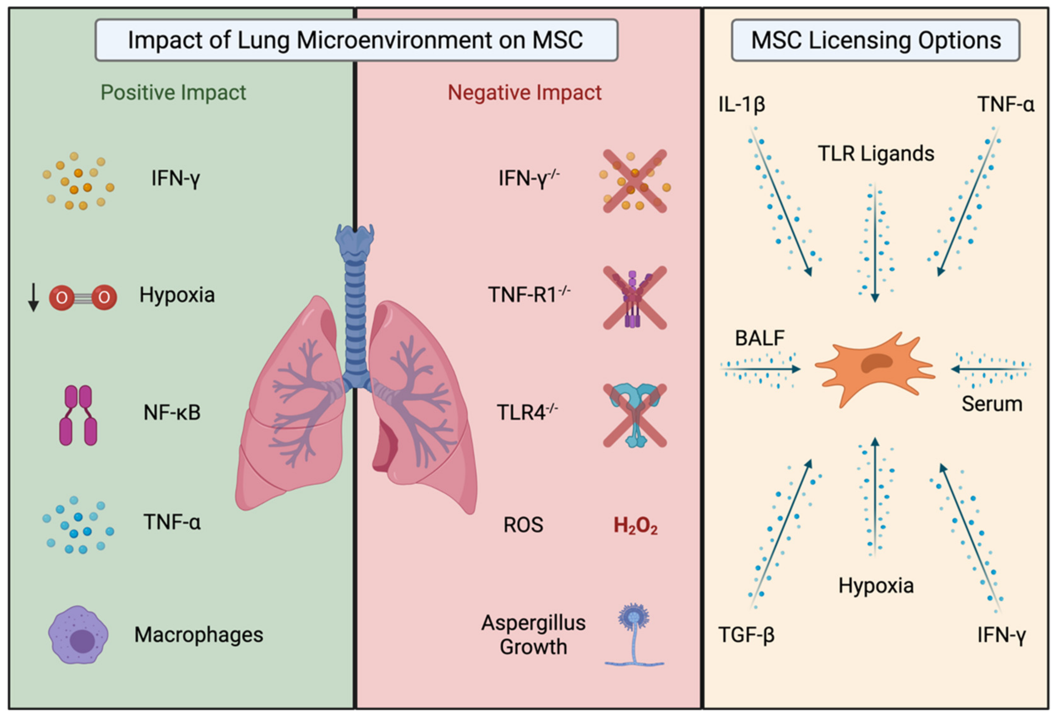
Table 1.
Pre-clinical models of lung disease that received licensed MSCs.
| Murine Model | MSC Source | Route of Administration | Licensing Method | Licensing Location | Outcome | Ref |
|---|---|---|---|---|---|---|
| IPF (bleomycin) | Mouse BM-MSC | I.T. | Hypoxia (1.5% O2) | in vitro | ↑ HIF1α, HGF, VEGF ↓ IL-6, pro-IL-1β | [67] |
| ALI | Rat BM-MSC | In perfusate | Hypoxia (1% O2) | in vitro | ↑ IL-10, PGE2 ↓ Lung injury score ↓ TNF-α, IL-1β, MIP-2 | [68] |
| ALI | Human UC-MSC | I.V. | TGF-β1 | in vitro | ↑ MSC survival ↑ Expression of RhoA ↓ LPS-induced injury | [69] |
| ALI | Human UC-MSC (EVs) | I.V. | IFN-γ | in vitro | ↑ Animal survival ↑ eNOS ↓ Lung injury score ↓ TNF-α | [70] |
| ALI | Rat lung MSCs | I.V. | Culture on lung ECM-cyclic stretch | in vitro | ↑ Lung elastance ↓ TNF-α, CXCL2 + neutrophils | [71] |
| ALI | Human UC-MSC | I.N. | Heatshock (42 °C for 1 h) | in vitro | ↑ HSP70 expression ↑ IL-10 + PGE2 ↓ NLRP3 inflammasome formation ↓ IL-1β secretion by macrophages | [72] |
| ALI | Human BM-MSC | I.V. | Co-culture w/ serum from ARDS patients | ex vivo | ↑ IL-10 + IL-1RN ↓ IL-6, IL-1α, IL-8, IL-1β, IFN-γ, TGFβ3, TGFβ2 + TNFAIP6 | [73] |
| ALI | Human BM-MSC | AM treated with MSC- EVs-I.V. | Co-culture w/ BALF from ARDS patients | ex vivo | ↑ M2 macrophage marker expression ↑ Phagocytic capacity of human MDMs ↓ Cytokine production | [74] |
| ALI | Human BM-MSC | I.T. | Cco-culture w/ plasma from ARDS patients | ex vivo | ↑ IL-6 production ↑ N-cadherin expression at mRNA + protein levels ↓ CD105 + CD90 marker expression at day 5 | [47] |
| ALI | Mouse BM-MSC | I.V. | Co-culture w/ serum from ALI mice | ex vivo | ↑ Expression of anti-inflammatory mediators (TGF-β + IL-10) in AM in vitro ↓ iNOS + IL-6 | [75] |
| Asthma | Mouse BM-MSC | I.T. | Co-culture w/ serum or BALF from asthmatic mice | ex vivo | ↑ TGF-β, IFN-γ, IL-10,TSG-6, IDO-1, IL-1RN, iNOS, TNF-α, IL-1β + arginase-2 ↑ caspase-3, bax ↓ bcl-2Serum:↓ IL-4, IL-13 + eotaxin | [2] |
Footnote ↑ = Increased, ↓ = Decreased. Idiopathic Pulmonary Fibrosis (IPF), Acute Lung Injury (ALI), Acute Respiratory Distress Syndrome (ARDS), Bone-Marrow Mesenchymal Stromal Cells (BM-MSC), Umbilical-Cord Mesenchymal Stromal Cells (UC-MSC), Intratracheal (I.T.), Intranasal (I.N.), Intravenous (I.V.), Alveolar Macrophages (AM), Extracellular Vesicles (EV), Extracellular Matrix (ECM), Bronchoalveolar Lavage Fluid (BALF).
Table 2.
In vivo studies utilising MSC licensing.
| Murine Model | MSC Source | Route of Administration | Licensing Method | Licensing Location | Outcome | Ref |
|---|---|---|---|---|---|---|
| GvHD | Human BM-MSC | I.V. | IFN-γ | in vitro | ↑ Animal survival ↓ Cellular infiltration ↓ Pathology severity in small intestine + liver | [43] |
| GvHD | Human MAPC | I.V. | IFN-γ / PPARδ antagonist | in vitro | ↑ Efficacy on day 0 ↑ Retention in spleen + liver | [49] |
| GvHD | Human BM-MSC | I.V. | Cyclosporine A + IFN-γ | in vitro | ↑ IDO production + activity ↑ Animal survival | [54] |
| GvHD | Human BM-MSC | I.V. | N/A | N/A | ↑ Immunosuppression ↑ IDO production in recipient phagocytes | [76] |
| Corneal allograft | Mouse BM-MSC | I.V. | TGF-β1 | in vitro | ↑ CD73 expression ↑ Treg expansion ↑ Immunosuppression ↑ Allograft survival ↓ Syngeneic T cell proliferation | [77] |
| Corneal allograft | Rat BM-MSC | I.V. | IFN-γ + TNF-α + IL-1β | in vitro | ↑ Nitric oxide production ↑ Suppression of syngeneic lymphocytes ↑ Allograft survival ↑ Myeloid cells in lung ↑ FoxP3+ Treg population in lung + spleen | [78] |
| Sepsis | BM-MSC | I.V. | N/A | N/A | ↑ Animal survival ↑ Organ function ↓ TNF-α + IL-6 | [79] |
| Arthritis | Human BM-MSC | I.V. | PPARβ/δ antagonist orIFN-γ + TNF-α | in vitro | ↑ Immunosuppression ↑ NF-kB activity ↑ p65 binding on iNOS promoter ↓ Severity of disease | [48] |
| Myocardial Infarction | Human BM-MSC | I.V. | TNF-α | in vitro | ↑ TSG-6 mRNA in lung ↓ Infarct size ↓ Pro-inflammatory proteases ↓ MMP9 | [64] |
Footnote: ↑ = Increased, ↓ = Decreased. Graft versus Host Disease (GvHD), Bone-Marrow Mesenchymal Stromal Cells (BM-MSC), Multipotent Adult Progenitor Cells (MAPC), Intravenous (I.V.).
Publisher’s Note: MDPI stays neutral with regard to jurisdictional claims in published maps and institutional affiliations. |
© 2021 by the authors. Licensee MDPI, Basel, Switzerland. This article is an open access article distributed under the terms and conditions of the Creative Commons Attribution (CC BY) license (https://creativecommons.org/licenses/by/4.0/).
Share and Cite
MDPI and ACS Style
Dunbar, H.; Weiss, D.J.; Rolandsson Enes, S.; Laffey, J.G.; English, K. The Inflammatory Lung Microenvironment; a Key Mediator in MSC Licensing. Cells 2021, 10, 2982. https://doi.org/10.3390/cells10112982
AMA Style
Dunbar H, Weiss DJ, Rolandsson Enes S, Laffey JG, English K. The Inflammatory Lung Microenvironment; a Key Mediator in MSC Licensing. Cells. 2021; 10(11):2982. https://doi.org/10.3390/cells10112982
Chicago/Turabian StyleDunbar, Hazel, Daniel J Weiss, Sara Rolandsson Enes, John G Laffey, and Karen English. 2021. "The Inflammatory Lung Microenvironment; a Key Mediator in MSC Licensing" Cells 10, no. 11: 2982. https://doi.org/10.3390/cells10112982
Note that from the first issue of 2016, this journal uses article numbers instead of page numbers. See further details here.