Article

Characterization of an Mtbp Hypomorphic Allele in a Diethylnitrosamine-Induced Liver Carcinogenesis Model

1
Department of Pediatrics, Children’s Mercy Research Institute, Kansas City, MO 64108, USA
2
Department of Cancer Biology, University of Kansas Medical Center, Kansas City, KS 66160, USA
3
Department of Pathology & Laboratory Medicine, University of Kansas Medical Center, Kansas City, KS 66160, USA
4
Path Forward Solutions, LLC, Frederick, MD 21701, USA
*
Author to whom correspondence should be addressed.
Cancers 2023, 15(18), 4596; https://doi.org/10.3390/cancers15184596
Submission received: 29 July 2023 / Revised: 11 September 2023 / Accepted: 13 September 2023 / Published: 16 September 2023

Abstract

:

Simple Summary

The MDM2 binding protein MTBP is implicated in various cellular functions and cancer-related processes, which vary depending on the cellular context and its localization within the cell. Moreover, the in vivo physiological function of MTBP remains unclear. To overcome embryonic lethality due to complete deletion of the Mtbp gene in mice, we created mice with an Mtbp hypomorphic allele (MtbpH) that expresses Mtbp protein at approximately 30% of the wild-type level. In a carcinogen-induced liver cancer model, MtbpH/− mice showed worse overall survival than wild-type mice. MEFs generated from the MtbpH/− mice displayed an increased nuclear localization of p-Erk1/2 protein and enhanced migratory potential. Thus, the newly generated MtbpH/− mice and MEFs can be used to study the in vivo physiological function of Mtbp and validate its diverse functions observed in human cells.

Abstract

MTBP is implicated in cell cycle progression, DNA replication, and cancer metastasis. However, the function of MTBP remains enigmatic and is dependent on cellular contexts and its cellular localization. To understand the in vivo physiological role of MTBP, it is important to generate Mtbp knockout mice. However, complete deletion of the Mtbp gene in mice results in early embryonic lethality, while its heterozygous deletion shows modest biological phenotypes, including enhanced cancer metastasis. To overcome this and better characterize the in vivo physiological function of MTBP, we, for the first time, generated mice that carry an Mtbp hypomorphic allele (MtbpH) in which Mtbp protein is expressed at approximately 30% of that in the wild-type allele. We treated wild-type, Mtbp+/−, and MtbpH/− mice with a liver carcinogen, diethylnitrosamine (DEN), and found that the MtbpH/− mice showed worse overall survival when compared to the wild-type mice. Consistent with previous reports using human liver cancer cells, mouse embryonic fibroblasts (MEFs) from the MtbpH/− mice showed an increase in the nuclear localization of p-Erk1/2 and migratory potential. Thus, MtbpH/− mice and cells from MtbpH/− mice are valuable to understand the in vivo physiological role of Mtbp and validate the diverse functions of MTBP that have been observed in human cells.

1. Introduction

MDM2 binding protein (MTBP) was originally identified using the yeast two-hybrid system as a protein capable of binding to MDM2 [1]. Abundant literature indicates that the biological functions of MTBP are independent of MDM2, and MTBP’s function appears to be dependent on cellular contexts or the intracellular location of MTBP [2,3,4,5,6]. Until today, diverse functions of MTBP have been reported. These include mitotic progression, DNA replication, proliferation, cell migration, and cancer metastasis [2,5,7,8,9,10,11,12]. Specifically, our previous studies and others have demonstrated that MTBP suppresses migration and metastasis of osteosarcoma, gastric cancer, head and neck cancer, and hepatocellular carcinoma (HCC) cells [2,9,10,11,12,13].
The first observation of the migratory- and metastasis-suppressive role of MTBP was made using heterozygous Mtbp knockout mice in which tumors from Mtbp+/−p53+/− mice showed a higher frequency of metastasis of osteosarcoma and HCC (~20%) when compared with tumors from p53+/− mice (~3%) [10]. Clinical studies also support that decreased MTBP levels are correlated with increased metastasis and/or poor prognosis in gastric cancer, head and neck cancer, esophageal squamous cell carcinoma, and HCC [9,11,13,14]. Although the underlying mechanisms need to be clarified, the involvement of actinin-4 (ACTN4, an actin-bundling protein involved in cell motility and migration) for osteosarcoma and lung cancer as well as Erk1/2 for HCC has been shown [2,11,12,15]. Intriguingly, the C-terminal region of MTBP is required for its inhibitory binding to ACTN4 and Importin 7, a protein that shuttles phosphorylated Erk1/2 into the nucleus [2,11,12]. Such migration-suppressive function of MTBP may be mediated by the cytoplasmic portion of MTBP, since a nuclear localization signal (NLS) mutant MTBP, localizing to the cytoplasm, retains the ability to inhibit cancer cell migration [11,12]. These results suggest that cytoplasmic MTBP may function as a migration/metastasis suppressor, while nuclear MTBP may play roles in the regulation of mitotic progression and DNA replication [5,7]. Additionally, there are reports that show oncogenic roles of MTBP where MTBP overexpression enhances cell proliferation, transformation, and migration through interactions with Myc and Ets-1 [4,16,17,18,19]. Moreover, Grieb et al. [20] recently reported that Mtbp heterozygous mice showed an increase in longevity (a 20% increase in median survival) and levels of metabolic markers in the liver as compared to wild-type mice. Thus, physiological and pathological functions of MTBP may vary depending on cellular contexts and intracellular localization of MTBP.
To understand the in vivo function of MTBP and validate the diverse and sometimes controversial observations in MTBP functions, it is essential to generate and analyze Mtbp knockout mice. However, early embryonic lethality following homozygous deletion of Mtbp in mice made it impossible to analyze the phenotypes induced by complete Mtbp deletion [10]. Indeed, all in vivo studies have been made using Mtbp+/− mice, while the phenotypes observed in Mtbp+/− mice are modest [10]. In order to overcome these existing challenges, we generated mice carrying a low-level expression (hypomorphic) allele for Mtbp using a hygromycin cassette [21]. This approach has successfully been used to uncover the in vivo phenotypes of several other genes, including Bub1, Bub3, and BubR1, whose complete deletion causes embryonic lethality, while the deletion of one allele shows unnoticeable or modest phenotypes, similar to Mtbp [22,23,24]. Since mRNA and protein levels of MTBP in human HCC tissues are reduced to 30% compared to those in adjacent non-tumor liver tissues [11], we analyzed Mtbp hypomorphic (MtbpH) mice in a carcinogen-induced liver carcinogenesis model. In addition, we generated and analyzed mouse embryonic fibroblasts (MEFs) derived from MtbpH/ mice for their migratory potential.

2. Materials and Methods

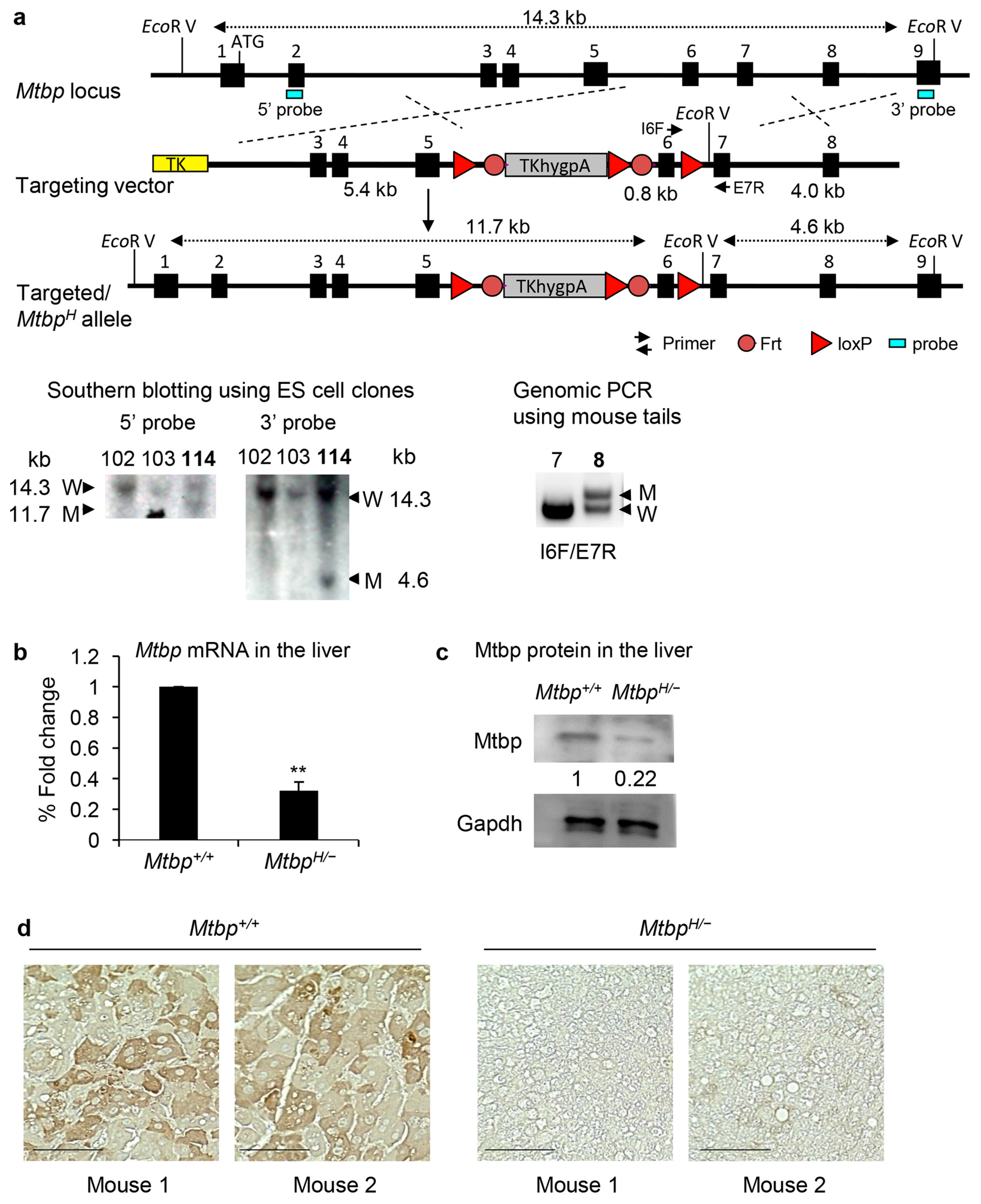
2.1. Generation of Mtbp Hypomorphic Mice

To generate the Mtbp hypomorphic (MtbpH) allele, we obtained the hyg cassette (TKhygpA) from Dr. Wieringa [21,25]. This cassette was modified to be flanked by loxP and Frt sequences, which was inserted into the Mtbp intron 5. We also inserted another loxP site into intron 6 to have the option to generate a conditional Mtbp knockout allele. Successfully targeted 129Sv/Ev ES clones were injected into blastocysts in the National Cancer Institute (NCI) genetically engineered mouse facility. The resulting heterozygous (MtbpH/+) mice were backcrossed to C57BL/6 mice for 8 generations so that they had over 99% of the C57BL/6 background.
Additionally, we generated Mtbp knockout (Mtbp) mice by inserting the pGKneo cassette with the SV40 poly(A) sequence in the same direction as the Mtbp gene (Supplementary Figure S1). Although there was a loxP site in intron 7 in the targeting vector, this site was not inserted into the ES cell genome. The homozygous mice turned out to be early embryonic lethal, consistent with a previous report [10], confirming that this allele is a null allele (Mtbp). All mouse studies were conducted in compliance with Institutional Animal Care and Use Committee protocols of the University of Kansas Medical Center (KUMC).

2.2. Genotyping

Genomic DNA was isolated from the tails, which were incubated in tail lysis buffer containing 0.2 mg/mL of proteinase K at 55 °C overnight, using the standard genomic DNA isolation protocol. Mouse genotyping for the hypomorphic MtbpH allele was performed with PCR amplification using primer sets of I6F (5′-cac agg act tac cat gtc ctg tct gt-3′) and E7R (5′-ata tcc aga gtt gtc acc cct acg gt-3′), while the Mtbp allele was detected using primers of NeoSVF (5’-gaa ttc gcc ctt cga cta gcc ata atc agc-3′), I5F (5′-cta gct atg ctg gag aat tag caa gc-3′), and E6R (5’-gag ggt ctt tgt cag aag gca aca g-3′). The PCR products were resolved on 1.5% agarose gels.

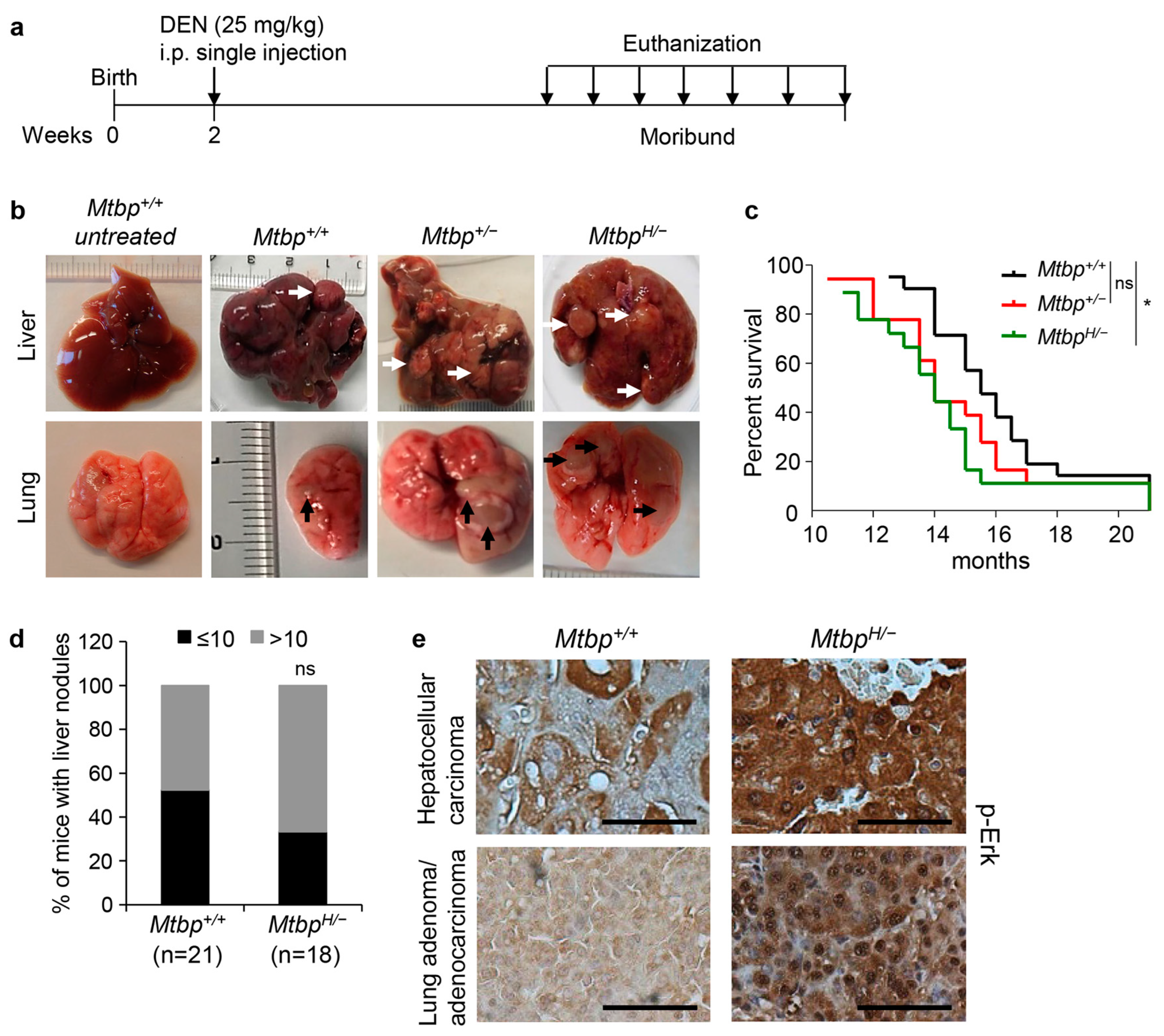
2.3. DEN-Induced Liver Carcinogenesis

Diethylnitrosamine (DEN, N0258, Sigma-Aldrich, St. Louis, MO, USA,) was injected once intraperitoneally to 2-week-old wild-type (Mtbp+/+), Mtbp+/−, and MtbpH/− mice (25 mg/kg body weight). The mice were euthanized when they became moribund.

2.4. Antibodies

The antibodies used for western blotting included mouse monoclonal anti-Mtbp (sc-137201, Santa Cruz, CA, USA) and rabbit polyclonal anti-Gapdh (sc-27117, Santa Cruz, CA, USA) antibodies. For the immunofluorescence studies, rabbit monoclonal anti-p-Erk1/2 Thr202/Tyr204 (#4370, Cell Signaling, Danvers, MA, USA) antibody was used. For IHC, rabbit monoclonal anti-p-Erk1/2 Thr202/Tyr204 (#4370, Cell Signaling) and goat polyclonal anti-Mtbp (sc-47174, Santa Cruz, CA, USA) antibodies were used.

2.5. Immunofluorescence

The cells plated onto poly-D-lysine/laminin-coated glass coverslips (BD Biosciences) were incubated with 4% paraformaldehyde for 20 min. After blocking with 1% BSA in PBS with 0.1% Tween 20 (PBS-T), the cells were incubated with primary antibodies overnight at 4 °C and subsequently with the appropriate secondary antibodies. The samples were mounted in the ProLong Gold Antifade Reagent with DAPI (Invitrogen, Waltham, MA, USA). The results were analyzed using a Nikon epifluorescence microscope (Nikon, Tokyo, Japan).

2.6. Quantitative Reverse Transcriptase PCR (qRT-PCR)

The total RNA from the mouse cells isolated using Quick-RNA Miniprep Kit (Zymo Research, Irvine, CA, USA) was reverse-transcribed with the M-MLV Reverse Transcriptase Kit (Invitrogen, Waltham, MA, USA) using 1 µg total RNA from each sample. The mouse mRNA expression for Mtbp and Gapdh was analyzed with quantitative RT-PCR (qRT-PCR) with TaqMan probes for Mtbp (Mm00519571_m1_g1, Thermo Fisher Scientific, Waltham, MA, USA) and Gapdh (Mm99999915_g1) using Applied Biosystems ViiA7 (Life Technologies, Carlsbad, CA, USA). Mtbp mRNA levels were normalized to those of Gapdh mRNA.

2.7. 3T3 Assay

MEFs were prepared from 13.5-day-old mouse embryos [10]. The MEFs were cultured in high-glucose DMEM medium with 10% fetal bovine serum (FBS) in a 37 °C incubator with 5% CO2. The MEFs (3 × 10⁵, passage 1) seeded onto a 6 cm dish were cultured for three days, and the total number of cells was counted. The counted cells (3 × 10⁵) were again seeded on a 6 cm dish as passage 2. This process was repeated until passage 5.

2.8. Transwell Migration Assay

The migration assays were performed with 24-well Transwell chambers (6.5 mm diameter, 8 mm pore size, Corning) using Mtbp+/+, Mtbp+/+, and MtbpH/− MEFs. The cells (5 × 10⁴) in 100 µL of 0.5% FBS-containing DMEM were seeded on the upper part of the chamber, while in the lower part of the chamber, 10% FBS-containing DMEM was added as a chemoattractant, allowing for cell migration for 14 h. The non-migrating cells were removed gently from the upper part of the chamber of the membrane using cotton swabs, while the migrating cells on the lower surface were stained with the Diff-Quik Stain Set (Dade Behring, Deerfield, IL, USA). Stained cells in the entire fields were counted using an inverted microscope.

2.9. Immunohistochemistry (IHC)

Tumors fixed with 10% buffered formalin for 24 h were embedded in paraffin at the Department of Pathology and Laboratory Medicine in KUMC. The tissue sections were deparaffinized in xylene and rehydrated through a series of graded alcohols. Endogenous peroxidases were inactivated with 3% hydrogen peroxide in PBS for 20 min at 25 °C. After washing with PBS, the slides were incubated in blocking solution (PBS with 0.1% Triton X-100, 3% bovine serum albumin) with 5% normal donkey serum for 10 min at 25 °C. Following antigen retrieval with sodium citrate buffer (10 mM sodium citrate, pH 6.0) for 20 min, IHC was performed using the Vector R.T.U. Vectastain Kit (PK-7800, Vector Laboratories, Newark, CA, USA). The sections were incubated with primary antibodies overnight at 4 °C and subsequently with biotinylated secondary antibodies at room temperature for 30 min. The Vector ImmPact DAB Peroxidase Substrate Kit (SK-4105, Vector Laboratories, Newark CA, USA,) was used for color development followed by hematoxylin counterstaining. All the tumors were pathologically examined.

2.10. Statistical Analysis

The experimental results of the RT-PCR and migration assays were analyzed using the Student’s t-test. The statistical analyses for immunofluorescence and IHC were performed using the two-tailed Fisher’s exact test. The statistical analysis of the survival curves was performed using the log-rank test. A p-value of 0.05 or lower was considered statistically significant. The statistical analyses were performed with the Graph Pad Prism software Version 9.4.1 (San Diego, CA, USA).

3. Results

3.1. Generation of Mtbp Hypomorphic (MtbpH/−) Mice

Complete deletion of Mtbp in mice results in early embryonic lethality, making the study of the in vivo physiological functions of Mtbp challenging [10]. To overcome this, we generated mice that carried the Mtbp hypomorphic (MtbpH) allele by inserting the hygromycin (hyg) cassette surrounded by the loxP and Frt sequences into the intron 5 [21,25] (Figure 1a). When the hyg cassette is inserted into an intron of a gene in the same transcriptional direction, the HSV-tk polyadenylation signal in the hyg cassette (TKhygpA) causes early attenuation of gene transcription from the endogenous promoter [21,25]. This strategy results in only 20~30% expression of the wild-type allele in most cases [21,24,25,26]. Embryonic stem (ES) cells with the MtbpH allele were selected with Southern blotting (Figure 1a) followed by the generation of chimeric mice. The successful germline transmission of the targeted MtbpH allele was confirmed with genomic PCR (Figure 1a).
We additionally generated mice with a pGKneo-SV40 poly(A) cassette surrounded by the loxP and Frt sequences into the intron 5 as well as a loxP site in the intron 7 (Supplementary Figure S1a–c). However, the loxP site in the intron 7 was not incorporated into the genome of the mouse ES cells. When we analyzed mice with the targeted allele, we never obtained mice homozygous for the pGKneo-SV40 poly(A) cassette at 3 weeks of age. This observation indicates that the Mtbp allele with the pGKneo-SV40 poly(A) cassette is a null allele (Mtbp), consistent with a previous report about embryonic lethality of Mtbp knockout mice [10]. Hence, the Mtbp allele with the pGKneo-SV40 poly(A) cassette was hereafter named as a Mtbp allele. These genetically engineered mice were backcrossed to C57BL/6 for eight generations followed by the generation of MtbpH/− mice by crossing MtbpH/+ mice with Mtbp+/− mice. The MtbpH/− mice were viable and fertile and did not exhibit any obvious phenotypes, including cancer-prone phenotypes.
To investigate the expression of Mtbp in the MtbpH/− mice, qRT-PCR was performed using mRNA from mouse livers taken from the Mtbp+/+ and MtbpH/− mice (Figure 1b). The level of Mtbp mRNA in the MtbpH/− liver was approximately 30% of that in the Mtbp+/+ liver. Consistently, the Mtbp protein level in the MtbpH/− liver, detected with western blotting and immunohistochemistry (IHC), was 20~30% of that in the Mtbp+/+ liver (Figure 1c,d). These data demonstrate the successful generation of Mtbp hypomorphic mice.

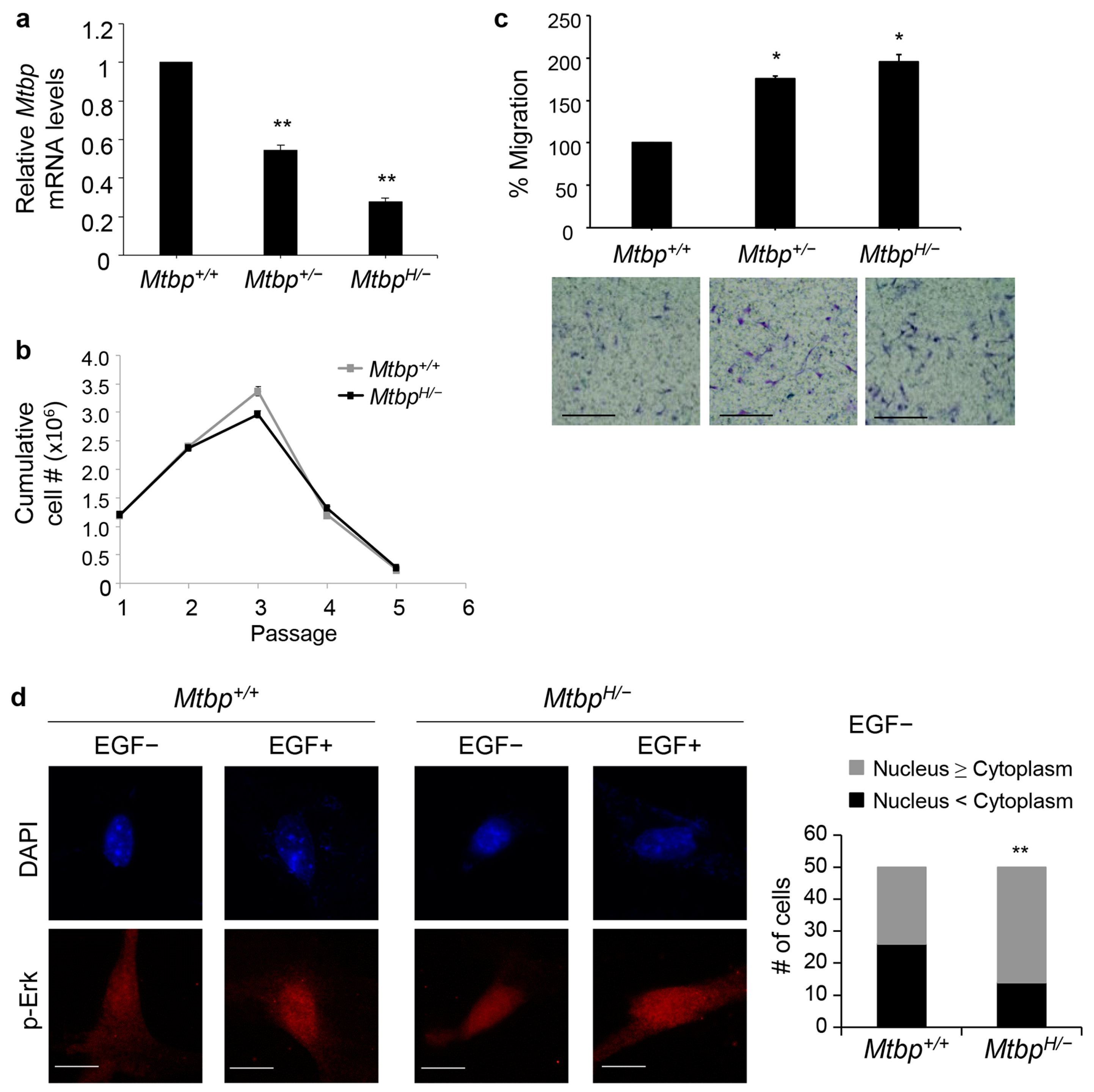
3.2. Increased Cell Migration of MtbpH/− MEFs

To further validate the phenotypes of hypomorphic Mtbp, we generated MEFs from the Mtbp+/+, Mtbp+/−, and MtbpH/− mice. Mtbp mRNA expression in the MEFs was confirmed with qRT-PCR. Consistent with the results in the liver, the Mtbp mRNA levels in the Mtbp+/− and MtbpH/− MEFs were ~50% and ~30% of the Mtbp+/+ MEFs, respectively (Figure 2a), further confirming that the Mtbp allele with the pGKneo-SV40 poly(A) cassette functions was a null allele, while the MtbpH/− allele was a hypomorphic allele. To examine whether reduced Mtbp expression could contribute to immortalization of MEFs, we performed 3T3 assays using Mtbp+/+ and MtbpH/− MEFs. The reduced Mtbp levels did not contribute to cellular immortalization of the MtbpH/− MEFs (Figure 2b), suggesting that Mtbp does not function as a typical tumor suppressor. Given the known metastasis-suppressive function of Mtbp [10,12], these MEFs were examined for their migratory potential using transwell migration assays. The migratory potential of the MEFs was negatively correlated with the levels of Mtbp, although the Mtbp+/− and MtbpH/− MEFs had similar migratory potential (Figure 2c). Moreover, the MtbpH/− MEFs had an increase in nuclear phosphorylated Erk (p-Erk) in the absence of EGF stimulation as compared with the Mtbp+/+ MEFs (Figure 2d). These observations are corroborated by previous reports where MTBP inhibits cellular migration by inhibiting nuclear translocation of p-Erk, while Mtbp haploinsufficiency enhances cancer cell migration and metastasis [10,12].

3.3. Reduced Mtbp Enhances Liver Carcinogenesis

To address the in vivo role of Mtbp in liver carcinogenesis, we used the diethylnitrosamine (DEN)-induced liver carcinogenesis model [27]. Two-week-old Mtbp+/+, Mtbp+/−, and MtbpH/− mice were intraperitoneally injected with DEN (25 mg/kg body weight) and were observed for tumor development (Figure 3a). The mice were euthanized when they became moribund. Major organs, including liver, lungs, intestine, lymph nodes, kidney, spleen, and brain, were investigated for primary tumors and metastatic nodules during dissection. All DEN-injected mice developed tumors in the liver, while several mice had nodules in the lungs as well (Figure 3b). The MtbpH/− mice became moribund significantly earlier than the Mtbp+/+ mice, supporting a tumor-suppressive role of Mtbp in liver carcinogenesis (Figure 3c). The Mtbp+/− mice also became moribund earlier than the Mtbp+/+ mice; however, the difference was not statistically significant. When we counted tumor nodules per liver, the MtbpH/− mice tended to have higher tumor nodules (>10 nodules per liver in 12 out of 18 mice: 66.7%) than the Mtbp+/+ mice (10 out of 21 mice: 47.6%), although the difference was not statistically significant (Figure 3d). Many liver nodules were fused and uncountable, which could be due to multifocal tumors or intrahepatic metastases.
Intriguingly, eight Mtbp+/+ mice (38.1%) developed pulmonary nodules, while twelve MtbpH/− mice (66.7%) formed pulmonary nodules (Table 1). Since DEN can induce lung tumors in mice [28], we stained lung tumor sections for TTF-1 (thyroid transcription factor-1), a marker of lung and thyroid adenocarcinomas, to distinguish whether these tumors originated from the liver or lung. All pulmonary nodules observed in the Mtbp+/+ mice turned out to be lung adenoma or adenocarcinoma, while one pulmonary nodule that developed in an MtbpH/− mouse was confirmed as HCC metastasis (Table 1).
Moreover, IHC staining of liver and lung tumors for p-Erk revealed higher intensity and increased nuclear localization of p-Erk in the MtbpH/− tumors when compared to the Mtbp+/+ tumors (Figure 3e). These results are consistent with our previous finding that MTBP inhibits p-Erk nuclear localization [12]. Thus, the newly generated Mtbp hypomorphic mice are useful to identify and validate the in vivo physiological function of Mtbp.

4. Discussion

In this study, we generated Mtbp hypomorphic mice (MtbpH/−) that express ~30% of the Mtbp levels expressed in wild-type mice and investigated the in vivo role of Mtbp in DEN-induced liver carcinogenesis. The results from the MtbpH/− mice and MEFs corroborated with the previously reported inhibitory roles of MTBP in HCC progression and migration using human cancer cell lines [2,9,10,11,12,13], showing the significance and usefulness of the MtbpH/− mice. Thus, the generation of mice that carry a hypomorphic allele provides a powerful tool to overcome the embryonic lethality caused by complete deletion of a gene of interest and examine the in vivo physiological function of the encoded protein. Indeed, previous studies have generated hypomorphic mice for genes whose knockout is embryonic lethal, but their heterozygous mice showed modest phenotypes. These include Mdm2, Bub1, BubR1, and Pkd1 [25,29,30,31].
In addition to the inhibitory function on cell migration and cancer metastasis, MTBP has also been proposed to play roles in origin firing for DNA replication [5,8,32], mitotic progression [7], MDM2 stabilization [33,34], regulation of Myc [35,36], and promotion of cancer progression [6,17,18]. Additionally, whole-genome sequencing in dogs has identified a variant in the MTBP gene associated with proinflammatory processes [37]. Recently, Grieb et al. [20] showed that Mtbp heterozygous mice exhibit increased levels of metabolic markers in the liver and increased longevity. It would be intriguing to examine whether Mtbp alters the metabolism of DEN in the liver by using MtbpH/− mice, which could consequently alter the DEN-induced liver tumorigenesis. Thus, MTBP appears to have diverse functions that depend on cellular and tissue-type contexts. Clinically, several studies using human cancer patients have suggested MTBP as a potential biomarker for favorable prognosis in patients with specific cancer types, including HNSCC, gastric cancer, and esophageal squamous cell carcinoma [9,11,13,14]. On the other hand, MTBP expression appears to serve as a biomarker for poor prognosis in patients with glioblastoma and triple-negative breast cancer [6,33]. Although the MtbpH/− mice did not show a significant increase in metastases of the DEN-induced liver tumors, we observed an increase in liver and lung nodules in the MtbpH/− mice as compared with the Mtbp+/+ mice, which may be due to intrahepatic and intrapulmonary metastases or increased incidence of multifocal tumors. In either case, the results from the MtbpH/− mice support the tumor-suppressive role of MTBP. MtbpH/− mice would also be a useful tool to study the in vivo role of Mtbp in the progression of other cancer types. Moreover, it would be interesting to generate mice that express a high level of Mtbp and examine whether Mtbp overexpression inhibits or promotes the progression of certain types of tumors.
Additionally, the MtbpH allele contains Frt and loxP sequences surrounding the TKhygpA cassette in the intron 5, while it also contains a loxP site in the intron 6 (Figure 1a). Hence, crossing MtbpH/− mice with mice expressing Flp recombinase is expected to make Mtbp+/− mice, while crossing MtbpH/− mice with mice expressing Cre recombinase will generate complete Mtbp knockout mice. When these recombinases are expressed in a specific tissue or a specific condition, mice with the MtbpH allele would be valuable to examine tissue-type or context-dependent physiological functions of Mtbp.

5. Conclusions

MTBP is involved in diverse cellular functions and cancer-related processes. However, its in vivo physiological function remains unclear. This is mainly because complete deletion of Mtbp in mice leads to early embryonic lethality. Our newly generated Mtbp hypomorphic mice and their derived cells are extremely useful to investigate the in vivo physiological function of Mtbp, including its role in cancer progression and metastasis. Indeed, the Mtbp hypomorphic mice show enhanced DEN-induced liver carcinogenesis with increased nuclear localization of p-Erk1/2, consistent with the previous reports.

Supplementary Materials

The following supporting information can be downloaded at: https://www.mdpi.com/article/10.3390/cancers15184596/s1, Figure S1: Generation of mice carrying a Mtbp null (Mtbp) allele.

Author Contributions

Conceptualization, T.V.D. and T.I.; methodology, A.R., E.A.T., A.K. and S.W.; validation, formal analysis, and investigation, A.R., A.K. and T.I.; data curation, A.R. and T.I.; writing—original draft preparation, A.R. and T.I.; writing—review and editing, A.R., E.A.T., A.K., S.W., T.V.D. and T.I.; funding acquisition, T.I. All authors have read and agreed to the published version of the manuscript.

Funding

This work has been supported by grants R01 CA174735 (TI) and R01 CA214916 (TI) and NIH P30 CA168524 (RAJ) from the NIH.

Institutional Review Board Statement

The animal study protocol was approved by the IACUC of KUMC (ACUP 2012-2081, date of approval on 4 September 2012).

Informed Consent Statement

Not applicable.

Data Availability Statement

The data can be shared up on request.

Acknowledgments

The authors thank B. Wieringa at the University of Nijmegen, The Netherlands, for providing the hyg cassette and Yuki Tochigi at Nippon Veterinary and Life Science University, Japan for genotyping the Mtbp+/− mice.

Conflicts of Interest

The authors declare no conflict of interest.

References

  1. Boyd, M.T.; Vlatkovic, N.; Haines, D.S. A novel cellular protein (MTBP) binds to MDM2 and induces a G1 arrest that is suppressed by MDM2. J. Biol. Chem. 2000, 275, 31883–31890. [Google Scholar] [CrossRef] [PubMed]
  2. Agarwal, N.; Adhikari, A.S.; Iyer, S.V.; Hekmatdoost, K.; Welch, D.R.; Iwakuma, T. MTBP suppresses cell migration and filopodia formation by inhibiting ACTN4. Oncogene 2013, 32, 462–470. [Google Scholar] [CrossRef] [PubMed]
  3. Iwakuma, T.; Agarwal, N. MDM2 binding protein, a novel metastasis suppressor. Cancer Metastasis Rev. 2012, 31, 633–640. [Google Scholar] [CrossRef] [PubMed]
  4. Odvody, J.; Vincent, T.; Arrate, M.P.; Grieb, B.; Wang, S.; Garriga, J.; Lozano, G.; Iwakuma, T.; Haines, D.S.; Eischen, C.M. A deficiency in Mdm2 binding protein inhibits Myc-induced B-cell proliferation and lymphomagenesis. Oncogene 2010, 29, 3287–3296. [Google Scholar] [CrossRef] [PubMed]
  5. Boos, D.; Yekezare, M.; Diffley, J.F. Identification of a heteromeric complex that promotes DNA replication origin firing in human cells. Science 2013, 340, 981–984. [Google Scholar] [CrossRef] [PubMed]
  6. Grieb, B.C.; Chen, X.; Eischen, C.M. MTBP is overexpressed in triple-negative breast cancer and contributes to its growth and survival. Mol. Cancer Res. 2014, 12, 1216–1224. [Google Scholar] [CrossRef]
  7. Agarwal, N.; Tochigi, Y.; Adhikari, A.S.; Cui, S.; Cui, Y.; Iwakuma, T. MTBP plays a crucial role in mitotic progression and chromosome segregation. Cell Death Differ. 2011, 18, 1208–1219. [Google Scholar] [CrossRef]
  8. Kohler, K.; Sanchez-Pulido, L.; Hofer, V.; Marko, A.; Ponting, C.P.; Snijders, A.P.; Feederle, R.; Schepers, A.; Boos, D. The Cdk8/19-cyclin C transcription regulator functions in genome replication through metazoan Sld7. PLoS Biol. 2019, 17, e2006767. [Google Scholar] [CrossRef] [PubMed]
  9. Vlatkovic, N.; El-Fert, A.; Devling, T.; Ray-Sinha, A.; Gore, D.M.; Rubbi, C.P.; Dodson, A.; Jones, A.S.; Helliwell, T.R.; Jones, T.M.; et al. Loss of MTBP expression is associated with reduced survival in a biomarker-defined subset of patients with squamous cell carcinoma of the head and neck. Cancer 2011, 117, 2939–2950. [Google Scholar] [CrossRef]
  10. Iwakuma, T.; Tochigi, Y.; Van Pelt, C.S.; Caldwell, L.C.; Terzian, T.; Parant, J.M.; Chau, G.P.; Koch, J.G.; Eischen, C.M.; Lozano, G. Mtbp haploinsufficiency in mice increases tumor metastasis. Oncogene 2008, 27, 1813–1820. [Google Scholar] [CrossRef]
  11. Bi, Q.; Ranjan, A.; Fan, R.; Agarwal, N.; Welch, D.R.; Weinman, S.A.; Ding, J.; Iwakuma, T. MTBP inhibits migration and metastasis of hepatocellular carcinoma. Clin. Exp. Metastasis 2015, 32, 301–311. [Google Scholar] [CrossRef] [PubMed]
  12. Ranjan, A.; Iyer, S.V.; Ward, C.; Link, T.; Diaz, F.J.; Dhar, A.; Tawfik, O.W.; Weinman, S.A.; Azuma, Y.; Izumi, T.; et al. MTBP inhibits the Erk1/2-Elk-1 signaling in hepatocellular carcinoma. Oncotarget 2018, 9, 21429–21443. [Google Scholar] [CrossRef] [PubMed]
  13. Wang, W.; Chen, Z.; Jin, J.; Long, Z.; Liu, X.; Cai, H.; Zhou, Y.; Huang, H.; Wang, Y. MDM2 binding protein as a predictor of metastasis and a novel prognostic biomarker in patients with gastric cancer. Oncol. Lett. 2017, 14, 6409–6416. [Google Scholar] [CrossRef] [PubMed]
  14. Shi, X.; Li, Y.; Sun, Y.; Zhao, X.; Sun, X.; Gong, T.; Liang, Z.; Ma, Y.; Zhang, X. Genome-wide analysis of lncRNAs, miRNAs, and mRNAs forming a prognostic scoring system in esophageal squamous cell carcinoma. PeerJ 2020, 8, e8368. [Google Scholar] [CrossRef] [PubMed]
  15. Wu, A.; Tang, J.; Guo, Z.; Dai, Y.; Nie, J.; Hu, W.; Liu, N.; Ye, C.; Li, S.; Pei, H.; et al. Long Non-Coding RNA CRYBG3 Promotes Lung Cancer Metastasis via Activating the eEF1A1/MDM2/MTBP Axis. Int. J. Mol. Sci. 2021, 22, 3211. [Google Scholar] [CrossRef] [PubMed]
  16. Xiao, Z.; Chen, M.; Yang, J.; Yang, C.; Lu, X.; Tian, H.; Liu, C. MTBP regulates migration and invasion of prostate cancer cells in vitro. Nan Fang Yi Ke Da Xue Xue Bao J. South. Med. Univ. 2019, 39, 6–12. [Google Scholar] [CrossRef]
  17. Pan, B.; Han, H.; Wu, L.; Xiong, Y.; Zhang, J.; Dong, B.; Yang, Y.; Chen, J. MTBP promotes migration and invasion by regulation of ZEB2-mediated epithelial-mesenchymal transition in lung cancer cells. Onco Targets Ther. 2018, 11, 6741–6756. [Google Scholar] [CrossRef]
  18. Wang, H.; Chu, F.; Zhijie, L.; Bi, Q.; Lixin, L.; Zhuang, Y.; Xiaofeng, Z.; Niu, X.; Zhang, D.; Xi, H.; et al. MTBP enhances the activation of transcription factor ETS-1 and promotes the proliferation of hepatocellular carcinoma cells. Front. Oncol. 2022, 12, 985082. [Google Scholar] [CrossRef]
  19. Shayimu, P.; Yusufu, A.; Rehemutula, A.; Redati, D.; Jiapaer, R.; Tuerdi, R. MTBP promoted the proliferation, migration and invasion of colon cancer cells by activating the expression of ZEB2. Anim. Cells Syst. 2021, 25, 152–160. [Google Scholar] [CrossRef]
  20. Grieb, B.C.; Boyd, K.; Mitra, R.; Eischen, C.M. Haploinsufficiency of the Myc regulator Mtbp extends survival and delays tumor development in aging mice. Aging 2016, 8, 2590–2602. [Google Scholar] [CrossRef]
  21. van Deursen, J.; Ruitenbeek, W.; Heerschap, A.; Jap, P.; ter Laak, H.; Wieringa, B. Creatine kinase (CK) in skeletal muscle energy metabolism: A study of mouse mutants with graded reduction in muscle CK expression. Proc. Natl. Acad. Sci. USA 1994, 91, 9091–9095. [Google Scholar] [CrossRef] [PubMed]
  22. Kalitsis, P.; Fowler, K.J.; Griffiths, B.; Earle, E.; Chow, C.W.; Jamsen, K.; Choo, K.H. Increased chromosome instability but not cancer predisposition in haploinsufficient Bub3 mice. Genes Chromosomes Cancer 2005, 44, 29–36. [Google Scholar] [CrossRef] [PubMed]
  23. Wang, Q.; Liu, T.; Fang, Y.; Xie, S.; Huang, X.; Mahmood, R.; Ramaswamy, G.; Sakamoto, K.M.; Darzynkiewicz, Z.; Xu, M.; et al. BUBR1 deficiency results in abnormal megakaryopoiesis. Blood 2004, 103, 1278–1285. [Google Scholar] [CrossRef] [PubMed]
  24. Jeganathan, K.; Malureanu, L.; Baker, D.J.; Abraham, S.C.; van Deursen, J.M. Bub1 mediates cell death in response to chromosome missegregation and acts to suppress spontaneous tumorigenesis. J. Cell Biol. 2007, 179, 255–267. [Google Scholar] [CrossRef]
  25. Dawlaty, M.M.; van Deursen, J.M. Gene targeting methods for studying nuclear transport factors in mice. Methods 2006, 39, 370–378. [Google Scholar] [CrossRef] [PubMed]
  26. Shawlot, W.; Deng, J.M.; Fohn, L.E.; Behringer, R.R. Restricted beta-galactosidase expression of a hygromycin-lacZ gene targeted to the beta-actin locus and embryonic lethality of beta-actin mutant mice. Transgenic Res. 1998, 7, 95–103. [Google Scholar] [CrossRef]
  27. Vesselinovitch, S.D.; Mihailovich, N. Kinetics of diethylnitrosamine hepatocarcinogenesis in the infant mouse. Cancer Res. 1983, 43, 4253–4259. [Google Scholar] [PubMed]
  28. Mervai, Z.; Egedi, K.; Kovalszky, I.; Baghy, K. Diethylnitrosamine induces lung adenocarcinoma in FVB/N mouse. BMC Cancer 2018, 18, 157. [Google Scholar] [CrossRef]
  29. Lantinga-van Leeuwen, I.S.; Dauwerse, J.G.; Baelde, H.J.; Leonhard, W.N.; van de Wal, A.; Ward, C.J.; Verbeek, S.; Deruiter, M.C.; Breuning, M.H.; de Heer, E.; et al. Lowering of Pkd1 expression is sufficient to cause polycystic kidney disease. Hum. Mol. Genet. 2004, 13, 3069–3077. [Google Scholar] [CrossRef]
  30. Mendrysa, S.M.; O’Leary, K.A.; McElwee, M.K.; Michalowski, J.; Eisenman, R.N.; Powell, D.A.; Perry, M.E. Tumor suppression and normal aging in mice with constitutively high p53 activity. Genes Dev. 2006, 20, 16–21. [Google Scholar] [CrossRef]
  31. Hartman, T.K.; Wengenack, T.M.; Poduslo, J.F.; van Deursen, J.M. Mutant mice with small amounts of BubR1 display accelerated age-related gliosis. Neurobiol. Aging 2007, 28, 921–927. [Google Scholar] [CrossRef] [PubMed]
  32. Zonderland, G.; Vanzo, R.; Gadi, S.A.; Martin-Doncel, E.; Coscia, F.; Mund, A.; Lerdrup, M.; Benada, J.; Boos, D.; Toledo, L. The TRESLIN-MTBP complex couples completion of DNA replication with S/G2 transition. Mol. Cell 2022, 82, 3350–3365.e7. [Google Scholar] [CrossRef] [PubMed]
  33. Song, Y.; Zhang, L.; Jiang, Y.; Hu, T.; Zhang, D.; Qiao, Q.; Wang, R.; Wang, M.; Han, S. MTBP regulates cell survival and therapeutic sensitivity in TP53 wildtype glioblastomas. Theranostics 2019, 9, 6019–6030. [Google Scholar] [CrossRef] [PubMed]
  34. Brady, M.; Vlatkovic, N.; Boyd, M.T. Regulation of p53 and MDM2 activity by MTBP. Mol. Cell Biol. 2005, 25, 545–553. [Google Scholar] [CrossRef] [PubMed]
  35. Grieb, B.C.; Gramling, M.W.; Arrate, M.P.; Chen, X.; Beauparlant, S.L.; Haines, D.S.; Xiao, H.; Eischen, C.M. Oncogenic protein MTBP interacts with MYC to promote tumorigenesis. Cancer Res. 2014, 74, 3591–3602. [Google Scholar] [CrossRef]
  36. Grieb, B.C.; Eischen, C.M. MTBP and MYC: A Dynamic Duo in Proliferation, Cancer, and Aging. Biology 2022, 11, 881. [Google Scholar] [CrossRef]
  37. Metzger, J.; Nolte, A.; Uhde, A.K.; Hewicker-Trautwein, M.; Distl, O. Whole genome sequencing identifies missense mutation in MTBP in Shar-Pei affected with Autoinflammatory Disease (SPAID). BMC Genom. 2017, 18, 348. [Google Scholar] [CrossRef]
Figure 1. Generation and characterization of MtbpH/− mice. (a) Genomic organization of the murine Mtbp gene, MtbpH targeting vector, and targeted allele. The vector is constructed as a conditional allele using two different recombinase systems, Flp/Frt and Cre/loxP. The hyg cassette (TKhygpA) attenuates transcription of the endogenous Mtbp gene, resulting in the MtbpH allele. Representative results of Southern blotting (bottom, left) following Eco RV restriction enzyme digestion of the genomic DNA from ES cell clones (#102, 103, 114) using the 5′ and 3′ probe set in exon 2 and exon 9, respectively. Genomic PCR (bottom, right) using the genomic DNA from mice (#7, 8) with primers of I6F and E7R, showing successful germline transmission. (b) Results of qRT-PCR for Mtbp using mRNA from Mtbp+/+ and MtbpH/− mouse livers. Data are normalized with values of Gapdh mRNA. Error bars: means + S.E. from three independent experiments. Student’s t test: **, p < 0.01. (c) Western blotting for Mtbp and Gapdh using protein extracts from liver tissues isolated from Mtbp+/+ and MtbpH/− mice. (d) IHC for Mtbp using liver tissues from Mtbp+/+ and MtbpH/− mice (2 representative images from each genotype). Scale bar, 25 μm. The uncropped blots are shown in Supplementary Materials.
Cancers 15 04596 g001
Figure 2. Increased cell migration of MtbpH/− MEFs. (a) Results of qRT-PCR for Mtbp using mRNA from Mtbp+/+, Mtbp+/−, and MtbpH/− MEFs. Data are normalized with values of Gapdh mRNA. Error bars: means ± S.E. from three independent experiments. Student’s t test: **, p < 0.01. (b) 3T3 assays using Mtbp+/+ and MtbpH/− MEFs. Error bars: means + S.E. from three independent experiments. Student’s t test: not significant. (c) Transwell migration assays using Mtbp+/+, Mtbp+/−, and MtbpH/− MEFs. Cells were plated on the upper chambers of the Transwell. Migrating cells in the entire fields were counted 14 h later. A summary graph (top) and representative images (bottom). Scale bar, 25 μm. Error bars: means ± S.E. from three independent experiments. Student’s t test: *, p < 0.05. (d) Immunofluorescence studies for p-Erk following treatment with vehicle (EGF-) or 50 ng/mL of EGF (EGF+) for 30 min using Mtbp+/+ and MtbpH/− MEFs. Scale bar, 25 μm. Graph showing the number of cells with different Mtbp locations (Nucleus ≥ Cytoplasm or Nucleus < Cytoplasm) in the absence of EGF treatment (n = 50). Fisher’s exact test (two tailed): **, p < 0.01.
Cancers 15 04596 g002
Figure 3. Increased liver carcinogenesis in DEN-treated MtbpH/− mice. (a) Schematic of DEN-induced liver carcinogenesis studies. Mice were injected with DEN at 25 mg/kg of body weight two weeks after birth. (b) Representative images of the liver (top) and lung (bottom) of a non-DEN-treated Mtbp+/+ mouse and DEN-treated Mtbp+/+, Mtbp+/−, and MtbpH/− mice with tumors. White and Black arrows indicate tumor nodules in liver and lung, respectively. (c) Kaplan–Meier survival curves of Mtbp+/+ (n = 21), Mtbp+/− (n = 18), and MtbpH/− (n = 18) mice injected with DEN. Log-rank test: * p < 0.05; ns: not significant. (d) Numbers of mice with liver nodules in Mtbp+/+ (n = 21) and MtbpH/− (n = 18) mice. Fisher’s exact test (two tailed); ns: not significant. (e) IHC for p-Erk using HCC tumors from Mtbp+/+ and MtbpH/− mice as well as a lung adenoma from a Mtbp+/+ mouse or a lung adenocarcinoma from a MtbpH/− mouse. Scale bar, 25 μm.
Cancers 15 04596 g003
Table 1. DEN-induced lung tumors and metastases from the liver in Mtbp+/+ and MtbpH/ mice.
GenotypeGender
(Male: M,
Female: F)
# of Mice# of Mice with Lung Nodules# of Mice with Metastatic Nodules
Mtbp+/+M1250
F930
Total218 (38.1%)0
MtbpH/−M1081
F840
Total1812 (66.7%)1
#: number.
Disclaimer/Publisher’s Note: The statements, opinions and data contained in all publications are solely those of the individual author(s) and contributor(s) and not of MDPI and/or the editor(s). MDPI and/or the editor(s) disclaim responsibility for any injury to people or property resulting from any ideas, methods, instructions or products referred to in the content.

