
Is Gross Extrathyroidal Extension to Strap Muscles (T3b) Only a Risk Factor for Recurrence in Papillary Thyroid Carcinoma? A Propensity Score Matching Study


Abstract
:Simple Summary
Abstract
1. Introduction
2. Materials and Methods
2.1. Patients
2.2. ETE Definition
2.3. Follow-Up Assessment
2.4. Primary and Secondary Endpoints
2.5. Statistical Analysis
3. Results
3.1. Comparison of Baseline Clinicopathological Characteristics According to the ETE Status before Propensity Score Matching
3.2. Univariate and Multivariate Analyses of Risk Factors for Recurrence before Propensity Score Matching
3.3. Comparison of Baseline Clinicopathological Characteristics between the mETE and gETE Groups after Propensity Score Matching
3.4. Univariate and Multivariate Analyses of Risk Factors for Recurrence after Propensity Score Matching
3.5. Sub-Analysis of Clinicopathological Characteristics According to ETE Status in PTMC
4. Discussion
5. Conclusions
Author Contributions
Funding
Institutional Review Board Statement
Informed Consent Statement
Data Availability Statement
Conflicts of Interest
References
- Chéreau, N.; Buffet, C.; Trésallet, C.; Tissier, F.; Golmard, J.-L.; Leenhardt, L.; Menegaux, F. Does extracapsular extension impact the prognosis of papillary thyroid microcarcinoma? Ann. Surg. Oncol. 2014, 21, 1659–1664. [Google Scholar] [CrossRef] [PubMed]
- Amit, M.; Boonsripitayanon, M.; Goepfert, R.P.; Tam, S.; Busaidy, N.L.; Cabanillas, M.E.; Dadu, R.; Varghese, J.; Waguespack, S.G.; Gross, N.D. Extrathyroidal extension: Does strap muscle invasion alone influence recurrence and survival in patients with differentiated thyroid cancer? Ann. Surg. Oncol. 2018, 25, 3380–3388. [Google Scholar] [CrossRef] [PubMed]
- Edge, S.B.; Compton, C.C. The American Joint Committee on Cancer: The 7th edition of the AJCC cancer staging manual and the future of TNM. Ann. Surg. Oncol. 2010, 17, 1471–1474. [Google Scholar] [CrossRef] [PubMed]
- Tuttle, R.M.; Haugen, B.; Perrier, N.D. Updated American Joint Committee on cancer/tumor-node-metastasis staging system for differentiated and anaplastic thyroid cancer: What Changed and Why? Thyroid 2017, 27, 751–756. [Google Scholar] [CrossRef]
- Edge, S.B. AJCC Cancer Staging Manual, 8th ed.; Springer: New York, NY, USA, 2017. [Google Scholar]
- Haugen, B.R.; Alexander, E.K.; Bible, K.C.; Doherty, G.M.; Mandel, S.J.; Nikiforov, Y.E.; Pacini, F.; Randolph, G.W.; Sawka, A.M.; Schlumberger, M. 2015 American Thyroid Association management guidelines for adult patients with thyroid nodules and differentiated thyroid cancer: The American Thyroid Association guidelines task force on thyroid nodules and differentiated thyroid cancer. Thyroid 2016, 26, 37–52. [Google Scholar] [CrossRef] [Green Version]
- Arora, N.; Turbendian, H.K.; Scognamiglio, T.; Wagner, P.L.; Goldsmith, S.J.; Zarnegar, R.; Fahey, T.J., III. Extrathyroidal extension is not all equal: Implications of macroscopic versus microscopic extent in papillary thyroid carcinoma. Surgery 2008, 144, 942–948. [Google Scholar] [CrossRef]
- Diker-Cohen, T.; Hirsch, D.; Shimon, I.; Bachar, G.; Akirov, A.; Duskin-Bitan, H.; Robenshtok, E. Impact of minimal extrathyroid extension in differentiated thyroid cancer: Systematic review and meta-analysis. J. Clin. Endocrinol. Metab. 2018, 103, 2100–2106. [Google Scholar] [CrossRef]
- Ito, Y.; Tomoda, C.; Uruno, T.; Takamura, Y.; Miya, A.; Kobayashi, K.; Matsuzuka, F.; Kuma, K.; Miyauchi, A. Prognostic significance of extrathyroid extension of papillary thyroid carcinoma: Massive but not minimal extension affects the relapse-free survival. World J. Surg. 2006, 30, 780–786. [Google Scholar] [CrossRef]
- Danilovic, D.L.; Castroneves, L.A.; Suemoto, C.K.; Elias, L.O.; Soares, I.C.; Camargo, R.Y.; Correa, F.A.; Hoff, A.O.; Marui, S. Is there a difference between minimal and gross extension into the strap muscles for the risk of recurrence in papillary thyroid carcinomas? Thyroid 2020, 30, 1008–1016. [Google Scholar] [CrossRef] [Green Version]
- Park, J.S.; Chang, J.W.; Liu, L.; Jung, S.-N.; Koo, B.S. Clinical implications of microscopic extrathyroidal extension in patients with papillary thyroid carcinoma. Oral Oncol. 2017, 72, 183–187. [Google Scholar] [CrossRef]
- Hay, I.D.; Johnson, T.R.; Thompson, G.B.; Sebo, T.J.; Reinalda, M.S. Minimal extrathyroid extension in papillary thyroid carcinoma does not result in increased rates of either cause-specific mortality or postoperative tumor recurrence. Surgery 2016, 159, 11–21. [Google Scholar] [CrossRef] [PubMed] [Green Version]
- Moon, H.J.; Kim, E.-K.; Chung, W.Y.; Yoon, J.H.; Kwak, J.Y. Minimal extrathyroidal extension in patients with papillary thyroid microcarcinoma: Is it a real prognostic factor? Ann. Surg. Oncol. 2011, 18, 1916–1923. [Google Scholar] [CrossRef] [PubMed]
- Rivera, M.; Ricarte-Filho, J.; Tuttle, R.M.; Ganly, I.; Shaha, A.; Knauf, J.; Fagin, J.; Ghossein, R. Molecular, morphologic, and outcome analysis of thyroid carcinomas according to degree of extrathyroid extension. Thyroid 2010, 20, 1085–1093. [Google Scholar] [CrossRef] [PubMed] [Green Version]
- Nixon, I.J.; Wang, L.Y.; Migliacci, J.C.; Eskander, A.; Campbell, M.J.; Aniss, A.; Morris, L.; Vaisman, F.; Corbo, R.; Momesso, D. An international multi-institutional validation of age 55 years as a cutoff for risk stratification in the AJCC/UICC staging system for well-differentiated thyroid cancer. Thyroid 2016, 26, 373–380. [Google Scholar] [CrossRef]
- Kim, M.; Kim, Y.N.; Kim, W.G.; Park, S.; Kwon, H.; Jeon, M.J.; Ahn, H.S.; Jung, S.H.; Kim, S.W.; Kim, W.B. Optimal cut-off age in the TNM Staging system of differentiated thyroid cancer: Is 55 years better than 45 years? Clin. Endocrinol. 2017, 86, 438–443. [Google Scholar] [CrossRef]
- Green, F.; Page, D.; Fleming, I.; Fritz, A.; Balch, C.; Haller, D.; Morrow, M. AJCC Cancer Staging Manual; Springer: New York, NY, USA, 2002; Volume 6, pp. 32–34. [Google Scholar]
- Kim, J.W.; Roh, J.-L.; Gong, G.; Cho, K.-J.; Choi, S.-H.; Nam, S.Y.; Kim, S.Y. Extent of extrathyroidal extension as a significant predictor of nodal metastasis and extranodal extension in patients with papillary thyroid carcinoma. Ann. Surg. Oncol. 2017, 24, 460–468. [Google Scholar] [CrossRef]
- Youngwirth, L.M.; Adam, M.A.; Scheri, R.P.; Roman, S.A.; Sosa, J.A. Extrathyroidal extension is associated with compromised survival in patients with thyroid cancer. Thyroid 2017, 27, 626–631. [Google Scholar] [CrossRef]
- Harries, V.; McGill, M.; Yuan, A.; Wang, L.Y.; Tuttle, R.M.; Shaha, A.R.; Shah, J.P.; Wong, R.J.; Patel, S.G.; Ganly, I. Does macroscopic extrathyroidal extension to the strap muscles alone affect survival in papillary thyroid carcinoma? Surgery 2021, 171, 1341–1347. [Google Scholar] [CrossRef]
- Andersen, P.E.; Kinsella, J.; Loree, T.R.; Shaha, A.R.; Shah, J.P. Differentiated carcinoma of the thyroid with extrathyroidal extension. Am. J. Surg. 1995, 170, 467–470. [Google Scholar] [CrossRef]
- Nixon, I.J.; Ganly, I.; Patel, S.; Palmer, F.L.; Whitcher, M.M.; Tuttle, R.M.; Shaha, A.R.; Shah, J.P. The impact of microscopic extrathyroid extension on outcome in patients with clinical T1 and T2 well-differentiated thyroid cancer. Surgery 2011, 150, 1242–1249. [Google Scholar] [CrossRef] [Green Version]
- Radowsky, J.S.; Howard, R.S.; Burch, H.B.; Stojadinovic, A. Impact of degree of extrathyroidal extension of disease on papillary thyroid cancer outcome. Thyroid 2014, 24, 241–244. [Google Scholar] [CrossRef] [PubMed]
- Shin, J.H.; Ha, T.K.; Park, H.K.; Ahn, M.S.; Kim, K.H.; Bae, K.B.; Kim, T.H.; Choi, C.S.; Kim, T.K.; Bae, S.K. Implication of minimal extrathyroidal extension as a prognostic factor in papillary thyroid carcinoma. Int. J. Surg. 2013, 11, 944–947. [Google Scholar] [CrossRef] [PubMed] [Green Version]
- Czarniecka, A.; Oczko-Wojciechowska, M.; Barczyński, M. BRAF V600E mutation in prognostication of papillary thyroid cancer (PTC) recurrence. Gland. Surg. 2016, 5, 495. [Google Scholar] [CrossRef] [Green Version]
- Yan, C.; Huang, M.; Li, X.; Wang, T.; Ling, R. Relationship between BRAF V600E and clinical features in papillary thyroid carcinoma. Endocr. Connect. 2019, 8, 988–996. [Google Scholar] [CrossRef] [PubMed] [Green Version]
- Frasca, F.; Nucera, C.; Pellegriti, G.; Gangemi, P.; Attard, M.; Stella, M.; Loda, M.; Vella, V.; Giordano, C.; Trimarchi, F. BRAF (V600E) mutation and the biology of papillary thyroid cancer. Endocr. Relat. Cancer 2008, 15, 191. [Google Scholar] [CrossRef]
- Xing, M.; Alzahrani, A.S.; Carson, K.A.; Shong, Y.K.; Kim, T.Y.; Viola, D.; Elisei, R.; Bendlová, B.; Yip, L.; Mian, C. Association between BRAF V600E mutation and recurrence of papillary thyroid cancer. J. Clin. Oncol. 2015, 33, 42. [Google Scholar] [CrossRef] [PubMed] [Green Version]
- Chakraborty, A.; Narkar, A.; Mukhopadhyaya, R.; Kane, S.; D’Cruz, A.; Rajan, M. BRAF V600E mutation in papillary thyroid carcinoma: Significant association with node metastases and extra thyroidal invasion. Endocr. Pathol. 2012, 23, 83–93. [Google Scholar] [CrossRef]
- Li, C.; Lee, K.C.; Schneider, E.B.; Zeiger, M.A. BRAF V600E mutation and its association with clinicopathological features of papillary thyroid cancer: A meta-analysis. J. Clin. Endocrinol. Metab. 2012, 97, 4559–4570. [Google Scholar] [CrossRef] [Green Version]
- Lee, D.Y.; Hwang, S.M.; An, J.H.; Son, K.R.; Baek, S.-K.; Kim, S.G.; Chae, Y.S.; Jung, K.-Y. Predicting extrathyroidal extension in patients with papillary thyroid microcarcinoma according to a BRAF mutation. Clin. Exp. Otorhinolaryngol. 2017, 10, 174. [Google Scholar] [CrossRef]
- Harwood, J.; Clark, O.H.; Dunphy, J.E. Significance of lymph node metastasis in differentiated thyroid cancer. Am. J. Surg. 1978, 136, 107–112. [Google Scholar] [CrossRef]
- Hughes, C.J.; Shaha, A.R.; Shah, J.P.; Loree, T.R. Impact of lymph node metastasis in differentiated carcinoma of the thyroid: A matched-pair analysis. Head Neck J. Sci. Spec. Head Neck 1996, 18, 127–132. [Google Scholar] [CrossRef]
- Smith, V.A.; Sessions, R.B.; Lentsch, E.J. Cervical lymph node metastasis and papillary thyroid carcinoma: Does the compartment involved affect survival? Experience from the SEER database. J. Surg. Oncol. 2012, 106, 357–362. [Google Scholar] [CrossRef] [PubMed]
- Nixon, I.J.; Wang, L.Y.; Palmer, F.L.; Tuttle, R.M.; Shaha, A.R.; Shah, J.P.; Patel, S.G.; Ganly, I. The impact of nodal status on outcome in older patients with papillary thyroid cancer. Surgery 2014, 156, 137–146. [Google Scholar] [CrossRef] [PubMed]
- Liu, F.H.; Kuo, S.F.; Hsueh, C.; Chao, T.C.; Lin, J.D. Postoperative recurrence of papillary thyroid carcinoma with lymph node metastasis. J. Surg. Oncol. 2015, 112, 149–154. [Google Scholar] [CrossRef] [PubMed] [Green Version]
- Beasley, N.J.; Lee, J.; Eski, S.; Walfish, P.; Witterick, I.; Freeman, J.L. Impact of nodal metastases on prognosis in patients with well-differentiated thyroid cancer. Arch. Otolaryngol. Head Neck Surg. 2002, 128, 825–828. [Google Scholar] [CrossRef] [Green Version]
- Mazzaferri, E.L.; Jhiang, S.M. Long-term impact of initial surgical and medical therapy on papillary and follicular thyroid cancer. Am. J. Med. 1994, 97, 418–428. [Google Scholar] [CrossRef]
- Scheumann, G.F.; Gimm, O.; Wegener, G.; Hundeshagen, H.; Dralle, H. Prognostic significance and surgical management of locoregional lymph node metastases in papillary thyroid cancer. World J. Surg. 1994, 18, 559–567. [Google Scholar] [CrossRef]
- Mercante, G.; Frasoldati, A.; Pedroni, C.; Formisano, D.; Renna, L.; Piana, S.; Gardini, G.; Valcavi, R.; Barbieri, V. Prognostic factors affecting neck lymph node recurrence and distant metastasis in papillary microcarcinoma of the thyroid: Results of a study in 445 patients. Thyroid 2009, 19, 707–716. [Google Scholar] [CrossRef]
- Sawka, A.M.; Thephamongkhol, K.; Brouwers, M.; Thabane, L.; Browman, G.; Gerstein, H.C. A systematic review and metaanalysis of the effectiveness of radioactive iodine remnant ablation for well-differentiated thyroid cancer. J. Clin. Endocrinol. Metab. 2004, 89, 3668–3676. [Google Scholar] [CrossRef] [Green Version]
- Besic, N.; Pilko, G.; Petric, R.; Hocevar, M.; Zgajnar, J. Papillary thyroid microcarcinoma: Prognostic factors and treatment. J. Surg. Oncol. 2008, 97, 221–225. [Google Scholar] [CrossRef]
- Arora, N.; Turbendian, H.K.; Kato, M.A.; Moo, T.A.; Zarnegar, R.; Fahey, T.J., III. Papillary thyroid carcinoma and microcarcinoma: Is there a need to distinguish the two? Thyroid 2009, 19, 473–477. [Google Scholar] [CrossRef] [PubMed]
- Chow, S.M.; Law, S.C.; Chan, J.K.; Au, S.K.; Yau, S.; Lau, W.H. Papillary microcarcinoma of the thyroid—Prognostic significance of lymph node metastasis and multifocality. Cancer 2003, 98, 31–40. [Google Scholar] [CrossRef] [PubMed]
- Lombardi, C.P.; Bellantone, R.; De Crea, C.; Paladino, N.C.; Fadda, G.; Salvatori, M.; Raffaelli, M. Papillary thyroid microcarcinoma: Extrathyroidal extension, lymph node metastases, and risk factors for recurrence in a high prevalence of goiter area. World J. Surg. 2010, 34, 1214–1221. [Google Scholar] [CrossRef] [PubMed]
- Ahn, D.; Sohn, J.; Jeon, J.; Jeong, J. Clinical impact of microscopic extrathyroidal extension in patients with papillary thyroid microcarcinoma treated with hemithyroidectomy. J. Endocrinol. Investig. 2014, 37, 167–173. [Google Scholar] [CrossRef] [PubMed]


| No ETE (A) (n = 2411) | Minimal ETE (B) (n = 1791) | Gross ETE (C) (n = 250) | p-Value (A vs. B) | p-Value (A vs. C) | p-Value (B vs. C) | |
|---|---|---|---|---|---|---|
| Age (years) | 45.7 ± 11.8 (range, 13–88) | 47.1 ± 12.1 (range, 12–80) | 49.8 ± 13.4 (range, 11–81) | 0.521 | 0.016 | 0.050 |
| Female | 1943 (80.6%) | 1435 (80.1%) | 208 (83.2%) | 0.724 | 0.353 | 0.269 |
| Extent of surgery | <0.001 | <0.001 | <0.001 | |||
| Less than TT | 863 (35.8%) | 269 (15.0%) | 5 (2.0%) | |||
| TT and/or mRND | 1548 (64.2%) | 1522 (85.0%) | 245 (98.0%) | |||
| Tumor size (cm) | 0.8 ± 0.6 (range, 0.2–6.0) | 1.0 ± 0.7 (range, 0.2–6.0) | 1.8 ± 1.0 (range, 0.2–6.0) | 0.003 | <0.001 | <0.001 |
| Multifocality | 740 (30.7%) | 822 (45.9%) | 120 (48.0%) | <0.001 | <0.001 | 0.543 |
| Bilaterality | 432 (17.9%) | 534 (29.8%) | 98 (39.2%) | <0.001 | <0.001 | 0.003 |
| Lymphatic invasion | 355 (14.7%) | 665 (37.1%) | 146 (58.4%) | <0.001 | <0.001 | <0.001 |
| Vascular invasion | 16 (0.7%) | 60 (3.4%) | 20 (8.0%) | <0.001 | <0.001 | 0.001 |
| Perineural invasion | 9 (0.4%) | 70 (2.9%) | 29 (11.6%) | <0.001 | <0.001 | <0.001 |
| BRAFV600E positive | 1618/2176 (74.4%) | 1413/1634 (86.5%) | 183/215 (85.1%) | <0.001 | <0.001 | 0.598 |
| Harvested LNs | 8.9 ± 9.4 | 15.6 ± 19.5 | 24.7 ± 25.8 | <0.001 | <0.001 | <0.001 |
| Positive LNs | 1.0± 2.5 | 3.2 ± 5.5 | 5.5 ± 6.8 | <0.001 | <0.001 | <0.001 |
| T stage | 0.039 | <0.001 | <0.001 | |||
| T1 | 2299 (95.4%) | 1677 (93.6%) | 0 | |||
| T2 | 99 (4.1%) | 104 (5.8%) | 0 | |||
| T3a | 13 (0.5%) | 10 (0.6%) | 0 | |||
| T3b | 0 | 0 | 250 (100%) | |||
| N stage | <0.001 | <0.001 | <0.001 | |||
| N0 | 1623 (67.3%) | 727 (40.6%) | 59 (23.6%) | |||
| N1a | 722 (29.9%) | 804 (44.9%) | 117 (46.8%) | |||
| N1b | 66 (2.7%) | 260 (14.5%) | 74 (29.6%) | |||
| M stage | 0.579 | 0.179 | 0.324 | |||
| M1 | 1 (0.0%) | 2 (0.1%) | 1 (0.4%) | |||
| TNM stage | <0.001 | <0.001 | <0.001 | |||
| Stage I | 2261 (93.8%) | 1565 (87.4%) | 152 (60.8%) | |||
| Stage II | 149 (6.2%) | 225 (12.6%) | 97 (38.8%) | |||
| Stage IV | 1 (0.0%) | 1 (0.1%) | 1 (0.4%) | |||
| RAI therapy | 849 (35.2%) | 1309 (73.1%) | 229 (91.6%) | <0.001 | <0.001 | <0.001 |
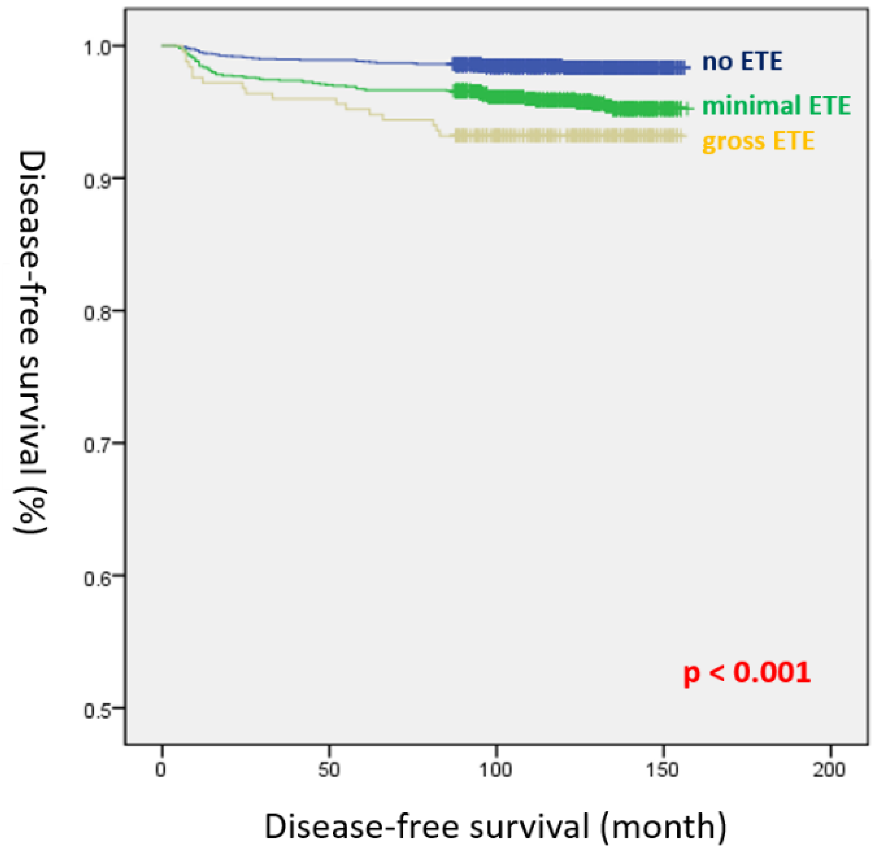
| Recurrence | 38 (1.6%) | 75 (4.2%) | 17 (6.8%) | <0.001 | <0.001 | 0.072 |
| Univariate | Multivariate | |||
|---|---|---|---|---|
| HR (95% CI) | p-Value | HR (95% CI) | p-Value | |
| Gender | ||||
| Female | ref. | |||
| Male | 1.610 (1.097–2.365) | 0.015 | ||
| Age (years) | ||||
| >45 | ref. | |||
| ≤45 | 1.676 (1.182–2.378) | 0.004 | ||
| Tumor size | ||||
| ≤1cm | ref. | |||
| >1cm | 2.944 (2.086–4.154) | <0.001 | ||
| ETE | 3.409 (2.140–5.432) | <0.001 | ||
| No ETE | ref. | ref. | ||
| Minimal ETE | 2.662 (1.802–3.934) | <0.001 | 1.362 (0.891–2.083) | 0.154 |
| Gross ETE | 4.350 (2.455–7.708) | <0.001 | 1.826 (0.984–3.520) | 0.072 |
| Multifocality | 1.689 (1.197–2.382) | 0.003 | ||
| Bilaterality | 1.717 (1.196–2.464) | 0.003 | ||
| Lymphatic invasion | 3.832 (2.708–5.423) | <0.001 | ||
| Vascular invasion | 3.063 (1.498–6.263) | 0.002 | ||
| Harvested LNs | 1.021 (1.015–1.026) | <0.001 | ||
| Positive LNs | 1.074 (1.062–1.086) | <0.001 | 1.059 (1.041–1.077) | <0.001 |
| T stage | ||||
| T1 | ref. | |||
| T2 | 3.191 (1.851–5.501) | <0.001 | ||
| T3a | 5.624 (1.782–17.749) | 0.003 | ||
| T3b | 2.870 (1.713–4.809) | <0.001 | ||
| N stage | ||||
| N0 | ref. | ref. | ||
| N1a | 5.216 (3.246–8.384) | <0.001 | 2.978 (1.785–4.968) | <0.001 |
| N1b | 9.010 (5.236–15.506) | <0.001 | 2.341 (1.175–4.662) | 0.016 |
| RAI therapy | 5.332 (3.241–8.774) | <0.001 | 2.587 (1.498–4.468) | 0.001 |
| Minimal ETE (n = 213) | Gross ETE (n = 213) | p-Value | |
|---|---|---|---|
| Age (years) | 49.5 ± 12.3 (range, 19–74) | 48.6 ± 12.4 (range, 19–74) | 0.484 |
| Female | 162 (76.1%) | 177 (83.1%) | 0.092 |
| Extent of surgery | 1.000 | ||
| Less than TT | 4 (1.9%) | 4 (1.9%) | |
| TT and/or mRND | 209 (98.1%) | 209 (98.1%) | |
| Tumor size (cm) | 1.6 ± 0.9 (range, 0.2–5.0) | 1.6 ± 0.8 (range, 0.2–5.0) | 0.390 |
| Multifocality | 119 (55.9%) | 103 (48.4%) | 0.146 |
| Bilaterality | 85 (39.9%) | 84 (39.4%) | 1.000 |
| Lymphatic invasion | 122 (57.3%) | 117 (54.9%) | 0.696 |
| Vascular invasion | 11 (5.2%) | 11 (5.2%) | 1.000 |
| Perineural invasion | 16 (7.5%) | 20 (9.4%) | 0.601 |
| BRAFV600E positive | |||
| Harvested LNs | 23.0 ± 25.2 | 22.4 ± 22.3 | 0.801 |
| Positive LNs | 5.0 ± 7.2 | 5.2 ± 6.5 | 0.810 |
| T stage | <0.001 | ||
| T1 | 163 (76.5%) | 0 (0.0%) | |
| T2 | 44 (20.7%) | 0 (0.0%) | |
| T3a | 6 (2.8%) | 0 (0.0%) | |
| T3b | 0 (0.0%) | 213 (100.0%) | |
| N stage | 0.990 | ||
| N0 | 48 (22.5%) | 47 (22.1%) | |
| N1a | 110 (51.6%) | 110 (51.6%) | |
| N1b | 55 (25.8%) | 56 (26.3%) | |
| M stage | 1.000 | ||
| M1 | 1 (0.5%) | 0 (0.0%) | |
| TNM stage | 0.589 | ||
| Stage I | 137 (64.3%) | 140 (65.7%) | |
| Stage II | 75 (35.2%) | 73 (34.3%) | |
| Stage IV | 1 (0.5%) | 0 (0.0%) | |
| RAI therapy | 195 (91.5%) | 196 (92.0%) | 1.000 |
| Recurrence | 10 (4.7%) | 13 (6.1%) | 0.668 |
| Univariate | Multivariate | |||
|---|---|---|---|---|
| HR (95% CI) | p-Value | HR (95% CI) | p-Value | |
| Gender | ||||
| Female | ref. | |||
| Male | 3.139 (1.376–7.162) | 0.007 | ||
| Age (years) | ||||
| >45 | ref. | |||
| ≤45 | 1.123 (0.486–2.593) | 0.787 | ||
| Tumor size | ||||
| ≤1cm | ref. | |||
| >1cm | 3.702 (0.868–15.788) | 0.077 | ||
| ETE | ||||
| Minimal ETE | ref. | |||
| Gross ETE | 1.301 (0.570–2.966) | 0.532 | ||
| Multifocality | 2.151 (0.885–5.228) | 0.091 | ||
| Bilaterality | 2.015 (0.884–4.596) | 0.096 | ||
| Lymphatic invasion | 5.511 (1.637–18.550) | 0.006 | 3.694 (1.039–13.142) | 0.044 |
| Vascular invasion | 0.814 (0.110–6.039) | 0.841 | ||
| Harvested LNs | 1.013 (1.001–1.025) | 0.037 | ||
| Positive LNs | 1.070 (1.037–1.105) | <0.001 | 1.126 (1.043–1.215) | 0.003 |
| T stage | ||||
| T1 | ref. | |||
| T2 | 6.516 (1.557–27.268) | 0.010 | ||
| T3a | 19.596 (3.273–117.313) | 0.001 | ||
| T3b | 3.361 (0.958–11.795) | 0.058 | ||
| N stage | ||||
| N0 | ref. | |||
| N1a | 1.809 (0.510–6.412) | 0.358 | ||
| N1b | 2.323 (0.616–8.757) | 0.213 | ||
| RAI therapy | 1.947 (0.262–14.444) | 0.515 | ||
| No ETE (A) (n = 1981) | Minimal ETE (B) (n = 1167) | Gross ETE (C) (n = 56) | p-Value (A vs. B) | p-Value (A vs. C) | p-Value (B vs. C) | |
|---|---|---|---|---|---|---|
| Age (years) | 45.9 ± 11.4 (range, 16–88) | 47.6 ± 11.4 (range, 20–80) | 51.3 ± 11.3 (range, 27–74) | <0.001 | <0.001 | 0.018 |
| Female | 1616 (81.6%) | 967 (82.9%) | 45 (80.4%) | 0.389 | 0.955 | 0.761 |
| Extent of surgery | <0.001 | <0.001 | 0.006 | |||
| Less than TT | 782 (39.5) | 253 (21.7%) | 3 (5.4%) | |||
| TT and/or mRND | 1199 (60.5%) | 914 (78.3%) | 53 (94.6%) | |||
| Tumor size (cm) | 0.6 ± 0.2 | 0.7 ± 0.2 | 0.8 ± 0.2 | <0.001 | <0.001 | <0.001 |
| Multifocality | 578 (29.2%) | 492 (42.2%) | 30 (53.6%) | <0.001 | <0.001 | 0.122 |
| Bilaterality | 320 (16.2%) | 288 (24.7%) | 21 (37.5%) | <0.001 | <0.001 | 0.046 |
| Lymphatic invasion | 254 (12.8%) | 333 (28.5%) | 22 (39.3%) | <0.001 | <0.001 | 0.114 |
| Vascular invasion | 4 (0.2%) | 23 (2.0%) | 1 (1.8%) | <0.001 | 0.321 | 1.000 |
| Perineural invasion | 8 (0.4%) | 31 (2.7%) | 4 (7.1%) | <0.001 | <0.001 | 0.120 |
| BRAFV600E positive | 1375/1783 (77.1%) | 910/1060 (85.8%) | 38/50 (76.0%) | <0.001 | 0.988 | 0.085 |
| Harvested LNs | 8.2 ± 8.2 | 11.1 ± 13.1 | 18.9 ± 20.6 | <0.001 | <0.001 | 0.007 |
| Positive LNs | 0.8 ± 2.0 | 1.8 ± 3.2 | 3.2 ± 4.9 | <0.001 | <0.001 | 0.035 |
| T stage | 1.000 | <0.001 | <0.001 | |||
| T1 | 1981 (100.0%) | 1167 (100.0%) | 0 | |||
| T2 | 0 | 0 | 0 | |||
| T3a | 0 | 0 | 0 | |||
| T3b | 0 | 0 | 56 (100%) | |||
| N stage | <0.001 | <0.001 | 0.017 | |||
| N0 | 1382 (69.8%) | 586 (50.2%) | 23 (41.1%) | |||
| N1a | 563 (28.4%) | 494 (42.3%) | 23 (41.1%) | |||
| N1b | 36 (1.8%) | 87 (7.5%) | 10 (17.9%) | |||
| M stage | 0.789 | NA | 1.000 | |||
| M1 | 0 (0.0%) | 1 (0.1%) | 0 (0.0%) | |||
| TNM stage | <0.001 | <0.001 | <0.001 | |||
| Stage I | 1877 (94.8%) | 1056 (90.5%) | 31 (55.4%) | |||
| Stage II | 104 (5.2%) | 110 (9.4%) | 25 (44.6%) | |||
| Stage IV | 0 (0.0%) | 1 (0.1%) | 0 (0.0%) | |||
| RAI therapy | 566 (28.6%) | 735 (63.0%) | 48 (85.7%) | <0.001 | <0.001 | 0.001 |
| Recurrence | 29 (1.5%) | 31 (2.7%) | 1 (1.8%) | 0.026 | 1.000 | 1.000 |
| Univariate | Multivariate | |||
|---|---|---|---|---|
| HR (95% CI) | p-Value | HR (95% CI) | p-Value | |
| Gender | ||||
| Female | ref. | |||
| Male | 1.377 (0.758–2.501) | 0.293 | ||
| Age (years) | ||||
| >45 | ref. | |||
| ≤45 | 1.684 (1.011–2.804) | 0.045 | ||
| Tumor size | ||||
| ≤1cm | ||||
| >1cm | ||||
| ETE | ||||
| No ETE | ref. | ref. | ||
| Minimal ETE | 1.813 (1.092–3.008) | 0.021 | 1.039 (0.607–1.779) | 0.889 |
| Gross ETE | 1.208 (0.165–8.866) | 0.853 | 0.522 (0.069–3.928) | 0.527 |
| Multifocality | 1.663 (1.005–2.753) | 0.048 | ||
| Bilaterality | 1.124 (0.609–2.075) | 0.708 | ||
| Lymphatic invasion | 3.238 (1.949–5.379) | <0.001 | ||
| Vascular invasion | 3.892 (0.951–15.929) | 0.059 | ||
| Harvested LNs | 1.017 (1.001–1.034) | 0.035 | ||
| Positive LNs | 1.133 (1.088–1.181) | <0.001 | 1.123 (1.034–1.219) | 0.006 |
| T stage | ||||
| T1 | ref. | |||
| T2 | ||||
| T3a | ||||
| T3b | 0.975 (0.505–1.885) | 0.941 | ||
| N stage | ||||
| N0 | ref. | |||
| N1a | 4.340 (2.457–7.678) | <0.001 | ||
| N1b | 4.476 (1.290–3.497) | 0.003 | ||
| RAI therapy | 5.020 (2.719–9.269) | <0.001 | 3.890 (2.030–7.452) | <0.001 |
Publisher’s Note: MDPI stays neutral with regard to jurisdictional claims in published maps and institutional affiliations. |
© 2022 by the authors. Licensee MDPI, Basel, Switzerland. This article is an open access article distributed under the terms and conditions of the Creative Commons Attribution (CC BY) license (https://creativecommons.org/licenses/by/4.0/).
Share and Cite
Kim, Y.; Kim, Y.-S.; Bae, J.S.; Kim, J.S.; Kim, K. Is Gross Extrathyroidal Extension to Strap Muscles (T3b) Only a Risk Factor for Recurrence in Papillary Thyroid Carcinoma? A Propensity Score Matching Study. Cancers 2022, 14, 2370. https://doi.org/10.3390/cancers14102370
Kim Y, Kim Y-S, Bae JS, Kim JS, Kim K. Is Gross Extrathyroidal Extension to Strap Muscles (T3b) Only a Risk Factor for Recurrence in Papillary Thyroid Carcinoma? A Propensity Score Matching Study. Cancers. 2022; 14(10):2370. https://doi.org/10.3390/cancers14102370
Chicago/Turabian StyleKim, Yongseon, Yong-Seok Kim, Ja Seong Bae, Jeong Soo Kim, and Kwangsoon Kim. 2022. "Is Gross Extrathyroidal Extension to Strap Muscles (T3b) Only a Risk Factor for Recurrence in Papillary Thyroid Carcinoma? A Propensity Score Matching Study" Cancers 14, no. 10: 2370. https://doi.org/10.3390/cancers14102370