
Re-Irradiation for Head and Neck Cancer: Cumulative Dose to Organs at Risk and Late Side Effects

 ,
,  ,
,
Abstract
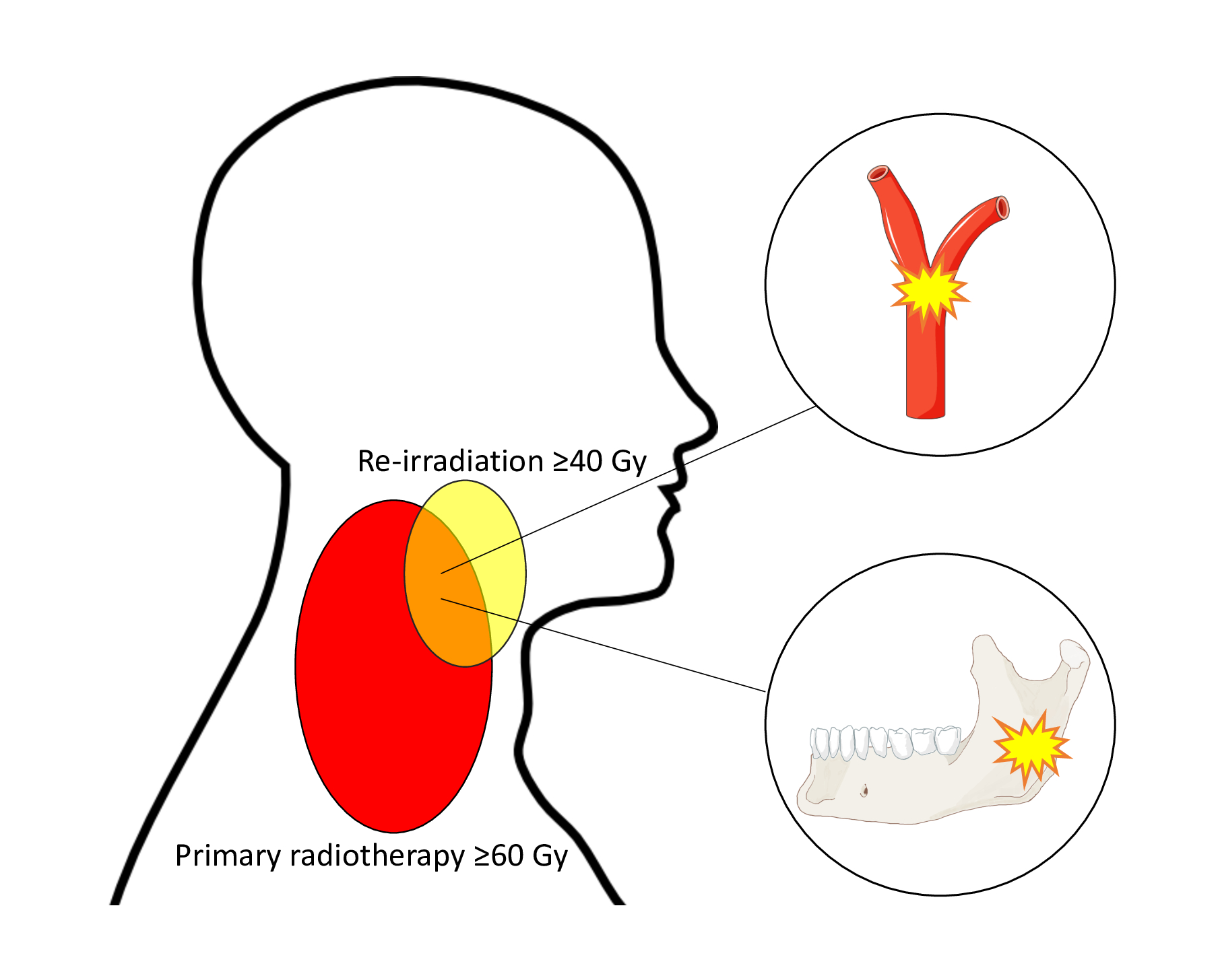
:Simple Summary
Abstract

1. Introduction
2. Materials and Methods
2.1. Patients
2.2. Toxicity
2.3. Organs at Risk
2.4. Dose Accumulation
2.5. Statistics
3. Results
3.1. Patient and Treatment Characteristics
3.2. Toxicity
3.3. Predictors of Toxicity
4. Discussion
5. Conclusions
Author Contributions
Funding
Institutional Review Board Statement
Informed Consent Statement
Data Availability Statement
Conflicts of Interest
Appendix A

| Patient and Treatment Characteristics at First Presentation | Number | Percent (Range) |
|---|---|---|
| Male | 35 | 65 |
| Female | 19 | 35 |
| Median age (years) | 59 | (33 to 81) |
| Tumor site | ||
| Larynx | 6 | 11 |
| Oropharyngeal * | 18 | 33 |
| Nasopharyngeal | 1 | 2 |
| Hypopharyngeal | 5 | 9 |
| Oral cavity | 17 | 31 |
| Unknown primary | 2 | 4 |
| Sino/nasal | 3 | 6 |
| Salivary gland | 2 | 4 |
| Histology | ||
| Squamous cell carcinoma | 46 | 85 |
| Adenoid cystic carcinoma | 5 | 9 |
| Other † | 3 | 6 |
| Tumor Stage | ||
| I | 5 | 9 |
| II | 11 | 20 |
| III | 7 | 13 |
| IVa | 28 | 52 |
| IVb | 3 | 6 |
| T-stage | ||
| 0 | 2 | 4 |
| 1 | 9 | 17 |
| 2 | 22 | 41 |
| 3 | 4 | 7 |
| 4 | 17 | 31 |
| Median treatment dose (Gy) | 68 | (60–79) |
| Patient and Treatment Characteristics at Re-Irradiation | Number | Percent (Range) |
|---|---|---|
| Median age at end of re-irradiation (years) | 63 | (40 to 89) |
| Median time between radiations (months) | 36 | (5 to 177) |
| Performance status | ||
| 0 | 30 | 56 |
| 1 | 21 | 39 |
| 2 | 2 | 4 |
| 3 | 1 | 2 |
| Tumor | ||
| Local recurrence | 37 | 69 |
| Secondary primary tumor | 17 | 31 |
| Surgery before re-irradiation | ||
| No | 32 | 59 |
| Primary tumor | 11 | 20 |
| Neck dissection | 8 | 15 |
| Primary tumor and neck dissection | 3 | 6 |
| Systemic medical treatment | ||
| No | 41 | 76 |
| Induction chemotherapy | 11 | 20 |
| Concurrent chemotherapy (cisplatin) | 1 | 2 |
| Concurrent cetuximab | 1 | 2 |
| Radiotherapy technique at re-irradiation | ||
| VMAT/IMRT | 45 | 83 |
| VMAT/IMRT + brachytherapy | 3 | 6 |
| 3 D conformal | 4 | 7 |
| 3 D conformal + brachytherapy | 1 | 2 |
| Brachytherapy | 1 | 2 |
| Median re-irradiation dose (Gy) | 59 | (40 to 71) |
| Median cumulative near max dose, D1cc (Gy) | 129 | (106 to 478) |
| Median PTV at re-irradiation (cm³) | 145 | (13 to 668) |
| Median re-treated volume, V100 (cm³) | 90 | (2 to 283) |
Appendix B
Appendix B.1. Reference Cohort
Appendix B.2. Comparison of morbidity to the Reference Cohort

References
- Overgaard, J.; Hansen, H.S.; Specht, L.; Overgaard, M.; Grau, C.; Andersen, E.; Bentzen, J.; Bastholt, L.; Hansen, O.; Johansen, J.; et al. Five compared with six fractions per week of conventional radiotherapy of squamous-cell carcinoma of head and neck: DAHANCA 6 and 7 randomised controlled trial. Lancet 2003, 362, 933–940. [Google Scholar] [CrossRef]
- Farrag, A.; Voordeckers, M.; Tournel, K.; De Coninck, P.; Storme, G. Pattern of failure after helical tomotherapy in head and neck cancer. Strahlenther. Onkol. 2010, 186, 511–516. [Google Scholar] [CrossRef]
- Pagh, A.; Grau, C.; Overgaard, J. Failure pattern and salvage treatment after radical treatment of head and neck cancer. Acta Oncol. 2016, 55, 625–632. [Google Scholar] [CrossRef] [Green Version]
- Goodwin, W.J., Jr. Salvage surgery for patients with recurrent squamous cell carcinoma of the upper aerodigestive tract: When do the ends justify the means? Laryngoscope 2000, 110, 1–18. [Google Scholar] [CrossRef]
- Ward, M.C.; Riaz, N.; Caudell, J.J.; Dunlap, N.E.; Isrow, D.; Zakem, S.J.; Dault, J.; Awan, M.J.; Vargo, J.A.; Heron, D.E.; et al. Refining Patient Selection for Reirradiation of Head and Neck Squamous Carcinoma in the IMRT Era: A Multi-institution Cohort Study by the MIRI Collaborative. Int. J. Radiat. Oncol. Biol. Phys. 2018, 100, 586–594. [Google Scholar] [CrossRef]
- Ho, J.C.; Phan, J. Reirradiation of head and neck cancer using modern highly conformal techniques. J. Sci. Spec. Head Neck 2018, 40, 2078–2093. [Google Scholar] [CrossRef]
- Curtis, K.K.; Ross, H.J.; Garrett, A.L.; Jizba, T.A.; Patel, A.B.; Patel, S.H.; Wong, W.W.; Halyard, M.Y.; Ko, S.J.; Kosiorek, H.E.; et al. Outcomes of patients with loco-regionally recurrent or new primary squamous cell carcinomas of the head and neck treated with curative intent reirradiation at Mayo Clinic. Radiat. Oncol. 2016, 11, 55. [Google Scholar] [CrossRef] [Green Version]
- Duprez, F.; Berwouts, D.; Madani, I.; Bonte, K.; Boterberg, T.; De Gersem, W.; Deron, P.; Huvenne, W.; De Neve, W. High-dose reirradiation with intensity-modulated radiotherapy for recurrent head-and-neck cancer: Disease control, survival and toxicity. Radiother. Oncol. 2014, 111, 388–392. [Google Scholar] [CrossRef]
- Leong, Y.H.; Soon, Y.Y.; Lee, K.M.; Wong, L.C.; Tham, I.W.K.; Ho, F.C.H. Long-term outcomes after reirradiation in nasopharyngeal carcinoma with intensity-modulated radiotherapy: A meta-analysis. Head Neck 2018, 40, 622–631. [Google Scholar] [CrossRef]
- Sulman, E.P.; Schwartz, D.L.; Le, T.T.; Ang, K.K.; Morrison, W.H.; Rosenthal, D.I.; Ahamad, A.; Kies, M.; Glisson, B.; Weber, R.; et al. IMRT reirradiation of head and neck cancer-disease control and morbidity outcomes. Int. J. Radiat. Oncol. Biol. Phys. 2009, 73, 399–409. [Google Scholar] [CrossRef]
- Strojan, P.; Beitler, J.J.; Silver, C.E.; Mendenhall, W.M.; Shaha, A.R.; Rinaldo, A.; Takes, R.P.; Ferlito, A. When is re-irradiation in head and neck squamous cell carcinoma not indicated? Eur. Arch. Otorhinolaryngol. 2014, 271, 3107–3109. [Google Scholar] [CrossRef] [Green Version]
- Strojan, P.; Corry, J.; Eisbruch, A.; Vermorken, J.B.; Mendenhall, W.M.; Lee, A.W.; Haigentz, M., Jr.; Beitler, J.J.; De Bree, R.; Takes, R.P.; et al. Recurrent and second primary squamous cell carcinoma of the head and neck: When and how to reirradiate. Head Neck 2015, 37, 134–150. [Google Scholar] [CrossRef] [Green Version]
- Ward, M.C.; Lee, N.Y.; Caudell, J.J.; Zajichek, A.; Awan, M.J.; Koyfman, S.A.; Dunlap, N.E.; Zakem, S.J.; Hassanzadeh, C.; Marcrom, S.; et al. A competing risk nomogram to predict severe late toxicity after modern re-irradiation for squamous carcinoma of the head and neck. Oral Oncol. 2019, 90, 80–86. [Google Scholar] [CrossRef] [PubMed]
- Tanvetyanon, T.; Padhya, T.; McCaffrey, J.; Zhu, W.; Boulware, D.; Deconti, R.; Trotti, A. Prognostic factors for survival after salvage reirradiation of head and neck cancer. J. Clin. Oncol. 2009, 27, 1983–1991. [Google Scholar] [CrossRef] [PubMed]
- Hoebers, F.; Heemsbergen, W.; Moor, S.; Lopez, M.; Klop, M.; Tesselaar, M.; Rasch, C. Reirradiation for head-and-neck cancer: Delicate balance between effectiveness and toxicity. Int. J. Radiat. Oncol. Biol. Phys. 2011, 81, e111–e118. [Google Scholar] [CrossRef] [PubMed]
- Takiar, V.; Garden, A.S.; Ma, D.; Morrison, W.H.; Edson, M.; Zafereo, M.E.; Gunn, G.B.; Fuller, C.D.; Beadle, B.; Frank, S.J.; et al. Reirradiation of Head and Neck Cancers With Intensity Modulated Radiation Therapy: Outcomes and Analyses. Int. J. Radiat. Oncol. Biol. Phys. 2016, 95, 1117–1131. [Google Scholar] [CrossRef] [PubMed]
- Bots, W.T.C.; van den Bosch, S.; Zwijnenburg, E.M.; Dijkema, T.; van den Broek, G.B.; Weijs, W.L.J.; Verhoef, L.C.G.; Kaanders, J.H.A.M. Reirradiation of head and neck cancer: Long-term disease control and toxicity. Head Neck 2017, 39, 1122–1130. [Google Scholar] [CrossRef] [PubMed]
- Garg, S.; Kilburn, J.M.; Lucas, J.T., Jr.; Randolph, D.; Urbanic, J.J.; Hinson, W.H.; Kearns, W.T.; Porosnicu, M.; Greven, K. Reirradiation for second primary or recurrent cancers of the head and neck: Dosimetric and outcome analysis. Head Neck 2016, 38, E961–E969. [Google Scholar] [CrossRef]
- Lee, J.Y.; Suresh, K.; Nguyen, R.; Sapir, E.; Dow, J.S.; Arnould, G.S.; Worden, F.P.; Spector, M.E.; Prince, M.E.; McLean, S.A.; et al. Predictors of severe long-term toxicity after re-irradiation for head and neck cancer. Oral Oncol. 2016, 60, 32–40. [Google Scholar] [CrossRef]
- Dionisi, F.; Fiorica, F.; D’Angelo, E.; Maddalo, M.; Giacomelli, I.; Tornari, E.; Rosca, A.; Vigo, F.; Romanello, D.; Cianchetti, M.; et al. Organs at risk’s tolerance and dose limits for head and neck cancer re-irradiation: A literature review. Oral Oncol. 2019, 98, 35–47. [Google Scholar] [CrossRef] [PubMed]
- Emami, B.; Lyman, J.; Brown, A.; Coia, L.; Goitein, M.; Munzenrider, J.E.; Shank, B.; Solin, L.J.; Wesson, M. Tolerance of normal tissue to therapeutic irradiation. Int. J. Radiat. Oncol. Biol. Phys. 1991, 21, 109–122. [Google Scholar] [CrossRef]
- Nieder, C.; Grosu, A.L.; Andratschke, N.H.; Molls, M. Update of human spinal cord reirradiation tolerance based on additional data from 38 patients. Int. J. Radiat. Oncol. Biol. Phys. 2006, 66, 1446–1449. [Google Scholar] [CrossRef]
- McDonald, M.W.; Moore, M.G.; Johnstone, P.A. Risk of carotid blowout after reirradiation of the head and neck: A systematic review. Int. J. Radiat. Oncol. Biol. Phys. 2012, 82, 1083–1089. [Google Scholar] [CrossRef]
- Embring, A.; Onjukka, E.; Mercke, C.; Lax, I.; Berglund, A.; Bornedal, S.; Wennberg, B.; Friesland, S. Overlapping volumes in re-irradiation for head and neck cancer—An important factor for patient selection. Radiat. Oncol. 2020, 15, 147. [Google Scholar] [CrossRef]
- Ruifrok, A.C.; van der Kogel, A.J. A “reappraisal” of the LQ model for the understanding of dose-fractionation in radiotherapy. Int. J. Radiat. Oncol. Biol. Phys. 1993, 25, 926–929. [Google Scholar] [CrossRef]
- LENT SOMA tables. Radiot. Oncol. 1995, 35, 17–60. [CrossRef]
- Cox, J.D.; Stetz, J.; Pajak, T.F. Toxicity criteria of the Radiation Therapy Oncology Group (RTOG) and the European Organization for Research and Treatment of Cancer (EORTC). Int. J. Radiat. Oncol. Biol. Phys. 1995, 31, 1341–1346. [Google Scholar] [CrossRef]
- Hammerlid, E.; Haugen Cange, H.; Söderström, K.; Brun, E.; Farnebo, L.; Olin, M.; Högmo, A.; Sjödin, H.; on behalf of Regionalt Cancercentrum. Nationellt Vårdprogram Huvud—och Halscancer (National Guidelines for Head and Neck Cancer). 2019. Available online: https://kunskapsbanken.cancercentrum.se/diagnoser/huvud-och-halscancer/vardprogram/behandling-vid-huvud-och-halscancer/#chapter-9-2-Onkologisk-behandling (accessed on 23 June 2021).
- Jensen, K.; Friborg, J.; Hansen, C.R.; Samsoe, E.; Johansen, J.; Andersen, M.; Smulders, B.; Andersen, E.; Nielsen, M.S.; Eriksen, J.G.; et al. The Danish Head and Neck Cancer Group (DAHANCA) 2020 radiotherapy guidelines. Radiot. Oncol. 2020, 151, 149–151. [Google Scholar] [CrossRef]
- Pfeister, D.; Spencer, S.; Brizel, D.; Busse, P.; Caudell, J.; Cmelak, A.; Foote, R.; Galloway, T.; Hitchcock, Y.; Mell, L.; et al. Head and Neck Cancers. NCCN Clinical Practice Guidelines in Oncology. National Comprehensive Cancer Network, 2021. Available online: https://jnccn.org/view/journals/jnccn/18/7/article-p873.xml(accessed on 23 June 2021).
- Caudell, J.J.; Ward, M.C.; Riaz, N.; Zakem, S.J.; Awan, M.J.; Dunlap, N.E.; Isrow, D.; Hassanzadeh, C.; Vargo, J.A.; Heron, D.E.; et al. Volume, Dose, and Fractionation Considerations for IMRT-based Reirradiation in Head and Neck Cancer: A Multi-institution Analysis. Int. J. Radiat. Oncol. Biol. Phys. 2018, 100, 606–617. [Google Scholar] [CrossRef] [PubMed]
- Phan, J.; Sio, T.T.; Nguyen, T.P.; Takiar, V.; Gunn, G.B.; Garden, A.S.; Rosenthal, D.I.; Fuller, C.D.; Morrison, W.H.; Beadle, B.; et al. Reirradiation of Head and Neck Cancers With Proton Therapy: Outcomes and Analyses. Int. J. Radiat. Oncol. Biol. Phys. 2016, 96, 30–41. [Google Scholar] [CrossRef] [PubMed]
- Olteanu, L.A.M.; Duprez, F.; De Neve, W.; Berwouts, D.; Vercauteren, T.; Bauters, W.; Deron, P.; Huvenne, W.; Bonte, K.; Goethals, I.; et al. Late mucosal ulcers in dose-escalated adaptive dose-painting treatments for head-and-neck cancer. Acta Oncol. 2018, 57, 262–268. [Google Scholar] [CrossRef] [PubMed]
- van der Geer, S.J.; van Rijn, P.V.; Kamstra, J.I.; Langendijk, J.A.; van der Laan, B.; Roodenburg, J.L.N.; Jan, L.N.; Dijkstra, P.U. Prevalence and prediction of trismus in patients with head and neck cancer: A cross-sectional study. Head Neck 2019, 41, 64–71. [Google Scholar] [CrossRef] [PubMed]


| Subject | Late Side Effects Grade ≥3 at Two Years After Re-Irradiation |
|---|---|
| 10 | None |
| 11 | Mucosa, Osteoradionecrosis |
| 13 | None |
| 19 | None |
| 26 | None |
| 27 | Skin, Mucosa, Xerostomia, Dysphagia |
| 35 | None |
| 37 | None |
| 42 | Xerostomia |
| 45 | None |
| 52 | Osteoradionecrosis, Trismus |
| Subject | Tumor Site at First Presentation | Late Side Effects Grade ≥3 after Re-RT | Surgery at First Presentation | Surgery Before Re-RT | PS at Re-RT | Grade ≥3 side effect at 1 st RT | Time between RT and re-RT (months) | Age at Re-RT (years) | PTV at Re-RT (cm³) | V100 at Re-RT (cm³) | Concomitant Systemic Therapy at Re-RT | ||||||||
|---|---|---|---|---|---|---|---|---|---|---|---|---|---|---|---|---|---|---|---|
| Skin | Mucosa | Larynx | Salivary Glands | Dysphagia | ORN | Trismus | Blowout | Other | |||||||||||
| 52 | Sino/nasal | No | No | No | No | No | Grade 4 | Grade 3 | No | No | Primary tumor | No | 0 | No | 88 | 41 | 62 | 71 | No |
| 42 | Nasopharyngeal | No | No | No | Grade 3 | No | No | No | No | Grade 3 | No | No | 0 | No | 60 | 55 | 248 | 91 | No |
| 23 | Oral cavity | No | Grade 3 | No | No | No | No | No | No | No | Primary tumor | No | 1 | No | 73 | 69 | 262 | 119 | No |
| 27 | Oral cavity | Grade 3 | Grade 3 | No | Grade 3 | Grade 3 | No | No | No | No | Primary tumor | Primary tumor | 0 | No | 108 | 62 | 198 | 256 | No |
| 30 | Oral cavity | No | No | No | No | No | No | Grade 3 | No | No | No | No | 1 | No | 78 | 60 | 161 | 167 | No |
| 41 | Oral cavity | No | No | No | No | Grade 3 | No | No | No | No | Primary tumor + ND | No | 0 | Yes | 127 | 60 | 44 | 82 | No |
| 7 | Oropharyngeal † | No | Grade 3 | No | Grade 3 | Grade 3 | No | No | No | No | No | Primary tumor | 0 | No | 138 | 74 | 309 | 87 | No |
| 17 | Oropharyngeal † | No | No | No | No | No | No | Grade 3 | No | Grade 3 | No | No | 1 | Yes | 177 | 71 | 102 | 141 | No |
| 22 | Oropharyngeal * | No | No | No | No | Grade 4 | No | Grade 4 | No | No | No | No | 0 | No | 36 | 68 | 128 | 142 | No |
| 31 | Oropharyngeal * | Grade 3 | No | No | Grade 3 | Grade 3 | No | Grade 3 | No | No | No | Primary tumor | 0 | No | 50 | 61 | 333 | 120 | No |
| 37 | Oropharyngeal * | No | No | No | No | No | Grade 4 | No | No | No | No | Primary tumor | 0 | No | 136 | 59 | 219 | 154 | No |
| 38 | Oropharyngeal * | No | No | No | No | No | Grade 3 | No | Grade 5 | No | No | Primary tumor | 1 | No | 31 | 56 | 106 | 138 | No |
| 6 | Hypopharyngeal | No | Grade 4 | No | No | No | No | No | No | No | No | No | 0 | No | 43 | 74 | 93 | 64 | No |
| 20 | Hypopharyngeal | No | Grade 3 | No | No | No | No | No | No | No | No | No | 0 | Yes | 21 | 64 | 149 | 109 | No |
| 24 | Hypopharyngeal | No | No | No | No | Grade 3 | No | No | No | No | No | Primary tumor + ND | 0 | No | 71 | 64 | 26 | 56 | No |
| 9 | Larynx | No | No | No | No | No | No | No | Grade 5 | No | Primary tumor + ND | ND | 1 | No | 15 | 74 | 66 | 92 | No |
| 19 | Larynx | No | No | No | No | Grade 4 | No | No | No | No | No | Primary tumor + ND | 0 | No | 15 | 68 | 452 | 76 | No |
| 5 | Salivary gland | Grade 3 | No | No | No | No | No | No | No | No | Primary tumor + ND | No | 0 | No | 81 | 77 | 283 | 89 | No |
| 11 | Unknown primary | No | Grade 4 | No | No | No | Grade 3 | No | No | No | ND | Primary tumor | 0 | No | 149 | 76 | 154 | 166 | No |
| Median (whole cohort) | 95% was PS 0–1 | 36 | 63 | 145 | 90 | ||||||||||||||
| Author, Year, Reference Number | Cumulative Maximum Dose * | |
|---|---|---|
| Carotid Blowout | Osteoradionecrosis | |
| Embring et al., 2021(current article) | 119 Gy | 119 Gy |
| Garg et al., 2016 [18] | 120 Gy | - |
| Bots et al., 2017 [17] | - | 114 Gy |
Publisher’s Note: MDPI stays neutral with regard to jurisdictional claims in published maps and institutional affiliations. |
© 2021 by the authors. Licensee MDPI, Basel, Switzerland. This article is an open access article distributed under the terms and conditions of the Creative Commons Attribution (CC BY) license (https://creativecommons.org/licenses/by/4.0/).
Share and Cite
Embring, A.; Onjukka, E.; Mercke, C.; Lax, I.; Berglund, A.; Bornedal, S.; Wennberg, B.; Dalqvist, E.; Friesland, S. Re-Irradiation for Head and Neck Cancer: Cumulative Dose to Organs at Risk and Late Side Effects. Cancers 2021, 13, 3173. https://doi.org/10.3390/cancers13133173
Embring A, Onjukka E, Mercke C, Lax I, Berglund A, Bornedal S, Wennberg B, Dalqvist E, Friesland S. Re-Irradiation for Head and Neck Cancer: Cumulative Dose to Organs at Risk and Late Side Effects. Cancers. 2021; 13(13):3173. https://doi.org/10.3390/cancers13133173
Chicago/Turabian StyleEmbring, Anna, Eva Onjukka, Claes Mercke, Ingmar Lax, Anders Berglund, Sara Bornedal, Berit Wennberg, Emmy Dalqvist, and Signe Friesland. 2021. "Re-Irradiation for Head and Neck Cancer: Cumulative Dose to Organs at Risk and Late Side Effects" Cancers 13, no. 13: 3173. https://doi.org/10.3390/cancers13133173