Time on Therapy for at Least Three Months Correlates with Overall Survival in Metastatic Renal Cell Carcinoma

 , ,
, ,
Abstract
:1. Introduction
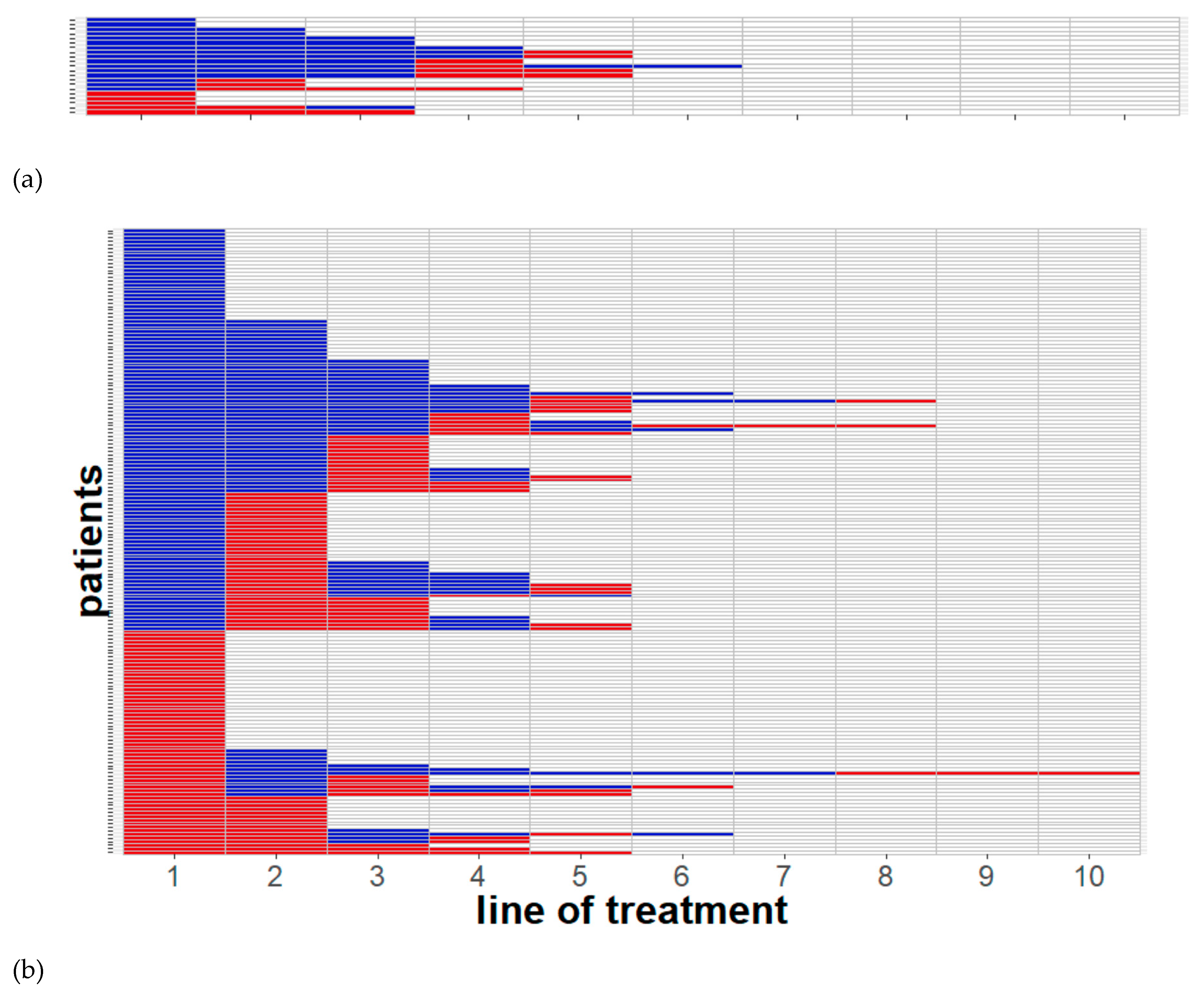
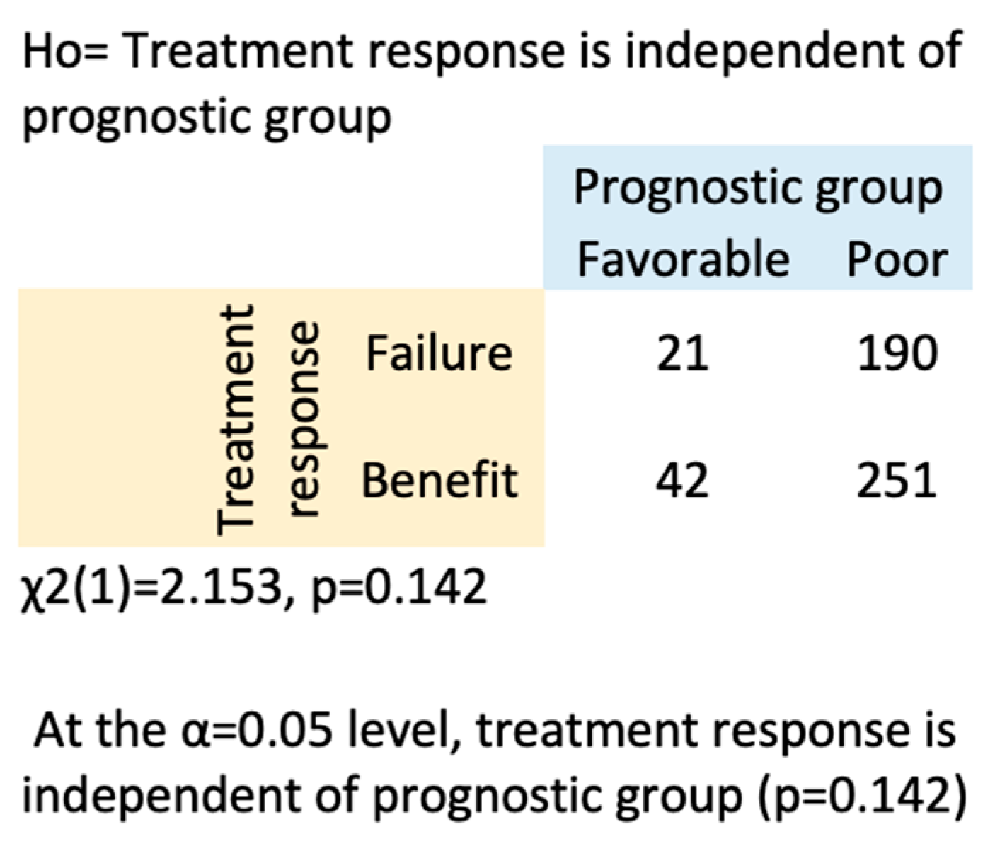
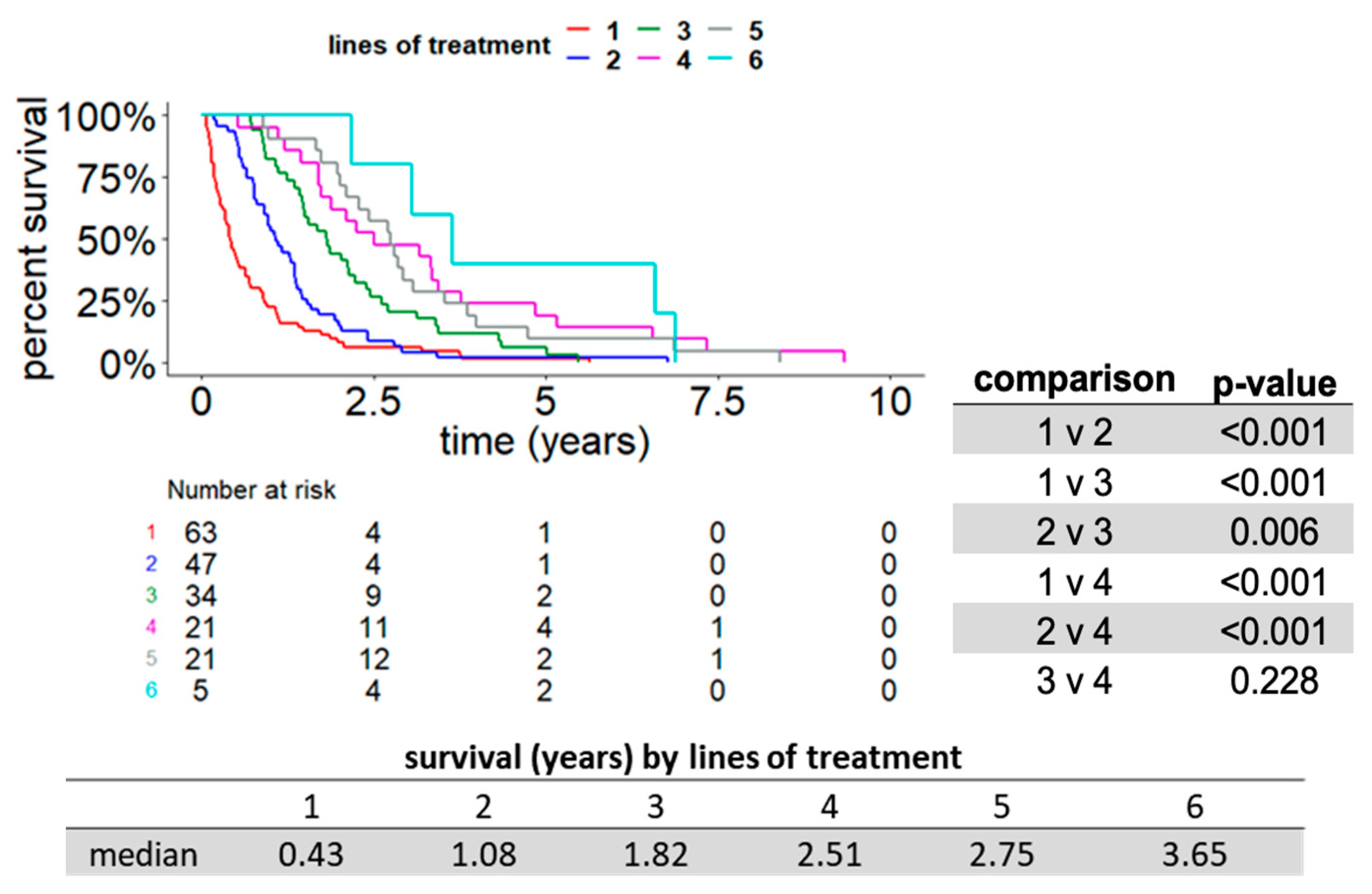
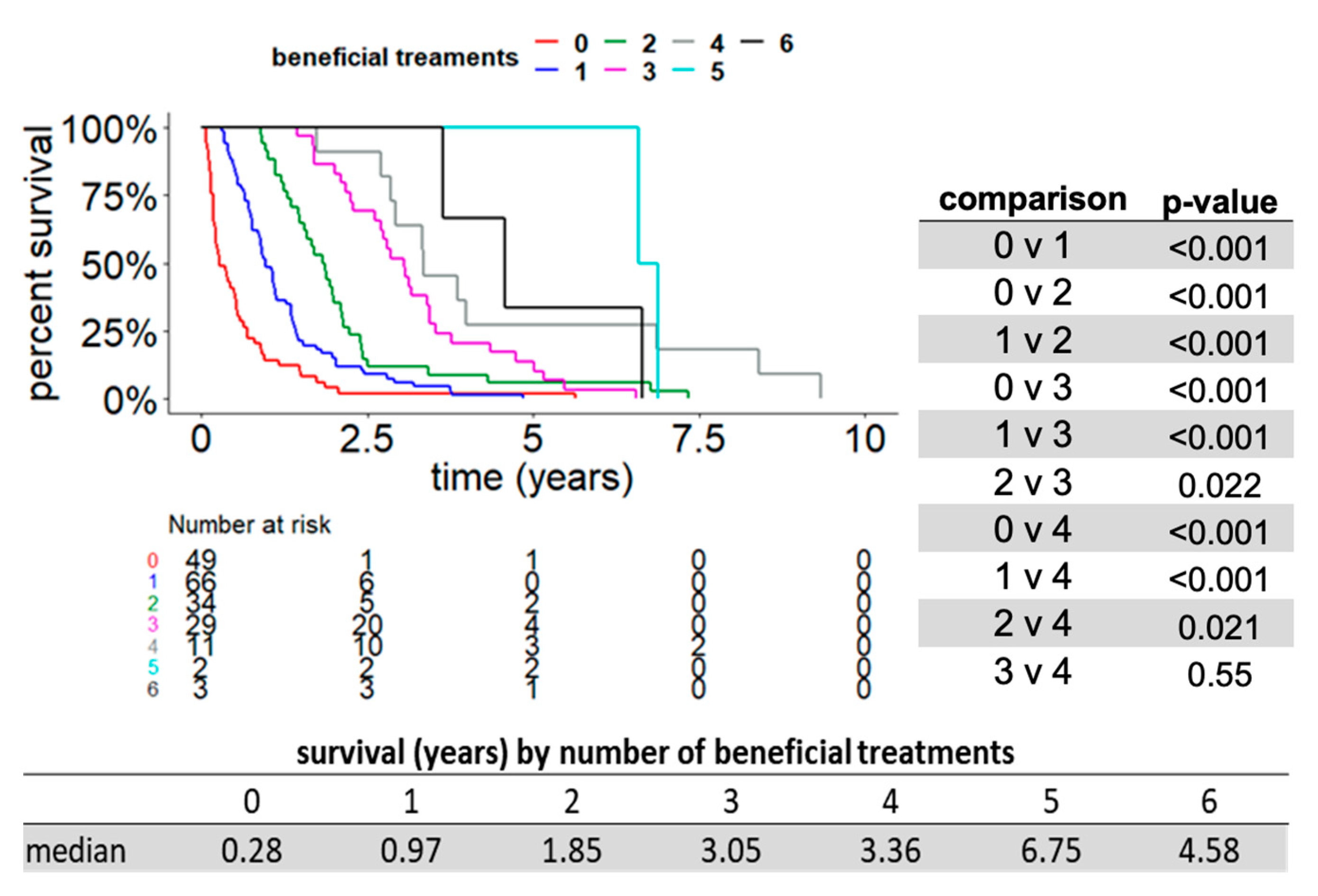
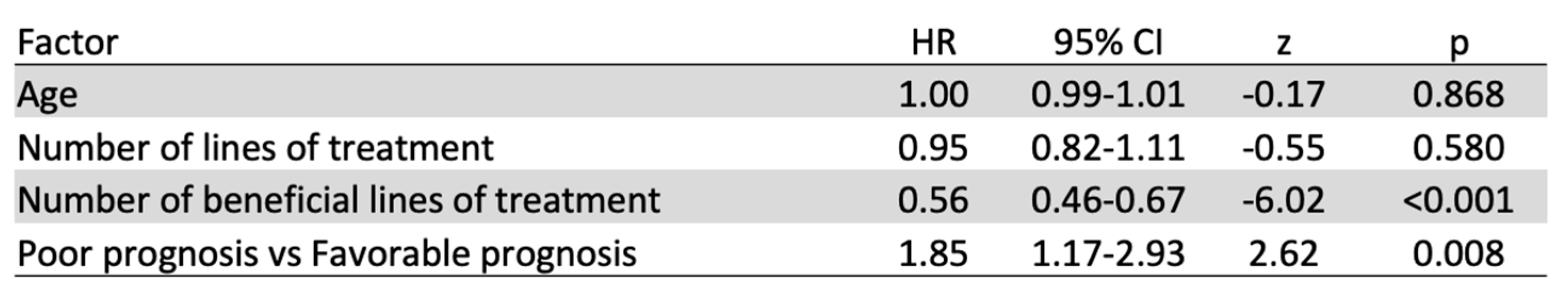
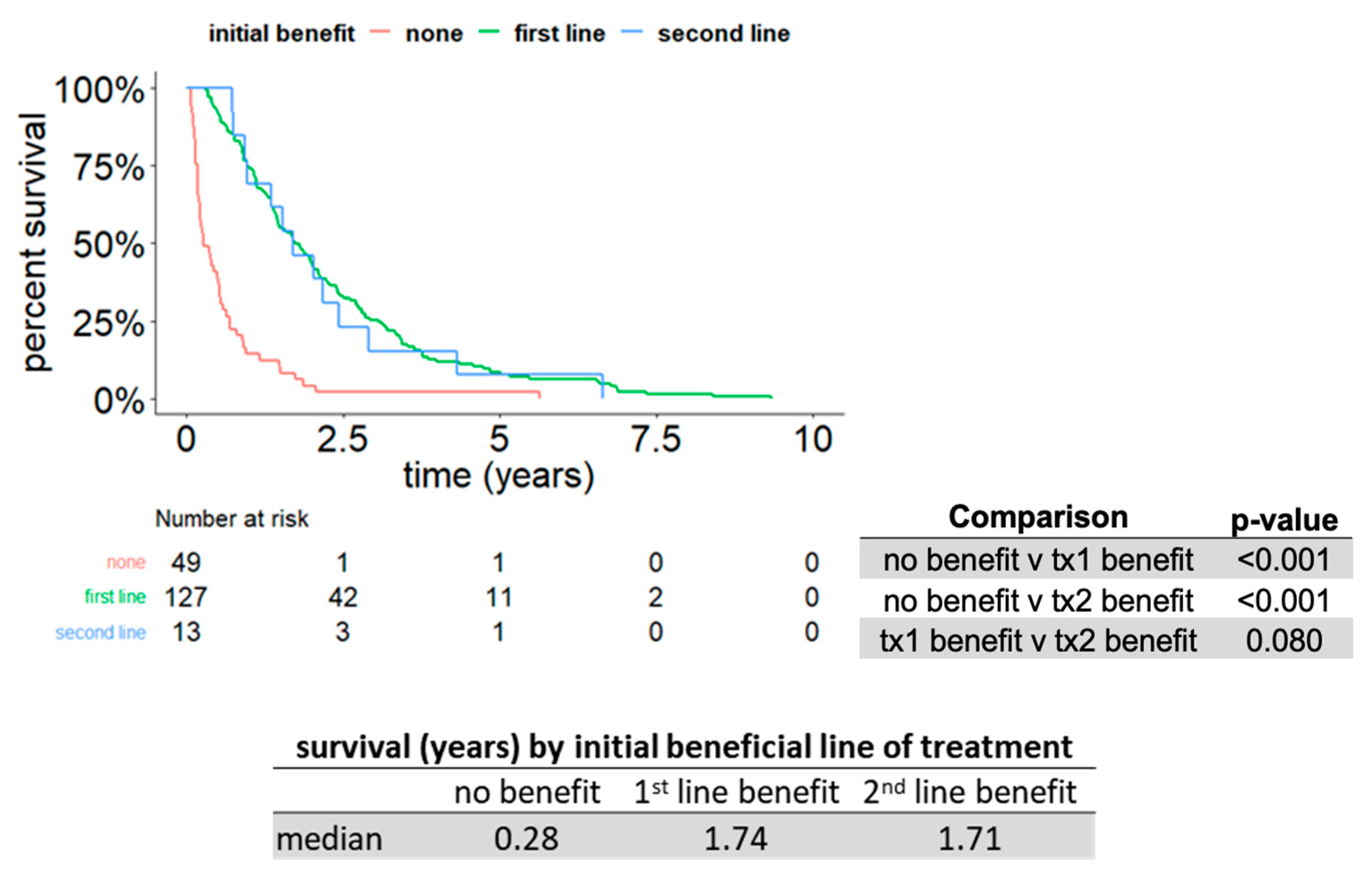
2. Results
3. Discussion
4. Materials and Methods
4.1. Patients
4.2. Data Source
4.3. Outcome Variables
4.4. Statistical Analysis
5. Conclusions
Supplementary Materials
Author Contributions
Funding
Acknowledgments
Conflicts of Interest
References
- American Cancer Society | Cancer Facts & Statistics. Available online: https://cancerstatisticscenter.cancer.org (accessed on 26 January 2019).
- Maia, M.C.; Dizman, N.; Salgia, M.; Pal, S.K. Therapeutic Sequencing in Metastatic Renal Cell Carcinoma. Kidney Cancer 2017, 1, 15–29. [Google Scholar] [CrossRef] [PubMed]
- Wagstaff, J.; Jones, R.; Hawkins, R.; Porfiri, E.; Pickering, L.; Bahl, A.; Brown, J.; Buchan, S. Treatment patterns and clinical outcomes in patients with renal cell carcinoma in the UK: Insights from the RECCORD registry. Ann. Oncol. 2016, 27, 159–165. [Google Scholar] [CrossRef] [PubMed]
- Kidney and Renal Pelvis Statistics. American Cancer Society. Available online: https://cancerstatisticscenter.cancer.org/#!/cancer-site/Kidney%20and%20renal%20pelvis (accessed on 31 October 2018).
- Heng, D.Y.; Mackenzie, M.J.; Vaishampayan, U.N.; Bjarnason, G.A.; Knox, J.J.; Tan, M.H.; Wood, L.; Wang, Y.; Kollmannsberger, C.; North, S.; et al. Primary anti-vascular endothelial growth factor (VEGF)-refractory metastatic renal cell carcinoma: Clinical characteristics, risk factors, and subsequent therapy. Ann. Oncol. 2012, 23, 1549–1555. [Google Scholar] [CrossRef] [PubMed]
- Bedke, J.; Gauler, T.; Grünwald, V.; Hegele, A.; Herrmann, E.; Hinz, S.; Janssen, J.; Schmitz, S.; Schostak, M.; Tesch, H.; et al. Systemic therapy in metastatic renal cell carcinoma. World J. Urol. 2016, 35, 179–188. [Google Scholar] [CrossRef] [PubMed]
- Choueiri, T.K.; Escudier, B.; Powles, T.; Tannir, N.M.; Mainwaring, P.N.; Rini, B.I.; Hammers, H.J.; Donskov, F.; Roth, B.J.; Peltola, K.; et al. Cabozantinib versus everolimus in advanced renal cell carcinoma (METEOR): Final results from a randomised, open-label, phase 3 trial. Lancet Oncol. 2016, 17, 917–927. [Google Scholar] [CrossRef]
- Motzer, R.J.; Tannir, N.M.; McDermott, D.F.; Frontera, O.A.; Melichar, B.; Choueiri, T.K.; Plimack, E.R.; Barthélémy, P.; Porta, C.; George, S.; et al. Nivolumab plus Ipilimumab versus Sunitinib in Advanced Renal-Cell Carcinoma. N. Engl. J. Med. 2018, 378, 1277–1290. [Google Scholar] [CrossRef] [PubMed]
- Rini, B.I.; Plimack, E.R.; Stus, V.; Gafanov, R.; Hawkins, R.; Nosov, D.; Pouliot, F.; Alekseev, B.; Soulières, D.; Melichar, B.; et al. Pembrolizumab plus Axitinib versus Sunitinib for Advanced Renal-Cell Carcinoma. N. Engl. J. Med. 2019, 380, 1116–1127. [Google Scholar] [CrossRef] [PubMed]
- Motzer, R.J.; Escudier, B.; McDermott, D.F.; George, S.; Hammers, H.J.; Srinivas, S.; Tykodi, S.S.; Sosman, J.A.; Procopio, G.; Plimack, E.R.; et al. Nivolumab versus Everolimus in Advanced Renal Cell Carcinoma. N. Engl. J. Med. 2015, 373, 1803–1813. [Google Scholar] [CrossRef]
- Calvo, E.; Schmidinger, M.; Heng, D.Y.; Grünwald, V.; Escudier, B. Improvement in survival end points of patients with metastatic renal cell carcinoma through sequential targeted therapy. Cancer Treat. Rev. 2016, 50, 109–117. [Google Scholar] [CrossRef]
- Smaldone, M.C.; Egleston, B.; Hollingsworth, J.M.; Hollenbeck, B.K.; Miller, D.C.; Morgan, T.M.; Kim, S.P.; Malhotra, A.; Handorf, E.; Wong, Y.-N.; et al. Understanding Treatment Disconnect and Mortality Trends in Renal Cell Carcinoma Using Tumor Registry Data. Med. Care 2017, 55, 398–404. [Google Scholar] [CrossRef] [Green Version]
- Méjean, A.; Escudier, B.; Thezenas, S.; Beauval, J.-B.; Geoffroy, L.; Bensalah, K.; Thiery-Vuillemin, A.; Cormier, L.; Lang, H.; Guy, L.; et al. CARMENA: Cytoreductive nephrectomy followed by sunitinib versus sunitinib alone in metastatic renal cell carcinoma—Results of a phase III noninferiority trial. J. Clin. Oncol. 2018, 36, LBA3. [Google Scholar] [CrossRef]
- Merza, H.; Bilusic, M. Current Management Strategy for Metastatic Renal Cell Carcinoma and Future Directions. Curr. Oncol. Rep. 2017, 19, 27. [Google Scholar] [CrossRef] [PubMed]
- Motzer, R.J.; Penkov, K.; Haanen, J.; Rini, B.; Albiges, L.; Campbell, M.T.; Venugopal, B.; Kollmannsberger, C.; Negrier, S.; Uemura, M.; et al. Avelumab plus Axitinib versus Sunitinib for Advanced Renal-Cell Carcinoma. N. Engl. J. Med. 2019, 380, 1103–1115. [Google Scholar] [CrossRef] [PubMed]
- Hall, P.S.; Harshman, L.C.; Srinivas, S.; Witteles, R.M. The Frequency and Severity of Cardiovascular Toxicity from Targeted Therapy in Advanced Renal Cell Carcinoma Patients. JACC Heart Fail. 2013, 1, 72–78. [Google Scholar] [CrossRef] [PubMed]
- Harshman, L.C.; Yu, R.J.; Allen, G.I.; Srinivas, S.; Gill, H.S.; Chung, B.I. Surgical outcomes and complications associated with presurgical tyrosine kinase inhibition for advanced renal cell carcinoma (RCC). Urol. Oncol. Semin. Orig. Investig. 2013, 31, 379–385. [Google Scholar] [CrossRef]
- Heng, D.Y.C. General Session 8: Evolving Management of Metastatic Renal Cell Carcinoma. In Proceedings of the Genitourinary Cancers Symposium, San Francisco, CA, USA, 14–16 February 2019; Available online: https://meetinglibrary.asco.org/record/166877/video (accessed on 1 April 2018).
- Ko, J.J.; Xie, W.; Kroeger, N.; Lee, J.; Rini, B.I.; Knox, J.J.; Bjarnason, G.A.; Srinivas, S.; Pal, S.K.; Yuasa, T.; et al. The International Metastatic Renal Cell Carcinoma Database Consortium model as a prognostic tool in patients with metastatic renal cell carcinoma previously treated with first-line targeted therapy: A population-based study. Lancet Oncol. 2015, 16, 293–300. [Google Scholar] [CrossRef]
- Flanigan, R.C.; Salmon, S.E.; Blumenstein, B.A.; Bearman, S.I.; McGrath, P.C.; Caton, J.R.; Munshi, N.; Roy, V.; Crawford, E.D. Nephrectomy Followed by Interferon Alfa-2b Compared with Interferon Alfa-2b Alone for Metastatic Renal-Cell Cancer. N. Engl. J. Med. 2001, 345, 1655–1659. [Google Scholar] [CrossRef]
- Mickisch, G.H.; Garin, A.; van Poppel, H.; de Prijck, L.; Sylvester, R.; European Organisation for Research and Treatment of Cancer (EORTC) Genitourinary Group. Radical nephrectomy plus interferon-alfa-based immunotherapy compared with interferon alfa alone in metastatic renal-cell carcinoma: A randomised trial. Lancet 2001, 358, 966–970. [Google Scholar] [CrossRef]
- Gao, X. Combination Immunotherapy and Targeted Therapy: Will New Combinations Raise the Tail of the Survival Curve? Kidney Cancer J. 2018, 16, 76–82. [Google Scholar]
- Motzer, R.J.; Powles, T.; Atkins, M.B.; Escudier, B.; McDermott, D.F.; Suárez, C.; Bracarda, S.; Stadler, W.M.; Donskov, F.; Lee, J.-L.; et al. IMmotion151: A Randomized Phase III Study of Atezolizumab Plus Bevacizumab vs. Sunitinib in Untreated Metastatic Renal Cell Carcinoma (mRCC). J. Clin. Oncol. 2018, 36, 578. [Google Scholar] [CrossRef]
- BAVENCIO®(avelumab) Plus INLYTA®(axitinib) Significantly Improved Progression-Free Survival in Previously Untreated Patients with Advanced Renal Cell Carcinoma in Phase III Study. Available online: https://pipelinereview.com/index.php/2018091269104/Antibodies/BAVENCIO-avelumab-Plus-INLYTA-axitinib-Significantly-Improved-Progression-Free-Survival-in-Previously-Untreated-Patients-with-Advanced-Renal-Cell-Carcinoma-in-Phase-III-Study.html (accessed on 1 April 2018).
- Rini, B.I.; Escudier, B.; Tomczak, P.; Kaprin, A.; Szczylik, C.; E Hutson, T.; Michaelson, M.D.; A Gorbunova, V.; E Gore, M.; Rusakov, I.G.; et al. Comparative effectiveness of axitinib versus sorafenib in advanced renal cell carcinoma (AXIS): A randomised phase 3 trial. Lancet 2011, 378, 1931–1939. [Google Scholar] [CrossRef]
- Di Lorenzo, G.; Cartenì, G.; Autorino, R.; Bruni, G.; Tudini, M.; Rizzo, M.; Aieta, M.; Gonnella, A.; Rescigno, P.; Perdonà, S.; et al. Phase II study of sorafenib in patients with sunitinib-refractory metastatic renal cell cancer. J. Clin. Oncol. 2009, 27, 4469–4474. [Google Scholar] [CrossRef] [PubMed]
- Dudek, A.Z.; Zolnierek, J.; Dham, A.; Lindgren, B.R.; Szczylik, C. Sequential therapy with sorafenib and sunitinib in renal cell carcinoma. Cancer 2009, 115, 61–67. [Google Scholar] [CrossRef] [PubMed]
- Eichelberg, C.; Heuer, R.; Chun, F.K.; Hinrichs, K.; Zacharias, M.; Huland, H.; Heinzer, H. Sequential Use of the Tyrosine Kinase Inhibitors Sorafenib and Sunitinib in Metastatic Renal Cell Carcinoma: A Retrospective Outcome Analysis. Eur. Urol. 2008, 54, 1373–1378. [Google Scholar] [CrossRef] [PubMed]
- Sablin, M.; Negrier, S.; Ravaud, A.; Oudard, S.; Balleyguier, C.; Gautier, J.; Celier, C.; Medioni, J.; Escudier, B. Sequential Sorafenib and Sunitinib for Renal Cell Carcinoma. J. Urol. 2009, 182, 29–34. [Google Scholar] [CrossRef] [PubMed]
- Sepulveda, J.; Maroto, P.; Andres, R.; Padilla, I.D.; Coronado, C.; Delarosa, F.; Castellano, D.E. Sorafenib as a second-line and sequential therapy for patients with metastatic renal cell carcinoma (mRCC): Analysis for safety and activity on sunitinib progressive pts. J. Clin. Oncol. 2008, 26, 16100. [Google Scholar] [CrossRef]
- Shepard, D.R.; Rini, B.I.; Garcia, J.A.; Hutson, T.E.; Elson, P.; Gilligan, T.; Nemec, C.; Lopez, R.; Borner, D.; Dreicer, R.; et al. A multicenter prospective trial of sorafenib in patients (pts) with metastatic clear cell renal cell carcinoma (mccRCC) refractory to prior sunitinib or bevacizumab. J. Clin. Oncol. 2008, 26, 5123. [Google Scholar] [CrossRef]
- Tamaskar, I.; Garcia, J.A.; Elson, P.; Wood, L.; Mekhail, T.; Dreicer, R.; Rini, B.I.; Bukowski, R.M. Antitumor Effects of Sunitinib or Sorafenib in Patients with Metastatic Renal Cell Carcinoma Who Received Prior Antiangiogenic Therapy. J. Urol. 2008, 179, 81–86. [Google Scholar] [CrossRef] [PubMed]
- Vickers, M.M.; Choueiri, T.K.; Rogers, M.; Percy, A.; Finch, D.; Zama, I.; Cheng, T.; North, S.; Knox, J.J.; Kollmannsberger, C.; et al. Clinical Outcome in Metastatic Renal Cell Carcinoma Patients after Failure of Initial Vascular Endothelial Growth Factor-Targeted Therapy. Urology 2010, 76, 430–434. [Google Scholar] [CrossRef] [PubMed]
- Zimmermann, K.; Schmittel, A.; Steiner, U.; Asemissen, A.M.; Knoedler, M.; Thiel, E.; Miller, K.; Keilholz, U. Sunitinib Treatment for Patients with Advanced Clear-Cell Renal-Cell Carcinoma after Progression on Sorafenib. Oncology 2009, 76, 350–354. [Google Scholar] [CrossRef] [PubMed]
- Al-Marrawi, M.Y.; For the International mRCC Database Consortium; Rini, B.I.; Harshman, L.C.; Bjarnason, G.A.; Wood, L.; Vaishampayan, U.; MacKenzie, M.; Knox, J.J.; Agarwal, N.; et al. The association of clinical outcome to first-line VEGF-targeted therapy with clinical outcome to second-line VEGF-targeted therapy in metastatic renal cell carcinoma patients. Target. Oncol. 2013, 8, 203–209. [Google Scholar] [CrossRef] [PubMed] [Green Version]
- Miyake, H.; Imai, S.; Harada, K.-I.; Fujisawa, M. Absence of Significant Correlation of Adverse Events between First- and Second-Line Tyrosine Kinase Inhibitors in Patients with Metastatic Renal Cell Carcinoma. Clin. Genitourin. Cancer 2016, 14, e19–e24. [Google Scholar] [CrossRef] [PubMed]
- Michel, M.S.; Vervenne, W.; De Santis, M.; Von Weikersthal, L.F.; Goebell, P.J.; Lerchenmueller, J.; Zimmermann, U.; Bos, M.M.; Freier, W.; Schirrmacher-Memmel, S.; et al. SWITCH: A randomized sequential open-label study to evaluate efficacy and safety of sorafenib (SO)/sunitinib (SU) versus SU/SO in the treatment of metastatic renal cell cancer (mRCC). J. Clin. Oncol. 2014, 32, 393. [Google Scholar] [CrossRef]
- Schmidinger, M. Improving Outcomes in Metastatic Clear Cell Renal Cell Carcinoma by Sequencing Therapy. Am. Soc. Clin. Oncol. Educ. Book 2014, 34, e228–e238. [Google Scholar] [CrossRef] [PubMed]
- Surveillance, Epidemiology and End Results (SEER) Program (National Cancer Institute). www.seer.cancer.gov, SEER*Stat Database: Mortality—All COD, Aggregated With State, Total USA (1969–2015), National Cancer Institute, DCCPS, Surveillance Research Program, released December 2017. In SEER Database. 1 December. Available online: www.cdc.gov/nchs (accessed on 1 October 2018).
- Motzer, R.J.; Hutson, T.E.; Tomczak, P.; Michaelson, M.D.; Bukowski, R.M.; Rixe, O.; Oudard, S.; Négrier, S.; Szczylik, C.; Kim, S.T.; et al. Sunitinib versus Interferon Alfa in Metastatic Renal-Cell Carcinoma. N. Engl. J. Med. 2007, 356, 115–124. [Google Scholar] [CrossRef] [PubMed]
- Motzer, R.J.; Hutson, T.E.; Cella, D.; Reeves, J.; Hawkins, R.; Guo, J.; Nathan, P.; Staehler, M.; De Souza, P.; Merchan, J.R.; et al. Pazopanib versus Sunitinib in Metastatic Renal-Cell Carcinoma. N. Engl. J. Med. 2013, 369, 722–731. [Google Scholar] [CrossRef] [PubMed]
- Toriihara, A.; Duan, H.; Thompson, H.M.; Park, S.; Hatami, N.; Baratto, L.; Fan, A.C.; Iagaru, A. 18F-FPPRGD2 PET/CT in patients with metastatic renal cell cancer. Eur. J. Nucl. Med. Mol. Imaging 2019, 46, 1518–1523. [Google Scholar] [CrossRef] [PubMed]
- Fan, A.C.; Sundaram, V.; Kino, A.; Schmiedeskamp, H.; Metzner, T.J.; Kamaya, A. Early Changes in CT Perfusion Parameters: Primary Renal Carcinoma versus Metastases after Treatment with Targeted Therapy. Cancers 2019, 11, 608. [Google Scholar] [CrossRef]
- Wells, J.C.; Stukalin, I.; Norton, C.; Srinivas, S.; Lee, J.L.; Donskov, F.; Bjarnason, G.A.; Yamamoto, H.; Beuselinck, B.; Rini, B.I.; et al. Third-line Targeted Therapy in Metastatic Renal Cell Carcinoma: Results from the International Metastatic Renal Cell Carcinoma Database Consortium. Eur. Urol. 2017, 71, 204–209. [Google Scholar] [CrossRef]
- Stukalin, I.; Wells, J.C.; Fraccon, A.; Pasini, F.; Porta, C.; Lalani, A.-K.A.; Srinivas, S.; Bowman, I.A.; Brugarolas, J.; Lee, J.-L.; et al. Fourth-Line Therapy in Metastatic Renal Cell Carcinoma (mRCC): Results from the International mRCC Database Consortium (IMDC). Kidney Cancer 2018, 2, 1–6. [Google Scholar] [CrossRef]
- Heng, D.Y.; Xie, W.; Regan, M.M.; Warren, M.A.; Golshayan, A.R.; Sahi, C.; Eigl, B.J.; Ruether, J.D.; Cheng, T.; North, S.; et al. Prognostic Factors for Overall Survival in Patients with Metastatic Renal Cell Carcinoma Treated with Vascular Endothelial Growth Factor-Targeted Agents: Results from a Large, Multicenter Study. J. Clin. Oncol. 2009, 27, 5794–5799. [Google Scholar] [CrossRef] [PubMed]
- Kyriakopoulos, C.E.; Chittoria, N.; Choueiri, T.K.; Kroeger, N.; Lee, J.-L.; Srinivas, S.; Knox, J.J.; Bjarnason, G.A.; Ernst, S.D.; Wood, L.A.; et al. Outcome of Patients with Metastatic Sarcomatoid Renal Cell Carcinoma: Results from the International Metastatic Renal Cell Carcinoma Database Consortium. Clin. Genitourin. Cancer 2015, 13, e79–e85. [Google Scholar] [CrossRef] [PubMed]
- Heng, D.Y.; MacKenzie, M.J.; Vaishampayan, U.N.; Knox, J.J.; Bjarnason, G.A.; Tan, M.; Wood, L.; Donskov, F.; Rini, B.I.; Choueiri, T.K.; et al. Primary anti-VEGF-refractory metastatic renal cell carcinoma (mRCC): Clinical characteristics, risk factors, and subsequent therapy. J. Clin. Oncol. 2011, 29, 305. [Google Scholar] [CrossRef]
- Benjamini, Y.; Hochberg, Y. Controlling the false discover rate: A practical and powerful approach to multiple testing. J. R. Stat. Soc. Ser. B Methodol. 1995, 57, 289–300. [Google Scholar]
- The R Development Core Team. A Language and Environment for Statistical Computing. 2018. Available online: http://www.R-project.org (accessed on 1 April 2019).


| Parameter | Median | Range |
|---|---|---|
| Age (years) | 60 | (18–91) |
| Therapy lines/patient | 2.0 | (1–10) |
| Beneficial therapy lines/patient | 1.0 | (0.0–6.0) |
| Individual therapy line duration (months) | 9.9 | (0.03–70.1) |
| OS (months) | 16.4 | (0.83–113.5) |
| Sex | n | % |
|---|---|---|
| Male | 139 | 72 |
| Female | 55 | 28 |
© 2019 by the authors. Licensee MDPI, Basel, Switzerland. This article is an open access article distributed under the terms and conditions of the Creative Commons Attribution (CC BY) license (http://creativecommons.org/licenses/by/4.0/).
Share and Cite
Chen, V.J.; Hernandez-Meza, G.; Agrawal, P.; Zhang, C.A.; Xie, L.; Gong, C.L.; Hoerner, C.R.; Srinivas, S.; Oermann, E.K.; Fan, A.C. Time on Therapy for at Least Three Months Correlates with Overall Survival in Metastatic Renal Cell Carcinoma. Cancers 2019, 11, 1000. https://doi.org/10.3390/cancers11071000
Chen VJ, Hernandez-Meza G, Agrawal P, Zhang CA, Xie L, Gong CL, Hoerner CR, Srinivas S, Oermann EK, Fan AC. Time on Therapy for at Least Three Months Correlates with Overall Survival in Metastatic Renal Cell Carcinoma. Cancers. 2019; 11(7):1000. https://doi.org/10.3390/cancers11071000
Chicago/Turabian StyleChen, Viola J., Gabriela Hernandez-Meza, Prashasti Agrawal, Chiyuan A. Zhang, Lijia Xie, Cynthia L. Gong, Christian R. Hoerner, Sandy Srinivas, Eric K. Oermann, and Alice C. Fan. 2019. "Time on Therapy for at Least Three Months Correlates with Overall Survival in Metastatic Renal Cell Carcinoma" Cancers 11, no. 7: 1000. https://doi.org/10.3390/cancers11071000